Embed Size (px)
Citation preview
1521-0103/357/2/300–310$25.00 http://dx.doi.org/10.1124/jpet.115.231100THE JOURNAL OF PHARMACOLOGY AND EXPERIMENTAL THERAPEUTICS J Pharmacol Exp Ther 357:300–310, May 2016Copyright ª 2016 by The American Society for Pharmacology and Experimental Therapeutics
Rescue of Synaptic Phenotypes and Spatial Memory in YoungFragile X Mice
Miao-Kun Sun, Jarin Hongpaisan, and Daniel L. AlkonBlanchette Rockefeller Neurosciences Institute, Morgantown, West Virginia
Received November 30, 2015; accepted March 2, 2016
ABSTRACTFragile X syndrome (FXS) is characterized by synaptic immatu-rity, cognitive impairment, and behavioral changes. The disorderis caused by transcriptional shutdown in neurons of the fragile Xmental retardation 1 gene product, fragile X mental retardationprotein. Fragile X mental retardation protein is a repressor ofdendritic mRNA translation and its silencing leads to dysregu-lation of synaptically driven protein synthesis and impairments ofintellect, cognition, and behavior, and FXS is a disorder thatcurrently has no effective therapeutics. Here, young fragile Xmice were treated with chronic bryostatin-1, a relatively selectiveprotein kinase C« activator, which induces synaptogenesisand synaptic maturation/repair. Chronic treatment withbryostatin-1 rescues young fragile X mice from the disorderphenotypes, including normalization of most FXS abnormalities in
1) hippocampal brain-derived neurotrophic factor expression, 2)postsynaptic density-95 levels, 3) transformation of immaturedendritic spines tomature synapses, 4) densities of the presynapticand postsynapticmembranes, and 5) spatial learning andmemory.The therapeutic effects were achieved without downregulation ofmetabotropic glutamate receptor (mGluR) 5 in the hippocampusand aremore dramatic than those of a late-onset treatment in adultfragile X mice. mGluR5 expression was in fact lower in fragile Xmice and its expression was restored with the bryostatin-1treatment. Our results show that synaptic and cognitive functionof young FXS mice can be normalized through pharmacologicaltreatment without downregulation of mGluR5 and that bryostatin-1–like agentsmay represent a novel class of drugs to treat fragile Xmental retardation at a young age and in adults.
IntroductionFragile X syndrome (FXS), the most common form of
inherited intellectual disability (Santoro et al., 2012), ischaracterized by synaptic immaturity, cognitive deficits(Koekkoek et al., 2005), and autistic-like behavior (Sabaratnamet al., 2003). The disorder is typically caused by an expansionof an untranslated CGG repeat in the 59 untranslated regionof the X-linked gene fragile X mental retardation 1 (FMR1;Verkerk et al., 1991). This triplet expansion leads to DNAmethylation ofFMR1 and transcriptional shutdown of the gene.The loss of the fragile Xmental retardation protein (FMRP), anRNA-binding protein, causes dysregulation of the translation ofdendritic mRNAs (Ashley et al., 1993; Darnell and Klann,2013). FMRP represses translation probably by interactingwith a specific subset of mRNAs, directly binding to theribosomewith high affinity, and thereby precluding the bindingof tRNA and translation elongation factors on the ribosome(Chen et al., 2014). The key functional role of the binding ofFMRP to RNA is supported by the evidence that a missensemutation in the KH2 domain (Ile304Asn) of human FMRPabolishes its binding to polyribosome and leads to a severe formof FXS (Siomi et al., 1994).
Signal processing at synapses is dramatically altered due tothe lack of FMRP, resulting in an impaired ability in synapto-genesis, synaptic maturation, and synaptic plasticity to meetcognitive demands (Nelson and Alkon, 2015) and behavioralcontrol. The most compelling is the evidence involving themetabotropic glutamate receptor (mGluR) 5 (Bhattacharyaand Klann, 2012), based on the observation of an abnormallyenhanced mGluR5-dependent long-term depression in fragileX mice (Huber et al., 2002). The leading mGluR theory of FXSindicates that overactive mGluR5 signaling, normally bal-anced by FMRP, underlies much of the brain pathology of FXS(Santoro et al., 2012; Hajós, 2014). Drug development for thetreatment of FXS thus has been focused on achieving thisbalance by reducing mGluR5 hyperactivity in the brain.Consistently, genetic reduction of mGluR5 expression orpharmacologic mGluR5 antagonism has been reported tocorrect many FXS phenotypes in fragile X mice (Huber et al.,2002; Hajós, 2014).We have found that chronic activation of protein kinase C
(PKC) « in the hippocampus can rescue synapses and spatialcognition, as well as other FXS phenotypes, in adult fragile Xmice after the disorder has established (Sun et al., 2014).The downside of late-onset treatment is that the therapymay miss a critical period of youth, important in age-relatedsociocognitive and behavioral development. It is also notclear whether the observed therapeutic effects involve
Financial disclosure: The authors declare no conflict of interest.dx.doi.org/10.1124/jpet.115.231100.
ABBREVIATIONS: ANOVA, analysis of variance; BDNF, brain-derived neurotrophic factor; DiI, 1,19-dioctadecyl-3,3,39,39-tetramethylindocarbo-cyanine perchlorate; FMRP, fragile X mental retardation protein; FXS, fragile X syndrome; GAP, growth-associated protein; mGluR, metabotropicglutamate receptor; PBS, phosphate-buffered saline; PKC, protein kinase C; PSD, postsynaptic density; TB, fragile X mice; TC, fragile X mice withbryostatin-1; WB, wild-type mice with bryostatin-1; WC, wild-type mice.
300
at ASPE
T Journals on M
arch 22, 2022jpet.aspetjournals.org
Dow
nloaded from
downregulation of the mGluR5 in the hippocampus. If thesyndrome is a lasting consequence of brain development withdeficit in synaptic maturation, it is possible that early in-tervention with agents that facilitate synaptic maturationcould achieve a better outcome, unfolding full potential oftherapeutics. This is highly achievable, considering the evi-dence that newborn screening for FXS is technically feasible(Tassone, 2014). In this study, we, therefore, evaluate thera-peutic potential of a PKC« activator on synapses, cognitivefunction, and other FXS phenotypes, including mGluR5expression in the hippocampus of young fragile X mice.
Materials and MethodsAnimals and Drug Treatment. Two types of male mice (9 to 10
per group; at an age of close to 3 weeks when obtained; JacksonLaboratories, Bar Harbor, ME) were used: FVB.129P2-Pde6b1
Tyrc-ch Fmr1tm1Cgr/J (fragile X mice) and FVB.129P2-Pde6b1
Tyrc-ch/AntJ (age-matched wild-type controls). These mice do notsuffer from blindness. Theywere housed in a temperature-controlled(20°C–24°C) room, allowed free access to food and water, and kept ona 12-hour light/dark cycle.
All mice were randomly assigned to different groups. Bryostatin-1was administered (20 mg/m2, tail i.v., two doses per week for 6 weeks),starting at an age of close to 4 weeks (26 to 27 days). The treatmentwould catch the last week or so in brain development (a month in amouse with a lifespan of 2 years is roughly equal to 3.3 years of braindevelopment in humans; Sengupta, 2013) before FXS phenotypes areestablished in fragile X mice (Bhattacharya and Klann, 2012).Although synaptogenesis and synaptic maturation occur throughouta mammalian lifespan, mouse brain development is anatomicallycomplete at approximately 5 weeks of age, when all FXS phenotypesare established in fragile X mice (Bhattacharya and Klann, 2012;Michalon et al., 2012). The age would roughly correspond to mid-adolescence (approximately 15 years of age) in humans in term ofsynaptic maturation in the prefrontal cortex (Huttenlocher andDabholkar, 1997). The same behavioral training procedures wereperformed in all animal groups in this study, so that differences inobserved results would indeed reflect effects of drug treatments. Thedose was based on our preliminary dose-response studies that smallerdoses were not effective against disorder-induced synaptic andcognitive impairments. Nontreated groups received the same volumeof vehicle at the same frequency. Synaptic and memory functions andother phenotypic features were evaluated 9 days after the last dose.
Tissue Preparation for Confocal Microscopy. Animals weredeeply anesthetized with sodium pentobarbital (120 mg/kg, i.p.) andperfused through the heart with phosphate-buffered saline (PBS)for, 4 minutes at room temperature and then with 4% paraformalde-hyde in PBS (approximately 40 ml) to remove any negative effects ofhypothermia on dendritic spines (Kirov et al., 2004). Brains were thenkept in 4% paraformaldehyde in PBS for 10 minutes at 4°C and storedin PBS at 4°C. Right dorsal hippocampi were sectioned with avibratome (Leica VT1200S; Leica Microsystems, Wetzlar, Germany)into 200-mm thickness and kept in series in PBC at 4°C for three-dimensional reconstruction.
Three-Dimensional Reconstruction Analysis of DendriticSpine Morphology on Dendritic Shafts. The tips of glass elec-trodes, prepared for electrophysiological experiments, were immersedfor 10 seconds in 5% (w/v) 1,19-dioctadecyl-3,3,39,39-tetramethylindo-carbocyanine perchlorate (DiI) in dichloromethane (Sigma-Aldrich,St. Louis, MO) and air dried at room temperature for 1 hour (twice).The tips of DiI-coated electrodes were inserted, broken, and left in thestrata oriens of the CA1 area of hippocampal sections at 200-mmthickness (three to four electrode tips per slice). After maintenance inPBS at 4°C overnight, the hippocampal sections were resectioned to7-mm thickness and kept in PBS at 4°C. In the case that DiI was not
well diffused along dendritic shafts, the 7-mm thickness sections inPBS were changed from 4°C to room temperature. At each time ofconfocal scanning, only one section was freshly mounted on a silane-coated microscope slide (Silanated slides; KD Medical, Columbia,MD), using 4% paraformaldehyde in PBS as a mounting medium.
Dendrites were imaged by a Zeiss Axio Observer Z1 microscopeequipped with a 710-confocal scanning microscopy (.510 nm/568 nmexcitation/emission) using a 100� Plan-APO Chromat oil immersionobjective (1.4 numerical aperture; Carl Zeiss, Jena, Germany). Aseries of randomized confocal images (1024 � 1024 pixels) wereconfocally scanned at every 0.145 mm. The image resolution wasaccording to Nyquist sampling, and the pixel size was 48, 48, and145 (x, y, and z in nanometers, respectively). Using the ImageJ pluginDeconvolution Laboratory (Biomedical Imaging Group, EPFL;Lausanne, Switzerland), stacks of confocal images were deconvolutedby the Tikhonov-Miller algorithm. A confocal stack of point spreadfunction, used for deconvolution, was prepared from Fluoro-max dyedred aqueous fluorescent beads [63-nm diameter, 1% (w/v) stocksolution; Thermo Fisher Scientific, Fremont, CA], further diluted to1:10,000, smeared, and air dried on a microscopic slide coated with0.01% poly(L-lysine).
Dendritic spines were automatically detected and counted with theNeuronStudio (beta) program (Rodriguez et al., 2008; http://research.mssm.edu/cnic/tools-ns.html). Dendritic projections from dendriticshafts at 0.2–3 mm in length were classified as dendritic spines.Dendritic spines with a head-to-neck diameter ratio (neck ratio)greater than 1.1 were either considered as thin or mushroom-shapedspines (see below). Spines that did not meet the neck ratio value(head-to-neck diameter ratio of greater than 1.1) and had a length ofspine to head diameter (thin ratio) above 2.5 were automaticallyclassified as thin; otherwise, they were classified as stubby. Spinesthat met the neck ratio value and had a head diameter (mushroomsize) of 600 nm or greater (Sorra and Harris, 2000) were automat-ically labeled as mushroom shaped; otherwise, they were labeled asstubby. Filopodia were identified manually (long profiles withoutenlarged heads) from the projection structures that was longer thandendritic spines and did not have enlarged heads.
Immunohistochemistry. Although histochemical levels of post-synaptic density (PSD)-95 or synaptophysin have been found tocorrespond well with those detected at the biochemical levels (Glantzet al., 2007; Horling et al., 2015), Western blots measure the totalprotein levels in the tissue and may have results that are differentfrom histochemistry at a single structure or cell level, owing to proteinexpression or transport to a specific structure (Hongpaisan et al.,2013). Our study, however, focused on the structure changes, and wethus used immunohistochemistry to confirm the morphologic studiesusing electron microscopy (see below) and confocal microscopy of DiIstaining.
Hippocampal sections at 200-mm thickness were further fixed in 4%paraformaldehyde in PBS for 2 to 3 days at 4°C and resectioned into5-mm thickness. Sections at 5-mm thickness of the right dorsalhippocampus (one section every 400 mm; four serial sections fromeach animal) were processed as described below. Hippocampal sliceswere incubated free floating with Image-iTTM FX signal enhancer(Thermo Fisher Scientific/Life Technologies, Grand Island, NY) for30 minutes at room temperature. Sections were incubated overnight atroom temperature with the following primary antibodies (Hongpaisanand Alkon, 2007): mouse monoclonal anti-synaptophysin (1:2000;EMD Millipore, Billerica, MA), rabbit polyclonal anti-spinophilin(1:100; EMD Millipore), mouse monoclonal anti–growth-associatedprotein (GAP)-43/B-50 (1:2000; EMD Millipore), mouse monoclonalanti–PSD-95 (1:100; Santa Cruz Biotechnology, Santa Cruz, CA),mouse monoclonal anti–brain-derived neurotrophic factor (BDNF)(1:50; Santa Cruz Biotechnology), and/or rabbit monoclonal anti-mGluR5 (1:100; Abcam, Cambridge, MA). Tissue sections were thenincubated with either Alexa Fluor 568 secondary antibody (1:200;Thermo Fisher Scientific) and/or a biotinylated secondary antibody(1:20; Vector Laboratories, Burlingame, CA) for 3 hours at room
Rescuing Synapses and Spatial Memory in Young Fragile X Mice 301
at ASPE
T Journals on M
arch 22, 2022jpet.aspetjournals.org
Dow
nloaded from

temperature and then with streptavidin-conjugated Alexa Fluor 488(1:100; Thermo Fisher Scientific) for 3 hours at room temperature.Sections were mounted with VECTASHIELD mounting mediumwith 49,6-diamidino-2-phenylindole to counterstain nuclei (VectorLaboratories).
The random area in the hippocampal CA1 stratum radiatum thatappeared immediately after switching to a higher-magnification lens(63� Plan-APO Chromat oil immersion objectives; 1.4 numericalaperture) was imaged for appropriate fluorescence (e.g., Alexa Fluor488 and/or 568). Confocal images of hippocampal sections wereacquired in line-scan mode and with a pinhole of approximately 1.00airy unit. Confocal images with similar levels of 49,6-diamidino-2-phenylindole fluorescence intensity among experimental conditionswere quantified with the ImageJ program (http://rsb.info.nih.gov/ij/;National Institutes of Health, Bethesda, MD), with control data beingset at 100%.
Quantification for Immunostained Density of Pre- or Post-synaptic Structure. Confocal images of the density of postsynapticdendritic spines, presynaptic axonal terminals, postsynaptic mem-branes, or presynaptic membranes per 45 � 45 � 0.6 mm3 volume ofthe CA1 stratum radiatum were analyzed as previously described(Hongpaisan et al., 2011).
Electron Microscopy. Under anesthesia with pentobarbiturate,mice were perfused through the heart with PBS (5 ml) and 2%glutaraldehyde and 3%paraformaldehyde in PBS (80ml). Brainswereremoved and stored in fixative at 4°C. Hippocampal slices wereprocessed for Epon embedding, resectioned to 90-nm thickness,collected on a grid, and stained with uranyl acetate and lead citrate.Electron micrographs were taken of the middle of each section with aJEOL 1010 transmission electron microscope (JEOL Inc., Peabody,MA). Random sampling was achieved by orienting the hippocampalCA1 area under low-power magnification. The random area thatimmediately appeared after switching to a higher magnification(7000� magnification) was imaged with a charge-coupled devicecamera. Electron micrographs were quantified on the Preview pro-gram on a MacPro 4.1 computer with a 30-inch monitor (Apple,Cupertino, CA). Electronmicrographs were collected within 14� 14�0.09 mm3 of the CA1 stratum radiatum. We defined axospinoussynapses as those located between dendritic spine structures that donot contain mitochondria and with axon boutons containing pre-synaptic vesicles.
Spatial Learning and Memory and Visible Platform Test. Amodified Morris water maze task (two trials per day for 8 days), adifficult task for revealing mild impairments, was used to evaluatespatial learning and memory.
Water maze training sessions began on the ninth day after the lastdose of bryostatin-1, a time gap to separate potential acute effectsfrom the chronic therapeutic impacts. The maze pool had a diameterof 114 cm and height of 60 cmandwas filledwith 40 cmH2O (226 1°C),mixed with 200 ml nontoxic white tempera (BesTemp; Certified ColorCorp., Orange, CA). Mice were trained to find a hidden platform,centered in one of the quadrants and submerged 2 cm below the watersurface. At the start of each trial, mice were placed individually in thewater facing themazewall, using different starting positions each trial,and were allowed to swim until they found the platform, where theyremained for 20 seconds. If a mouse failed to find the platform within1.5 minutes, it would be guided there by the investigator, with90 seconds scored. The swim path was recorded with a video-trackingsystem. After the training trials, a probe trial was given 24 hours afterthe last training trial, with the platform removed to assess memoryretention for its location by the distance the mouse moved in thequadrants. The video-tracking system tracked the animal’s movementsin each quadrant (1 minute).
The sensorimotor ability of mice was evaluated with a visibleplatform test. The platform was placed at a new location and wasmarked with a pole that protruded 9 inches above the water surface.
Statistical analysis was performed using analysis of variance(ANOVA), followed by the Newman–Keuls multiple comparisons test.
P , 0.05 was considered statistically significant. All procedures wereconducted according to the National Institutes of Health Animal Careand Use Committee guidelines and were approved by the EthicalCommittee of West Virginia University.
ResultsBryostatin-1 Improves Levels of PSD-95 and Metabo-
tropic Glutamate Receptors in Young Fragile X Mice.Significant differences among experiment groups for PSD-95membranes, primarily accumulated in the postsynaptic mem-branes (F3,222 5 6.121, P , 0.001; Fig. 1, A and B), wereobserved. In wild-type mice, bryostain-1 significantly in-creased postsynaptic membranes (P , 0.01; Fig. 1B). FragileX mice showed significant decreases in the densities ofpostsynaptic membranes (P , 0.05; Fig. 1B). Bryostatin-1improved the formation of postsynaptic membranes (P ,0.001; Fig. 1B) in fragile X mice. The data using immuno-histochemistry confirmed the results from electron micros-copy analysis (see below).Accumulation of mGluR5 into postsynaptic densities was
examined with the colocalization of mGluR5 and PSD-95, usingimmunohistochemistry and confocal microscopy (Fig. 1A).Significant differences among the groups of wild-type mice(WC), wild-type mice with bryostatin-1 (WB), fragile X mice(TC), and fragile X mice with bryostatin-1 (TB) were observedfor the density of mGluR5-containing postsynaptic membranes(colocalization area of mGluR5 and PSD-95; ANOVA, F1,187 57.814, P , 0.001; Fig. 1C) and mGluR5 level in postsynapticmembranes (mGluR5 fluorescence intensity in the colocaliza-tion area;F1,1875 3.478,P, 0.01; Fig. 1D). Datawith significantdifference with ANOVA were subsequently analyzed with atwo-tailed paired t test. Bryostatin-1 had no effect in wild-typemice, whereas decreases in the number of mGluR5-containingpostsynapticmembranes (P, 0.01; Fig. 1C) and theirmGluR5level (P , 0.05; Fig. 1D) were significantly improved withbryostatin-1 in transgenic mice (P , 0.01; Fig. 1, C and D).Bryostatin-1 also enhanced the density of mGluR5-containingpostsynaptic membranes in transgenic mice, compared withwild-type controls (Fig. 1C, compare TB with WC).Bryostatin-1 Prevents the Loss of BDNF in Young
Fragile X Mice. Immunohistochemistry of BDNF in the CA1stratum radiatum, imagedwith a confocal microscope, showeda significant difference among experimental groups (F1,120 527.484, P , 0.001; Fig. 1E). Bryostatin-1 did not increaseBDNF expression in wild-type mice (Fig. 1F). However, intransgenic mice, the reduction of BDNF (P , 0.001) wassignificantly improved (P , 0.001) and was even elevatedabove wild-type levels (Fig. 1E, compare TB with WC; P ,0.05) with bryostatin-1.Bryostatin-1 Increases Presynaptic Vesicle Concen-
tration within Presynaptic Boutons. Densities of pre-synaptic axonal boutons and their presynaptic vesiclecontent in the hippocampal CA1 stratum radiatum wereevaluated with confocal immunohistochemistry (Fig. 2A).ANOVA showed no significant difference among experimen-tal groups for presynaptic axonal bouton density, as de-termined by counting the grains of presynaptic vesicleprotein synaptophysin (Fig. 2B). However, significant differ-ences among experiment groups were seen for presynapticvesicle concentration within the presynaptic axonal boutons,as determined with the fluorescent intensity of synaptophysin
302 Sun et al.
at ASPE
T Journals on M
arch 22, 2022jpet.aspetjournals.org
Dow
nloaded from

(F3,142 5 2.726, P , 0.05; Fig. 2C). Although bryostatin-1 hadno effect on presynaptic bouton density in all experimentalconditions (Fig. 2B), bryostatin-1 increased presynaptic vesicleconcentration within the presynaptic boutons both in wild-type(P , 0.05) and fragile X (P , 0.01) mice (Fig. 2C).Electron microscopy was further used to determine the
concentration of presynaptic vesicles at a single axonalbouton level (Fig. 2E). There was a significant difference
among experimental groups (F3,190 5 8.018, P , 0.001;Fig. 2D). Electron microscopy confirmed the results ofconfocal immunohistochemistry that no significant changein presynaptic vesicles level was observed in fragile Xmice (Fig. 2D, compare TC with WC), and bryostyatin-1significantly increased the presynaptic vesicle level inboth wild-type and fragile X mice (Fig. 2D, WB versus WC,and TB versus TC).
Fig. 1. Bryostatin-1 raised the levels of mGluR5 and BDNF in the hippocampal CA1 stratum radiatum of young Fragile X mice (at age 12 weeks). Micewere treated with bryostatin-1 or drug vehicle and behaviorally trained for spatial memory. Immunohistochemistry and confocal microscopy wereperformed in the hippocampal CA1 stratum radiatum. (A) Confocal immunohistochemistry of PSD-95 and colocalization of mGluR5 and PSD-95. (B)Overall density of PSD of postsynaptic membranes per 45 � 45 � 0.6 mm3 volume of the CA1 stratum radiatum. (C) Density of postsynaptic membranescontaining mGluR5. (D) Level of mGluR5 within postsynaptic membranes. (E and F) Levels of BDNF. Data are shown as means 6 S.E.M. (n = 40–55random CA1 stratum radiatum areas from four mice per experimental condition). All images are from the stratum radiatum only. *P, 0.05; **P, 0.01;***P , 0.001. TB, fragile X mice with bryostatin-1; TC, fragile X mice with vehicle; WB, wild-type with bryostatin-1; WC, wild-type with vehicle.
Rescuing Synapses and Spatial Memory in Young Fragile X Mice 303
at ASPE
T Journals on M
arch 22, 2022jpet.aspetjournals.org
Dow
nloaded from

Bryostatin-1 Increases Synaptic Densities. Density ofaxodendritic synapses in the hippocampal CA1 stratum radia-tum was determined with electron microscopy (Fig. 3A). Therewas a significant difference among experimental groups(F3,19556.426,P,0.001;Fig. 3B). Inwild-typemice, bryostatin-1significantly increased synaptic density (P, 0.05; Fig. 3B). Infragile X mice, synaptic loss (P , 0.05) was significantlyreversed (P , 0.001) with bryostatin-1 treatment (Fig. 3B).Changes in presynaptic membranes in the hippocampal
CA1 stratum radiatum were further studied, using confocalimmunohistochemistry (Fig. 3C). Significant differencesamong experiment groups for GAP-43, predominantly located
in presynaptic membranes (F3,117 5 13.149, P , 0.001; Fig.3D), were observed. In wild-type mice, bryostain-1 signifi-cantly increased presynaptic membranes (P , 0.05; Fig. 3D).Fragile Xmice showed significant decreases in the densities ofpresynaptic membranes (P , 0.001; Fig. 3D). Bryostatin-1improved the formation of presynaptic membranes (P , 0.05;Fig. 3D). The data using immunohistochemistry confirmed theresults from electron microscopy (see below).In wild-type mice, bryostatin-1 increased the synaptic
density (Fig. 3, A–E) but not the dendritic spine density (Fig.3, I and J), suggesting that some dendritic spines form morethan one synapse.
Fig. 2. Bryostatin-1 raised the levels of vesicle concentration in presynaptic axonal boutons in the hippocampal CA1 stratum radiatum of young FragileX mice (at age 12 weeks). We have concerns about potential problems of single-section analysis for presynaptic vesicle concentration using electronmicroscopy. Therefore, we used the immunohistochemistry to measure the presynaptic vesicle membrane protein synaptophysin intensity within thepresynaptic boutons. Both electron microscopy and immunohistochemistry gave the same results. (A) Immunohistochemistry of the presynaptic vesiclemembrane protein synaptophysin, imaged with a confocal microscope. (B) Density of presynaptic axonal boutons (synaptophysin grain counting). (C)Concentration of presynaptic vesicles within the axonal boutons. (D and E) Concentrations of presynaptic vesicles within the axonal boutons (D), basedon electron microscopy at higher magnification (E). White arrows indicate presynaptic vesicles, red arrows indicate synapses, and yellow highlightedareas indicate dendritic spines. Data are shown as means 6 S.E.M. (n = 40–55 random CA1 stratum radiatum areas from four mice per experimentalcondition). All images are from the stratum radiatum only. *P , 0.05; **P , 0.01. TB, fragile X mice with bryostatin-1; TC, fragile X mice with vehicle;WB, wild-type with bryostatin-1; WC, wild-type with vehicle.
304 Sun et al.
at ASPE
T Journals on M
arch 22, 2022jpet.aspetjournals.org
Dow
nloaded from

Fig. 3. Bryostatin-1 increased synaptic density and induced memory-dependent mushroom dendritic spine formation and dendritic spine maturation inthe apical dendrites of the hippocampal CA1 pyramidal neurons in young fragile X mice (at age 12 weeks). Mice were treated with bryostatin-1 or drugvehicle and behaviorally trained for spatial memory. (A and B) Electron microscopy (yellow highlighted dendritic spines with synapses) (A) was used todetermine the densities of synapses (per 14 � 14 � 0.09 mm3 of stratum radiatum) (B). (C and D) Confocal immunohistochemistry of GAP-43accumulated in presynaptic membranes (C) and quantification presynaptic membranes per 45 � 45 � 0.6 mm3 volume of the CA1 stratum radiatum (D).(E) Confocal laser scanning micrograph of DiI-stained dendritic spines on dendrites shafts. (F–H) Numbers (per 100-mm dendritic shaft) of mushroomspines (F), immature spines or filopodia (G), and mushroom, thin, and stubby together (H). (I and J) Immunohistochemistry and confocal microscopy ofthe dendritic spine–specific protein spinophilin (I) were used to determined the density of dendritic spines (per 45 � 45 � 0.6 mm3 of stratum radiatum)
Rescuing Synapses and Spatial Memory in Young Fragile X Mice 305
at ASPE
T Journals on M
arch 22, 2022jpet.aspetjournals.org
Dow
nloaded from
Bryostatin-1 Increases Mushroom-Shaped DendriticSpine Formation. Change in the dendritic spine numberat a single apical dendrite level in the hippocampal CA1pyramidal neuron was studied with DiI staining and imagedwith a confocal microscopy (Fig. 3E). A significant differenceamong experimental groups for mushroom dendritic spineswas seen (F3,142 5 7.186, P , 0.001; Fig. 3F).In wild-type mice, bryostatin-1 significantly elevated the
number of mushroom spines per 100-mmdendritic shaft abovewild-type levels (P , 0.05; Fig. 3F, compare WB with WC),correlated with the enhancement of memory retention afterwater maze training. In fragile X mice, a significant decreasein mushroom dendritic spines (P , 0.01; Fig. 3F, compare TCwith WC) was significantly improved with bryostatin-1 treat-ment (P, 0.001; Fig. 3F, compare TBwith TC). The number ofmushroom spines in fragile X mice treated with bryostatin-1was not different from that in wild-type mice treated withbryostatin-1 (Fig. 3F, compare TB with WB).Bryostatin-1 Improves the Maturation of Dendritic
Spines in Young Fragile X Mice. Significance differences
among experimental groups were found for immature spinesor filopodia (F3,142 5 24.386, P , 0.001; Fig. 3G) and maturedendritic spines (mushroom plus thin plus stubby spines;F3,142 5 3.956, P , 0.01; Fig. 3H) were seen at a single apicaldendritic shaft of the CA1 pyramidal neuron. In wild-typemice, bryostatin-1 treatment did not change the numbers offilopodia and mature dendritic spines (Fig. 3, G and H). Infragile X mice, an increased number of immature spines (P ,0.001; Fig. 2H) and a decreased number ofmushroomplus thinplus stubby spines (P, 0.05; Fig. 3H) was observed comparedwith wild-type mice. These changes were reversed to the wild-type control level with bryostatin-1 treatment.Changes in overall dendritic spine density (45� 45� 0.6mm3
volume of stratum radiatum), a hippocampal part thatcorrelated with the apical dendrites of CA1 pyramidalneurons, were studied with immunohistochemistry of thedendritic spine–specific protein spinophilin (Fig. 3I). A signif-icant difference among experiment groups was seen (F3, 154 54.748, P , 0.01). Bryostatin-1 had no effect on dendritic spinedensity in wild-type mice but improved the loss of dendritic
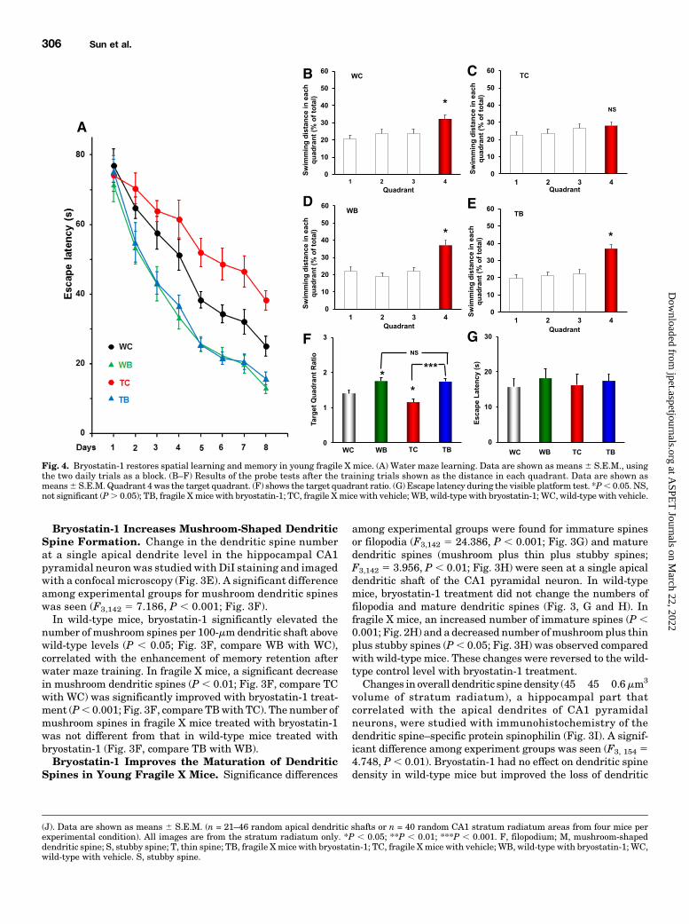
Fig. 4. Bryostatin-1 restores spatial learning and memory in young fragile X mice. (A) Water maze learning. Data are shown as means6 S.E.M., usingthe two daily trials as a block. (B–F) Results of the probe tests after the training trials shown as the distance in each quadrant. Data are shown asmeans6 S.E.M. Quadrant 4 was the target quadrant. (F) shows the target quadrant ratio. (G) Escape latency during the visible platform test. *P, 0.05. NS,not significant (P. 0.05); TB, fragile Xmice with bryostatin-1; TC, fragile Xmice with vehicle; WB, wild-type with bryostatin-1; WC, wild-type with vehicle.
(J). Data are shown as means 6 S.E.M. (n = 21–46 random apical dendritic shafts or n = 40 random CA1 stratum radiatum areas from four mice perexperimental condition). All images are from the stratum radiatum only. *P , 0.05; **P , 0.01; ***P , 0.001. F, filopodium; M, mushroom-shapeddendritic spine; S, stubby spine; T, thin spine; TB, fragile Xmice with bryostatin-1; TC, fragile Xmice with vehicle; WB, wild-type with bryostatin-1; WC,wild-type with vehicle. S, stubby spine.
306 Sun et al.
at ASPE
T Journals on M
arch 22, 2022jpet.aspetjournals.org
Dow
nloaded from
spine density in fragile X mice (P , 0.05; Fig. 3J). These dataon the densities of dendritic spines per brain volume unitswere correlated with changes in the number of dendriticspines on dendritic spine shafts studied with DiI staining(compare Fig. 3, G and H).Taken together, the results suggest that bryostatin-1
improves the maturation of dendritic spines from the imma-ture spines in the young fragile X mice.Bryostatin-1 Prevents Deficits in Spatial Learning
and Memory of Young Fragile X Mice But Did Not AlterSensorimotor Ability. There were significant learning dif-ferences among the four groups (F3,560 5 58.528, P , 0.001;Fig. 4A), indicating different learning among the groups.Spatial learning was significantly impaired in fragile X mice(fragile X mice with vehicle versus wild-type with vehicle:F1,272 5 22.117, P , 0.001). Bryostatin-1 treatment signif-icantly improved the learning performance of fragile X mice(fragile X mice with vehicle versus fragile X mice withbryostatin-1: F1,272 5 110.621, P , 0.001) to the level ofthe control mice with bryostatin-1 treatment (wild-type withbryostatin-1 versus fragile X mice with bryostatin-1: F1,272 50.669, P . 0.05), indicating that the chronic bryostatin-1treatment not only improved spatial learning in the wild-type mice but also repaired the learning deficits of fragile Xmice when tested at an adult age after an early 6-weektreatment.The results of the probe test (Fig. 4, B–E) were analyzed
using the target quadrant ratio (dividing the target quadrantdistance by the average of the nontarget quadrant valuesduring the probe test; Fig. 4F). There were significant differ-ences in the target quadrant ratio among the groups (F3,38 58.790,P, 0.001), indicating differences in the spatial memoryamong the groups. Detailed analysis reveals that the memoryrecall of the transgenic mice was impaired (fragile Xmice withvehicle versus wild-type with vehicle: F1, 18 5 5.155, P, 0.05)and that bryostatin-1 treatment in the transgenic micesignificantly improved the memory recall, compared with thatof fragile X mice without the treatment (F1,17 5 21.145, P ,0.001), to the level of the control mice with bryostatin-1(control mice with bryostatin-1 versus fragile X mice withbryostatin-1: F1,17 5 0.063, P . 0.05).A visible platform test, determined after the probe test,
revealed no significant differences among the groups (F3,35 50.179, P . 0.05; Fig. 4G), indicating that there were nosignificant group differences in sensorimotor ability andescape motivation of the mice among different groups.Therefore, the differences in learning and memory-recallperformance among the groups cannot be attributed tothe differences in their sensorimotor ability and escapemotivation.
DiscussionThe main findings of this study of an early-onset treat-
ment in young adult mice can be summarized as follows.First, the loss of FMR1 protein in fragile X mice suppressesBDNF expression, dendritic spine and synaptic maturation,PSD-95 andmGluR5 accumulation, andmemory-dependentmushroom-shaped dendritic spine formation in the apicaldendrites of the hippocampal CA1 neurons, resulting inspatial learning and memory deficits. Second, the synapticimmaturity and cognitive dysfunction, core features of FXS,
and other FXS phenotypes can be rescued with an earlytreatment of bryotatin-1 in order to produce therapeuticeffects that are better than those of a late-onset treatment(see below). Finally, therapeutic effects can be achievedwithout downregulation of mGluR5 in the hippocampus.It has been well established that FMRP mediates activity-
dependent control of synaptic structure and function (Huberet al., 2002). Experimental studies suggest that the lack ofFMRP leads to overactivity ofmGluR5, a decreasedGABAergicsystem (Olmos-Serrano et al., 2010), and elevated activity ofglycogen synthase kinase 3b (Guo et al., 2012). Synapticimmaturity and cognitive dysfunction are core symptoms inFXS and learned-induced formation of dendritic spines isseverely impaired in fragile X mice (Padmashri et al., 2013).Fragile X mice show an abundance of dense, immaturedendritic spines (Scotto-Lomassese et al., 2011), as in FXSpatients (Grossman et al., 2006). The hyperabundance ofimmature-looking lengthened dendritic spines could be theresult of failed/delayed maturation (Cruz-Martín et al., 2010)and activity-dependent synaptic elimination (Pfeiffer et al.,2010). It has been reported that the left hippocampus inyoung male adult fragile X permutation carriers exhibitsreduced structural connectome (Leow et al., 2014), consistentwith evidence of a range of cognitive impairment, includingspatial processing (Hocking et al., 2012; Wong et al., 2012).The deficit may also involve a loss of some dendritic channels(Routh et al., 2013) but can be rescued with chronicbryostatin-1 treatment at a young age, as shown in our studyand previously in adults (Sun et al., 2014). The treatment inour study began at least 1 month earlier than our previousstudy (Sun et al., 2014), in which the same dose wasadministered at an adult age (2 months of age) for a longertreatment period (13 weeks). Histologic analyses were allperformed at an adult age. Earlier treatment may lead tomore favorable outcomes for several reasons. First, thetherapeutic outcomes seem to be significantly better in theearly-onset treatment group with the same doses of treat-ment, including the higher enhancement of BDNF expres-sion andmushroom-shaped dendritic spine formation (Fig. 5,A and B), better synaptic maturation (Fig. 5, C and D), andgreater improvements in performance in the water mazetask (Fig. 5, E and F), suggesting that the brain networkunder the treatment has regained the capacity to meetcognitive demands. Second, an early intervention mightfacilitate a more “normal” development of sociocognitive skillsand behaviors.Our results of reducedmGluR5 labeling in the hippocampus
of fragile X mice and increased labeling with bryostatin-1treatment are somewhat unexpected. The data suggest thatsyndromic features of FXS might not be caused by anupregulated mGluR5 signaling pathway and synaptic andcognitive function can be rescued without downregulation ofmGluR5 signaling. Desensitization of mGluR5, if induced(Gereau and Heinemann, 1998) with the treatment, andincreased internalization would result in decreased surfaceexpression of the receptor (Ko et al., 2012), a response notobserved in this study. There is no evidence suggesting adirect association of FMRP with the metabotropic glutamatereceptor 5 mRNA, although Lohith et al. (2013) revealed thatboth mGluR5 binding and protein expression were increasedin the prefrontal cortex of FXS patients and carriers. In thefragile X mouse hippocampus, Western blot analysis was
Rescuing Synapses and Spatial Memory in Young Fragile X Mice 307
at ASPE
T Journals on M
arch 22, 2022jpet.aspetjournals.org
Dow
nloaded from

Fig. 5. Chronic bryostatin-1 achieves better therapeutic effects in early-onset than in late-onset treatment in the fragile X mice. (A–E) Effects on BDNFand synapses. Data (means 6 S.E.M.) are compared ratios between transgenic mice with bryostatin-1 (TB) and their matched wild-type mice. (A–C)Early-onset treatment (data in our study) not only recovered but also elevated the following expression of BDNF (A), density of mushroom spines (B), anddensity of axodendritic synapses (C) in fragile X mice to the level higher than the wild-type control level (dashed lines). (D) Early-onset treatment wasalso more effective to reduce the number of immature dendritic filopodia. Data for the late-onset treatment were from Sun et al. (2014) (paired t tests). (Eand F) Chronic bryostatin-1 achieves better therapeutic effects in the early-onset than the late-onset treatment in the fragile X mice. (E) Water mazelearning. Data are compared using their matched daily trial results (the escape latency of the treated mice/that of the untreated multiplies 100; i.e., thedaily escape latency of the untreated fragile X mice as 100%) and are shown as means 6 S.E.M. (F) Memory retention. Data are compared using their
308 Sun et al.
at ASPE
T Journals on M
arch 22, 2022jpet.aspetjournals.org
Dow
nloaded from

reported to show no difference in mGluR5 protein expression(Dölen et al., 2007), although a reduction was observed in thedetergent-insoluble fraction of synaptic membranes isolatedfrom the forebrain of fragile X mice (Giuffrida et al., 2005).There are also some observations of reduced expression ofmGluR5 in fragile X mice (Giuffrida et al., 2005), but nodifference in mGluR5 expression in total hippocampal homog-enates has been reported by others (Huber et al., 2002).The lack of differences in mGluR5 expression (Huber et al.,2002; Giuffrida et al., 2005) between fragile X mice andcontrols but the presence of a reduced association of mGluR5with PSD in fragile X mice might suggest an increased non-PSD association of mGluR5 labeling in fragile X mice.However, the evidence that supports the notion that mGluR5overactivity reflects neuronal pathology in FXS seems verysolid. Reducing mGluR5 expression or the use of mGluR5inhibitors has been shown to correct a broad range of fragileX phenotypes in fragile X mice (Huber et al., 2002; Hajós,2014). Although one would expect an increased colocalizationof postsynaptic mGluR5 when it is overactive, our study doesnot rule out the possibility that the observed reduction inmGluR5 labeling in fragile X mice might partially reflect thereduced PSD labeling in the brain area. Nevertheless, ourresults indicate that the desired therapeutic effects can beachieved in fragile X mice without downregulation ofmGluR5 in the hippocampus. The effective treatment actu-ally improves mGluR5 labeling in the hippocampus. It isprobably appropriate to mention here that while preclinicalstudies with mGluR5 antagonism appear promising, thera-peutic values of mGluR5 inhibitors for FXS are still not clear.The mGluR5 inhibition exaggerates spine immaturity infragile X mice (Cruz-Martín et al., 2010), an effect that wouldbe considered opposite to the intended therapeutic outcomes.In addition, basimglurant and mavoglurant, two differentand potent mGluR5 inhibitors, did not show therapeuticbenefit in recent phase II clinical trials in FXS patients(Scharf et al., 2015).Consistent with our previous study in fragile Xmice, chronic
bryostatin-1 rescues other FXS phenotypes, such as PSD-95(Zalfa et al., 2007; Tang and Alger, 2015) and BDNF levels inthe hippocampus. PSD-95, a major synapse organizer, playsan important role in the stabilization of spines and synapsesas well as in activity-regulated formation of PSD. BDNF isimportant for local protein synthesis and synaptic plasticity(Neumann et al., 2015). Although images of the BDNFfluorescence labeling were used to measure the BDNF levelsin this study, the labeling intensity was found to reliablyreflect the BDNF levels in the same brain areas determinedwith an immunochemical assay, such as the enzyme-linkedimmunosorbent assay, in our previous studies (Sun et al.,2014). Reduction of BDNF expression in fragile Xmice inducescognitive deficits (Louhivuori et al., 2011; Uutela et al., 2012).Infusion of BDNF has been found to restore synaptic functionin slices from fragile X mice (Lauterborn et al., 2007). Thedecrease in BDNF accumulation in the CA1 stratum radiatumwas significantly prevented with both the early-onset (this
study) and late-onset (Sun et al., 2014) bryostatin-1 treat-ment. However, early-onset bryostatin-1 treatment, but notlate-onset bryostatin-1 treatment, also enhanced the BDNFlevel (higher than the control wild-type without bryostatin-1treatment).It is interesting that in the wild-type mice, the early-onset
bryostatin-1 treatment significantly elevated the spatialmemory retention (in this study) above wild-type levels,whereas the late-onset bryostatin-1 treatment did notenhance memory retention (Sun et al., 2014), with thesame lag period to separate acute effects from the chronictherapeutic effects. However, we did not find a significantenhancement of PKC«-induced BDNF levels after early-onset bryostatin-1 treatment. This might implicate thatother PKC«-dependent pathways might also be involved insynaptic plasticity. The direct interaction of PKC« withactin is important for synaptic function and neurite growthand synaptic formation (Prekeris et al., 1996). PKC« mayalso induce synaptogenesis directly by activating structuralchanges through its phosphorylation substrates, GAP-43,the myristoylated alanine-rich C kinase substrate, andadducin (Matsuoka et al., 1998).Bryostatin-1, a highly potent and relatively specific PKC«
activator with pharmacological profiles of synaptogenesisand synaptic maturation/repairing (Hongpaisan and Alkon,2007; Hongpaisan et al., 2011; Sun et al., 2015), rescuessynaptic and memory functions and other phenotypic fea-tures in young fragile X mice. At low concentrations (about1 nm), its PKC activation ismostly on PKC« and less on PKCa.At higher concentrations, however, activities of other PKCisoforms might also be affected. At a lower dose (10 mg/m2,two doses per week for 3 weeks), bryostatin-1 alone wasfound to have no significant effects on spatial learning andmemory in rats (Sun and Alkon, 2008). We previouslyobserved that its chronic administration did not alterswimming speed of rodents in the swimming test (Sun andAlkon, 2008). Evidence is accumulating, supporting anessential role of some PKC isoforms in various phases andtypes of learning and memory (Alkon et al., 2005, 2007).Bryostatin-1–like agents enhance the synthesis of proteinsrequired for memory processing and synaptic repair/synaptogenesis, reduce Ab formation through activation ofa-secretase and increase Ab degradation via the endothelin-converting enzyme, and are antiapoptotic. Since intellectualability, as well as retardation (Wang et al., 2012), involvesmultiple players in signal processing, these agents withmultitargeting actions (Sun et al., 2015) may represent amore effective class of therapeutics than agents that target asingle factor in this complex pathologic process (Vislay et al.,2013).
Authorship Contributions
Conducted experiments: Sun, Hongpaisan.Performed data analysis: Sun, Hongpaisan.Wrote or contributed to the writing of the manuscript: Sun,
Hongpaisan, Alkon.
target quadrant distance in the probe test (the target quadrant distance of the treatedmice/that of the untreated multiplies 100; i.e., the target quadrantdistance of the untreated fragile X mice as 100%) and are shown as means6 S.E.M. The levels of learning andmemory recall of late-onset treated fragileX mice are about those of the age-matched untreated control mice (arrows). *P, 0.05. TB, fragile X mice with bryostatin-1; WC, wild-type with vehicle.
Rescuing Synapses and Spatial Memory in Young Fragile X Mice 309
at ASPE
T Journals on M
arch 22, 2022jpet.aspetjournals.org
Dow
nloaded from

References
Alkon DL, Epstein H, Kuzirian A, Bennett MC, and Nelson TJ (2005) Protein syn-thesis required for long-term memory is induced by PKC activation on days beforeassociative learning. Proc Natl Acad Sci USA 102:16432–16437.
Alkon DL, Sun MK, and Nelson TJ (2007) PKC signaling deficits: a mechanistichypothesis for the origins of Alzheimer’s disease. Trends Pharmacol Sci 28:51–60.
Ashley CT, Jr, Wilkinson KD, Reines D, and Warren ST (1993) FMR1 protein: con-served RNP family domains and selective RNA binding. Science 262:563–566.
Bhattacharya A and Klann E (2012) Fragile X syndrome therapeutics S(C)TEPthrough the developmental window. Neuron 74:1–3.
Chen E, Sharma MR, Shi X, Agrawal RK, and Joseph S (2014) Fragile X mentalretardation protein regulates translation by binding directly to the ribosome. MolCell 54:407–417.
Cruz-Martín A, Crespo M, and Portera-Cailliau C (2010) Delayed stabilization ofdendritic spines in fragile X mice. J Neurosci 30:7793–7803.
Darnell JC and Klann E (2013) The translation of translational control by FMRP:therapeutic targets for FXS. Nat Neurosci 16:1530–1536.
Dölen G, Osterweil E, Rao BS, Smith GB, Auerbach BD, Chattarji S, and Bear MF(2007) Correction of fragile X syndrome in mice. Neuron 56:955–962.
Gereau RW, 4th and Heinemann SF (1998) Role of protein kinase C phosphorylationin rapid desensitization of metabotropic glutamate receptor 5. Neuron 20:143–151.
Giuffrida R, Musumeci S, D’Antoni S, Bonaccorso CM, Giuffrida-Stella AM, OostraBA, and Catania MV (2005) A reduced number of metabotropic glutamate subtype5 receptors are associated with constitutive homer proteins in a mouse model offragile X syndrome. J Neurosci 25:8908–8916.
Glantz LA, Gilmore JH, Hamer RM, Lieberman JA, and Jarskog LF (2007) Syn-aptophysin and postsynaptic density protein 95 in the human prefrontal cortexfrom mid-gestation into early adulthood. Neuroscience 149:582–591.
Grossman AW, Aldridge GM, Weiler IJ, and Greenough WT (2006) Local proteinsynthesis and spine morphogenesis: Fragile X syndrome and beyond. J Neurosci26:7151–7155.
Guo W, Murthy AC, Zhang L, Johnson EB, Schaller EG, Allan AM, and Zhao X (2012)Inhibition of GSK3b improves hippocampus-dependent learning and rescues neuro-genesis in a mouse model of fragile X syndrome. Hum Mol Genet 21:681–691.
Hajós M (2014) Portraying inhibition of metabotropic glutamate receptor 5 in fragileX mice. Biol Psychiatry 75:177–178.
Hocking DR, Kogan CS, and Cornish KM (2012) Selective spatial processing deficitsin an at-risk subgroup of the fragile X premutation. Brain Cogn 79:39–44.
Hongpaisan J and Alkon DL (2007) A structural basis for enhancement of long-termassociative memory in single dendritic spines regulated by PKC. Proc Natl AcadSci USA 104:19571–19576.
Hongpaisan J, Sun MK, and Alkon DL (2011) PKC « activation prevents synapticloss, Ab elevation, and cognitive deficits in Alzheimer’s disease transgenic mice.J Neurosci 31:630–643.
Hongpaisan J, Xu C, Sen A, Nelson TJ, and Alkon DL (2013) PKC activation duringtraining restores mushroom spine synapses and memory in the aged rat. NeurobiolDis 55:44–62.
Horling K, Schlegel G, Schulz S, Vierk R, Ullrich K, Santer R, and Rune GM (2015)Hippocampal synaptic connectivity in phenylketonuria. Hum Mol Genet 24:1007–1018.
Huber KM, Gallagher SM, Warren ST, and Bear MF (2002) Altered synaptic plas-ticity in a mouse model of fragile X mental retardation. Proc Natl Acad Sci USA 99:7746–7750.
Huttenlocher PR and Dabholkar AS (1997) Regional differences in synaptogenesis inhuman cerebral cortex. J Comp Neurol 387:167–178.
Kirov SA, Petrak LJ, Fiala JC, and Harris KM (2004) Dendritic spines disappearwith chilling but proliferate excessively upon rewarming of mature hippocampus.Neuroscience 127:69–80.
Ko SJ, Isozaki K, Kim I, Lee JH, Cho HJ, Sohn SY, Oh SR, Park S, Kim DG, and KimCH, et al. (2012) PKC phosphorylation regulates mGluR5 trafficking by enhancingbinding of Siah-1A. J Neurosci 32:16391–16401.
Koekkoek SK, Yamaguchi K, Milojkovic BA, Dortland BR, Ruigrok TJ, Maex R, DeGraaf W, Smit AE, VanderWerf F, and Bakker CE, et al. (2005) Deletion of FMR1in Purkinje cells enhances parallel fiber LTD, enlarges spines, and attenuatescerebellar eyelid conditioning in Fragile X syndrome. Neuron 47:339–352.
Lauterborn JC, Rex CS, Kramár E, Chen LY, Pandyarajan V, Lynch G, and Gall CM(2007) Brain-derived neurotrophic factor rescues synaptic plasticity in a mousemodel of fragile X syndrome. J Neurosci 27:10685–10694.
Leow A, Harvey D, Goodrich-Hunsaker NJ, Gadelkarim J, Kumar A, Zhan L, RiveraSM, and Simon TJ (2014) Altered structural brain connectome in young adultfragile X premutation carriers. Hum Brain Mapp 35:4518–4530.
Lohith TG, Osterweil EK, Fujita M, Jenko KJ, Bear MF, and Innis RB (2013) Ismetabotropic glutamate receptor 5 upregulated in prefrontal cortex in fragile Xsyndrome? Mol Autism 4:15.
Louhivuori V, Vicario A, Uutela M, Rantamäki T, Louhivuori LM, Castrén E,Tongiorgi E, Akerman KE, and Castrén ML (2011) BDNF and TrkB in neuronaldifferentiation of Fmr1-knockout mouse. Neurobiol Dis 41:469–480.
Matsuoka Y, Li X, and Bennett V (1998) Adducin is an in vivo substrate for proteinkinase C: phosphorylation in the MARCKS-related domain inhibits activity inpromoting spectrin-actin complexes and occurs in many cells, including dendriticspines of neurons. J Cell Biol 142:485–497.
Michalon A, Sidorov M, Ballard TM, Ozmen L, Spooren W, Wettstein JG, Jaeschke G,Bear MF, and Lindemann L (2012) Chronic pharmacological mGlu5 inhibitioncorrects fragile X in adult mice. Neuron 74:49–56.
Nelson TJ and Alkon DL (2015) Molecular regulation of synaptogenesis during as-sociative learning and memory. Brain Res 1621:239–251.
Neumann JT, Thompson JW, Raval AP, Cohan CH, Koronowski KB, and Perez-Pinzon MA (2015) Increased BDNF protein expression after ischemic or PKCepsilon preconditioning promotes electrophysiologic changes that lead to neuro-protection. J Cereb Blood Flow Metab 35:121–130.
Olmos-Serrano JL, Paluszkiewicz SM, Martin BS, Kaufmann WE, Corbin JG,and Huntsman MM (2010) Defective GABAergic neurotransmission and pharma-cological rescue of neuronal hyperexcitability in the amygdala in a mouse model offragile X syndrome. J Neurosci 30:9929–9938.
Padmashri R, Reiner BC, Suresh A, Spartz E, and Dunaevsky A (2013) Alteredstructural and functional synaptic plasticity with motor skill learning in a mousemodel of fragile X syndrome. J Neurosci 33:19715–19723.
Pfeiffer BE, Zang T, Wilkerson JR, Taniguchi M, Maksimova MA, Smith LN, CowanCW, and Huber KM (2010) Fragile X mental retardation protein is required forsynapse elimination by the activity-dependent transcription factor MEF2. Neuron66:191–197.
Prekeris R, Mayhew MW, Cooper JB, and Terrian DM (1996) Identification andlocalization of an actin-binding motif that is unique to the epsilon isoform of pro-tein kinase C and participates in the regulation of synaptic function. J Cell Biol132:77–90.
Rodriguez A, Ehlenberger DB, Dickstein DL, Hof PR, and Wearne SL (2008) Auto-mated three-dimensional detection and shape classification of dendritic spinesfrom fluorescence microscopy images. PLoS One 3:e1997.
Routh BN, Johnston D, and Brager DH (2013) Loss of functional A-type potassiumchannels in the dendrites of CA1 pyramidal neurons from a mouse model of fragileX syndrome. J Neurosci 33:19442–19450.
Sabaratnam M, Murthy NV, Wijeratne A, Buckingham A, and Payne S (2003)Autistic-like behaviour profile and psychiatric morbidity in Fragile X Syndrome: aprospective ten-year follow-up study. Eur Child Adolesc Psychiatry 12:172–177.
Santoro MR, Bray SM, and Warren ST (2012) Molecular mechanisms of fragile Xsyndrome: a twenty-year perspective. Annu Rev Pathol 7:219–245.
Scharf SH, Jaeschke G, Wettstein JG, and Lindemann L (2015) Metabotropic glu-tamate receptor 5 as drug target for Fragile X syndrome. Curr Opin Pharmacol 20:124–134.
Scotto-Lomassese S, Nissant A, Mota T, Néant-Féry M, Oostra BA, Greer CA, LledoPM, Trembleau A, and Caillé I (2011) Fragile X mental retardation protein regu-lates new neuron differentiation in the adult olfactory bulb. J Neurosci 31:2205–2215.
Sengupta P (2013) The laboratory rat: Relating its age with human’s. Int J Prev Med4:624–630.
Siomi H, Choi M, Siomi MC, Nussbaum RL, and Dreyfuss G (1994) Essential role forKH domains in RNA binding: impaired RNA binding by a mutation in the KHdomain of FMR1 that causes fragile X syndrome. Cell 77:33–39.
Sorra KE and Harris KM (2000) Overview on the structure, composition, function,development, and plasticity of hippocampal dendritic spines. Hippocampus 10:501–511.
Sun MK and Alkon DL (2008) Synergistic effects of chronic bryostatin-1 anda-tocopherol on spatial learning and memory in rats. Eur J Pharmacol 584:328–337.
Sun MK, Hongpaisan J, Lim CS, and Alkon DL (2014) Bryostatin-1 restores hippo-campal synapses and spatial learning and memory in adult fragile x mice.J Pharmacol Exp Ther 349:393–401.
Sun MK, Nelson TJ, and Alkon DL (2015) Towards universal therapeutics formemory disorders. Trends Pharmacol Sci 36:384–394.
Tang AH and Alger BE (2015) Homer protein-metabotropic glutamate receptorbinding regulates endocannabinoid signaling and affects hyperexcitability in amouse model of fragile X syndrome. J Neurosci 35:3938–3945.
Tassone F (2014) Newborn screening for fragile X syndrome. JAMA Neurol 71:355–359.
Uutela M, Lindholm J, Louhivuori V, Wei H, Louhivuori LM, Pertovaara A, AkermanK, Castrén E, and Castrén ML (2012) Reduction of BDNF expression in Fmr1knockout mice worsens cognitive deficits but improves hyperactivity and sensori-motor deficits. Genes Brain Behav 11:513–523.
Verkerk AJ, Pieretti M, Sutcliffe JS, Fu YH, Kuhl DP, Pizzuti A, Reiner O, RichardsS, Victoria MF, and Zhang FP, et al. (1991) Identification of a gene (FMR-1) con-taining a CGG repeat coincident with a breakpoint cluster region exhibiting lengthvariation in fragile X syndrome. Cell 65:905–914.
Vislay RL, Martin BS, Olmos-Serrano JL, Kratovac S, Nelson DL, Corbin JG,and Huntsman MM (2013) Homeostatic responses fail to correct defective amyg-dala inhibitory circuit maturation in fragile X syndrome. J Neurosci 33:7548–7558.
Wang X, Snape M, Klann E, Stone JG, Singh A, Petersen RB, Castellani RJ,Casadesus G, Smith MA, and Zhu X (2012) Activation of the extracellular signal-regulated kinase pathway contributes to the behavioral deficit of fragilex-syndrome. J Neurochem 121:672–679.
Wong LM, Goodrich-Hunsaker NJ, McLennan Y, Tassone F, Harvey D, Rivera SM,and Simon TJ (2012) Young adult male carriers of the fragile X premutationexhibit genetically modulated impairments in visuospatial tasks controlled forpsychomotor speed. J Neurodev Disord 4:26.
Zalfa F, Eleuteri B, Dickson KS, Mercaldo V, De Rubeis S, di Penta A, Tabolacci E,Chiurazzi P, Neri G, and Grant SG, et al. (2007) A new function for the fragile Xmental retardation protein in regulation of PSD-95 mRNA stability. Nat Neurosci10:578–587.
Address correspondence to: Miao-Kun Sun, Blanchette Rockefeller Neuro-science Institute, 8 Medical Center Drive, Morgantown, WV 26505. E-mail:[email protected] and [email protected]
310 Sun et al.
at ASPE
T Journals on M
arch 22, 2022jpet.aspetjournals.org
Dow
nloaded from