Embed Size (px)
Citation preview
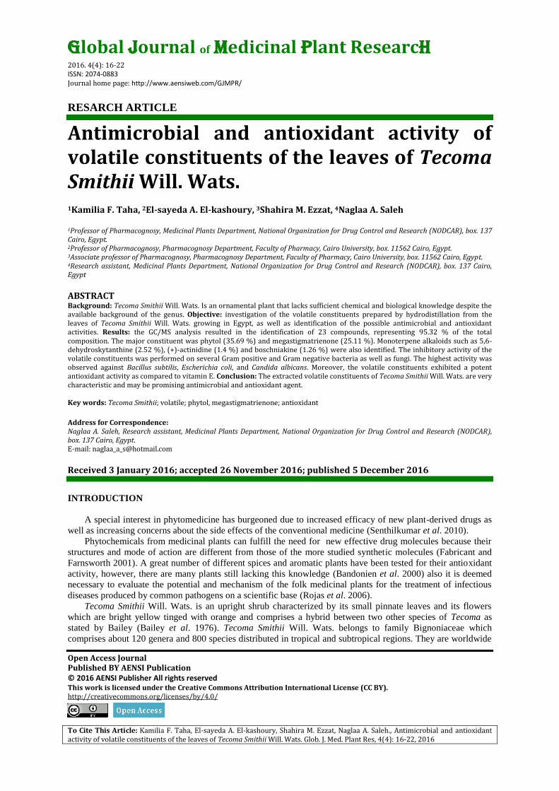
Global Journal of Medicinal Plant Research
2016. 4(4): 16-22 ISSN: 2074-0883 Journal home page: http://www.aensiweb.com/GJMPR/
RESARCH ARTICLE
Open Access Journal Published BY AENSI Publication © 2016 AENSI Publisher All rights reserved This work is licensed under the Creative Commons Attribution International License (CC BY). http://creativecommons.org/licenses/by/4.0/
To Cite This Article: Kamilia F. Taha, El-sayeda A. El-kashoury, Shahira M. Ezzat, Naglaa A. Saleh., Antimicrobial and antioxidant activity of volatile constituents of the leaves of Tecoma Smithii Will. Wats. Glob. J. Med. Plant Res, 4(4): 16-22, 2016
Antimicrobial and antioxidant activity of volatile constituents of the leaves of Tecoma Smithii Will. Wats.
1Kamilia F. Taha, 2El-sayeda A. El-kashoury, 3Shahira M. Ezzat, 4Naglaa A. Saleh
1Professor of Pharmacognosy, Medicinal Plants Department, National Organization for Drug Control and Research (NODCAR), box. 137 Cairo, Egypt. 2Professor of Pharmacognosy, Pharmacognosy Department, Faculty of Pharmacy, Cairo University, box. 11562 Cairo, Egypt. 3Associate professor of Pharmacognosy, Pharmacognosy Department, Faculty of Pharmacy, Cairo University, box. 11562 Cairo, Egypt. 4Research assistant, Medicinal Plants Department, National Organization for Drug Control and Research (NODCAR), box. 137 Cairo, Egypt ABSTRACT Background: Tecoma Smithii Will. Wats. Is an ornamental plant that lacks sufficient chemical and biological knowledge despite the available background of the genus. Objective: investigation of the volatile constituents prepared by hydrodistillation from the leaves of Tecoma Smithii Will. Wats. growing in Egypt, as well as identification of the possible antimicrobial and antioxidant activities. Results: the GC/MS analysis resulted in the identification of 23 compounds, representing 95.32 % of the total composition. The major constituent was phytol (35.69 %) and megastigmatrienone (25.11 %). Monoterpene alkaloids such as 5,6-dehydroskytanthine (2.52 %), (+)-actinidine (1.4 %) and boschniakine (1.26 %) were also identified. The inhibitory activity of the volatile constituents was performed on several Gram positive and Gram negative bacteria as well as fungi. The highest activity was observed against Bacillus subtilis, Escherichia coli, and Candida albicans. Moreover, the volatile constituents exhibited a potent antioxidant activity as compared to vitamin E. Conclusion: The extracted volatile constituents of Tecoma Smithii Will. Wats. are very characteristic and may be promising antimicrobial and antioxidant agent. Key words: Tecoma Smithii; volatile; phytol, megastigmatrienone; antioxidant
Address for Correspondence: Naglaa A. Saleh, Research assistant, Medicinal Plants Department, National Organization for Drug Control and Research (NODCAR), box. 137 Cairo, Egypt. E-mail: [email protected]
Received 3 January 2016; accepted 26 November 2016; published 5 December 2016
INTRODUCTION
A special interest in phytomedicine has burgeoned due to increased efficacy of new plant-derived drugs as
well as increasing concerns about the side effects of the conventional medicine (Senthilkumar et al. 2010).
Phytochemicals from medicinal plants can fulfill the need for new effective drug molecules because their
structures and mode of action are different from those of the more studied synthetic molecules (Fabricant and
Farnsworth 2001). A great number of different spices and aromatic plants have been tested for their antioxidant
activity, however, there are many plants still lacking this knowledge (Bandonien et al. 2000) also it is deemed
necessary to evaluate the potential and mechanism of the folk medicinal plants for the treatment of infectious
diseases produced by common pathogens on a scientific base (Rojas et al. 2006).
Tecoma Smithii Will. Wats. is an upright shrub characterized by its small pinnate leaves and its flowers
which are bright yellow tinged with orange and comprises a hybrid between two other species of Tecoma as
stated by Bailey (Bailey et al. 1976). Tecoma Smithii Will. Wats. belongs to family Bignoniaceae which
comprises about 120 genera and 800 species distributed in tropical and subtropical regions. They are worldwide
17 Kamilia F. Taha., 2016/ Global Journal of Medicinal Plant Research, 4(4): 16-22, 2016
spread as ornamental plants due to their characteristic large colorful flowers (Von Poser et al. 2000).
The genus Tecoma was widely used in traditional medicine; Tecoma stans leaves as antidiabetic (Raju et al.
2011) and to control urinary disorder (Shapiro and Gong 2002), flowers and barks for the treatment of cancers
and as an antimicrobial agent (Binuti and Lajubutu 1994) and roots are used as diuretic and vermifuge (Singh et
al. 2011) and anti-snake bites (Kandakatla et al. 2010). Tecoma undulata bark was used as a remedy for syphilis
(Singh and Gupta 2011). Well established researches on Tecoma reported antidiabetic (Ramírez et al. 2012),
anti-inflammatory and analgesic (Dash et al. 2011), antipyretic (El-Emary et al. 2002), antimicrobial (Salem et
al. 2013), antioxidant (Govindappa et al. 2011), antiplatelet aggregation (Villar et al. 1997), hepatoprotective
(Kameshwaran et al. 2013), and cytotoxic (Al-Azzawi 2012) activities. Scientists claimed various classes of
compounds to be present in genus Tecoma like alkaloids (Al-Azzawi et al. 2012), flavonoids (Hashem 2007),
phenolic acids (Al-Azzawi et al. 2012), iridoids, phenylpropanoids (Abdel-Mageed et al. 2011), sterols and
triterpenes (El-Emary et al. 2002). Despite the availability of literature about the genus Tecoma, nothing was
traced concerning the phytochemistry and biological activities of Tecoma Smithii Will. Wats.
In the present study; the chemical composition, antimicrobial, and antioxidant activities of the
hydrodistilled volatiles of Tecoma Smithii Will. Wats. fresh leaves are investigated. The antimicrobial activity is
determined adopting agar disc diffusion and the minimum inhibitory concentration methods, while the
antioxidant activity is assessed by using 2,2-diphenyl-1-picrylhydrazyl (DPPH) radical scavenging in vitro
assay.
MATERIAL AND METHODS
Plant material:
Fresh leaves of Tecoma Smithii Will. Wats. used in this study were collected during November (autumn)
2012 from plants cultivated in a field belonging to the Applied Research Center for Medicinal Plants (ARCMP),
National Organization for Drug Control and Research (NODCAR). The plant was kindly authenticated by Dr.
Wafaa M. Amer, Professor of Flora, Botany Department, Faculty of Science, Cairo University. A voucher
specimen (no. 4122012) is kept at the Herbarium of the Department of Pharmacognosy, Faculty of Pharmacy,
Cairo University.
Preparation of the volatile constituents:
A sample of fresh leaves (2 kg), was subjected to hydrodistillation in a Clevenger apparatus according to
the Egyptian Pharmacopoeia procedures. The isolated liquid was dried over anhydrous sodium sulfate and saved
in a refrigerator till analysis.
GC/MS analysis:
The hydrodistilled volatile constituents of the leaves were diluted in n-hexane (HPLC- grade) and subjected
to HP Agilent® GC-MS system, comprising a 6890 gas chromatograph coupled with a 5973A Agilent® mass
spectrometer. Column HP-5MS, (HP Agilent ®, USA) cross-linked (5% phenyl)- methylpolysiloxane capillary
column, 30 m x 0.25 mm, film thickness 0.25 μm. The temperature program was 60°C, kept isothermal for 3
minutes, then increased at rate 8°C/minute to 300°C and kept at 300°C for 15 minutes. The injection
temperature was set 250°C, injection volume 1.0 μl, Inlet pressure: 37.1 kPa. The carrier gas: helium linear
velocity (ū): 1 ml/minute. Injection mode: split-less. MS interface temperature: 250°C; MS mode: EI; detector
voltage: 70 eV; mass range: 50 - 500 m/z at 3.62/scan. Data handling was carried out by means of GC/MSD
ChemStation software Agilent®
Identification of the individual volatile components was confirmed by comparison of the mass
fragmentation patterns with MS library database (Wiley7n and Wiley7Nist05) and mass spectra literature data
(Adams 2004) as well as comparison of their retention indices (RI) determined with reference to a homologous
series of n-alkanes which are subjected to GLC analysis under the same experimental conditions.
The antimicrobial activity:
In vitro antimicrobial activity of the hydrodistilled volatile constituents was tested against representatives of
Gram-positive bacteria; Bacillus subtilis (ATCC 6051) and Staphylococcus aureus (ATCC 12600), Gram-
negative bacteria; Pseudomonas aeruginosa (ATCC 101450) and Escherichia coli (ATCC 11775) and fungi;
Candida albicans (ATCC 26555) and Aspergillus niger (laboratory collection strains). The test bacteria/ fungi
(100 µl, each) were grown in 10 ml of fresh media until they reached a count of approximately 108 cells/ml for
bacteria or 105 cells/ml for fungi (Pfaller et al. 1988). The microbial suspension (100 µl) was spread onto agar
plates corresponding to the broth in which they were maintained, then colonies of each organism were selected
from primary agar plates for further testing (National-Committee-for-Clinical-Laboratory-Standards-(NCCLS)
2012). The antimicrobial assay was performed by a modified Kirby-Bauer disc diffusion method (Bauer et al.
1966).
18 Kamilia F. Taha., 2016/ Global Journal of Medicinal Plant Research, 4(4): 16-22, 2016
Disc diffusion method:
Mueller-Hinton agar was inoculated with 100 µl of the inoculum (1x10⁸ CFU/ml) and poured in the petri
dish plate. A stock solution of the hydrodistilled volatile constituents (100 mg/ml in DMSO) was prepared and
10 µl were loaded over 8.0 mm diameter blank paper discs (each disc was thus loaded with 1 mg of the volatile
constituents). Other discs were impregnated with 10 μl of DMSO (Sigma®, USA) as a negative control.
Standard commercially available discs of tetracycline (Sedico®, Egypt) and amphotericin B (Bristol-Myers
Squibb®, Switzerland) (5 µg/disc) were used as positive controls for antibacterial and antifungal activities,
respectively. All discs were placed on agar surface and incubated, where the plates inoculated with Gram
positive and Gram negative bacteria were incubated at 35-37ºC for 24-48 hours, while those inoculated with
Aspergillus niger and Candida albicans were incubated at 25ºC for 48 hours and at 30ºC for 24-48 hours,
respectively. The antimicrobial activity was determined by measuring the diameter of the zone of inhibition in
millimeter (Bauer et al. 1966).
The Minimum Inhibitory Concentration (MIC):
The MIC measurement (also called broth micro-dilution method) is a quantitative method for testing the
antimicrobial activity. It is defined as the least concentration of an antimicrobial agent (among a series of
dilutions) that will inhibit the growth of a microorganism. The MIC of the volatile constituents was determined
by a whole-cell assay in a 96-well microtiter format. Bacillus subtilis, Staphylococcus aureus, Pseudomonas
aeruginosa, Escherichia coli and Candida albicans were grown on sabouraud dextrose broth media (SDP;
Difco.®) while Aspergillus niger was grown on Czepek`s dox. Both media had an initial cell optical density at
600 nm (OD600) of 0.001, then they were inoculated with serial dilutions (0-16 g/ml) of the volatile
constituents sample in the same media. Growth inhibition was measured by determining the microbial biomass
(i.e.: by determination of the OD600) at 48 hours. The lowest concentration which led to an OD600 of ≤ 0.001
(that caused 100 % inhibition, MIC100) was recorded.
The antioxidant activity:
The free radical scavenging activity of the hydrodistilled volatile constituents was assessed using 2,2-
diphenyl-1-picrylhydrazyl (DPPH, Sigma®, USA) radical in vitro assay with reference to a standard antioxidant
vitamin E (was chosen for its oily nature which is comparable to that of the tested volatiles) and adopting the
method described by Mensor et al. (Mensor et al. 2001) with some modifications. A methanolic solution of the
volatile constituents was prepared (25 g/l), this solution was further diluted to final concentrations of 17.5, 15,
12.5, 10, 7.5, 5 and 2.5 g/l in methanol. Serial dilutions of vitamin E (available as 0.4 g soft gelatin capsule,
Pharco Pharmaceutical Co., Egypt) were prepared as 400, 350, 300, 250, 200, 150 and 100 g/l. One ml of a
methanolic solution of DPPH (0.3 mM) was added to 2.5 ml of each of the prepared dilutions of the test and
standard. Each sample was compared to a blank, where the DPPH is replaced with 1 ml methanol. A negative
control was prepared by mixing 1 ml the DPPH solution with methanol (2.5 ml). All samples were incubated in
the dark for 30 minutes at room temperature. The absorbance of the color of the resulting mixture was recorded
at 518 nm using a UV/ visible spectrophotometer (UVD-2960 Labomed Inc.®, USA), The decrease of the
absorbance of the DPPH solution indicates an increase in DPPH radical scavenging activity of the sample The
experiment was performed in triplicate and the average absorbance for each concentration (Abs sample) was taken.
The values of absorbance were converted into percentage antioxidant activity (AA %) using the formula:
AA % = (Abs control – Abs sample / Abs control) × 100 (equation 1)
The concentrations of the sample (the tested hydrodistilled volatiles or vitamin E) and the corresponding
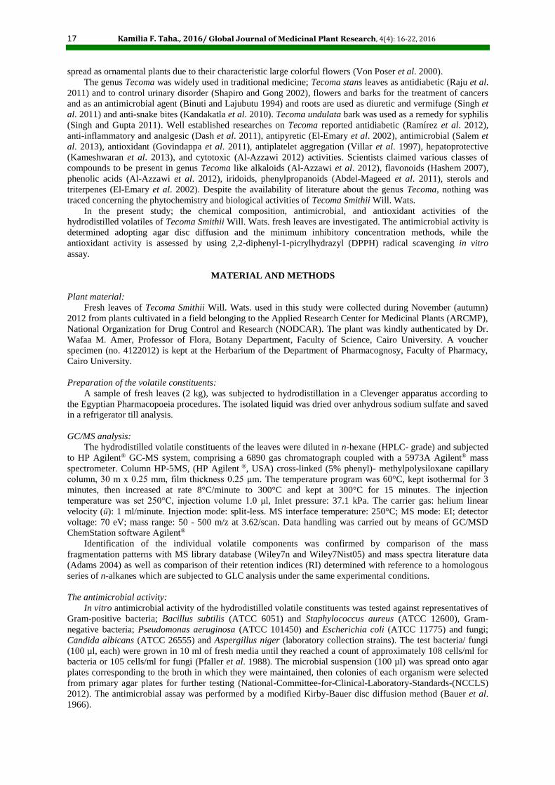
antioxidant activity (AA %) were plotted and the graph resulted in a logarithmic regression pattern (Fig.1).
Fig. 1: Log graphs representing the antioxidant activity (AA %) in response to concentration of vitamin E (a)
and the volatile constituents of the leaves of Tecoma Smithii Will. Wats. (b).
19 Kamilia F. Taha., 2016/ Global Journal of Medicinal Plant Research, 4(4): 16-22, 2016
RESULTS AND DISCUSSION
Chemical composition of the hydrodistillate:
The volatile constituents sample obtained by hydrodistillation of the fresh leaves of Tecoma Smithii Will.
Wats. (0.09 % w/w) was slightly viscous, oily, orange-yellow in color, with an intense characteristic odor and
readily soluble in aqueous ethanol (70 % v/v). The specific gravity and refractive index were 0.927 and 1.455 at
room temperature, respectively. Compounds identified and their relative percentages are listed in Table 1.
Twenty-three components were identified under the adopted operating conditions representing 95.32 % of the
total composition. The percentage of the identified non-terpenoid constituents was 74.32 %, while that of the
identified terpenoids was 21.00 %. The overall chromatographic profile appeared to be dominated by the
oxygenated constituents (69.95 %), which include alcohols (39.79 %), ketones (25.83 %) and aldehydes (4.33
%). Monoterpene alkaloids represented an appreciable percentage of the volatile constituents (5.18 %), include
5,6-dehydroskytanthine, (+)-actinidine and boschniakine.
Table 1: Identified constituents in the hydrodistillate of the fresh leaves of Tecoma Smithii Will. Wats. by GC-MS.
RI Identified compound M+ bp Content %
779 1,7-Octadiene 124 67 5.49
1022 Limonene 136 68 4.19
1088 Linalool 154 71 2.16 1095 5,6-Dehydroskytanthine* 165 58 2.52
1192 Myrtenal 150 79 1.35
1231 (+)-Actinidine* 147 132 1.4 1238 Cuminaldehyde 148 148 1.49
1411 β-Damascone 192 177 0.72
1450 Isoeugenol 164 164 0.68 1485 Boschniakine* 160 160 1.26
1525 δ-Cadinene 204 161 0.72
1560 Megastigmatrienone 190 133 25.11 1799 n-Octadecane 254 57 1.08
1846 Neophytadiene 278 68 2.79
1948 Phytol 296 71 35.69 1998 1,4-Eicosadiene 278 82 0.95
2000 n-Eicosane 282 57 0.41
2058 9,12,15-Octadecatrien-1-ol (linolenyl alcohol) 306 79 1.26 2080 Cycloisolongifolene,8,9-dehydro-9-formyl 405 230 1.49
2101 n-Heneicosane 296 57 0.77
2202 n-Docosane 310 57 0.81 2301 n-Tricosane 324 57 1.31
2905 n-Nonacosane 408 57 1.67
Notes: bold numbers indicate major components percentages, bp: base peak, M+: molecular ion peak, RI: Retention index as determined on
an HP-5MS column using the homologous of n-alkanes, *: compounds identified by comparison of their MS with published data.
The major constituent was phytol (35.69 %), which is a hydrophobic acyclic diterpene alcohol, occurs as a
side chain in the chlorophyll molecule. It was reported to be a component of many hydrodistilled plant
constituents like tea (Pripdeevech and Machan 2011). The second major component was megastigmatrienone
(25.11 %) which is a non-terpenoid ketone derived from carotenoids (Mendes 2009), it is a key flavor
compound in tobacco and also has been detected in wine, where it may contribute to a tobacco/incense aroma
(Slaghenaufi et al. 2014). Other components were 1,7-octadiene (5.49 %), which is a component of thyme
essential oil (Porte and Godoy 2008), limonene (4.19 %), a famous monoterpene hydrocarbon reported to be
present in several volatile oils such as citrus fruits (de Andrade Dutra et al. 2016), neophytadiene (2.79 %) and
β-damascone (0.72 %) identified in tobacco leaves (Zhang et al. 2012).
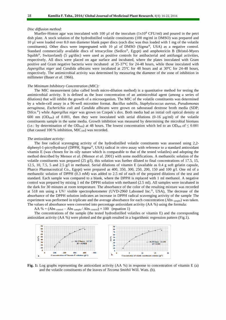
The antimicrobial activity:
The antimicrobial activity was evaluated by measuring the diameter of zones of inhibition due to the
samples, also the percentage potency of the tested hydrodistilled volatiles to the appropriate reference standard
antimicrobial drug was, in each case, calculated (Table 2 and Fig.2). The hydrodistilled volatiles of the leaves of
Tecoma Smithii Will. Wats. showed a powerful inhibitory activity against all tested bacteria and fungi, therefore
it was subjected to the broth micro-dilution method for the determination of the Minimum Inhibitory
Concentration (MIC).
20 Kamilia F. Taha., 2016/ Global Journal of Medicinal Plant Research, 4(4): 16-22, 2016
Table 2: The diameter of zones of inhibition of the volatile constituents sample against standard antimicrobial agents; tetracycline and
amphotericin B.
Tested solutions
Gram-positive bacteria Gram-negative bacteria Fungi
Staphylococcus aureus
Bacillus subtilis
Escherichia coli
Pseudomonas aeruginosa
Candida albicans
Aspergillus niger
Volatile constituents 18 (64 %) 30 (103 %) 29 (97 %) 22 (73 %) 20 (100 %) 15 (88 %)
Tetracycline 28 29 30 30 - - Amphotericin B - - - - 20 17
Notes: The numbers represent the diameter of the zone of inhibition in mm, while the numbers in brackets represent the percentage of
potency (zone diameter) of the sample referring to the standard antimicrobial agent.
Fig. 2: Histogram representing the antimicrobial activity of the volatile constituents of the leaves of Tecoma
Smithii Will. Wats. against standard antimicrobial agents (tetracycline and amphotericin B)
The MIC results of the hydrodistilled volatiles sample against the microorganisms under investigation
revealed that the sample is most effective against Bacillus subtilis and Candida albicans (MIC = 4 g/ml)
followed by Aspergillus niger and Escherichia coli (MIC = 6 g/ml and 6.5 g/ml, respectively) and finally
Staphylococcus aureus and Pseudomonas aeruginosa (MIC = 12.5 g/ml).
The antioxidant activity determined by DPPH method:
We use the equation of the logarithmic trendline representing the relation between the sample concentration
and AA % to conclude the IC50 value (The Inhibitory Concentration: sample concentration that produces a 50 %
quenching of the UV absorption of DPPH). The IC50 values of the hydrodistilled volatiles and vitamin E were
7.05 g/l and 198 g/l, respectively. As long as the IC50 values are indicative of the antioxidant potential (the
lower IC50, the higher the antioxidant potential), thus the volatile constituents of the leaves of Tecoma Smithii
Will. Wats. exhibited a stronger antioxidant activity than the standard antioxidant (vitamin E.)
The biological activity of the hydrodistilled volatiles of Tecoma Smithii Will. Wats. can be correlated to its
composition; where phytol, the major constituent (35.69 %), was proved to exhibit an antioxidant activity which
is comparable to Trolox (a synthetic analogue of α-tocopherol, used as standard antioxidant) which may be
attributed to its hydroxyl group (OH) by reacting with a free radical to produce a less reactive species (Santos et
al. 2013). Another research claimed that phytol could reduce the production of hydroxyl, superoxide anion,
methoxy, carbon dioxide anion, nitric oxide radical and DPPH radicals and proved activity against eight
bacterial and eight fungal strains tested (Pejin et al. 2014).
Limonene was detected among the composition of the hydrodistillate. The antioxidant activity of limonene
was proved (Roberto et al. 2010). Limonene also possesses an antimicrobial activity against a wide spectrum of
human pathogenic fungi and bacteria (Bevilacqua et al. 2010). The presence of alkaloids in the hydrodistillate
have been shown to possess an antimicrobial and antioxidant activities which may be attributed to their ability to
intercalate DNA (Salem et al. 2013).
Conclusion:
The present study indicated that the volatile constituents obtained by hydrodistillation of the fresh leaves of
Tecoma Smithii Will. Wats. (Bignoniaceae) grown in Egypt had a potent antibacterial (against Gram-positive,
Gram-negative bacteria, and fungi) and antioxidant activities, these activities can be attributed to the
constituents especially the major component phytol. This is the first study for the investigation of the chemical
composition and biological activity of the hydrodistilled volatile constituents of Tecoma Smithii Will. Wats. and
the results obtained should encourage more intensive researches to be carried out.
21 Kamilia F. Taha., 2016/ Global Journal of Medicinal Plant Research, 4(4): 16-22, 2016
REFERENCES
Egyptian Pharmacopoeia; 4th ed.: 2005. Ministry of Health and Population, Cairo, Egypt.
Abdel-Mageed, W.M., E.Y. Backheet, A.A. Khalifa, Z.Z. Ibraheim and S.A. Ross, 2011. Antiparasitic
antioxidant phenylpropanoids and iridoid glycosides from Tecoma mollis. Fitoterapia, 83(3): 500-07.
Adams, R.P., 2004. Identification of essential oil components by gas chromatography / mass spectroscopy:
Allured Publishing Corporation: Illinois; USA.
Al-Azzawi, A.M., 2012. Genotoxic and cytotoxic study of Tecoma stans Bignoniaceae. Pakistan Journal of
Biological Sciences, 15(2): 92-97.
Al-Azzawi, A.M., E. Al-Khateeb, K. Al-Sameraei and A.G. Al-Juboori, 2012. Antibacterial activity and the
histopathological study of crude extracts and isolated tecomine from Tecoma stans Bignoniaceae in Iraq.
Pharmacognosy Research, 4(1): 37-43.
Bailey, L.H., E.Z. Bailey and L.H.B. Hortorium, 1976. Hortus third: A concise dictionary of plants
cultivated in the United States and Canada New York: the Macmillan Company.
Bandonien, D., A. Pukalskas, P. Venskutonis and D. Gruzdien, 2000. Preliminary screening of antioxidant
activity of some plant extracts in rapeseed oil. Food Research International, 33(9): 785-91.
Bauer, A.W., W.M. Kirby, C. Sherris and M. Turck, 1966. Antibiotic susceptibilty testing by a standardized
single method. American Journal of Clinical Pathology, 45: 493-96.
Bevilacqua, A., M.R. Corbo and M. Sinigaglia, 2010. In vitro evaluation of the antimicrobial activity of
eugenol, limonene, and citrus extract against bacteria and yeasts, representative of the spoiling microflora of
fruit juices. Journal of Food Protection, 73(5): 888-94.
Binuti, O.A. and B.A. Lajubutu, 1994. Antimicrobial potentials of some plant species of the Bignoniaceae
family. Afr. J. Med. Med. Sci., 23: 269-73.
Dash, S., C. Das, D.C. Sahoo and A.C. Sahoo, 2011. Phytochemical composition, anti-inflammatory and
analgesic activities of Tecoma stans Linn. (Bignoniaceae). Nature of Pharmaceutical Technology, 1(2): 5-8.
de Andrade Dutra, K., J.V. de Oliveira, D.M.d.A.F. Navarro and J.P.O. Santos, 2016. Control of
Callosobruchus maculatus (FABR.)(Coleoptera: Chrysomelidae: Bruchinae) in Vigna unguiculata (L.) WALP.
with essential oils from four Citrus spp. plants. Journal of Stored Products Research, 68: 25-32.
El-Emary, N.A., A.A. Kalifa, E.Y. Backheet and W.M. Abdel-Mageed, 2002. Phytochemical and biological
studies on the leaves of Tecoma mollis Humb. and Bonpl. cultivated in Egypt. Bulletin-of-Pharmaceutical-
Sciences Assiut-Univ., 25(2): 118-207.
Fabricant, D. S. and N.R. Farnsworth, 2001. The Value of Plants Used in Traditional Medicine for Drug
Discovery. Environmental Health Perspectives, 109: 69-75.
Govindappa, M., T.S. Sadananda, R. Channabasava, M.K. Jeevitha, K.S. Pooja and V.B. Raghavendra,
2011. Antimicrobial, antioxidant activity and phytochemical screening of Tecoma stans (L.) Juss. ex Kunth.
Journal of Phytology and Phytopharmacology, 3(3): 68-76.
Hashem, F.A., 2007. Free radical scavenging activity of the flavonoids isolated from Tecoma radicans.
Journal of Basic and Applied Sciences, 3(1): 49-53.
Kameshwaran, S., A.R. Kothai, C. Jothimanivannan and R. Senthilkumar, 2013. Evaluation of
hepatoprotective activity of Tecoma stans flowers. Pharmacologia, 4(3): 236-42.
Kandakatla, S., K.N.V. Rao and D. Banji, 2010. Ethno-medicinal review on "Pachagota" Tecoma stans (L.)
Juss. ex Kunth. Herbal Technology Industry, 0-2.
Mendes, M.M., 2009. Carotenoid breakdown products the nor-isoprenoids in wine aroma. Archives of
Biochemistry and Biophysics, 483(2): 236-45.
Mensor, L.I., F.S. Menezes, G.G. Leitão, A.S. Reis, T. dos Santos, C.S. Coube and S.G. Leitão, 2001.
Screening of Brazillian plant extracts for antioxidant activity by the use of DPPH free radical method.
Phytotherapy Research, 15(2): 127-30.
National-Committee-for-Clinical-Laboratory-Standards-(NCCLS), 2012. Methods for dilution
antimicrobial susceptibility tests for bacteria that grow aerobically; Approved standard- ninth edition. 32(2).
Pejin, B., A. Savic, M. Sokovic, J. Glamoclija, A. Ciric, M. Nikolic, K. Radotic and M. Mojovic, 2014.
Further in vitro evaluation of antiradical and antimicrobial activities of phytol. Natural product research, 28(6):
372-76.
Pfaller, M.A., L. Burmeister, M.A. Bartlett and M.G. Rinaldi, 1988. Multicenter evaluation of four methods
of yeast inoculum preparation. Journal of Clinical Microbiology, 26: 1437-41.
Porte, A. and R.L.O. Godoy, 2008. Chemical composition of Thymus vulgaris L. (thyme) essential oil from
the Rio de Janeiro State (Brazil). Journal of the Serbian Chemical Society, 73(3): 307-10.
Pripdeevech, P. and T. Machan, 2011. Fingerprint of volatile flavour constituents and antioxidant activities
of teas from Thailand. Food Chemistry, 125: 797-802.
22 Kamilia F. Taha., 2016/ Global Journal of Medicinal Plant Research, 4(4): 16-22, 2016
Raju, S., S. Kavimani, V.U. Maheshwara-rao and K.S. Reddy, 2011. Tecoma stans (L.) Juss. ex Kunth
(Bignoniaceae): Ethnobotany, phytochemistry and pharmacology. Journal of Pharmaceutical and Biomedical
Sciences, 8(7): 1-5.
Ramírez, G., M. Zavala, J. Pérez and A. Zamilpa, 2012. In vitro screening of medicinal plants used in
Mexico as antidiabetics with glucosidase and lipase inhibitory activities. Evidence-Based Complementary and
Alternative Medicine, pp: 1-6.
Roberto, D., P. Micucci, T. Sebastian, F. Graciela and C. Anesini, 2010. Antioxidant activity of limonene
on normal murine lymphocytes: relation to H2O2 modulation and cell proliferation. Basic & clinical
pharmacology & toxicology, 106(1): 38-44.
Rojas, J.J., V.J. Ochoa, S.A. Ocampo and J.F. Muñoz, 2006. Screening for antimicrobial activity of ten
medicinal plants used in Colombian folkloric medicine: A possible alternative in the treatment of non-
nosocomial infections. BMC Complementary and Alternative Medicine, 6(1): 1-6.
Salem, M.Z., Y.M. Gohar, L. Camacho, N.A. El-Shanhorey and A. Salem, 2013. Antioxidant and
antibacterial activities of leaves and branches extracts of Tecoma stans (L.) Juss. ex Kunth against nine species
of pathogenic bacteria. Afr J Microbiol Res., 7(5): 418-26.
Santos, C.C.d.M.P., M.S. Salvadori, V.G. Mota, L.M. Costa, A.A.C. de Almeida, G.A.L. de Oliveira, J.P.
Costa, D.P. de Sousa, R.M. de Freitas and R.N. de Almeida, 2013. Antinociceptive and antioxidant activities of
phytol in vivo and in vitro models. Neuroscience Journal.
Senthilkumar, C.S., M.S. Kumar and M.R. Pandian, 2010. In vitro antibacterial activity of crude leaf
extracts from Tecoma stans (L.) Juss. ex Kunth., Coleus forskohlii and Pogostemon patchouli against human
pathogenic bacteria. International Journal of PharmTech Research, 2(1): 438-42.
Shapiro, K. and W.C. Gong, 2002. Natural Products Used for Diabetes. Journal of the American
Pharmaceutical Association, 42(2): 217-26.
Singh, D. and R.S. Gupta, 2011. Hepatoprotective activity of methanol extract of Tecomella undulata
against alcohol and paracetamol induced hepatotoxicity in rats. Life Sciences and Medicine Research, pp: 1-8.
Singh, V., L. Kumar, G.S. Chakraborthy and A. Mazumder, 2011. Pharmacological and phytochemical
findings of Tecoma stans, a review. Journal of Advanced Pharmacy and Healthcare Research, 1(3): 75-81.
Slaghenaufi, D., M.-C. Perello, S. Marchand-Marion, S. Tempere and G. de Revel, 2014. Quantitative solid
phase microextraction – Gas chromatography mass spectrometry analysis of five megastigmatrienone isomers in
aged wine. Analytica Chimica Acta, 813: 63-69.
Villar, R., J.M. Callega, C. Morales and A. Caceres, 1997. Screening of 17 guatemalan medicinal plants for
platelet antiaggregant activity. Phytotherapy Research, 11(6): 441-45.
Von Poser, G.L., J. Schripsema, A.T. Henriques and S.R. Jensen, 2000. The distribution of iridoids in
Bignoniaceae. Biochemical Systematic & Ecology, 28(4): 351-66.
Zhang, X., H. Gao, L. Zhang, D. Liu and X. Ye, 2012. Extraction of essential oil from discarded tobacco
leaves by solvent extraction and steam distillation, and identification of its chemical composition. Industrial
Crops and Products, 39: 162-69.