Embed Size (px)
DESCRIPTION
To begin development biology
Citation preview

The Reproductive Process

Reproduction
Reproduction is one of the ubiquitous properties of life.
Evolution is inextricably linked to reproduction.
Two modes of reproduction are recognized: Asexual
Sexual

Asexual vs. Sexual Reproduction
Asexual reproduction – the production
of offspring whose genes all come from
one parent without the fusion of egg and
sperm.
Usually diploid eggs are produced by
mitosis which then develop directly.

Asexual vs. Sexual Reproduction
Sexual reproduction –the production of offspring by the fusion of haploid gametes (eggs & sperm) from two parents to form a diploid zygote (fertilized egg). Dioecious
Gametes arise by meiosis.
Genetic variability is increased by the random combinations of genes from the parents.

Asexual Reproduction
Bacteria and
many protozoa
can reproduce by
binary fission –
separating into
two or more
individuals
approximately the
same size.

Asexual Reproduction
Budding is a form of
asexual reproduction
where new
individuals form as
offshoots of a parent.
The offspring may
separate or remain
attached to form
colonies.

Asexual Reproduction
Fragmentation results when an
organism’s body is broken into several
pieces and each piece grows into a new
organism.
Regeneration – the regrowth of lost body
parts.

Asexual Reproduction
Fragmentation occurs in some sponges, cnidarians, polychaete annelids, tunicates. Sea stars can
regenerate lost limbs, but only species in the genus Linckia can form new individuals from broken arms.

Asexual Reproduction - Advantages
Animals living far from
members of their own species
can reproduce without having
to search for a mate.
Numerous offspring quickly –
ideal for colonizing a new
area.
Advantageous in a stable,
favorable environment
because it reproduces a
successful genotype
precisely.

Sexual Reproduction
Generally involves two parents.
Special germ cells unite to form a zygote.
Sexual reproduction recombines parental characters. A richer, more diversified population results.
In haploid asexual organisms mutations are expressed and selected quickly.
In sexual reproduction a normal gene on the homologous chromosome may mask a gene mutation.

Parthenogenesis
Parthenogenesis involves the
development of an embryo from an
unfertilized egg or one where sperm &
egg nuclei did not fuse.
Ameiotic parthenogenesis – no meiosis,
egg is formed by mitosis (diploid)
Meiotic parthenogenesis – haploid ovum
formed by meiosis, it may be activated by a
male (or not).

Parthenogenesis
In some animals (aphids, rotifers,
Daphnia) the females can produce two
types of eggs.
One must be fertilized.
One type will develop directly into haploid
adults – parthenogenesis.
Haploid females produce eggs by mitosis.

Sequential Hermaphroditism
In sequential
hermaphroditism, an
individual reverses its
sex during its lifetime.
In wrasses, sex reversal
is associated with age,
size and social
conditions.
Fish are female first.
The largest female
becomes male if the
previous male dies.

Sequential Hermaphroditism
There are also sequential
hermaphrodites that are male first, later
changing to female.
This occurs in species that produce more
eggs at a bigger size – so it is
advantageous to have larger females.
Oysters

Fertilization
Fertilization – fusion
of egg and sperm
into a single diploid
cell, the zygote.
External
Internal

External Fertilization
External fertilization –fertilization takes place outside the female’s body.
A wet environment is required so gametes don’t dry out and so sperm may swim to the eggs.

Internal Fertilization
Internal fertilization allows terrestrial
animals to reproduce away from water.
Cooperative behavior leading to copulation
is required.

Ensuring Survival of Offspring
Species with internal
fertilization produce
fewer zygotes, but
protect them more from
predation.
Tough eggshells
Embryo may develop in
reproductive tract of
female
Parental care of eggs &
offspring

Gamete Production & Delivery
Gametes (eggs & sperm) are required
for sexual reproduction.
Usually, gametes are produced in
gonads (ovaries & testes).
Germ cells are set aside early in
development. They will produce only
gametes.

Migration of Germ Cells
Germ cells arise in
the yolk-sac
endoderm of
vertebrates – not in
the gonads.
They migrate to the
gonads using
amoeboid
movement.

Gametogenesis
Gametogenesis – the production of gametes.
Spermatogenesis – each primary spermatocyte divides to form 4 sperm.
Oogenesis – each primary oocyte divides to form 1 ovum and 2-3 polar bodies. In oogenesis, cytokinesis is unequal, most of the
cytoplasm goes to one daughter cell which becomes the ovum. The other cells, polar bodies, degenerate.

Spermatogenesis
Outermost layer of the seminiferous
tubules contain spermatogonia, diploid
cells that grow to become primary
spermatocytes.
After the first meiotic division, they are
called secondary spermatocytes.
When meiosis is complete the haploid
cells are spermatids.

Spermatogenesis

Spermatogenesis
Spermatids mature
into motile sperm
with a tail for
locomotion, and a
head containing an
acrosome as well as
the nucleus.

Oogenesis
In the ovary, early germ cells called oogonia
are diploid.
Oogonia grow to become primary oocytes.
After the first meiotic division, the cytoplasm
divides unequally and only one secondary
oocyte and one polar body result.
Following the second meiotic division, one
ootid and another polar body result.
The ootid develops into a functional ovum.

Oogenesis

Oogenesis
Meiosis is usually arrested at the
beginning of meiosis and is not
completed until ovulation or fertilization.

Basal body(centriole)
Spermhead
Sperm-bindingreceptors
Acrosome
Jelly coat Vitelline layer
Egg plasmamembrane
Hydrolytic enzymes
Acrosomalprocess
Actinfilament
Spermnucleus
Sperm plasmamembrane
Fused
plasma
membranes
Fertilizationenvelope
Corticalgranule
Perivitellinespace
EGG CYTOPLASM
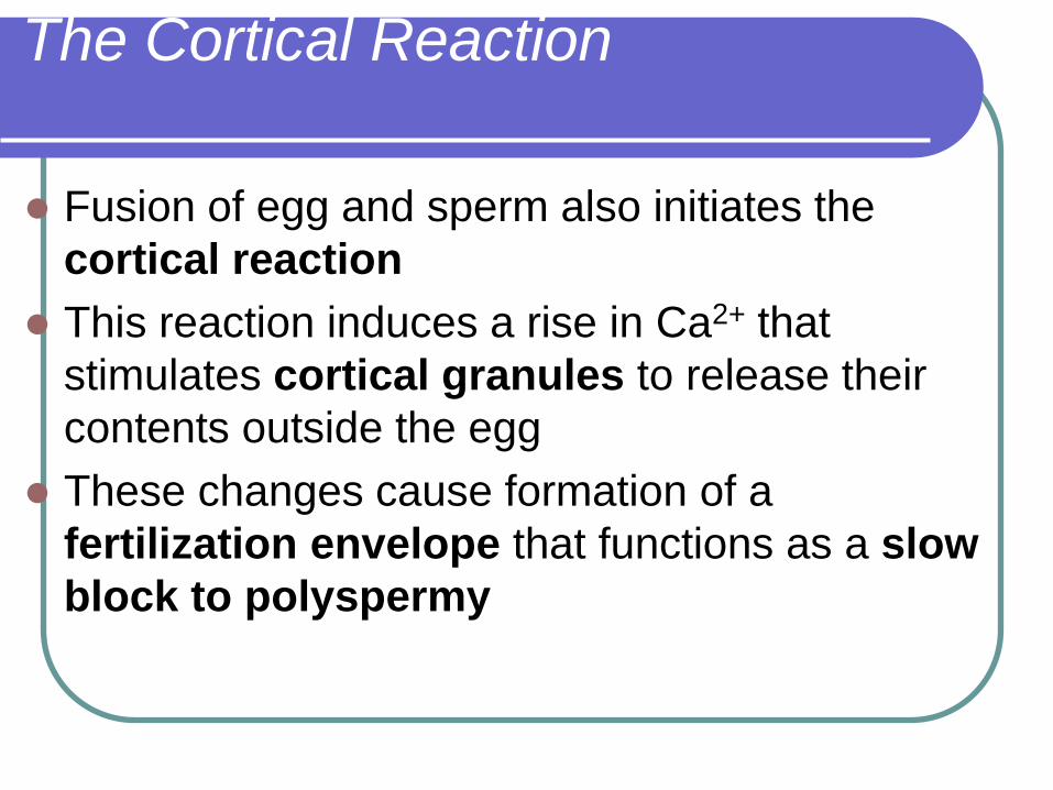
The Cortical Reaction
Fusion of egg and sperm also initiates the
cortical reaction
This reaction induces a rise in Ca2+ that
stimulates cortical granules to release their
contents outside the egg
These changes cause formation of a
fertilization envelope that functions as a slow
block to polyspermy

EXPERIMENT
10 sec afterfertilization
1 sec beforefertilization
RESULTS
CONCLUSION
25 sec 35 sec 1 min500 µm
10 sec afterfertilization
20 sec 30 sec500 µm
Point ofspermnucleusentry
Spreadingwave of Ca2+
Fertilizationenvelope

Activation of the Egg
The sharp rise in Ca2+ in the egg’s cytosol
increases the rates of cellular respiration and
protein synthesis by the egg cell
With these rapid changes in metabolism, the
egg is said to be activated
The sperm nucleus merges with the egg
nucleus and cell division begins

Blastula(crosssection)
BlastocoelAnimal pole
4-cellstageforming
2-cellstageforming
Zygote 8-cellstage
Vegetalpole
0.25 mm 0.25 mm

Stage 1 of fertilization:
The acrosome reaction must be completed before the sperm can fuse with the secondary oocyte Occurs when sperms come into contact with the corona
radiata of the oocyte
Perforations develop in the acrosome
Point fusions of the sperm plasma membrane and the external acrosomal membrane occur
The acrosome reaction is associated with the release of acrosome enzymes that facilitate fertilization
Passage of sperm through the corona radiata depends on enzyme action: hyaluronidase released from sperm acrosome
Tubal mucosal enzymes
Flagella action also aids corona radiata penetration

Corona radiata are follicular cells
surrounding zona pellucida

Ovum and sperms: (In vitro)
Advanced Fertility Center of Chicago
http://www.advancedfertility.com/
The surfaces of unfertilized eggs are usually smooth in appearance. The mottled look
of this egg is not normally seen, but apparently all the ova from this woman had this
appearance.
From this photograph, it should be
clear that the heads of human
sperm are less than 1/20 the
diameter of human eggs.
Arrows point to sperm heads
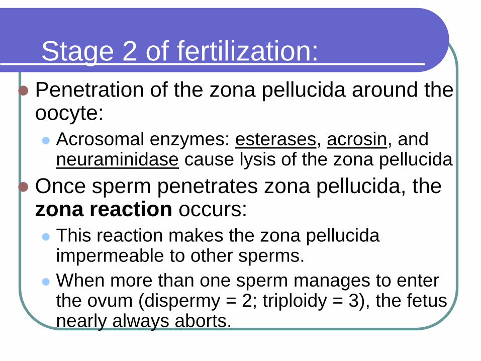
Stage 2 of fertilization:
Penetration of the zona pellucida around the oocyte:
Acrosomal enzymes: esterases, acrosin, and neuraminidase cause lysis of the zona pellucida
Once sperm penetrates zona pellucida, the zona reaction occurs:
This reaction makes the zona pellucida impermeable to other sperms.
When more than one sperm manages to enter the ovum (dispermy = 2; triploidy = 3), the fetus nearly always aborts.

Stages 3 & 4 of fertilization:
Fusion of plasma membranes of oocyte and
sperm
Head and tail of a sperm enter the cytoplasm of
the oocyte, but the sperm plasma membrane
remains behind.
2nd meiotic division of oocyte is completed
The secondary oocyte was previously arrested
in metaphase of the 2nd meiotic division, and
now forms the mature ovum and another polar
body.

Stage 5 of fertilization:
Formation of male and female pronuclei:
Chromosomal material of the sperm decondensates and enlarges
Chromosomal material of the ovum decondensates following the completion of meiosis
At this stage, the male and female pronuclei are indistinguishable.
As they grow, the pronuclei replicate their DNA still 1N (haploid)- 23 chromosomes, each in chromatid pairs

The male and female
pronuclei are
indistinguishable from one
another.
The second polar body can
be seen (arrow).
The plasma membranes of
the two pronuclei are
dissolving and one diploid
nucleus will remain.
Fusion of the pronuclei:(in vitro)
Advanced Fertility Center of Chicago
http://www.advancedfertility.com/

Stage 6 of fertilization:
Membranes of the pronuclei break down,
chromosomes condense and arrange
themselves for mitotic cell division
On membrane dissolution, there is 1 cell with
46 chromosomes = diploid (2N)
The first cleavage follows shortly, leaving 2
cells, each with 46 chromosomes.
Mitosis in the new zygote uses centrioles derived
from the sperm. The oocyte has no centrioles.

Triploidy (in vitro)
There are 3
pronuclei within this
one zygote. In the
laboratory, such
embryos are
discarded. In vivo,
such embryos almost
always abort
spontaneously.

Morula forms (solid ball of cells)
Blastula forms (hollow ball of cells)
Cells begin to grow before dividing


Placental Circulation
The placenta contains closely entwined
embryonic & maternal blood vessels for the
exchange of nutrients and wastes.

First Trimester
Organogenesis is occurring during the
first trimester.
The heart starts beating about the fourth
week.
At 8 weeks, all major organs are present in
rudimentary form.
Now called a fetus.

Second Trimester
The fetus grows to about 30 cm and is very active.
Hormone levels stabilize, hCG declines, the corpus luteum disintegrates and the placenta takes over production of progesterone.

Third Trimester
Fetal activity may decrease as space
becomes limited.
Fetus grows to about 50 cm and 3-4 kg.
Development of organs is completed.
Neural development continues even after
birth.

Labor & Delivery
Birth, parturition,
occurs through
strong rhythmic
contractions of the
uterus.
Dilation
Expulsion
Delivery of placenta

Multiple Births
Humans are usually uniparous – one offspring at a time.
Multiparous animals have several.
Fraternal twins result from ovulation & fertilization of two eggs.
Identical twins result from the splitting of one zygote.