Embed Size (px)
Citation preview

Collectively, these results demonstrate that (i)lethal synergy of influenza virus and bacterialcoinfection can result from loss of tolerance toinfection-induced tissue damage, (ii) morbidityand mortality of coinfection can be independentof pathogen burden or excessive inflammatoryresponse, and (iii) promoting tissue repair can,in principle, rescue coinfected animals frommorbidity and mortality, even without affect-ing pathogen burden. Finally, our influenza–L. pneumophila coinfection model demonstratesthe distinction between resistance and toleranceas separate host defense strategies that can bothcontribute to morbidity and mortality of infec-tious disease.
References and Notes1. L. Råberg, D. Sim, A. F. Read, Science 318, 812
(2007).2. L. Råberg, A. L. Graham, A. F. Read, Philos. Trans. R. Soc.
London Ser. B Biol. Sci. 364, 37 (2009).3. D. S. Schneider, J. S. Ayres, Nat. Rev. Immunol. 8, 889
(2008).4. R. Medzhitov, D. S. Schneider, M. P. Soares, Science 335,
936 (2012).5. C. Beadling, M. K. Slifka, Curr. Opin. Infect. Dis. 17, 185
(2004).6. J. A. McCullers, Clin. Microbiol. Rev. 19, 571 (2006).
7. J. M. Hament, J. L. Kimpen, A. Fleer, T. F. Wolfs,FEMS Immunol. Med. Microbiol. 26, 189 (1999).
8. V. T. Peltola, J. A. McCullers, Pediatr. Infect. Dis. J. 23(suppl.), S87 (2004).
9. M. Iannuzzi et al., J. Med. Case Rep. 5, 520 (2011).10. A. Shahangian et al., J. Clin. Invest. 119, 1910 (2009).11. A. R. Iverson et al., J. Infect. Dis. 203, 880 (2011).12. K. Sun, D. W. Metzger, Nat. Med. 14, 558 (2008).13. A. Didierlaurent et al., J. Exp. Med. 205, 323 (2008).14. K. H. Berger, R. R. Isberg, Mol. Microbiol. 7, 7 (1993).15. D. B. Mendel et al., Antimicrob. Agents Chemother. 42,
640 (1998).16. T. Ichinohe, Expert Rev. Vaccines 9, 1315 (2010).17. A. García-Sastre, C. A. Biron, Science 312, 879 (2006).18. T. Ren, D. S. Zamboni, C. R. Roy, W. F. Dietrich,
R. E. Vance, PLoS Pathog. 2, e18 (2006).19. A. B. Molofsky et al., J. Exp. Med. 203, 1093 (2006).20. D. S. Zamboni et al., Nat. Immunol. 7, 318 (2006).21. K. A. Archer, C. R. Roy, Infect. Immun. 74, 3325
(2006).22. R. Spörri, N. Joller, U. Albers, H. Hilbi, A. Oxenius,
J. Immunol. 176, 6162 (2006).23. N. L. La Gruta, K. Kedzierska, J. Stambas, P. C. Doherty,
Immunol. Cell Biol. 85, 85 (2007).24. J. S. M. Peiris, K. P. Y. Hui, H.-L. Yen, Curr. Opin. Immunol.
22, 475 (2010).25. N. Schmitz, M. Kurrer, M. F. Bachmann, M. Kopf, J. Virol.
79, 6441 (2005).26. T. Decker, M. Müller, S. Stockinger, Nat. Rev. Immunol. 5,
675 (2005).27. M. F. Fontana, S. Shin, R. E. Vance, Infect. Immun. 80,
3570 (2012).
28. D. K. Bhalla, J. Toxicol. Environ. Health B Crit. Rev. 2, 31(1999).
29. L. M. Crosby, C. M. Waters, Am. J. Physiol. Lung Cell.Mol. Physiol. 298, L715 (2010).
30. H. R. Wong, J. R. Wispé, Am. J. Physiol. 273, L1(1997).
31. L. A. Monticelli et al., Nat. Immunol. 12, 1045 (2011).
Acknowledgments: We thank S. Holley and C. Annicelli fortechnical assistance; T. Ichinohe, M. Linehan, and A. Iwasakifor viral strains and advice; T. Ren, M. Fontana, R. Vance,K. Archer, S. Shin, and C. Roy for L. pneumophila strains andadvice; M. Gillum for assistance with experiments; and M. Muellerand C. Lassnig for mouse infection infrastructure. The datapresented in the manuscript are tabulated in the main paperand in the supplementary materials. This work was supported bythe Howard Hughes Medical Institute (R.M.), NIH grants R01046688 and AI R01 055502 (R.M.), the Ellison Foundation (R.M.),the New England Regional Center of Excellence (R.M.), andFWF (Austrian Science Fund) grant P25235-B13 (A.M.J.). A.M.J.was a Berger Foundation fellow of the Damon Runyon CancerResearch Foundation. The authors have no conflicts of interest.
Supplementary Materialswww.sciencemag.org/cgi/content/full/science.1233632/DC1Supplementary TextFigs. S1 to S6References (32, 33)
4 December 2012; accepted 15 April 2013Published online 25 April 2013;10.1126/science.1233632
Repeated Cortico-StriatalStimulation Generates PersistentOCD-Like BehaviorSusanne E. Ahmari,1,2,3,4* Timothy Spellman,5 Neria L. Douglass,1,2 Mazen A. Kheirbek,1,2
H. Blair Simpson,1,3,4 Karl Deisseroth,6 Joshua A. Gordon,1,2 RenéHen1,2
Although cortico-striato-thalamo-cortical (CSTC) circuit dysregulation is correlated withobsessive compulsive disorder (OCD), causation cannot be tested in humans. We usedoptogenetics in mice to simulate CSTC hyperactivation observed in OCD patients. Whereasacute orbitofrontal cortex (OFC)–ventromedial striatum (VMS) stimulation did not producerepetitive behaviors, repeated hyperactivation over multiple days generated a progressiveincrease in grooming, a mouse behavior related to OCD. Increased grooming persisted for2 weeks after stimulation cessation. The grooming increase was temporally coupled with aprogressive increase in light-evoked firing of postsynaptic VMS cells. Both increased groomingand evoked firing were reversed by chronic fluoxetine, a first-line OCD treatment. Brief butrepeated episodes of abnormal circuit activity may thus set the stage for the developmentof persistent psychopathology.
OCDischaracterized by intrusive distressingthoughts (obsessions) and/or repetitivemental or behavioral acts (compulsions)
and is a leading cause of illness-related disability(1, 2). Although the pathophysiology underlyingOCD is unclear, multiple lines of evidence im-plicate dysregulation within cortico-striato-thalamo-cortical (CSTC) circuits (3–6). Specifically,functional imaging studies suggest that hyper-activity in orbitofrontal cortex (OFC) and ventro-medial striatum (VMS) is associated with OCDpathology (5, 7, 8). Furthermore, successful treat-ments are associated with reductions in hyperac-tivity (9,10).However, it is not known ifOFC-VMS
hyperactivity can directly cause OCD symptoms,because increased activity could represent adaptive,homeostatic, or unrelated processes compensat-ing for other primary abnormalities. We there-fore used an optogenetic strategy to directly testwhether hyperstimulation of glutamatergic OFC-VMS projections leads to OCD-like behaviorsin mice.
A Cre-inducible adenovirus-associated vec-tor (AAV) carrying the gene encoding channel-rhodopsin (ChR2) fused to enhanced yellowfluorescent protein (EYFP) [pAAV-Ef1a-DIO-ChR2 (H134R)-EYFP; referred to as DIO-ChR2](11) was stereotactically injected into OFC of
EMX-Cre transgenic mice to ensure specific ChR2expression in cortical glutamatergic neurons (Fig.1A) (12). Cortical Cre expression led to sustainedexpression of ChR2-EYFP (Fig. 1B). Unilateral473-nm stimulation through chronic fiber-opticimplants in OFC yielded lateralized increasedactivation of the immediate early gene c-fos (P <0.009) (Fig. 1, C and D), which demonstratedin vivo cellular activation by laser stimulation.Two weeks postinjection, EYFP staining was seenin OFC cell bodies and axons projecting to VMS(Fig. 1E), which indicated targeting of OFC-VMS projections. In vitro recordings in cortico-striatal slices demonstrated VMS field responsesafter 473-nm laser stimulation of OFC axon ter-minals in striatum (Fig. 1F). To verify adequatestimulation of ChR2-expressing OFC-VMS ter-minals in vivo, we implanted stereo opto-electrodes(optrodes) into VMS that permit combined fiber-optic stimulation and 32-channel simultaneousrecording of multiple single units (Fig. 1G). Inawake behaving mice, in vivo recordings dem-onstrated robust VMS field responses after473-nm laser stimulation of OFC axon termi-nals in striatum (Fig. 1, H and I), which showed
1Department of Psychiatry, Columbia University College ofPhysicians and Surgeons, New York, NY 10032,USA. 2Division ofIntegrative Neuroscience, New York State Psychiatric Institute,New York, NY 10032, USA. 3Division of Clinical Therapeutics,New York State Psychiatric Institute, New York, NY 10032, USA.4Anxiety Disorders Clinic and OCD Research Program, New YorkState Psychiatric Institute, New York, NY 10032, USA. 5Depart-ment of Physiology, Columbia University College of Physiciansand Surgeons, New York, NY 10032, USA. 6Departments ofPsychiatry and Bioengineering, Stanford University School ofMedicine, Stanford, CA 94305, USA.
*Corresponding author. E-mail: [email protected]
7 JUNE 2013 VOL 340 SCIENCE www.sciencemag.org1234
REPORTS
on
June
20,
201
3w
ww
.sci
ence
mag
.org
Dow
nloa
ded
from

feasibility of activation of specific cortical-VMSprojections.
Because patients with OCD have hyper-activity inOFC-striatal circuits (3–6), we predictedthat direct elevation of OFC-VMS activity wouldlead to increases in OCD-related behaviors in-cluding grooming, anxiety, and prepulse inhi-bition (PPI) deficits (13). We injected DIO-ChR2into the left OFC of EMX-Cremice and implantedfiber-optic probes unilaterally in left VMS (Fig.2A). After waiting 3 to 4 weeks for surgical re-covery and stable viral expression, we habituatedmice to the open field and fiber-optic stimula-tion apparatus. We then repeatedly elevated ac-tivity in OFC-VMS projections by stimulatingfor 5min at 10Hz for five consecutive days (10ms,1 to 5mW) (14). Grooming behavior was recordedwith digital video and scored by blind raters for
5 min before (Pre), during (Stim), and after (Post)stimulation (Fig. 2B). Whereas acute OFC-VMSstimulation did not produce grooming, a smallbut significant progressive increase in groom-ing time was noted during the prestimulationperiod on consecutive days (Fig. 2C) [repeatedmeasures analysis of variance (ANOVA), maineffect: P < 0.048; F = 4.43; Fisher’s protectedleast significant difference (PLSD): for day 3,P <0.03; for day 5, P < 0.047]. Because the pre-stimulation measurement on days 2 to 5 served asa 24-hour time stamp for effects of stimulationthe day before, this suggested that repeated stim-ulation led to chronic circuit changes that ultimate-ly resulted in sustained, stimulation-independentOCD-like behavior. Although it is possible thatstress from handling contributed to the groomingincrease in the prestimulation period, stress was
minimized by habituation to fiber-optic tether-ing daily for a week before data collection andwas identical for controls and ChR2+ mice.To resolve the time-course of the grooming in-crease, we examined a new cohort an hour afterstimulation (Groom1 hour post) (Fig. 2D). We ob-served a dramatic progressive increase in groom-ing over consecutive days using this measure(main effect: P < 0.02; F = 7.32) (Fig. 2E). Al-though total grooming time increased, there wasnot a significant increase in stereotyped syntacticgrooming chains in ChR2+ animals on day 5 ofstimulation (table S1). No differences in groom-ing time were observed between controls andChR2+ animals on day 1 of stimulation, whichindicated that ChR2 expression without laserstimulation did not lead to an increase in groom-ing. Notably, increased grooming persisted even
Fig. 1. Injection of ChR2-EYFPAAV into OFC leads to functional ChR2expression in projections from OFC to VMS. (A) Schematic diagram ofDIO-ChR2 injections. (Left) Reference sagittal section indicates injection po-sition in ventromedial OFC (VO/MO) of EMX-Cre mice (2.6 mm AP, 1.7 mm DV,0.5 mm ML). Blue shading: Cre expression in cortex and hippocampus. (Right)Cre-expressing glutamatergic cells in OFC irreversibly invert the ChR2-EYFPopen reading frame, which leads to cell type–specific ChR2-EYFP expression(green shading). EF-1a, elongation factor 1a; ITR, inverted terminal repeat;WPRE, woodchuck hepatitis virus posttranslational regulatory element;DLO, dorsolateral orbitofrontal cortex; LO, lateral orbitofrontal cortex; PrL,prelimbic cortex. (B) Confocal image of YFP-immunostaining shows unilateralChR2 expression at OFC injection site. Scale bar, 500 mm. (C) c-Fos im-munostaining demonstrates 473-nm light–induced activation of OFC inawake behaving mice through chronic fiber-optic implant. (Inset) Referencecoronal section. Blue square, stimulated; black, unstimulated. (D) Quantifica-
tion of c-Fos–positive cells in stimulated versus unstimulated OFC (P < 0.009)(n = 4 controls; 4 ChR2 mice; five sections each). (E) Targeting of OFC-VMSprojections evidenced by axonal YFP staining under fiber-optic implant site(arrow). Scale bar, 100 mm. (Inset) Low magnification. Scale bar, 500 mm.(F) Extracellular field recordings from striatal slices. Increased populationspike amplitude with increasing laser power. (Inset) Individual populationspike after 0.1-ms light pulse (3 mW); calibration bars: vertical 0.5 mV, hor-izontal 1 ms. n = 4 slices from each of three animals. (G) Schematic diagramof stereo-optrode implant in VMS. (Stereotaxic coordinates: 0.98 mm AP,3.5 mm DV, 1.25 mm ML). CPu, caudate putamen; AcbC, accumbens core;AcbSh, accumbens shell. (H) In vivo recordings in awake behaving animalsshow field responses to 473-nm stimulation of VMS terminals. Mean re-sponse to 20 flashes delivered at 0.5 Hz. Calibration bar: vertical 0.5 mV,horizontal 20 ms. (I) Raw responses to train of 10 flashes at 10 Hz. Calibrationbar: vertical 0.5 mV, horizontal 100 ms.
www.sciencemag.org SCIENCE VOL 340 7 JUNE 2013 1235
REPORTS
on
June
20,
201
3w
ww
.sci
ence
mag
.org
Dow
nloa
ded
from
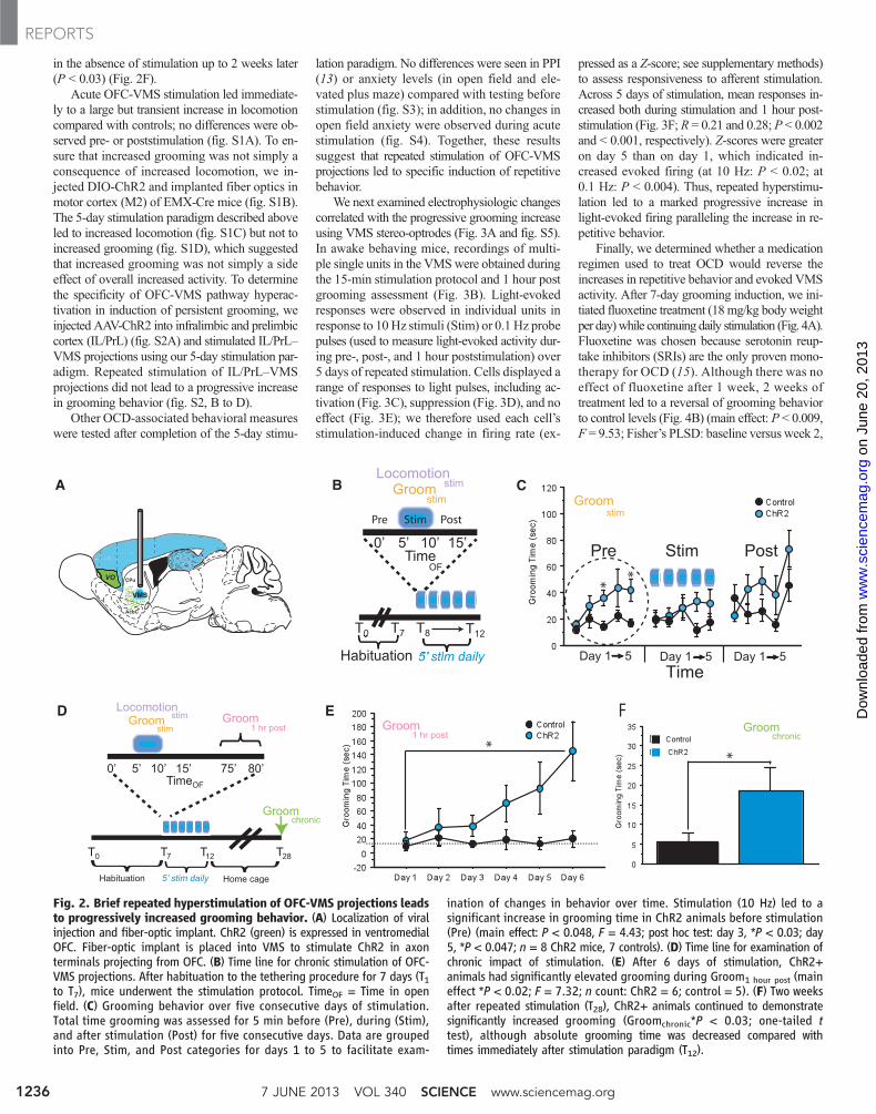
in the absence of stimulation up to 2 weeks later(P < 0.03) (Fig. 2F).
Acute OFC-VMS stimulation led immediate-ly to a large but transient increase in locomotioncompared with controls; no differences were ob-served pre- or poststimulation (fig. S1A). To en-sure that increased grooming was not simply aconsequence of increased locomotion, we in-jected DIO-ChR2 and implanted fiber optics inmotor cortex (M2) of EMX-Cre mice (fig. S1B).The 5-day stimulation paradigm described aboveled to increased locomotion (fig. S1C) but not toincreased grooming (fig. S1D), which suggestedthat increased grooming was not simply a sideeffect of overall increased activity. To determinethe specificity of OFC-VMS pathway hyperac-tivation in induction of persistent grooming, weinjected AAV-ChR2 into infralimbic and prelimbiccortex (IL/PrL) (fig. S2A) and stimulated IL/PrL–VMS projections using our 5-day stimulation par-adigm. Repeated stimulation of IL/PrL–VMSprojections did not lead to a progressive increasein grooming behavior (fig. S2, B to D).
Other OCD-associated behavioral measureswere tested after completion of the 5-day stimu-
lation paradigm. No differences were seen in PPI(13) or anxiety levels (in open field and ele-vated plus maze) compared with testing beforestimulation (fig. S3); in addition, no changes inopen field anxiety were observed during acutestimulation (fig. S4). Together, these resultssuggest that repeated stimulation of OFC-VMSprojections led to specific induction of repetitivebehavior.
We next examined electrophysiologic changescorrelated with the progressive grooming increaseusing VMS stereo-optrodes (Fig. 3A and fig. S5).In awake behaving mice, recordings of multi-ple single units in the VMSwere obtained duringthe 15-min stimulation protocol and 1 hour postgrooming assessment (Fig. 3B). Light-evokedresponses were observed in individual units inresponse to 10 Hz stimuli (Stim) or 0.1 Hz probepulses (used to measure light-evoked activity dur-ing pre-, post-, and 1 hour poststimulation) over5 days of repeated stimulation. Cells displayed arange of responses to light pulses, including ac-tivation (Fig. 3C), suppression (Fig. 3D), and noeffect (Fig. 3E); we therefore used each cell’sstimulation-induced change in firing rate (ex-
pressed as a Z-score; see supplementary methods)to assess responsiveness to afferent stimulation.Across 5 days of stimulation, mean responses in-creased both during stimulation and 1 hour post-stimulation (Fig. 3F; R = 0.21 and 0.28; P < 0.002and < 0.001, respectively). Z-scores were greateron day 5 than on day 1, which indicated in-creased evoked firing (at 10 Hz: P < 0.02; at0.1 Hz: P < 0.004). Thus, repeated hyperstimu-lation led to a marked progressive increase inlight-evoked firing paralleling the increase in re-petitive behavior.
Finally, we determined whether a medicationregimen used to treat OCD would reverse theincreases in repetitive behavior and evoked VMSactivity. After 7-day grooming induction, we ini-tiated fluoxetine treatment (18mg/kg body weightper day)while continuing daily stimulation (Fig. 4A).Fluoxetine was chosen because serotonin reup-take inhibitors (SRIs) are the only proven mono-therapy for OCD (15). Although there was noeffect of fluoxetine after 1 week, 2 weeks oftreatment led to a reversal of grooming behaviorto control levels (Fig. 4B) (main effect: P < 0.009,F = 9.53; Fisher’s PLSD: baseline versus week 2,
Fig. 2. Brief repeated hyperstimulation of OFC-VMS projections leadsto progressively increased grooming behavior. (A) Localization of viralinjection and fiber-optic implant. ChR2 (green) is expressed in ventromedialOFC. Fiber-optic implant is placed into VMS to stimulate ChR2 in axonterminals projecting from OFC. (B) Time line for chronic stimulation of OFC-VMS projections. After habituation to the tethering procedure for 7 days (T1to T7), mice underwent the stimulation protocol. TimeOF = Time in openfield. (C) Grooming behavior over five consecutive days of stimulation.Total time grooming was assessed for 5 min before (Pre), during (Stim),and after stimulation (Post) for five consecutive days. Data are groupedinto Pre, Stim, and Post categories for days 1 to 5 to facilitate exam-
ination of changes in behavior over time. Stimulation (10 Hz) led to asignificant increase in grooming time in ChR2 animals before stimulation(Pre) (main effect: P < 0.048, F = 4.43; post hoc test: day 3, *P < 0.03; day5, *P < 0.047; n = 8 ChR2 mice, 7 controls). (D) Time line for examination ofchronic impact of stimulation. (E) After 6 days of stimulation, ChR2+animals had significantly elevated grooming during Groom1 hour post (maineffect *P < 0.02; F = 7.32; n count: ChR2 = 6; control = 5). (F) Two weeksafter repeated stimulation (T28), ChR2+ animals continued to demonstratesignificantly increased grooming (Groomchronic*P < 0.03; one-tailed ttest), although absolute grooming time was decreased compared withtimes immediately after stimulation paradigm (T12).
7 JUNE 2013 VOL 340 SCIENCE www.sciencemag.org1236
REPORTS
on
June
20,
201
3w
ww
.sci
ence
mag
.org
Dow
nloa
ded
from

P < 0.003). This delayed response is consistentwith the delayed onset of effective SRI treatmentin OCDpatients.We also repeated this experimentusing a vehicle control group (Fig. 4C). Again,2 weeks of fluoxetine led to reversal of inducedgrooming (Fig. 4D). Moreover, in a separate co-hort of stereo-optrode–implanted animals, the in-crease in light-evoked activity induced by repeated10 Hz stimulation was normalized after chronicfluoxetine (Fig. 4, E and F).
Repeated hyperactivation of OFC-VMS pro-jections generates a progressive increase in groom-ing, temporally linked to a cumulative increasein VMS light-evoked firing. Acute stimulationof the OFC-VMS pathway was not sufficientto produce OCD-relevant excessive grooming(3, 16–18). The behavioral change was persist-ent, becoming stimulation-independent with-in 6 days. Although classic theories suggestthat abnormal repetitive behaviors, including
OCD symptoms, directly result from hyper-activity in CSTC loops (19–21), causation hasbeen difficult to prove. Though genetic and phar-macologic manipulations of norepinephrine anddopamine can lead to transient increases in re-petitive behaviors (22), the interventions werenot limited to specific circuits, and associatedelectrophysiologic changes were observed in mul-tiple brain regions. Our optogenetic system per-mits activation of specific cortico-striatal circuitsand genetic definition of the activated cell-typeas cortical glutamatergic projection neurons.
Our in vivo electrophysiology data suggesta circuit-based mechanism for establishment ofrepetitive behaviors. Repeated hyperstimulationled to a marked progressive increase in light-evoked firing paralleling the increase in groom-ing, suggesting plasticity at OFC-VMS synapsesthat builds over consecutive days. We speculatethat brief episodes of light-induced activity leadto long-lasting changes that prime OFC-VMSsynapses, decreasing the activation threshold duringsubsequent bouts of stimulation. In turn, increasedactivity at OFC-VMS synapses may transmit in-formation through the CSTC circuit (23–25) andlead to multiple downstream events that ultimate-ly reinforce repetitive behaviors, including (i) plas-ticity in downstream structures such as thalamusand prefrontal cortex (26), and (ii) increased mo-tivational saliencymediated by the ventral tegmen-tal area (22). This mechanismwould be consistentwith the observed fluoxetine effects, since selectiveSRIs have been shown to reduce primary rewardprocessing (27, 28).
OCD is a heterogeneous disorder. Our studytherefore may have greater relevance for partic-ular OCD subtypes. For example, dimensionalmodels of OCD have been proposed in whichdifferent types of obsessions and compulsionsare associated with different circuits (29, 30). Be-cause our results suggest that repeated stimulationof OFC-VMS projections led to specific induc-tion of repetitive grooming, our model may be ofparticular importance for OCD patients with pre-dominant contamination concerns.
Our findings yield new insight into howpsychopathology could develop. Only 5 minof stimulation per day was sufficient to lead tosustained significant behavioral effects. Thisraises the possibility that pathological changes,including compulsions in OCD, may result fromsmall but repeated bursts of abnormal neu-ronal activity and also offers suggestions fornew treatment approaches or refinements ofexisting therapies for disorders characterizedby repetitive behaviors. For example, our dataare consistent with recent clinical studies dem-onstrating efficacy of ventral capsule–ventralstriatum deep brain stimulation in OCD (31, 32),which is thought to act via inhibition of OFChyperactivity. Optogenetic approaches couldbe used to dissect circuit mechanisms under-lying deep brain stimulation and other treat-ments, with a goal of identifying new treatmenttargets.
Fig. 3. Repeateddailystim-ulation of OFC-VMS pro-jections leads to increasedevoked firing. (A) (Left) Sche-matic diagramof stereo-optrodeimplant site. (Right) Placementvisualized via implanting astereo-optrode dipped inHoecsht stain (1:1000). Scalebar, 500 mm. (B) Stimula-tion protocol used for in vivorecording. (C to E) Represent-ative peristimulus spike his-tograms (5-ms time bins) ofthree neurons recorded dur-ing 10 Hz stimulation (left)and 0.1 Hz probe pulses(1 hour poststimulation onright). Baseline spontaneousfiring rate for each cell isshown as pink dashed line.Cells exhibited varied stimu-lus responsiveness, includingevoked activation (C), evokedsuppression (D), and no re-sponse (E). (F) Light-evokedfiring (measured by peristim-ulus z-scores) across 5 daysof stimulation both during10 Hz stimulation (Stim) andduring 0.1 Hz probe pulses1 hour after stimulation(1 hour post) (*P < 0.021 andP < 0.004). Negative Z-scoresfor 0.1 Hz on days 1 and 2indicate net suppression ofevoked firing rate duringGroom1 hour post after thefirst two epochs of 10 Hzstimulation.
www.sciencemag.org SCIENCE VOL 340 7 JUNE 2013 1237
REPORTS
on
June
20,
201
3w
ww
.sci
ence
mag
.org
Dow
nloa
ded
from

References and Notes1. C. J. L. Murray, A. D. Lopez, The Global Burden of Disease:
A Comprehensive Assessment of Mortality and Disabilityfrom Diseases, Injuries, and Risk Factors in 1990 andProjected to 2020, Global burden of disease and injuryseries; vol. 1 (Harvard School of Public Health, HarvardUniv. Press, Cambridge, MA, 1996).
2. R. C. Kessler et al., Arch. Gen. Psychiatry 62, 593 (2005).3. J. T. Ting, G. Feng, Curr. Opin. Neurobiol. 21, 842 (2011).4. R. Marsh, T. V. Maia, B. S. Peterson, Am. J. Psychiatry
166, 664 (2009).5. M. R. Milad, S. L. Rauch, Trends Cogn. Sci. 16, 43
(2012).6. C. Pittenger, M. H. Bloch, K. Williams, Pharmacol. Ther.
132, 314 (2011).7. S. Saxena, R. G. Bota, A. L. Brody, Semin. Clin.
Neuropsychiatry 6, 82 (2001).8. J. Y. Rotge et al., Biol. Psychiatry 65, 75 (2009).9. S. L. Rauch, C. R. Savage, N. M. Alpert, A. J. Fischman,
M. A. Jenike, Biol. Psychiatry 42, 446 (1997).10. J. L. Abelson et al., Biol. Psychiatry 57, 510 (2005).
11. H. C. Tsai et al., Science 324, 1080 (2009).12. J. A. Gorski et al., J. Neurosci. 22, 6309 (2002).13. S. E. Ahmari, V. B. Risbrough, M. A. Geyer, H. B. Simpson,
Neuropsychopharmacology 37, 1216 (2012).14. J. Mattis et al., Nat. Methods 9, 159 (2012).15. L. M. Koran, G. L. Hanna, E. Hollander, G. Nestadt,
H. B. Simpson; American Psychiatric Association, Am. J.Psychiatry 164 (suppl.), 5 (2007).
16. S. V. Shmelkov et al., Nat. Med. 16, 598, 1p, 602 (2010).17. J. M. Welch et al., Nature 448, 894 (2007).18. S. K. Chen et al., Cell 141, 775 (2010).19. T. R. Insel, J. T. Winslow, Psychiatr. Clin. North Am.
15, 813 (1992).20. D. R. Rosenberg, M. S. Keshavan, Biol. Psychiatry 43,
623 (1998).21. L. R. Baxter Jr. et al., Am. J. Psychiatry 145, 1560 (1988).22. K. Dzirasa et al., J. Neurosci. 30, 6387 (2010).23. S. K. Bourne, C. A. Eckhardt, S. A. Sheth, E. N. Eskandar,
Front Integr Neurosci 6, 29 (2012).24. A. M. Graybiel, S. L. Rauch, Neuron 28, 343 (2000).25. S. N. Haber, S. L. Rauch, Neuropsychopharmacology
35, 1 (2010).
26. E. G. Antzoulatos, E. K. Miller, Neuron 71, 243(2011).
27. B. Abler, G. Grön, A. Hartmann, C. Metzger, M. Walter,J. Neurosci. 32, 1329 (2012).
28. C. McCabe, Z. Mishor, P. J. Cowen, C. J. Harmer,Biol. Psychiatry 67, 439 (2010).
29. D. Mataix-Cols et al., Arch. Gen. Psychiatry 61, 564 (2004).30. J. F. Leckman et al., Depress. Anxiety 27, 507 (2010).31. B. D. Greenberg et al., Mol. Psychiatry 15, 64 (2010).32. P. P. de Koning, M. Figee, P. van den Munckhof,
P. R. Schuurman, D. Denys, Curr. Psychiatry Rep. 13,274 (2011).
Acknowledgments: We thank H. B. Simpson and C. Kellendonkfor critical discussions and reading of the manuscript andD. Flicker and M. Cloidt for behavioral scoring assistance.S.E.A. is supported by National Institute of Mental Health(NIMH) K08MH087718; the Louis V. Gerstner, Jr., ScholarsProgram; the Irving Institute for Clinical and TranslationalResearch; the Gray Matters Foundation; the Leon LevyFoundation; and a Brain and Behavior Research FoundationNARSAD Young Investigator Award. M.A.K. is supported by
Fluoxetine 18mg/kg
Stim
Fluoxetine 18mg/kg
Stim
1 hour PostStim
1 hour Post 1 hour Post
Fig. 4. Perseverative grooming and elevated evoked firing rate areresolved by chronic, but not acute, fluoxetine treatment. (A) Exper-imental time line for fluoxetine wash-out experiment. (B) Two weeks offluoxetine treatment reduced grooming to level of controls. Main effect:P < 0.009; F = 9.53; Fisher’s PLSD: baseline versus week 2, ***P < 0.003.Increased grooming was reestablished after a 1-week fluoxetine wash-out.Main effect: P < 0.09; F = 3.58. n values: ChR2+ mice = 8; controls = 7. (C)Experimental time line for fluoxetine versus vehicle experiment. (D) Two
weeks of fluoxetine treatment reduced grooming to levels of vehicle-treatedanimals. Main effect: P < 0.14; F = 2.59; Fisher’s PLSD: baseline versusweek 2, *P < 0.04. Fluoxetine: n = 7; vehicle: n = 6. (E) (Left) In stereo-optrode–implanted animals, peristimulus Z-scores for 10 Hz stimuli nor-malized after 2 weeks of fluoxetine (P < 0.028); after 2-week wash-out,Z-scores returned to pretreatment levels. (Right) Peristimulus Z-scores for0.1 Hz probe pulses showed a nonsignificant decrease after fluoxetine treat-ment, which returned to pretreatment levels after wash-out.
7 JUNE 2013 VOL 340 SCIENCE www.sciencemag.org1238
REPORTS
on
June
20,
201
3w
ww
.sci
ence
mag
.org
Dow
nloa
ded
from
NIMH K01MH099371, the Sackler Institute, and a NARSADYoung Investigator Award. K.D. is supported by the HowardHughes Medical Institute, NIH, California Institute forRegenerative Medicine, and the Defense Advance ResearchProjects Agency The Reorganization and Plasticity to AccelerateInjury Recovery (REPAIR) Program. J.G. is supported byNIH R01 MH096274, the Hope for Depression Research
Foundation, and the International Mental Health ResearchOrganization. R.H. is supported by the Hope for DepressionResearch Foundation.
Supplementary Materialswww.sciencemag.org/cgi/content/full/340/6137/1234/DC1Materials and Methods
Figs. S1 to S5Table S1References (33–43)
3 January 2013; accepted 3 April 201310.1126/science.1234733
Geniculocortical Input Drives GeneticDistinctions Between Primary andHigher-Order Visual AreasShen-Ju Chou,1*† Zoila Babot,1* Axel Leingärtner,1‡ Michele Studer,2§Yasushi Nakagawa,1|| Dennis D. M. O’Leary1¶
Studies of area patterning of the neocortex have focused on primary areas, concluding thatthe primary visual area, V1, is specified by transcription factors (TFs) expressed by progenitors.Mechanisms that determine higher-order visual areas (VHO) and distinguish them from V1 areunknown. We demonstrated a requirement for thalamocortical axon (TCA) input by geneticallydeleting geniculocortical TCAs and showed that they drive differentiation of patterned geneexpression that distinguishes V1 and VHO. Our findings suggest a multistage process for areapatterning: TFs expressed by progenitors specify an occipital visual cortical field that differentiatesinto V1 and VHO; this latter phase requires geniculocortical TCA input to the nascent V1 thatdetermines genetic distinctions between V1 and VHO for all layers and ultimately determinestheir area-specific functional properties.
Theneocortex is patterned into functionallydistinct fields that include primary sensoryareas, which receivemodality-specific sen-
sory input from thalamocortical axons (TCAs)that originate from the principal sensory nuclei ofthe dorsal thalamus (dTh), and higher-order sen-sory areas that are connected with the primaryareas through intracortical projections (1). Studiesof mechanisms that pattern the neocortex intoareas, known as arealization, have focused onprimary areas and have led to the prevailingmodelthat genetic mechanisms intrinsic to the neocor-tex are predominant in arealization (2). Tran-scription factors (TFs) expressed in neocorticalprogenitors determine the size and position ofprimary areas (2–5) and regulate guidance in-formation that governs the area-specific target-ing of TCAs (6). However, roles for TCAs inarealization remain vague (7–10), and importantfeatures of arealization, such as differential gene
expression in the embryonic neocortex that re-lates to nascent areas, develop independently ofTCA input (9, 10).
Higher-order areas outnumber primary areasby roughly 10-fold; for example, in mouse, ninehigher-order visual areas (VHO) are positionedaround the primary visual area (V1) within theoccipital neocortex (11). However, mechanismsthat specify and regulate differentiation of the par-ticular properties of higher-order areas and distin-guish them from primary areas have yet to beexplored (11, 12). To perform genetic manipu-lations of dTh neurons required for these studies,we created RORa-IRES-Cre mice (RORaCre;fig. S1, A and B) with RORa function intact andexpression of Cre recombinase driven by RORaregulatory elements (13). Crossing this RORaCre
mouse to conditional reporter lines (fig. S1) re-vealed Cre-mediated recombination in neuronsof the principal sensory nuclei in dTh at embry-onic day 14.5 (E14.5), shortly after they becomepostmitotic (14), with robust recombination inthe dorsal lateral geniculate nucleus (dLG) (fig.S1, C to K), which forms the geniculocorticalTCA projection that relays visual informationfrom the eyes selectively to V1. Little or no re-combinationwas detected in the neocortex throughthe end of the first postnatal week, encompassingthe differentiation of cortical areas and the timeframe of our study (fig. S1, C to K).
We crossed RORaCre mice to mice in whichthe third exon of the COUP-TF1 gene is flankedby loxP sites, i.e. floxed (fl) COUP-TF1 [COUP-TF1fl/fl is described in (5)], because COUP-TF1 isstrongly expressed in dLG, COUP-TF1 deletion
diminishes axon growth (15), and most TCAsfail to reach the cortex in COUP-TF1–null mice(16). COUP-TF1–null mice are not useful for ourstudies because of viability issues and defects incortical development (16). In contrast, theconditional knockout (cKO) mice (RORaCre/+
or RORaCre/Cre; COUP-TF1fl/fl) were viable andretained robust COUP-TF1 expression in theneocortex (fig. S2, A and B), but COUP-TF1wasdeleted from dLG by E15.5 (fig. S2, A and B),and dLG size in cKO mice progressively de-creased from the wild-type (WT) size embryon-ically to virtually absent by postnatal day 7 (P7)(figs. S2, C and D, and S3).
To visualize TCA projections in the cortex,we first used serotonin [5-hydroxytryptamine(5-HT)] immunostaining on tangential sectionsof flattened P7 cortices. In P7 WT mice, 5-HTstaining revealed the geniculocortical TCA pro-jection from dLG to V1, as well as TCA projec-tions from the ventroposterior nucleus (VP) to theprimary somatosensory area (S1) and from themedial geniculate nucleus (MG) to the primaryauditory area (A1) (Fig. 1A). In P7 cKO mice,5-HT staining showed that TCA projections toS1 and A1 were intact, but the geniculocorticalTCA projection to V1 was absent (Fig. 1A). Theloss of geniculocortical input to V1 in P7 cKOmice was confirmed by anterograde and retro-grade axon tracing from dLG and V1 (fig. S4, Aand B) and by crossing the cKO mice to aROSA26-GAP43-eGFP reporter line that labelsTCAs by RORaCre reporter activation (fig. S5A).Thus, conditional deletion of COUP-TF1 fromdLG using the RORaCre line resulted in de-letion of the geniculocortical TCA projectionby P7, but COUP-TF1 remained intact in thecortex.
To determine the time course of the geniculo-cortical TCA projection in cKO mice as com-pared to WT mice, we bred RORaCre mice oneither a WT (COUP-TF1fl/+; RORaCre/+) or cKO(COUP-TF1fl/fl; RORaCre/+ or COUP-TF1fl/fl;RORaCre/Cre) background, to a conditional re-porter line (Ai14 tdTomato) (17). Activation ofthe tdTomato reporter with the RORaCre linelabeled, at high resolution, geniculocortical TCAsfrom the dLG and TCAs from VP and MG pro-jecting to S1 and A1 (Fig. 1B). GeniculocorticalTCAs extend tangentially in the subplate andunderlie the cortical plate (CP) of nascent V1by E16.5, invade after birth the overlying V1CP, and over the first postnatal week arborizein V1 layer 4, their predominant target layer(18). At E16.5, before TCAs invade the CP, theappearance of tdTomato-labeled TCAs was in-distinguishable betweenWTand cKOmice, with
1Molecular Neurobiology Laboratory, The Salk Institute forBiological Studies, La Jolla, CA, USA. 2Institute of BiologyValrose, INSERM, Nice, France.
*These authors contributed equally to this work.†Present address: Institute of Cellular and Organismic Biology,Academia Sinica, Taipei, Taiwan.‡Present address: University Cancer Center Hamburg, Univer-sity Medical Center Hamburg-Eppendorf, Hamburg, Germany.§Present address: Institute of Biology Valrose, iBV, UMRINSERM1091/CNRS7277/UNS, Nice, F-06108, France; and Uni-versity of Nice Sophia-Antipolis, UFR Sciences, Nice, F-06108,France.||Present address: Department of Neuroscience, Stem CellInstitute and Developmental Biology Center, University ofMinnesota, Minneapolis, MN 55455, USA.¶Corresponding author. E-mail: [email protected]
www.sciencemag.org SCIENCE VOL 340 7 JUNE 2013 1239
REPORTS
on
June
20,
201
3w
ww
.sci
ence
mag
.org
Dow
nloa
ded
from


![An Introduction to Genomic Ranges Classes · | b chr2 [2, 8] + | 0.888888888888889 c chr2 [3, 9] + | 0.777777777777778-----seqinfo:](https://img.dokumen.tips/doc/110x75/60d780e78509dc5ecd7adc88/an-introduction-to-genomic-ranges-classes-b-chr2-2-8-0888888888888889.jpg)