Embed Size (px)
Citation preview

A new type of human genetic disease mutation was unexpectedly discovered more than 18 years ago — expansion of a repeated microsatellite sequence. At least 22 inherited disorders, all involving the neuraxis, are now known to be caused by expanded repeats (TABLE 1). Repeat expansion diseases include some of the most common inherited diseases, such as Huntington’s disease (HD) and myotonic dystrophy. In 1991, two repeat expansion mutations — for the X‑linked disorders fragile X mental retardation syndrome (FMR1) and spinal and bulbar muscular atrophy (SBMA) — were reported to produce disease phenotypes by encoding proteins with expanded poly‑amino acid tracts1,2. We now recognize SBMA as the first member of a subcategory of repeat expansion disorders known as the ‘CAG/polyglutamine’ repeat diseases. Although it was initially thought that FMR1 was caused by an expanded polyarginine tract, further work indicated that the CGG repeat expansion in FMR1 is located in the 5′ UTR. The expansion reduces expres‑sion of FMR1 by promoting DNA hypermethylation at the promoter3–5.
Since these seminal discoveries, various repeat expan‑sion mutations have been identified (TABLE 1), and at least four mechanisms of disease are now known: loss of func‑tion of the gene containing the repeat; gain of function due to production of a protein containing a polyglutamine tract expansion; gain of function due to production of RNA containing an expanded CUG tract; and gain of function due to production of a protein containing a polyalanine tract expansion. Classification of the repeat expansion diseases into these four mechanistic categories
typically reflects both the sequence composition of the repeat and the location of the repeat in a gene (TABLE 1).
Despite these advances, there are still difficulties — as there were at the genesis of the field — in determining exactly how repeat expansion mutations cause inherited human diseases. Therefore, current classification schemes are at risk of becoming outdated in the future. Indeed, a number of recent findings have revealed the potential complexity of the molecular mechanisms underlying disease pathogenesis. For example, post-translational modification of disease proteins has been identified as a key step in the pathogenic cascade of CAG/polyglutamine disease, and autophagy has been implicated in the degra‑dation of misfolded proteins. Molecular genetic studies, driven by recent advances in our understanding of the transcriptome, suggest that bidirectional transcription and chromatin structure could be involved in repeat dis‑ease pathology and genetic instability. In this Review we examine the most compelling of these paradigm‑shifting advances, focusing on RNA toxicity, autophagy, post‑translational modification, bidirectional transcription and genomic structure. We consider advances in these areas for the relevant subcategory of repeat expansion disease and the broader repeat disease field, and note how these findings might be translated into novel, widely applicable therapies.
RNA as a driver of toxicityA shared mechanism for myotonic dystrophy type 1 and 2. One of the most striking developments in the repeat dis‑ease field has been the realization that expanded repeats in
*Division of Genetics, Department of Pediatrics, Department of Cellular and Molecular Medicine and Institute for Genomic Medicine, University of California-San Diego, La Jolla, California 92093, USA. ‡Rady Children’s Hospital, San Diego, California 92123, USA.§Department of Developmental Neurobiology, St Jude Children’s Research Hospital, Memphis, Tennessee 38105, USA.Correspondence to A.R.L.S. e-mail: [email protected]:10.1038/nrg2748 Published online 23 February 2010
Post-translational modificationA covalent chemical modification of a protein that takes place after translation.
Repeat expansion disease: progress and puzzles in disease pathogenesisAlbert R. La Spada*‡ and J. Paul Taylor§
Abstract | Repeat expansion mutations cause at least 22 inherited neurological diseases. The complexity of repeat disease genetics and pathobiology has revealed unexpected shared themes and mechanistic pathways among the diseases, such as RNA toxicity. Also, investigation of the polyglutamine diseases has identified post-translational modification as a key step in the pathogenic cascade and has shown that the autophagy pathway has an important role in the degradation of misfolded proteins — two themes that are likely to be relevant to the entire neurodegeneration field. Insights from repeat disease research are catalysing new lines of study that should not only elucidate molecular mechanisms of disease but also highlight opportunities for therapeutic intervention for these currently untreatable disorders.
REVIEWS
NATURe RevIeWS | Genetics vOlUMe 11 | ApRIl 2010 | 247
© 20 Macmillan Publishers Limited. All rights reserved10

Table 1 | Clinical and molecular characteristics of inherited neurological repeat expansion disorders
Disease Main clinical features causal repeat (gene)
Repeat location
Mechanism or category
comments
DM1 Muscle weakness, myotonia, cardiac-endocrine-GI disease, MR
CTG (DM1, also known as DMPK)
3′ UTR RNA GOF A very common form of muscular dystrophy
DM2 Muscle weakness, myotonia, cardiac-endocrine-GI disease
CTG (ZNF9, also known as CNBP)
Intron RNA GOF A striking phenocopy of DM1
DRPLA Seizures, choreoathetosis, ataxia, cognitive decline
CAG (ATN1) Coding region
Polyglutamine GOF
Very rare, most patients are in Japan
FMR1 MR, facial dysmorphism, autism CGG (FMR1) 5′ UTR Hypermethylation of promoter, LOF
Most common inherited MR
FMR2 MR, hyperactivity GCC (FMR2) 5′ UTR LOF Needs to be ruled out in X-linked MR
FRDA Ataxia, sensory loss, weakness, diabetes mellitus, cardiomyopathy
GAA (FXN) Intron LOF, phenocopy of mitochondrial disease
Most common inherited ataxia in Caucasian ethnicity
FXTAS Ataxia, intention tremor, parkinsonism
CGG (FMR1) 5′ UTR RNA GOF Premutation carriers only
HD Chorea, dystonia, cognitive decline, psychiatric disease
CAG (HTT) Coding region
Polyglutamine GOF
One of the most common inherited diseases in humans
HDL2 Chorea, dystonia, cognitive decline
CTG (JPH3) 3′ UTR, coding region
RNA GOF, poly-amino acid GOF and/or LOF?
A striking phenocopy of HD
Myoclonic epilepsy of Unverricht and Lundborg
Photosensitive myoclonus, tonic–clonic seizures, cerebellar degeneration
CCCCGCCCCGCG (CSTB)
Promoter LOF Rare autosomal recessive disorder found in Finland and N. Africa
OPMD Eyelid weakness, dysphagia, proximal limb weakness
GCG (PABPN1) Coding region
Polyalanine GOF Modest expansion causes disease
SBMA Proximal limb weakness, lower motor neuron disease
CAG (AR) Coding region
Polyglutamine GOF
Phenotype includes LOF androgen insensitivity
SCA1 Ataxia, dysarthria, spasticity, ophthalmoplegia
CAG (ATXN1) Coding region
Polyglutamine GOF
Accounts for 6% of all dominant ataxia
SCA2 Ataxia, slow eye movement, hyporeflexia, motor disease, occasional parkinsonism
CAG (ATXN2) Coding region
Polyglutamine GOF
ATXN2 protein may not reside in the nucleus
SCA3 Ataxia, dystonia, lower motor neuron disease
CAG (ATXN3) Coding region
Polyglutamine GOF
Most common dominant ataxia
SCA6 Ataxia, dysarthria, sensory loss, occasionally episodic
CAG (CACNA1A) Coding region
Polyglutamine GOF
Causal gene encodes a subunit of a P/Q-type Ca2+ channel
SCA7 Ataxia, dysarthria, cone-rod dystrophy retinal disease
CAG (ATXN7) Coding region
Polyglutamine GOF
Clinically distinct as patients have retinal disease
SCA8 Ataxia, dysarthria, nystagmus, spasticity
CTG/CAG (ATXN8) Untranslated RNA, coding region
RNA GOF and polyglutamine GOF
Many cases of reduced penetrance
SCA10 Ataxia, dysarthria, seizures, dysphagia
ATTCT (ATXN10) Intron RNA GOF? Huge repeats; only Mexican ancestry?
SCA12 Tremor, ataxia, spasticity, dementia CAG (PPP2R2B) Promoter, 5′ UTR?
Unknown Causal gene encodes a phosphatase
SCA17 Ataxia, dementia, chorea, seizures, dystonia
CAG (TBP) Coding region
Polyglutamine GOF
Causal gene encodes a common transcription factor (TBP)
Syndromic/non-syndromic X-linked mental retardation
MR alone, with seizures or with dysarthria and dystonia
GCG (ARX) Coding region
Probably LOF Associated with West syndrome or Partington syndrome
AR, androgen receptor; ARX, aristaless-related homeobox; ATN1, atrophin 1; ATXN, ataxin; CACNA1A, voltage-dependent P/Q-type calcium channel subunit α-1A; CSTB, cystatin B; DM, myotonic dystrophy; DMPK, DRPLA, dentatorubral-pallidoluysian atrophy; FMR1, fragile X mental retardation syndrome; FMR2, fragile X E mental retardation; FRDA, Friedreich’s ataxia; FXN, frataxin; FXTAS, fragile X tremor ataxia syndrome; GI, gastrointestinal; GOF, gain of function; HD, Huntington’s disease; HDL2, Huntington’s disease-like 2; HTT, huntingtin; JPH3, junctophilin 3; LOF, loss of function; MR, mental retardation; OPMD, oculopharyngeal muscular dystrophy; PABPN1, poly(A)-binding protein, nuclear 1; PPP2R2B, protein phosphatase 2 regulatory subunit B, β isoform; SBMA, spinal and bulbar muscular atrophy; SCA, spinocerebellar ataxia; TBP, TATA box-binding protein; ZNF9, zinc finger 9.
R E V I E W S
248 | ApRIl 2010 | vOlUMe 11 www.nature.com/reviews/genetics
© 20 Macmillan Publishers Limited. All rights reserved10

HaploinsufficiencyA condition in a diploid organism in which a single functional copy of a gene results in a phenotype, such as a disease.
Contiguous gene syndromeA multi-symptom disorder caused by the deletion of a large sequence of DNA that encodes several genes.
Phenocopy A phenotype that is closely similar to a phenotype determined by a different gene.
AnticipationThe tendency of certain diseases to have an earlier age of onset and increasing severity in successive generations.
Myotonia The failure of muscle to relax immediately after voluntary contraction has stopped.
Myopathy A disease of the muscle.
PremutationAn unstable mutation that has no phenotypic effect but that is highly likely to mutate to a full mutation during transmission through the germ line, as is seen with some expanding trinucleotide repeats.
Inclusions Accumulations of proteins and other materials that are visualized as discrete entities at the light microscope level, often after the application of special stains or antibodies.
transcripts can cause cellular toxicity and neurodegenera‑tion by altering the splicing machinery. In 1992, a CTG repeat expansion in the 3′ UTR of a protein kinase gene was found to be the cause of myotonic dystrophy type 1 (DM1), the most common form of adult muscular dystrophy6–8. This discovery posed a puzzling question: how does a dominantly inherited repeat expansion in the non‑coding region of a gene produce a disease phenotype that affects many different tissues? Although some evi‑dence emerged for reduced dosage of the protein kinase DM1 (also known as DMpK) as the cause of certain DM1 features, haploinsufficiency could not account for most fac‑ets of DM1 pathophysiology9. As the DM1 gene is located in a gene‑rich region, some argued that DM1 resulted from altered expression of the DM1 protein kinase in combination with altered expression of adjacent genes and proposed that the CTG repeat expansion mimicked a contiguous gene syndrome10. This ‘field theory’ of DM1 pathogenesis gained support when knockout mice lack‑ing the gene downstream of DM1, SIX5, were found to develop cataracts, which are also a feature of DM111,12.
However, the field theory of DM1 pathogenesis could not easily be reconciled with a DM1 phenocopy caused by a different genetic locus13. This disorder, known as myotonic dystrophy type 2 (DM2), has a phenotype sim‑ilar to DM1. A distinction is occasional congenital pres‑entation and mental retardation in patients with DM1 who receive large CTG repeat expansions because of maternal anticipation14,15. DM2 is caused by expansion of a CCTG tetranucleotide repeat in the zinc finger 9 (ZNF9, also known as CNBP) gene16, the normal func‑tion of which does not match any of the genes at the DM1 locus. Hence, the parsimonious conclusion was that RNA transcripts containing a CUG repeat expan‑sion — whether in a triplet repeat or a CCUG repeat — initiate a shared pathogenic cascade.
A role for RNA toxicity in DM1 was first suggested by RNA foci in cells from patients with DM1 (REF. 17), and indistinguishable RNA foci were found in samples from patients with DM2 (REF. 16). The validity of the RNA toxicity model was supported by a mouse model of myotonic dystrophy in which a 250 CTG repeat in the 3′ UTR of an unrelated transgene — skeletal actin — was shown to cause myotonia and myopathy18. In another study, preceding the identification of DM2, altered splic‑ing due to increased function of CUG‑binding protein 1 (CUGBp1) emerged as a plausible mechanism for the RNA gain‑of‑function toxicity19. Subsequent work has implicated reduced function of muscleblind 1 (MBNl1) due to its sequestration into CUG repeat‑rich foci as a contributing factor in the splicing abnormalities in DM1 and DM2 (REF. 20). MBNl1 is one of a family of three proteins that have been shown to bind specifi‑cally to double‑stranded RNA hairpins formed by CUG repeats21. Although increased CUG repeat‑containing RNAs seem to increase CUGBp1 levels, the formation of CUG and CCUG ribonuclear foci by expanded CUG repeat‑containing RNAs results in MBNl1 sequestration and depleted function. Therefore, the disease model is increased CUGBp1 function combined with decreased MBNl1 function.
Studies in Drosophila melanogaster showed that loss of function of muscleblind prevents terminal differentia‑tion of retinal and muscle cells22,23. In mammals, MBNl1 serves an analogous function, as it favours the splicing of target genes into adult isoforms so, in DM1 and DM2, reduced MBNl1 function allows fetal isoform produc‑tion to persist in adult tissues and affects the expression level of many target transcripts24. A recent study found that loss of MBNl1 can explain the majority of splicing alterations in DM1 and identified extracellular matrix genes as a common target of expanded CUG repeat‑containing RNAs, linking DM1 pathogenesis with other connective tissue diseases and muscular dystrophies24. The pleiotropic phenotype of the myotonic dystrophies is therefore believed to result from the altered splicing of genes that produce proteins that function in path‑ways that are linked to disease features. For example, altered splicing of the chloride channel gene and insu‑lin receptor genes are linked to myotonia and glucose intolerance, respectively.
Fragile X tremor ataxia syndrome: two mechanisms at the FMR1 locus. Advances in our understanding of myo‑tonic dystrophy pathogenesis have set the stage for the characterization of other RNA gain‑of‑function repeat diseases. One of the most intriguing of these is fragile X tremor ataxia syndrome (FXTAS)25,26. This disorder occurs primarily in male carriers of the FMR1 premutation allele and seems to result from an entirely different molec‑ular mechanism from FMR1. CGG expansions exceeding ~200 repeats produce a mental retardation phenotype by reducing expression of FMR1, whereas CGG expansions of 55–200 repeats result in higher expression of a tran‑script containing the CGG tracts. The RNA molecules with the expanded CGG tract initiate a cascade of events that culminate in central nervous system (CNS) neurode‑generation, which is characterized by ubiquitin‑positive inclusions in the nuclei of neurons and glia27. Studies in D. melanogaster showed that CGG repeat expansions are sufficient to produce neurodegeneration28, and features of FXTAS histopathology were also recapitulated in a knock‑in mouse model29. In light of the CUGBp1 model of myo‑tonic dystrophy, researchers investigated the function of CGG RNA‑binding proteins. Through a combination of biochemical and genetic approaches, mainly in flies, three proteins were found that bind CGG repeats and have reduced function in the disease models: purine‑rich binding protein‑α (pURA), heterogeneous nuclear ribonucleoproteins A2/B1 (HNRNpA2B1) and CUGBp1 (REFs 30,31). The extensive work on myotonic dystrophy and FXTAS, which combined molecular approaches with model organism studies and proteomics, has emphasized that mutant transcripts can produce neuronal dysfunc‑tion by disturbing the balance and availability of RNA‑binding proteins. Altered function of these proteins seems to be the crux of the molecular pathology in these diseases (FIG. 1).
Toxic RNAs in CAG/polyglutamine diseases? If uninter‑rupted CUG repeat expansions in RNA can produce neurotoxicity, is it possible that other types of repeat
R E V I E W S
NATURe RevIeWS | Genetics vOlUMe 11 | ApRIl 2010 | 249
© 20 Macmillan Publishers Limited. All rights reserved10
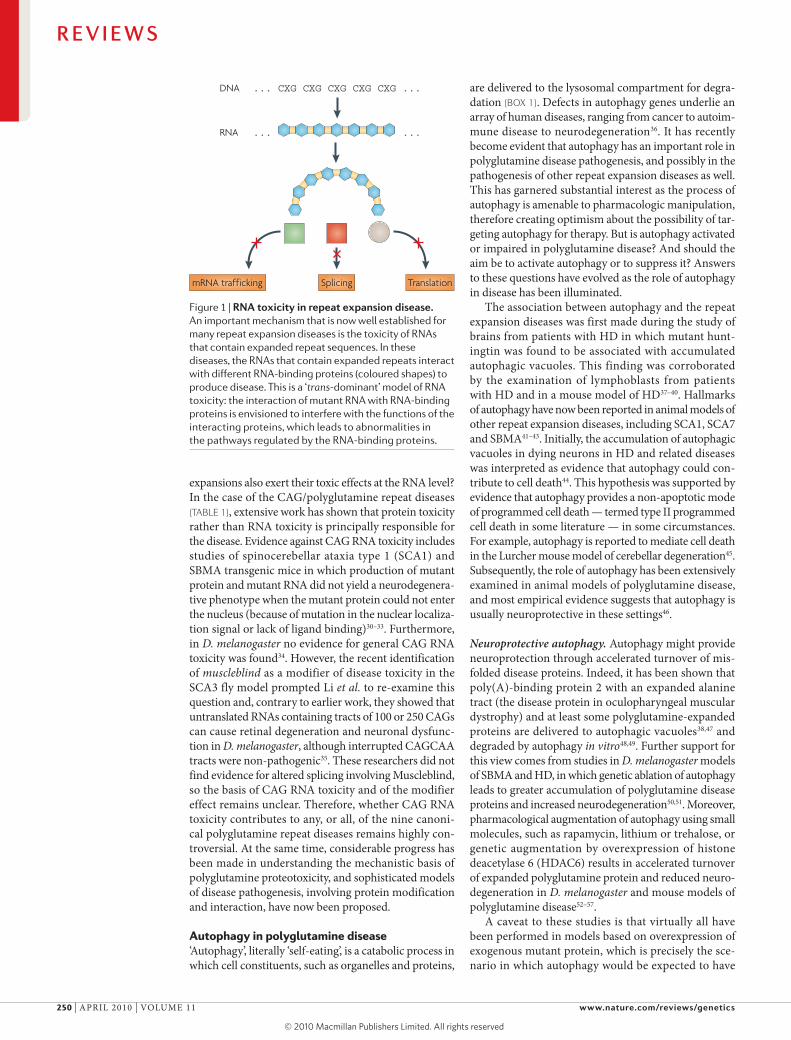
Nature Reviews | Genetics
DNA ... CXG CXG CXG CXG CXG ...
RNA
mRNA trafficking TranslationSplicing
... ...
Figure 1 | RnA toxicity in repeat expansion disease. An important mechanism that is now well established for many repeat expansion diseases is the toxicity of RNAs that contain expanded repeat sequences. In these diseases, the RNAs that contain expanded repeats interact with different RNA-binding proteins (coloured shapes) to produce disease. This is a ‘trans-dominant’ model of RNA toxicity: the interaction of mutant RNA with RNA-binding proteins is envisioned to interfere with the functions of the interacting proteins, which leads to abnormalities in the pathways regulated by the RNA-binding proteins.
expansions also exert their toxic effects at the RNA level? In the case of the CAG/polyglutamine repeat diseases (TABLE 1), extensive work has shown that protein toxicity rather than RNA toxicity is principally responsible for the disease. evidence against CAG RNA toxicity includes studies of spinocerebellar ataxia type 1 (SCA1) and SBMA transgenic mice in which production of mutant protein and mutant RNA did not yield a neurodegenera‑tive phenotype when the mutant protein could not enter the nucleus (because of mutation in the nuclear localiza‑tion signal or lack of ligand binding)30–33. Furthermore, in D. melanogaster no evidence for general CAG RNA toxicity was found34. However, the recent identification of muscleblind as a modifier of disease toxicity in the SCA3 fly model prompted li et al. to re‑examine this question and, contrary to earlier work, they showed that untranslated RNAs containing tracts of 100 or 250 CAGs can cause retinal degeneration and neuronal dysfunc‑tion in D. melanogaster, although interrupted CAGCAA tracts were non‑pathogenic35. These researchers did not find evidence for altered splicing involving Muscleblind, so the basis of CAG RNA toxicity and of the modifier effect remains unclear. Therefore, whether CAG RNA toxicity contributes to any, or all, of the nine canoni‑cal polyglutamine repeat diseases remains highly con‑troversial. At the same time, considerable progress has been made in understanding the mechanistic basis of polyglutamine proteotoxicity, and sophisticated models of disease pathogenesis, involving protein modification and interaction, have now been proposed.
Autophagy in polyglutamine disease‘Autophagy’, literally ‘self‑eating’, is a catabolic process in which cell constituents, such as organelles and proteins,
are delivered to the lysosomal compartment for degra‑dation (BOX 1). Defects in autophagy genes underlie an array of human diseases, ranging from cancer to autoim‑mune disease to neurodegeneration36. It has recently become evident that autophagy has an important role in polyglutamine disease pathogenesis, and possibly in the pathogenesis of other repeat expansion diseases as well. This has garnered substantial interest as the process of autophagy is amenable to pharmacologic manipulation, therefore creating optimism about the possibility of tar‑geting autophagy for therapy. But is autophagy activated or impaired in polyglutamine disease? And should the aim be to activate autophagy or to suppress it? Answers to these questions have evolved as the role of autophagy in disease has been illuminated.
The association between autophagy and the repeat expansion diseases was first made during the study of brains from patients with HD in which mutant hunt‑ingtin was found to be associated with accumulated autophagic vacuoles. This finding was corroborated by the examination of lymphoblasts from patients with HD and in a mouse model of HD37–40. Hallmarks of autophagy have now been reported in animal models of other repeat expansion diseases, including SCA1, SCA7 and SBMA41–43. Initially, the accumulation of autophagic vacuoles in dying neurons in HD and related diseases was interpreted as evidence that autophagy could con‑tribute to cell death44. This hypothesis was supported by evidence that autophagy provides a non‑apoptotic mode of programmed cell death — termed type II programmed cell death in some literature — in some circumstances. For example, autophagy is reported to mediate cell death in the lurcher mouse model of cerebellar degeneration45. Subsequently, the role of autophagy has been extensively examined in animal models of polyglutamine disease, and most empirical evidence suggests that autophagy is usually neuroprotective in these settings46.
Neuroprotective autophagy. Autophagy might provide neuroprotection through accelerated turnover of mis‑folded disease proteins. Indeed, it has been shown that poly(A)‑binding protein 2 with an expanded alanine tract (the disease protein in oculopharyngeal muscular dystrophy) and at least some polyglutamine‑expanded proteins are delivered to autophagic vacuoles38,47 and degraded by autophagy in vitro48,49. Further support for this view comes from studies in D. melanogaster models of SBMA and HD, in which genetic ablation of autophagy leads to greater accumulation of polyglutamine disease proteins and increased neurodegeneration50,51. Moreover, pharmacological augmentation of autophagy using small molecules, such as rapamycin, lithium or trehalose, or genetic augmentation by overexpression of histone deacetylase 6 (HDAC6) results in accelerated turnover of expanded polyglutamine protein and reduced neuro‑degeneration in D. melanogaster and mouse models of polyglutamine disease52–57.
A caveat to these studies is that virtually all have been performed in models based on overexpression of exogenous mutant protein, which is precisely the sce‑nario in which autophagy would be expected to have
R E V I E W S
250 | ApRIl 2010 | vOlUMe 11 www.nature.com/reviews/genetics
© 20 Macmillan Publishers Limited. All rights reserved10

Box 1 | Autophagy
Autophagy refers to a set of biological processes in which cell constituents, such as organelles and proteins, are delivered to the lysosomal compartment for degradation. Autophagy can be broadly divided into three types: microautophagy, which involves the direct engulfment of small volumes of cytosol by lysosomes; chaperone-mediated autophagy, which involves receptor-mediated translocation of proteins into the lysosomal lumen; and macroautophagy, which is described below. Autophagy is an evolutionarily conserved process for rapid mobilization of macromolecules when nutrient availability is limited133. Amino acid starvation, for example, induces autophagy and results in increased degradation of non-essential proteins to provide amino acids for the synthesis of essential proteins. In lower organisms autophagy is not essential when nutrients are abundant, but in mammals the role of autophagy has broadened to include additional functions beyond adapting to starvation. These include essential roles in development, immunity and tumour suppression, among others36.
The first step in macroautophagy involves the expansion of an isolation membrane (see figure) that engulfs a portion of the cell. This membrane eventually fuses to form a new double-membraned structure known as an autophagosome. The source of the membrane is not clear, but it might arise from endoplasmic reticulum (ER) or the Golgi complex. The process of autophagy is controlled by parallel activation cascades that involve ubiquitin-like protein modification and are strikingly similar to the activation cascade that regulates the ubiquitin proteasome system. The first arm of the cascade produces a large (~350 kDa) multimeric complex (ATG12–ATG5–ATG16) that is thought to act as a structural support for membrane expansion. A second arm of the cascade conjugates the micro-tubule-associated protein light chain 3α (LC3) with the phospholipid phosphotidylethanolamine (PE). As PE is a component of the autophagosomal membrane, the lipidation reaction results in studding of the inner and outer membranes of autophagosomes with LC3. LC3 also contributes to membrane expansion, but an additional important function is its ability to bind autophagy receptors, such as sequestosome 1 (SQSTM1, also known as p62) or next to BRCA1 gene 1 (NBR1), which permits selective autophagy.
Once formed, new autophagosomes move through a stepwise maturation process that results in acidification and delivery of lysosomal hydrolases, permitting degradation of the luminal contents. In mammals, autophagosomes first fuse with endosomes and multivesicular bodies to form amphisomes, which subsequently fuse with lysosomes to create degradative vacuoles termed autolysosomes. Finally, the breakdown products from the autolysosome are translocated back across the lysosomal membrane for reuse in metabolic processes in the cytosol, or in some cases are extruded from the cell, and the membrane is recycled.
Autophagosome
Expansion ofisolation membrane
Membrane recycling Membrane recycling
AmphisomeRecycling ofmacromolecules
Golgicomplex
LysosomeLate endosome Multivesicular body
AutolysosomeER
Nature Reviews | Genetics
ATG12–ATG5–ATG16 complex promotesmembrane expansion
Autophagy receptor (for example, p62 or NBR1)
LC3 promotes membrane expansionand interacts with autophagy receptors
Targeting signal for degradation (for example, ubiquitin)
Protein targeted for autophagy(for example, a polyglutamine-expanded protein)
Autophagy targets damaged organelles(for example, mitochondria)
Autophagy targets large and small cytosolic macromolecules (for example, ribosomes or protein aggregates)
R E V I E W S
NATURe RevIeWS | Genetics vOlUMe 11 | ApRIl 2010 | 251
© 20 Macmillan Publishers Limited. All rights reserved10

the greatest effect. It remains important to examine the role of autophagy further in animal models that more faithfully recapitulate endogenous protein expression levels and patterns, using knock‑in or bacterial artificial chromosome (BAC) transgenic approaches, for example. Nevertheless, the view of autophagy as a neuroprotec‑tive process is consistent with the emerging appreciation that autophagy is cytoprotective in numerous contexts — such as under conditions of oxidative stress, growth‑factor deficiency or nutrient limitation — through accel‑erated turnover of damaged organelles and maintenance of metabolic homeostasis by mobilization of intracellular energy stores58,59.
Is autophagy a disease target? Despite compelling evi‑dence that autophagy affords neuroprotection by degrad‑ing disease proteins, the question remains: why is this protection incomplete? Is it possible that autophagy is not only a modifier of disease but also a target of disease? It has been suggested that the increased numbers of neu‑ronal autophagic vacuoles in some repeat expansion dis‑eases may reflect a defect in autophagy flux rather than autophagy induction60. It is now appreciated that there is significant basal autophagy in many mammalian tissues. The demand for basal autophagy differs among tissues; it is particularly important in the liver and in post‑mitotic cells, such as neurons and myocytes61–64. Indeed, neu‑rons are especially vulnerable to perturbations in the autophagy–lysosomal system — not only because they are highly metabolically active but also because of their unique cellular architecture. In neurons, lysosomes are concentrated in the soma adjacent to the nucleus. Therefore, autophagosomes produced in dendrites, axons or synaptic terminal regions must be transported substantial distances to enable fusion with lysosomes, which makes autophagy in neurons particularly vulner‑able to defects in vesicular trafficking66. The importance of basal autophagy in the CNS was shown by conditional knockout of key autophagy genes, which resulted in neurodegeneration with the accumulation of ubiquitin‑ positive inclusions similar to those seen human neuro‑degenerative diseases (including many repeat expansion diseases)66,67. The increased vulnerability of neurons to impairment of the autophagy–lysosomal system might account for the high frequency of neurologi‑cal phenotypes produced by mutations that target the endosomal–lysosomal system68.
It has yet to be shown convincingly that autophagy is impaired in repeat expansion disease. Given the broad range of cellular abnormalities associated with poly‑glutamine diseases and other repeat expansion diseases, it would not be surprising to find impairment of autophagy. Impaired autophagy — a sort of ‘cellular indigestion’ — could result from overburdening the autophagy– lysosomal system with misfolded, aggregated proteins that are difficult to degrade. Alternatively, autophagy could be directly impaired if a disease protein is important for the process of autophagy. As noted above, huntingtin associates with components of the endosomal–lysosomal system and is trafficked with autophagosomes in axons, which suggests that huntingtin might normally regulate
autophagy8,69. Recently, it was shown in vitro and in a mouse knock‑in model of HD that polyglutamine expan‑sion in huntingtin impairs the activity of the GTpase Ras‑related protein 11A (RAB11A) and leads to a defect in endosome recycling70,71. Therefore is it possible that polyglutamine expansion impairs a normal function of huntingtin and contributes to impaired autophagy? This would be consistent with the emerging theme that altered native protein function underlies polyglutamine disease pathogenesis.
Post-translational modificationNon-polyglutamine determinants of disease. A striking feature of polyglutamine diseases is the selective vul‑nerability of the CNS despite widespread expression of many polyglutamine disease proteins in non‑neural cell types. However, there is striking divergence in clinical phenotypes among the polyglutamine diseases: neurol‑ogists can easily distinguish the movement disorder of HD from the weakness in SBMA or the ataxia in SCA1. The disease‑specific features reflect selective loss of dif‑ferent populations of neurons: despite wide expression within the CNS, polyglutamine expansion in huntingtin selectively affects striatal neurons and cortical neurons, whereas the same genetic mutation in the androgen receptor or ataxin 1 targets motor neurons or purkinje neurons, respectively. On the basis of these observations it was predicted that features other than polyglutamine, unique to each disease protein, must influence patho‑genesis72. This prediction has been borne out in recent advances that have highlighted the importance of host protein context in polyglutamine disease pathogenesis. principal among these have been insights into the influ‑ence of post‑translational modification in pathogenesis and, in at least one case, in determining cell‑type spe‑cificity. Here, we summarize some important insights into the role of post‑translational phosphorylation, acetylation and sumoylation in polyglutamine disease pathogenesis (TABLE 2).
Phosphorylation of ataxin 1 maintains a balance. The importance of post‑translational modification in polyglutamine disease was first shown by the Orr and Zoghbi laboratories in a series of papers examining ataxin 1 phosphorylation. Orr and colleagues detected polyglutamine length‑dependent phosphorylation of ataxin 1 that mapped to serine 776 (S776)73. Their inter‑est was heightened after observing that an antibody spe‑cific for phospho‑S776‑ataxin 1 preferentially stained pathological, nuclear‑localized ataxin 1 in their SCA1 mouse model. They generated transgenic mice express‑ing polyglutamine‑expanded ataxin 1 with an alanine substituted for serine at position 776 (S776A) to prevent phosphorylation. The mice expressing polyglutamine‑expanded ataxin 1‑S776A had minimal behavioural and histopathological abnormalities, showing the importance of this phosphorylation in pathogenesis.
phosphorylation alters the conformation of a target protein and can profoundly influence target‑protein func‑tion, often by regulating protein–protein interactions74. Ataxin 1 interacts with two discrete heterotypic protein
R E V I E W S
252 | ApRIl 2010 | vOlUMe 11 www.nature.com/reviews/genetics
© 20 Macmillan Publishers Limited. All rights reserved10

Endocytosis The process whereby cells engulf extracellular material through invagination of the plasma membrane to create an endocytic vesicle.
Neurotrophic factor A small protein that promotes the growth and/or survival of neurons.
complexes, one defined by the transcription factor capicua and the other defined by the RNA‑binding protein RBM17. phosphorylation of ataxin 1 regulates the balance of ataxin 1 association with these distinct functional complexes. The evidence suggests that poly‑glutamine expansion upsets this balance and drives pathogenesis. Specifically, polyglutamine expansion promotes phosphorylation of S776, which favours asso‑ciation with the RBM17 complex and so might contrib‑ute to SCA1 neuropathology through a gain‑of‑function mechanism. Concomitantly, polyglutamine expansion attenuates the formation and function of the capicua complex, contributing to SCA1 through a partial loss‑of‑function mechanism75,76. This discovery has important implications, as this model suggests that polyglutamine disease pathogenesis might involve subtle alteration of native protein function rather than an entirely novel gain of function. This insight highlights the importance of understanding the normal interactions and functions of each disease protein to understand the pathogenesis of each polyglutamine disease.
Phosphorylation and huntingtin trafficking. Huntingtin is subject to phosphorylation at multiple serines, and phosphorylation of S421 has emerged as a par‑ticularly important determinant of HD pathogenesis. phosphorylation of S421 has been reported to be car‑ried out by multiple kinases, including the serine/threo‑nine kinase AKT1 and serum/glucocorticoid‑induced
kinase 1 (SGK1)77–79. Significant levels of phospho‑S421‑huntingtin are present in normal human and mouse striatum but are reduced by polyglutamine expansion. The basis of reduced phosphorylation is unclear but is presumed to be due to altered huntingtin conforma‑tion with polyglutamine expansion. Through the use of phosphorylation‑site mutants and manipulation of the relevant kinases, phosphorylation of S421 has been shown to substantially reduce toxicity in cultured stri‑atal neurons and in a rodent model of HD77–80. What is the basis for this? Huntingtin normally associates with
vesicle membranes and microtubules and contributes to endocytosis and transport of endocytic vesicles81–86. Indeed, huntingtin might mediate retrograde trans‑port of signalling endosomes that carry brain‑derived neurotrophic factor (BDNF)86. This is crucial, as BDNF signalling supplied by cortico‑striatal projection neurons to the striatum — the brain region most severely affected in HD — is an important survival factor87,88. Although it has been known for some time that polyglutamine expan‑sion impairs the ability of huntingtin to support BDNF signalling, the basis for this was unclear86. However, an explanation is offered by the recent findings that the phosphorylation status of S421 modulates the recruit‑ment of motor proteins to endocytic vesicles, is a key determinant of huntingtin regulation of vesicular trans‑port and is altered by polyglutamine expansion. Indeed, constitutive phosphorylation of S421 can overcome the BDNF signalling defect caused by polyglutamine expan‑sion and can suppress toxicity in HD models89,90. These data show that the alteration of post‑translational modi‑fication by repeat expansion can affect the ratio of native protein–protein interactions. Therefore, as in the case of SCA1, the pathogenesis of HD seems to be mediated, at least in part, by the alteration of native protein function.
Additional phosphorylation sites in huntingtin have been implicated as modifiers of disease, although less is known about how they are influenced by polyglutamine expansion or the mechanisms by which they influence pathogenesis. For example, phosphorylation at S343 and S536 is associated with reduced cleavage of huntingtin, and phosphorylation at threonine 3 is associated with reduced aggregation of huntingtin amino‑terminal fragments91–93. Notably, the phosphorylation statuses of S13 and S16 have recently been implicated as important determinants of HD pathogenesis. Gu and colleagues generated transgenic mice that express full‑length mutant huntingtin with S13 and S16 mutated to either aspartate (which mimics constitutive phosphorylation) or alanine (which is resistant to phosphorylation)94. Both mutant proteins retain normal huntingtin function, as
Table 2 | Post-translational modifications in repeat expansion disease
Disease Protein sumoylation Acetylation Phosphorylation
HD Huntingtin K6*, K9*, K15*, K91* (REFs 102,103)
K444* (REF. 101) S421*, S434*, S536*, S1181*, S1201*, S2076, S2653, S2657 (REF. 93), T3* (REF. 92)
SBMA Androgen receptor
K385*, K511* (REF. 134) K630*, K632*, K633* (REF. 135)
S215*, S792* (REF. 136)
DRPLA Atrophin 1 Unknown lysine(s)* (REF. 137) ‡ S734
SCA1 Ataxin 1 K16, K194, K610, K697, K746 ‡ S776* (REFs 73,75), S239, T236
SCA2 Ataxin 2 ‡ ‡ S860, S864
SCA3 Ataxin 3 Unknown lysines(s)* (REF. 138) ‡ S256
SCA6 CACNA1A ‡ ‡ ‡
SCA7 Ataxin 7 ‡ K257* (REF. 99) S840, S849, T854
SCA17 TBP § § §
*Post-translational modifications for which empirical evidence indicates a role in pathogenesis. ‡There are no data pertaining to this post-translational modification. §This post-translational modification occurs, but there are no data pertaining to its role in disease pathogenesis. CACNA1A, voltage-dependent P/Q-type calcium channel subunit α-1A; DRPLA, dentatorubral-pallidoluysian atrophy; HD, Huntington’s disease; K, lysine; S, serine; SBMA, spinal and bulbar muscular atrophy; SCA, spinocerebellar ataxia; T, threonine; TBP, TATA box-binding protein.
R E V I E W S
NATURe RevIeWS | Genetics vOlUMe 11 | ApRIl 2010 | 253
© 20 Macmillan Publishers Limited. All rights reserved10

shown by their ability to rescue huntingtin knockout phenotypes. Mice expressing the phosphorylation‑resistant protein exhibited typical neurodegeneration with associated motor and behavioural phenotypes. By contrast, mice expressing the phosphomimetic protein did not exhibit neurodegeneration, showing the dra‑matic impact of this post‑translational modification on toxicity. How might phosphorylation of S13 and S16 influence pathogenesis? Thompson and colleagues recently found that these serines are phosphorylated by the IκB kinase (IKK) complex. Through promoting modification of adjacent lysine residues, phosphoryla‑tion by IKK targets huntingtin for degradation by a proc‑ess that requires both the proteasome and lysosome95. phosphorylation has also been implicated in influencing the toxicity of SCA3, dentatorubral‑pallidoluysian atro‑phy (DRplA) and SBMA. For example, phosphoryla‑tion of ataxin 3 at S256 by glycogen synthase kinase 3β reduces aggregation of polyglutamine‑expanded ataxin 3 (REF. 96), phosphorylation of polyglutamine‑expanded androgen receptor by mitogen‑activated protein kinase at S516 is associated with increased cleavage and toxic‑ity in cell models of SBMA97, and S734 of atrophin 1 is phosphorylated by Jun N‑terminal kinase, although the significance of this in the pathogenesis of DRplA has not yet been examined.
Acetylation as a determinant of stability. post‑translational protein acetylation takes place on the ε‑amino group of lysines and prevents this group from becoming positively charged, therefore affecting the electrostatic properties of the protein99. Acetylation has diverse consequences, including altered protein–DNA and protein–protein interactions. Acetylation can compete with ubiquitylation at lysine residues, such that acetylation increases protein stability98. For example, acetylation of ataxin 7 at lysine 257 (K257) was recently found to stabilize the protein and promote accumu‑lation of the ataxin 7 N‑terminal truncation prod‑uct, which is believed to be the toxic, disease‑causing species99. Acetylation can also, occasionally, reduce protein stability100.
Krainc and colleagues found that huntingtin sta‑bility is regulated by acetylation at K444 (REF. 101). Interestingly, they found that acetylation facilitates trafficking of mutant huntingtin into autophagosomes, therefore serving as a signal for degradation by the autophagy–lysosomal system rather than influenc‑ing proteasomal degradation. Acetylation accelerates clearance of the mutant huntingtin by autophagy and reverses its toxic effects in mouse primary striatal and cortical neurons and in a Caenorhabditis elegans model of HD. When polyglutamine‑expanded huntingtin was rendered resistant to acetylation, the protein steadily accumulated and increased degeneration in cultured neurons and in mouse brain. This study provided the first evidence that acetylation can target proteins for autophagic degradation. However, the mechanism of targeting for degradation remains to be determined, as does the extent to which proteins other than huntingtin use this mechanism.
Sumoylation as a determinant of cell-type vulnerability. Sumoylation is a reversible post‑translational modifica‑tion in which a small ubiquitin‑like modifier (SUMO) is covalently conjugated to a lysine residue in a target pro‑tein. It provides an efficient way to modulate the subcel‑lular localization, activity and stability of a wide variety of substrates. A role for sumoylation in polyglutamine disease was first suggested because of SUMO immuno‑reactivity in pathological inclusions characteristic of HD, SCA3 and DRplA. Thompson and colleagues reported that sumoylation of huntingtin at K6 and K9 stabilizes N‑terminal fragments of huntingtin, reduces its ability to form aggregates and promotes its capacity to repress transcription102. They also noted that sumoylation of a polyglutamine‑expanded huntingtin fragment exacer‑bated neurodegeneration in a D. melanogaster model of HD.
Recently, huntingtin sumoylation was found to depend upon the small guanine nucleotide‑binding protein RHeS by a mechanism that is independent of RHeS GTpase activity103. RHeS preferentially binds polyglutamine‑expanded huntingtin and promotes sumoylation at various lysines, including K9, K15 and K91. Sumoylation of mutant huntingtin at K15 and K91 correlated with reduced aggregation and increased cytotoxicity. RHeS is expressed only in the striatum, and endogenous RHeS copurified with huntingtin in extracts from transgenic mice, suggesting that the interaction of these proteins takes place under physi‑ological conditions. Therefore, RHeS–huntingtin inter‑actions may account for the localized neuropathology of HD, which is largely restricted to the corpus stria‑tum. Other polyglutamine disease proteins — includ‑ing ataxin 1, androgen receptor, TATA box‑binding protein (TBp) and atrophin 1 — are sumoylated under physiological conditions, and in some cases this has been correlated with increased toxicity in cell culture disease models. However, the extent to which sumoyla‑tion influences pathogenesis in these diseases is yet to be explored.
Bidirectional transcriptionThe two faces of spinocerebellar ataxia type 8. SCA8 is a progressive, dominantly inherited disease characterized by ataxia in combination with signs of corticospinal tract dysfunction104. SCA8 was found to be caused by expan‑sion of a CTG repeat in a gene that is transcribed and not translated105. Therefore the researchers proposed that expression of the expanded repeat was neurotoxic in the form of RNA analogous to the mechanism observed in DM1 and DM2. Initially, this hypothesis was not widely accepted, but D. melanogaster and mouse models were generated that showed that RNA with the CTG expansion was indeed toxic106,107.
However, further analysis of the SCA8 BAC transgenic mice revealed an unexpected finding — 1C2‑positive intranuclear inclusions of the type that would be detected in polyglutamine expansion diseases106. CTG is read as CAG on the opposite strand, and re‑examination of the antisense sequence indicated the potential translation of a protein that consists almost
R E V I E W S
254 | ApRIl 2010 | vOlUMe 11 www.nature.com/reviews/genetics
© 20 Macmillan Publishers Limited. All rights reserved10

NucleosomeThe basic unit of chromatin. A nucleosome contains approximately 146 bp of DNA wrapped around a histone octamer.
entirely of polyglutamine. examination of samples from patients with SCA8, SCA8 transgenic mice and cell cul‑ture showed that a transcript (ATXN8) encodes the pre‑dicted polyglutamine‑rich protein — now called ataxin 8 (REF. 106). The authors proposed that polyglutamine pathology and RNA gain‑of‑function toxicity combine to cause the SCA8 phenotype. Recently this group has shown that the ataxin 8 antisense transcript (ataxin 8 opposite strand (ATXN8OS)) might sequester the MBNl1 protein108. MBNl1 sequestration was shown to alter the splicing of Gabt4 (also known as Slc6a11), which encodes a transporter of the inhibitory neuro‑transmitter GABA (γ‑aminobutyric acid). Altered splic‑ing of Gabt4 resulted in increased GABT4 protein and correlated with the predicted loss of GABAergic inhibi‑tion in the cerebellar granule layer of SCA8 mice. The discovery of MBNl1 sequestration and a splicing defect in this SCA8 model reveals unexpected similarity to the pathogenic mechanism responsible for DM1 and DM2. A caveat to this study is that so far the splicing of only a limited number of genes has been examined in detail. As the splicing defect and the relationship to MBNl1 deficiency are more fully explored it will be interesting to learn the extent to which the splicing defect underlies the neurodegenerative phenotype. For example, to what extent will restoration of MBNl1 levels in SCA8 mice rescue the neurodegenerative phenotype?
Multiple mechanisms in Huntington’s disease-like 2? Another repeat disease that is difficult to classify by pathogenic mechanism is Huntington’s disease‑like 2 (HDl2), a phenocopy of HD109. HDl2 is caused by a CTG repeat expansion in the junctophilin 3 (JPH3) gene; affected individuals have 40 or more CTGs109. JpH3 belongs to a family of proteins that link plasma mem‑brane voltage sensors with intracellular ion channels110. It has been a daunting challenge to determine how the JPH3 CTG repeat expansion produces molecular pathol‑ogy, as it lies in the variably spliced exon 2A. Three splice variants have been reported: one places the CTG repeat in a polyleucine translational frame, one places it in a polyalanine translational frame and one places it in the 3′ UTR111.
Database queries do not show evidence for transcrip‑tion of the opposite, CAG repeat‑containing strand. Nevertheless, ubiquitin‑positive neuronal intranuclear inclusions (that incidentally do not contain huntingtin) are observed, which suggests the deposition of mis‑folded protein due to polyglutamine or some other poly‑amino acid tract. RNA foci containing MBNl1 have been detected in HDl2 brain, and the expression of CUG‑expanded JPH3 in cell culture also causes RNA foci containing MBNl1 (REF. 111). Furthermore, altered splicing of genes encoding tau and amyloid precursor protein occurs in HDl2 frontal cortex. Therefore, it seems that CUG RNA toxicity, together with some form of proteotoxicity, may combine in HDl2 to produce neurodegeneration. Hence, SCA8 and HDl2 seem to belong to a category of diseases that involve simultane‑ous RNA gain‑of‑function and protein gain‑of‑function molecular pathology (FIG. 2).
In the case of HDl2, a third possible simultaneous mechanism is partial loss of function of JpH3, as Jph3‑null mice and heterozygous‑null mice show impaired coordination and shortened lifespan112, and patients with HDl2 express reduced levels of JpH3 (REF. 111). However, knockout of Jph3 in mice does not cause RNA foci or ubiquitin‑positive inclusions, suggesting that Jph3 haploinsufficiency alone does not account for HDl2.
Genomic structure and antisense transcriptionUp to this point, we have considered the potential con‑tribution of the transcriptional and translational gene products to disease pathogenesis. However, unique aspects of the repeat disease loci are also emerging at the level of DNA sequence and chromatin organization. Most repeat expansion diseases involve gene loci with highly polymorphic repeats in normal individuals; both the median and mode of repeat size are quite large, even though the repeats are non‑pathogenic. CTG repeats are potent nucleosome positioning elements113, so it seems
Nature Reviews | Genetics
RNA ... CUG CUG CUG CUG CUG CUG ...
DNA ... CTG CTG CTG CTG CTG CTG ...... CAG CAG CAG CAG CAG CAG ...
RNA ... CAG CAG CAG CAG CAG CAG ...
Toxicity
Toxicity
N Q
C
QQQQQQ
QQQQQQQ
QQQ
QQQQQQQQQ
QQQQQ
QQQQQQQQQQQQ. . . . . . . .
. . . .
.
. . . . . . .
Figure 2 | sense and antisense toxicity. For a subset of repeat expansion diseases, sense and antisense transcripts are produced from the repeat region. In these cases, one transcript may cause toxicity through an RNA gain-of-function pathway and the other transcript, which is generated in opposite orientation, may yield a toxic poly-amino acid tract-containing protein. In the example shown here for spinocerebellar ataxia type 8 (SCA8), a transcript containing a CUG repeat expansion is suggested to produce toxicity, and a polyglutamine protein encoded from an antisense transcript is predicted to simultaneously produce toxicity. Even for diseases, such as SCA3, that do not exhibit bidirectional overlapping transcription, concomitant RNA toxicity from the RNA transcript containing the CAG repeat expansion has been proposed. Therefore the potential toxic contribution of different gene products (RNA and proteins) from the sense strand must be considered in addition to the potential toxic contribution of different gene products from antisense overlapping transcription. C, carboxyl terminus; N, amino terminus.
R E V I E W S
NATURe RevIeWS | Genetics vOlUMe 11 | ApRIl 2010 | 255
© 20 Macmillan Publishers Limited. All rights reserved10

HeterochromatinParts of chromosomes with an unusual degree of contraction and that consequently have different staining properties from euchromatin at nuclear divisions. Largely composed of repetitive DNA, heterochromatin forms dark bands after Giemsa staining.
plausible that expanded repeats could influence the chromatin organization. Filippova et al. identified bind‑ing sites for the CCCTC‑binding factor CTCF — an evo‑lutionarily conserved zinc finger DNA‑binding protein with activity in chromatin insulation, transcription regu‑lation and genomic imprinting114 — adjacent to and/or flanking some triplet repeat loci115. CTCF‑binding sites can now be identified by directed approaches (such as electrophoretic mobility shift assays) and discovery‑driven approaches (such as chromatin immunoprecipi‑tation followed by microarray (ChIp–chip))116–118, and evidence for CTCF binding in close proximity to the repeat sequences is emerging. At the DM1 locus, CTCF binding prevents the spread of heterochromatin into the DM1 gene by blocking antisense transcription119. Hyperexpansion of the CTG repeat — as in congenital DM1 — correlates with loss of CTCF binding and expansion of the region of antisense transcription, which results in heterochromatinization of DM1 and adjacent genes. In some cases, CTCF binding seems to be associ‑ated with antisense transcription119,120, but in DM1 and Friedreich’s ataxia CTCF depletion may permit produc‑tion of an antisense transcript, resulting in heterochro‑matin formation and gene silencing119,121. CTCF has also been implicated in genetic instability, which occurs in most repeat expansion diseases122. Abrogation of CTCF binding at the SCA7 locus by point mutation or methyla‑tion of the binding site promotes CAG repeat instability in SCA7 transgenic mice, both in the germ line and in somatic tissues123.
Surveys of mammalian transcriptomes are uncovering tremendous numbers and varieties of non‑coding RNAs, including antisense transcripts at the start sites of sense transcription and in adjacent 5′ promoter regions124–127, and this phenomenon seems to be prominent at repeat disease loci. For example, at the FMR1 locus, an anti‑sense transcript overlapping the CGG repeat in oppo‑site orientation has been identified128. This transcript (antisense fragile X mental retardation 1 (ASFMR1)) is upregulated in patients carrying premutation alleles and seems to be silenced in full mutation carriers. Altered antisense transcription might be a characteristic of some repeat disease loci and raises important questions about the relationship between non‑coding and cod‑ing transcription and how it is influenced by changes in repeat length and chromatin structure. CTCF‑ and
non‑coding‑transcript‑mediated regulation of genomic architecture could have an important role in transcription dysregulation in many repeat expansion diseases, espe‑cially as evidence of altered epigenetic regulatory processes in certain of the polyglutamine diseases is increasing129,130. We expect that a considerable amount of information on this topic will emerge over the next few years.
Conclusionsenormous strides have been made since the initial discovery of a novel, esoteric type of mutation as the cause of SBMA and FMR1. Repeat expansion research has revealed unexpected complexities, including toxic‑ity mediated at the protein, RNA and genomic levels. perturbations of numerous cellular functions have been implicated in disease pathogenesis — for example, tran‑scription, splicing, translation, mitochondrial function and protein quality control, in addition to specific neu‑ronal functions, such as axonal transport and synaptic activity. This complexity has attracted a diverse group of scientists and has catalysed the development of inter‑disciplinary approaches for elucidating the mechanisms of disease. As a result, research in the repeat expansion field has often been at the vanguard of key advances in molecular genetics. For example, repeat disease research has a leading role in elucidating the basis of defective RNA metabolism and autophagy in human disease.
A nagging question for the repeat disease field is why the development of meaningful treatments has not kept pace with the remarkable trajectory of advances in the study of mechanisms and pathways. Although this disconnect applies to most neurological disorders, the repeat diseases offer a wonderful opportunity for rational therapy development because presymptomatic definitive diagnosis based on repeat allele expansion can normally be made. Discoveries over the past few years have yielded a promising crop of candidate targets and pathways and have led to some very encouraging therapeutic developments, including the use of oligo‑nucleotide and morpholino antisense repeats targeted against the DM1 CTG expansion and the use of small molecules to augment autophagy131–133. There can be no doubt that the successful application of one or more of these strategies as a therapy for a repeat expansion dis‑ease would be the most crucial advance that could come out of this field.
1. La Spada, A. R., Wilson, E. M., Lubahn, D. B., Harding, A. E. & Fischbeck, K. H. Androgen receptor gene mutations in X-linked spinal and bulbar muscular atrophy. Nature 352, 77–79 (1991).
2. Verkerk, A. J. et al. Identification of a gene (FMR-1) containing a CGG repeat coincident with a breakpoint cluster region exhibiting length variation in fragile X syndrome. Cell 65, 905–914 (1991).
3. Ashley, C. T. et al. Human and murine FMR-1: alternative splicing and translational initiation downstream of the CGG-repeat. Nature Genet. 4, 244–251 (1993).
4. Bell, M. V. et al. Physical mapping across the fragile X: hypermethylation and clinical expression of the fragile X syndrome. Cell 64, 861–866 (1991).
5. Heitz, D. et al. Isolation of sequences that span the fragile X and identification of a fragile X-related CpG island. Science 251, 1236–1239 (1991).
6. Aslanidis, C. et al. Cloning of the essential myotonic dystrophy region and mapping of the putative defect. Nature 355, 548–551 (1992).
7. Brook, J. D. et al. Molecular basis of myotonic dystrophy: expansion of a trinucleotide (CTG) repeat at the 3′ end of a transcript encoding a protein kinase family member. Cell 68, 799–808 (1992).
8. Harley, H. G. et al. Expansion of an unstable DNA region and phenotypic variation in myotonic dystrophy. Nature 355, 545–546 (1992).
9. Jansen, G. et al. Abnormal myotonic dystrophy protein kinase levels produce only mild myopathy in mice. Nature Genet. 13, 316–324 (1996).
10. Harris, S., Moncrieff, C. & Johnson, K. Myotonic dystrophy: will the real gene please step forward! Hum. Mol. Genet. 5, 1417–1423 (1996).
11. Klesert, T. R. et al. Mice deficient in Six5 develop cataracts: implications for myotonic dystrophy. Nature Genet. 25, 105–109 (2000).
12. Sarkar, P. S. et al. Heterozygous loss of Six5 in mice is sufficient to cause ocular cataracts. Nature Genet. 25, 110–114 (2000).
13. Ranum, L. P., Rasmussen, P. F., Benzow, K. A., Koob, M. D. & Day, J. W. Genetic mapping of a second myotonic dystrophy locus. Nature Genet. 19, 196–198 (1998).
14. Ricker, K. et al. Proximal myotonic myopathy. Clinical features of a multisystem disorder similar to myotonic dystrophy. Arch. Neurol. 52, 25–31 (1995).
15. Day, J. W. et al. Myotonic dystrophy type 2: molecular, diagnostic and clinical spectrum. Neurology 60, 657–664 (2003).
16. Liquori, C. L. et al. Myotonic dystrophy type 2 caused by a CCTG expansion in intron 1 of ZNF9. Science 293, 864–867 (2001).
17. Timchenko, L. T. Myotonic dystrophy: the role of RNA CUG triplet repeats. Am. J. Hum. Genet. 64, 360–364 (1999).
R E V I E W S
256 | ApRIl 2010 | vOlUMe 11 www.nature.com/reviews/genetics
© 20 Macmillan Publishers Limited. All rights reserved10

18. Mankodi, A. et al. Myotonic dystrophy in transgenic mice expressing an expanded CUG repeat. Science 289, 1769–1773 (2000).This paper shows that expression of a CUG repeat expansion in a non-repeat disease RNA is sufficient to produce a myotonic dystrophy-like phenotype in mice. This was an important step in validating the RNA gain-of-function toxicity model.
19. Philips, A. V., Timchenko, L. T. & Cooper, T. A. Disruption of splicing regulated by a CUG-binding protein in myotonic dystrophy. Science 280, 737–741 (1998).This study showed a role for altered splicing in the pathogenesis of myotonic dystrophy and also offered an explanation for how the DM1 gene defect could affect a variety of different cell types and tissues.
20. Kanadia, R. N. et al. A muscleblind knockout model for myotonic dystrophy. Science 302, 1978–1980 (2003).This work implicated muscleblind in the splicing pathology caused by the CUG repeat expansions in myotonic dystrophy and provided evidence for the genesis and nature of the splicing alterations in this disease.
21. Miller, J. W. et al. Recruitment of human muscleblind proteins to (CUG)n expansions associated with myotonic dystrophy. EMBO J. 19, 4439–4448 (2000).
22. Artero, R. et al. The muscleblind gene participates in the organization of Z-bands and epidermal attachments of Drosophila muscles and is regulated by Dmef2. Dev. Biol. 195, 131–143 (1998).
23. Begemann, G. et al. muscleblind, a gene required for photoreceptor differentiation in Drosophila, encodes novel nuclear Cys3His-type zinc-finger-containing proteins. Development 124, 4321–4331 (1997).
24. Du, H. et al. Aberrant alternative splicing and extracellular matrix gene expression in mouse models of myotonic dystrophy. Nature Struct. Mol. Biol. 24 Jan 2010 (doi:10.1038/nsmb.1720).
25. Hagerman, R. J. et al. Intention tremor, parkinsonism, and generalized brain atrophy in male carriers of fragile X. Neurology 57, 127–130 (2001).
26. Jacquemont, S. et al. Fragile X premutation tremor/ataxia syndrome: molecular, clinical, and neuroimaging correlates. Am. J. Hum. Genet. 72, 869–878 (2003).
27. Greco, C. M. et al. Neuronal intranuclear inclusions in a new cerebellar tremor/ataxia syndrome among fragile X carriers. Brain 125, 1760–1771 (2002).
28. Jin, P. et al. RNA-mediated neurodegeneration caused by the fragile X premutation rCGG repeats in Drosophila. Neuron 39, 739–747 (2003).
29. Willemsen, R. et al. The FMR1 CGG repeat mouse displays ubiquitin-positive intranuclear neuronal inclusions; implications for the cerebellar tremor/ataxia syndrome. Hum. Mol. Genet. 12, 949–959 (2003).
30. Jin, P. et al. Pur-α binds to rCGG repeats and modulates repeat-mediated neurodegeneration in a Drosophila model of fragile X tremor/ataxia syndrome. Neuron 55, 556–564 (2007).
31. Sofola, O. A. et al. RNA-binding proteins hnRNP A2/B1 and CUGBP1 suppress fragile X CGG premutation repeat-induced neurodegeneration in a Drosophila model of FXTAS. Neuron 55, 565–571 (2007).
32. Katsuno, M. et al. Testosterone reduction prevents phenotypic expression in a transgenic mouse model of spinal and bulbar muscular atrophy. Neuron 35, 843–854 (2002).
33. Klement, I. A. et al. Ataxin-1 nuclear localization and aggregation: role in polyglutamine-induced disease in SCA1 transgenic mice. Cell 95, 41–53 (1998).
34. McLeod, C. J., O’Keefe, L. V. & Richards, R. I. The pathogenic agent in Drosophila models of ‘polyglutamine’ diseases. Hum. Mol. Genet. 14, 1041–1048 (2005).
35. Li, L. B., Yu, Z., Teng, X. & Bonini, N. M. RNA toxicity is a component of ataxin-3 degeneration in Drosophila. Nature 453, 1107–1111 (2008).
36. Kundu, M. & Thompson, C. B. Autophagy: basic principles and relevance to disease. Annu. Rev. Pathol. 3, 427–455 (2008).
37. Sapp, E. et al. Huntingtin localization in brains of normal and Huntington’s disease patients. Ann. Neurol. 42, 604–612 (1997).
38. Kegel, K. B. et al. Huntingtin expression stimulates endosomal–lysosomal activity, endosome tubulation, and autophagy. J. Neurosci. 20, 7268–7278 (2000).
39. Petersen, A. et al. Expanded CAG repeats in exon 1 of the Huntington’s disease gene stimulate dopamine-mediated striatal neuron autophagy and degeneration. Hum. Mol. Genet. 10, 1243–1254 (2001).
40. Nagata, E., Sawa, A., Ross, C. A. & Snyder, S. H. Autophagosome-like vacuole formation in Huntington’s disease lymphoblasts. Neuroreport 15, 1325–1328 (2004).
41. Vig, P. J., Shao, Q., Subramony, S. H., Lopez, M. E. & Safaya, E. Bergmann glial S100B activates myo-inositol monophosphatase 1 and co-localizes to Purkinje cell vacuoles in SCA1 transgenic mice. Cerebellum 8, 231–244 (2009).
42. Zander, C. et al. Similarities between spinocerebellar ataxia type 7 (SCA7) cell models and human brain: proteins recruited in inclusions and activation of caspase-3. Hum. Mol. Genet. 10, 2569–2579 (2001).
43. Montie, H. L. et al. Cytoplasmic retention of polyglutamine-expanded androgen receptor ameliorates disease via autophagy in a mouse model of spinal and bulbar muscular atrophy. Hum. Mol. Genet. 18, 1937–1950 (2009).
44. Yuan, J., Lipinski, M. & Degterev, A. Diversity in the mechanisms of neuronal cell death. Neuron 40, 401–413 (2003).
45. Yue, Z. et al. A novel protein complex linking the δ2 glutamate receptor and autophagy. Neuron 35, 921–933 (2002).
46. Nedelsky, N. B., Todd, P. K. & Taylor, J. P. Autophagy and the ubiquitin-proteasome system: collaborators in neuroprotection. Biochim. Biophys. Acta 1782, 691–699 (2008).
47. Taylor, J. P. et al. Aggresomes protect cells by enhancing the degradation of toxic polyglutamine-containing protein. Hum. Mol. Genet. 12, 749–757 (2003).
48. Ravikumar, B., Duden, R. & Rubinsztein, D. C. Aggregate-prone proteins with polyglutamine and polyalanine expansions are degraded by autophagy. Hum. Mol. Genet. 11, 1107–1117 (2002).
49. Qin, Z. H. et al. Autophagy regulates the processing of amino terminal huntingtin fragments. Hum. Mol. Genet. 12, 3231–3244 (2003).
50. Berger, Z. et al. Rapamycin alleviates toxicity of different aggregate-prone proteins. Hum. Mol. Genet. 15, 433–442 (2006).
51. Pandey, U. B. et al. HDAC6 rescues neurodegeneration and provides an essential link between autophagy and the UPS. Nature 447, 859–863 (2007).
52. Ravikumar, B. et al. Inhibition of mTOR induces autophagy and reduces toxicity of polyglutamine expansions in fly and mouse models of Huntington disease. Nature Genet. 36, 585–595 (2004).This study showed that pharmacological induction of autophagy ameliorated neurodegeneration in animal models of HD.
53. Pandey, U. B., Batlevi, Y., Baehrecke, E. H. & Taylor, J. P. HDAC6 at the intersection of autophagy, the ubiquitin-proteasome system and neurodegeneration. Autophagy 3, 643–645 (2007).
54. Sarkar, S. et al. A rational mechanism for combination treatment of Huntington’s disease using lithium and rapamycin. Hum. Mol. Genet. 17, 170–178 (2008).
55. Tanaka, M. et al. Trehalose alleviates polyglutamine-mediated pathology in a mouse model of Huntington disease. Nature Med. 10, 148–154 (2004).
56. Davies, J. E., Sarkar, S. & Rubinsztein, D. C. Trehalose reduces aggregate formation and delays pathology in a transgenic mouse model of oculopharyngeal muscular dystrophy. Hum. Mol. Genet. 15, 23–31 (2006).
57. Sarkar, S., Davies, J. E., Huang, Z., Tunnacliffe, A. & Rubinsztein, D. C. Trehalose, a novel mTOR-independent autophagy enhancer, accelerates the clearance of mutant huntingtin and α-synuclein. J. Biol. Chem. 282, 5641–5652 (2007).
58. Kiffin, R., Bandyopadhyay, U. & Cuervo, A. M. Oxidative stress and autophagy. Antioxid. Redox Signal. 8, 152–162 (2006).
59. Lum, J. J. et al. Growth factor regulation of autophagy and cell survival in the absence of apoptosis. Cell 120, 237–248 (2005).
60. McCray, B. A. & Taylor, J. P. The role of autophagy in age-related neurodegeneration. Neurosignals 16, 75–84 (2008).
61. Komatsu, M. et al. Loss of autophagy in the central nervous system causes neurodegeneration in mice. Nature 441, 880–884 (2006).This study, and the related study by Hara et al. (reference 62), showed the importance of basal, quality-control autophagy in the CNS. Impairment of basal autophagy was found to cause neurodegeneration with accumulation of ubiquitin-positive inclusions.
62. Hara, T. et al. Suppression of basal autophagy in neural cells causes neurodegenerative disease in mice. Nature 441, 885–889 (2006).
63. Komatsu, M. et al. Impairment of starvation-induced and constitutive autophagy in Atg7-deficient mice. J. Cell Biol. 169, 425–434 (2005).
64. Komatsu, M. et al. Essential role for autophagy protein Atg7 in the maintenance of axonal homeostasis and the prevention of axonal degeneration. Proc. Natl Acad. Sci. USA 104, 14489–14494 (2007).
65. Hollenbeck, P. J. Products of endocytosis and autophagy are retrieved from axons by regulated retrograde organelle transport. J. Cell Biol. 121, 305–315 (1993).
66. Hara, T. et al. Suppression of basal autophagy in neural cells causes neurodegenerative disease in mice. Nature 441, 885–889 (2006).
67. Kamaya, H., Hayes, J. J. Jr & Ueda, I. Dissociation constants of local anesthetics and their temperature dependence. Anesth. Analg. 62, 1025–1030 (1983).
68. Nixon, R. A., Yang, D. S. & Lee, J. H. Neurodegenerative lysosomal disorders: a continuum from development to late age. Autophagy 4, 590–599 (2008).
69. Atwal, R. S. et al. Huntingtin has a membrane association signal that can modulate huntingtin aggregation, nuclear entry and toxicity. Hum. Mol. Genet. 16, 2600–2615 (2007).
70. Li, X. et al. Mutant huntingtin impairs vesicle formation from recycling endosomes by interfering with Rab11 activity. Mol. Cell. Biol. 29, 6106–6116 (2009).
71. Li, X. et al. Disruption of Rab11 activity in a knock-in mouse model of Huntington’s disease. Neurobiol. Dis. 36, 374–383 (2009).
72. La Spada, A. R. & Taylor, J. P. Polyglutamines placed into context. Neuron 38, 681–684 (2003).
73. Emamian, E. S. et al. Serine 776 of ataxin-1 is critical for polyglutamine-induced disease in SCA1 transgenic mice. Neuron 38, 375–387 (2003).
74. Johnson, L. N. & Lewis, R. J. Structural basis for control by phosphorylation. Chem. Rev. 101, 2209–2242 (2001).
75. Chen, H. K. et al. Interaction of Akt-phosphorylated ataxin-1 with 14-3-3 mediates neurodegeneration in spinocerebellar ataxia type 1. Cell 113, 457–468 (2003).
76. Lim, J. et al. Opposing effects of polyglutamine expansion on native protein complexes contribute to SCA1. Nature 452, 713–718 (2008).This study showed that polyglutamine expansion alters the ratio of ataxin 1 between two alternative protein complexes. This discovery has important implications: it suggests that polyglutamine disease pathogenesis might involve subtle alteration of native protein function rather than an entirely novel gain of function.
77. Humbert, S. et al. The IGF-1/Akt pathway is neuroprotective in Huntington’s disease and involves huntingtin phosphorylation by Akt. Dev. Cell 2, 831–837 (2002).
78. Rangone, H. et al. The serum- and glucocorticoid-induced kinase SGK inhibits mutant huntingtin-induced toxicity by phosphorylating serine 421 of huntingtin. Eur. J. Neurosci. 19, 273–279 (2004).
79. Warby, S. C. et al. Huntingtin phosphorylation on serine 421 is significantly reduced in the striatum and by polyglutamine expansion in vivo. Hum. Mol. Genet.14, 1569–1577 (2005).
80. Pardo, R. et al. Inhibition of calcineurin by FK506 protects against polyglutamine-huntingtin toxicity through an increase of huntingtin phosphorylation at S421. J. Neurosci. 26, 1635–1645 (2006).
81. Difiglia, M. et al. Huntingtin is a cytoplasmic protein associated with vesicles in human and rat-brain neurons. Neuron 14, 1075–1081 (1995).
82. Engelender, S. et al. Huntingtin-associated protein 1 (HAP1) interacts with the p150Glued subunit of dynactin. Hum. Mol. Genet. 6, 2205–2212 (1997).
83. Kalchman, M. A. et al. HIP1, a human homologue of S. cerevisiae Sla2p, interacts with membrane-associated huntingtin in the brain. Nature Genet. 16, 44–53 (1997).
R E V I E W S
NATURe RevIeWS | Genetics vOlUMe 11 | ApRIl 2010 | 257
© 20 Macmillan Publishers Limited. All rights reserved10

84. Li, S. H., Gutekunst, C. A., Hersch, S. M. & Li, X. J. Interaction of huntingtin-associated protein with dynactin P150Glued. J. Neurosci. 18, 1261–1269 (1998).
85. Li, X. J. et al. A huntingtin-associated protein enriched in brain with implications for pathology. Nature 378, 398–402 (1995).
86. Gauthier, L. R. et al. Huntingtin controls neurotrophic support and survival of neurons by enhancing BDNF vesicular transport along microtubules. Cell 118, 127–138 (2004).
87. Altar, C. A. et al. Anterograde transport of brain-derived neurotrophic factor and its role in the brain. Nature 389, 856–860 (1997).
88. Baquet, Z. C., Gorski, J. A. & Jones, K. R. Early striatal dendrite deficits followed by neuron loss with advanced age in the absence of anterograde cortical brain-derived neurotrophic factor. J. Neurosci. 24, 4250–4258 (2004).
89. Zala, D. et al. Phosphorylation of mutant huntingtin at S421 restores anterograde and retrograde transport in neurons. Hum. Mol. Genet.17, 3837–3846 (2008).
90. Colin, E. et al. Huntingtin phosphorylation acts as a molecular switch for anterograde/retrograde transport in neurons. EMBO J. 27, 2124–2134 (2008).
91. Warby, S. C. et al. Phosphorylation of huntingtin reduces the accumulation of its nuclear fragments. Mol. Cell. Neurosci. 40, 121–127 (2009).
92. Aiken, C. T. et al. Phosphorylation of threonine-3: implications for huntingtin aggregation and neurotoxicity. J. Biol. Chem. 284, 29427–29436 (2009).
93. Schilling, B. et al. Huntingtin phosphorylation sites mapped by mass spectrometry. Modulation of cleavage and toxicity. J. Biol. Chem. 281, 23686–23697 (2006).
94. Gu, X. et al. Serines 13 and 16 are critical determinants of full-length human mutant huntingtin induced disease pathogenesis in HD mice. Neuron 64, 824–840 (2009).
95. Thompson, L. M. et al. IKK phosphorylates huntingtin and targets it for degradation by the proteasome and lysosome. J. Cell Biol. 187, 1083–1099 (2009).
96. Fei, E. et al. Phosphorylation of ataxin-3 by glycogen synthase kinase 3β at serine 256 regulates the aggregation of ataxin-3. Biochem. Biophys. Res. Commun. 357, 487–492 (2007).
97. LaFevre-Bernt, M. A. & Ellerby, L. M. Kennedy’s disease. Phosphorylation of the polyglutamine-expanded form of androgen receptor regulates its cleavage by caspase-3 and enhances cell death. J. Biol. Chem. 278, 34918–34924 (2003).
98. Glozak, M. A., Sengupta, N., Zhang, X. & Seto, E. Acetylation and deacetylation of non-histone proteins. Gene 363, 15–23 (2005).
99. Mookerjee, S. et al. Posttranslational modification of ataxin-7 at lysine 257 prevents autophagy-mediated turnover of an N-terminal caspase-7 cleavage fragment. J. Neurosci. 29, 15134–15144 (2009).
100. Jeong, J. W. et al. Regulation and destabilization of HIF-1α by ARD1-mediated acetylation. Cell 111, 709–720 (2002).
101. Jeong, H. et al. Acetylation targets mutant huntingtin to autophagosomes for degradation. Cell 137, 60–72 (2009).
102. Steffan, J. S. et al. SUMO modification of huntingtin and Huntington’s disease pathology. Science 304, 100–104 (2004).
103. Subramaniam, S., Sixt, K. M., Barrow, R. & Snyder, S. H. Rhes, a striatal specific protein, mediates mutant-huntingtin cytotoxicity. Science 324, 1327–1330 (2009).
104. Day, J. W., Schut, L. J., Moseley, M. L., Durand, A. C. & Ranum, L. P. Spinocerebellar ataxia type 8: clinical features in a large family. Neurology 55, 649–657 (2000).
105. Koob, M. D. et al. An untranslated CTG expansion causes a novel form of spinocerebellar ataxia (SCA8). Nature Genet. 21, 379–384 (1999).
106. Moseley, M. L. et al. Bidirectional expression of CUG and CAG expansion transcripts and intranuclear polyglutamine inclusions in spinocerebellar ataxia type 8. Nature Genet. 38, 758–769 (2006).This paper showed that expansion of the SCA8 triplet repeat is sufficient to produce neurological disease, and demonstrated that transcription of a non-coding RNA on one strand and transcription of a coding CAG RNA on the opposite strand, which gives rise to a polyglutamine protein, both occur at the SCA8 disease locus.
107. Mutsuddi, M., Marshall, C. M., Benzow, K. A., Koob, M. D. & Rebay, I. The spinocerebellar ataxia 8 noncoding RNA causes neurodegeneration and associates with staufen in Drosophila. Curr. Biol. 14, 302–308 (2004).
108. Daughters, R. S. et al. RNA gain-of-function in spinocerebellar ataxia type 8. PLoS Genet. 5, e1000600 (2009).
109. Margolis, R. L. et al. A disorder similar to Huntington’s disease is associated with a novel CAG repeat expansion. Ann. Neurol. 50, 373–380 (2001).
110. Takeshima, H., Komazaki, S., Nishi, M., Iino, M. & Kangawa, K. Junctophilins: a novel family of junctional membrane complex proteins. Mol. Cell 6, 11–22 (2000).
111. Rudnicki, D. D. et al. Huntington’s disease-like 2 is associated with CUG repeat-containing RNA foci. Ann. Neurol. 61, 272–282 (2007).
112. Nishi, M. et al. Motor discoordination in mutant mice lacking junctophilin type 3. Biochem. Biophys. Res. Commun. 292, 318–324 (2002).
113. Wang, Y. H., Gellibolian, R., Shimizu, M., Wells, R. D. & Griffith, J. Long CCG triplet repeat blocks exclude nucleosomes: a possible mechanism for the nature of fragile sites in chromosomes. J. Mol. Biol. 263, 511–516 (1996).
114. Ohlsson, R., Renkawitz, R. & Lobanenkov, V. CTCF is a uniquely versatile transcription regulator linked to epigenetics and disease. Trends Genet. 17, 520–527 (2001).
115. Filippova, G. N. et al. CTCF-binding sites flank CTG/CAG repeats and form a methylation-sensitive insulator at the DM1 locus. Nature Genet. 28, 335–343 (2001).This study indicated that altered CTCF binding at the DM1 locus could be involved in DM1 pathology. It also implicated CTCF in a trinucleotide repeat disease for the first time, and suggested that CTCF-binding sites are commonly associated with repeat tracts that are susceptible to disease-causing expansion.
116. Barski, A. et al. High-resolution profiling of histone methylations in the human genome. Cell 129, 823–837 (2007).
117. Kim, T. H. et al. Analysis of the vertebrate insulator protein CTCF-binding sites in the human genome. Cell 128, 1231–1245 (2007).
118. Nguyen, P. et al. CTCFL/BORIS is a methylation-independent DNA-binding protein that preferentially binds to the paternal H19 differentially methylated region. Cancer Res. 68, 5546–5551 (2008).
119. Cho, D. H. et al. Antisense transcription and heterochromatin at the DM1 CTG repeats are constrained by CTCF. Mol. Cell 20, 483–489 (2005).
120. Chao, W., Huynh, K. D., Spencer, R. J., Davidow, L. S. & Lee, J. T. CTCF, a candidate trans-acting factor for X-inactivation choice. Science 295, 345–347 (2002).
121. De Biase, I., Chutake, Y. K., Rindler, P. M. & Bidichandani, S. I. Epigenetic silencing in Friedreich ataxia is associated with depletion of CTCF (CCCTC-binding factor) and antisense transcription. PLoS ONE 4, e7914 (2009).
122. Pearson, C. E., Nichol Edamura, K. & Cleary, J. D. Repeat instability: mechanisms of dynamic mutations. Nature Rev. Genet. 6, 729–742 (2005).
123. Libby, R. T. et al. CTCF cis-regulates trinucleotide repeat instability in an epigenetic manner: a novel basis for mutational hot spot determination. PLoS Genet. 4, e1000257 (2008).
124. Core, L. J., Waterfall, J. J. & Lis, J. T. Nascent RNA sequencing reveals widespread pausing and divergent initiation at human promoters. Science 322, 1845–1848 (2008).
125. He, Y., Vogelstein, B., Velculescu, V. E., Papadopoulos, N. & Kinzler, K. W. The antisense transcriptomes of human cells. Science 322, 1855–1857 (2008).
126. Preker, P. et al. RNA exosome depletion reveals transcription upstream of active human promoters. Science 322, 1851–1854 (2008).
127. Seila, A. C. et al. Divergent transcription from active promoters. Science 322, 1849–1851 (2008).
128. Ladd, P. D. et al. An antisense transcript spanning the CGG repeat region of FMR1 is upregulated in premutation carriers but silenced in full mutation individuals. Hum. Mol. Genet. 16, 3174–3187 (2007).
129. Seong, I. S. et al. Huntingtin facilitates polycomb repressive complex 2. Hum. Mol. Genet. 19, 573–583 (2009).
130. Kim, M. O. et al. Altered histone monoubiquitylation mediated by mutant huntingtin induces transcriptional dysregulation. J. Neurosci. 28, 3947–3957 (2008).
131. Mulders, S. A. et al. Triplet-repeat oligonucleotide-mediated reversal of RNA toxicity in myotonic dystrophy. Proc. Natl Acad. Sci. USA 106, 13915–13920 (2009).
132. Wheeler, T. M. et al. Reversal of RNA dominance by displacement of protein sequestered on triplet repeat RNA. Science 325, 336–339 (2009).
133. Nedelsky, N. B., Todd, P. K. & Taylor, J. P. Autophagy and the ubiquitin-proteasome system: collaborators in neuroprotection. Biochim. Biophys. Acta 1782, 691–699 (2008).
134. Mukherjee, S., Thomas, M., Dadgar, N., Lieberman, A. P. & Iniguez-Lluhi, J. A. Small ubiquitin-like modifier (SUMO) modification of the androgen receptor attenuates polyglutamine-mediated aggregation. J. Biol. Chem. 284, 21296–21306 (2009).
135. Thomas, M. et al. Androgen receptor acetylation site mutations cause trafficking defects, misfolding, and aggregation similar to expanded glutamine tracts. J. Biol. Chem. 279, 8389–8395 (2004).
136. Palazzolo, I. et al. Akt blocks ligand binding and protects against expanded polyglutamine androgen receptor toxicity. Hum. Mol. Genet. 16, 1593–1603 (2007).
137. Terashima, T., Kawai, H., Fujitani, M., Maeda, K. & Yasuda, H. SUMO-1 co-localized with mutant atrophin-1 with expanded polyglutamines accelerates intranuclear aggregation and cell death. Neuroreport 13, 2359–2364 (2002).
138. Shen, L. et al. Research on screening and identification of proteins interacting with ataxin-3. Zhonghua Yi Xue Yi Chuan Xue Za Zhi 22, 242–247 (2005).
AcknowledgementsThe authors’ repeat disease research is supported by the US National Institutes of Health (grants R01 NS041648, R01 GM059356 and R01 EY014061 to A.R.L.S., and grants R01 NS053825 and R01 AG031587 to J.P.T.) and by grants from the Muscular Dystrophy Association to A.R.L.S. and J.P.T.
Competing interests statementThe authors declare no competing financial interests.
DATABASESEntrez Gene: http://www.ncbi.nlm.nih.gov/geneCNBP | FMR1 | JPH3 | SIX5 | Slc6a11OMIM: http://www.ncbi.nlm.nih.gov/omimfragile X mental retardation syndrome | myotonic dystrophy type 1 | myotonic dystrophy type 2 | SCA8 | spinal and bulbar muscular atrophyUniProtKB: http://www.uniprot.organdrogen receptor | ataxin 1 | CTCF | CUGBP1 | DMPK | HNRNPA2B1 | MBNL1 | PURA
FURTHER INFORMATIONAlbert R. La Spada’s homepage: http://cmm.ucsd.edu/laspadaJ. Paul Taylor’s homepage: http://www.stjude.org/taylor_jp
All links ARe Active in the online PDf
R E V I E W S
258 | ApRIl 2010 | vOlUMe 11 www.nature.com/reviews/genetics
© 20 Macmillan Publishers Limited. All rights reserved10