Embed Size (px)
DESCRIPTION
Genetica, biotecnologia
Citation preview

1
Análisis de DNA
Wolfgang Lettl (1919 - 2008)
Repaso: métodos de detección de ADN y ARN [la documentación se encuentra en clases previas]
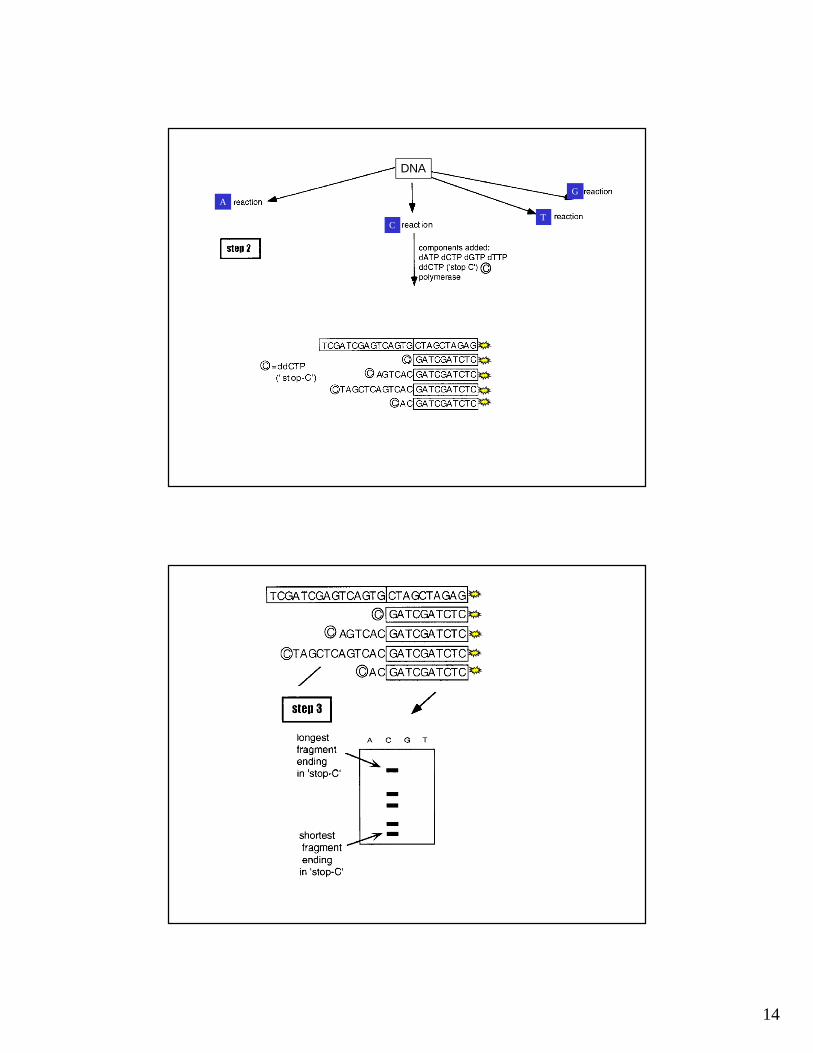
Secuenciamiento de nucleótidos [Método de Sanger ]

2
2.8 1.2
MAPA DE RESTRICCIÓN

3
SOUTHERN BLOTTING
SOUTHERN BLOTTING

4
SOUTHERN BLOTTING
SOUTHERN BLOTTING

5
A B
MstI(Enz restricción)
probe
Gen de la beta globina
A
B
electroforesis Southern blot
DNA “normal”
DNA “normal”
DNA “normal”

6
A B
MstI(Enz restricción)
probe
Gen de la beta globina
A
B
electroforesis Southern blot
DNA “normal”
DNA “normal”
DNA “normal”
A
B
electroforesis Southern blot
DNA “normal”
DNA “normal”
DNA “SC”
DNA “SC”
DNA “X”
A B
MstI(Enz restricción)
Gen de la beta globina
DNA “normal”
probe
C
MstI(Enz restricción)
Gen de la beta globina
DNA “SC”
C

7
10 micrograms per lane of tail DNA was digested with BamHI and EcoRI, then electrophoresed on 1.2% agarose, and alkaline capillary transferred to Hybond N+. The blot was hybridized with random prime-32P-dCTP-labeled probe from the transgene. This probe hybridizes with 6 and 5 kb endogenous bands seen in each lane, and the 2 kb transgene band seen in the 1X, 5X, and 10X transgene DNA-spiked tail DNA lanes, two MLC-Sno control
lanes containing tail DNA from another, single-copy, transgenic line, and four new transgenic mice identified in this Southern blot. Inclusion of the copy standards allowed us to calculate the approximate number of transgenes integrated for each founder as one for #10054, six for #10116, sixteen for #10050, and sixty for #10052. Thus,
four out of 30 mice screened (13%) were transgenic, which is lower than our recent track record of 20% transgenic.
DETECCION DE PRESENCIA Y NUMERO DE COPIAS DE UN TRANSGEN
Detección de cromosoma 21 en cromosomas en metafase y núcleos en interfase (linfocitos de sangre
periférica)

8
IDENTIFICACIÓN DE CROMOSOMAS HUMANOS
Fluorescent In-Situ Hybridization (FISH) -"Chromosome Painting"
DNA probes specific to regions of particular chromosomes are attached to fluorescent markers and hybridized with a chromosome spread. The picture shows a computer-generated "false colour"
image, in which small variations in fluorescence wavelength among probes are enhanced as distinct primary colours. The combination of probes that hybridize to a particular chromosome
produces a unique pattern for each chromosome. This makes it particulalry easy to detect segmental deletions and translocations among chromosomes.
NORTHERN BLOTTING
+ Probe (DNA marcado)
Aislar mRNA
Electroforesis Blot

9
10
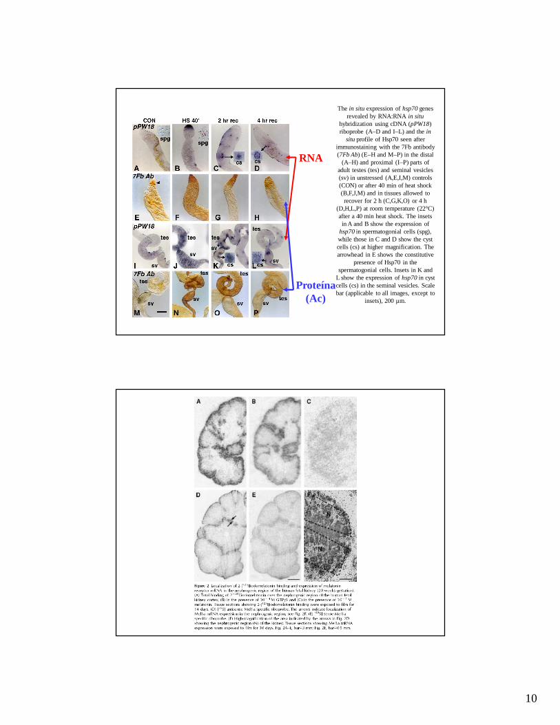
The in situ expression of hsp70 genes revealed by RNA:RNA in situ
hybridization using cDNA (pPW18) riboprobe (A–D and I–L) and the in
situ profile of Hsp70 seen after immunostaining with the 7Fb antibody (7Fb Ab) (E–H and M–P) in the distal
(A–H) and proximal (I–P) parts of adult testes (tes) and seminal vesicles (sv) in unstressed (A,E,I,M) controls (CON) or after 40 min of heat shock (B,F,J,M) and in tissues allowed to recover for 2 h (C,G,K,O) or 4 h
(D,H,L,P) at room temperature (22°C) after a 40 min heat shock. The insets in A and B show the expression of
hsp70 in spermatogonial cells (spg), while those in C and D show the cyst cells (cs) at higher magnification. The arrowhead in E shows the constitutive
presence of Hsp70 in the spermatogonial cells. Insets in K and
L show the expression of hsp70 in cyst cells (cs) in the seminal vesicles. Scale bar (applicable to all images, except to
insets), 200 µm.
RNA
Proteína(Ac)

11
21
Primers , dNTP, DNA polimerasa, Mg++
DesnaturalizaciónDesnaturalización
Unión Unión “primers”“primers”
ExtensiónExtensión
9595ººCC
5050--6060ººCC
7272ººCC
PCR
22
Medición de los niveles de expresión de genes
Diseño de los “primers” para la síntesis de cDNA
mRNA
mRNA
mRNAAl azarAl azar
Oligo (dT)Oligo (dT)
EspecíficoEspecífico
RT-PCR[amplificación de cDNA]
Síntesis de cDNA por Transcripción Reversa (RT) seguida de amplificación por PCR

12

13
14
DNA
A
CT
G

15
Se utilizan 4 carriles, uno par cada nucleótido cuando la marcación es la misma para todos.

16
►Deoxinucleotido marcado (interno, se incorpora en cadena sintetizada, generalmente radioactivo. En la actualidad esta en desuso)
►“Primer” marcado (radioactivo o fluorescente)
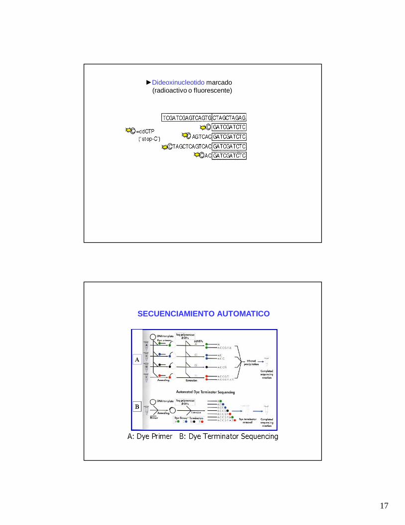
►Dideoxinucleotido marcado (radioactivo o fluorescente)
Tres formas de marcar el ADN cuando se realiza secuenciamiento de nucleótidos
►“Primer” marcado (radioactivo o fluorescente)
►Deoxinucleotido marcado
17
►Dideoxinucleotido marcado (radioactivo o fluorescente)
SECUENCIAMIENTO AUTOMATICO

18
Laser Capilar

19
RESULTADOS (secuenciamiento automático-electroforetograma)

20
EVOLUCION DE METODOS DE SECUENCIAMIENTO
1990-2003 3.000.000.000 u$s
2007 (2 MESES) 1.000.000 u$s
Sanger-electroforesis capilar; ~ 67,000 bases / h
454 Life Sciences Corp.; ~ 6.000.000 bases / h
May 31, 2007 James D. Watson, co-discoverer of the DNA helix and father of the Human Genome Project, prepares to autograph his book at the Baylor College of Medicine's Human Genome Sequencing Center.


![Repaso - cms.dm.uba.arcms.dm.uba.ar/.../probabilidades_y_estadistica_C/Adicionales/repaso… · Repaso Proba (C)-2015. Desigualdad de Markov Y 0 P(Y ) E[Y] Desigualdad de Tchebishev](https://img.dokumen.tips/doc/110x75/60ab951e202af13a8362c0d9/repaso-cmsdmubaarcmsdmubaarprobabilidadesyestadisticacadicionalesrepaso.jpg)