Embed Size (px)
Citation preview

RESEARCH ARTICLE Open Access
Renal kallikrein excretion and epigenetics inhuman acute kidney injury: Expression,mechanisms and consequencesSun Woo Kang1†, Pei-an Betty Shih2†, Roy O Mathew3†, Manjula Mahata2, Nilima Biswas2, Fangwen Rao2,Liying Yan4, Josee Bouchard5, Rakesh Malhotra6, Ashita Tolwani7, Srikrishna Khandrika6, Ravindra L Mehta6* andDaniel T O’Connor2,6*
Abstract
Background: Renal kallikrein (KLK1) synthesis and urinary excretion are reportedly diminished during AKI (acutekidney injury) in animal models, and provision of kallikrein abrogates renal injury in this setting, but data in humanAKI is limited. Therefore we first examined KLK1 renal excretion in human AKI, and then probed potentialendocrine and epigenetic mechanisms for its alterations.
Methods: KLK1 enzymatic activity excretion was evaluated in urine from patients with established or incipient AKI,versus healthy/non-hospital as well as ICU controls. Endocrine control of KLK1 excretion was then probed bycatecholamine and aldosterone measurements in established AKI versus healthy controls. To examine epigeneticcontrol of KLK1 synthesis, we tested blood and urine DNA for changes in promoter CpG methylation of the KLK1gene, as well as LINE-1 elements, by bisulfite sequencing.
Results: Patients with early/incipient AKI displayed a modest reduction of KLK1 excretion, but unexpectedly,established AKI displayed substantially elevated urine KLK1 excretion, ~11-fold higher than healthy controls, and ~3-fold greater than ICU controls. We then probed potential mechanisms of the change. Established AKI patients hadlower SBP, higher heart rate, and higher epinephrine excretion than healthy controls, though aldosterone excretionwas not different. Promoter KLK1 CpG methylation was higher in blood than urine DNA, while KLK1 methylation inblood DNA was significantly higher in established AKI than healthy controls, though KLK1 methylation in urinetended to be higher in AKI, directionally consistent with earlier/incipient but not later/established changes in KLK1excretion in AKI. On multivariate ANOVA, AKI displayed coordinate changes in KLK1 excretion and promotermethylation, though directionally opposite to expectation. Control (LINE-1 repetitive element) methylation in bloodand urine DNA was similar between AKI and controls.
Conclusions: Unexpectedly, increased KLK1 excretion in AKI patients was found; this increase is likely to be due inpart to increments in adrenergic tone during BP depression. Epigenetic changes at KLK1 may also play a role inearly changes of KLK1 expression and thus AKI susceptibility or recovery.
* Correspondence: [email protected]; [email protected]† Contributed equally2Departments of Medicine and Pharmacology, and Institute for GenomicMedicine, University of California at San Diego, CA, USA6Division of Nephrology and Hypertension, Department of Medicine,University of California San Diego Medical Center, San Diego, CA, USAFull list of author information is available at the end of the article
Kang et al. BMC Nephrology 2011, 12:27http://www.biomedcentral.com/1471-2369/12/27
© 2011 Kang et al; licensee BioMed Central Ltd. This is an Open Access article distributed under the terms of the Creative CommonsAttribution License (http://creativecommons.org/licenses/by/2.0), which permits unrestricted use, distribution, and reproduction inany medium, provided the original work is properly cited.
BackgroundThe incidence of acute kidney injury (AKI) in hospitalizedpatients is estimated to be 5-10%, and is much higher inthe critically ill [1,2]. Despite the potential for recovery ofkidney function, acute kidney injury is associated with sub-stantial morbidity and even mortality. AKI, due to ische-mia or nephrotoxic agent exposure, may lead to death orsublethal injury of proximal tubular cells, after which sur-viving cells may repolarize and/or de-differentiate, prolifer-ate, migrate to denuded areas, re-differentiate, and restorenephron structure (including the tubular epithelium) andfunction [3,4].The serine protease kallikrein (KLK1; E.C.-3.4.21.35;
OMIM 147910), excreted from kidney into urine, catalyzesthe cleavage of low molecular weight kininogen to lysyl-bradykinin (kallidin), which exhibits both vasodilator andnatriuretic pharmacological properties in the kidney; ifthese properties occur in vivo, the potential of the systemfor regulating blood pressure is clear [5].Renal kallikrein levels were markedly reduced in an ami-
noglycoside-induced AKI animal model [6], and KLK1gene transfer protected against aminoglycoside-inducednephropathy by diminishing apoptosis and inflammation[7]. In addition, KLK1 infusion during aminoglycosidetreatment attenuated drug-induced renal dysfunction, cor-tical damage, and apoptosis in the rat [8]. Previously, wehave reported that urinary KLK1 excretion was diminishedin renal allograft recipients with a clinical diagnosis ofacute tubular necrosis (ATN) [9]; since urinary KLK1 ori-ginates in the kidney, reduced urinary kallikrein levels mayreflect impaired renal function. However, this finding hasnot been pursued in humans.In mammals, cytosine methylation occurs almost
exclusively at CpG dinucleotides, which are enriched atCpG islands, often are located at 5’-/promoter regions offunctional genes [10]. Cytosine methylation may result intranscriptional repression either by interfering with tran-scription factor binding or by inducing a repressive chro-matin structure [11]. Apoptotic pathways are targets forsuch “epigenetic” silencing, and several apoptosis-linkedgenes that are regulated directly or indirectly by methyla-tion have been described [12].In this study, we first probed whether KLK1 excretion is
altered in human AKI, and if so what mechanisms (endo-crine or epigenetic) might be driving the change. Sincekidney repair after injury may recapitulate normal mor-phogenesis, we hypothesized that urinary kallikrein levelswould be associated with severity of AKI and with epige-netic changes in the renal kallikrein-1 (KLK1) promoter.We considered that changes in kallikrein excretion or theKLK1 promoter might predict renal functional recoveryand thus serve as biomarkers of recovery from AKI,thereby facilitating timely diagnosis and treatment. We
therefore examined patients with AKI for urinary expres-sion of KLK1 enzymatic activity, as well as genomic DNAfrom blood and urine for CpG methylation pattern at theKLK1 gene promoter.
MethodsEstablished AKI casesCases were ascertained from a single center prospectivenon-concurrent observational cohort of inpatients whowere identified as having suffered acute kidney injury(AKI) during a hospital admission. The institutional reviewboard of the University of California, San Diego (UCSD)reviewed and approved the study as well as the consentdocument. Primary providers referred potentially eligiblepatients to investigators and study coordinators. Patientswere eligible for enrollment if they were greater than 18years of age and met the serum creatinine criteria foracute kidney injury as set out by the Acute Kidney InjuryNetwork (AKIN) [13]: an abrupt rise by ≥0.3 mg/dl withina 48-hour period. Patients were excluded if they receivedchronic renal replacement therapy (hemodialysis or perito-neal dialysis) within the 6 months prior to admission; hadever been in receipt of a kidney transplant; pregnant orbreast feeding; currently incarcerated or otherwise institu-tionalized (nursing home, rehabilitation); were placedunder hospice/comfort care; or did not have reasonableexpectation of survival past the present hospitalization. Ifeligible, the patient or authorized representative was pre-sented the study and consent for participation wasobtained. All relevant patient data were derived from theelectronic and paper medical records, as well as directinterview of the patient or surrogate. Basic demographicinformation and co-morbid conditions were recorded onenrollment. Etiology of AKI was determined by chartreview of provider diagnosis, and verification by study per-sonnel; diagnosis followed the categories set by the Projectto Improve Care of Acute Renal Disease (PICARD) studyinvestigators [1]. Daily assessment included medicationreview, physical exam (as recorded in medical record orassessed by study personnel when missing), vital signs,intake and output, and labs. Clinical data elements werecollected daily and the need for and utilization pattern ofrenal replacement therapy was also monitored andrecorded. Blood and urine samples were collected at timeof entry, daily for 7 days maximum, and at hospitaldischarge. Twenty-four hour urine collections were per-formed at study entry and hospital discharge for creati-nine/urea clearance (in approximation of glomerularfiltration rate - GFR) as well as urinary kallikrein activity.One sample of whole blood was collected for genomicDNA preparation (and genetic analysis of the KLK1 locus)at the time of study entry. At the time of discharge,follow-up appointments with either the primary care
Kang et al. BMC Nephrology 2011, 12:27http://www.biomedcentral.com/1471-2369/12/27
Page 2 of 15

physician or a nephrologist (not all patients were seen by anephrologist during the hospitalization), extent of renalrecovery, and dialysis dependence (if needed during hospi-talization) were ascertained. Recovery: pre-defined studyendpoints were 12 months of follow-up, start of mainte-nance hemodialysis or peritoneal dialysis, receipt of a kid-ney transplant, or death; recovery of renal function wasthe primary outcome, defined as a return to within 10% ofbaseline eGFR or lowest eGFR prior to AKI event. Recov-ery was assessed at 6 months of follow-up.
Healthy (non-hospital) controlsIn addition, we obtained data for a control group of 38healthy adult subjects. Each healthy control was selectedfrom only one member from each of 38 twin pair sets.Twin pairs were recruited by a population-based twin reg-istry in southern California [14], and by newspaper adver-tisement. These twins are of European, African-American,and Asian biogeographic ancestry. Ethnicity was estab-lished by self-identification. Self-reported zygosity wasconfirmed by extensive SNP genotyping. There was noclinical evidence for kidney disease or any other cardiovas-cular disorder in any of the controls. Untimed (spot) urinecollections were performed, and one sample of EDTA-anticoagulated whole blood was collected for geneticanalysis of the KLK1 locus at the time of study entry.
Replication: Incipient (early) AKI cases in an intensive careunit (ICU), with ICU control subjectsAn additional sample of controls was ascertained forreplication of findings, in an ICU setting. In brief, thereplication sample consisted of n = 44 subjects (“ICUcontrols”) who did not develop AKI during a 7-dayobservation period after hospital ICU admission, as wellas n = 11 subjects (ICU cases) who did develop AKI, asdefined above. Patients were screened at ICU admissionfor potential study participation at three academic medi-cal centers (University of California San Diego, Univer-sity of Alabama, and Université de Montréal) betweenJuly 2006 and December 2008. Patients were eligible forenrollment if they were age 18 or older and had a lifeexpectancy of at least 48 hours. Controls were excludedif they had AKI according to the AKIN criteria [13],were admitted to the ICU > 48 hours prior to screening,transferred from another ICU, had a serum creatinine >2 mg/dl ≤ 3 days before ICU admission, were prisoners,received dialysis within the 12 months prior to admis-sion, had a functioning kidney transplant, were on antic-oagulants or warfarin within the last 7 days, sufferedfrom decompensated cirrhosis, had CKD stage 5, wereanemic (hemoglobin < 9.0 mg/dl or hematocrit < 27%),or were already enrolled in another research project.The study was approved by the Institutional Review
Boards at each institution, and written informed consent
was obtained from all participants or their health caresurrogates. Following informed consent, data on pastmedical history were collected once, and clinical, labora-tory and process-of-care elements were collected daily.Each institution’s local laboratory measured serum crea-tinine values. AKI was defined as an increase in serumcreatinine level of more than 0.3 mg/dL or more than50% from a reference creatinine within 48 hours (AKINcriteria)[13]. Patients without AKI within the first 4 dayshad continued blood and urine samples twice daily, fora total study observation period of 7 days.
Biochemical assaysUrine was assayed for kallikrein by an alkaline amidoly-tic activity assay as previously described (4), using thechromogenic substrate S-2266: [D] Val-Leu-Arg-parani-troanilide (Kabi Pharmacia; Franklin, OH, USA) [5,15].The activity of kallikrein per liter of urine (units perliter, U/L) is calculated from the formula: U/L =(9.55·A), where A = absorbance, after a 30-min assayincubation of the paranitroanilide product in a spectro-photometer at 405 nm [5,15]. Inclusion or exclusion ofthe kallikrein inhibitor aprotinin (Trasylol, Miles Inc.,West Haven, CT, USA) indicated that a fraction ofhuman urinary S-2266 amidolytic activity in the absenceof aprotinin was non-kallikrein (likely urokinase) [15];thus, aprotinin (20 kallikrein inhibitory U/mL) was sys-tematically included in the assay blank, to assure specifi-city for kallikrein measurement. The inter-assaycoefficient of variation was 18.1%, and activities fromn = 20 samples measured on two separate occasionscorrelated highly (Spearman R = 0.92, P< 0.01). In n =87 subjects, activity correlated (Spearman R = 0.82, P<0.0001) when results were compared for kallikreinexcretion normalized to time versus creatinine excre-tion. Specificity of the S-2266 amidolytic assay forglandular (KLK1, including renal, pancreas, and salivary)kallikrein activity in urine arises from two features: first,the substrate S-2266 is cleaved only by a particular sub-set of serine proteases, including KLK1; and second, theinclusion of aprotinin in the assay blank, which specifi-cally inhibits serine proteases including KLK1; nonethe-less, only an immunoassay specific for lysyl-bradykinin(the kinin product of KLK1) generation could provideabsolute assurance of specificity for KLK1.Clinical chemistries (serum or urine, electrolytes or
creatinine) were measured by spectrophotometric autoa-nalyzer. Urine aldosterone concentration was deter-mined by enzyme immunoassay (Alpco Diagnostics,Salem, NH, USA). Urine catecholamine concentrationwas determined by commercial ELISA kit (Labor Diag-nostika Nord GmBH & Co. KG, Nordhorn, Germany).Urine values were normalized to creatinine concentra-tion in the same sample.
Kang et al. BMC Nephrology 2011, 12:27http://www.biomedcentral.com/1471-2369/12/27
Page 3 of 15

DNA extraction and bisulfite treatment of CpG sitesA sample of EDTA-anticoagulated whole blood wasobtained from participants and stored at -70°C prior toDNA extraction. Timed and spot urine samples wereobtained and frozen at -70°C before assay. Blood DNAwas prepared from blood leukocytes with a commercialkit (QIAamp® DNA Mini Kit; Qiagen, USA). UrineDNA was prepared from the urine pellet with spun col-umns (urine DNA isolation kit; Norgen, Canada). BothDNAs were subjected to sodium metabisulfite(Na2S2O5) treatment (Imprint™ DNA Modification Kit;Sigma, USA), and then eluted in 20 μL elution buffer.Bisulfite converts cytosine residues to uracil, but leaves5-methylcytosine residues unaffected.
KLK1 promoter CpG methylation analysisPyrosequencing for allele discrimination (Pyrosequencing;Qiagen, USA) provides real-time extension-based DNAanalysis that can evaluate multiple CpG sites [16]. CpGmethylation analysis at the 5’/upstream/proximal promo-ter region of human kallikrein serine protease 1 (KLK1)gene was performed. The KLK1 gene was analyzed by asingle PCR amplicon spanning 4 CpGs in a 263 bp region,with two biotinylated sequencing (extension) primers(Figure 1). The consecutive 4 CpGs were located -203 to-135 bp from the transcription initiation site: sequentiallyat -203, -196, -154, and -135 bp (Figure 1).As a control, global CpG methylation analysis was com-
pleted using PyroMark LINE-1 reagents (Pyrosequencing;Qiagen, USA). We thus determined the methylation statusof three CpG sites in LINE-1 repetitive (LTR-like) ele-ments, wherein methylation levels of CpG sites representglobal methylation status across the genome, because ofthe repetitive nature of LINE-1 elements [17,18].PCR amplification was performed using 10X PCR buf-
fer, 3.0 mM MgCl2, 200 μM of each dNTP, 0.2 μM each
of forward and reverse primers, HotStar DNA polymer-ase (Qiagen, USA) 1.25 U, and ~10 ng of bisulfite-converted DNA per 50 μl reaction. PCR cycling condi-tions were: 94°C 15 min; 45 cycles of 94°C 30 s, 56°C30 s, 72°C 30 s; 72°C 5 min; and then products wereheld at 4°C. The PCR was performed with one of thePCR primers biotinylated to convert the PCR product tosingle-stranded DNA template. PCR products (each10 μl) were sequenced by Pyrosequencing PSQ96 HSSystem (Pyrosequencing, Qiagen, USA). The methylationstatus of each locus was analyzed individually as a T/CSNP using Pyro-QCpG™ software (Pyrosequencing).AKI case samples were evaluated at study entry (base-
line, time of diagnosis). After bisulfite modification andPCR amplification, KLK1 blood DNA promoter methyla-tion data from 13 AKI patients and 30 controls wereobtained. KLK1 urine DNA promoter methylation datafrom 9 AKI and 22 controls were available. LINE-1 bloodDNA methylation data from 14 AKI patients and 32 con-trols were available for evaluation. LINE-1 urine DNAmethylation data from 15 AKI patients and 32 controlswere obtained.
Statistical analysesResults are expressed as the mean value ± one standarderror of the mean (SEM) for continuous variables. Forcomparisons of two groups, unpaired two-sided t-testsor one-way ANOVA (enabling adjustment for covariatesof age, sex, and ethnicity) were performed. Non-para-metric Wilcoxon Rank Sum test was used to confirmparametric tests in the face of relatively small samplesizes. Proportions were evaluated by Fisher’s exact test(2 × 2 tables) or chi-square test (3 × 2 tables). Statisticalanalyses were performed in R2.10.1 <http://www.r-pro-ject.org/ > or SPSS-17 (Statistical Package for the SocialSciences; Chicago, IL, USA). A P value of < 0.05 was
Figure 1 Map of CpG sites studied in the human KLK1 proximal promoter. Letters highlighted in gray are the target regionscomplementary to amplified PCR primers. The CpGs analyzed are numbered as Pos#1 - Pos#4 and colored yellow. Green underlined sequencesare target regions complementary to sequencing primers. Green highlighted A is the transcriptional initiation ("cap”) site. Sequences followingthe transcription initiation site are colored pink.
Kang et al. BMC Nephrology 2011, 12:27http://www.biomedcentral.com/1471-2369/12/27
Page 4 of 15

considered significant. Multiple linear regression wasperformed with default criteria of entry (p < 0.05) andexit (p > 0.10) from the multivariate regression model,using stepwise or forward options. Recovery from AKIwas pre-defined as return (within 6 months follow-up)to within 10% of baseline eGFR, or lowest eGFR prior toAKI event.
ResultsRenal KLK1 excretion and eGFR in the 4 subject groups:AKI cases and controlsDemographic and anthropometric description of thestudy samples is presented in Table 1. Baseline demo-graphic characteristics (age, sex) were similar acrossgroups.As compared to healthy/outpatient controls (Table 1,
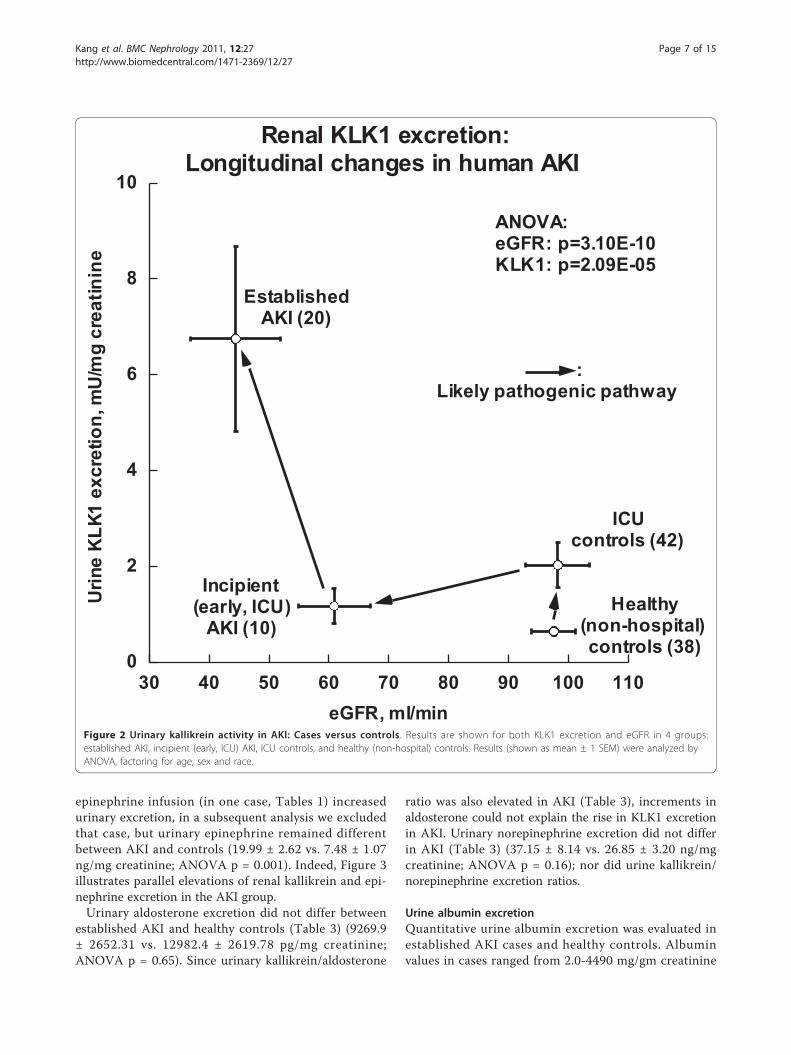
Figure 2), ICU/inpatient controls displayed unalteredeGFR, though a modest ~3.2-fold increase in urinaryKLK1 excretion. ICU subjects with incipient AKI had amodest fall in eGFR (down ~38% compared to ICU con-trols), coupled with a ~43% fall in KLK1 excretion.However, subjects with established (more severe) AKIexhibited a ~6.9-fold increase in KLK1 excretion (p =2.09E-05), coupled with a further ~27% fall in eGFR(p = 3.10E-10).
Established AKI: Clinical characteristicsWe then turned our attention to clinical features (Table 2)of the AKI subjects that might account for the KLK1 ele-vation. In clinical laboratory findings at study enrollment(entry), patients with AKI had significantly higher serumcreatinine (2.67 ± 0.43 vs. 0.9 ± 0.04 mg/dl; p < 0.001),and fractional excretion of sodium (FeNa+; 1.7 ± 0.4 vs.0.8 ± 0.06%; p = 0.05), than healthy controls, with lowereGFR (44.4 ± 7.5 vs. 97.5 ± 3.6 ml/min; p = 0.0001).At study enrollment, AKI patients had lower SBP (119.8
± 4.4 vs. 131.4 ± 1.7 mmHg; p = 0.02) and higher heartrate (89.3 ± 3.6 vs. 68.0 ± 1.6 beats/min; p = 1.73E-05)than healthy controls. Within the AKI group (n = 20),acute kidney injury was attributed to ischemia in 7patients, nephrotoxins in 4, sepsis in 1, and multifactorialcauses in 8 (Table 2); KLK1 excretion did not vary by AKIcausal group (ANOVA p = 0.83). Six patients had diabetesmellitus, 10 had hypertension, 1 had coronary artery dis-ease, 6 had chronic liver disease, 4 had chronic lung dis-ease, 8 had chronic kidney disease (previous eGFR <60 ml/min), 13 required admission to an ICU during hos-pitalization, 5 required mechanical ventilation at enroll-ment, and 5 had oliguria at enrollment. At study entry,3 patients required infusion of vasopressors (2 norepi-nephrine, 1 dopamine and epinephrine combination), and3 took diuretics (2 furosemide, 1 thiazide). In the evalua-tion of primary outcome, 17 patients attained recovery of
renal function (see Methods) at 6 months of follow-up(Table 1).
Established AKI: Kallikrein, catecholamines, andaldosteroneHere we probed potential hormonal mechanismswhereby KLK1 excretion was elevated in establishedAKI, focusing on such known KLK1 stimulators as cate-cholamines [19] and aldosterone.
Urinary kallikrein enzymatic activityFrom the 20 established AKI patients and 38 healthy con-trols, urine was available for kallikrein measurement(Table 3) in 18 patients and 37 controls. Unexpectedly,established AKI subjects displayed substantially elevatedkallikrein excretion (Figure 2, Table 1), about ~10 timeshigher than that of controls (activity: 6132.9 ± 2302 vs.623.0 ± 88.2 mU/L, p < 0.001; urine kallikrein activity/creatinine ratio: 6.74 ± 1.92 vs. 0.63 ± 0.08 U/gm, p <0.001). To exclude the possibility that diuretic treatmentat study entry increased urinary kallikrein excretion [20],we conducted statistical analysis again by exclusion of the3 diuretic cases (Tables 1); urinary kallikrein excretionremained significantly different between AKI and controls(urine kallikrein/urine creatinine ratio: 7.14 ± 2.18 vs. 0.63± 0.08 mU/mg; p = 0.001). We measured the urinary non-kallikrein amidolytic activity (likely urokinase) by inclusionor exclusion of the kallikrein inhibitor aprotinin. The per-centage of kallikrein activity within total S-2266 amidolyticactivity was not different between AKI patients and con-trols (78.0 ± 4.8% vs. 69.0 ± 2.4%; p = 0.072).Since black and white subjects differ in reported KLK1
excretion [5,15], we evaluated the role of ethnicity inour samples (Table 1). Although cases and controls eachincluded several biogeographic ancestries, KLK1 excre-tion did not differ significantly in black versus whitecases (p = 0.26) or black versus white controls (p =0.69), perhaps reflecting the relatively small samplesizes. Disease analyses were adjusted for biogeographicancestry as a covariate.
Urinary aldosterone and catecholamine excretionsWe measured urinary aldosterone, epinephrine and nore-pinephrine excretions (Table 3), since these hormonesare known to increase urinary kallikrein. Established AKIsubjects exhibited substantially elevated epinephrineexcretion, ~2.7 times higher than that of healthy controls(Table 3, Figure 3; 20.1 ± 2.4 vs. 7.48 ± 1.07 ng/mg creati-nine; ANOVA p = 0.00016). Urinary kallikrein/epinephr-ine ratio did not differ between groups (Table 3); parallelelevations of KLK1 and epinephrine suggest that the epi-nephrine excess in AKI may be sufficient to account forthe increased KLK1. To exclude the possibility that
Kang et al. BMC Nephrology 2011, 12:27http://www.biomedcentral.com/1471-2369/12/27
Page 5 of 15

Table 1 Clinical characteristics of cases and controls: Established versus incipient AKI cases, and ICU versus healthycontrols
AKI Controls
P < 0.05 (*) Established Incipient (early, ICU) ICU Healthy(non-hospital)
Characteristics n = 20 n = 11 n = 44 n = 38
Age, years * 48.8 ± 3.5 68.1 ± 3.8 52.9 ± 2.3 46.3 ± 1.5
Sex (male/female) 15/5 5/6 26/18 28/10
Ethnicity, n *
White 11 5 28 20
Black 3 4 8 6
Hispanic 5 0 7 6
Other 1 2 1 6
Lab findings at enrollment
sCr, mg/dl * 2.67 ± 0.43 1.29 ± 0.12 0.81 ± 0.05 0.9 ± 0.04
eGFR, ml/min * 44.4 ± 7.3 60.9 ± 5.7 98.2 ± 4.9 97.5 ± 3.5
uCr/sCr, ratio * 42.4 ± 7.2 83.3 ± 18.1 106.6 ± 18.3 119.6 ± 5.2
Urine kallikrein(U/gm creatinine)
* 6.74 ± 1.92 1.17 ± 0.16 2.04 ± 0.47 0.63 ± 0.08
Vital signs at enrollment
Systolic BP, mmHg 0.08 119.8 ± 4.4 126.9 ± 8.7 124.3 ± 3.4 131.4 ± 1.7
Diastolic BP, mmHg * 70.7 ± 3.4 61.8 ± 5.1 68.6 ± 2.3 74.7 ± 1.5
Heart rate, beats/min * 89.3 ± 3.6 86.5 ± 7.4 81.3 ± 2.4 68.0 ± 1.6
Co-morbid conditions (Y/N)
Diabetes mellitus 6/14 6/5 12/32 0/38
Hypertension * 10/10 8/3 20/22 8/30
Coronary artery disease 1/19 4/7 10/34 0/38
Congestive heart failure * 0/20 3/8 4/40 0/38
Chronic liver disease * 6/14 0/11 4/40 0/38
Chronic lung disease * 4/16 3/8 7/37 0/38
Chronic kidney disease * 8/12 3/8 1/43 0/38
HIV positive 2/18 0/11 3/41 0/38
Malignancy 3/17 1/10 12/32 0/38
Smoker * 8/12 3/8 16/28 4/34
Primary treating service, n
Medical/Surgical 14/6 11/0 34/10 -
Other characteristics while hospitalized (Y/N)
ICU admission * 13/7 11/0 44/0 -
Ventilator at enrollment 5/15 3/8 13/31 -
Pressor infusions * 3/17 9/2 12/32 -
Norepinephrine * 2/18 8/3 11/33 -
Epinephrine + dopamine * 1/19 9/2 12/32 -
Diuretics at enrollment * 3/17 5/6 2/42 -
AKI outcomes
Required dialysis for AKI 3/17 1/10 - -
Recovery from AKI * 17/3 11/0 - -
Remained dialysis-dependent at follow-up
1/19 0/11 - -
Final eGFR at follow-up 66.5 ± 8.0 76.6 ± 5.5 - -
eGFR Δ at follow-up +16.5 ± 2.2 +22.2 ± 5.4 - -
sCr Δ at follow-up 0.08 -1.0 ± 0.3 -0.25 ± 0.05 - -
ICU = intensive care unit, AKI = acute kidney injury, n = number of study subjects, BMI = body mass index, s = serum, u = urine, Cr = creatinine, FeNa+ =fractional excretion of sodium, SBP = systolic blood pressure, DBP = diastolic blood pressure, HR = heart rate, bpm = beats per minute. Δ = change at discharge(from initial value). *P values calculated with Fisher’s exact test for categorical variables and ANOVA (log-transformed and covariates adjusted: age, sex) forcontinuous variables. *: Symbol indicates P ≤ 0.05 comparing across all available groups. Recovery of renal function was the primary outcome, defined as a returnto within 10% of baseline eGFR or lowest eGFR prior to AKI event. Plus-minus values are mean ± SEM.
Kang et al. BMC Nephrology 2011, 12:27http://www.biomedcentral.com/1471-2369/12/27
Page 6 of 15
epinephrine infusion (in one case, Tables 1) increasedurinary excretion, in a subsequent analysis we excludedthat case, but urinary epinephrine remained differentbetween AKI and controls (19.99 ± 2.62 vs. 7.48 ± 1.07ng/mg creatinine; ANOVA p = 0.001). Indeed, Figure 3illustrates parallel elevations of renal kallikrein and epi-nephrine excretion in the AKI group.Urinary aldosterone excretion did not differ between
established AKI and healthy controls (Table 3) (9269.9± 2652.31 vs. 12982.4 ± 2619.78 pg/mg creatinine;ANOVA p = 0.65). Since urinary kallikrein/aldosterone
ratio was also elevated in AKI (Table 3), increments inaldosterone could not explain the rise in KLK1 excretionin AKI. Urinary norepinephrine excretion did not differin AKI (Table 3) (37.15 ± 8.14 vs. 26.85 ± 3.20 ng/mgcreatinine; ANOVA p = 0.16); nor did urine kallikrein/norepinephrine excretion ratios.
Urine albumin excretionQuantitative urine albumin excretion was evaluated inestablished AKI cases and healthy controls. Albuminvalues in cases ranged from 2.0-4490 mg/gm creatinine
0
2
4
6
8
10
30 40 50 60 70 80 90 100 110
Renal KLK1 excretion:Longitudinal changes in human AKI
Urin
e K
LK1
excr
etio
n, m
U/m
g cr
eatin
ine
eGFR, ml/min
EstablishedAKI (20)
Incipient(early, ICU)
AKI (10)Healthy
(non-hospital)controls (38)
ICUcontrols (42)
ANOVA:eGFR: p=3.10E-10KLK1: p=2.09E-05
:Likely pathogenic pathway
Figure 2 Urinary kallikrein activity in AKI: Cases versus controls. Results are shown for both KLK1 excretion and eGFR in 4 groups:established AKI, incipient (early, ICU) AKI, ICU controls, and healthy (non-hospital) controls. Results (shown as mean ± 1 SEM) were analyzed byANOVA, factoring for age, sex and race.
Kang et al. BMC Nephrology 2011, 12:27http://www.biomedcentral.com/1471-2369/12/27
Page 7 of 15

(mean, 1090 mg/gm), but kallikrein and albumin excre-tions did not correlate (Pearson r = 0.006, p = 0.54), ren-dering it unlikely that elevated kallikrein activity in AKIarose simply from pathological excretion of plasma pro-teases. In healthy controls, albumin excretion was 6.27 ±0.39 mg/gm creatinine.
ICU controls: Urinary kallikrein activity and clinical findings44 “ICU controls” were available to evaluate the specifi-city of urinary kallikrein elevation in AKI. Table 1shows demographic, laboratory and clinical findings ofthese ICU controls. The kallikrein increment in AKIpersisted when studied with ICU controls (6.74 ± 1.92vs. 2.04 ± 0.47 U/gm creatinine; p = 0.028), though kal-likrein excretion was modestly elevated in ICU- com-pared to healthy controls (2.04 ± 0.47 vs. 0.63 ± 0.08 U/gm creatinine; p = 0.005).Compared with established AKI, ICU controls had sig-
nificantly lower serum creatinine (0.81 ± 0.05 vs. 2.67 ±
0.43 mg/dl; p = 0.0005). Compared with healthy con-trols, ICU controls were older (54.2 ± 2.2 vs. 46.3 ± 1.5years; p = 0.005), with higher heart rate (81.3 ± 2.4 vs.68.0 ± 1.6 bpm; p < 0.0001), but lower DBP (68.6 ± 2.3vs. 74.7 ± 1.5 mmHg; p = 0.029). Even though not sig-nificantly different, SBPs of ICU controls tended to belower than those in healthy controls (124.3 ± 3.4 vs.131.4 ± 1.7 mmHg; p = 0.066). The ICU controls hadvariable primary diseases (Table 1). 13 among themrequired mechanical ventilation, while 12 required vaso-pressor infusion during ICU admission (Table 1).
KLK1 promoter DNA CpG methylation patternsKLK1 promoter CpG methylation (positions in Figure 1)was studied in established AKI and healthy controls.Promoter KLK1 CpG methylation (Figure 4) was higherin blood than urine DNA (blood 66.38 ± 1.00 vs. urine33.43 ± 4.67%; ANOVA p < 0.0001). Promoter KLK1methylation in blood DNA was also higher in AKI than
Table 2 Mechanistic studies: Characteristics of the AKI study subjects versus healthy controls
Characteristics Established AKI patients(n = 20)
Healthy controls(n = 38)
Pvalue*
Age, years 48.8 ± 3.5 46.3 ± 1.5 0.52
Sex (male/female), n 15/5 30/8 1
Ethnicity, n 0.6
White 11 20
Black 3 6
Hispanic 5 6
Other 1 6
Weight, kg 91.2 ± 6.2 93.9 ± 3.7 0.293
BMI, kg/m2 31.1 ± 2.2 27.8 ± 1 0.177
Laboratory findings at enrollment
sCr, mg/dl 2.7 ± 0.47 0.9 ± 0.04 0.0007
eGFR, ml/min 44.4 ± 7.5 97.5 ± 3.6 <0.0001
uNa+/uCr, mEq/gm 127.6 ± 22.5 125.4 ± 9.4 0.347
uCr/sCr, ratio 42.4 ± 7.2 119.6 ± 11.4 <0.0001
FeNa+, % 1.7 ± 0.4 0.8 ± 0.1 0.05
Vital signs at enrollment
Systolic BP, mmHg 119.8 ± 4.4 131.4 ± 1.7 0.02
Diastolic BP, mmHg 70.7 ± 3.4 74.7 ± 1.5 0.3
Heart rate, beats/min 89.3 ± 3.6 68.0 ± 1.6 <0.0001
Contributing causes to AKI, n (with urine KLK1 activity excretion, U/gm creatinine,mean ± SEM)
0.83
Ischemia 7 (6.0 ± 3.7) - -
Nephrotoxins 4 (7.2 ± 3.9) - -
Septic 1 (4.6) - -
Multifactorial causes/other 8 (7.6 ± 2.9) - -
AKI = acute kidney injury, n = number of study subjects, BMI = body mass index, s = serum, u = urine, Cr = creatinine, FeNa+ = fractional excretion of sodium,SBP = systolic blood pressure, DBP = diastolic blood pressure, HR = heart rate, bpm = beats per minute, ICU = intensive care unit. *P values were calculated withFisher’s exact test for categorical variables, and ANOVA (log-transformed, adjusted for covariates: age, sex) for continuous variables. Plus-minus values are mean ±one SEM. Bold: p ≤ 0.05.
Kang et al. BMC Nephrology 2011, 12:27http://www.biomedcentral.com/1471-2369/12/27
Page 8 of 15

Table 3 Mechanistic studies: Urinary biochemistries in established AKI cases versus healthy controls
AKI patients n available Healthy controls n available T-test P ANOVA P† Wilcoxon rank P
Urine kallikrein (U/gm creatinine) 6.74 ± 1.92 18 0.63 ± 0.08 37 0.0058 0.00029 0.0012
Urine aldosterone (pg/mg creatinine) 9269.9 ± 2652.31 16 12982.4 ± 2619.78 38 0.325 0.65 0.1282
Urine epinephrine (ng/mg creatinine) 20.1 ± 2.4 14 7.48 ± 1.07 32 1.11E-06 0.00016 1.79E-05
Urine norepinephrine, (ng/mg creatinine) 37.15 ± 8.14 14 26.85 ±3.2
32 0.344 0.16 0.4424
Urine kallikrein/urine aldosterone ratio (mU/μg) 872.0 ± 277.5 15 160.5 ±76.7
38 0.00027 8.45E-05 3.62E-05
Urine kallikrein/urine epinephrine ratio (mU/ng) 0.32 ± 0.13 13 0.18 ±0.04
31 0.8465 0.8 0.899
Plus-minus values are covariate-adjusted mean ± one SEM. N is the number of study subjects available to conduct each experiment. †Results are analyzed by one-way ANOVA, factoring for age, sex and race. Bold: p≤ 0.05.
Kanget
al.BMCNephrology
2011,12:27http://w
ww.biom
edcentral.com/1471-2369/12/27
Page9of
15

controls (70.32 ± 2.27 vs. 65.36 ± 1.05%; ANOVA p =0.011), while promoter KLK1 methylation in urine DNAtrended to be higher in AKI than controls (40.95 ± 7.06vs. 30.35 ± 5.88%; ANOVA p = 0.22; Figure 4).Global CpG methylation, examined by LINE-1, was
high in both blood and urine, though even higher inblood (blood 73.11 ± 0.38 vs. urine 69.37 ± 0.86%, p =0.0002). LINE-1 CpG methylation in blood DNA wassimilar between AKI and controls (71.71 ± 0.44 vs. 73.67
± 0.41%; ANOVA p = 0.08), as was LINE-1 methylationin urine DNA (AKI 69.53 ± 1.54 vs. control 69.29 ±1.05%; ANOVA p = 0.79) (Figure 4). In AKI blood DNA,KLK1-specific methylation was similar to global LINE-1methylation (70.32 ± 2.27 vs. 71.71 ± 0.44%; p = 0.56). Incontrols, however, KLK1 CpG methylation in blood DNAwas significantly lower than global LINE-1 methylationin blood DNA (65.36 ± 1.05 vs. 73.67 ± 0.41%; p <0.0001) (Figure 4). In both AKI and controls, KLK1
0
2
4
6
8
6 8 10 12 14 16 18 20 22
Renal kallikrein and epinephrineexcretions in AKI: Parallel elevations
Ren
al k
allik
rein
exc
retio
n(m
IU/m
g cr
eatin
ine)
Renal epinephrine excretion(ng/mg creatinine)
Controls(kallikrein 37,
epinephrine 32)
AKI cases(kallikrein 18,
epinephrine 14)
Epinephrine: p=1.6E-04Kallikrein: p=2.9E-04
Figure 3 Coordinate effects of AKI on kallikrein and epinephrine excretion. Results (shown as mean ± 1 SEM) were analyzed by one-wayANOVA, factoring for age, sex and race. The numbers studied for kallikrein are 18 established AKI patients and 37 healthy controls. The numbersstudied for epinephrine are 14 established AKI patients and 32 healthy controls.
Kang et al. BMC Nephrology 2011, 12:27http://www.biomedcentral.com/1471-2369/12/27
Page 10 of 15

specific methylation in urine DNA was significantly lowerthan global LINE-1 methylation in urine DNA(AKI 40.95 ± 7.06 vs. 69.53 ± 1.54%; p = 0.003; controls30.35 ± 5.88 vs. 69.29 ± 1.05%; p < 0.0001) (Figure 4).Urine KLK1 promoter CpG methylation did not pre-
dict KLK1 activity excretion. However, in a multivariateanalysis, disease status jointly predicted both KLK1 pro-moter methylation and enzyme activity excretion, withhigher values for each in cases (multivariate p = 0.004;Figure 5. Since increased KLK1 promoter methylationwould be predicted to decrease gene expression, thisjoint effect cannot explain (and indeed runs counter to)
the elevated KLK1 excretion observed in our AKI cases(Figure 5).
DiscussionOverviewSeveral previous lines of investigation link alterations inKLK1 expression to AKI, in both experimental animalsand humans. A decrease in urinary kallikrein excretionoccurs in rodents with AKI after methemoglobin [21];rats treated with aminoglycosides have dramaticallyreduced levels of urinary kallikrein [6], and a transientdecrease in urinary kallikrein excretion occurs during
Significance (by ANOVA):
Blood versus urine KLK1methylation: p<0.0001
AKI versus control KLK1methylation in blood DNA: p=0.011
Blood versus urine DNA LINE-1methylation: p=0.0002
Urine DNA KLK1 versus urine LINE-1 methylation: AKI p=0.003, and controls p<0.0001
Figure 4 CpG methylation analyzed by bisulfite sequencing: Results at the KLK1 promoter, as well as a global control (LINE-1repetitive elements), in genomic DNA from urine or blood (mononuclear cells). Results (shown as mean ± one SEM), from established AKIcases or healthy controls, were analyzed by ANOVA, factoring for age, sex and race. LINE-1 reagents (Pyromark, Biotage) were used to analyzethe 3 CpG sites in LINE-1 repetitive elements, while the KLK1 gene was analyzed by a separate PCR covering 4 promoter CpGs. Promoter KLK1specific methylation was substantially higher in blood than urine DNA (blood 66.38 ± 1.00 vs. urine 33.43 ± 4.67%; ANOVA p < 0.0001). PromoterKLK1 methylation in blood DNA was higher in AKI than controls (AKI, 70.32 ± 2.27 vs. controls, 65.36 ± 1.05%; ANOVA p = 0.011). Promoter KLK1methylation in urine DNA did not differ in AKI versus controls (AKI, 40.95 ± 7.06 vs. controls, 30.35 ± 5.88%; ANOVA p = 0.22). Global LINE-1methylation was greater in both blood than urine DNA (blood 73.11 ± 0.38 vs. urine 69.37 ± 0.86%, p = 0.0002). LINE-1 methylation in bloodDNA did not differ in AKI and controls (AKI, 71.71 ± 0.44 vs. controls, 73.67 ± 0.41%; ANOVA p = 0.08). LINE-1 methylation in urine DNA did notdiffer in cases/controls (AKI, 69.53 ± 1.54 vs. controls, 69.29 ± 1.05%; ANOVA p = 0.79).
Kang et al. BMC Nephrology 2011, 12:27http://www.biomedcentral.com/1471-2369/12/27
Page 11 of 15

chromate-induced AKI in the rat [22]. Renal kallikreinmRNA expression was specifically reduced in the post-ischemic rodent kidney, with persistently altered expres-sion even after functional recovery from ischemic acuterenal failure [23]. In humans, we reported previouslythat urinary kallikrein excretion was diminished in acutetubular necrosis (ATN) after renal transplantation [9].Studies in rodents suggest beneficial effects of exogen-
ous KLK1 replacement in the setting of experimentalAKI. KLK1 gene transfer protected against aminoglyco-side-induced nephropathy, with inhibition of apoptosis
and inflammation [7]. KLK1 replacement after gentami-cin attenuated drug-induced renal dysfunction, corticaldamage, and apoptosis in the rat [8]. Furthermore, KLK1reduced gentamicin-induced renal dysfunction and fibro-sis, with decreased myofibroblast and collagen accumula-tion [8]. These findings indicate that the renal kallikrein/kinin system prevents and promotes recovery of amino-glycoside-induced renal injury by inhibiting apoptosis,inflammatory cell recruitment, and fibrotic lesions.Thus, we expected that AKI patients would have
diminished urinary kallikrein excretion, since urinary
0
2
4
6
8
25 30 35 40 45 50
AKI: Coordinate effects on KLK1 promoter CpGmethylation and enzymatic activity excretion in urine
Urin
e K
LK1
activ
ity e
xcre
tion,
U/g
m c
reat
inin
e
Urine DNA KLK1 promoter CpG methylation, %
Multivariate ANOVA:Pillai's trace F=6.9, p=0.004
Healthycontrol
AKIcases
Figure 5 AKI: Coordinate effects of disease on KLK1 promoter CpG methylation and KLK1 enzymatic activity excretion in urine. Themultivariate analysis compared established AKI cases with healthy controls.
Kang et al. BMC Nephrology 2011, 12:27http://www.biomedcentral.com/1471-2369/12/27
Page 12 of 15

kallikrein originates in the kidney; further, we antici-pated that kallikrein increments might be associatedwith superior outcomes in AKI. Unexpectedly our estab-lished AKI subjects displayed substantially elevated (byas much as ~11-fold; ANOVA p = 0.00029; Figure 2;Tables 1,3) kallikrein excretion.
Origin of increased KLK1 excretion in established AKIAlthough previous reports of KLK1 excretion in AKIindicated diminished excretion in both rodent models[6,21,22] and AKI/ATN in the setting of human renaltransplantation [9], we noted a ~11-fold elevation ofKLK1 excretion in our established AKI subjects (Tables1-3; Figure 2). Why might KLK1 excretion be elevatedin human AKI? Here we examined hormonal factorsknown to increase KLK1 excretion: catecholamines andaldosterone [5,19,20,24-26]. We found that an elevationin epinephrine excretion paralleled that for kallikrein(Figure 3), while norepinephrine and aldosterone excre-tions were unchanged (Table 3). In experimental ani-mals, kallikrein excretion is regulated by adrenergicreceptors, with stimulation by b-receptors and inhibitionby a-receptors [19]. While diuretics can also elevate kal-likrein excretion [20,27], the KLK1 excretion increment
in AKI persisted after exclusion of the 3 subjects ondiuretics.Why was epinephrine elevated in our AKI subjects?
AKI patients had lower systolic BP than controls (119.8 ±4.4 vs. 131.4 ± 1.7 mmHg; p = 0.02) and higher heart rate(89.3 ± 3.6 vs. 68.0 ± 1.6 bpm; p = 1.73E-05) (Table 2).While the mechanism cannot be readily probed in thesetting of acute human illness, we suspect that that lowerBP in AKI may stimulate baroreceptors, with resultingincrease in endogenous production of epinephrine, andconsequently increased heart rate and kallikrein excre-tion (Figure 6).
KLK1 epigeneticsPromoter KLK1 specific CpG methylation was higher inblood than urine DNA (blood 66.38 ± 1.00 vs. urine33.43 ± 4.67%; ANOVA p < 0.0001; Figure 4), consistentwith kidney-specific expression of the KLK1 gene, inthat renal kallikrein is synthesized in the distal tubuleand released into urine and peritubular interstitium [20],and cytosine methylation results in transcriptionalrepression either by interfering with transcription factorbinding or by inducing a repressive chromatin structure[11].
Hypotension
Baroreceptors
Epinephrine
Renalkallikrein (KLK1)
Human AKI: Hypothetical schema for stepseventuating in increased renal kallikrein (KLK1) excretion
Azotemia
Diuretics
Adrenergicpressors
Azotem
1)
Endogenous Exogenousg
KLK1 promoter
Geneticvariation
CpGmethylation
enetic CpG
kaFigure 6 Hypothetical schema integrating experimental findings in this study of KLK1 in AKI. This diagram is presented not as establishedfact, but rather to generate hypotheses for further investigation. Endogenous factors may influence KLK1 synthesis and renal excretion: lower BPin AKI may activate baroreceptors, thus increasing endogenous secretion of epinephrine, thereby increasing both heart rate and urinary kallikreinexcretion. KLK1 promoter genetic variants or CpG methylation can influence renal kallikrein production. Finally, exogenous factors such asadrenergic pressor infusions or diuretic treatment can also increase renal kallikrein production; indeed, since a subset of our AKI cases receivedsuch treatments (Table 1), we cannot exclude this possibility.
Kang et al. BMC Nephrology 2011, 12:27http://www.biomedcentral.com/1471-2369/12/27
Page 13 of 15

KLK1 promoter methylation in blood DNA was higherin AKI than controls (70.32 ± 2.27 vs. 65.36 ± 1.05%; p =0.011), and there was also a trend towards higher urineKLK1 methylation in AKI than controls, but the differencewas not significant (40.95 ± 7.06 vs. 30.35 ± 5.88%; p =0.22). While a multivariate analysis indicated a joint effectof AKI on both KLK1 excretion and urine KLK1 CpGmethylation (Figure 5), elevated KLK1 methylation wouldbe predicted to decrease KLK1 excretion in AKI, as occursin early/incipient AKI (Figure 2). Increased KLK1 excre-tion in later/established AKI (Figure 2) thus highlights theinfluence of epinephrine (Figure 3) to elevate KLK1, evenin the face of opposing epigenetic influence.LINE-1 methylation results enabled comparisons of
KLK1 to global genomic patterns of CpG methylation[18]. In AKI blood DNA, KLK1-specific methylation wassimilar to global LINE-1 methylation (70.32 ± 2.27 vs.71.71 ± 0.44%; p = 0.56). In control blood DNA, however,KLK1-specific methylation was lower than global LINE-1methylation (65.36 ± 1.05 vs. 73.67 ± 0.41%; p < 0.0001)(Figure 4).We investigated the 4 most proximal consecutive CpG
sites in KLK1 promoter (Figure 1). This proximal promo-ter region is unusually polymorphic, containing a poly-guanine length polymorphism coupled with multiple base-substitution variants that constitute at least ten differentalleles or haplotypes [28]. Functional/transfection analysisof several alleles in this region suggests that different var-iants lead to alterations in expression of the KLK1 gene[29]. Since genetic variation may contribute to AKI sus-ceptibility [30], this hypothesis warrants future studies ofKLK1 promoter variants in larger cohorts, assessing theeffects of such variants on both susceptibility and recoveryin AKI, since exogenous KLK1 does exert protective effectsagainst aminoglycoside-induced AKI [7,8].It should be noted that the sources of DNA for these
epigenetic studies in blood and urine are likely to be het-erogeneous - blood DNA could emerge from any leuko-cyte subpopulation, while DNA in urine can emerge fromcell types other than renal. Nor have we establishedwhether the promoter CpG methylation events weobserved have functional consequences for transcription.
Advantages and limitations of this studyOur conclusions are derived from analysis of four subjectgroups (Figure 2): two degrees of AKI (established versusincipient/early), and two kinds of controls (ICU versushealthy/non-hospital). Furthermore, we were able to probeclinical and biochemical characteristics of cases and con-trols to identify elevated epinephrine as a likely driver ofincreased KLK1 excretion (Figure 3, Table 3). While wewere able to evaluate epigenetic factors in control of KLK1excretion in the form of promoter CpG methylation
(Figures 1, 4), we did not study other epigenetic mechan-isms, such as histone modifications, nor could we probegene expression more directly by evaluating transcriptabundance in tissue, since biopsies would have beenhazardous. Furthermore, the established AKI cases wereascertained at a later time point than either AKI cases orcontrols in the ICU cohorts. Finally, the results would ben-efit from replication, given the numbers of subjects studied(Figure 2, Table 1), as well as evaluation of additionalmediators of AKI risk and repair.
ConclusionsIn conclusion, human patients with established AKI displayan unexpected increase in urinary KLK1 enzymatic activityexcretion (Figure 2); the effect is reproducible across con-trol groups, and seems to be driven by epinephrine excessin the setting of hemodynamic instability (Figures 3, 6).AKI and controls differed in KLK1 promoter CpG methy-lation in blood DNA (AKI > controls), and KLK1 CpGmethylation differed systematically from global control(LINE-1 element) methylation, suggesting a potential roleof epigenetic factors in AKI susceptibility (Figure 4).
List of AbbreviationsAKI: Acute Kidney Injury; ATN: Acute Tubular Necrosis; BP: Blood Pressure;CpG: 5’-Cytosine-phosphate-Guanine-3’; KLK1: Kallikrein-1 (glandular/renalkallikrein); LINE-1: Long Interspersed Nuclear Element, type-1.
AcknowledgementsThe work was supported by the National Institutes of Health: HL58120;RR00827 (UCSD General Clinical Research Center); MD000220 (UCSDComprehensive Research Center in Health Disparities (CRCHD); andDK079337 (UAB/UCSD O’Brien Kidney Disease Research Center). SWK wassupported by the Inje Research and Scholarship Foundation. SWK, PBS, andROM were supported by post-doctoral research fellowships from theNational Kidney Foundation.
Author details1Department of Nephrology, Inje University, Busan, South Korea.2Departments of Medicine and Pharmacology, and Institute for GenomicMedicine, University of California at San Diego, CA, USA. 3Veterans AffairsMedical Center, Albany, NY, USA. 4EpigenDx, Worcester, MA, USA. 5Service deNéphrologie, Département de médecine, Hôpital du Sacré-Coeur deMontréal, Université de Montréal, Montreal, Quebec, Canada. 6Division ofNephrology and Hypertension, Department of Medicine, University ofCalifornia San Diego Medical Center, San Diego, CA, USA. 7Division ofNephrology, University of Alabama at Birmingham, AL, USA.
Authors’ contributionsSWK carried out the molecular genetic studies, analyzed all data and draftedthe manuscript. PBS performed the statistical analysis. ROM was in charge ofthe patient database and the design of the study. MM, SK and NB carriedout the biochemical assays. FR was in charge of the healthy controldatabase. LY performed the methylation analysis. JB, RM, and AT were incharge of the ICU patient database. RLM and DTOC were involved in thedesign of the study, analyzed all data and responsible for the project. Allauthors read and approved the final manuscript.
Competing interestsThe authors declare that they have no competing interests.
Received: 1 June 2011 Accepted: 16 June 2011 Published: 16 June 2011
Kang et al. BMC Nephrology 2011, 12:27http://www.biomedcentral.com/1471-2369/12/27
Page 14 of 15

References1. Mehta RL, Pascual MT, Soroko S, Savage BR, Himmelfarb J, Ikizler TA,
Paganini EP, Chertow GM: Spectrum of acute renal failure in the intensivecare unit: the PICARD experience. Kidney Int 2004, 66(4):1613-1621.
2. Nash K, Hafeez A, Hou S: Hospital-acquired renal insufficiency. Am JKidney Dis 2002, 39(5):930-936.
3. Nony PA, Schnellmann RG: Mechanisms of renal cell repair andregeneration after acute renal failure. J Pharmacol Exp Ther 2003,304(3):905-912.
4. Liu KD, Brakeman PR: Renal repair and recovery. Crit Care Med 2008, 36(4Suppl):S187-192.
5. Song CK, Martinez JA, Kailasam MT, Dao TT, Wong CM, Parmer RJ,O’Connor DT: Renal kallikrein excretion: role of ethnicity, gender,environment, and genetic risk of hypertension. J Hum Hypertens 2000,14(7):461-468.
6. Higa EM, Schor N, Boim MA, Ajzen H, Ramos OL: Role of the prostaglandinand kallikrein-kinin systems in aminoglycoside-induced acute renalfailure. Braz J Med Biol Res 1985, 18(3):355-365.
7. Bledsoe G, Crickman S, Mao J, Xia CF, Murakami H, Chao L, Chao J:Kallikrein/kinin protects against gentamicin-induced nephrotoxicity byinhibition of inflammation and apoptosis. Nephrol Dial Transplant 2006,21(3):624-633.
8. Bledsoe G, Shen B, Yao YY, Hagiwara M, Mizell B, Teuton M, Grass D,Chao L, Chao J: Role of tissue kallikrein in prevention and recovery ofgentamicin-induced renal injury. Toxicol Sci 2008, 102(2):433-443.
9. O’Connor DT, Barg AP, Amend W, Vincenti F: Urinary kallikrein excretionafter renal transplantation: relationship to hypertension, graft source,and renal function. Am J Med 1982, 73(4):475-481.
10. Weber M, Davies JJ, Wittig D, Oakeley EJ, Haase M, Lam WL, Schubeler D:Chromosome-wide and promoter-specific analyses identify sites ofdifferential DNA methylation in normal and transformed human cells.Nat Genet 2005, 37(8):853-862.
11. Jaenisch R, Bird A: Epigenetic regulation of gene expression: how thegenome integrates intrinsic and environmental signals. Nat Genet 2003,33(Suppl):245-254.
12. Jones PA: Cancer. Death and methylation. Nature 2001, 409(6817):143-144.13. Mehta RL, Kellum JA, Shah SV, Molitoris BA, Ronco C, Warnock DG, Levin A:
Acute Kidney Injury Network: report of an initiative to improveoutcomes in acute kidney injury. Crit Care 2007, 11(2):R31.
14. Cockburn M, Hamilton A, Zadnick J, Cozen W, Mack TM: The occurrence ofchronic disease and other conditions in a large population-based cohortof native Californian twins. Twin Res 2002, 5(5):460-467.
15. Wong CM, O’Connor DT, Martinez JA, Kailasam MT, Parmer RJ: Diminishedrenal kallikrein responses to mineralocorticoid stimulation in AfricanAmericans: determinants of an intermediate phenotype forhypertension. Am J Hypertens 2003, 16(4):281-289.
16. Brakensiek K, Wingen LU, Langer F, Kreipe H, Lehmann U: Quantitativehigh-resolution CpG island mapping with Pyrosequencing revealsdisease-specific methylation patterns of the CDKN2B gene inmyelodysplastic syndrome and myeloid leukemia. Clin Chem 2007,53(1):17-23.
17. Gonzalgo ML, Liang G: Methylation-sensitive single-nucleotide primerextension (Ms-SNuPE) for quantitative measurement of DNAmethylation. Nat Protoc 2007, 2(8):1931-1936.
18. Yang AS, Estecio MR, Doshi K, Kondo Y, Tajara EH, Issa JP: A simple methodfor estimating global DNA methylation using bisulfite PCR of repetitiveDNA elements. Nucleic Acids Res 2004, 32(3):e38.
19. Olsen UB: Changes of Urinary Kallikrein and Kinin Excretions Induced byAdrenaline Infusion in Conscious Dogs. Scand J Clin Lab Inv 1980,40(2):173-178.
20. O’Connor DT: Response of the renal kallikrein-kinin system, intravascularvolume, and renal hemodynamics to sodium restriction and diuretictreatment in essential hypertension. Hypertension 1982, 4(5 Pt 2):III72-78.
21. Martin R, Nesse A, de Muchnik EE: Urinary kallikrein and pathophysiologyof acute renal failure in the rat. Medicina (B Aires) 1976, 36(3):223-228.
22. Girolami JP, Orfila C, Pecher C, Cabos-Boutot G, Bascands JL, Moatti JP,Adam A, Colle A: Inverse relationship between renal and urinarykallikrein during chromate-induced acute renal failure in rat: urinarykallikrein excretion as a possible recovery index. Biol Chem Hoppe Seyler1989, 370(12):1305-1313.
23. Basile DP, Fredrich K, Alausa M, Vio CP, Liang M, Rieder MR, Greene AS,Cowley AW Jr: Identification of persistently altered gene expression inthe kidney after functional recovery from ischemic acute renal failure.Am J Physiol Renal Physiol 2005, 288(5):F953-963.
24. Ohman KP: Circulating kallikreins in normotensive and hypertensivehumans: effects of mineralocorticoid administration. Blood Press 1997,6(4):214-222.
25. O’Connor DT, Preston RA: Urinary kallikrein activity, renal hemodynamics,and electrolyte handling during chronic beta blockade with propranololin hypertension. Hypertension 1982, 4(5):742-749.
26. Yasujima M, Abe K, Tanno M, Sato K, Kasai Y, Seino M, Chiba S, Goto T,Omata K, Tajima J, et al: Chronic Effects of Norepinephrine andVasopressin on Urinary Prostaglandin-E and Kallikrein Excretions inConscious Rats. Clin Exp Hypertens A 1984, 6(7):1297-1310.
27. Olshan AR, O’Connor DT, Preston RA, Frigon RP, Stone RA: Involvement ofkallikrein in the antihypertensive response to furosemide in essentialhypertension. J Cardiovasc Pharmacol 1981, 3(1):161-167.
28. Hua H, Zhou S, Liu Y, Wang Z, Wan C, Li H, Chen C, Li G, Zeng C, Chen L,et al: Relationship between the regulatory region polymorphism ofhuman tissue kallikrein gene and essential hypertension. J HumHypertens 2005, 19(9):715-721.
29. Song Q, Chao J, Chao L: DNA polymorphisms in the 5’-flanking region ofthe human tissue kallikrein gene. Hum Genet 1997, 99(6):727-734.
30. Alam A, O’Connor DT, Perianayagam MC, Kolyada AY, Chen Y, Rao F,Mahata M, Mahata S, Liangos O, Jaber BL: Phenylethanolamine N-methyltransferase gene polymorphisms and adverse outcomes in acutekidney injury. Nephron Clin Pract 2010, 114(4):c253-259.
Pre-publication historyThe pre-publication history for this paper can be accessed here:http://www.biomedcentral.com/1471-2369/12/27/prepub
doi:10.1186/1471-2369-12-27Cite this article as: Kang et al.: Renal kallikrein excretion andepigenetics in human acute kidney injury: Expression, mechanisms andconsequences. BMC Nephrology 2011 12:27.
Submit your next manuscript to BioMed Centraland take full advantage of:
• Convenient online submission
• Thorough peer review
• No space constraints or color figure charges
• Immediate publication on acceptance
• Inclusion in PubMed, CAS, Scopus and Google Scholar
• Research which is freely available for redistribution
Submit your manuscript at www.biomedcentral.com/submit
Kang et al. BMC Nephrology 2011, 12:27http://www.biomedcentral.com/1471-2369/12/27
Page 15 of 15