Embed Size (px)
Citation preview
“HOW I DO IT”
Removing large or sessile colonic polyps
AUTHORSHIP
How I do it: Removing large or sessile colonic polyps
Brian Saunders MD FRCP
St Mark’s Academic Institute
Harrow
Middlesex
UK
Comment
Gregory G. Ginsberg, MD
University of Pennsylvania Health Systems
Philadelphia
USA
Summary
David J. Bjorkman, MD, MSPH
Dean, University of Utah School of Medicine
Salt Lake City
Utah
USA

OMED “How I Do It”
Removing large or sessile colonic polyps 2
Introduction
Endoscopic mucosal resection (EMR) has become the standard technique for resection of large sessile
and flat colorectal lesions. Its simplicity is the key. By working with the natural tissue planes of the
colonic wall, surprisingly large lesions can be removed without the need for heavy sedation or inpatient
stay. The submucosa is composed of loose areolar tissue which can be filled with fluid, “ballooning” the
mucosa away from the underlying muscularis propria and making polypectomy inherently safer and
easier.
The term EMR encompasses several techniques, from simple saline injection for snaring a small sessile
polyp through to widespread piecemeal excision of hemicircumferential 10-cm lesions. Good EMR
technique ensures high levels of safety and complete endoscopic excision, offering a powerful tool for
cancer prevention. It represents a major step towards the evolution of “colonoscopic surgery”, the
ultimate form of minimally invasive surgery – an operation from within.
Basic EMR technique for sessile polyps 1–2 cm in size, or for small flat adenomas smaller than 1 cm,
should be within the armamentarium of all colonoscopists. However, effective endoscopic removal of
large or complex lesions by EMR can only be achieved by appropriate referral to expert endoscopists
skilled in the technique, and all too often patients with lesions that could be removed endoscopically
undergo surgery because there is a lack of an appropriate referral pathway. Surgery carries a greater
immediate patient risk and invariably results in a loss of intestinal length and function. Conversely, the
use of poor endoscopic technique by inexperienced endoscopists may be equally harmful, risking
incomplete removal or major endoscopic complication. An excellent way of learning both basic and
advanced EMR techniques is by means of the various animal models which have gained widespread
approval and should be part of all training programmes.
Approximately 3%–6% of colorectal adenomas detected at colonoscopy are large sessile polyps and up
to 20% of all polyps are flat or minimally elevated. The detection of these lesions is likely to increase
with the introduction of population screening for colorectal cancer (CRC). Thus a significant number of
“HOW I DO IT”
Removing large or sessile colonic polyps
How I Do It
Brian Saunders

OMED “How I Do It”
Removing large or sessile colonic polyps 3
lesions are potentially suitable for removal by either basic EMR at routine colonoscopy or by piecemeal
excision by an expert colonoscopist at a specialist clinic (Figures 1 and 2).
Indications
I would consider using an EMR technique for any sessile polyp larger than 1 cm in size, anywhere in the
colon and for any polyp in the right colon that is larger than 5 mm. Utilizing this strategy I have never
encountered a polypectomy-related perforation in more than 10 000 procedures to date, including more
than 400 large sessile polyp resections. True “depressed” (IIc) lesions are rare in the colon but should
always be removed by EMR (if possible) regardless of size as these lesions may contain high grade
dysplasia and are difficult to ensnare without submucosal lifting. Sessile or flat lesions larger than 2 cm
are usually removed piecemeal, although large lesions can now be removed en bloc with the new
technique of endoscopic submucosal dissection (ESD).
ESD involves using a viscous injection solution for sustained submucosal lifting, a diathermy knife, and
a plastic hood to help retract the polyp as it is dissected away from the muscularis propria. Although
feasible anywhere in the colon, currently this technique is technically challenging and time consuming
and carries a relatively high rate of major complication. Detailed description of ESD is beyond the remit
of this paper but at present I would only consider this technique for large, flat or minimally elevated
lesions in the rectum or distal sigmoid colon. In the future, and with improved accessories, ESD may
become the preferred method of resection for all large benign lesions and very early submucosally
invasive cancers, due to its inherent advantages of dissecting the deep submucosal layer to produce
clear lateral and deep resection margins and a more accurate, “oncologically correct” specimen for
histological assessment.
Contraindications to EMR
There are very few. If a polyp is located in an area of the colon where access and visibility is restricted,
for instance in the sigmoid colon in the presence of diverticular disease, then submucosal injection with
“ballooning” of the mucosa towards the opposite bowel wall, can make polypectomy more difficult due to
decreased endoscopic access and visibility.
EMR should not be attempted if the polyp fails to lift with adequate submucosal injection. This is the
“non-lifting sign” and indicates malignant invasion deep into the submucosal layer. In this situation
biopsies should be taken and tattoos placed around the lesion for surgical identification. Non-lifting does
not always indicate a malignant process if there has been a previous polypectomy attempt. In this
situation, diathermy injury has caused scarring to the submucosal layer and lifting will either not occur or
will only be partial. Complete endoscopic removal of a polyp can still be achieved in these
circumstances, but often only with a combination of conventional piecemeal snare excision and thermal

OMED “How I Do It”
Removing large or sessile colonic polyps 4
ablation, followed by tattooing of the site and close endoscopic follow up at 6–8 weeks.
Clinical scenario
Polyps suitable for EMR may be detected during any colonoscopic examination. Generally speaking all
polyps smaller than 2 cm should be removed at the time of a routine diagnostic examination. However
larger or more complex lesions, if potentially suitable for piecemeal EMR, should be scheduled for a
therapeutic clinic carried out by an expert colonoscopist familiar with all aspects of EMR.
In my own practice I have two exclusively therapeutic clinics per week, lasting 3.5 hours, with only two
or three patients scheduled. Senior nursing staff familiar with the EMR equipment are allocated to these
sessions and there is provision for an overnight hospital stay for elderly patients or those with significant
co-morbidity.
Consent, sedation, and patient information
Fully informed consent for the procedure is obtained from the patient. My explanation includes the
following features:
• The patient’s polyp needs to be removed because if left it is likely to turn into a cancer. This
sounds obvious but sets the tone for a procedure which should not be taken lightly. I describe it
as “internal surgery” to make the distinction from just another endoscopy.
• I explain that EMR is a good alternative to conventional surgery for most people as it avoids the
need for an anaesthetic, a prolonged hospital stay, abdominal wounds, and the risks of a
surgical anastomosis. It also preserves intestinal length and long-term function.
• It is important that the patient appreciates that a piecemeal EMR procedure carries more risks
than a routine colonoscopy and polypectomy – particularly of bleeding (for up to 2 weeks after
the procedure) and of perforation, both of which could result in the need for surgery and, rarely,
surgery with a stoma.
• The patient should be aware that although the EMR may be successful in removing the polyp
locally, if subsequent histological examination shows microscopic cancer then surgery might still
be recommended.
• An early repeat colonoscopy is necessary 3 months after piecemeal EMR to check for complete
healing and any residual polyp. Further check colonoscopies will also be advised at intervals
determined by findings. So the patient is committing him- or herself to several procedures and
bowel preparations (often the part of the examination that is most disagreeable to the patient).
With regard to the procedure itself:

OMED “How I Do It”
Removing large or sessile colonic polyps 5
• I always give the patient a choice of sedation. Most patients have light conscious sedation with
small doses (1–3 mg of midazolam plus 25–50 mg of pethidine) whilst some prefer to have no
sedation. Deep propofol sedation or anaesthesia is rarely necessary, apart from in patients who
are very anxious. The EMR procedure is actually more difficult and hazardous with a patient
who is unresponsive under propofol medication, as repositioning the patient is difficult and there
is no feedback regarding pain (see later). I always explain this to patients who request
anaesthesia.
• It is explained to the patient that the procedure can sometimes take over an hour, with the need
to change the patient’s position several times. I encourage patients to watch the procedure on
the monitor; most are so fascinated that the time flies!
• I emphasize that if the patient experiences any sharp pain during the procedure they should let
me know immediately. (Serosal irritation and hence pain may occur before perforation thereby
warning the endoscopist to desist.)
• Finally I always leave some time for reflection and ask the patient if they have any questions
about the procedure.
Patient preparation before the procedure
All patients attending for an EMR procedure should undergo full oral bowel preparation, even if the
lesion is in the rectum. A clean bowel facilitates visualization and assessment prior to EMR and reduces
the risk of explosive gases in the bowel. I recommend the following bowel preparation:
• 48-h fibre-restricted diet
• oral senna 22.5 mg, at 1400 p.m. the day before the procedure
• 1.5 sachets of magnesium citrate powder (Citramag; Sanochemia Diagnostics Ltd Bristol, UK)
made up to 1.5 L with water and drunk slowly between 1600 and 2000 p.m. the day before the
procedure
• 0.5 L magnesium citrate, drunk between 0600 and 0700 a.m. on the day of the EMR if the
procedure is scheduled for 0930 a.m.–1230 p.m.. or taken at 0930 for a 1330–1630 p.m.
procedure
This preparation is generally well tolerated, effective, and low cost, compared with other products. It is
important that some of the bowel preparation should be administered on the morning of the procedure
to ensure good cleansing in the proximal colon.
Patients due to undergo a wide piecemeal EMR are told that they may require hospital admission after

OMED “How I Do It”
Removing large or sessile colonic polyps 6
the procedure, and are asked to bring an overnight bag and to arrange an escort home after the
procedure.
Patients are instructed to stop taking warfarin (with or without heparin cover) clopidogrel and aspirin;
consultation with the patient’s cardiologist may be necessary. Iron should be stopped for at least 7 days
prior to the procedure. Antibiotic prophylaxis is given when indicated.
Equipment required
I always use carbon dioxide as the insufflation gas as this is more comfortable for the patient than air,
during and after a prolonged procedure. CO2 is absorbed through the colonic wall and excreted via the
lungs, therefore there is no prolonged gaseous distension post procedure. I always use an
antispasmodic in the form of hyoscine (10–30 mg) or glucagon (500 micrograms–1 mg). Reducing
colonic spasm improves the stability of access onto the polyp.
Before commencing a wide endoscopic resection, I check that the following equipment is available:
• 50-ml syringe filled with water for direct washing
• 20-ml syringe with simethicone bubble breaker solution
• 20-ml syringe with 0.2% indigo carmine for surface dye application
• Olympus variable-stiffness colonoscope with Scopeguide imager capability
• 160-cm paediatric variable-stiffness colonoscope
• Olympus gastroscope
• Olympus twin-channel colonoscope (CF 2T-200)
• ERBE diathermy Unit (Vio-300D) or Olympus PSD (PSD-30)
• ERBE argon plasma coagulation (APC) equipment
• Snare-master Olympus snares both mini (1 cm) and large (2.5 cm)
• Wilson Cook 260-cm 25-G injection needle (LDVI-25)
• Submucosal injection solution: 1/200,000 adrenaline plus a few drops of methylene blue
(60 ml solution in total in 3 × 20-ml syringes). The methylene blue stains the submucosa
and exquisitely defines the edge of the polyp whilst dilute adrenaline provides an invariably
“dry” field in which to work. Some bleeding ooze is inevitable if saline is used, and although
this is rarely significant it reduces visibility and can delay the procedure. Very rarely I will
use hyaluronic acid if a more sustained lift is required. This is expensive, and a very cheap
and effective alternative that I have employed is hypermellose 0.5% (as used for “artificial

OMED “How I Do It”
Removing large or sessile colonic polyps 7
tears” for dry eyes) drawn up into 2.5-ml syringes and injected through a 21-G (wide-bore)
needle.
• 5 ml of 1/10,000 adrenaline made up to 50 ml with sterile water provides a useful topical
adrenaline solution to prevent oozing from the submucosal surface after EMR. A dry field
makes APC more effective at the end of the procedure.
• Endoscopic clips: at least six Olympus “quick-clips” available
• Roth retrieval net (2.5 cm diameter).
EMR procedure
Assessment of the lesion
First and foremost, it is important to carefully assess the lesion to be removed. I will only attempt to
resect a lesion endoscopically if I judge that it can be completely removed in a single procedure. Partial
removal of a polyp is usually a disaster, leading to inevitable polyp regrowth over a “fixed” scar that may
then prove impossible for resection at a later date.
Additional washing and patient repositioning is necessary to optimize visibility and access to the lesion.
Ideally access should be obtained with the patient in such a position that luminal fluid can seen to be
pooling away from the lesion. This facilitates piecemeal EMR, as resected pieces will then fall away
from the operating field due to gravity.
A visual assessment of the size and extent of the lesion must be made, and a plan for resection
formulated. For lesions that pass over a haustral fold, optimal access to the proximal side might only be
achieved in retroflexion, which sometimes necessitates a change of instrument to a paediatric
colonoscope or even a gastroscope.
Ulceration, marked friability or major fold deformity alert the endoscopist to the possibility of an
advanced malignancy unsuitable for EMR. Gentle palpation with the biopsy forceps may also provide
useful information regarding the degree of fixation and hard (malignant) or soft (benign) feel of the
lesion. Ultimately the lifting characteristics of the lesion (see above) will determine whether endoscopic
resection is feasible and safe.
There is a small literature on the use of pit patterns (with magnification) and of miniprobe high frequency
ultrasound for assessment of lesions, but in my opinion these techniques add very little in practical
terms over commonsense assessment of the lesion visually and of its lifting characteristics. They are
also time-consuming and add to the expense and complexity of the procedure, so I no longer use them.
Resection of the lesion
Lesions extending to two-thirds of the bowel wall circumference can be removed with piecemeal EMR;

OMED “How I Do It”
Removing large or sessile colonic polyps 8
however there is a risk of significant stenosis. Very large or circumferential lesions are often better
removed with laparoscopic bowel resection.
To delineate the margins and surface of flat or very subtle lesions I use indigo carmine dye injected
directly down the biopsy channel.
Having decided to go ahead with EMR, I pass the injection catheter down the instrument channel and
target the proximal side of the lesion to lift it forward into the field of view. The exception to this is where
an exophytic lesion fills the lumen, obscuring the view onto the proximal side, and where retroflexion is
not possible. In this situation I would inject the distal (leading) edge of the lesion, aiming to debulk the
polyp and in so doing obtain access onto the proximal side.
Hitting the submucosal space is crucial. I aim for the needle to enter the mucosa at an angle of about
45° rather than at 90°, thereby allowing more submucosa to be encountered, as the needle passes in a
more tangential direction into the bowel wall. Therefore there is a greater chance of hitting the desired
space. For this purpose also, I ask the assistant to start fluid injection, with constant pressure, whilst the
needle tip is still in the bowel lumen before it enters the bowel wall. This ensures that as soon as the
submucosal layer is entered, fluid will course into the submucosal space and “ballooning” of the mucosa
will occur, creating several millimeters of space between the underlying muscle and the mucosa to be
resected.
Often it is necessary to jab the needle slightly to penetrate the relatively tough mucosa. If an effective lift
is not occurring, it is likely that the needle is too deep and must be withdrawn, and the process must be
started again. Injection of a sterile solution into the peritoneal cavity does not appear to cause any harm,
although the patient may experience a sharp pain as the serosa is breached, which disappears
immediately once the needle is pulled back.
If the lesion is obviously benign I will inject through it; however if there is any doubt, the injection should
be started through adjacent normal mucosa to avoid the rare event of tumour “seeding”.
For very large lesions, injection may have to be done in stages, with the polyp being removed in
segments. It is a mistake however to try to snare an area of the polyp that has not been adequately
lifted, as the diathermy effect will obscure the tissue plane and make complete excision more difficult. I
sometimes use 60–80 ml of injection solution for a single large polyp and there is no limit to the volume
that may be used. If the decision has been made to remove the polyp piecemeal, then I am willing to
resect multiple smaller pieces 1–2 cm in size rather than try to remove the polyp in three or four very
large pieces; in my opinion this approach is inherently safer and equally effective.
Before the snare excision is attempted, the scope is rotated so that the lesion is at the 6 o’clock position.
It is kept in this position by an endoscopy assistant, who holds the shaft of the colonoscope thereby
leaving the endoscopist’s right hand free to manipulate the relevant accessories.

OMED “How I Do It”
Removing large or sessile colonic polyps 9
The snaring of the first piece is crucial: if it is too small or superficial, the submucosal plane is not
correctly defined, whilst snaring too large a piece risks complication. It is always gratifying to see the
methylene blue-stained submucosa after snaring, which means that the correct tissue plane has been
reached.
When snaring polyp pieces, I carefully place the fully opened snare over the area to be resected, push
the snare downwards and then gently aspirate luminal gas as the snare is closed by a second
endoscopy assistant. This relaxes the bowel wall and allows a reasonably sized piece of polyp to be
ensnared without much slippage. All snares used for EMR have a mark (usually a pen line) made on the
snare handle which corresponds to the point at which the snare wire is almost fully closed onto the
outer plastic catheter sheath. If the endoscopy assistant can close the snare easily to this mark without
undue resistance, it is very unlikely that muscularis propria is caught in the snare. Once the snare has
been closed to the mark, it is handed to me and I perform the cutting with my right hand, whilst angling
the scope tip up and away from the bowel wall (left-hand controls).
When taking a large piece of polyp I often slightly relax the tension on the snare, which allows any
trapped muscle or deep submucosa to slip away; I then tighten again back down to the mark.
To cut through the mucosa I use short bursts of diathermy current, applying increasing tension on the
snare to control the rate at which the tissue is resected. With this approach low power coagulating
current can be used (25–30 W with the ERBE system, or 15 W with the Olympus PSD), providing
effective haemostasis and cutting using snare tension. If the patient develops pain at any time during
resection, I stop immediately and reposition the snare, checking all diathermy connections.
Once the mucosa has been resected, the polyp piece falls away from the field of view, revealing the
blue-stained submucosa which should be visualized carefully, after gas insufflation, to check for
bleeding or perforation. The next area of the polyp to be snared can then be targeted, and the process
is repeated until the entire polyp has been resected. Generally the polypectomy progresses from one
side across the bowel wall, but the endoscopist must to some extent be opportunistic, resecting more
easily accessed areas of the polyp in order to make room to work in.
It is practically never necessary to increase power settings or use a cutting current, as the rate of cut is
determined by the squeeze pressure on the snare handle which is closely controlled by the endoscopist.
I liken this to a surgeon with a scalpel: light pressure will cause some cutting but harder pressure
causes a deep cut. Using this technique the endoscopist is in charge, like the surgeon, rather than
relying on an “intelligent” (perhaps!) diathermy machine or a less experienced endoscopy assistant.
In order to grasp smaller fragments of polyp, the snare often has to be exchanged for a mini-snare.
Even for larger polyps, if working space is restricted then it may be easier to place a mini-snare than a
larger snare. Snaring can take place in forward or retroflexed positions and the same process is applied.

OMED “How I Do It”
Removing large or sessile colonic polyps 10
Rarely, a very flat polyp proves difficult to grasp with the snare despite good positioning and adequate
aspiration of air during snare closure. In this case I change instrument to a double-channel scope and
utilize a grasping forceps down one channel with a snare down the other. The snare is first backloaded
onto the extended grasping forceps which is then used to grab the polyp and pull it back into the open
snare which is then closed to capture the polyp. Great care is required with this technique to ensure that
the bowel wall is not ensnared.
I do not use barbed or spiked snares as I find these reduce “feel” and hence control during final snare
closure and mucosal cutting.
During piecemeal EMR, additional injection of fluid may be necessary; and frequent washing to maintain
a view and aspiration of excess gas are also required.
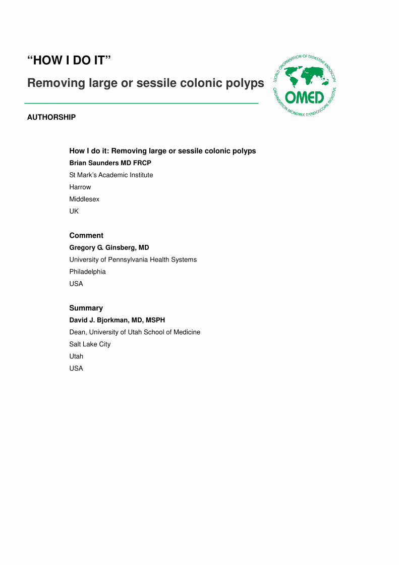
Once the polyp has been completely removed, I like to use the argon beamer to “touch up” the
polypectomy margins and destroy any remaining visible polyp fragments (Figure 3). Settings of 30–
40 W in the right colon and 50–65 W in the left colon are appropriate. Newer APC delivery devices
(ERBE VIO) are much more energy-efficient, and lower power settings of 20–25W in the right colon and
35–45 W in the left colon should be used. Be careful not to use diathermy on exposed areas of muscle
– you will see muscle contraction if this occurs – and remember to aspirate excess gas.
At the end of the procedure, I always look carefully for any bleeding points and photodocument the end
result. I have to be satisfied that clear visualization of the polypectomy site shows no remaining polyp
tissue. Tattooing the site may also be important, with at least two large India ink tattoos, placed just
distally to the lesion. Finally I retrieve all the polyp pieces with a Roth retrieval net. During withdrawal
this can be held away from the colonoscope tip to allow an adequate inspection of the mucosa distal to
the polypectomy site.
Complications of EMR
Bleeding
Oozing from the polypectomy base can be effectively treated by topical administration of an adrenaline
wash. Focal bleeding sites should be clipped or treated with APC. Severe arterial bleeding is rare during
colonic EMR, but if it occurs the first priority is to maintain a view of the mucosa by changing the
patient’s position so that blood pools, with gravity, away from the polypectomy site. Endoclips or APC
can then be applied, the combination often being more effective than a single modality.
Delayed bleeding can occur for up to 2 weeks post procedure. Patients usually require nothing more
than close observation as bleeding will stop spontaneously in the vast majority of cases. However,
emergency colonoscopy after a rapid oral purge may be necessary, and always with surgical back-up as

OMED “How I Do It”
Removing large or sessile colonic polyps 11
a last resort.
Perforation
Microperforations, if detected, can be closed with endoclips although it is better to not perforate in the
first place! Delayed perforation is an absolute indication for laparotomy; however it is important not to
confuse free perforation with post-polypectomy syndrome. In the latter situation, serosal irritation from a
full thickness burn has caused localized peritonism, low grade fever, and raised inflammatory markers,
without free gas in the abdomen. Conservative management in this case is usually successful, with
intravenous antibiotics, bed rest and close clinical assessment.
Post-resection surveillance
Most patients, even after a large piecemeal EMR, can be discharged the same day provided they are
pain-free and fully ambulatory. I advise patients to avoid aspirin and proprietary nonsteroidal anti-
inflammatory (NSAID) drugs for 2 weeks post procedure. I do not impose any dietary or lifestyle
restrictions other than to suggest that they do not undertake prolonged air travel and remain within
reasonable access of modern medical facilities for 2 weeks. At the time of discharge patients are given
a copy of their colonoscopy report which also contains emergency contact telephone numbers. I always
stress to patients that should they get severe pain, fever, or bleeding then they must inform us or their
local clinician. Delayed bleeding is the main risk, and patients need to be aware that they may be well
for 10 days but then still get significant bleeding.
Standard adenoma surveillance intervals apply when a small sessile polyp is removed en bloc by
single-snare EMR. Surveillance intervals are determined in the usual way by the number and size of the
adenomas removed. Piecemeal excision however necessitates an early repeat assessment at 3 months
to check for healing and the presence of residual polyp. If there is any doubt about completeness of
resection of a large lesion, the patient must return even earlier, at 2 months, before any chance of a
large recurrence. Any small area of recurrence can usually then be definitively destroyed at the second
procedure with a further repeat check arranged for 3 months’ time.
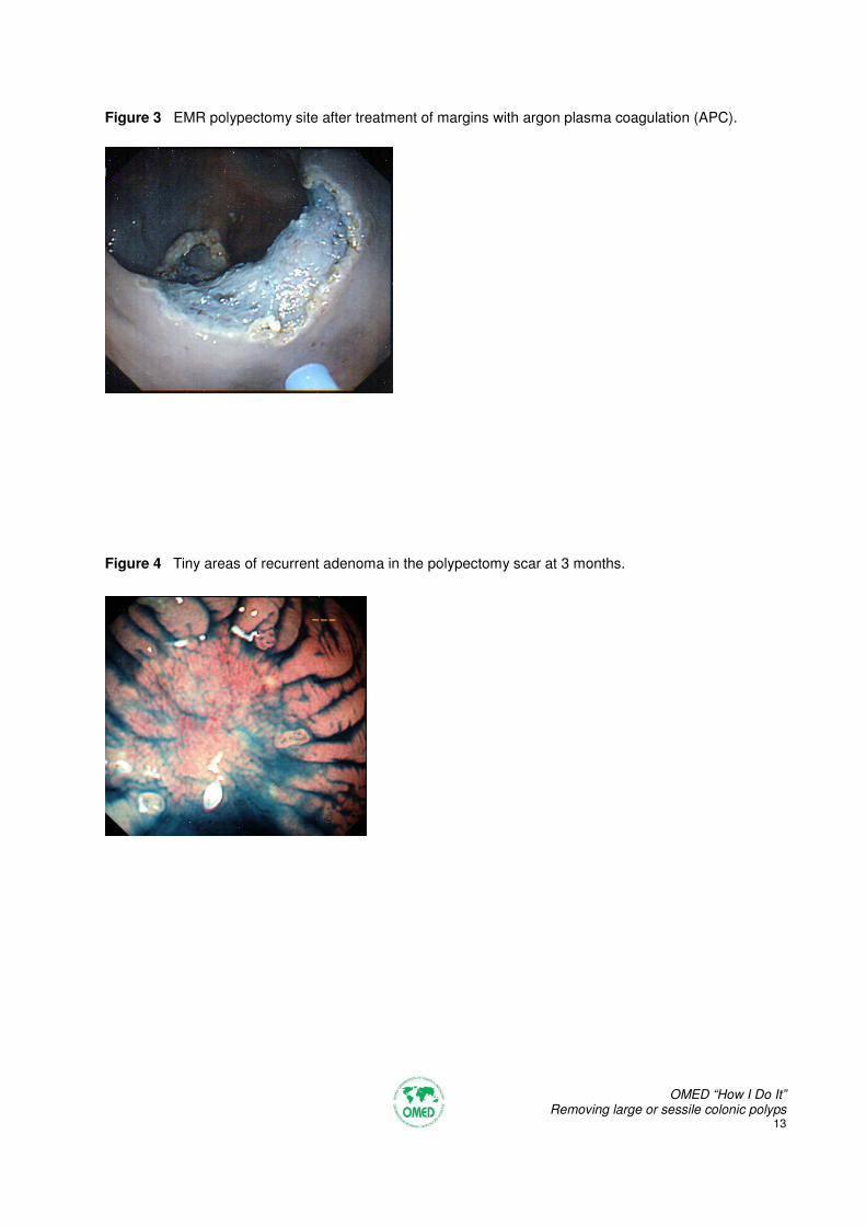
Whenever I perform a check examination post-EMR, I use indigo carmine dye to help highlight the scar
and surrounding mucosa. Occasionally tiny areas of recurrence are only visible after dye application
(Figure 4). APC is invaluable for treating small areas of recurrence at a previous polypectomy scar.

OMED “How I Do It”
Removing large or sessile colonic polyps 12
Figures
Figure 1 Endoscopic mucosal resection (EMR) of 6-cm sessile tubulovillous adenoma (3/4 complete
and showing “clean” resection plane through the submucosa.
Figure 2 EMR of flat (IIb) adenoma containing tiny focus of superficially submucosally invasive cancer
(clear resection margin), A=before, B=after EMR.
A B
OMED “How I Do It”
Removing large or sessile colonic polyps 13
Figure 3 EMR polypectomy site after treatment of margins with argon plasma coagulation (APC).
Figure 4 Tiny areas of recurrent adenoma in the polypectomy scar at 3 months.

OMED “How I Do It”
Removing large or sessile colonic polyps 14
Recommended reading
Waye JD. Advanced polypectomy. Gastrointest Endosc Clin N Am 2005 Oct;15: 733–756
Conio M, Ponchon T, Blanchi S, Filiberti R. Endoscopic mucosal resection. Am J Gastroenterol 2006;
101: 653–663; Epub 2006 Feb 8
Uraoka T, Saito Y, Matsuda T et al. Endoscopic indications for endoscopic mucosal resection of
laterally spreading tumours in the colorectum. Gut 2006; 55: 1592–15927; Epub 2006 May 8
Bories E, Pesenti C, Monges G et al. Endoscopic mucosal resection for advanced sessile adenoma and
early-stage colorectal carcinoma. Endoscopy 2006; 38: 231–235
Su MY, Hsu CM, Ho YP et al. Endoscopic mucosal resection for colonic non-polypoid neoplasms. Am J
Gastroenterol 2005; 100: 2174–2179
Conio M, Repici A, Demarquay JF et al. EMR of large sessile colorectal polyps. Gastrointest Endosc
2004; 60: 234–241
Tamura S, Nakajo K, Yokoyama Y et al. Evaluation of endoscopic mucosal resection for laterally
spreading rectal tumors. Endoscopy 2004; 36: 306–312
Kudo S, Tamegai Y, Yamano H et al. Endoscopic mucosal resection of the colon: the Japanese
technique. Gastrointest Endosc Clin N Am 2001; 11: 519–535
Kudo S, Kashida H, Tamura T et al. Colonoscopic diagnosis and management of nonpolypoid early
colorectal cancer. World J Surg 2000; 24: 1081–1090
Matsuda K, Masaki T, Abo Y et al. Rapid growth of residual colonic tumor after incomplete mucosal
resection. J Gastroenterol 1999; 34: 260–263
Kudo S, Kashida H, Nakajima T et al. Endoscopic diagnosis and treatment of early colorectal cancer.
World J Surg 1997; 21: 694–701
Suzuki N, Saunders BP, Brown G. Flat colorectal neoplasms: endoscopic detection, clinical relevance
and management. Tech Coloproctol 2004; 8 Suppl 2: s261–s266
Brooker JC, Saunders BP, Shah SG, Williams CB. Endoscopic resection of large sessile colonic polyps
by specialist and non-specialist endoscopists. Br J Surg 2002; 89: 1020–1024
Brooker JC, Saunders BP, Shah SG et al. Treatment with argon plasma coagulation reduces
recurrence after piecemeal resection of large sessile colonic polyps: a randomized trial and
recommendations. Gastrointest Endosc 2002; 55: 371–375

OMED “How I Do It”
Removing large or sessile colonic polyps 15
“HOW I DO IT”
Removing large or sessile colonic polyps
Comment
Gregory G. Ginsberg
Large, flat, and otherwise laterally spreading adenomas make up the class of “defiant” polyps, i.e. those
colonic polyps not resectable using a standard snare polypectomy technique. As Dr. Saunders
delineates, however, the majority of these polyps may be resected with curative intent by expert
endoscopists using adjunctive techniques. Dr. Saunders is a master colonoscopist and so I was
gratified in reviewing his submission to find that we are largely in agreement with regard to strategies
and techniques for the endoluminal resection of large sessile colonic polyps. Where the expertise is
available, patients with large sessile colonic polyps should be afforded the opportunity for colonoscopic
eradication in lieu of operative resection. I prefer the term “endoluminal resection” (ELR) over
“endoscopic mucosal resection” because, in fact, the submucosa is resected as well.
Indications
I incorporate submucosal injection to facilitate snare polypectomy for flat, sessile, and broad-based
polyps greater than 1–2 cm in diameter, throughout the colon. The majority of large sessile colonic
polyps that I encounter have been referred by another gastroenterologist or colorectal surgeon and so
the expectations on the patient and physician sides are uniquely delineated. For the incidental large
sessile polyp, I believe that it is within the scope of common practice to employ these techniques for
resection of sessile polyps of up to about 2.5 cm, at the time of the index colonoscopy. However, for
larger lesions, consideration should be given to photographic and written documentation of the lesion(s)
including number, size, location, and configuration. Cold forceps biopsy should be performed for
histological sampling. As Dr. Saunders implies, the use of electrosurgical energy should be avoided
outside of a commitment to complete curative resection. Partial or incomplete thermal snare resection
or “biopsy” results in a fibroinflammatory reparative response that tacks down the remnant margin of the
lesion. This results in the “pseudo-non-lifting sign” and makes subsequent completion of resection more
hazardous and more difficult. Tattooing to mark the lesion for subsequent recognition is usually
welcomed. However, some tattooing agents (e.g. India ink) if placed too near the lesion may also
promote a local fibroinflammatory response compromising subsequent ELR and so inert agents placed
apart from the lesion are preferred.
Unlike Dr. Saunders, I have encountered polypectomy-related perforations, with two in approximately
500 large sessile polyp resections. These both occurred with moderate-size (~2 cm) sessile proximal

OMED “How I Do It”
Removing large or sessile colonic polyps 16
ascending colon polyps, during attempts to resect them en bloc rather than in a piecemeal fashion. I
concur with Dr. Saunders that, short of applying ESD techniques, piecemeal resection is preferred for
larger sessile lesions.
Contraindications
Colonoscopic ELR should not be attempted when morphologic and/or tactile features indicate invasive
carcinoma. These include ulceration, firm texture, and fixation. We routinely apply endoscopic
ultrasound when considering rectal lesions for ELR and demur when there is evidence of invasion into
the submucosa or beyond. Endoscopic ultrasound is not used in the evaluation of lesions proximal to
the rectum. Except when there has been prior application of electrosurgical energy, the non-lifting sign
is a contraindication to proceeding with attempted ELR.
Beyond the rectum, sessile lesions that extend beyond more than 30% to 50% of the luminal
circumference and those that extend beyond 7 cm in length should be considered for operative
resection. Similarly lesions that are present within the appendiceal orifice and the iliocecal valve
typically defy completion endoscopic resection.
Clinical scenario, consent, and sedation
As stated above and in agreement with Dr. Saunders, large polyps potentially suitable for colonoscopic
ELR should be biopsied and the patient rescheduled for dedicated ELR after a discussion of the options
for management. Ideally, these procedures should be booked for a 30- to 60-minute block.
In addition to the points that Dr. Saunders emphasizes to patients, I quote an up to 20% risk of post-
polypectomy bleeding that may be acute or delayed, and that delayed bleeding may occur anytime from
12 hours to 12 days after the procedure.
It is our standard practice to perform colonoscopic ELR as an outpatient procedure, using narcotic and
benzodiazepine sedation. I advise that acute bleeding is treated endoscopically and may prompt a
recommendation for overnight hospital observation. Most delayed bleeding is short-lived, self-limited,
and not hemodynamically destabilizing. Patients with evidence of delayed bleeding are advised to report
to their local hospital emergency department for evaluation and initial management. For the rare patient
who requires directed therapy, we prefer to arrange transfer to our center, if feasible.
Patient preparation before the procedure
I similarly recommend a full oral bowel preparation to best ensure adequate visualization of the lesion
for resection. I simply employ a clear liquid diet on the day prior to the procedure and a 4-L polyethylene
glycol oral purgative. I no longer adjust patients’ aspirin or other antiplatelet medications. Warfarin

OMED “How I Do It”
Removing large or sessile colonic polyps 17
management is individualized.
Equipment
We use a variable-stiffness, pediatric caliber colonoscope as our standard instrument, along with air
insufflation. For distal colon and rectal lesions I use a therapeutic channel upper endoscope.
Antispasmodic agents are only rarely used. In addition to our standard electrosurgical generator, an
argon plasma beam-capable system is also kept in readiness. Our assistants prepare: four 10-mL
syringes with methylene blue-tinted normal saline solution, an injection needle, two standard
polypectomy snares, a mini-snare, and a specimen retrieval net. For rectal lesions, the Olympus EMR
kit is available in order to use the transparent aspiration cap and crescent snare, plus additional
crescent snares (as these tend to deform after a single use). This latter approach is useful for very distal
lesions that approach the anal verge and for those lesions displaying the pseudo-non-lifting sign. Acute
bleeding is similarly treated with local injection of diluted epinephrine solution and clips are applied
when this is insufficient.
EMR procedure
I concur with Dr. Saunders’ detailed description of EMR techniques. Key to assessment and planning
are satisfactory visualization and favorable orientation of the lesion. To achieve this, it is worth the
expenditure of time and effort to reposition the patient, axially rotate the colonoscope, and/or visualize
the lesion from the retroflexed position. I, too, preferentially inject around the base of the lesion, rather
than directly into it when circumstances permit.
In contrast to Dr. Saunders’ standard technique, a trained gastrointestinal technician operates the
opening and closing of the snare, while the endoscopist activates the electrosurgical current with a foot
pedal and applies traction with a to-and-fro motion on the shaft of the snare. Training technical
assistants for EMR requires interest, dedication, patience, and acceptance of an incremental approach.
We develop EMR specialists in that same way ERCP technicians were developed in the past. Like Dr.
Saunders, we rely generally on a coagulation current from a standard electrosurgical generator.
Post-resection surveillance
We have discontinued routine restrictions on aspirin and nonsteroidal anti-inflammatory drug (NSAID)
use related to endoscopic procedures, in accordance with American Society for Gastrointestinal
Endoscopy (ASGE) guidelines that indicate insufficient evidence of increased bleeding risk. However,
these recommendations may be individualized. Patients are allowed to resume their diets.
When incomplete resection is suspected, 6 weeks is the minimum duration before follow-up

OMED “How I Do It”
Removing large or sessile colonic polyps 18
examination. This is sufficient for healing of the resection site in the vast majority of cases. In a patient
returning sooner than this, the endoscopist is likely to encounter hyperplastic reparative changes that
are indistinguishable in appearance from residual adenoma. Otherwise, all rectal lesions and colon
lesions with high grade dysplasia (HGD) or ImCa dictate a 6-month follow-up and all others are followed
up at 1 year. Subsequent surveillance follows established guidelines. Our experience indicates that
increased lesion size and piecemeal resection are predictors of residual/recurrent adenoma. Given
compliance with surveillance, residual/recurrent lesions are identified and effectively eradicated
endoscopically.
OMED “How I Do It”
Removing large or sessile colonic polyps 19
“HOW I DO IT”
Removing large or sessile colonic polyps
Summary
David J. Bjorkman
Advances in endoscopic therapy have expanded the spectrum of lesions that can be excised without
resorting to surgery, perhaps most commonly in the setting of neoplastic lesions of the colon. While it
has been standard therapeutic practice to remove small and pedunculated polyps when they are
discovered, large and sessile lesions have often been referred for surgical resection.
These two excellent and detailed summaries demonstrate that, with appropriate care and caution, many
lesions previously referred for surgery can be resected endoscopically. The techniques described here
are almost identical. The basic principle is to raise the lesion by injecting fluid into the submucosal
space, then use electrocautery to snare portions of the lesion until it has been completely excised.
There are a few points made by both Dr. Saunders and Dr. Ginsberg that deserve emphasis. First,
patient selection and preparation are critical. This approach has a higher risk of complications and
patients must be willing to accept these. Second, the bowel must be thoroughly prepared to ensure that
there is a clean field for the resection. A third important point is the extensive list of specialized
endoscopes, accessories, medications/solutions, and trained support personnel outlined by both
authors. This is not a procedure that can be performed with limited resources. One must be fully
prepared to deal with any situation that may arise during the procedure. Finally, after beginning the
excision, the endoscopist is committed to completing it, regardless of how long it may take. One cannot
perform EMR in multiple procedures. Healing and fibrosis from a first partial excision would cause
scarring that would prevent the required lifting of the lesion at subsequent procedures.
Both authors have given us very detailed instructions on how this procedure can be performed, but
have also cautioned us that the endoscopist should know the limits of his or her resources and
experience. When in doubt, these lesions should be referred to experienced endoscopists, such as Drs.
Saunders and Ginsberg. The principles they have taught us can also be applied to our therapy of
smaller sessile lesions and can improve our own endoscopic skills.