Embed Size (px)
Citation preview

HAL Id: tel-00684264https://tel.archives-ouvertes.fr/tel-00684264
Submitted on 3 Apr 2012
HAL is a multi-disciplinary open accessarchive for the deposit and dissemination of sci-entific research documents, whether they are pub-lished or not. The documents may come fromteaching and research institutions in France orabroad, or from public or private research centers.
L’archive ouverte pluridisciplinaire HAL, estdestinée au dépôt et à la diffusion de documentsscientifiques de niveau recherche, publiés ou non,émanant des établissements d’enseignement et derecherche français ou étrangers, des laboratoirespublics ou privés.
Relations structure-fonction des transporteursnucléotidesPankaj Panwar
To cite this version:Pankaj Panwar. Relations structure-fonction des transporteurs nucléotides. Sciences agricoles. Uni-versité de Grenoble, 2012. Français. �NNT : 2012GRENV005�. �tel-00684264�
THÈSE Pour obtenir le grade de
DOCTEUR DE L’UNIVERSITÉ DE GRENOBLE Spécialité : Biologie Structurale et Nanobiologie
Arrêté ministériel : 7 août 2006
Présentée par
Pankaj PANWAR Thèse dirigée par Eva PEBAY-PEYROULA Préparée à l’Institut de Biologie Structurale Jean-Pierre Ebel dans l'École Doctorale Chimie et Sciences du Vivant Relations structure – fonction des transporteurs nucléotides Thèse soutenue publiquement le 26 janvier 2012, devant le jury composé de :
Bruno MIROUX Directeur de recherche, Paris, rapporteur
Jean-Jacques LACAPÉRE Directeur de recherche, Paris, rapporteur
Andrea DESSEN Directrice de recherche, Grenoble, Présidente François DEHEZ Chargé de recherche, Nancy, Examinateur
Norbert ROLLAND Directeur de recherche, Grenoble, Examinateur
M.R.N MURTHY Professeur, India, Examinateur
Eva PEBAY-PEYROULA Professeur, Grenoble, Directrice de thèse
Contents
Contents i
Abbreviations vi
1 Introduction 1
1.1 Transport across biological membranes . . . . . . . . . . . . 1
1.1.1 Principle of membrane transport . . . . . . . . . . . 2
1.2 Adenine nucleotide transport . . . . . . . . . . . . . . . . . 5
1.2.1 Mitochondrial localized adenylate transporters . . . . 7
1.2.2 Adenylate transporters in chloroplast . . . . . . . . . 9
1.3 Mitochondrial AAC . . . . . . . . . . . . . . . . . . . . . . . 11
1.3.1 Physiological role and location of the carrier . . . . . 11
1.3.2 Structure of carrier . . . . . . . . . . . . . . . . . . . 12
1.3.3 Inhibitors of the carrier . . . . . . . . . . . . . . . . . 14
1.3.4 Transport mechanism . . . . . . . . . . . . . . . . . . 14
1.3.5 Oligomeric status and transport mechanisms . . . . . 16
1.4 Chloroplast ATP/ADP carrier - NTT1 . . . . . . . . . . . . 17
1.4.1 Physiological role of the carrier . . . . . . . . . . . . 18
1.4.2 Isoforms of the carrier . . . . . . . . . . . . . . . . . 20
1.4.3 Homologues in other organisms . . . . . . . . . . . . 21
1.4.4 Characteristics of transport . . . . . . . . . . . . . . 23
1.5 Mitochondrial AAC v/s chloroplast NTT . . . . . . . . . . 24
i
Contents ii
1.5.1 Functional differences in context of physiological de-
mands . . . . . . . . . . . . . . . . . . . . . . . . . . 25
1.5.2 Evolution of nucleotide carriers . . . . . . . . . . . . 27
1.6 Crystallographic studies on membrane transporter . . . . . . 27
1.6.1 Difficulties associated with membrane proteins . . . . 28
1.6.1.1 Production of membrane proteins . . . . . 28
1.6.1.2 Purification and homogenous preparation . 29
1.6.1.3 Crystallization and structure determination
of membrane proteins . . . . . . . . . . . . 30
1.6.2 Curious case of detergents . . . . . . . . . . . . . . . 30
1.6.3 Lipidic mesophase (LMP) for membrane protein crys-
tallization . . . . . . . . . . . . . . . . . . . . . . . . 31
1.6.4 Beyond X-ray crystallography . . . . . . . . . . . . . 34
1.6.5 MD simulations and docking . . . . . . . . . . . . . . 35
1.6.6 Structural themes of TM transporters . . . . . . . . . 36
1.7 Objective of the thesis . . . . . . . . . . . . . . . . . . . . . 36
2 Materials and methods 39
2.1 Production of NTT1 . . . . . . . . . . . . . . . . . . . . . . 39
2.1.1 Overexpression . . . . . . . . . . . . . . . . . . . . . 39
2.1.2 Membrane preparation . . . . . . . . . . . . . . . . . 40
2.1.3 Quantification of total protein . . . . . . . . . . . . . 40
2.1.4 Solubilization . . . . . . . . . . . . . . . . . . . . . . 41
2.2 Purification and characterization of NTT1 . . . . . . . . . . 41
2.2.1 Affinity chromatography . . . . . . . . . . . . . . . . 41
2.2.2 Size exclusion chromatography . . . . . . . . . . . . . 42
2.2.3 Sodium dodecyl sulfate- polyacrylamide gel electrophore-
sis (SDS-PAGE) . . . . . . . . . . . . . . . . . . . . . 42
2.2.4 Western Blot detection . . . . . . . . . . . . . . . . . 43
2.2.5 Silver staining . . . . . . . . . . . . . . . . . . . . . . 44
2.2.6 Negative stain Electron microscopy . . . . . . . . . . 44

Contents iii
2.3 Crystallization of NTT1 . . . . . . . . . . . . . . . . . . . . 45
2.3.1 Crystallization and data collection . . . . . . . . . . 45
2.3.2 Quantification of detergent by FTIR . . . . . . . . . 45
2.4 Structure determination of Streptavidin . . . . . . . . . . . . 46
2.4.1 Purification . . . . . . . . . . . . . . . . . . . . . . . 46
2.4.2 Crystallization . . . . . . . . . . . . . . . . . . . . . 46
2.4.3 Data collection and processing . . . . . . . . . . . . . 47
2.4.4 Model building and refinement . . . . . . . . . . . . . 47
2.5 Virtual screening for novel mitochondrial AAC Inhibitors . 48
2.5.1 Ligand selection . . . . . . . . . . . . . . . . . . . . . 48
2.5.2 Molecular docking . . . . . . . . . . . . . . . . . . . 49
2.5.3 Uptake studies with radiolabelled ATP . . . . . . . . 50
3 Results 52
3.1 Towards understanding of NTT1 structure and function . . . 52
3.1.1 Expression . . . . . . . . . . . . . . . . . . . . . . . . 52
3.1.2 Solubilization . . . . . . . . . . . . . . . . . . . . . . 53
3.1.3 Initial purification protocol . . . . . . . . . . . . . . . 53
3.1.3.1 Cleavage of His-tag . . . . . . . . . . . . . . 55
3.1.4 Modification in the purification protocol . . . . . . . 58
3.1.4.1 Modification of the purification sequence . . 59
3.1.4.2 Modification with Ni-NTA chromatography 59
3.1.5 Size exclusion chromatography (SEC) . . . . . . . . . 64
3.1.5.1 Large scale purification . . . . . . . . . . . . 68
3.1.6 Characterization of protein homogeneity . . . . . . . 69
3.1.6.1 Negative staining EM . . . . . . . . . . . . 69
3.1.7 Initial Crystallization and data collection . . . . . . . 70
3.1.8 Quantification of detergents by FT-IR . . . . . . . . 70
3.1.8.1 Optimization of detergent quantity . . . . . 75
3.1.9 Latest trials of crystal optimization . . . . . . . . . . 76
3.1.10 Stability profiling of NTT1 using thermal denaturation 77

Contents iv
3.2 New inhibitors for mitochondrial ADP/ATP Carrier . . . . . 79
3.2.1 Virtual screening - computational quirk or useful tool? 80
3.2.2 Glide as a tool for molecular docking . . . . . . . . . 82
3.2.2.1 XP v/s SP docking mode . . . . . . . . . . 84
3.2.3 Known inhibitors and validation of docking method 85
3.2.3.1 Binding of known inhibitors . . . . . . . . . 85
3.2.4 Docking of CATR to the AAC . . . . . . . . . . . . . 87
3.2.5 Test with reference compounds . . . . . . . . . . . . 89
3.2.6 Classification of docked ligands . . . . . . . . . . . . 90
3.2.6.1 Interaction fingerprint based analysis of dock-
ing results . . . . . . . . . . . . . . . . . . . 90
3.2.6.2 Identification of key interactions . . . . . . 93
3.2.7 Functional tests with top-scoring compounds . . . . . 94
3.2.8 New ADP analogues for transport studies . . . . . . 96
3.2.8.1 ADP binding to AAC . . . . . . . . . . . . 96
3.2.8.2 Docking with ADP analogues . . . . . . . . 98
3.2.8.3 Functional tests with ADP analogues . . . . 99
4 Discussion 102
4.1 Towards understanding of NTT1 structure and function . . . 102
4.1.1 NTT1 production for structural studies . . . . . . . . 102
4.1.2 Homogeneity and crystallization . . . . . . . . . . . . 103
4.1.3 Role of detergent in crystallization . . . . . . . . . . 104
4.2 New inhibitors for mitochondrial AAC . . . . . . . . . . . . 105
4.2.1 Need for new inhibitors . . . . . . . . . . . . . . . . . 105
4.2.2 Possible binding mode of C-15 . . . . . . . . . . . . 106
4.2.3 Binding of ADP analogues to AAC . . . . . . . . . . 106
4.2.4 Correlation between Gscore and experimental obser-
vations . . . . . . . . . . . . . . . . . . . . . . . . . . 107
4.3 Connecting link between AAC and NTT1 . . . . . . . . . . . 110
4.3.1 Functional mechanism . . . . . . . . . . . . . . . . . 110

Contents v
4.3.2 Structural correlation . . . . . . . . . . . . . . . . . . 112
5 Conclusions and perspectives 113
Annexes 114
A Contamination from affinity column: Encounter with a
new villain in the world of membrane protein crystallization115
A.1 Introduction . . . . . . . . . . . . . . . . . . . . . . . . . . . 116
A.2 Methods . . . . . . . . . . . . . . . . . . . . . . . . . . . . . 118
A.2.1 Expression and purification of NTT1 . . . . . . . . . 118
A.2.2 Crystallization, data collection and structure deter-
mination . . . . . . . . . . . . . . . . . . . . . . . . 119
A.2.3 Detergent elution of engineered streptavidin from Strep-
Tactin® . . . . . . . . . . . . . . . . . . . . . . . . . 119
A.3 Results and discussion . . . . . . . . . . . . . . . . . . . . . 120
A.3.1 Purification and crystallization . . . . . . . . . . . . 120
A.3.2 Structure of the engineered streptavidin bound to desthio-
biotin . . . . . . . . . . . . . . . . . . . . . . . . . . 121
A.3.3 Elution of streptavidin from Strep-Tactin® matrix . 122
A.4 Conclusions and perspectives . . . . . . . . . . . . . . . . . 124
A.5 Figure legends . . . . . . . . . . . . . . . . . . . . . . . . . . 128
B Plasmid and NTT1 gene construct 130
B.1 NTT1 protein sequence . . . . . . . . . . . . . . . . . . . . . 136
B.2 List of detergents used during experiments . . . . . . . . . . 138
B.3 Isolation and purification of thylakoid lipids . . . . . . . . . 139
Bibliography 141

Abbreviations
AAC.................................... ADP/ATP carrier
ADP.................................... Adenosine diphosphate
AMP................................... Adenosine monophosphate
ATP.................................... Adenosine triphosphate
BM ..................................... Bongkrekic acid
CATR ................................ Carboxyactractyloside
CMC .................................. Critical micellar concentration
EM ..................................... Electron microscope
GTP ................................... Guanosine triphosphate
IMS .................................... Intermembrane space
LCP .................................... Lipid cubic phase
LMP ................................... Lipid mesophase
Mant-ADP ......................... 3-O´-N-methylanthraniloyl-ADP
MD ...................................... Molecular dynamic
N-ADP ............................... 3-O´-naphthoyl-ADP
N-ATP ................................ 3-O´-naphthoyl-ATP
NMR ................................... Nuclear magnetic resonance
NTT1................................... Nucleotide transporter1
PDC...................................... Protein-detergent complex
TM ...................................... Transmembrane
VDAC ................................ Voltage dependent anionic channel
vi

Résumé du chapitre 1
L’objectif principal de ma thèse était l’étude structurelle et fonctionnelle
de protéines de transport de nucléotides en étudiant deux transporteurs
différents. Le NTT1 chloroplaste, catalyse l’échange d’ATP éléctroneutre
dans le cytosol avec l’ADP et le Pi dans le stroma. La séquence d’acide
aminé de NTT1 présente un degré élevé de similitude avec le transporteur
ATP/ADP de la bactérie Rickettsia prowazekii ou Chlamydia trachomatis.
Une autre classe importante de transporteur de nucléotides, choisie dans
la présente étude est AAC mitochondriale. Le transporteur est un consti-
tuant majeur et essentiel de la membrane mitochondriale interne. AAC
est responsable de l’importation de l’ADP et de l’exportation de l’ATP
néo-synthétisé. Des différences importantes au niveau phylogénétique et
fonctionnelle ont été rapportées pour AAC et NTT1. Les études compara-
tives fonctionnelles et structurelles de ces transporteurs seraient bénéfiques
pour notre compréhension fondamentale des processus de transport de nu-
cléotides et pourrait permettre le développement de médicament efficace
contre les bactéries pathogènes.
Dans un effort pour comprendre et analyser le rôle de NTT1 dans le
transport des nucléotides, la détermination de sa structure cristalline est
une étape essentielle. Comme pré-requis pour la cristallisation, la purifi-
cation et l’expression de protéine fonctionnelle sont nécessaires. Les résul-
tats obtenus dans le cadre de cette étude sont présentés dans la section
3.1.6. Quand j’ai commencé le doctorat, Aurélien Deniaud, un post-doc de
l’équipe, a lancé la production et la caractérisation des NTT1. J’ai travaillé
avec lui pendant un an (Deniaud et al., Soumis), puis amélioré les proto-
coles et de caractérisation. La cristallisation des protéines membranaires
requiert une connaissance approfondie des interactions protéine-détergent
et ses charactérisations. Nos premiers essais de cristallisations ont entraînés
l’obtention de cristaux qui sont bien formés et diffractent bien. Néanmoins,
j’ai résolu la structure, qui est le premier du mutant triple de streptavidine

conçu pour se lier Strep-tag II, dans un complexe avec la desthiobiotine.
Le manuscrit en annexe décrivant ce travail sera soumis prochainement.
La caractérisation des complexes protéines-détergents par des techniques
biochimiques et biophysiques fournis des informations précieuses sur la sta-
bilité des protéines, l’homogénéité et cette information peut mener à la
réussite de la cristallisation de la protéine. Les essais de cristallisation et
les caractérisations biophysique associés sont discutés et présentés dans la
section 3.1.7.
La structure des AAC mitochondriaux liés à l’inhibiteur CATR disponible
fournis un outil précieux pour comprendre le processus de transport des nu-
cléotides. Toutefois, d’autres données structurelles (structure d’une autre
conformation) sont nécessaires pour déchiffrer l’ensemble du mécanisme de
transport et de comprendre la spécificité des transporteurs. Comme outil
pour obtenir de nouvelles structures de protéine-ligand, le criblage de com-
posés qui pourraient potentiellement se lier avec son AAC est nécessaire. La
recherche de petites molécules inhibitrices couplées à une validation fonc-
tionnelle pourrait fournir des ligands intéressants, qui peuvent stabiliser
l’AAC dans des états intermédiaires importants impliqués dans la fonction
de transport. Ces objectifs constituent la deuxième partie de mon travail.
La stratégie utilisée dans ce contexte et les résultats obtenus est présentée
dans la section 3.2.
Résumé du chapitre 2
2.1 Production de NTT1
2.1.1 Sur-expression
2.1.2 Préparation de membrane
2.1.3 Quantification de la concentration de protéines totales
2.1.4 Solubilisations
2.2 Purification et caractérisation de NTT1
2.2.1 Chromatographie d’affinité
2.2.2 Chromatographie d’exclusion
2.2.3 Électrophorèse sur gel de polyacrylamide en présence de dodécyl-
sulfate de sodium
2.2.4 Détection par Western Blot
2.2.5 Coloration à l’argent
2.2.6 Microscopie électronique à coloration négatif
2.3 La cristallisation de NTT1
2.3.1 Cristallisation et collecte de données
2.3.2 Quantification des détergents par FTIR
2.4 Détermination de la structure de la streptavidine
2.4.1 Purification
2.4.2 Cristallisation
2.4.3 Collecte et traitement des donnés
2.4.4 Construction du modèle et raffinement

2.5 Criblage virtuel pour de nouveaux inhibiteurs
mitochondriaux AAC
2.5.1 Choix des ligands
2.5.2 Docking moléculaire
2.5.3 Etude des interactions des ligands
Résumé du chapitre 3
Production, caractérisation et cristallisation de NTT1
La protéine NTT1 a pu être exprimée et produite dans E. coli. Plusieurs
détergents ont été testés pour extraire la protéine des membranes. Les ren-
dements étaient les meilleurs avec le LAPAO qui a été sélectionné pour la
purification. Un premier protocole de purification a donné lieu à la présence
d’un contaminant présent en très faible quantité et donc difficilement dé-
tectable. Ce contaminant, la streptavidine, a été cristallisé par erreur et
sa structure a pu être résolue à haute résolution. Un nouveau protocole de
purification a ensuite été développé. Il a permis l’obtention d’une protéine
suffisamment pure et une chromatographie par filtration sur gel a permis de
séparer deux fractions, l’une majoritairement monomérique et l’autre plutôt
dimérique comme indiqué par les analyses en microscopie électronique. La
fraction monomérique a conduit à des petits cristaux diffractant à 10 Å.
Afin d’assurer la reproductibilité des conditions de cristallisation, plusieurs
analyses permettant la caractérisation de la solution de protéines purifiées
ont été mises au point. Parmi celles-ci, l’absorption IR par transformée
de Fourier a permis de quantifier la quantité de détergent présente dans
la solution, donnée essentielle pour la cristallisation. Cette quantité a pu
ensuite être suivie en fonction des étapes de purification et un nouveau pro-
tocole a permis d’optimiser cette quantité. Plus récemment, une approche
permettant de contrôler la stabilité thermique de la protéine a été explorée.
Nouveaux inhibiteurs de AAC
Une approche de docking moléculaire basée sur le programme Glide a été
mise en place. Elle a permis d’explorer plusieurs millions de molécules de
la base ZINC. Plusieurs filtres et des scores basés sur des énergies ont per-
mis de restreindre le nombre de molécules et d’en sélectionner quelques

dizaines. La méthode a été testée sur les inhibiteurs d’AAC ainsi que
des dérivés d’ADP connus pour se lier à AAC sans être transportés. En
particulier la localisation de l’inhibiteur CATR connue expérimentalement
car présent dans la structure résolue par cristallographie, a été retrouvée
validant ainsi cette approche. Les meilleurs composés identifiés par cette
approche numérique ont ensuite été testés expérimentalement. L’un d’entre
eux, noté C15, inhibe à plus de 60% le transport.
Résumé du chapitre 4
Études structurales de NTT1
Le protocole mis au point durant le travail de thèse et présenté dans le
chapitre 3 a permis d’obtenir des premiers cristaux. Les études ont mis
en évidence l’importance de l’homogénéité de la solution de protéine avant
cristallisation. En particulier, la sélection de la fraction majoritairement
monomérique et un des points clés. Par ailleurs, le choix du détergent pour
la solubilisation et la purification est connu pour être un des paramètres
critiques en cours de cristallisation. Un premier détergent, le LAPAO, sem-
ble être le meilleur pour la solubilisation, d’autres pourraient être explorés
pour la cristallisation après échange lors de la purification. La quantité
de détergent présente dans la solution, et plus exactement le rapport pro-
téine/détergent est également un facteur déterminant. Les méthodes mises
en place permettront pour les études futures de suivre ce paramètre et donc
de le contrôler lors de la mise au point des protocoles.
Nouveaux inhibiteurs d’AAC
Afin de comprendre le mécanisme de transport au niveau moléculaire, les
structures de plusieurs conformations d’AAC sont nécessaires. L’identification
de nouveaux inhibiteurs pourra permettre de bloquer d’autres conforma-
tions du transporteur et ainsi permettre leur cristallisation. Ces nouveaux
inhibiteurs seront donc des outils pour explorer l’espace conformationnel
d’AAC. Le mode de liaison du composé C15 présente des analogies avec
celui de CATR, néanmoins la molécule C15 est plus petite que CATR.
Parmi les analogues d’ADP inhibant AAC, nous avons identifié la molécule
AP5, un inhibiteur de kinase. À ce stade, même si les molécules identifiées
par docking présentent des taux d’inhibition plus ou moins fort, il a été dif-
ficile de faire une corrélation directe entre taux d’inhibition expérimental et

le score obtenu par les simulations numériques. Celles-ci devront donc être
affinées en prenant en compte des facteurs plus fins comme l’intervention
de molécules d’eau dans la liaison des inhibiteurs.
Comparaison NNT1 et AAC
D’après l’analyse des séquences en acides aminés les structures globales des
deux transporteurs sont probablement différentes, avec 6 hélices transmem-
branaires connues pour AAC et 11 à 12 prédites pour NTT1. Les mécan-
ismes de transport diffèrent aussi, électrogénique pour AAC, neutre pour
NTT1. Néanmoins, la comparaison des taux d’inhibitions de certains com-
posés identifiés par docking pour AAC et testés expérimentalement pour
AAC et NTT1 montre quelques analogies. Lorsque la structure de NTT1
sera connue, il sera donc très intéressant de pouvoir comparer les sites de
fixation des nucléotides.

Résumé du chapitre 5
Dans cette étude, nous avons conçu et réalisé un protocole pour l’expression,
la purification et la cristallisation de NTT1, qui est une protéine mem-
branaire intégrale. Le rôle significatif de NTTI comme transporteur ATP/ADP
et son homologie étroite avec les transporteurs des bactéries pathogènes rend
NTT1 une cible intéressante à étudier. Un protocole pour la purification
de NTT1 a été optimisé, ce qui est compatible avec les études structurales.
Une caractérisation plus poussée des protéines par SEC et par EM avec col-
oration négative, montre une homogénéité significative dans la préparation,
ce qui est idéal pour les études structurales.
La pureté et l’homogénéité des protéines nous ont permis d’identifier
les conditions de cristallisation de NTT1. La cristallisation des protéines
membranaires exige un équilibre délicat entre la protéine et le contenu de
détergent dans la solution de cristallisation. Nous avons adopté pour une
stratégie de quantification de détergents dans la solution de cristallisation
de NTT1 et développé une méthode pour réduire la quantité de détergent
dans la solution après concentration de la protéine. Plusieurs additifs de
cristallisation pourraient être explorées, telles que les dérivés de nucléotides
ou de lipides chloroplastiques. Plusieurs optimisations des conditions de
cristallisation permettent de se rapprocher de la détermination de la struc-
ture de NTT1. Grace à l’obtention de milligrammes de NTT1 purifiée et à
l’identification des conditions de cristallisation, il sera possible d’étudier la
structure et la dynamique NTT1 entière, ce qui améliora la compréhension
du mécanisme de transport de nucléotides.
Pour développer la compréhension générale du transport de nucléotides,
un autre transporteur de nucléotides d’AAC mitochondrial a été choisi pour
les études. Le criblage virtuel de ‘druglike subset’ dans la base de donnés
publique ZINC a été réalisée pour rechercher des inhibiteurs de l’activité bi-
ologique des mitochondries AAC. L’applicabilité de la procédure d’amarrage
a été validé en comparant la liaison Glide pose de CATR avec la structure

cristalline. Les structures de Glide reproduit ainsi les géométries prévues
du complexe. Après le criblage virtuel, les complexes pour les 500 premières
du classement des composés ont été inspectés visuellement et 23 composés
ont été achetés. Un composé a été livré en quantité insuffisante. Enfin,
22 composés ont été testés in vivo de l’inhibition de l’ATP de transport
par l’AAC. Notamment, quatre des 22 composés ont montrés plus de 40%
d’inhibition lorsqu’ils sont appliqués à une concentration de 100 μM.
Un des composés C-15 a montré une inhibition d’environ 60% dans
l’ordre du micromolaires. La visualisation des positions de C-15 dans le
docking montre des liaisons hydrogènes potentiellement favorables et des
liaisons van der Waals avec le site actif de l’AAC. Des travaux complémen-
taires sur les produits dérivés du C-15 est hautement souhaitable et pourrait
conduire à des inhibiteurs plus puissants pour AAC. De ce premier com-
posé, d’autres molécules pourraient être conçus en modifiant des groupes
chimiques qui permettraient d’améliorer l’interaction avec la protéine. Le
design sera guidé par la structure de l’inhibiteur dans le transporteur. Une
meilleure compréhension des relations structure activité serait bénéfique
pour les travaux sur la cristallisation de complexe AAC-inhibiteur. Le
présent travail a conduit à la découverte d’inhibiteurs de petites molécules
d’AAC.
Chapter 1
Introduction
1.1 Transport across biological membranes
Cell membranes are crucial to the life of the cell. The plasma membrane en-
closes the cell and defines its boundary. Inside the eukaryotic cell along with
plasma membrane, membranes of other organelles including Golgi appara-
tus, mitochondria and endoplasmic reticulum maintain the characteristic
difference between content of each organelle and the cytosol. Hydropho-
bic architecture of the lipid bilayer of cell membranes prevents the passage
of polar molecules on their own. This barrier allows the cell or specific
organelles to maintain concentration of solutes in cytosol or their respec-
tive fluids. However such barrier requires specific mechanisms to transfer
lipid insoluble molecules and ions across the membrane in order to ingest
essential nutrients, excrete metabolic waste products and regulate intracel-
lular ion concentration. To transport small water soluble organic molecules
and inorganic ions specialized transmembrane proteins are utilized. Some
specialized mammalian cells such as intestinal cells can devote about two
third of their energy consumption on membrane transport, which reflects
the paramount importance of the process.
1
1.1. Transport across biological membranes 2
1.1.1 Principle of membrane transport
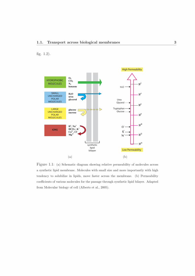
The rate of diffusion for any molecule across the lipid bilayer depends par-
tially on its size and mainly on its relative solubility in lipid. In general, the
smaller the molecule and more soluble it is in the oil, faster it moves across
the membrane. Small molecules such as O2 and CO2 rapidly diffuse across
the bilayer whereas small uncharged polar molecules such as water and urea
also diffuse across the bilayer, albeit much slowly. On the other hand bilayer
is impermeable to charged inorganic molecules. The charge and high degree
of hydration of these molecules prevent them to be transported naturally,
in fact synthetic bilayers are 109 times more permeable to water molecules
than small charged molecules such as Na+ and K+ (see fig. 1.1). Another
factor that affects diffusion of the molecules across the membrane is differ-
ence in their concentration on the two sides of the membrane. Multiplying
this concentration difference by the permeability coefficient gives the flow
rate of the molecules across the membrane.
To transport naturally impermeable molecules such as ions, sugars,
amino acids, nucleotides and other metabolites, membrane transport pro-
teins are employed by the cells. Membrane transport proteins provide a
continuous protein path that allows the specific molecule to pass through
lipid bilayer without directly interacting with it. Transporters (also known
as carriers) and channels are the two major classes of membrane transport
proteins. Upon binding to the substrate transporters, undergo large con-
formational changes and facilitate transport of the molecule. On the other
hand, channels form pores that extend across the lipid bilayer and these
pores can be opened by a variety of mechanisms. When open, most of
the pores interact with solute and undergo relatively small conformational
changes and allow the solute to pass through membrane. Due to generally
large conformational changes associated with transporters, solute transport
by them is usually slower than channel-mediated solute transport. Trans-
port mediated by both kinds of proteins can be either passive or active (see
1.1. Transport across biological membranes 3
fig. 1.2).
!"#$%&
"$
!'()*#$+
"$
'&,"#$'-
*$
./,"$
/-01234$
3015234$
!,($
054&$
/-61452-$
(,$'(,$%,$7489484$
HYDROPHOBIC
MOLECULES
!
synthetic
lipid
bilayer
LARGE
UNCHARGED
POLAR
MOLECULES
SMALL
UNCHARGED
POLAR
MOLECULES
IONS
(a)
High Permeability
Low Permeability
H2O
Urea
Glycerol
Tryptophan
Glucose
!"#!$%
!"#!&%
!"#!"%
!"#'%
!"#(%
!"#$%
!"#&%
)*%
+,*%
-.%#%
(b)
Figure 1.1: (a) Schematic diagram showing relative permeability of molecules across
a synthetic lipid membrane. Molecules with small size and more importantly with high
tendency to solubilize in lipids, move faster across the membrane. (b) Permeability
coefficients of various molecules for the passage through synthetic lipid bilayer. Adapted
from Molecular biology of cell (Alberts et al., 2005).

1.1. Transport across biological membranes 4
lipid
bilayer
simple
di!usion
channel-
mediated
carrier-
mediated
transported molecule
channel
protein
carrier
protein
PASSIVE TRANSPORT ACTIVE TRANSPORT
!
ENERG
Y
OUTSIDE
INSIDE
electrochemical gradient
with no membrane potential
electrochemical gradient
with membrane potential
negative inside
electrochemical gradient
with membrane potential
positive inside
concentration
gradient
(A)
(B)
(a)
Figure 1.2: (a) Passive transport of molecules can occur either by simple diffusion
through the lipid bilayer or by facilitated diffusion through channels and passive trans-
porters. In contrast, active transporter utilizes energy to pump solutes against their
concentration or electrochemical gradient. (b) Combination of electrochemical gradient
with membrane potential forms electrochemical potential, which guides the solute trans-
port; it can be additive (middle) or it can be subtractive in nature (left). Adapted from
Molecular biology of cell (Alberts et al., 2005).

1.2. Adenine nucleotide transport 5
1.2 Adenine nucleotide transport
Among all the metabolites, nucleotides are of pivotal importance due to
their energy storing capacity in the form of ATP and GTP. Nucleotides,
apart from being the universal energy currency, also constitute the core
component of all forms of genetic information material. Due to both by
size and charge, adenylate nucleotides do not cross biomembranes freely,
making the involvement of specific transporter proteins necessary for their
transport. Oxidative mitochondrial metabolism and associated nucleotide
transport have been studied extensively, however our knowledge of nu-
cleotide transport across other membrane bound organelles is still rudi-
mentary. In the past, it was assumed that adenine nucleotide transport in
cells mainly takes place at the mitochondrial membrane. However, in recent
years an increasing number of studies have shown that the majority of cel-
lular membranes contain adenine nucleotide transport systems (see figure
1.3) [Mayinger et al., 1995, Abeijon et al., 1997]. Based on transmembrane
(TM) helices predictions and sequence homology, nucleotide transporters
can be divided into two broad categories, as shown below.

1.2. Adenine nucleotide transport 6
Figure 1.3: Occurrence of adenine nucleotide transporters in various membrane-bound
organelles. The ATP/ADP carrier (1), the ATP/AMP carrier (2) and the ATP-Mg/Pi
carrier (3) are located in the inner mitochondrial membrane and represent typical mem-
bers of the mitochondrial carrier family (MCF) (depicted as gray spheres). Apart from
mitochondria, MCF carriers are widespread in other organelles such as the ADP-glucose
importer (4) residing in the inner envelope membrane of heterotrophic plastids, thylakoid-
located ATP/ADP carrier (6) and the ATP/ADP carrier from plant ER (7) as well as the
adenine-nucleotide carriers from peroxisomes (8) and glyoxysomes (9) and of the plasma
membrane (10). Adenylate carriers other than MCF (depicted in white spheres) are also
found in some organelles e.g.. the ADP/ATP carrier at inner chloroplast membrane (11),
nucleotide carrier (12) and nucleotide-sugar carrier (13) of Golgi complex. Adapted from
[Haferkamp et al., 2011].

1.2. Adenine nucleotide transport 7
1.2.1 Mitochondrial localized adenylate transporters
The transport of metabolites across the inner mitochondrial membrane is
carried out by a family of proteins called Mitochondrial Carriers (MCF).
These carriers link the biochemical pathways of the cytosol and the mito-
chondrial matrix by transporting metabolites, nucleotides, inorganic ions
and cofactors across the mitochondrial inner membrane. Mitochondrial
carriers share a common structure consisting of around 300 amino acids
and contain three homologue repeats of around 100 amino acids. Each
of the repeat is predicted as two TM helices. Despite a low sequence
conservation, a consensus sequence, P-X-(D/E)-X-X-(K/R)-X-(K/R)-(20-
30 residues)-(D/E)-G-X-X-X-X-A-(K/R)-G1 is found in all the members
[Saraste & Walker, 1982]. To date, nine human diseases have been found
to be caused by defects in mitochondrial carriers (Palmieri et al., 2008).
All the carriers located at the inner membrane of mitochondria are specific
for selective metabolite transport whereas outer membrane of mitochon-
dria harbors several pores (i.e. VDAC2) that allow a rather non specific
transport of diverse small molecules. This process of differential transport
allows accumulation of several ions and metabolites at intermembrane space
(IMS), eventually depending on the metabolic demand they are being im-
ported or exported.
A subgroup of carriers transport adenine nucleotides and related com-
pounds, such as thiamine pyrophosphate, FAD, NAD, coenzyme A and
pyrimidine nucleotides. Mitochondria being the powerhouse of cell gener-
ates ATP by the process of oxidative phosphorylation. ATP generation
is fueled by the substrate ADP and phosphate, which enter inside the or-
ganelle and regenerated ATP must leave from matrix to outside. Export of
this neo-synthesized ATP by counter exchange of cellular ADP is carried1Where A is an aromatic residue2VDAC is a voltage-dependent anion-selective channel that act as a general diffusion
pore for small hydrophilic molecules

1.2. Adenine nucleotide transport 8
out by a MCF member: the ADP/ATP carrier (AAC) (see fig. 1.4). AAC
is one of the most abundant carrier in mitochondrial inner membrane and
by ATP transport the carrier supplies energy from mitochondria to cytosol
and subsequently to other organelles [Klingenberg, 2008, Nury et al., 2006].
!!"#$
!!!%%"
!!!
&
!!!
&&
!!!
&&&
!!!
&'
!!!("%
!!)*+,-./0
!!%10/*2!3!"4%
!!!567689
!!!%():
!!!%()
!!!;< !!!;<!!!;<
!!!;<
!!!;$#!!!#$
!!!=,>!!!?,11
!!!@%A<!!!@%A;
!!!%A)B
!!!%A)
!!!%%"
#C&A%(&'D!);#?);#EF5%(&#@
!!!
%()3GH
!!!)6
Figure 1.4: Schematic representation of the mitochondrial adenylate carriers and their
physiological implication on oxidative phosphorylation process. The NADH generated by
lipid oxidation (β-oxidation) and in the Krebs cycle (TCA) is oxidized by the respiratory
chain complexes (complexes I, II, III, and IV in green, cytochrome C in blue), and protons
are pumped to the intermembrane space. The proton electrochemical gradient is used by
the ATP synthase to generate ATP from ADP and phosphate (Pi). The ATP generated
is transported to the cytosol in exchange for ADP through the ADP/ATP carrier (AAC).
Another adenylate carrier ATP-Mg/Pi catalyzes electroneutral transport; the direction
of ATP-Mg and Pi transport is dependent on their relative concentration across the
membrane.

1.2. Adenine nucleotide transport 9
Apart from AAC two more adenylate carriers are located in mitochon-
drial inner membrane. One of them is ATP-Mg/Pi carrier, that carries out
an electroneutral exchange of ATP-Mg2- for HPO42-, between the cytosol
and mitochondrial matrix and thus contributes to the net uptake or net loss
of adenine nucleotides [Traba et al., 2009, Aprille, 1993]. The direction and
magnitude of net adenine nucleotide transport is dependent on the relative
ATP-Mg and Pi concentrations in the mitochondria. The ATP-Mg/Pi car-
rier is insensitive to classical AAC inhibitors carboxyatractyloside (CATR)
and bongkrekic acid (BA). Another identified adenylate mitochondrial car-
rier is AMP/ATP carrier, albeit found only in plant mitochondria. Its
primary function is probably to catalyze the exchange between cytosolic
AMP and intra mitochondrial ATP (Palmieri et al., 2008). f
1.2.2 Adenylate transporters in chloroplast
Like mitochondrion membrane, plastid membrane is also formed by two
significantly different membranes. The outer membrane harbors various
pores (porins) whereas inner envelope membrane forms a metabolic bar-
rier between cytosol and chloroplast stroma, therefore contains most of the
active transporters. Thylakoids that reside in the chloroplast stroma, fur-
ther extend the compartmentalization by forming third chloroplast mem-
brane. Thylakoid membrane is the site of photosynthesis and contains sev-
eral metabolite transporters. Thylakoids contain chlorophylls, the complete
photosynthetic electron transport chain and the ATP synthase, which is re-
quired for ATP regeneration on the stroma side.
The first information of adenine nucleotide transport across chloroplast
inner membrane was obtained by import measurements on intact chloro-
plast from the algae Acetabularia mediterranea [Heldt, 1969, Strotmann & Berger, 1969].
Roughly 30 years later an Arabidopsis transporter protein family NTT
(see fig. 1.5) was found responsible for the adenine nucleotide transport
[Kampfenkel et al., 1995, Neuhaus & Emes, 2000]. NTT family is a small

1.2. Adenine nucleotide transport 10
family and all the known NTTs strictly transport ATP and ADP only.
These transporters functionally and phylogenetically differ from mitochon-
drial AAC. Members of this family have been predicted to contain 11-12
TM helices. [Neuhaus & Emes, 2000].
!!!"#$ !!!"%$!&!$'
!!!"#$!!!(
!!!)#!*!!+)#!*,-.
!!!"#$
!!!"%$
!!!"/$
!!!"#$
!!!"%$
!!!"/$
$01'23
2045367'83!
9:27;39'9
!!!
"%$!&!$'
!!!<=5>'2
!!!<:453
?? ?
@&@-A A-
#;:5=B6'8
13=47'629
#""<
!!!"%$
!!!"%$!!!"#$
!!!"#$@&
@&
!!!"%$
!!!"#$
C##
!!!C"%$&!!!C"%$@
D504693
!!<A-
Figure 1.5: Graphical presentation of all the chloroplast adenylate carriers. Adenine
nucleotide carriers (NTT) located at the inner envelope, exchange cytosolic ATP for
chloroplast ADP and Pi, and this ATP is used for night reactions, including synthesis of
lipids and starch. The thylakoid adenine nucleotide carriers (TAAC) exchange stroma
ATP (generated by photophosphorylation in the plastid ATP synthase) for thylakoid
ADP, and this ATP is used for thylakoid reactions. The ubiquitous BT1 (BT1-2) trans-
porter exports adenine nucleotides, which are exclusively synthesized de novo in plastids,
to the cytosol.
One of the NTT members in chloroplast, NTT1 mediates catalysis of
ATP import by the counter exchange of ADP and Pi, thus provides energy
to the chloroplast. Another adenylate transporter in Arabidopsis is Brit-

1.3. Mitochondrial AAC 11
tle1 (AtBT1), which acts as a uniporter and known to transport ADP, AMP
and ATP across the inner chloroplast membrane [Kirchberger et al., 2008,
Sullivan & Kaneko, 1995]. AtBT1 is a member of MCF, which provides
adenine nucleotides, exclusively synthesized de novo inside plastids to the
cytosol and other organelles. At the thylakoid membrane one more adeny-
late carrier thylakoid AAC (TAAC) is located that allows exchange of ATP
inside the thylakoid by ADP exchange to the stroma [Thuswaldner et al., 2007].
TAAC, links stroma adenylate pool to thylakoid, which catalyzes ATP-
dependent process including protein phosphorylation, folding, import and
degradation [Thuswaldner et al., 2007].
1.3 Mitochondrial AAC
1.3.1 Physiological role and location of the carrier
Membrane potential across the inner membrane regulates the AAC me-
diated exchange of ATP versus ADP between the matrix space and the
cytosol (Heldt et al., 1972). This process is the last step of oxidative
phosphorylation and the energy generated by the process is supplied to
the cytoplasm. An important characteristic of the carrier is that, due to
counter exchange, it is unable to modify the net content of adenine nu-
cleotides. Therefore ADP is always available for mitochondrial ATP gen-
eration machinery and along with phosphate carrier, the carrier maintains
the metabolite pool for energy generation. The affinity of the AAC for ADP
or ATP is high with Km of around 2 μM in yeast and 4-8 μM in humans
[De Marcos Lousa et al., 2002].
Three isoforms of bovine AAC have been identified [Powell et al., 1989],
whereas in humans 4 isoforms of the AAC have been reported [Stepien et al., 1992,
Skárka et al., 2003]. Existence of these isoforms could be correlated with
adaptations of the functional parameters to the particular tissue needs. One
of the human isoforms hAAC1 is exclusively expressed in muscles and its

1.3. Mitochondrial AAC 12
expression is induced during myoblast differentiation [Stepien et al., 1992].
hAAC2 expression is strongly stimulated by growth factor in fibroblasts
[Ku et al., 1990] and also increased in neoplastic cells (Heddi et al., 1996).
AAC3 is highly expressed in cancer cells (Li et al., 1989; Powell et al., 1989),
whereas hAAC4 is expressed at low levels and is confined to liver and testis,
marginally to brain [Dolce et al., 2005].
Pathophysiological role of the carrier has been studied by knock-out mu-
tations in mice. Transgenic mice lacking mAAC1 showed characteristics of
mitochondrial myopathy and cardiomyopathy [Graham et al., 1997]. While
mAAC2 inactivation was found lethal before birth [Kokoszka & Coskun, 2001].
The high expression of hAAC3 in tumor cells has been proposed as con-
ductive for maintaining membrane potential despite the glycolytic activity
of this particular cell type [Chevrollier et al., 2005]. Several studies have
indicated a potentially important role for AAC in programmed cell death
[Kroemer & Galluzzi, 2007].
1.3.2 Structure of carrier
Three dimensional structure of the bovine AAC isoform1 was the first
among entire family [Pebay-Peyroula et al., 2003]. Since most of the family
members are expected to contain similar structure, this structure has im-
proved the understanding of metabolites transport to a great extent. The
backbone of AAC appears in canonical shape, closed toward the matrix
and opened widely toward the IMS. The backbone also exhibits a pseudo-
threefold symmetry consistent with the triplicated sequence of the gene.
The overall architecture of AAC consists of six TM helices labelled H1 to
H6, connected by three loops M1 to M3 on the matrix side and two loops C1
and C2 on the IMS (see fig. 1.6) . Matrix loops are partially structured and
contain short amphipathic helices (labelled h1-2, h3-4, and h5-6) spanning
over 12 residues. The prolines of odd-numbered TM helices kink helices,
bringing the C terminal ends together at the base of the cavity. Infact all

1.3. Mitochondrial AAC 13
the six TM helices are also tilted with respect to the membrane plane.

ERROR: ioerrorOFFENDING COMMAND: image
STACK:
-mark- -savelevel-