Embed Size (px)
Citation preview
RELATIONS OB^ THE BLUE STAIN FUNGUS,
CERATOCrSTIS IPS (RUMBOLD)C. MOREAU, TO IPS BARK BEETLES
(COLEOPTERA: SCOLYTIDAE)OCCURRING IN FLORIDA
By
WILLIAM C. YEARIAN, JR.
«^>"
A DISSERTATION PRESENTED TO THE GRADUATE COUNCti. lOF
THE UNIVERSITY OF FLORIDA, , , \
IN PARTIAL FULFILLMENT OF THE REQUIREMENTS FOR THE
DEGREE OF DOCTOR OF PHILOSOPHY ,. ^,
UNIVERSITY OF FLORIDA
June, 1966

y 3?/"
AGRI-
CULTURAL
LIBRARY
v-;..:'"'^( I
• ( \
;" • Yf,Ai,

ACKNOWLEDGMENT
The wrlt«r wishes to acknowledg* his Indebtedness to the
Co-Chalrmen of his Supervisory Committee, Dr. R. C. Wilkinson and
Dr. L. A. Hetrick, for their advice and assistance throughout the
course of this study and the preparation of the manuscript.
Special thanks are also due Dr. J. T. Crelghton, Dr. C. M. Kaufman,
and Dr. K. R. Swinford for their criticism on the preparation of
the manuscript. The writer also wishes to express his appreciation
to Dr. L. C. Kultert for his Interest and encouragement. Sincere
gratitude Is extended to Mr. W. J. Coleman for assistance In both
the field and laboratory phases of the work.
Thanks are accorded Dr. J. W. Kimbrough, Department of Plant
Pathology, University of Florida and Dr. C. L. Wilson, Department of
Plant Pathology, University of Arkansas for confirmation of Cerato-
cystis Ips (Rumboid) C. Moreau.
The generous assistance of Mr. Charles C. Russell In the
preparation of photographs that appear In the manuscript Is gratefully
acknowledged. Special thanks are also uue Miss Linda Weir for typing
of the manuscript.
This study was conducted under Florida Agricultural Experiment
Station State Project 1188. Additional funds were made available by
the Southern Forest Disease and Insect Research Council, Southern
Puipwood Conservation Association.
II

A special kind of thanks Is due my wife, LaVerne, for her constant
encouragement during the course of this work. To the above and all
others who have given help during this study, the writer wishes to
express deepest appreciation.
Ill

TABLE OF CONTENTS
PAGEACKNOWLEDGMENT
1
1
LIST OF TABLES v|
LIST OF FIGURES vMt
INTRODUCTION|
LITERATURE REVIEW 3
Transmission of Ceratocystis Species 3
Specificity of Ceratocystis spp. - Bark Beetle
Association g
Characteristics of Bark Beetle - Associated
Ceratocystis Species g
Nature of the Ceratocystis spp. - Bark BeetleAssociation 10
Pathogenicity of Ceratocystis Associates of
Bark Beetles, 12
MATERIALS AND METHODS I5
Fungus Isolations , 15
Frequency of C. ips Transmission 15
Rearing Ips Species in Laboratory Colonies 19
Rearing Ips Species Free of C. Ips^^ 21
Mass Attraction 24
Ips Development In C, ips Stained Bolts 25
Brood Development and Fecundity 27
iv

RESULTS AND DISCUSSION 32
Fungus isolations 32
Frequency of C. Ips Transmission 37
Larval and Pupal Development on Phloem-BasedRear t ng Med I urn 39
Mass Attraction 42
Ips Development In C. Ips Stained Bolts...... 44
Effect of C. ips on Brood Development andFecundity 4S
Egg gal lery construction 45
Ceratocystis Ips In brood developmenttest bolts 48
Brood size and mortality 49
Brood composition 53
Pupal weight 62
Sex ratio 62
Fecundity. 62
Discussion of the Effect of C. Ips and OtherOrganisms on Brood Development and Fecundity 64
CONCLUS I ONS 73
LITERATURE CITED 75
BIOGRAPHICAL SKETCH 80

LIST OF TABLESPAGE
1. Phloem-based, seml-artlf Iclal rearing medium forIps bark beetles. Amounts sufficient to make100 grams 23
2. Relative frequency of Ceratocystis Ips
Isolation from Ips avulsus , Ips cal llgraphus,
and Ips grandlcol Ms. 56
3. Relative frequency of Ceratocystis Ips
transmi ss I on by Ips avulsus , Ips cal llgraphus,
and Ips grandlcol I Is. 38
4. Mean larval and pupal development of Ips avulsus,
Ips cal llgraphus, and Ips grandlcol Us on phloem-based semT-arttf Iclal rearing meadlum at 30° C 40
5. Attractiveness of W and BSF Ips cal tigraphusmale Infested slash pine bolts 43
6. Mean egg gallery length and spacing betweeneggs of W, BSF, and BS Ips avulsus, Ips
cal llgraphus and Ips grandlcol! Is femalesIn slash pine bolts at 30° C 47
7. Mean size and mortality of W, BSF, and BS Ips avulsus ,
Ips cal 1 1 graphslash pine bol
Ips cal llgraphus, and Ips grandlcol lis broods In' ts at 300C. 52
8. Mean composition of W, BSF, and BS Ips avulsus , Ips
cal llgraphus, and Ips grandlcol lis broods In sFash
pine bolts at 5, 10, 15, and 20 days at 30° C 57
9. Relative composition (percentage) of W, BSF, and BS
Ips avulsus , Ips cal llgraphus, and Ips grandlcol lis
broods In slash pine bolts at 5, 10, 15, and 20
days at 30O C 59
10. Chl-square comparison of W, BSF, and BS Ips avulsus,Ips cal llgraphus, and Ips grandlcol I Is broodcoR4>osltIon at 5, 10, 15, and 20 days at 30° C 61
vl

11. Msan weight (mg.) of 100 W, BSF, and BS tps avulsus,jps calllgraphus, and Ids grand I col Ms pupae rearedIn slash pine bolts at 30" C... , 63
12. Sex ration of W, BSF, and BS Ips avulsus , Ipscal llgraphus, Ips grandlcolHs adults reared Inslash pine bolts at 30" C 64
13. Mean length of egg gallery, total number of eggslaid, and number of eggs deposited per centimeterof gallery by W, BSF, and BS Ips avulsus ,
Ips cal llgraphus, and Ips grand I col Us femalesat 30 days In slash pine bolts at 30" C 65
vtl

LIST OF FIGURES
PAGE1. Screen cage, 6' X 6' X 6', containing male
Infested loblolly pine bolts used to attractwild beetles 18
2. Gelatin capsule used to hold bark beetles priorto entrance Into pine bolt. Capsule held In
place with "Duxseal" 20
3. Rotating turntable used to expose male Infestedpine bolts In attraction studies..... 26
4. Pine bolt rearing unit used In brooddevelopment studies 30
5. Per I thee I urn of Ceratocystis Ips . Approx. 75X 33
6. Ascospores of Ceratocystis Ips. Approx. 2000X 34
7. Section of 15-day old BSF Ips grandlcolMs gallery5 centimeters from nuptial chamber. Noteperlthecia of Ceratocystis Ips lining gallery andmasses of ascospores at tips of perlthecia.Approx. 40X 50
8. Section of 15-day old BSF Ips grandlcollls gallery5 centimeters from nuptial chamber. Note absenceof Ceratocystis Ips. Approx. lOX 51
9. Mean composition of W, BSF, and BS Ips avulsusbroods In slash pine bolts at 5, 10, 15, and20 days at 30® C 54
10. Mean composition of W, BSF, and BS Ips cal II-graphus broods In slash pine bolts at 5, 10,
15, and 20 days at 30© C 55
Hi Mean composition of W, BSF, and BS Ips grandlcol lis
broods In slash pine bolts at 5, 10, 15, and 20days at 30° C 56
vtll

12. Ips avulsus pupal chamber covared with masses
of conldia, probably Tubarcu I ar I e 1 1 a I ps .
Approx. 5X 71
tx

INTROCXX^TION
Th« association b«twe«n blue stain fungi, particularly of the
genus Ceratocysti
s
Ellis and Halstaad, and scolytid bark beetles in
the genera Dendroctonus Erlchson and ips DeGeer has long been known.
Because of the constant presence of blue stain fungi in the phloem
and sapwood of trees attacked by bark beetles, early workers
generally assumed the fungi were commonly introduced under the
bark by insects. Leach, Orr, and Christensen (1934) used a series
of caged and uncaged logs of red pine, Plnus resinosa Ait., to
prove that Ips grandicollis (Eichh.) and Ips plni (Say) transmitted
the blue stain fungus, Ceratocystis Ips (Rumbold) C. Moreau. They
concluded that certain blue stain fungi, such as C. ips, were rarely,
if ever, transmitted In any other way.
Most previous research on blue stain fungi and bark beetles was
directed at determining the species of fungi associated with various
bark beetles and the effect of the fungi on the host trees and their
wood products. No comprehensive study was made of the relationship
between the organisms involved, and the nature of the association
was open to considerable question. Craighead (1928), Leach (1940),
Leach et al. (1934) and Nelson (1934) considered the association to
be one of mutualism, with the fungi benefiting from transmission by
the beetles to a suitable host and the insects benefiting from
creation of a more favorable environment for brood development by
I

action of tho fungi on tha wood and tnnar bark. Hoist (1937) In tha
Unltad Statas and Grosnann (1930) In Garmany raarad carta In bark
baetlos from egg to adult fraa of their blue stain associates and
concluded the fungi were not essential for development within a
single generation of the Insects. Hetrick (1949) observed an
Infestation of the southern pine beetle, Dendroctonus frontalis Zliiai»,
In which the trees showed no signs of blue staining. Since beetle
brood development appeared to be normal, he concluded that blue
stain organisms were not essential for successful attack by the
beetles.
In light of the uncertainty of the exact nature of the blue
stain fungus-bark beetle association, a project was Initiated to
study the Ips bark beetles occurring In Florida; Ips avulsus (Etchh.),
Ips cal ligraphus (Germ.), and Ips grandicoi lis (Eichh.), and their
blue stain fungus associate, Ceratocystis ips (Rumbold) C. Moreau.
The principal objective of the study was to conqaare development of
all stages of the beetles in one or more generations In the presence
or absence of the fungus. The results of this investigation
together with observations on the biology and behavior of the beetles
are reported herein.

LITERATURE REVIEW
Thatcher (i960) r«vl«w«d current knowUdg* of the bark beetles
attacking southern pines which Included the Ips species occurring
in Florida. These beetles have not been studied Intensively and no
detailed data were available on their biology and habits. A
discussion of blue stain In timber was recently presented by FIndlay
(1959a, b) In which he covered the fungi involved and their effect
on strength and quality of wood, dissemination, prevention, and
control.
Bark beetle-associated blue stain fungi have been described
in several genera, but most have belonged to the genus (^eratocystls .
Species of Graph I um and Leptographlum , many of which were shown to
be imperfect forms of (^ratocystls, and Tubercularlel la ips Leach,
Orr, and Christensen also have been found to cause blue stain
(Mathre, i964a). The present study was restricted to the Ceratocystis
associates of Ips species bark beetles in Florida. Hunt (1956)
revised the genus Ceratocystis , and all previous citations from the
literature have been given in terms of his monograph.
Transmission of Ceratocystis Species
The bluing of timber was first described by Hartig in 1878
(Hartig, 1894). He recognized the fungal nature of the stain,
Ceratocyst Is pill fera (Fries) C. Moreau, and noted that the fungus

coomonly occurred In tr««s attacked by Insacts. Von Schrank (1903)
studlad blua stain fungi In pondarosa pina, PInus ponderosa Laws,
attacked by Dendroctonua ponderosae Hopk, He Isolated Ceratocystis
plUfera from the galleries of the beetle which suggested the beetle
transmitted the fungus. Von Schrenk was unable to Isolate the fungus
from the Intestinal tract and feces of the beetle, however, and
discounted the probability of Insect transmission. He concluded
that the spores of the fungus were distributed by the wind and
entered the region between the bark and wood through holes made
by the beetles.
Little attention was given the blue stain fungus-bark beetle
association until Craighead (1928) suggested that the fungi were
transmitted by bark beetles. Supported by Isolations of Ceratocystis
species from wood adjacent to beetle galleries and directly from the
beetles, subsequently workers (BakshI, 1950; Bramble and Hoist, 1935,
1940; Davidson, 1935, 1955, 1958; Ellis, 1939; Grosraann, 1930;
Leach et ai., 1934; Nelson, 1934; Nelson and Seal, 1929; Robinson,
1962; Roblnson-Jeffery and Grinchenko, 1964; Rumbold, 1929, 1931,
1936, 1941; Verrali, 1941; and others) generally assumed that bark
beetles played an Important role In dissemination of blue stain
fungi. Rumbold (1931) showed that C. pi Hfera was a secondary
Invader. She was of the opinion that wood used by Von Schrenk (1903)
to start his cultures was too old, and the primary fungus, probably
Ceratocystis minor (Hedge.) Hunt, had lost Its vitality by the time
the wood reached the laboratory. Grosmann (1930) stated that
Von Schrenk's failure to Isolate the fungus from the beetle could

b« explained in that he used liquid agar which, owing to its high
teaperature of solidification, was too hot at the time of
Innoculation. She also added that Von Schrenk had obviously
neglected to prevent bacterial growth by the addition of acid to
the culture medium and to punctually Isolate the germinating
fungus spores. Grosmann noted that even though no exact proof
had been presented, repeated observations that scolytlds appeared
mostly in association with blue stain fungi led to the recognition
that bark beetles played a significant role as transmitters of
blue stain fungi
.
Leach et al. (1934) studied the interrelationships of bark
beetles and blue stain fungi in felled red pine, PInus resinosa Alt»,
and demonstrated transmission of Ceratocystis Ips (Rumbold) C. Moreau
by ips grand I CO I lis (Etchh.) and Ips pini (Say). In a series of
caged and uncaged logs with sealed and unsealed ends, C. ips
occurred only in logs attacked by the bark beetles. The fungus
was abundant in all uncaged logs and absent from the caged logs
except when a few beetles accidentally gained entrance, in logs
lightly attacked by the beetles, the fungus was limited to the
immediate vicinity of the beetle tunnels. Histological preparations
of adult beetles revealed ascospores of the fungus adhering In
clumps to various parts of the body wall and distributed throughout
the intestinal tract. Fragments of peri thee I a were also found in
the gut, and examination of fecal pellets showed viable ascospores
of the fungus to be present. In view of their findings. Leach et al.
( i 934 ) cone I uded that these bark beet Ies un 1 versa 11y I ntroduced
blue stain fungi Into logs and trees.

Not all Ceratocystfs species have been found to be disseminated
by barK beetles. Verrall (1941) studied dissemination of various
blue stain fungi In lumber yards In Louisiana and Mississippi and
found air currents. Insects, milling machinery, and rainwater to be
disseminating agents. C. Ips was occasionally Isolated from the
air, but Verrall stated Ips species bark beetles were the most
Important transporters of the fungus since he Isolated C. ips
from 62 per cant of the bark beetles he captured In the yards.
Leach et ai. (1934) also presented evidence that mites associated
with bark beetles often carried C. Ips spores on their bodies and
distributed the spores through the beetle gallery system.
Specificity of Ceratocystis spp. - Bark Beetle Association
Depending upon their association with bark beetles, Mathiesen
(1950) divided the Swedish Ceratocystis species Into the following
types: (I) those species spread primarily by wind and only rarely
by bark beetles: e.g., C. coerulescens (MUnch) BakshI, C. f loccosa
(Hathiesen) Hunt, C. pi lifera , and C. plurlannulata (Hedge.) C.
Horeau; (2) those species associated with several bark beetles
usually In older material, but also spread by wind: e.g., C. minor ,
C. minuta (Slem.) Hunt, C. piceae (Munch) BakshI, and C. polonlcum
(Siem.) C. Moreau; and (3) those always associated with certain
species of bark beetles: e.g., C. brunneo-cl 1 1 ate (Mathlesen-K.)
Hunt with I ps sexdentatus Boern,, C. cana (Munch) C. Moreau with
Blastophagus minor Hart, and C. clavata (Mathiesen) Hunt with Ips
acumlnatus 6yl I. In North America, Dendroctonus and Ips species

have been considered the most Important bark beetles of coniferous
trees. Several Ceratocystis species have been found associated with
Dendroctonus bark beetles (Bramble and Hoist, 1940; Davidson, 1933,
1958; Mathre, 1964a; Robinson, 1962; Robinson-Jeffrey and Grlnchenko,
1964; and Rumbold, 1931, 1936, 1941), but C. minor was the most common
found species. Ceratocystis Ips has been the only species of the
genus consistently associated with Ips bark beetles.
Ceratocystis Ips was described by Rumbold (1931) as an associate
of Ips cal llgraphus and Ips grandlcol Ms . The fungus was also found
In association with Ips avulsus (Rumbold, 1931), Ips confusus (Lee.)
(Mathre, 1964a), Ips emarginatus (Lee.) (Mathre, 1964a; Rumbold, 1936),
Ips Integer (Elchh.) (Rumbold, 1936), ips lecontel (Lee.) (Ellis,
1939), Ips oregoni (Elchh.) (Mathre, 1964a; Rumbold, 1936), Ips pinl
(Leach et al., 1934), Ips piastographus (Lee.) (Rumbold, 1936) and
Ips ponderosa Sw. (Mathre, 1964a). C. Ips was also found associated
with Orthotomlcus caelatus (Elchh.) (Verrall, 1941), Dendroctonus
valens Lee. and Dendroctonus ponderosa (Mathre, 1964a) In this
country. In addition to North America, C. Ips was reported In
association with Ips sexdentatus In Germany (Francke-Grosmann, 1963)
and Poland (Siemaszko, 1939), ips species In Japan (NIslkado and
YamautI, 1933), and Orthotomlcus proxireus (Elchh.) In Sweden
(Mathiesen-Kaarik, I960). Certain Ips species In Europe were found
to be associated with several Ceratocystis species other than C. Ips,
however.

Characteristics of Bark B—tl» - Associated Ceratocystis Species
The blue stain fungi associated with bark beetles character-
istically have oonldia and ascospores covered with a sticky mucilage:
a characteristic associated with adherence of these reproductive
bodies to the body wall of the Insects. This trait has not been
observed In wind-borne Ceratocystis species. Leach et al. (1934)
showed the sticky covering also protected the spores as they passed
through the beetles* digestive tract. Hathlesen-K^arlk (I960)
demonstrated that the sticky layer rendered the spores resistant to
desiccation. She found the spores remained viable for several
months on beetles In hibernation.
The bark beetle associated Ceratocystis have been shown to be
ecologically adapted to their vectors. Mathleson-Kiiarik (I960)
and Grosmann (1930) studied the development of certain European
bark beetles and found developmental time for the fungi corresponded
very closely with that of the beetles. Leach et al. (1934) made
similar observations In the United States on C. Ips In association
*'''*''* ip» grandioollls and ips pint . The fungus began to grow
Immediately after introduction by the beetles and was isolated from
wood near the galleries within a week. Isolation was possible before
visible signs of staining were present. The fungus rapidly spread
radially and longitudinally from the egg gallery. Tangential
growth was slow and took place primarily through the larval galleries.
Isolations showed that the larvae moved ahead of C. Ips and very
little. If any, of the fungus was consumed. Shortly after the
larvae pupated the fungus began to sporulate. Perlthecia and

coremla were abundant In the old egg galleries, larval tunnels, and
pupal chambers. Perlthecia also formed In large numbers In the Inner
bark not consumed by the larvae. Francke-Grosmann (1963) noted that
C. Ips was especially adapted to endozolc dissemination since the
spores of embedded perlthecia could be spread only when young
beetles ate the Inner bark and carried the spores In their gut to
new breeding places. Newly formed Ips grandlcol I Is and Ips pinl
adults began feeding before leaving the logs In which they had
developed, and according to Leach et al. (1934) the young beetles
wandered through old egg galleries and tunneled through Inner bark
containing embedded perlthecia. Many spores were Ingested or
adhered to the outside of the body wall. They stated that the
beetles emerged from the logs thoroughly Infested with spores of
the fungus.
Through studies on the physiology of certain Ceratocystis
species, Hathlesen-Kaarik (I960) attempted to differentiate
between the insect and non- Insect associated species. She found
Insect associated forms to be deficient of certain vitamins while
wind-blown forms were vitamin-autotrophic. The insect associated
species were able to utilize a greater number of carbohydrates and
showed a wider enzymatic capacity than the wind-borne species. She
was not able to demonstrate consistent physiological differences
between the two groups of fungi, but she stated each fungus had Its
own special moisture and nutritional requirements and developmental
time. Mathlesen-Kaarlk (I960) concluded that all these factors
together might produce optimal conditions for some particular fungus

10
or fungi fn the galiertes of a specific bark beetle; other fungi
might occasionally be Introduced Into the galleries of this Insect,
but were not compatible for long periods of time and would disappear
In succeeding Insect generations. She noted that this would fully
explain the existence of non-special I zed associations between certain
widespread bluing fungi and a number of different but ecologically
similar bark beetles.
Nature of the Ceratocystis spp. - Bark Beetle Association
The blue stain fungus-bark beetle association has been referred
to as mutualism by some workers and commensal Ism by others. It has
been clearly demonstrated In seme Instances that the fungi benefit
from the association. Leach et al. (1934) and Mathlesen (1950)
proved that certain Ceratocyst I
s
species were almost exclusively
disseminated by bark beetles. Leach et al. stated that the fungi
obviously benefited from dissemination by the beetles and from
Introduction Into the Inner bark of logs and susceptible trees.
St. George and Beal* Indicated that the action of Dendroctonus
frontalis on the Inner bark created conditions favorable for
C. minor growth since the fungus failed to become established when
Insect broods failed to develop. Pathogenicity trials (Mathre,
1964b, and Nelson, 1934) with C. jj^ and C. minor added further
•St. George, R. A. and J. A. Beal. 1927. Progress report onthe southern pine beetle ( Dendroctonus frontalis ZIram.), U. S. Bur,Entcmol. Plant Quar. DIv. Forest Insect Invest. Ashvllle, N. C,(typewritten 40 pp.) (Cited from Olxon and Osgood, 1961, p. 18).

It
support to this assumption. The fungi were unable to Infect trees
when the Innoculum was Introduced through needle holes Intended to
simulate Initial bark beetle attack. Grosmann (1930) noted that
blue stain fungi rapidly spread longitudinally and radially In the
wood and phloem from the egg galleries of the beetles, but tangential
spread was stow and took place primarily through tangential ly
extended larval galleries In the phloem.
Benefits derived by the beetles from the association have not
been clearly defined. Craighead (1928) suggested that blue stain
fungi created conditions favorable for brood development and that
the fungi possibly furnished essential nourishment for the beetles.
Nelson (1934) found C. minor Infection resulted In a reduction of
the water content of the wood, and concluded the fungus was probably
necessary for optimum brood development of Dendroctonus frontalis .
Leach et al. (1934) stated:
... The b I ue-sta I n I ng fungi, by Inhibiting the flow ofsap. In all probability, make living trees more favorablefor beetle development and by aiding In decomposition ofthe Inner bark cause it to separate from the wood, creatinga more favorable environment for development of Insectbroods.
They pointed out that until a brood of beetles was reared In a fungus-
free log. It could not be safely concluded that blue stain fungi
were not necessary for normal development of the beetles.
Other workers have expressed the opinion that both blue stain
fungi and bark beetles can exist and develop fully without their
associates. Grosmann (1930) found both tps typographus L. and
Biastophagus piniperda L. to be Independent of their blue stain
associates. She noted that larvae of the beetles moved ahead of

12
the spread of the fungi through the Inner bark and very little, If
any, fungal material was consumed by the larvae. She observed young
adults of both beetles feeding on blue stain fungi, but she was
unable to detect any difference In the maturation period or vigor
of beetles that had or had not fed on the fungi. Hoist (1937)
aseptlcal ly reared Dendroctonus frontalis, Ips cal llgraphus and
I ps grandlcol Us In small numbers on strips of phloem sealed between
two glass plates. Since the reared Insects were within size limits
given by Blackman (1922), Hoist concluded that blue stain fungi
were not essential for development from egg to adult within a single
generation. Hetrick (1949) observed a Dendroctonus frontalis Infes-
tation in Florida In which the trees showed no sign of blue staining.
He stated that the trees died rapidly and normal broods developed.
Based upon this and other field observations, Hetrick concluded
that blue stain fungi were not essential for successful attack by
the bark beetle.
Pathogenicity of (^ratocystls Associates of Bark Beetles
Craighead (1928) pointed out that the girdling action of bark
beetles alone did not fully explain the rapid death of attacked
trees since mechanically girdled trees usually remained green for a
year or more. The constant presence of blue stain fungi In beetle-
attacked trees prompted Craighead to suggest that the fungi played
a role In causing death of the tree. Subsequent pathogenicity trials
with certain Ceratocystis species proved Craighead's suggestion to
be correct. Nelson (1934) and Nelson and Bee I (1929) were able to

13
kill small loblolly (PInus taeda L.), pitch (P. riglda Mill.), short-
leaf (P. echlnata Ml 1 1.), and Virginia (P. virglnlana. Mill.) pinas
with C. minor . Bramble and Hoist (1940) and Caird (1935) were also
able to kill shortleaf pines with the fungus. Nelson (1934) Innocu-
lated trees with C. Ips, but the results were Inconclusive since the
trees were attacked by bark beetles. Mathre (1964b) recently detnon-
strated that C. Ips , as well as C. ml nor
»
C. monlta (Rumboid) Hunt,
and C. schrenklana (Hedge.) C. Moreau killed small ponderosa pines,
P. ponderosa. In order to kill the trees, Mathre (1964b) and Nelson
(1934) found It necessary to apply the Innoculum over a large area
which completely encircled the tree. Trees Innoculated In small
patches or needle holes, so as to simulate Initial bark beetle
attacks, were resistant to Infection. Additional tests with
secondary fungi associated with bark beetles; C. pill fera (Mathre,
1964a), Trichoderma spp. (Mathre, 1964a and Bramble and Hoist, 1940),
and Dacromyces spp. (Bramble and Hoist, 1940) showed them to be non-
pathogenic.
Mathre (1964b) found oleorestn exudation pressure (o.e.p.) to
be a good indicator of the susceptibility of large ponderosa pines
to Infection by C. Ips and C. minor . Trees with an o.e.p. greater
than 33 p.s.i. were resistant to infection. Small trees were
susceptible to Infection regardless of the o.e.p., probably because
of the small resin volume to the exposed surface area ratio In such
trees.
Mathre (1964b) Indicated the role played by C. minor in killing
trees attacked by the western pine beetle, Dendroctonus brevlcomis
Lee., was not clear. It was shown that the beetle preferred to

14
attack large trees with an o.e.p. below 60 p.s.l., but was able to
mass attack and kill large, high o.e.p. trees (Miller and Keen, I960
and Vlte' and Wood, 1961). Mathre speculated that In such cases, the
reduction of o.e.p. was not due to C. minor since the fungus probably
would not Infect the trees until the o.e.p. was reduced to near zero.
He added further that the trees were usually stained In scattered
patches and wedges only and that water (induction was probably not
stopped until the bole was completely Invaded by the fungus. Ips
bark beetles, on the other hand, seldom attacked trees unless the
o.e.p. was near zero; a condition suitable for rapid Infection by
C. Ips . Trees attacked by Ips species were completely stained by
"•"he fungus and water conduction was stopped. Mathre (1964b) stated
"these observations suggest C. minor may not be necessary to kl 1
1
trees attacked by the western pine beetle, whereas C, Ips may play
a role In killing trees attacked by Ips beetles."

MATERIALS AND METHODS
Fungus Isolation*
Blue stain fungus Isolations ware made from the trood and Inner
bark of loblolly, Pinus taeda L.; long leaf, P. patustris Ml II.; pond,
P^. serotlna MIchx.; sand, P. clause (Chapm.) Vasey; spruce, P. glabra
Walt.; and typical slash, P. elllottH var. elllotti Engelre. pines
Infested with one or a combination of the three species of Ips bark
beetles occurring In Florida. Additional isolations were made from
surface sterilized eggs, larvae, pupae, and adults; non-sterl i Ized
adults; and galleries, including frass, of the three beetle species.
All isolations were made on a malt extract, yeast extract agar
medium consisting of 10 grams malt extract, 2 grams yeast extract,
and 20 grams agar per liter of water, isolations from wood were made
by cutting small blocks of stained wood from beetle infested host
material, dipping them In 70 per cent ethyl alcohol, flaming to
reduce surface contamination, cutting small chips from the blocks,
and placing the chips in 2 per cent water agar. Sufficient nutrients
were present in the wood chips to support fungus growth, yet yeast
and bacterial contaminates were confined to the chips and immediate
vicinity. After a 10-day Incubation period at 24^ C, a small piece
of water agar bearing fungus mycelia was removed and transferred to
the culture medium. Inner bark isolations were carried out as above,
with the exception that surfaces were not decontaminated by dipping
15

16
In alcohol and flaming. Cultures from beetle galleries were obtained
by touching a small piece of sterile agar to the spore bearing tips
of perithecia extending Into the galleries and placing the seeded
agar on the medium. Frass plugs were teased from the egg niches and
placed on the culture medium. Isolations from surface sterilized
eggs, larvae, pupae, and adults and nonsteri I ized adults were made
by crushing the specimens with sterile forceps and streaking the
fragments on the surface of the medium. Surface sterilization was
accomplished by washing the specimens for 5 minutes in the following
modification of the sterilizing solution given by Vanderzant and
Davlch (1958); 0.25 grams mercuric chloride, 6.5 grams sodium chloride,
1.25 milliliters hydrochloric acid, 250 mi 1 11 liters 95 per cent ethyl
alcohol and 750 milliliters distilled water. The wash was followed
with a 3-mInute rinse In sterile water.
Frequency of C. ips Transmission
Beetles used in studies on the frequency of Ceratocystis ips
transmission were collected by two methods. Ips caliigraphus and
Ips grandlcoiils adults were collected utilizing the male attraction
phenomenon (Anderson, 1948, and Wilkinson, 1964). Longleaf or lob-
lolly pine bolts, 5 feet long and 4 to 8 Inches Inside bark diameter,
were cut from felled trees. The bolts were transferred to an insect-
tight room held at 40 to 50 per cent relative humidity and cured
(dehydrated) until the phloem moisture content was approximately
100 per cent (o.d.w.). After curing, which usually required 7 days,
5 bolts were removed and placed In a 6' X 6' X 6* screen (16 mesh)

17
cage (Figure I) located tn a pine woods on the University of Florida
Agronofny Farm. A minimum of 100 wild Ips cal I igraphus or Ips grand I
-
col I Is males were released in the cage and allowed to Infest the
bolts. After a 48-hour period, the cage was visited hourly during
the light period of the day, and the attracted beetles were Individ-
ually collected from the outer surface of the cage and placed In 000
gelatin capsules. Ips avulsus adults were collected In 00 gelatin
capsules as they emerged from infested loblolly pine slash collected
In Marlon County, Florida.
Slash pine bolts, 6 inches long and 3 to 5 inches in diameter, were
cut from the clear portion of the bole of felled trees. Loose bark
scales were removed from the bolts with a pruning knife, and the ends of
the bolts were surface-decontaminated by dipping them for 30 seconds in
70 per cent alcohol containing the fungistatic agents, methyl parahydroxy-
benzoate and sorbic acid at rates of 1.0 per cent and 1.5 per cent,
respectively. The bolts were placed In an open rack and cured for 3 days
•t 30*> C. and 40 to 50 per cent relative humidity. Following curing,
the ends of the bolts were again dipped for 30 seconds. After evapora-
tion of the alcohol, ends of the bolts were painted with a commerclaliy
available, fungicidal and bactericidal, asphalt based pruning paint*
containing 0.84 per cent metallic-copper derived from brown copper oxide.
To determine the frequency of C. Ips transmission, the bolts
were artificially Infested with the collected beetles. "Starter
*"Seelskln Pruning Coat." Florida Agricultural Supply Company,Jacksonville, Florida.

18
Figure I. Screen cage, 6» X 6' X 6», containingmale Infested loblolly pine bolts used to attractmale Infestedwild beetles.

19
holes" large enough to admit a given tps species, 1/16, 5/64, and
7/64 I nches for Ips avulsus , Ips grandlcol I Is , and Ips cal llgraphus ,
respectively, were drilled obliquely Into the outer bark only and
care was taken not to score the phloem. Two holes were drilled per
bolt, on opposing sides, and midway the length of the bolt. The
drill bit was dipped In 70 per cent ethyl alcohol before boring each
hole. The beetles were sexed according to the presence (female) or
absence (male) of a strldulatory organ on the top of the head
(Wilkinson, 1962). A single adult was placed In the long half of an
00 gelatin capsule lined with sterile filter paper. The capsule was
placed over the starter holes and held In place with a ring of "Dux-
seal,"* an asbestos-based plastic sealing compound (Figure 2). One
hundred individuals of each sex of the three Ips species were intro-
duced in such manner. The infested bolts were placed In sterile
12-pound Kraft paper bags and held for 10 days at 50** C. Bolts
treated In the same manner, with the exception of introduction of
beetles, served as checks. Following the \0~4ay holding period, the
bolts were dissected and examined for the presence of C. Ips .
Galleries from which the fungus was isolated were considered positive.
Rearing Ips Species in Laboratory Colonies
For the most part. It was not necessary to maintain laboratory
colonies of the three Ips species. Natural infestations In all
stages of development were usually accessible throughout the study
*John8-Mansv 1 1 le Corporation, Chicago, Illinois.

20
>m wyi «>»
Figure 2. Gelatin capsule used to hold bark beetlesprior to entrance Into pine bolt. Capsule held In
place with "Duxseal."

21
p«riod. Nh«n t>««tl«s w«r« r««r«d In th« laboratory, a modification
of tha tachnlquas datcrlbad by Clark and Osgood (1964a, b) and
Hopping (1961) Mas usad. Slash or loblolly pinas wara usad as tha
host matartal. Faltad traas wara cut Into bolts 12 Inchas long,
transfarrad to tha laboratory, and curad for 5 days. Tha ands of
tha bolts wara dippad In moltan paraffin, aftar curing, and placad
In 29- or 50-pound lard cans with two glass amarganca Jars mountad
on vantllatlon scraans on opposing si das of tha cans. From 2 to 4
bolts, dapanding upon thair diamatar, wara placad In aach can.
From 10 to 20 ma Ias of a givan spaclas wara Introducad Into tha
can, and 20 to 40 famalas wara addad I to 2 days I star. Tha cans
wara hald at 30*^ C. Raarad baatlas wara althar collactad In tha
amarganca Jars or dissactad from tha bolts 20 to 25 days aftar Intro-
duction of tha famalas. Tha baatlas wara hald until naadad at 7^ C.
In patrl dishas llnad with moist flltar papar and containing frash
phloam.
Raaring Ips Spaclas Fraa of C. Ips
Savaral mathods wara tastad as a maans of obtaining adult Ips
bark baatlas fraa of C. Ips . Surfaca starl llzatlon of flald
collactad pupaa was unsatisfactory. A 5-mlnuta bath In tha
praviously mantlonad starlllzing solution provad lathal to most of
tha pupaa, and Isolations showad that soma of tha pupaa wara Intarnally
Infactad with tha fungus. Surfaca starl i Izatlon of adults fol lowad
by a 10-day faading parlod on macaratad phloam containing sorbic
acid, mathyl parahydroxybanzoata, and straptomycin suifata

22
failed to rid th« beetles of the fungus. Some beetles, free of
C. Ips, were reared from surface sterilized eggs utilizing the
"phloem sandwich** technique described by Hoist (1937). Larval
survival, however, was less than 5 per cent and all of the plates
became contaminated with mold and bacteria. Use of larval rearing
media provided a means of obtaining beetles free of C. tps and
other contaminates. Among 18 experimental media tested, the phloem-
based medium listed In Table I proved to be the most satisfactory
for larval development and was used to obtain adults for brood
development and fecundity studies.
Stash pine phloem was used as the phloem source in the diet.
The phloem was thoroughly macerated In a blender containing the
prescribed amount of water to be used In preparing the diet. The
phloem was separated from the liquid by filtration through a
double thickness of cheesecloth, and the water-phloem filtrate
was used in preparation of the medium. Procedures for preparing
the medium were as follows: (I) the agar was placed in the water-
phloem filtrate and heated until disolved; (2) the agar solution
was poured into a blender and the combined dry ingredients
(including macerated phloem) were slowly added and the whole
homogenized; (3) the hot medium was poured from the blender Into
20 X 90 millimeter petrl dishes at approximately 40 milliliters
par dish; (4) a disc of blotting paper the same diameter as the
dish was pressed to the surface of the medium and the medium
allowed to cool. The acidity of the medium was approximately
pH5.

23
Table I. Phloem-based, seml-artJf Iclal rearing medium for Ips barkbeetles. Amounts sufficient to make 100 grans. FromYearlan and Wilkinson (1965).
Constituent Amount
Pine phloem (fresh weight)
AgarSucroseFructoseVitamin Diet Fortification Mixture
(In dextrose)*Brewer's YeastSoybean proteinWesson's saltsSorbic acidMethyl parahydroxybenzoateStreptomycin sulfate
Water
(Grams)46.002.001.00i.OO
0.501.00
1.00
0.100.200.150.01
(Milliliters)47.00
^Nutritional Blochemicals Ck>rporatlon, Cleveland 28, Ohio.
Eggs of the three Ips species were obtained from infested host
material. The eggs were teased from the oviposltion niches with a
flattened dissecting needle and transferred to a petri dish with a
sabie brush. The eggs were surface-sterilized, as previously
described, and placed in a petri dish lined with sterile, moist
filter paper to Incubate at 30^ C. Ten newly-hatched larvae were
aseptically transferred to a rearing dish. Each larva was inserted
Into a slit cut In the paper covering. The slit was then closed
and sealed with excess agar coated on the paper surface. The
dishes were held at 30^ C. and 40 to 50 per cent relative humidity.

24
When the adults were approximately 7 days old, they were removed from
the medium In a sterile Isolation chamber and transferred to dishes
containing fresh medium, according to sex and species. The beetles
were held at 7° C. until needed.
Isolations from beetles reared as described above showed them
to be free of fungal, bacterial and yeast contaminates.
Observations were made on the duration of the larval and pupal
stages of the Ips species reared on the phloem-based medium. Five
larvae were Implanted per dish. The dishes were examined dally and
the stage of development recorded. Head capsule width was used as
the criterion for differentiating the larval Instars (Wilkinson, 1963).
When a larva was not visible through the bottom of the dish. It was
dissected from the medium, examined, and transferred to a new dish.
Pupal weight and survival were also recorded.
Mass Attracti on
The attractiveness of pine bolts artificially infested with wild
I ps cal llgraphus and Ips grand I col I Is males to both sexes of the same
species was demonstrated by Wilkinson (1964). A test was conducted
to compare the attractiveness of bolts Infested with males freed of
C. Ips with bolts infested with wild males. Slash pine bolts, 12
inches long and 6 to 8 Inches In diameter were cut and cured as .
previously described. Following curing, the ends of the bolts were
dipped in molten paraffin. Six bolts each were infested with two
*''** >P» calUgraphus males, introduced 4 and 6 inches, respectively,
from the top of the bolts. Six bolts were similarly Infested with two
Ips cal llgraphus males reared on the phloem-based rearing medium.

25
After a 24-hour holding period, the bolts were exposed for 24
hours on a constantly rotating turntable (Figure 3) located In a
pine woods on the University of Florida Agronomy Farm. The 46- Inch
diameter turntable was mounted on an "H" frame support and suspended
6 feet above ground level. A 120-volt generator served as the power
source for a 1/4 h.p. electric motor coupled to the turntable. A
series of gears reduced the turntable speed to 24 revolutions per
hour. The two types of bolts were alternately suspended around the
edge of the turntable, as shown in Figure 3.
At the end of the exposure period, the bolts were dissected and
the number of males, females, and new attacks present were recorded.
ips Development in C. ips Stained Bolts
A study was conducted to ascertain (I) whether the three Ips
species would attack, construct egg galleries, and oviposit in bolts
completely stained with C. Ips; and if oviposltion occurred, (2)
whether broods developed normally. Bolts 6 inches long were cut from
felled slash pine trees and cured for 3 days. Following curing, holes
0.25 Inches In diameter and 3 inches deep were drilled longitudinally
in the xylem of both ends of the bolts. The holes were spaced 0.5
inches apart in a circular pattern and 0.5 inches from the outer edge
of the phloem. The bolts were innoculated with a mixture prepared by
blending ten petrl dish cultures of C. ips with 500 milliliters of
water. The holes in the bolts were filled with the liquid mixture
(using a water dropper) and plugged with 00 corks. The ends of the

26
Figure 3. Rotating turntable used to expose maleInfested pine bolts In attraction studies.

27
bolts were painted with an asphalt pruning cxxnpound* to reduce
desslcation. Bolts treated in the same manner, but innoculated
with sterile liquid, served as a check. Sample C. ips Innoculated
bolts were dissected at 2-day Intervals to detect movement of the
fungus Into the phloem and after 8 days an estimated 95 per cent of
the phloem was stained.
Two beetle "starter holes" were drilled In the bark 0.5 Inches
from one end and on opposing sides of each bolt. Each hole In five
stained and unstained bolts was Infested with one male and two
females of the three Ips species. After 15 days, the bolts were
dissected and observations made.
Brood Development and Fecundity
Slash pine bolts were used exclusively for brood development
and fecundity studies. The bolts, 12 Inches long and 3 to 8 Inches
In diameter were selectively cut so as to be free of limbs and scars.
The bolts were surface decontaminated In the following manner: (I)
I ramed lately after cutting, loose bark scales were removed from the
bolts, and the ends of the bolts were beveled and dipped for 30
seconds in 70 per cent alcohol containing 200 p. p.m. mercuric chloride;
(2) the bolts were placed In an open rack and cured for 5 days at
30** C. and 30 to 40 per cent relative humidity; (3) the ends of the
bolts were painted with pruning paint; C4) the bolts were stored In
an insect-tight room held at 21° C. until needed; and (5) Immediately
*"Tree Kote." Walter E. Clark and Son. Orange, Connecticut.

28
prior to use, the bolts were sprayed, to the point of run-off, with
70 per cent ethyl alcohol containing the fungistatic agents, methyl
parahydroxybenzoate (1.0 per cent) and sorbic acid (1.5 per cent).
Comparison of brood development and fecundity was made between
three experimental types of beetles within each Ips species occurring
In Florida. The types were as follows:
(1) Blue stain-free beetles (BSF): Beetles derived from surface
sterilized eggs and reared through the larval, pupal, and callow
adult stages on the phloem-based medium (Table I), thus free of
C . ips.
(2) Blue stain beetles (BS): Beetles similarly reared on the phloem-
based medium, but Innoculated with C. Ips by placing them in
petrl dishes contafnlng sporulating cultures of the fungus for
24 hours.
(3) Wild beetles (W): Beetles collected In the field from naturally
infested trees, logs, or slash.
In order to obtain beetles reared on similar host material and
approximately the same age, beetles used in the studies were the F|
progeny of BSF, BS, and W beetles reared In surface decontaminated
slash pine bolts. In general, bolts 3 to 4 Inches in diameter were
infested with Ips avulsus , bolts 4 to 6 Inches in diameter were
Infested with Ips grand i col lis and bolts 6 to 8 Inches In diameter
were Infested with Ips cal I Igraphus . "Starter holes" were drilled
In the Lark as previously described. Four holes were located mid-
way along the length of the bolts and equidistant from each other

29
on the circumference. A male was Introduced Into an 00 gelatin capsule
lined with sterile filter paper, and a capsule was placed over each of
the holes and attached to the bark with "Duxseal." After the male was
established for 24 hours, two females were Introduced Into each capsule.
To reduce the possibility of contamination of the bolts through the
"starter holes," the above procedures were carried out In an Isolation
chamber. Each infested bolt was placed in a sterile 25-pound Kraft
paper bag. The top of the bag was folded several times and stapled.
The bolts were held at 30^ C. and 40 to 50 per cent relative humidity.
The bolts were dissected either 20 days dps avulsus ) or 25 days
dps calllgraphus and Ips grandlcol lis ) following introduction of the
females. The teneral adults were removed, sexed, and placed in petrl
dishes lined with moist, sterile filter paper, isolations were made
from BSF and BS beetles and wood as a checl( against accidental intro-
duction of contaminates.
In brood development studies, two males, followed by two females
per male, were Introduced Into surface sterilized bolts as described
above. The "starter holes" were drilled midway along the length on
opposing sides of the bolts (Figure 4). Lots of 20 bolts each were
Infested with BSF, BS, or M beetles of the three Ips species. Two
additional lots were Infested either with BSF or W Ips grandlcol Ms
adults. At 5-day intervals (up to 20 days) after introduction of the
females, five bolts were selected at random from each of the treat-
ments and dissected. The length of egg galleries and number of egg
niches, sggs, larvae, pupae and adults were recorded. Pupal weight
was also recorded. Observations were made on C. Ips development in
BS and W bolts, and isolations were made from BSF, BS, and W bolts.

30
Figure 4. Pine bolt rearing unit used In brood develop-ment studies.

3i
Bolts used In fecundity studies were prepared as those for brood
development. Only one female, however, was Introduced with each male.
Lots of ten bolts each were Infested with each of the three experi-
mental types of each Ips species. The bolts were dissected 10 days
after Introduction of the females. The females were removed and
re-Introduced Into fresh bolts with newly established males. Obser-
vations were carried out over a 30-day period. At each dissection,
the length of egg gallery, number of egg niches, and number of eggs
were recorded for each female.

RESULTS AND DISCUSSION
Fungus isolations
Ceratocystis Ips (Rumbold) C. Moreau was the only member of
the genus found associated with ips avulsus , Ips cal i Igraphus and
ips grand i CO 1 1 Is In Florida. A peritheclum and spores of C, ips
are shown in Figures 5 and 6, respectively. Another fungus,
probably Tubercularlel la Ips Leach, Orr, and Chrlstensen, was
commonly found in association with Ips avulsus . In addition,
several undetermined fungi, yeasts, and bacteria were frequently
isolated from the wood and galleries of trees attacked by the three
beetle species.
Ceratocystis Ips was easily Isolated from the wood and phloem
of beetle Infested trees. Peri thee la of the fungus were abundant in
old egg galleries of all three Ips species. The fruiting structures
were commonly found in uninvested phloem adjacent to the egg galleries
shortly before pupation by the larvae. Isolations from frass plugging
the egg niches and loose In the egg galleries consistently yielded
£. ips . Isolations from 100 frass plugs from egg niches of ips
avulsus , Ips cal I Igraphus and Ips grand I col I Is showed 23, 100, and 97
of them, respectively, contained the fungus. Per I thee I a were
frequently observed in the frass plugs shortly after the eggs hatched.
32

33
Figure 5. Perltheclum of Ceratocystis Ips . Approx,75X.

34
I
Figure 6. Ascospores of Ceratocystis Ips. Approx,2000X.
' ^

35
Isolations from each of the life stages of Ips avulsus , Ips
cal llgraphus , and Ips grandlcol Us showed the fungus to be prlmarl ly
associated with the late larval and adult stages (Table 2). Eggs and
first-stage larvae of the three beetle species were Internally free
of the fungus, but C. Ips was Isolated from surface-sterilized second
and third Instar larvae. Ips avulsus larvae showed the highest
Incidence of Internal contamination In both the second and third
Instars, 74 and 100 per cent, respectively. Surface-sterilized Ips
avu I sus pupae were found to be free of C. Ips , but Isolations from
100 Ips cal llgraphus and Ips grandlcol lis pupae yielded 23 and 4
Isolates, respectively. Surface-sterilized teneral adults showed a
high Incidence of C. Ips Isolation. No difference was observed In
the degree of Internal contamination of males and females of Ips
cal I Igraphus and Ips grandlcol lis , but 35 per cent of the Ips avulsus
males contained the fungus while only 8 per cent of the females were
Internally contaminated. C, Ips was Isolated from every non-
sterl 1 1 zed Ips cal I Igraphus and Ips grandlcol I Is adult cultured.
Only Ips avulsus with 85 per cent of the males and 83 per cent of the
females showed less than 100 per cent Incidence of contamination.
As previously mentioned, Ips avulsus larvae showed the highest
Incidence of Internal contamination. This can possibly be explained
by the larval habits of this species. The larvae were usually con-
fined to within I centimeter of the egg gallery. They tunneled more
or less obliquely to the egg gallery and fonned a "fan-shaped"
gallery. The phloem surrounding the larvae soon became stained with
C. Ips after the eggs hatched, and as the larvae enlarged the gallery

36
Table 2. Relative frequency of Ceratocystls Ips I sol at!

37
The data (Table 2) Indicating Ips cal I Igraphus and Ips grand I
-
col Ms pupae were Internally Infected with C. Ips appear questionable.
Previous workers (Leach et al., 1934, and Grosmann, 1930) failed to
Isolate Ceratocyst I
s
species from surface-sterilized Ips species
pupae. In the present study all third Instar Ips avulsus larvae
examined were found to be Infected with C. Ips , yet none of the
pupae of this species yielded the fungus. Incomplete surface
sterl I Izatlon of the Ips cal I Igraphus and Ips grandlcol Us pupae
cultured would explain the results obtained since the pupal
chambers of both species frequently contained fruiting structures
of the fungus and the pupae were doubtlessly externally contaminated
with C. Ips spores. Grosmann (1930), however, found viable yeast
celts In the Ips typographus pupae; therefore, the possible validity
of the above data cannot be totally discounted.
Frequency of C. Ips Transmission
Both male and female Ips avulsus , Ips cal I Igraphus and Ips
grandlcol I Is adults were found capable of transmitting Ceratocyst Is
Ips (Table 3). Males of all three species, that successfully
attacked the bolts, were also successful In transmitting the fungus.
SIml lar observations were made with Ips cal I Igraphus and Ips grand I
~
col I Is females, but Ips avulsus females showed less than 100 per cent
transmission. Of 92 successful attacks by Ips avulsus females, two
attacks were found that showed no sign of blue staining and from
which C. Ips was not Isolated. Since the males of this species

38
Table 3. Relative frequency of Ceratocystfs Ips transmission by Ips
avulsus y Ips cal I Igraphus , and Ips grandlccl Us .
Beetle Species Successful Attacks
C. Ips C. Ips C. Ips
Present Absent Trans-mission
Unsuccessful AttacksC. Ips C. Ips C. Ips
Present Absent Trans-mission
Ips avulsusMaleFemale

59
considered unsuccessful, most of the beetles fed for a brief period,
and contacted the phloem and xylem.
Upon dissection, wood of successfully attacked bolts was visibly
stained by C. tps . The stain was confined to the immediate vicinity
of the attack and did not extend deeply into unscored areas of the
phloem. Peri thee I a were beginning to form In the nuptial chamber made
by the males and In the basal portion of the galleries constructed by
the females. Stain was not visible In the wood or phloem of bolts
where attacks were not successful unless the beetle had chewed through
the phloem to the wood. In any case, staining was very slight, and
isolations were necessary to detect the fungus.
Larval and Pupal Development on Phloem-Based Rearing Medium
Ips avulsus , Ips cal llgraphus , and Ips grandlcol lis larval and
pupal developmental rates on the phloem-based rearing medium were
similar at 30^ C. (Table 4). The mean length of the larval period
was 8.4 days for Ips avulsus, 8.9 days for Ips cal I igraphus and 9.2
days for Ips grandlcol lis . With the exception of Ips avulsus , the
mean developmental time for the three larval Instars progressively
increased. Second instar Ips avulsus larvae required an average of
3.2 days to complete development while the third instar required
2.8 days. Although no data were recorded, the pre-pupal stage for
the three species was approximately one day. The pupal period for
Ips avulsus , Ips cal I Igraphus and Ips grandlcol I Is averaged 2.8,
3.7, and 3.6 days, respectively.

40
Table 4. Mean larval and pupal development of Ips avulsus ^ Ips
cal I Igraphus , and Ips grand I col Us on phloem-based,seml-artif Iclal rearing medium at 30° C.
Stage of
DevelopmentIps avulsus Ips cal I IgraphusMean No, Ob- Mean No. Ob-(days) served (days) served
Ips grand I CO 1 1 Is
Mean No. Ob-(days) served
Larva (total) 8.4t0.5l II
1st Instar
2nd Instar3rd Instar
Pupa
2.5±0.22 15
3.2t0.29 15
2.8±0.30 13
8.9±0.27 45
2.710.13 86
3.0tO.IO 643.7+0.22 49
2.8l0.36 9 3.3I0.IO 45
9.2t0.20 51
2.6±0.I0 872.8t0.i2 81
3.6±0.I8 71
3.0tO.I8 43
The Incubation period of newly deposited surface-stert llzed eggs
was found to average 2.3, 2.7, and 2.8 days for Ips avulsus, and Ips
cal I Igraphus , and Ips prandlcol I Is , respectively. With the mean
Incubation period added to the mean larval and pupal developmental
times, the average time required from egg to callow adult was 13.5
days for Ips avulsus, was 14.9 days for Ips cal I Igraphus , and was
15.0 days for Ips grandicol I Is . After 7 days, teneral adults reared
on the medium were capable of Infesting pine bolts and establishing
broods. Thus, allowing a day for attack, mating and Initiation of
oviposltlon, a complete life cycle, from egg to egg, averaged 21.5,
22.9, and 23.0 days for Ips avulsus , Ips cal I igraphus, and Ips
grandicol I Is, respectively. These developmental rates are comparable
to generalized life cycles for the three species given by Thatcher
(I960).

41
Larval mortality on the medium was high. Of 100 newly hatched
larvae Implanted at the beginning of the study, only 13 Ips avulsus,
50 Ips cal I Igraphus , and 49 Ips grandlcol Ms larvae reached the pupal
stage. Most of the mortality was attributed to handling since It was
necessary to remove many of the larvae from the medium daily to
determine the stage of development. Additional observations on
several hundred larvae, handled only when implanted In the medium,
showed larval survival to be 56.7 per cent for Ips avulsus, 80.6
per cent for Ips cal ilgraphus , and 68.3 per cent for Ips grandlcol lis.
Ips avulsus pupae reared on the medium weighed significantly less
(p".05) than wild (W) pupae; 1.66 and 2.20 milligrams, respectively.
Suggested reasons for this difference are discussed later. Ips
cal I igraphus and Ips grandlcol I is pupae reared on the med i urn averaged
10.77 and 5.17 milligrams and were significantly larger (p>.05) than
wild (W) pupae (see Table ID. The Increased weight of medium reared
Ips cal I Igraphus and Ips grandlcol lis pupae was attributed to two
factors. First, the medium contained carbohydrates, proteins,
vitamins, and minerals in addition to phloem (see Table I) and was
no doubt richer In available nutrients than natural food. Secondly,
the pupae were reared from surface-sterilized eggs, and the larvae
were free of Internal parasites, particularly nematodes. Massey
(1957, I960, 1962) and Nickle (1963a, b) found a high incidence of
nematode Infection In certain species of bark beetles and Indicated
the nematodes reduced the vigor of the beetles. Observations
Incidental to this study, showed Ips cal Ilgraphus and Ips grandlcol I Is
larvae, pupae, and adults were frequently Infected with a ntmber of

42
nematodes. The removal of these parasites by rearing the beetles
from surface-sterilized eggs» could account for the significant
increase in weight. Beetles reared on the medium and transferred to
surface decontaminated pine bolts also produced pupae significantly
larger than wild (W) pupae (Table II).
Mass Attraction
A comparison of the attractiveness of wild (W) and blue stain-
free (BSF) Ips calUgraphus male infested bolts is presented In
Table 5. Although BSF bolts attracted more females, Chi -square
analysis of the data failed to show a significant difference in the
relative attractiveness of the two types of bolts. Mhile one
observation is usually not sufficient to draw a conclusion, the
conclusiveness of the attraction test clearly indicated that £. ips
was not a factor In Ips calllgraphus mass attraction.
Person (1931) suggested that the odor caused by yeast fermentation
influenced mass attraction of bark beetles. Subsequent work (Anderson,
1948; Vite' and Gara, 1961, 1962; Wood and Vite' , 1961; and Wood,
1962) established that live male Ips bark beetles In freshly attacked
host material created the attraction. Wood and Vite' (1961) noted that
for yeasts and/or other microorganisms to be responsible for the
attraction they would have to be specifically associated with the male
beetles. No such association has been reported In the literature.
Vite' and Gara (1962) treated logs with materials toxic to yeasts,
but the logs were still attractive to other beetles when attacked by
the pioneering male bark beetles. Anderson (1948) innoculated pine

43
Table 5. Attractiveness of W and BSF Ips calUgraphus male Infestedslash pine bolts.
Number of Beetles Recovered New Attacks
Females Males* Total
BSF Bolts

44
Ips Development In C. tps Stained Bolts
Attempts to estab 1 1 sh Ips avulsus , Ips cal I Igraphus and Ips
grandlcol lis broods In bolts innocuiated with C. Ips 8 days prior to
Introduction of the beetles were unsuccessful. The males of all three
species constructed nuptial chambers, and females tunneled In the
stained phloem but, with two exceptions, deposited no eggs. An Ips
avulsus female laid two eggs shortly after Initiating the egg gallery.
The eggs hatched and at dissection, 15 days, the larvae had pupated.
The pupae weighed 2.05 and 2.14 milligrams, respectively, and were
within the range of pupal weights given In Table II. An tps cal 11-
graphus female laid one egg and the resultant pupa was recovered at
dissection. In handling, the pupa was crushed before it could be
weighed, but It appeared comparable in size to laboratory reared ips
call igraphus pupae. The remainder of the females of all three beetle
species abandoned the bolts without ovipositing although some
constructed galleries the entire length of the bolts.
Females of the three Ips species produced apparently normal
broods In the check bolts, thus It appeared that C. ips rendered the
infected bolts unsuitable as breeding material for the beetles. The
infected bolts appeared notably drier than the check bolts. Anderson
(1948) Induced Ips plni attack In logs dried for a year. He noted
that the females constructed egg galleries, but no brood was
produced; a situation very similar to that previously described for
the stained bolts. Mathlesen-KJRIrlk (1960) found that certain Insect
associated Ceratocystls species utilized several amino acids, sugars,
and vitamins and Mathre (1964c) demonstrated that C. jps_ significantly

49
reduced the fructose content of ponderosa pine sapwood. Whether a
similar alteration of the chemical composition of the xylem and
phloem was sufficient to render the bolts unsuitable cannot be
answered here.
Even though the presence of C. Ips In the bolts apparently
created conditions not conducive to oviposit Ion by the beetles, there
was no evidence that the fungus rendered the phloem unsuitable as
larval food. Although only three eggs were deposited, the resultant
larvae developed to the pupal stage. The developmental time and
slie of the pupae did not appear to be retarded, although White
(1962) has shown that blue stain fungi reduce the nutritional value
of Scot's pine ( PInus sylvestrls L. ) sapwood as food for the larvae
of Hylotnipes bajulus L. Additional observations are necessary to
ascertain the effect C. Ips may have on the nutritional value of
pine phloem as food for Ips bark beetles.
Effect of C. Ips on Brood Development and Fecundity
Egg gallery construction
In brood development test bolts seeded with two males and two
females per male, the females usually excavated their egg galleries
In opposite directions; one extending upward and the other downward
from the nuptial chamber. Occasionally both females cut their egg
galleries side by side and the galleries were separated only by a
narrow strip of phloem (usually less than 0.5 centimeter). In
general egg gallery excavation ceased when the female reached the
end of the test bolt. Some females continued to burrow around the

46
•nd of the bolt and eventually turned back parallel with the grain
of the wood forming a "U" shaped gallery. Seme Ips grand I col lis and
Ips cal Hgraphus females burrowed out the end of the bolt and re-entered
at anothjer site on the bolt.
''Egg gallery construction and oviposit Ion by all three species were
completed 10 days after Introduction of the females, and most of the
activity took place the first 5 days. Statistical analysis of gallery
length at 5, 10, 15, and 20 days failed to show a significant differ-
ence between mean lengths at any of the dissection dates. Thus, all
gallery length measurements, regardless of dissection date, were used
to determine mean gallery lengths for the W, BSF, and BS beetles of
each species. As shown In Table 6, W Ips avulsus females constructed
significantly (p».05) longer galleries than BSF and BS females, while
BSF Ips cal llgraphus and Ips grandlcol lis females constructed
significantly (p>.05) longer galleries than W and BS females.
The eggs were deposited at Irregular Intervals In niches cut In
both sides of the gallery. When either Ips cal I Igraphus or Ips
grandlcol I Is females constructed galleries side by side, most of the
eggs were deposited In the outer wall of their respective galleries,
and very few eggs were deposited In the narrow phloem strip
separating the galleries. Ips avulsus females also deposited more
eggs In the outer wall, when the galleries were side by side, but
several eggs were deposited In the adjacent gallery walls. Ips
avulsus females, particularly BSF and BS females, spaced their eggs
farther apart In the egg galleries than did the other two species
(Table 6). Ips grandlcol I Is eggs were more closely spaced than Ips
cal llgraphus eggs.

47
Table 6. M«an egg gallery length and spacing between eggs of W, BSF,
and BS Ips avu I sus , Ips cal I Igraphus , and Ips grand Icol I Is
females In slash pine volts at 30° C.
Species Number of Galleries Mean Length Spacing Between(cm.) Eggs (cm.)
Ips avu I susW 46 I3.5±l.4» 0.59
BSF 37 ll.6±l.3 0.68
BS 30 12.211.
I
0.86
Ips cal Ifgraphusi 40 I4.2t2.2 0.55
BSF 36 20.1+1.8* 0.48BS 37 i4.9tl.8 0.49
Ips grand Icol Ms73 ll.5±l.7 0.39
BSF 61 I3.9±2.2» 0.34
BS 33 M.9tl.8 0.31
•Significantly different (p«.05) frcw others within same species.
Duncan's New Multiple Range Test.
Although egg gallery excavation and ovtposltlon were teraioated
after approximately 10 days, the females remained In the galleries.
Most of the Ips avu t sus females abandoned their galleries between the
15- and 20-day dissection dates, but some remained until the 20-day
dissection. Ips grandlcollls and Ips cal Ifgraphus females remained
In the gallery for the entire 20-day observation period. Approximately
10 days after Introduction of the females, the males of all three
species vacated the nuptial chamber and re-attacked the bolts.

48
Ceratocystts Ips In brood developm»nt test bolts
Ceratocystis Ips was present In each W and BS bolt examined, but
careful examination of BSF bolts showed them to be free of the fungus.
Staining of the sapwood of BSF bolts was observed, but the stain did
not penetrate the sapwood as deeply as C. Jjgs and was somewhat darker
In color. Since Isolations from discolored sapwood did not yield
microbial growth and microscopic examination did not reveal fungus
hyphae or fruiting structures. It was assumed that the stain was
chemical In nature. An occasional BSF bolt became contaminated with
mold, and such bolts were discarded and the data were not Included In
the study.
Growth of C. Ips through the gallery systems of all three Ips
species was somewhat faster In the BS bolts than the W bolts, but
the developmental pattern was similar to that described by Leach
et ai. (1934). Staining was not visible In W bolts after 5 days,
but the fungus was easily Isolated from all sections of nuptial
chambers and egg galleries of the three beetle species. In most BS
bolts slight staining was visible In the nuptial chamber and basal
portion of the egg gallery. In a few bolts perithecia were present
In frass plugging the first egg niches. After 10 days the nuptial
chambers and basal portion of the egg galleries In both W and BS
bolts were heavily stained and bore scattered perithecia. The
fungus had begun to spread perpendicularly to the egg galleries
through the larval tunnels, and the older members of Ips avulsus
broods were overtaken by C. Ips . Fifteen days after Introduction of

49
the females the entire length of the egg galleries was stained and
lined with perlthecla. Figure 7 shows C. Ips perlthecia lining a
section of a 15-day old BS Ips grandlcollls gallery 5 centimeters
from the nuptial chamber. Figure 8 shows a comparable section of
^^ tps grandlcollls gallery. At 15 days the entire gallery system
of Ips avulsus broods, and most of the gallery system of Ips cal 11-
graphus and Ips grandlcol lis broods were covered by the fungus.
Perlthecla and coremla were beginning to form In the older larval
tunnels, pupal chambers, and unconsumed areas of phloem. By 20
days the fungus had grown over the entire gallery system of all
three tps species.
Brood size and mortality
Mean brood size (average number of eggs, larvae, pupae, and/or
adults per egg gallery) was determined using all observations since
statistical analysis failed to reveal any significant differences In
size of the broods at the different dissection dates. Wild (W) Ips
avu I sus females produced significantly (p".05) larger broods than BSF
and BS females (Table 7). Blue staln.free (BSF) Ips cal 11graphus
broods were significantly larger than W and BS broods. Both BSF and
BS Ips grandlcol I Is broods contained significantly (p".05) more
individuals than W broods.
Brood mortality was based upon the difference between the number
of eggs niches cut by the females and the number of offspring
recovered at dissection of the bolts. As shown In Table 7, mortality
In ail three species was highest in the W bolts, and overall mortality

49
the females the entire length of the egg galleries was stained and
lined with perlthecla. Figure 7 shows C. Ips perlthecia lining a
section of a 15-day old BS Ips grandlcotlls gallery 5 centimeters
from the nuptial chamber. Figure 8 shows a comparable section of
BSF Ips grandlcollls gallery. At 15 days the entire gallery system
of Ips avulsus broods, and most of the gallery system of I ps ca 1 11
-
graphus and Ips grandlcoj lis broods were covered by the fungus.
Perlthecla and coremta were beginning to form in the older larval
tunnels, pupal chambers, and unconsumed areas of phloem. By 20
days the fungus had grown over the entire gallery system of all
three ips species.
Brood size and mortality
Mean brood size (average number of eggs, larvae, pupae, and/or
adults per egg gallery) was determined using all observations since
statistical analysis failed to reveal any significant differences In
size of the broods at the different dissection dates. Wild (W) Ips
avu I sus females produced significantly (p».05) larger broods than BSF
and BS females (Table 7). Blue stain»free (BSF) Ips call I graphus
broods were significantly larger than W and BS broods. Both BSF and
BS Ips grandicollis broods contained significantly (p".05) more
individuals than W broods.
Brood mortality was based upon the difference between the numi»er
of eggs niches cut by the females and the number of offspring
recovered at dissection of the bolts. As shown In Table 7, mortality
in all three species was highest in the W bolts, and overall mortality

90
Figure 7. Section of 15-day old BS Ips grandlcollls
gallery 5 centimeters from nuptial chamber^ Note perl-
thecla of Ceratocystis Ips lining gallery and masses of
ascospores at tips of perlthecla. Approx. 40X.

51
Figure 8. Section of 15-day old BSF Ips qrandlcollls
gallery 5 centimeters from nuptial chamber. Note
absence of Ceratocystis Ips . Approx. I OX.

52
Tabl« 7. Mean size and mortality of W, BSF, and 6S Ips avulsus , Ips
cal I lgraphus » and Ips grandlcollls broods In slash pinabolts at 30© C.
Spaclas Number of Broods Brood Size Brood MortalityObserved (No. Offspring) {%)
Ips avulsusW 46 20.7±l.6 5.1
BSF 37 I6.4±l.7 3.2
BS 30 I4.0±2.0 2.7
Ips cal I IgraphusM 40 28.5t3.8 16.1
BSF 36 35.7l3.9» 13.5
BS 37 3I.8±3.9 8.6
Ips grandlcol lis
W 73 28.5±2.6 9.4BSF 61 39.9+2.9* 4.6
BS 33 39.6l3.9« 6.5
*Slgnlf leant ly larger (p".05) from others within same species.
Duncan's New Multiple Range Test.
was greatest In Ips calligraphus broods. The Increased mortality In
the W broods was attributed. In part, to parasites. A parasitic mite
was observed attacking the eggs of all three beetle species, and was
more connon In Ips calligraphus bolts. As previously mentioned, the
beetles In all stages, with the exception of the egg, were infected
with nematode parasites. The possible pathogenic effect of these
nematodes has not been demonstrated, however. A fungus was also
found infecting the eggs of the three beetle species.

53
Brood cowposltton
The composition of W, BSF, and BS Ips avulsus , tps cal llgraphus,
and Ips grandlcol Ms broods at the different dissection dates Is
depicted In Figures 9, 10, and II, respectively. The data are also
numerically presented In Table 8. As mentioned In the previous
section, there were significant differences In the size of W, BSF,
and BS broods within each beetle species. Such differences made
direct comparison of brood composition difficult. To obtain a clear
picture of the make-up of the broods and the shift In composition of
the broods with time, absolute brood composition data were converted
to relative values (Table 9), The relative composition of W, BSF,
and BS broods within species were compared at each dissection date
using Chl-square analysis (Table 10) to detect differences in rate
of development.
Although W Ips avulsus broods were significantly larger than BSF
and BS broods, the relative make-up of the three types of broods was
not significantly different after 5 days. At 10 and 15 days, however,
W Ips avulsus broods appeared to have developed faster, and the
relative composition of W broods was significantly different from BSF
and BS broods (Table 10). Wild (W) broods contained proportionally
fewer eggs and first tnstar larvae and more pupae at 10 days (Table 9).
After 15 days W broods were predominately pupae and callow and tenerai
adults while BSF and BS broods were composed of proportionally more
second and third Instar larvae and pupae. At 15 days, none of the
W Ips avulsus broods examined contained second instar larvae. By
the final dissection date (20 days) all three Ips avulsus broods were

94
4«

so
s«
E
k
N
F
I
E
Q
I
E
N
C
Y
30
20
5 DAYSW
D BSF
S BS
1 s_iE g p ng10 DAYS
n^ in^ ihi15 DAYS
^ = ini ^ni20 DAYS
egg
I I
1 2 3
lama I instar
i n ^ i n g,
pupa adult
STAGE OF DEVELOPMENT
55
Figure 10. Mean composition of W, BSF, and BS Ips
cal Hgraphus broods In slash pine bolts at 5, 10,
15, and 20 days at 30*^ C.

56
M
E 40
k
N ^
20
F
R 10
E
Q
U
[ 40
N
C
Y
S DAYS
lUU -O-^ r-
10 DAYS
in^ ini I I15 DAYS
jlLU-X20 DAYS
W
D BSF
S BS
I II I
i Inl aX
^1 T »
egg 1 2
larva I instar
In-, I n -I Ipupa adult
STAGE OF DEVELOPMENT
Figure II. Mean composition of W, BSF, and BS Ips
grand I CO I Us broods In slash pine bolts at 5, 10,
15, and 20 days at SO® C.

57
in
c(0ucm
•ocQ
<n3
Q.O(0
CT O
(0<0
O
o
c10
inr

58
^5
o
SI

59
Table 9. Relative composition (percentage) of W, BSF, and BS Ips
avulsus , Ips cal I Igraphus , and Ips grand icol lis broods I
n
slash pine bolts at 5, 10, 15, and 20 days at XP C.
Species Time Stage of Development(days)
Larva I I nstarEgg i 2 3 Pupa Adult
Ips avulsusW 5 43.2 42.8 13.9
10 3.2 3.5 29.9 54.3 9.1
15 - 8.9 17.1 73.820 100.0
BSF 5 40.6 27.8 31.6ID 12.5 11.3 27.2 47.1 1.7
15 16.0 56.6 11.7 15.5
20 100.0
BS 5 50.0 32.6 17.4
10 6.1 18.5 32.9 40.2 2.015 - — 15.8 36.4 23.3 24.220 100.0
Ips cal I IgraphusW 5 61.1 28.1 10.
1
- -10 18.3 15.2 26.8 39.615 8.6 62.5 23.2 5.520 10.5 8.4 80.9
BSF 5 50.4 34.7 14.8
10 15,2 15.7 28.8 40.1
15 13.6 57.2 16.4 12.6
20 3.9 8.6 87.4
BS 5 33.8 39.4 26.710 il.7 13.0 34.9 39.815 13,6 34.4 21.3 30.620 6.6 7.8 85.6
Ips grandicol I Is
W 5 50.5 44.1 5.3
to 22.3 19.4 41.2 17.1
15 1.5 12.0 54.7 31.6 -20 15.3 17.8 66.8

60
Table 9. Cont.
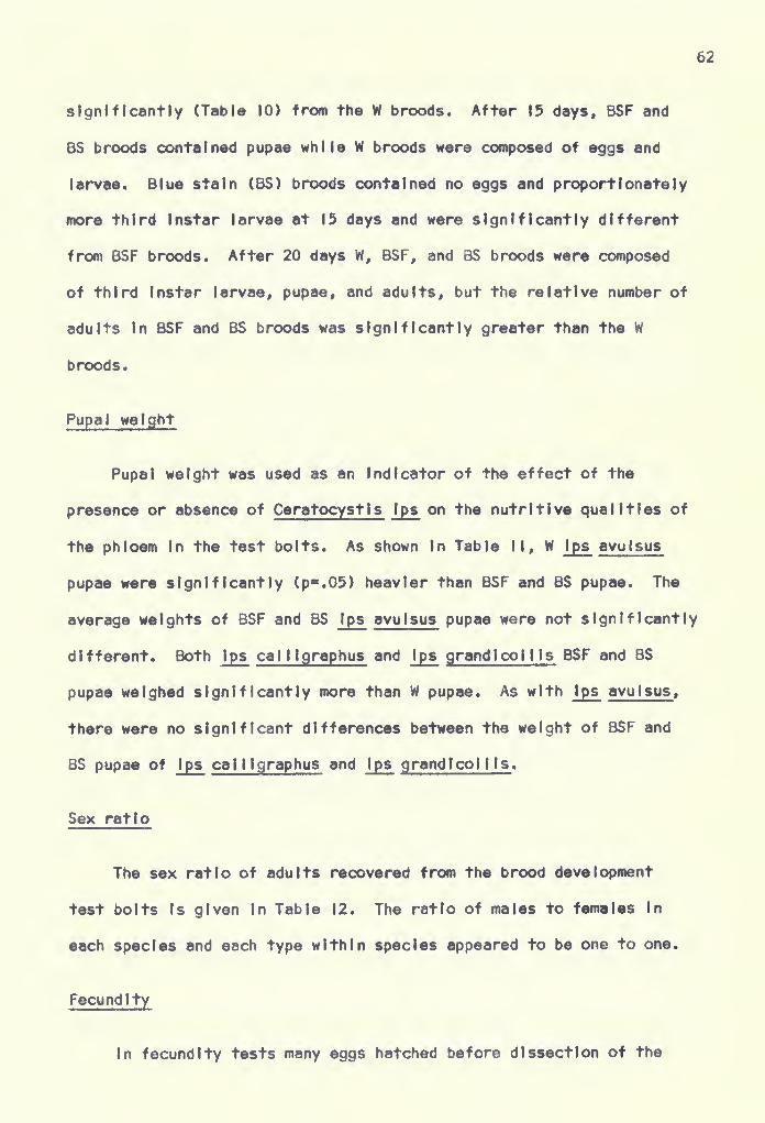
62
significantly (Table 10) from the W broods. After 15 days, BSF and
BS broods contained pupae while W broods were composed of eggs and
larvae. Blue stain (BS) broods contained no eggs and proportionately
more third Instar larvae at 15 days and were significantly different
from BSF broods. After 20 days W, BSF, and BS broods were composed
of third Instar larvae, pupae, and adults, but the relative number of
adults In BSF and BS broods was significantly greater than the W
broods.
Pupal weight
Pupal weight was used as an indicator of the effect of the
presence or absence of Ceratocystis tps on the nutritive qualities of
the phloem In the test bolts. As shown In Table II, W Ips avulsus
pupae were significantly (p".05) heavier than BSF and BS pupae. The
average weights of BSF and BS Ips avulsus pupae were not significantly
different. Both Ips cal ligraphus and Ips grand I col I Is BSF and BS
pupae weighed significantly more than W pupae. As with ips avulsus,
there were no significant differences between the weight of BSF and
BS pupae of ips cal ligraphus and ips grand I col lis .
Sex ratio
The sex ratio of adults recovered from the brood development
test bolts is given in Table 12. The ratio of males to females In
each species and each type within species appeared to be one to one.
Fecundity
in fecundity tests many eggs hatched before dissection of the
63
Table II. Mean weight (mg.) of 100 W, BSF, and BS Ips avulsus , Ips cal II-
graphu8» and Ips grandlcol t Is pupae reared In slash pine boltsat 30O C.
Beetle Species Type of Pupa
W BSF BS
Ips avulsus 2.20*0. II* l.7l±0.23 l.76±0.ll
Ips calllgraphus 9.74*0.25 l0.28io.40» IO.I7io.48»
Ips grandlcol I Is 3.71*0.11 4,54±0.I4» 4.23*0. I6»
• Significantly different (p"0.5) from others within same species.Ouncan*s New Multiple Range Test.
bolts, and actual egg counts were not feasible. Examination of
several hundred egg niches before the eggs hatched showed that each
niche, without exception, contained an egg. Consequently, the number
of egg niches cut per female was considered equal to the number of
eggs laid and was recorded as such.
Wild (W) Ips avulsus females constructed significantly (p".0l)
longer egg galleries and deposited significantly more eggs than BSF
and BS females (Table 13). The average number of eggs per centimeter
of egg gallery, however, was comparable for the three female types.
The number of eggs laid by both BSF and BS Ips call Igraphus and Ips
grandlcol! Is females was significantly (p".OI) greater than that
laid by W females. Blue stain-free (BSF) Ips cal I Igraphus females
also produced (p".05) more eggs than BS females. The average length
of egg gal lery cut by the three types of Ips cal I Igraphus and Ips

64
Table 12. Sex ratio

65
Tabia 13. Mean length of egg gallery, number of eggs laid, and numberof eggs deposited per centimeter of gallery by W, BSF, and
BS Ips avulsus , Ips cal I Igraphus , and Ips grandlcol I Is
females at % days In slash pine bolts at 30° C.
Number Mean Gallery Mean No. Mean No.
Females Length (cm.) Eggs Laid Eggs PerObserved cm. Gai-
lery
Ips avulsusW 10 5I.2*5.75«» 77.0t|3.8» 1.5BSF ii 3l.2i6.83 54.0*6.8 1.7BS It 36.8±6.I0 53.71 9.3 1.4
Ips cal I IgraphusW 9 50.8-4.16 II7.4±20.8 2.3BSF 13 52.7±4.I9 I5I.I±I0.6»» 2.9BS 12 47.8l5.65 I39.6i|0.5»« 2.9
Ips grandlcol UsW 15 37.3t5,42 I08.2±I9.2 2.9BSF 16 44.6*6. 21 I48.5l|6.l«» 3.4BS 14 40.l±5.77 I47.3±I3.9»» 3.7
* Significantly different (p".05) from others within the samespecies. Duncan's New Multiple Range Test.
•* Significantly different (p".0l) from others within the samespecies. Duncan's New Multiple Range Test.
dependent upon the beetles for dissemination and Introduction Into
a suitable host, but also upon the activities of the beetles In the
Inner bark to open avenues of spread once the fungus Is In the tree.
When Introduced Into host material over a small surface area as In
bolts unsuccessfully attacked In the transmission study (Table 3)
and pathogenicity trials simllating bark beetle attacks (Mathre,
1964b and Nelson, 1934), the fungus may become established but does

66
not spread from the site of Infection and sporulatlon does not occur.
Thus the action of adult Ips beetles In constructing galleries and
the feeding activities of the larvae are a requisite for optimum
development and spread of the fungus In the tree.
As previously mentioned. Leach et at. (1934) noted that until
a brood of beetles was reared In a fungus-free log, it could not be
safely concluded that blue stain fungi were not necessary for normal
development of the beetles, in this study it was clearly demonstrated
for the first time that all three Ips species were capable of develop-
ment through successive generations in pine bolts free of C. Ips .
ips avulsus was reared through three successive generations without
observable effects when compared to beetles reared In the presence
of the fungus. Similar results were obtained with Ips cal I Igraphus
and Ips grand I CO i I is over four generations.
When brood development was examined more closely. It was found
that Ips avulsus broods free of £. Ips (BSF) were comparable In size
to broods reared In the presence of the fungus (6S). Additionally,
no differences were detected between length of egg gallery, brood
mortality, brood composition (rate of development), or pupal weight
of the two brood types. The number of eggs laid by BSF and BS
females after 30 days was not significantly different. Thus, it
is apparent that C. ips has no effect on brood development and
fecundity of Ips avulsus under laboratory conditions.
It was also apparent that C. Ips had little or no effect on
brood development and fecundity of Ips cai I igraphus and Ips grand i-
coi I Is although the data were not as conclusive as with ips avulsus .

67
Ips cal llgraphus femal«s fr»« of C. Ips (BSF) constructed longer egg
galleries and produced significantly larger broods than females (BS)
In the presence of the fungus. Furthermore, BSF females laid
significantly more eggs than BS females after 2K) days. Conversely*
however, BS Ips cal I Igraphus broods appeared somewhat more advanced
than BSF broods at 5 and 15 days. This apparent Increase In rate of
development was not consistent throughout the observational period,
and at 10 and 20 days the relative composition of the broods was
comparable. No explanation (other than random error) can be given
for the significant differences between brood size and fecundity of
BSF and BS beetles. It should be pointed out that In both Instances
the differences barely met the minimum required for significance
and were possibly due to chance. Owing to the small number of
broods examined and the Inconsistency of the data, It can neither be
assumed that the absence of C. Ips results In construction of longer
egg galleries and Increased fecundity, nor the presence of the
fungus resulted In more rapid brood development. Considering all
measurements made on the various aspects of brood development and
fecundity. It was evident that the fungus has little or no effect on
the beetle.
With the exception of brood composition, no differences were
found between BSF and BS Ips grandlcol lis broods. At 5 and 20 days,
the composition of the broods was comparable, but at 10 days BSF
broods appeared somewhat more advanced than the BS brood. The
situation, however, was reversed at 15 days, and BS broods contained
more Individuals In the later stages of development. With this aspect

68
of brood developinent and fecundity being the only area In which
differences were detected and again due to the Inconsistency of the
differences, there Is little reason to suspect that C. Ips Is a
factor In brood development and fecundity of Ips grandlcol Ms ,
Although the results of this Investigation clearly Indicated
that C. Ips has little or no effect on brood development and fecundity
of the three Ips species studied, the question still arises as to the
applicability of these findings to field conditions. It Is well
documented In the I Iterature that Ceratocystis Infection resulted In
reduction of the water content of the xylem and phloem. Shepard and
Watson (1959) further demonstrated the fungi Indirectly reduced resin
production by causing collapse of the res In-secret I ng epithelial cells.
As previously mentioned, many authors believed reduced water content
and resin flow were necessary to create conditions favorable for
brood development by the beetles. The point In question, then. Is
whether C. Ips Is capable of lowering the moisture content and o.e.p.
of a tree, which at the time of attack was too high, to a level
suitable for brood development. Although no field studies were
conducted, observations In the laboratory Indicated C. Ips did not
become established rapidly enough to significantly influence moisture
content or resin flow In the early stages of brood development. When
forced to attack bolts with a high phloem moisture content, beetles
of all three species. In the presence or absence of the fungus, were
equally "pitched out." In some bolts, the adults became established,
but the young larvae were "drowned" In resin soon after ecloslon.
Under field conditions, therefore. It appears unlikely that £. Ips

69
would be a significant factor In the establishment of Ips broods. The
possibility that C. Ips or other Ceratocyst I
s
species would prevent,
as a result of excessive environmental moisture, a sudden Increase In
moisture content and o.e.p. In trees with well-established broods Is
not excluded, however.
In addition to comparing brood development of the three Ips
species In the presence or absence of C. Ips , the maintenance of wild
(M) colonies of beetles afforded a comparison of BS and BSF broods
with broods enjoying their "nomal" complement of associated organisms.
As previously noted, W broods of both Ips grand I col Ms and Ips cal 11-
graphus did not fair as well as the BS and BSF ones. These differences
were attributed to parasites; namely, parasitic mites and nematodes
and pathogenic fungi. Wild Ips avulsus broods, on the other hand,
appeared more vigorous than BS and BSF broods. The broods were larger
and development was more rapid. Wild Ips avulsus pupae weighed
significantly more than BS and BSF pupae, and the fecundity of W
females was greater. Since no basic differences between BS and BSF
Ips avulsus broods were exhibited, the apparent Increased vigor of
the W broods did not appear to be correlated with the presence or
absence of C. Ips .
The consistency of the difference between W and BS and BSF Ips
avulsus beetles with respect to brood development, pupal weight, and
fecundity suggested that some factor or factors were missing from the
BS and BSF bolts. Although determinations were not made, two
organisms were consistently Isolated from the gallery systems and
adjacent tissues In W Ips avulsus bolts; a yeast and a fungus.

70
probably Tubercutartel la Ips Leach, Orr, and Chrlstensen. Callahan
and Shlfrlne (I960) suggested that yeasts played a role In the
nutrition of bark beetles. They noted that all species of bark beetles
have associated yeasts and that the yeasts were eaten by both larvae
and adults. Callahan and Shlfrlne added further that It has not been
demonstrated that "yeast-feed I ng" Is not a prerequisite for fertility
of the adults. The results of this study clearly refute Callahan and
Shifrlne's hypothesis. Brood development, fecundity, and fertility
of both Ips cal I Igraphus and Ips grand I col I Is were not adversely
affected when the beetles were reared In the absence of yeasts. Based
upon observations made during this study, however, the above statement
cannot be made for Ips avulsus since broods In the presence of yeast
as well as other organisms, exhibited more rapid development and larger
numbers. The yeast associated with Ips avulsus appeared to be the
same as that associated with the other Ips species; thus It Is most
unlikely that yeasts were responsible for the Increased vigor of W
Ips avulsus broods.
In addition to Ceratocystis Ips and the undetermined yeasts,
another fungus was consistently found associated with Ips avulsus .
The growth habits and structural characteristics of the fungi were
similar to those given by Leach et al. (1934) for Tubercularlel la
Ips . The fungus was most frequently observed In the pupal chambers
where fructification took place. By the time callow adults were
present the walls of the pupal chamber were lined with a white mass
of conldia (Figure 12). The fungus no doubt served as a food source
for Ips avulsus adults as newly emerged beetles were observed

71
Figure 12. tps avuISM* pupal chamber covered with
masses of con! d la, probably Tubercularlella tps .
Approx. 5X.

72
"grazing" on the fungal watt. Mo«t Individuals did not stray from
the pupal chambar until amargence from the brood fv and apparently
restricted their pre-emergence feeding to the fungus.
Francke-Orosmann (1963) stated that among phloem- Inhabiting
Scolyttdae, some cases of association between fungi and beetles are
known which can be called ambrosia because they Indicate existence
of true symbiosis. As one example, she cited the association between
Trichosporlum tengens var. macrosporum Francke-Grosmann and Ips
acumlnatus Gyll. In this case the fungus possessed nutrlent-rlch,
globose conldia which completely cover the gallery walls. The young
beetles fed on the fungal mass which, according to Francke-Grosmann,
no doubt served as Important nutrients for the young beetles. Leach
et al. (1934) noted that Tubercular lei la Ips resembled the so-called
"ambrosia" fungi by possessing large globose conldia rich In nutrients.
The relationship of T. Ips to Ips pinl and Ips grandlcol lis In the
United States (Leach et al., 1934) and Ips sexdentatus In Europe
(Slemaszko, 1939) then appears very similar to that described above.
The fungus In question In this study, probably T. Ips , was only
found In association with Ips avulsus. Whether Its presence was
responsible for the Increased vigor of Ips avulsus brood must remain
problematical. The dependence of I ps avulsus upon the fungus for
nutritive factors. If any, was not obligatory since beetles were
successfully reared In Its absence. Additional data are necessary
before the ecological Impact of the fungus on the development of
Ips avu I sus can be assessed.

CONCLUSIONS
Results of this study allow th« following conclusions to b«
drawn
t
Caratocystls Ips was the only member of the genus consistently
associated with Ips bark beetles occurring In Florida. Another fungus,
probably Tubercular lei la Ips , was frequently associated with Ips
avulsus , but not with Ips calilgraphus and Ips grandlcoMls .
Ceratocystis Ips was transmitted by both males and females of
the three Ips species. Adults were both externally and Internally
contaminated with the fungus. Larvae, particularly the later Instars,
were also Internally contaminated. Ips avulsus larvae demonstrated
a higher Incidence of contamination than Ips call igraphus and Ips
grandlcollls.
Ceratocystis ips had no influence on the mass attraction
phenomenon of Ips cal I Igraphus males.
Pine bolts tnnoculated with Ceratocystis Ips prior to attack by
the three Ips species proved unsatisfactory as breeding material.
The presence, or absence, of Ceratocystis Ips had little or no
effect on brood development and fecundity of Ips avulsus , Ips call i-
graphus and Ips grandlcol I Is . Larvae were reared on semi-artif ictal
medium without adverse effects when compared to broods reared In
bolts In the presence of Ceratocystis Ips . The three Ips species
were reared through successive generations in bolts free of
73

74
Ceratocystis Ips without observable effects on vigor and fecundity
when compared to beetles In the presence of the fungus. Ips ca 1 11
-
graphus and Ips grand I col I Is In the absence of Ceratocystis Ips or
In the presence of Ceratocystis Ips alone produced larger brood with
lower mortality than wild beetles with the normal complement of
micro-organisms. These differences were attributed to parasites
present In wild populations only.
Wild Ips avulsus broods were larger and development was more
rapid than broods In the absence or presence of Ceratocystis Ips .
The presence of a fungus, probably Tubercular lei la Ips , with the
wild broods was suggested as a possible factor contributing to these
differences since the fungus resembled the "ambrosia" fungi. Ips
avulsus adults were observed feeding on the fungus, but their
dependence upon It for nutritive factors was not obligatory as
beetles were successfully reared In Its absence.

LITERATURE CITED
Anderson, R. F. 1948. Host selection of the pine engraver.
J. Econ. Entomol. 41: 596-602.
BakshI, F. K. 1950. Fungi associated with ambrosia beetles In
Great Britain. Brit. Mycol. Soc. Trans. 33: 111-20.
Blackman, M. W. 1922. Mississippi bark beetles. Miss. Agr. Exp.
Sta. Tech. Bull. II: 130 pp.
Bramble, W. C. and E. C. Hoist. 1935. Micro-organisms Infecting
pines attacked by Dendroctonus fronta lis . Phytopathology
25: 7.
1940. Fungi associated with Dendroctonus frontalis
In killing short leaf pines and their effect on conduction.
Phytopathology 30: 881-99.
Caird, R. W. 1935. Physiology of pines Infested with bark beetles.
Bot. Gaz. 96: 700-33.
Callahan, R. Z. and M. Shlfrlne. I960. The yeasts associated with
bark beetles. Forest Scl. 6: 146-54.
Clark, E. W. and E. A. Osgood, Jr. 1964a. Mass rearing the southern
pine beetle and the coarse writing engraver. Southeast. Forest.
Exp. Sta. U. S. Forest Serv. Res. Note. SE-30: 4pp.
1964b. A simple laboratory
technique for rearing Ips cal llgraphus . Southeast. Forest.
Exp, Sta. U. S. Forest Serv. Res. Note. SE-31: 3pp.
Craighead, F. C. 1928. Interrelation of tree-killing bark beetles
(Dendroctonus) and blue-stains. J. Forest, 26: 886-7.
Davidson, R. W. 1935. Fungi causing stain In logs and lumber In
the southern states. Including five new species. J. Agr. Res.
50: 799-807.
________^__^ 1955. Wood staining fungi associated with bark
beetles 'fn Engelmann spruce In Colorado. Mycol. 47: 58-67.
1958. Additional species of Ophlostomaceae from
Colorado. Mycol. 50: 661-70.
75

76
Dixon, J. C. and E. A. Osgood. 1961. Southern pine beetle: A
review of present knowledge. U. S. Dept. Agr., U. S. ForestServ., SE Forest Exp. Sta. Paper 128: 34 pp.
Ellis, D. E, 1939. Ceratostomel la Ips association with Ips
lecontel In Arizona. Phytopathology 29: 556-7,
FIndlay, W. P. K. 1959a. Sap-stain of timber I. Forest. Abstr.
20: 1-7.
1959b. Sap-stain of timber II. Forest. Abstr.
20: 167-74.
Francke-Grosmann, H. 1963. Some new aspects In forest entomology.
Am. Rev. Entomol. 8: 415-38.
Grosmann, H. 1930. Beltrage zur Kenntnis der Lebensgemelnschaft
zwischen Borkenkafern und Pllzen. Z. Parasltenk. 36: 56-102.
Hartig, R. 1894. Textbook of the diseases of trees. McMillan and
Co., London. 331 pp. (Translated to English by W. Soramervllle
and H. M. Ward.)
Hetrick, L. A. 1949. Some overlooked relationships of southern
pine beetle. J. Econ. Entomol. 42: 466-9.
Hoist, E. C. 1937. Aseptic rearing of bark beetles. J. Econ.
Entomol. 30: 676-77.
Hopping, G. R. 1961. Techniques for rearing Ips DeGeer (Coleoptera:
Scolytldae). Can. Entomol. 93: 1050-3.
Hunt, J. 1956. Taxonomy of the genus Ceratocystls . Lloydia
19: 1-58.
Leach, J. 6. 1940. Insect transmission of plant diseases. McGraw-
Hill Book Co., New York. 615 pp.
, L. W. Orr, and C. Chrlstensen. 1934. The Inter-
relationships of bark beetles and blue-staining fungi in felled
Norway pine timber. J. Agr. Res. 49: 315-41.
Massey, C. L. 1957. Four new species of Aphelenchulus (Nematode)
parasitic in bark beetles in the United States. Proc. Helm.
Soc. Wash. 24: 29-34.
I960. Nematode parasites and associates of the
California five-splned engraver, Ips confusus (Lee). Proc.
Helm. Soc. Wash. 27: 14-22.

77
1962. Life history of Aphelenchulus etongatus Mas8»y
(Neroatoda), an endoparasita of Ips confusus (Lee.) with
description of the male. J. Insect. Pathol. 4: 95-103.
Mathlesen, A. 1950. Dber einlge mit BorkenkSfern assocllerte
Blaue-Pllze In Schweden. Oikos 2: 275-308.
Mathlesen-Kaarlk, A. I960. Studies on the ecology, taxonomy, and
physiology of Swedish Insect-associated blue stain fungi,
especially the genus Ceratocystls . Oklos II: 1-25.
Mathre, D. E. 1964a. Survey of Ceratocystls spp. associated with
bark beetles In California. Contrlb. Boyce Thompson Inst.
22: 353-62.
___^___^__ 1964b. Pathogenicity of Ceratocyst ls Ips and
Ceratocystls minor to PInus ponderoiaT Contrlb. Boyce
Thooipson Inst. 22: 363-88.
1964c. Effect of Ceratocystls Ips and Ceratocystls
minor on the free sugar pool In ponderosa pine sapwood.
Contrlb. Boyce Thompson Inst. 22: 509-12.
Miller, J. M. and F. P. Keen. I960. Biology and control of the
western pine beetle; a summary of the first fifty years of
research. U. S. Oept. Agr. Misc. Pub. 800: 381 pp.
Nelson, R. M. 1934. Effect of blue stain fungi on southern pines
attacked by bark beetles. Phytopathol. Z. 7: 327-53.
and J. A. Beal. 1929. Experiments with blue stain
fungi In southern pines. Phytophthology 19: 1 101-6.
Nickle, W. R. 1963a. Notes on the genus Contortylenchus Ruhm 1956,
with observations on the biology and life history of C. elongatus
(Massey, I960) n. comb., a parasite of a bark beetle. Proc.
Helm. Soc. Wash. 30: 218-23.
1963b. Observations on the effect of nematodes on Ips
confusus (Lee.) and other bark beetles. J. Insect. Pathol.
5: 386-9.
NIslkado, Y. and K. Yamautl. 1933. Contributions to the knowledge
of sapstain of wood In Japan. I. Studies on Ceratostome 1 1 a
Ips Rumb. the cause of a blue stain In pine trees . Ohara Inst.
Landwirtsch. Forsch. Bar. 6: 467. (Abstr. In Rev. Appl.
Entomol. Sec. A. 21: 403. 1935).
Person, H. L. 1931. Theory In explanation of the selection of the
certain trees by the western pine beetle. J. Forest.
29: 696-9.

78
Robinson, R. C. 1962. Biu* stain fungi In todgepote pine (PInuscontorts Doug I. var. jatlfolla) Infssted by mountain pine beetle(Dendroctonus wontlcotae Hopk«). Can. J. Bot. 40: 609-14.
Roblnson-Jeffery, R. C. and A. H. H. Grinchenko. 1964. New fungusIn the genus Ceratocystis occurring In blue stained lodgepote
pine attacked by bark beetles. Can. J. Bot. 42: 527-32.
Rumbold, C. T. 1929. Blue-stalnlng fungi found In the United States.Phytopathology 19: 597-9.
1931. Two blue-stalnlng fungi associated with bark
beetle Infestation of pines. J. Agr. Res. 43: 847-73.
1936. Three blue-stalnlng fungi. Including two new
species, associated with bark beetles. J. Agr. Res. 52: 419-37.
1941. A blue stain fungus, Ceratostowella montluw .
n. sp., and sone yeasts associated with two species of Dendroctonus .
J. Agr. Res. 62: 589-601.
Slemaszko, W. 1939. Zespoly grzbyow towarzyszacych kornlkom poisklm.
(Fungi associated with bark beetles In Poland) Planta Polonica
7: 54 pp. (Abstr. in f^v. Appl. Mycol. 18: 488. 1939).
Shepard, R. F. and J. A. Watson. 1959. A blue stain fungus associated
with the mountain pine beetle (Dendroctonus montlcolae) . Bl-mon.
Prog. Rep. Olv. For. Biol. Rep. Agrl. C^n. 15: 2-3.
Thatcher, R. C. I960. Bark beetles affecting southern pines: A
review of current knowledge. U. S. Dept. Agr., U. S. Forest
Serv. South. Forest. Exp. Sta. Occasional Paper 180: 25 pp.
Vanderzant, E. S. and T. B. Davich. 1958. Laboratory rearing of the
boll weevil: A satisfactory larval diet and oviposltlon studies.
J* Econ. Entomoi. 51: 288-91.
Verrail, A. F. 1941. Dissemination of fungi that stain logs and
lumber. J. Agr. Res. 63: 549-58.
Vlte', J. P. and R. I. 6ara. 1961. A field method for observationof olfactory responses of bark beetles to volatile materials.
Contrlb. Boyce Thompson Inst. 21: 175-82.
1962. Volatile attractants from
ponderosa pine attacked by bark beetles. (Coleoptera: Scolytldae).
Contrlb. Boyce Thompson Inst. 21: 251-74.

79
Vlte', J. P. and D. L. Wood. 1961. A study of the appHcabllfty of
the measurement of oleoresfn exudation pressure tn determining
susceptibility of second groirth ponderosa pine to bark beetle
Infestation. Contrlb. Boyce Thompson Inst. 21: 67-8.
Von Schrenk, H. 1903. The "bluing*' and "red rot** of the western
yellow pine, with special reference to the Black Hills Forest
Reserve. U. S. Oept. Agr. Bur. Plant Ind. Bull. No. 36: 40 pp.
White, M. 6. 1962. The effect of blue stain In Scot's pine (Ptnus
sylvestrls L.) on growth of larvae of the house longhorn beetle.
(Hylotrupes bajulus L.). J. Inst. Wood Scl. 9: 27-31.
Wilkinson, R. C. 1962. Strldulating organs In three southeastern
Ips bark beetles. Fla. Entomol. 45: 43-4.
1963. Larval Instars and head capsule morphology
In three southeastern Ips bark beetles. Fla. Entomol. 46: 19-22.
1964. Attraction and development of Ips bark beetles
populations In artificially Infested pine bolts exposed on fire-
towers and turntables In Florida. Fla. Entomol. 47: 57-64.
Wood, D. L, 1962. The attraction created by males of a bark beetle,
Ips confusus (Lee.), attacking ponderosa pine. Pan-Pac. Entomol.
38? 141-5.
and J. P. Vlte' . 1961, Studies on the host selection
behavior of Ips confusus (Lee.) attacking PInus ponderosa .
Ck>ntrlb. Boyce Thompson. Inst. 21: 79-95.
Yearlan, W. C. and R. C. Wilkinson. 1963. An artificial rearing
medium for Ips calllgraphus Germ. Fla. Entomol. 46: 319-20.
1965. Two larval rearing
media for Ips bark beetlesi Fla. Entomol. 48: 25-7,

BIOGRAPHICAL SKETCH
William C. Yearlan, Jr., was born May 20« 1937, 9^ Laka Village,
Arkansas. He attended elementary and secondary schools at Lake Village,
Arkansas, and was graduated from Lakeside High School in 1955.
He attended Arkansas A & M College, College Heights, Arkansas,
from September, 1955, until June, 1957. He transferred to the University
of Arkansas In January, 1958, where he received the degree of Bachelor of
Science In Agriculture In January, I960. Upon receipt of the Crossett
Research Fellowship, he entered the Graduate School, University of
Arkansas In January, I960, and was awarded the Master of Science Degree
with a major In Entomology In January, 1961.
On June 30, I960, he married the former Marguerite LaVerne
Robertson of Eudora, Arkansas. They have no children.
He entered the University of Florida In February, 1961, and was
granted a research asslstantshlp with the Department of Entomology.
In April, 1962, he accepted a research asslstantshlp with the Depart-
ment of Entomology, Florida Agricultural Experiment Station. In the
summer of 1964, he was granted a research fellowship from the Southern
Forest Disease and Insect Research Council.
80

81
In May, I969« h« was appolntad to tha staff of tha Dapartmant of
EntoMoiogy, Unt varsity of Arkansas, as an Assistant Profassor.
At prasant ha Is a candldata for tha Dagraa of Doctor of Philosophy.


This dissertation was prepared under the direction of the
chaiman of the candidate's supervisory committee and has been
approved by all members of that committee. It was submitted to
the Dean of the College of Agriculture and to the Graduate Council,
and was approved as partial fulfillment of the requirements for
the degree of Doctor of Philosophy.
Date June 21, 1966
n. College of Agriculture
Supervisory Committee:
Dean, Graduate School
,/^€t^Afj
A^Co-chairmen
- ' i. :t