Embed Size (px)
Citation preview

RelA Mutant Enterococcus faecium with Multiantibiotic ToleranceArising in an Immunocompromised Host
Erin S. Honsa,a Vaughn S. Cooper,b Mohammed N. Mhaissen,a Matthew Frank,a Jessica Shaker,a Amy Iverson,a Jeffrey Rubnitz,c
Randall T. Hayden,d Richard E. Lee,e Charles O. Rock,a Elaine I. Tuomanen,a Joshua Wolf,a,f Jason W. Roscha
Department of Infectious Diseases, St. Jude Children’s Hospital, Memphis, Tennessee, USAa; Department of Microbiology and Molecular Genetics, University of Pittsburgh,Pittsburgh, Pennsylvania, USAb; Department of Oncology, St. Jude Children’s Hospital, Memphis, Tennessee, USAc; Department of Pathology, St. Jude Children’s Hospital,Memphis, Tennessee, USAd; Department of Chemical Biology and Therapeutics, St. Jude Children’s Hospital, Memphis, Tennessee, USAe; Department of Pediatrics,University of Tennessee Health Science Center, Memphis, Tennessee, USAf
E.S.H. and V.S.C. contributed equally to this article.
ABSTRACT Serious bacterial infections in immunocompromised patients require highly effective antibacterial therapy for cure,and thus, this setting may reveal novel mechanisms by which bacteria circumvent antibiotics in the absence of immune pressure.Here, an infant with leukemia developed vancomycin-resistant Enterococcus faecium (VRE) bacteremia that persisted for26 days despite appropriate antibiotic therapy. Sequencing of 22 consecutive VRE isolates identified the emergence of a singlemissense mutation (L152F) in relA, which constitutively activated the stringent response, resulting in elevated baseline levels ofthe alarmone guanosine tetraphosphate (ppGpp). Although the mutant remained susceptible to both linezolid and daptomycinin clinical MIC testing and during planktonic growth, it demonstrated tolerance to high doses of both antibiotics when growingin a biofilm. This biofilm-specific gain in resistance was reflected in the broad shift in transcript levels caused by the mutation.Only an experimental biofilm-targeting ClpP-activating antibiotic was able to kill the mutant strain in an established biofilm.The relA mutation was associated with a fitness trade-off, forming smaller and less-well-populated biofilms on biological sur-faces. We conclude that clinically relevant relA mutations can emerge during prolonged VRE infection, causing baseline activa-tion of the stringent response, subsequent antibiotic tolerance, and delayed eradication in an immunocompromised state.
IMPORTANCE The increasing prevalence of antibiotic-resistant bacterial pathogens is a major challenge currently facing the med-ical community. Such pathogens are of particular importance in immunocompromised patients as these individuals may favoremergence of novel resistance determinants due to lack of innate immune defenses and intensive antibiotic exposure. During thecourse of chemotherapy, a patient developed prolonged bacteremia with vancomycin-resistant Enterococcus faecium that failedto clear despite multiple front-line antibiotics. The consecutive bloodstream isolates were sequenced, and a single missense mu-tation identified in the relA gene, the mediator of the stringent response. Strains harboring the mutation had elevated baselinelevels of the alarmone and displayed heightened resistance to the bactericidal activity of multiple antibiotics, particularly in abiofilm. Using a new class of compounds that modulate ClpP activity, the biofilms were successfully eradicated. These data rep-resent the first clinical emergence of mutations in the stringent response in vancomycin-resistant entereococci.
Received 22 November 2016 Accepted 28 November 2016 Published 3 January 2017
Citation Honsa ES, Cooper VS, Mhaissen MN, Frank M, Shaker J, Iverson A, Rubnitz J, Hayden RT, Lee RE, Rock CO, Tuomanen EI, Wolf J, Rosch JW. 2017. RelA mutantEnterococcus faecium with multiantibiotic tolerance arising in an immunocompromised host. mBio 8:e02124-16. doi:10.1128/mBio.02124-16.
Editor Michael S. Gilmore, Harvard Medical School
Copyright © 2017 Honsa et al. This is an open-access article distributed under the terms of the Creative Commons Attribution 4.0 International license.
Address correspondence to Joshua Wolf, [email protected], or Jason W. Rosch, [email protected].
Current challenges in treatment of infections are focused on themarked reduction of new candidates in the antibacterial dis-
covery pipeline at a time of increasing rates of antimicrobial resis-tance, including emergence of methicillin-resistant Staphylococcusaureus (MRSA), �-lactam- and macrolide-resistant Streptococcuspneumoniae, and vancomycin-resistant enterococci (VRE) (1–4).VRE species are especially problematic clinically, as these bacteriaare resistant to all first-line antibiotics and infection is associatedwith a marked increase in risk of mortality (5–8). However, otherchallenges to therapeutic success are also emerging, particularly inthe immuncompromised host, where refractory bacteremia andprolonged antibiotic therapy increase the opportunity to select foralternative bacterial survival traits by mutation or genetic ex-
change (9–12). Once mutations arise, the permissive nature of thecompromised immune system may allow for the development ofsecondary mutations that compensate for any associated fitnesstrade-offs (13). As such, hosts more permissive for infection havebeen postulated to represent an important reservoir for the emer-gence of novel problematic pathogens (14).
In the absence of host defenses, bacterial killing by antibiotics isrequired for cure. Commonly this is ensured by administeringdrugs with the aim of ensuring that concentrations at the site ofinfection are above the MIC as reported by the clinical laboratory.This assumes the bactericidal concentration is close to the MIC.However, bacteria can persist when the MIC and minimal bacte-ricidal concentration (MBC) dissociate such that antibiotics in-
RESEARCH ARTICLE
cross
January/February 2017 Volume 8 Issue 1 e02124-16 ® mbio.asm.org 1
on July 19, 2020 by guesthttp://m
bio.asm.org/
Dow
nloaded from

hibit growth but fail to kill, a property called “tolerance” (15–17).As genome sequencing technologies improve, it has become evi-dent that mutations—particularly those involved in stress re-sponses— can impact efficacy of antibiotics (18). The bacterialstringent response, which slows metabolism under low-nutrientor stress conditions, can decrease the response to antibiotic ther-apy, allowing quiescent bacteria to survive and tolerate antibiotics,without a change in MIC (19). In S. aureus, induction of the strin-gent response increases tolerance to �-lactam antibiotics (20–22),and in Gram-negative bacteria, activation contributes to the pro-duction of biofilm persister cells (23, 24). Also, a report on Entero-coccus faecalis demonstrated that treatment of cells with mupiro-cin induced expression of transport and stress-related genes, andstrong repression of genes involved DNA, RNA, and protein syn-thesis, similar to a stringent response (25). Despite the importanceof the stringent response, there are few reports of clinical emer-gence of mutations in this pathway, none demonstrating reducedantibiotic efficacy (22), and no studies focusing on antibiotic tol-erance and stringent response in E. faecium. For Enterococcus spe-cies, although the stringent response pathway is well characterizedand antibiotic resistance is widespread, we report the first exampleof mutation in the stringent response pathway causing increasedbaseline alarmone levels, which was responsible for antibiotic tol-erance within a biofilm.
RESULTSClinical setting. The patient was a 6-week-old African-Americangirl born by normal vaginal delivery at term. Acute myeloid leu-kemia was diagnosed at 4 weeks of age, after she presented withfever and marked leukocytosis. A double-lumen central venouscatheter (CVC) was placed (7.0 French-scale Hickman catheter;Bard Access Systems, Salt Lake City, UT), and chemotherapy wasinitiated, resulting in prolonged profound neutropenia. Induc-tion chemotherapy comprised systemic cytarabine, daunorubicin,etoposide, and methylprednisolone plus intrathecal methotrex-ate, hydrocortisone, and cytarabine. The patient received intrave-nous cefepime as antibacterial prophylaxis. After 2 weeks of che-motherapy, during profound neutropenia, routine microscopicexamination of a peripheral blood smear revealed bacterial organ-isms, although the patient had no signs or symptoms of infection.Blood cultures were obtained from both lumens of the CVC, andempirical combination therapy with vancomycin and mero-penem was initiated. After blood cultures grew vancomycin-resistant Enterococcus faecium, the antibiotic regimen was changedto linezolid, which was eventually supplemented with daptomy-cin, gentamicin, and quinupristin-dalfopristin (Table 1).
Initial paired blood cultures drawn from the two CVC lumensshowed a differential time to positivity of 4.0 h, and culture of theexplanted device at the time of CVC removal (day 9) was positivefor E. faecium, suggesting that the infection was initially related tobiofilm on the surface of the CVC (26). However, over the 28 days,the bacteremia failed to clear despite targeted antibiotic therapy,as well as CVC removal and replacement on two occasions, bothallowing for CVC-free periods. Antibiotic therapy was chosen inaccordance with expert opinion and in vitro susceptibility testingthat indicated the strain remained sensitive to all antibiotics ad-ministered except the initial vancomycin and meropenem (27).During the persistent bacteremia, there was no significantchange in the reported antimicrobial susceptibility pattern ac-cording to testing performed in our clinical microbiology lab-
oratory. Every strain was susceptible to linezolid (MIC, 1 to2 �g/ml), quinupristin-dalfopristin (MIC, 0.5 �g/ml), and dap-tomycin (MIC, 3 to 4 �g/ml). High-level resistance to gentamicinwas not detected (MIC, �500 �g/ml).
A comprehensive diagnostic evaluation, including Doppler ul-trasonography of the upper and lower extremities to detect intra-vascular thrombosis, ultrasound of head and abdomen, and mag-netic resonance imaging (MRI) of the entire body failed, toidentify any additional source of persistent infection. Analysis ofcerebrospinal fluid (CSF) obtained through lumbar puncture onday 2 of infection was within normal limits, and bacterial culturewas sterile. Transthoracic echocardiography performed on days11 and 22 showed mild preexisting aortic regurgitation withoutclear evidence of vegetations. The initial source for the blood-stream infection is thought to have been luminal colonization andbiofilm formation within the CVC, and after device removal, thebloodstream infection persisted. Based on this history, we believethat the most likely focus of persistent infection was occult endo-carditis or septic thrombophlebitis.
The patient’s absolute neutrophil count (ANC) remained at0 cells/mm3 until day 20, when donor granulocyte transfusion wasinitiated. Granulocyte transfusion was repeated again on days 22,23, 24, 25, 26, and 27 until neutropenia resolved. Daily ANC val-ues are shown in Table 1. On day 27, the bacteremia cleared, andthe patient received an additional 6 weeks of linezolid treatmentfor possible endocarditis with full clinical recovery.
Genetic characterization. The initial VRE isolate was se-quenced and completely assembled to obtain a closed genomecomprising one 2.93-Mbp chromosome and three plasmids of171, 78.6, and 59.2 kbp, containing a total of approximately 3,208open reading frames (see Table S1 in the supplemental material).Analysis of the functional roles of predicted coding regions re-vealed multiple antibiotic resistance systems, including the vanBgene cluster for vancomycin, fluoroquinolones, aminoglycosides,�-lactams, and multiple putative drug efflux pumps (Table 2) (28,29). All 22 isolates were completely sequenced at high depth(mean coverage of 256.6�) and aligned to the closed referencegenome of the initial VRE isolate. In total, all genomes differed byonly five single nucleotide polymorphisms (SNPs), and no inser-tions or deletions were detected, indicating that a single strain wasresponsible for the prolonged bacteremia. Four of the five muta-tions were identified only once in single isolates: therefore, weconcluded these mutations were not clinically relevant. These mu-tations included Y175C missense mutation in rnz (RNase Z), theA221D missense mutation in ptsI (phosphoenolpyruvate phos-photransferase enzyme I), the P640H missense mutation in recF(recombination protein F), and a silent mutation in a hypotheticalplasmid protein (Table 1). However, one missense mutation inthe relA gene, predicted to encode an RelA(L152F) variant, wasfound in eight isolates. The relA mutation was first detected 3 daysafter starting antibiotic therapy and persisted through the clinicalcourse until the infection eventually resolved (Table 1). The ob-servation that the mutation was intermittently identified indi-cated that both the initial strain and the relA mutant strain coex-isted until the later stages of the infection. As will be discussed,there may have been initial sampling bias during collection of arepresentative isolate from each day listed in Table 1; however, webelieve this may have underestimated the true presence and per-sistence of the relA variant subpopulation.
RelA is a critical mediator of the bacterial stringent response
Honsa et al.
2 ® mbio.asm.org January/February 2017 Volume 8 Issue 1 e02124-16
on July 19, 2020 by guesthttp://m
bio.asm.org/
Dow
nloaded from

via production of the alarmone guanosine tetraphosphate(ppGpp) and has been implicated in resistance or tolerance toantibiotic stress in several bacterial pathogens (30). In our RelAmutants, the altered residue (L152F) is immediately adjacent toresidues essential for the hydrolase activity of the RelA enzyme(31). Modeling of the structural consequences of this mutationindicated that the native leucine is buried in the structure; how-ever, replacement of this residue with a phenylalanine would notallow for such tight packing, potentially causing a local change inthe active site. This mutation was initially detected on the fourth
day of the bloodstream infection and appeared to become moreprevalent later in the course of therapy, particularly once dapto-mycin was included in the antibiotic regimen (Table 1). Previousstudies have also identified mutations in relA in response to dap-tomycin exposure in vitro, although no impact on antibiotic effi-cacy has been demonstrated (32). These data indicate that the VREpopulation remained mostly homogeneous, except for a single-point mutation in the stringent response pathway. This relA mu-tation arose during the course of the infection and persistedthroughout the course of therapy despite treatment with antibiot-
TABLE 1 Summary of patient parameters and antimicrobial therapya
a ANC, absolute neutrophil count; CVC, central venous catheter; CSF, cerebrospinal fluid.
Stringent Response Mutant Is Antibiotic Tolerant
January/February 2017 Volume 8 Issue 1 e02124-16 ® mbio.asm.org 3
on July 19, 2020 by guesthttp://m
bio.asm.org/
Dow
nloaded from

ics to which the bacterium retained apparent susceptibility basedon MIC testing. It is worth noting that the VRE strains analyzed inthis paper were genetically identical (isogenic), except for the relAmissense mutation, and they all possessed multiple antibiotic re-sistance genes as shown in Table 2. Furthermore, no growth de-fects were identified between the wild type (WT) and the isogenicrelA mutants in the absence of stressors (see Fig. S1A in the sup-plemental material).
Effects of RelA mutation. The missense mutation in a highlyconserved domain essential for RelA hydrolase activity led us toascertain whether the identified mutation in relA conferred anydiscernible differences in levels of (p)ppGpp, the alarmone thatmediates the downstream stringent response pathways. Strainsthat were genetically identical with the exception of the single-base-pair relA mutation were allowed to incorporate 32P, and thebaseline levels of (p)ppGpp were measured. Strains carrying themutation in relA demonstrated approximately 3� greater basal
levels of ppGpp than the WT controls (Fig. 1A, thin-layer chro-matography [TLC] blot, and B, quantification [P � 0.001, Mann-Whitney test]). This observation suggested inappropriate activa-tion of the alarmone at baseline in the absence of an externalstimulus, potentially priming the cells to more rapidly adapt toadverse conditions such as antibiotic exposure. While we at-tempted to monitor differences in (p)ppGpp and GTP levels inlinezolid-stressed relA WT and mutant strains, we could not de-tect any induction of the stringent response (Fig. 1C). It should benoted that both WT and relA mutant bacteria stressed with mupi-rocin did not show a difference in activated stringent responsealarmone levels (WT shown in Fig. 1C [mutant identical to WT]).However, as will be discussed, we suggest the fitness benefit of therelA mutation is to provide a higher resting level of alarmone,which allowed these strains to better adapt and respond to antibi-otic stress during therapy (tolerance).
The most likely initial focus of infection in this case, the CVC,
TABLE 2 VRE isolates possessed multiple antibiotic resistance genes
Resistance function Antibiotic Resistance gene(s)
Alter cell wall charge Polymyxin pmrEAntibiotic-altering enzyme Macrolide ermB, msrCAntibiotic efflux Fluoroquinolone arlRAntibiotic efflux Tetracycline adeC, tetK, tetCAntibiotic efflux Wide range lmrCDAntibiotic efflux Lincosamide lsaEAntibiotic efflux Streptogramin msrC, isaAAntibiotic inactivation enzyme Lincosamide lnuBAminoglycoside-modifying enzymes Aminoglycoside aac(6=)-Ii, aac(6=)-Ie–aph(2==)-IaAminoglycoside-modifying enzymes Aminoglycoside aph(3=)-IIIa, aad(6=)Antibiotic target protection protein Tetracycline tet32, tetOAntibiotic target replacement proteins �-Lactams pbp1b, pbp2x, pbp2b, pbp1a, mecCMolecular bypass Glycopeptide vanB cassetteTarget Mutation Trimethoprim dfrE, dfrFTarget mutation Rifampin rpoBTarget mutation Fluoroquinolone gyrB
FIG 1 Basal levels of ppGpp are increased in VRE strains harboring mutant relA alleles. (A) Multiple relA WT or mutant (isogenic) strains were grown inlow-phosphate media, followed by incubation with 32P, and levels of resting (p)ppGpp were measured by densitometry. (B) Data for ppGpp levels are quantified,and means and standard deviations are shown. Levels were normalized relative to the wild-type strain (100%). *, P � 0.0079 by Mann-Whitney testing. (C)Controls: negative control without any stressor (Cont.), positive-control mupirocin, as well as linezolid.
Honsa et al.
4 ® mbio.asm.org January/February 2017 Volume 8 Issue 1 e02124-16
on July 19, 2020 by guesthttp://m
bio.asm.org/
Dow
nloaded from
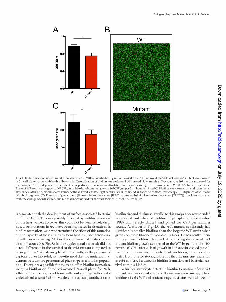
is associated with the development of surface-associated bacterialbiofilm (33–35). This was possibly followed by biofilm formationon the heart valves; however, this could not be conclusively diag-nosed. As mutations in relA have been implicated in alterations inbiofilm formation, we next determined the effect of this mutationon the capacity of these strains to form biofilm. Since traditionalgrowth curves (see Fig. S1B in the supplemental material) andtime-kill assays (see Fig. S2 in the supplemental material) did notdetect differences in the survival of the relA mutant compared toan isogenic relA WT strain (planktonic growth) in the presence ofdaptomycin or linezolid, we hypothesized that the mutation maydemonstrate a more pronounced phenotype in a biofilm popula-tion. To explore a possible fitness trade-off in biofilm formation,we grew biofilms on fibronectin-coated 24-well plates for 24 h.After removal of any planktonic cells and staining with crystalviolet, absorbance at 595 nm was determined as a quantification of
biofilm size and thickness. Parallel to this analysis, we resuspendednon-crystal violet-treated biofilms in phosphate-buffered saline(PBS) and serially diluted and plated for CFU-per-millilitercounts. As shown in Fig. 2A, the relA mutant consistently hadsignificantly smaller biofilms than the isogenic WT strain whengrown on these fibronectin-coated surfaces. Concurrently, iden-tically grown biofilms identified at least a log decrease of relAmutant biofilm growth compared to the WT isogenic strain (109
versus 108 CFU after 24 h of growth in fibronectin-coated plates).Each strain was grown under identical conditions, as well as inoc-ulated from titrated stocks, indicating that the missense mutationin relA conferred a defect in biofilm formation and bacterial sur-vival within a biofilm.
To further investigate defects in biofilm formation of our relAmutant, we performed confocal fluorescence microscopy. Here,biofilms of relA WT and mutant isogenic strains were allowed to
FIG 2 Biofilm size and live cell number are decreased in VRE strains harboring mutant relA alleles. (A) Biofilms of the VRE WT and relA mutant were formedin 24-well plates coated with bovine fibronectin. Quantification of biofilm was performed with crystal violet staining. Absorbance at 595 nm was measured foreach sample. Three independent experiments were performed and combined to determine the mean average (with error bars). *, P � 0.0074 by two-tailed t test.The relA WT consistently grew to 109 CFU/ml, while the relA mutant grew to 108 CFU/ml per 24-h biofilm. (B and C) Biofilms were formed on multichamberedglass slides. After 48 h, biofilms were stained with the Live/Dead BacLight bacterial viability kit and analyzed by confocal microscopy. (B) Representative imagesof a single segment. (C) The ratio of green to red (fluorescein isothiocyanate [FITC] to tetramethyl rhodamine isothiocyanate [TRITC]) signal was calculatedfrom the average of each section, and ratios were combined for the final average (n � 8). **, P � 0.002.
Stringent Response Mutant Is Antibiotic Tolerant
January/February 2017 Volume 8 Issue 1 e02124-16 ® mbio.asm.org 5
on July 19, 2020 by guesthttp://m
bio.asm.org/
Dow
nloaded from

form on an abiotic surface (microscope slide chamber) for 48 h.Live/dead staining was then performed, and confocal microscopywas used to measure fluorescein isothiocyanate (FITC) and te-tramethyl rhodamine isothiocyanate (TRITC) fluorescence ofz-stack 16-�m sections of each biofilm. To remove bias from bio-film sampling, each chamber for each strain was randomly ana-lyzed in four separate sections, and each strain was analyzed in twoseparate chambers on multiple days, allowing an n value of 8 foreach strain. Figure 2B shows that the WT strain had consistentlybrighter fluorescence in each section, compared to the relA mu-tant on the right. Furthermore, when the green and red signalswere quantified and a ratio determined (Fig. 2C), there was aconsistently higher fluorescent intensity for live cells for the WTstrains. This suggests that the biofilm of the mutant harbors lesstotal live cells, which correlates back to the CFU/ml data of estab-lished cells. Together, our biofilm analysis suggests that the abilityto form thicker, more robust biofilms is compromised in the relAmutant.
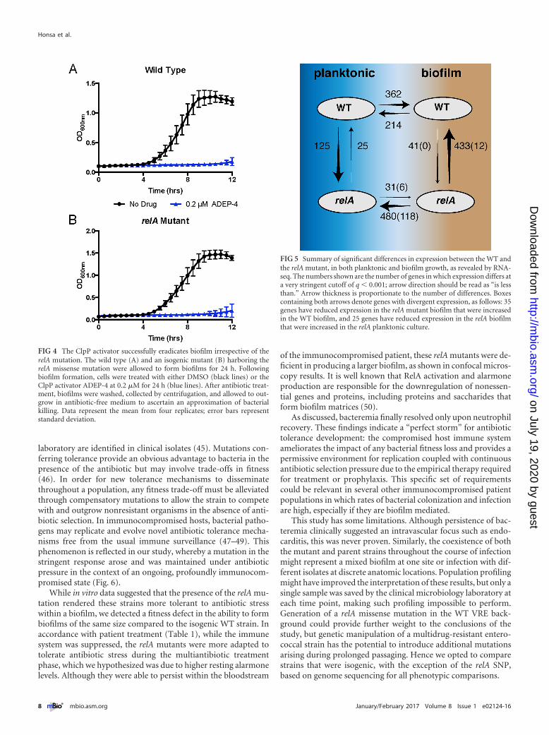
A mutation such as relA(L152F) that primes cells to enter thestringent response by increasing ppGpp levels is expected to affectexpression of many genes. Likewise, the phenotypic differencesbetween planktonic and biofilm growth of these strains should berelated to transcriptome changes. We tested these predictions byperforming RNA sequencing on these strains grown either in theliquid planktonic phase or as biofilms on a biotic surface. Thisexperiment revealed broad changes in expression in as much as15% of the ~2,940 genes in the genome in the relA mutant com-pared to the WT and during the shift from planktonic to biofilmgrowth. The interactions between genotype and growth environ-ment were most telling. Not only did this single relA mutationaffect expression (mostly by downregulation) of a broad array ofgenes in both conditions, it also changed how certain genes re-sponded during the shift from planktonic to biofilm growth. Infact, 35 genes responded in opposite directions in the shift to bio-film growth for the WT and the mutant (i.e., expression was up forthe WT, down for the mutant, or vice versa), and 25 genes weredivergently expressed for the relA mutant under biofilm andplanktonic growth conditions. Some of these genes with divergentexpression provide potential mechanistic explanations for thebiofilm-dependent resistance observed, including liaS, a majortarget of mutations causing resistance to daptomycin (36), whichwas upregulated in the WT biofilm relative to WT planktonicculture, but downregulated in the relA biofilm relative to plank-tonic relA cells. A small group of genes were uniquely upregulatedin the relA biofilm, including genes with chaperone functions,such as greA (EFAU004_01358) and cspA (EFAU004_01491),which would presumably enhance stress tolerance. However, theoverwhelming signature of the relA mutant was that it downregu-lated far more genes during biofilm growth than the WT strain(480 genes in comparison with 214 genes), and the direct compar-ison of the WT biofilm and the relA biofilm also revealed 433 genesdownregulated by the mutant. In summary, this single mutationnot only suppressed expression of many genes, it did so in abiofilm-dependent manner that actually reversed the tendency ofsome genes to be upregulated under this condition by the WT.
Given these biofilm-dependent changes in expression, it wassomewhat surprising to observe a fitness trade-off for the relAmutant in forming biofilms in standard media. However, in thepatient, the bacteria would have been constantly exposed to vari-ous antibiotic stresses during therapy. We therefore investigated
whether the relA mutation conferred enhanced tolerance toantibiotic-mediated killing in a biofilm model. Since no resistancewas detected against linezolid and daptomycin (MIC clinical dataand Table 2), we hypothesized that the relA mutation was notresponsible for a detectable rise in antibiotic resistance, but ratherallowed the mutants to respond faster to antibiotic stress in abiofilm, thus becoming tolerant to multiple antibiotics. Biofilmsof the two isogenic strains (WT or relA mutant) were allowed toform on Nunc peg plates in the rich medium Todd-Hewitt brothsupplemented with yeast extract, following previously publishedprotocols (37, 38). As a control, the relA mutation conferred nodiscernible difference in the outgrowth of planktonic cells re-moved from biofilms in the absence of antibiotics (Fig. 3A), indi-cating the relA mutation did not affect outgrowth from a biofilm.However, when the biofilms were subjected to prolonged antibi-otic exposure, high concentrations of vancomycin, linezolid, anddaptomycin were successful in eradicating the WT strain butfailed to eradicate strains harboring the relA mutation (Fig. 3B, C,and D). It should be noted that the subsequent outgrowth of eachstrain was in the absence of antibiotic, so no antibiotic carryovercould be responsible for the difference in response to antibioticstress. These data indicated that this mutation conferred a selec-tive advantage to the in vitro survival of antibiotic-tolerant per-sister cells within a biofilm, in the absence of linezolid or dapto-mycin resistance using standard CLSI susceptibility testing.Despite the defect of biofilm formation on fibronectin-coatedplates, the relA mutant still displayed enhanced tolerance to anti-biotics in the biofilm state. Taken together, these data suggest thatwhile the relA mutation led to increased alarmone resting levelsand subsequent tolerance to multiple-antibiotic stressors, the mu-tant strain was deficient in the ability to form biofilms comparedto its WT counterparts.
Eradication of bacterial biofilms is a major challenge duringtherapy due to their inherently greater tolerance to many antibi-otics and their capacity to both resist antibiotic penetration andresist immune clearance (39). This phenomenon is especially con-founded in immunocompromised patients. A new experimentalapproach to target such refractory bacterial communities has beenthe development of a class of compounds that cause unregulatedactivation of the housekeeping protease ClpP (40). We assayedthe bactericidal activity of one such investigational compound,ADEP-4, against isogenic strains possessing either WT or relA mu-tant strains in the context of a biofilm. While the absence of anti-biotic once again had no effect on the planktonic outgrowth ofbacteria, treatment with ADEP-4 successfully eradicated biofilmsof both strains (Fig. 4A and B). These data indicate that strainsharboring the relA mutation that conferred enhanced tolerance toclinically approved antibiotics during biofilm growth remainedsusceptible to this investigational class of compounds.
DISCUSSION
The stringent response is a highly conserved mechanism by whichbacteria can control broad metabolic changes required for sur-vival under adverse conditions and has been implicated in thevirulence of several prominent pathogens (41). It has also beenimplicated in the ability to promote antibiotic “tolerance”: bacte-rial cells that are able to survive antibiotic concentrations muchhigher than the MIC and can replicate once antibiotic pressure isremoved (15, 17, 19, 22, 24). It is hypothesized that this is due tothe absence or inactivity of antibiotic targets: for example, tolerant
Honsa et al.
6 ® mbio.asm.org January/February 2017 Volume 8 Issue 1 e02124-16
on July 19, 2020 by guesthttp://m
bio.asm.org/
Dow
nloaded from

persister cells may not be actively dividing, so the presence oflinezolid would not affect protein synthesis and therefore wouldnot cause bacterial cell death. As such, these tolerant cells canpersist during prolonged antibiotic exposure, further complicat-ing antibiotic treatment.
In Enterococcus, two enzymes are responsible for the produc-tion of the stringent response alarmone (p)ppGpp: the bifunc-tional RSH (i.e., Rel SpoT homolog [RelA for this study]) and themonofunctional RelQ alarmone synthase (42, 43). Recent studieshave highlighted the importance of subtle alterations in the levelsof basal ppGpp as opposed to the higher levels induced during thestringent response in mediating broad-range antibiotic tolerance(42). These data suggest that mutations in relA that alter basalppGpp levels may confer antibiotic tolerance of significance in animmunocompromised setting.
Emergence of a mutation in the relA gene has been observedduring persistent infection with S. aureus, but not in Enterococcus(22). Furthermore, the identification of a clinical VRE isolate withtolerance to multiple antibiotics due to a relA mutation has notbeen previously reported. It is important to note that ourantibiotic-tolerant relA VRE mutants could not be distinguishedfrom their isogenic WT counterparts by standard in vitro clinicalMIC testing or by traditional growth curves (Fig. S1A) and time-kill assays (Fig. S2) of exponentially replicating cells in liquid cul-ture. While the fitness advantage of the relA mutation was notpresent in planktonic culture, we determined that it was signifi-cant in the biofilm elimination model, whereby the mutant sur-
vived exposure to several antibiotics used to treat infection, sug-gesting an increase in antibiotic tolerance (Fig. 5). This haspreviously been reported for multiple bacterial pathogens, but notfor VRE (22, 44). Biofilms harboring the RelA missense mutationin the hydrolase domain had increased resting alarmone levels andwere recalcitrant to elimination by conventional antibiotic treat-ment. This included both linezolid and daptomycin, of which nogenetically encoded antibiotic resistance mechanisms existed (Ta-ble 2). However, both the WT and relA mutant were effectivelyeradicated by ADEP-4, which has been shown to constitutivelyactivate the housekeeping protease ClpP. These data indicate thatsuch unconventional antibiotic strategies may be extremely effec-tive for eliminating tolerant, persistent infections in an immuno-compromised host. The ClpP-sensitive, conventional antibiotic-tolerant biofilm population is reminiscent of the presumedclinical course, as the focus of infection is thought to have initiallybeen the CVC followed by possible endocarditis, both infectionstypically thought to be caused by multicellular biofilm communi-ties. Despite antibiotic therapy, bacterial clearance required hostimmune reconstitution; the infection only cleared upon neutro-phil recovery. This is an important reminder that clinical responsemay not correlate with in vitro susceptibility testing, especially inimmunocompromised hosts or biofilm-associated infections(34).
There exist a multitude of pathways by which bacteria canevolve greater antibiotic tolerance, as evidenced by in vitro selec-tion; however, only a subset of the mutations observed in the
FIG 3 The relA mutation contributes to tolerance to antibiotics in biofilms. Strains were grown and allowed to form biofilms by using a peg suspension system.Biofilms were then treated with the respective antibiotics (256 �g/ml vancomycin, 50 �g/ml linezolid, and 50 �g/ml daptomycin) for 24 h. After antibiotictreatment, biofilms were washed, collected by centrifugation, and allowed to outgrow in antibiotic-free medium to ascertain an approximation of bacterialkilling. Data represent the mean of three replicates; error bars represent standard deviation. For clarity, 6 h to 14 h is shown, as there was no growth prior to 6 hfor drug-treated VRE.
Stringent Response Mutant Is Antibiotic Tolerant
January/February 2017 Volume 8 Issue 1 e02124-16 ® mbio.asm.org 7
on July 19, 2020 by guesthttp://m
bio.asm.org/
Dow
nloaded from
laboratory are identified in clinical isolates (45). Mutations con-ferring tolerance provide an obvious advantage to bacteria in thepresence of the antibiotic but may involve trade-offs in fitness(46). In order for new tolerance mechanisms to disseminatethroughout a population, any fitness trade-off must be alleviatedthrough compensatory mutations to allow the strain to competewith and outgrow nonresistant organisms in the absence of anti-biotic selection. In immunocompromised hosts, bacterial patho-gens may replicate and evolve novel antibiotic tolerance mecha-nisms free from the usual immune surveillance (47–49). Thisphenomenon is reflected in our study, whereby a mutation in thestringent response arose and was maintained under antibioticpressure in the context of an ongoing, profoundly immunocom-promised state (Fig. 6).
While in vitro data suggested that the presence of the relA mu-tation rendered these strains more tolerant to antibiotic stresswithin a biofilm, we detected a fitness defect in the ability to formbiofilms of the same size compared to the isogenic WT strain. Inaccordance with patient treatment (Table 1), while the immunesystem was suppressed, the relA mutants were more adapted totolerate antibiotic stress during the multiantibiotic treatmentphase, which we hypothesized was due to higher resting alarmonelevels. Although they were able to persist within the bloodstream
of the immunocompromised patient, these relA mutants were de-ficient in producing a larger biofilm, as shown in confocal micros-copy results. It is well known that RelA activation and alarmoneproduction are responsible for the downregulation of nonessen-tial genes and proteins, including proteins and saccharides thatform biofilm matrices (50).
As discussed, bacteremia finally resolved only upon neutrophilrecovery. These findings indicate a “perfect storm” for antibiotictolerance development: the compromised host immune systemameliorates the impact of any bacterial fitness loss and provides apermissive environment for replication coupled with continuousantibiotic selection pressure due to the empirical therapy requiredfor treatment or prophylaxis. This specific set of requirementscould be relevant in several other immunocompromised patientpopulations in which rates of bacterial colonization and infectionare high, especially if they are biofilm mediated.
This study has some limitations. Although persistence of bac-teremia clinically suggested an intravascular focus such as endo-carditis, this was never proven. Similarly, the coexistence of boththe mutant and parent strains throughout the course of infectionmight represent a mixed biofilm at one site or infection with dif-ferent isolates at discrete anatomic locations. Population profilingmight have improved the interpretation of these results, but only asingle sample was saved by the clinical microbiology laboratory ateach time point, making such profiling impossible to perform.Generation of a relA missense mutation in the WT VRE back-ground could provide further weight to the conclusions of thestudy, but genetic manipulation of a multidrug-resistant entero-coccal strain has the potential to introduce additional mutationsarising during prolonged passaging. Hence we opted to comparestrains that were isogenic, with the exception of the relA SNP,based on genome sequencing for all phenotypic comparisons.
FIG 4 The ClpP activator successfully eradicates biofilm irrespective of therelA mutation. The wild type (A) and an isogenic mutant (B) harboring therelA missense mutation were allowed to form biofilms for 24 h. Followingbiofilm formation, cells were treated with either DMSO (black lines) or theClpP activator ADEP-4 at 0.2 �M for 24 h (blue lines). After antibiotic treat-ment, biofilms were washed, collected by centrifugation, and allowed to out-grow in antibiotic-free medium to ascertain an approximation of bacterialkilling. Data represent the mean from four replicates; error bars representstandard deviation.
FIG 5 Summary of significant differences in expression between the WT andthe relA mutant, in both planktonic and biofilm growth, as revealed by RNA-seq. The numbers shown are the number of genes in which expression differs ata very stringent cutoff of q � 0.001; arrow direction should be read as “is lessthan.” Arrow thickness is proportionate to the number of differences. Boxescontaining both arrows denote genes with divergent expression, as follows: 35genes have reduced expression in the relA mutant biofilm that were increasedin the WT biofilm, and 25 genes have reduced expression in the relA biofilmthat were increased in the relA planktonic culture.
Honsa et al.
8 ® mbio.asm.org January/February 2017 Volume 8 Issue 1 e02124-16
on July 19, 2020 by guesthttp://m
bio.asm.org/
Dow
nloaded from

Based on our results, we conclude that during prolongedinfection, bacterial populations may acquire evolutionarily ad-vantageous mutations conferring antibiotic tolerance. Thismay be especially likely within the permissive environment ofan immunocompromised host. This is the first clinical caseshowing in vivo development of a mutation in the enterococcalrelA gene during prolonged infection that functionally con-ferred tolerance to clinically relevant antibiotics without achange in clinically tested MIC. The case expands our under-standing of the role of the stringent response in susceptibilityand tolerance to a wide range of antibiotics, especially in bio-films, and demonstrates that these mutations can occur duringhuman infection. This mutation is especially clinically signifi-cant, as linezolid and daptomycin are the last line of defenseagainst infection with VRE, an important and already highlyresistant pathogen. Furthermore, ClpP activators retained bac-tericidal activity against the relA mutant within a biofilm, in-dicating a potential future therapeutic strategy for targetingsuch persistent infections.
MATERIALS AND METHODSInitial growth and storage of VRE isolates. Twenty-two VRE blood-stream isolates, one from each day of collection, were received from theclinical microbiology laboratory at St. Jude Children’s Research Hospital.A loop of each strain was grown in Todd-Hewitt broth supplemented with3% yeast extract (ThyB [Bacto BD]) at 37°C in 5% CO2. Glycerol stockswere generated for each strain and stored at �80°C until further analysis.Each glycerol stock was also grown on tryptic soy agar (21) containing 3%sheep blood (Merck, Darmstadt, Germany) to verify purity: colony mor-phology results indicated that all samples were pure.
Genomic DNA extraction and sequencing. Bacteria were grown inunshaken overnight cultures in ThyB medium and collected by centrifu-gation before genomic DNA was extracted by using the Purelink GenomicExtraction kit (Life Technologies, Inc.). Sequencing libraries were pre-pared and bar-coded by using the Nextera kit (Illumina) and pooled inone lane of an Illumina HiSeq2500, yielding a mean (standard deviation[SD]) of 6.6 � 106 (4.2 � 106) reads and 1.0 Gbp � 637 Mbp. To obtaina more robust genome assembly, the initial bloodstream isolate was alsosubjected to PacBio sequencing with 5- to 10-kb fragment libraries loadedon one SMRTcell (Johns Hopkins Sequencing Center), producing385.2 Mbp of sequence. Contiguous consensus sequences (contigs) wereassembled by using the PacBio SMRTanalysis toolkit, resulting in a ge-nome containing 3,242,672 Mbp in four circular contigs and 118.8�mean per-bp coverage. This polished genome served as a reference for themapping of all subsequent Illumina short reads, at a mean per-base-paircoverage of 256.6� (SD, 138�). Reference mapping and the detection ofSNPs, indels, and structural variants were conducted by using breseq soft-ware as described previously (51). Mutation calls were pooled by using acustom shell script, 23 of which were used to correct the PacBio genomeassembly at sites that were mostly restricted to one plasmid. A preliminaryset of nine putative unique calls were verified by manually inspecting thesequence pileups, which revealed 4 false mutations produced by pooralignments and 5 consensus mutations among the 22 bloodstream iso-lates, including one in the relA gene responsible for the stringent response.
relA sequencing. A single VRE isolate from each of infection days 1, 3,and 5 (wild type) and 4, 16, and 17 (mutant) was grown in 5 ml ThyBovernight at 37°C. Cultures were bead beaten with silica beads for 10 minto break open cells. An internal fragment of the relA gene of each isolatewas PCR amplified by using forward primer 5=-GTGGACGGCGTAACCAAATTAGGG-3= and reverse primer 5=-CCACTTACGTATTTTTCACGTTCTTCTC-3=. DreamTaq Green DNA polymerase (Thermo Scientific)
FIG 6 Pathway of stringent response with relA hydrolase mutation. During stress response, RelA (RSH/SpoT homolog in VRE) and RelQ are responsible for theproduction of the alarmone (red circles). In the relA mutants, a missense mutation adjacent to an active site in the hydrolysis domain led to increased resting levelsof ppGpp. Cells were “primed” to respond faster to stress in a biofilm, which included antibiotic stress in the patient’s case. We propose that the presence ofppGpp downregulated multiple nonessential pathways, as shown by the RNA-seq data, including biofilm components. Therefore, while the relA mutant was fitto survive in an immunocompromised patient, once the immune system was reconstituted, the fitness trade-off of a smaller, less robust biofilm would enhancesusceptibility to neutrophil killing.
Stringent Response Mutant Is Antibiotic Tolerant
January/February 2017 Volume 8 Issue 1 e02124-16 ® mbio.asm.org 9
on July 19, 2020 by guesthttp://m
bio.asm.org/
Dow
nloaded from

was used with a 51°C annealing temperature and a 1-min extension periodat 72°C for 30 cycles. PCRs were cleaned up by using Qiagen MinElute kitsand sequenced by using the forward primer with Sanger sequencing toconfirm the relA SNP.
Planktonic growth measurements. Ninety-six-well plates (Costar)were inoculated with approximately 2 � 106 CFU/ml of either the relAWT or isogenic relA mutant per well. Two hundred microliters ThyB wasused to measure the growth of each strain in the presence of linezolid(6.25 �g/ml) over 8 h. After 8 h, triplicates of each strain in either no drugor linezolid were serially diluted on ThyB agar and plates were incubatedat 37°C overnight. The number of CFU per milliliter was calculated, andgrowth of each strain was compared to growth at 8 h in no drug. Two-tailed t test was used to calculate significant differences in the growth ofeach strain, of which there were no significant differences observed.
Time-kill kinetics. VRE isolates corresponding to the WT (day 5) andrelA mutant (day 17) were grown overnight in ThyB (pH 6.5) at 37°C.Cultures were back-diluted 1:100 or 1:1,000 to produce an approximatestarting culture of 105 CFU/ml. To obtain the daptomycin kill kinetics,sterile-filtered (0.22-�m-pore filter) 1 mM CaCl2 was added to ThyBprior to the experiment. Aliquots (1 ml) of each culture were separatelyanalyzed over time (in triplicate), and either dimethyl sulfoxide (DMSO)/PBS (no-compound control [Sigma/Lonza]), 50 �g/ml daptomycin(Cubist Pharmaceuticals), or 200 �g/ml linezolid (Sigma) was added.Each sample was analyzed by performing serial dilution to determine thenumber of CFU per milliliter over time. The limit of detection was 1 �103 CFU/ml. Each time-kill assay was repeated three times, and a repre-sentative replicate is presented (see Fig. S2 in the supplemental material).
Biofilm antibiotic tolerance assays. Nunc plates with peg lids wereused to grow VRE biofilms and analyze the biofilm eradication capabilitiesof linezolid, vancomycin, and daptomycin. A 200-�l aliquot of ThyB(�1 mM CaCl2 for daptomycin) was inoculated into single wells of the96-well plates. Each condition was tested at least in triplicate for WT andthe isogenic relA mutant. Frozen stocks of VRE from days 5 (22) and 17(relA mutant) were inoculated into each well. Peg lids were seeded into theliquid culture, and the cells were allowed to grow overnight at 37°C. Peglids were then washed in sterile water to remove planktonic cells, placed innew Nunc plates containing ThyB and under the appropriate drug/stresscondition, and incubated at 37°C overnight. A second wash in sterilewater was then performed, and the peg lid was placed into a new Nuncplate with 100 �l ThyB. Centrifugation at 800 � g for 20 min was per-formed to remove biofilms from the peg. After centrifugation, peg lidswere discarded, and the plate was incubated at 37°C overnight in a Cyta-tion 3 (BioTek) plate reader recording absorbance at 600 nm over 24 h.This method allowed quantification of the growth of any viable cells fromthe biofilms. The average of at least three independent readings is reportedfor each strain and condition.
Stringent response measurement. Strains were grown at 37°C inC�Y medium (53) without potassium phosphate until an optical densityat 600 nm (OD600) measurement of 0.2 was obtained. To a 1-ml aliquot ofculture, 500 �Ci of [32P]orthophosphate (American Radiolabeled Chem-icals) was added. After a 2.5-h incubation, a 100-�l aliquot of culture wasadded to 50 �l of 13 M formic acid and then exposed to two freeze/thawcycles using dry ice. Samples were spotted onto a polyethyleneimine(PEI)-cellulose TLC plate (Analtech) and developed in a tank of 1.5 MKH2PO4 (pH 3.4). After the plate was dried, it was exposed to a phospho-imaging screen for 48 h; the signal intensity was read by using a TyphoonFLA 9500 (GE Healthcare Life Sciences) and quantified by using Im-ageQuant TL. Five replicates harboring a WT relA locus and five replicatesharboring the point mutation of interest were included in these experi-ments. As a control, each VRE strain was incubated with 100 �g/ml mupi-rocin (Sigma) as a positive control for stringent response induction. Inaddition, strains were also incubated with 10 �g/ml linezolid to determinewhether linezolid exposure induced the stringent response.
Biofilm quantification assays. Twenty-four-well plates (Costar) werecoated with bovine plasma fibronectin (Sigma) by an overnight incuba-
tion at 37°C. Each well was gently washed with 1 ml of 1� PBS, followedby addition of 1 ml fresh ThyB and inoculated from frozen titered stocksof the VRE WT or relA mutant. Biofilms were allowed to form by over-night growth without shaking at 37°C. Supernatant was removed, andeach biofilm was gently washed twice with 1� PBS to remove planktonicbacteria. PBS was discarded, and wells were allowed to dry. Four hundredmicroliters of 1% (wt/vol) crystal violet was added to each well or 1 �l PBSto disrupt biofilms for determination of CFU per milliliter. After 20 minfor staining, crystal violet was removed, and each well was washed multi-ple times with 1 � PBS until supernatant was clear. After drying, 1 ml 95%ethanol was added to each well, and the well was gently shaken for 10 minto allow complete solubilization of the crystal violet into solution. Absor-bance at 595 nm was measured using a Cytation3 plate reader (BioTek) toquantify biofilm. At least three independent determinations for each ex-perimental condition were performed. The number of CFU per milliliterfor each biofilm was determined via serial dilution and plating on ThyBagar.
Fluorescence microscopy of VRE biofilms. �-slide 8-chamber mi-croscopy slides (IBIDI) were inoculated (each chamber) with 300 �l ThyBper well plus 3-�l frozen stocks of the VRE relA WT or mutant. Slides wereincubated overnight at 37°C to allow biofilms to form. Two hundredmicroliters of medium was carefully removed from each chamber in orderto not disrupt formed biofilms, 200 �l fresh ThyB was added, and slideswere again incubated overnight at 37°C. One hour prior to confocal mi-croscopy, components A and B of the Thermo Scientific Live/DeadBacLight bacterial viability kit were thawed and equally mixed accordingto the manufacturer’s instructions. One microliter of this mixture wasthen carefully added to wells at a staggered interval of 30 min, to allowcomparable microscopy pictures of live/dead staining to occur. After a30-min incubation at room temperature in the dark, the first chamber wasanalyzed in a confocal microscope (Nikon C2). For each chamber, fourrandom segments were subject to z-stacking at a total height of 16 �mfrom the first fluorescent image corresponding to the bottom of the bio-film. Total green (fluorescein isothiocyanate [FITC]) and red (tetram-ethyl rhodamine isothiocyanate [TRITC]) filter sets from this volume(16 �m3) were analyzed for each segment, and each strain was analyzed inat least 2 separate chambers. This allowed an n value of 8, correspondingto four individual segments per chamber. The ratio of green to red signalwas calculated from the average of each section, and results were com-bined for the final average value reported. Prism6 was used to analyze alldata, and significance was calculated via two-tailed t test.
RNA extraction, cDNA synthesis and qRT-PCR, rRNA depletion,and RNA sequencing. Seventy-five-square-centimeter flasks (Corning)were coated overnight at 37°C with fibronectin (Sigma). After 24 h, eachflask was inoculated with 30 ml ThyB and either the relA WT or mutant.After 24 h, supernatant was removed, and biofilm cells were scraped offthe flask, eluted in 10 ml RNAprotect, and pelleted by a 10-min spin at10,000 � g. Planktonic cells were grown in 30 ml ThyB until mid-logphase (OD600 of 0.5), suspended in RNAprotect, and pelleted by a 10-minspin at 10,000 � g. Each biofilm sample was comprised of 3 flasks thatwere subsequently extracted and pooled to generate sufficient RNA forsequencing. All samples were run in triplicate for planktonic or biofilmpellets for each strain, and RNA extraction was performed immediatelyafter collection of the cell pellets.
RNA extraction was performed using the Qiagen RNeasy kit withslight modifications for initial cell lysis. Briefly, 250 �l silicon beads wasadded to each pellet containing RLT lysis buffer and 2-mercaptoethanoland bead beat (FastPrep MP Biomedicals) for one cycle of 45 s with asetting of 5.5. After cell lysis, the Qiagen RNeasy process was continuedwith a QiaShredder cleanup step and the RNeasy protocol was completed.Each RNA sample was eluted in a final volume of 30 �l of RNase-freewater and stored at �20°C. rRNA was depleted using the RiboMinus kit(Thermo-Fisher) following the manufacturer’s guidelines. For each sam-ple, 30 �g of RNA was utilized for the depletions. Following depletion,
Honsa et al.
10 ® mbio.asm.org January/February 2017 Volume 8 Issue 1 e02124-16
on July 19, 2020 by guesthttp://m
bio.asm.org/
Dow
nloaded from

libraries were prepared for sequencing using the Illumina stranded mRNAprotocol.
RNA-seq analysis. Four replicates each of the relA WT and mutantstrains were grown under planktonic and biofilm conditions, and RNAwas extracted from bulk cultures. RNA libraries were prepared for se-quencing and pooled on one lane of an Illumina HiSeq2000 at St. JudeChildren’s Research Hospital. These produced an average of 82 millionreads for the WT planktonic cultures, 91 million reads for the WT biofilmcultures, 82 million reads for the relA planktonic cultures, and 130 millionreads for the relA biofilm cultures. Differences in expression were evalu-ated using Rockhopper version 2.03 (52), which compares strand-aware,normalized sequence counts found throughout the reference genome.We used the well-annotated E. faecalis Aus04 reference genome(PRJNA86649) for this purpose, which had high identity to the strainsreported here. Resulting P values of differentially expressed genes wereused to compute false discovery rate (q) values derived from a Benjamini-Hochberg correction with a false discovery rate of �1%. Only q values of�0.001 are reported here in Table S2 in the supplemental material, almostcertainly underestimating the number of significant differences.
Ethics statement. This project was approved by the St. Jude Children’sResearch Hospital Institutional Review Board before any research proce-dures were performed (XPD14-052). Consent was obtained in writingfrom the patient’s legal guardian for use of clinical samples and access tothe medical record for research purposes.
Accession number(s). The chromosomal and plasmid sequences ofthe initial isolate are accessible at NCBI under GenBank accession no.CP018070, CP018071, CP018072, and CP018073.
SUPPLEMENTAL MATERIALSupplemental material for this article may be found at http://mbio.asm.org/lookup/suppl/doi:10.1128/mBio.02124-16/-/DCSupplemental.
Figure S1, DOCX file, 0.1 MB.Figure S2, DOCX file, 0.1 MB.Table S1, XLS file, 2.1 MB.Table S2, XLSX file, 1.4 MB.
ACKNOWLEDGMENTS
J.W.R. and V.C. are supported by 1U01AI124302. J.W.R. is supported by1RO1AI110618. C.R. is supported by R01GM034496, E.T. is supported byAI27913 and AI12111585, and R.L. is supported by R01AI111449. Wethank ALSAC for funding.
FUNDING INFORMATIONThis work, including the efforts of Jason W. Rosch, was funded by HHS |NIH | National Institute of Allergy and Infectious Diseases (NIAID)(1U01AI124302). This work, including the efforts of Jason W. Rosch, wasfunded by HHS | NIH | National Institute of Allergy and Infectious Dis-eases (NIAID) (1RO1AI110618). This work, including the efforts of Rich-ard Lee, was funded by HHS | NIH | National Institute of Allergy andInfectious Diseases (NIAID) (R01AI111449). This work, including theefforts of Charles O. Rock, was funded by HHS | NIH | National Instituteof General Medical Sciences (NIGMS) (R01GM034496). This work, in-cluding the efforts of Elaine I. Tuomanen, was funded by HHS | NIH |National Institute of Allergy and Infectious Diseases (NIAID) (AI27913and AI2111585).
REFERENCES1. Puskarich MA, Trzeciak S, Shapiro NI, Arnold RC, Horton JM, Stud-
nek JR, Kline JA, Jones AE, Emergency Medicine Shock ResearchNetwork (EMSHOCKNET). 2011. Association between timing of antibi-otic administration and mortality from septic shock in patients treatedwith a quantitative resuscitation protocol. Crit Care Med 39:2066 –2071.http://dx.doi.org/10.1097/CCM.0b013e31821e87ab.
2. Koomanachai P, Crandon JL, Nicolau DP. 2009. Newer developments inthe treatment of Gram-positive infections. Expert Opin Pharmacother10:2829 –2843. http://dx.doi.org/10.1517/14656560903357491.
3. Woodford N, Livermore DM. 2009. Infections caused by Gram-positive
bacteria: a review of the global challenge. J Infect 59(Suppl 1):S4 –S16.http://dx.doi.org/10.1016/S0163-4453(09)60003-7.
4. Talbot GH, Bradley J, Edwards JE, Jr, Gilbert D, Scheld M, Bartlett JG,Antimicrobial Availability Task Force of the Infectious Diseases Societyof America. 2006. Bad bugs need drugs: an update on the developmentpipeline from the Antimicrobial Availability Task Force of the InfectiousDiseases Society of America. Clin Infect Dis 42:657– 668. http://dx.doi.org/10.1086/499819.
5. Taur Y, Pamer EG. 2013. The intestinal microbiota and susceptibility toinfection in immunocompromised patients. Curr Opin Infect Dis 26:332–337. http://dx.doi.org/10.1097/QCO.0b013e3283630dd3.
6. Ubeda C, Taur Y, Jenq RR, Equinda MJ, Son T, Samstein M, Viale A,Socci ND, van den Brink MR, Kamboj M, Pamer EG. 2010.Vancomycin-resistant enterococcus domination of intestinal microbiotais enabled by antibiotic treatment in mice and precedes bloodstream in-vasion in humans. J Clin Invest 120:4332– 4341. http://dx.doi.org/10.1172/JCI43918.
7. van Vliet MJ, Tissing WJ, Dun CA, Meessen NE, Kamps WA, de BontES, Harmsen HJ. 2009. Chemotherapy treatment in pediatric patientswith acute myeloid leukemia receiving antimicrobial prophylaxis leads toa relative increase of colonization with potentially pathogenic bacteria inthe gut. Clin Infect Dis 49:262–270. http://dx.doi.org/10.1086/599346.
8. DiazGranados CA, Zimmer SM, Klein M, Jernigan JA. 2005. Compar-ison of mortality associated with vancomycin-resistant and vancomycin-susceptible enterococcal bloodstream infections: a meta-analysis. Clin In-fect Dis 41:327–333. http://dx.doi.org/10.1086/430909.
9. Montecalvo MA, Shay DK, Patel P, Tacsa L, Maloney SA, Jarvis WR,Wormser GP. 1996. Bloodstream infections with vancomycin-resistantenterococci. Arch Intern Med 156:1458 –1462.
10. Reperant LA, Kuiken T, Grenfell BT, Osterhaus AD. 2014. The immuneresponse and within-host emergence of pandemic influenza virus. Lancet384:2077–2081. http://dx.doi.org/10.1016/S0140-6736(13)62425-3.
11. Vega E, Donaldson E, Huynh J, Barclay L, Lopman B, Baric R, Chen LF,Vinjé J. 2014. RNA populations in immunocompromised patients as res-ervoirs for novel norovirus variants. J Virol 88:14184 –14196. http://dx.doi.org/10.1128/JVI.02494-14.
12. Carter R, Wolf J, van Opijnen T, Muller M, Obert C, Burnham C,Mann B, Li Y, Hayden RT, Pestina T, Persons D, Camilli A, Flynn PM,Tuomanen EI, Rosch JW. 2014. Genomic analyses of pneumococci fromchildren with sickle cell disease expose host-specific bacterial adaptationsand deficits in current interventions. Cell Host Microbe 15:587–599.http://dx.doi.org/10.1016/j.chom.2014.04.005.
13. Albarracín Orio AG, Piñas GE, Cortes PR, Cian MB, Echenique J. 2011.Compensatory evolution of pbp mutations restores the fitness cost im-posed by beta-lactam resistance in Streptococcus pneumoniae. PLoS Pat-hog 7:e1002000. http://dx.doi.org/10.1371/journal.ppat.1002000.
14. Karst SM, Baric RS. 2015. What is the reservoir of emergent humannorovirus strains? J Virol 89:5756 –5759. http://dx.doi.org/10.1128/JVI.03063-14.
15. Tuomanen E, Tomasz A. 1990. Mechanism of phenotypic tolerance ofnongrowing pneumococci to beta-lactam antibiotics. Scand J Infect DisSuppl 74:102–112.
16. Handwerger S, Tomasz A. 1985. Antibiotic tolerance among clinical iso-lates of bacteria. Rev Infect Dis 7:368 –386. http://dx.doi.org/10.1093/clinids/7.3.368.
17. Henriques Normark B, Novak R, Ortqvist A, Källenius G, Tuomanen E,Normark S. 2001. Clinical isolates of Streptococcus pneumoniae that ex-hibit tolerance of vancomycin. Clin Infect Dis 32:552–558. http://dx.doi.org/10.1086/318697.
18. van Opijnen T, Dedrick S, Bento J. 2016. Strain dependent geneticnetworks for antibiotic-sensitivity in a bacterial pathogen with a largepan-genome. PLoS Pathog 12:e1005869. http://dx.doi.org/10.1371/journal.ppat.1005869.
19. Tuomanen E, Cozens R, Tosch W, Zak O, Tomasz A. 1986. The rate ofkilling of Escherichia coli by beta-lactam antibiotics is strictly proportionalto the rate of bacterial growth. J Gen Microbiol 132:1297–1304. http://dx.doi.org/10.1099/00221287-132-5-1297.
20. Dordel J, Kim C, Chung M, Pardos de la Gándara M, Holden MT,Parkhill J, de Lencastre H, Bentley SD, Tomasz A. 2014. Novel deter-minants of antibiotic resistance: identification of mutated loci in highlymethicillin-resistant subpopulations of methicillin-resistant Staphylococ-cus aureus. mBio 5:e01000-13. http://dx.doi.org/10.1128/mBio.01000-13.
21. Mwangi MM, Kim C, Chung M, Tsai J, Vijayadamodar G, Benitez M,
Stringent Response Mutant Is Antibiotic Tolerant
January/February 2017 Volume 8 Issue 1 e02124-16 ® mbio.asm.org 11
on July 19, 2020 by guesthttp://m
bio.asm.org/
Dow
nloaded from

Jarvie TP, Du L, Tomasz A. 2013. Whole-genome sequencing reveals alink between �-lactam resistance and synthetases of the alarmone(p)ppGpp in Staphylococcus aureus. Microb Drug Resist 19:153–159.http://dx.doi.org/10.1089/mdr.2013.0053.
22. Gao W, Chua K, Davies JK, Newton HJ, Seemann T, Harrison PF,Holmes NE, Rhee HW, Hong JI, Hartland EL, Stinear TP, Howden BP.2010. Two novel point mutations in clinical Staphylococcus aureus reducelinezolid susceptibility and switch on the stringent response to promotepersistent infection. PLoS Pathog 6:e1000944. http://dx.doi.org/10.1371/journal.ppat.1000944.
23. Bernier SP, Lebeaux D, DeFrancesco AS, Valomon A, Soubigou G,Coppée JY, Ghigo JM, Beloin C. 2013. Starvation, together with the SOSresponse, mediates high biofilm-specific tolerance to the fluoroquinoloneofloxacin. PLoS Genet 9:e1003144. http://dx.doi.org/10.1371/journal.pgen.1003144.
24. Nguyen D, Joshi-Datar A, Lepine F, Bauerle E, Olakanmi O, Beer K,McKay G, Siehnel R, Schafhauser J, Wang Y, Britigan BE, Singh PK.2011. Active starvation responses mediate antibiotic tolerance in biofilmsand nutrient-limited bacteria. Science 334:982–986. http://dx.doi.org/10.1126/science.1211037.
25. Gaca AO, Abranches J, Kajfasz JK, Lemos JA. 2012. Global transcrip-tional analysis of the stringent response in Enterococcus faecalis. Microbi-ology 158:1994 –2004. http://dx.doi.org/10.1099/mic.0.060236-0.
26. Gaur AH, Flynn PM, Giannini MA, Shenep JL, Hayden RT. 2003.Difference in time to detection: a simple method to differentiate catheter-related from non-catheter-related bloodstream infection in immunocom-promised pediatric patients. Clin Infect Dis 37:469 – 475. http://dx.doi.org/10.1086/376904.
27. Arias CA, Murray BE. 2008. Emergence and management of drug-resistant enterococcal infections. Expert Rev Anti Infect Ther 6:637– 655.http://dx.doi.org/10.1586/14787210.6.5.637.
28. Seemann T. 2014. Prokka: rapid prokaryotic genome annotation. Bioin-formatics 30:2068 –2069. http://dx.doi.org/10.1093/bioinformatics/btu153.
29. McArthur AG, Waglechner N, Nizam F, Yan A, Azad MA, Baylay AJ,Bhullar K, Canova MJ, De Pascale G, Ejim L, Kalan L, King AM, KotevaK, Morar M, Mulvey MR, O’Brien JS, Pawlowski AC, Piddock LJ,Spanogiannopoulos P, Sutherland AD, Tang I, Taylor PL, Thaker M,Wang W, Yan M, Yu T, Wright GD. 2013. The comprehensive antibioticresistance database. Antimicrob Agents Chemother 57:3348 –3357. http://dx.doi.org/10.1128/AAC.00419-13.
30. Starosta AL, Lassak J, Jung K, Wilson DN. 2014. The bacterial transla-tion stress response. FEMS Microbiol Rev 38:1172–1201. http://dx.doi.org/10.1111/1574-6976.12083.
31. Hogg T, Mechold U, Malke H, Cashel M, Hilgenfeld R. 2004. Confor-mational antagonism between opposing active sites in a bifunctionalRelA/SpoT homolog modulates (p)ppGpp metabolism during the strin-gent response. Cell 117:57– 68. http://dx.doi.org/10.1016/S0092-8674(04)00260-0.
32. Hachmann AB, Sevim E, Gaballa A, Popham DL, Antelmann H, Hel-mann JD. 2011. Reduction in membrane phosphatidylglycerol contentleads to daptomycin resistance in Bacillus subtilis. Antimicrob AgentsChemother 55:4326 – 4337. http://dx.doi.org/10.1128/AAC.01819-10.
33. Costerton JW, Stewart PS, Greenberg EP. 1999. Bacterial biofilms: acommon cause of persistent infections. Science 284:1318 –1322. http://dx.doi.org/10.1126/science.284.5418.1318.
34. Donlan RM, Costerton JW. 2002. Biofilms: survival mechanisms of clin-ically relevant microorganisms. Clin Microbiol Rev 15:167–193. http://dx.doi.org/10.1128/CMR.15.2.167-193.2002.
35. Nallapareddy SR, Singh KV, Sillanpää J, Garsin DA, Höök M, Erland-sen SL, Murray BE. 2006. Endocarditis and biofilm-associated pili ofEnterococcus faecalis. J Clin Invest 116:2799 –2807. http://dx.doi.org/10.1172/JCI29021.
36. Sinel C, Cosquer T, Auzou M, Goux D, Giard JC, Cattoir V. 2016.
Sequential steps of daptomycin resistance in Enterococcus faecium andreversion to hypersusceptibility through IS-mediated inactivation of theliaFSR operon. J Antimicrob Chemother 71:2793–2797. http://dx.doi.org/10.1093/jac/dkw229.
37. Harrison JJ, Stremick CA, Turner RJ, Allan ND, Olson ME, Ceri H.2010. Microtiter susceptibility testing of microbes growing on peg lids: aminiaturized biofilm model for high-throughput screening. Nat Protoc5:1236 –1254. http://dx.doi.org/10.1038/nprot.2010.71.
38. Kristich CJ, Li YH, Cvitkovitch DG, Dunny GM. 2004. Esp-independentbiofilm formation by Enterococcus faecalis. J Bacteriol 186:154 –163. http://dx.doi.org/10.1128/JB.186.1.154-163.2004.
39. Høiby N, Bjarnsholt T, Givskov M, Molin S, Ciofu O. 2010. Antibioticresistance of bacterial biofilms. Int J Antimicrob Agents 35:322–332.http://dx.doi.org/10.1016/j.ijantimicag.2009.12.011.
40. Conlon BP, Nakayasu ES, Fleck LE, LaFleur MD, Isabella VM, ColemanK, Leonard SN, Smith RD, Adkins JN, Lewis K. 2013. Activated ClpPkills persisters and eradicates a chronic biofilm infection. Nature 503:365–370. http://dx.doi.org/10.1038/nature12790.
41. Dalebroux ZD, Svensson SL, Gaynor EC, Swanson MS. 2010. ppGppconjures bacterial virulence. Microbiol Mol Biol Rev 74:171–199. http://dx.doi.org/10.1128/MMBR.00046-09.
42. Gaca AO, Kajfasz JK, Miller JH, Liu K, Wang JD, Abranches J, LemosJA. 2013. Basal levels of (p)ppGpp in Enterococcus faecalis: the magic be-yond the stringent response. mBio 4:e00646-13. http://dx.doi.org/10.1128/mBio.00646-13.
43. Gaca AO, Colomer-Winter C, Lemos JA. 2015. Many means to a com-mon end: the intricacies of (p)ppGpp metabolism and its control of bac-terial homeostasis. J Bacteriol 197:1146 –1156. http://dx.doi.org/10.1128/JB.02577-14.
44. Khakimova M, Ahlgren HG, Harrison JJ, English AM, Nguyen D. 2013.The stringent response controls catalases in Pseudomonas aeruginosa andis required for hydrogen peroxide and antibiotic tolerance. J Bacteriol195:2011–2020. http://dx.doi.org/10.1128/JB.02061-12.
45. Beceiro A, Tomás M, Bou G. 2013. Antimicrobial resistance and virulence: asuccessful or deleterious association in the bacterial world? Clin MicrobiolRev 26:185–230. http://dx.doi.org/10.1128/CMR.00059-12.
46. Rozen DE, McGee L, Levin BR, Klugman KP. 2007. Fitness costs of fluoro-quinolone resistance in Streptococcus pneumoniae. Antimicrob Agents Che-mother 51:412– 416. http://dx.doi.org/10.1128/AAC.01161-06.
47. Handel A, Regoes RR, Antia R. 2006. The role of compensatory muta-tions in the emergence of drug resistance. PLoS Comput Biol 2:e137.http://dx.doi.org/10.1371/journal.pcbi.0020137.
48. Levin BR, Perrot V, Walker N. 2000. Compensatory mutations, antibi-otic resistance and the population genetics of adaptive evolution in bacte-ria. Genetics 154:985–997.
49. Tanaka MM, Valckenborgh F. 2011. Escaping an evolutionary lobstertrap: drug resistance and compensatory mutation in a fluctuating envi-ronment. Evolution 65:1376 –1387. http://dx.doi.org/10.1111/j.1558-5646.2011.01223.x.
50. He H, Cooper JN, Mishra A, Raskin DM. 2012. Stringent responseregulation of biofilm formation in vibrio cholerae. J Bacteriol 194:2962–2972. http://dx.doi.org/10.1128/JB.00014-12.
51. Deatherage DE, Barrick JE. 2014. Identification of mutations inlaboratory-evolved microbes from next-generation sequencing data usingbreseq. Methods Mol Biol 1151:165–188. http://dx.doi.org/10.1007/978-1-4939-0554-6_12.
52. McClure R, Balasubramanian D, Sun Y, Bobrovskyy M, Sumby P,Genco CA, Vanderpool CK, Tjaden B. 2013. Computational analysis ofbacterial RNA-seq data. Nucleic Acids Res 41:e140. http://dx.doi.org/10.1093/nar/gkt444.
53. Rosch JW, Sublett J, Gao G, Wang YD, Tuomanen EI. 2008. Calciumefflux is essential for bacterial survival in the eukaryotic host. Mol Micro-biol 70(2):435– 444. http://dx.doi.org/10.1111/j.1365-2958.2008.06425.x.
Honsa et al.
12 ® mbio.asm.org January/February 2017 Volume 8 Issue 1 e02124-16
on July 19, 2020 by guesthttp://m
bio.asm.org/
Dow
nloaded from