Embed Size (px)
Citation preview
ORIGINAL ARTICLE 15J o u r n a l o fJ o u r n a l o f
CellularPhysiologyCellularPhysiology
Reishi Immuno-ModulationProtein Induces Interleukin-2Expression via ProteinKinase-Dependent SignalingPathways Within Human T Cells
HSIEN-YEH HSU,1,2,3* KUO-FENG HUA,1,2 WEI-CHI WU,1 JASON HSU,4 SHIH-TING WENG,1TSAI-LENG LIN,5 CHUN-YI LIU,1 RUEY-SHYANG HSEU,5** AND CHING-TSAN HUANG5**1Department of Biotechnology and Laboratory Science in Medicine, National Yang-Ming University, Taipei, Taiwan2Institute of Biophotonics Engineering, National Yang-Ming University, Taipei, Taiwan3Department of Education and Research, Taipei City Hospital, Taiwan4Stephen M. Ross School of Business, University of Michigan, Ann Arbor, Michigan5Department of Biochemical Science and Technology, Institute of Microbiology and Biochemistry,
National Taiwan University, Taipei, Taiwan
Ganoderma lucidum, a medicinal fungus is thought to possess and enhance a variety of human immune functions. An immuno-modulatoryprotein, Ling Zhi-8 (LZ-8) isolated from G. lucidum exhibited potent mitogenic effects upon human peripheral blood lymphocytes (PBL).However, LZ-8-mediated signal transduction in the regulation of interleukin-2 (IL-2) gene expression within human T cells is largelyunknown. Here we cloned the LZ-8 gene of G. lucidum, and expressed the recombinant LZ-8 protein (rLZ-8) by means of a yeast Pichiapastoris protein expression system. We found that rLZ-8 induces IL-2 gene expression via the Src-family protein tyrosine kinase (PTK), viareactive oxygen species (ROS), and differential protein kinase-dependent pathways within human primary T cells and cultured JurkatT cells. In essence, we have established the nature of the rLZ-8-mediated signal-transduction pathways, such as PTK/protein kinase C(PKC)/ROS, PTK/PLC/PKCalpha/ERK1/2, and PTK/PLC/PKCalpha/p38 pathways in the regulation of IL-2 gene expression within humanT cells. Our current results of analyzing rLZ-8-mediated signal transduction in T cells might provide a potential application for rLZ-8 as apharmacological immune-modulating agent.
J. Cell. Physiol. 215: 15–26, 2008. � 2008 Wiley-Liss, Inc.
Ganoderma lucidum (Reishi or Ling-Zhi), an oriental fungus withreputed medical properties, has been widely used to promotehealth and longevity (Wang et al., 2002; Chen et al., 2004; Chienet al., 2004; Hsu et al., 2004); in recent studies, it has beenconsidered as an anti-tumor and immuno-modulating agent. Animmuno-modulatory protein, named Ling Zhi-8 (LZ-8) whichshown to exhibit certain mitogenic activity in vitro and someimmuno-modulating activity in vivo has been isolated previously
Hsien-Yeh Hsu and Kuo-Feng Hua contributed equally to this study.Contract grant sponsor: National Health Research Institutes,Taiwan;Contract grant number: NHRI-EX93-9211SI (support for the costof reprints).Contract grant sponsor: National Science Council, Taiwan;Contract grant numbers: NSC 94-2120-M-010-002, NSC 93-2314-B-010-003.Contract grant sponsor: Ministry of Education, Taiwan, on Programfor Promoting Academic Excellence of Universities;Contract grant number: A-91-B-FA09-2-4.Contract grant sponsor: A grant from Ministry of Education, Aimfor the Top University Plan;Contract grant number: 95A-C-D01-PPG-10.Contract grant sponsor: Thematic project, Academia Sinica,Taiwan.
� 2 0 0 8 W I L E Y - L I S S , I N C .
(Kino et al., 1989), and cloned (Murasugi et al., 1991) from themycelial extract of G. lucidum. LZ-8 consists of 110 amino acidresidues, and features a molecular mass of 12.4 kDa including anacetylated amino-end blocking group (Tanaka et al., 1989).Comparing the LZ-8 polypeptide chain with the variable regionof the immunoglobulin heavy chain, coincidentally or not, itappears that considerable similarity between these two entitiesexists both in their sequence and in their predicted secondary
*Correspondence to: Hsien-Yeh Hsu, Institute of Biotechnology inMedicine, National Yang-Ming University, 155 Li-Nong Street,Shih-Pai, Taipei, Taiwan. E-mail: [email protected]
**Correspondence to: Ruey-Shyang Hseu and Ching-Tsan Huang,Department of Biochemical Science and Technology, Institute ofMicrobiology and Biochemistry, National Taiwan University, Taipei,Taiwan. E-mail: [email protected]; [email protected]
Received 1 December 2006; Accepted 11 April 2007
DOI: 10.1002/jcp.21144
16 H S U E T A L .
structure (Tanaka et al., 1989). Hence, the molecular structureof LZ-8 may provide valuable clues for further investigation intothe mechanism of action of LZ-8 within the mammalian immunesystem (Tanaka et al., 1989). In addition, it has beendemonstrated that LZ-8 is capable of hemagglutinating sheepred blood cells, but no such activity by LZ-8 would appear tohave been observed as regards human red blood cells (Kinoet al., 1989). The presence of LZ-8, in vivo, prevents a systemicanaphylaxis reaction in bovine serum albumin sensitized miceif the LZ-8 has been administered, with a reduction of IgEproduction appearing to be the suggested mechanismunderlying such an effect (Kino et al., 1989).
From a previous study, LZ-8 has been shown to enhance theinternal expression of certain adhesion molecules, includingCD11b, ICAM-1, and CD2 within, respectively, the U937 cellline, vascular endothelial cells, and human T cells (Miyasakaet al., 1992). Moreover, it has also reported that stimulation ofhuman peripheral blood lymphocytes (PBL) with LZ-8 hasresulted in the induction of cytokine production includinginterleukin-2 (IL-2) by PBL, and the corresponding upregulationof IL-2 receptor expression by PBL (Haak-Frendscho et al.,1993). Having said this, however, it needs be noted here that thesignal transduction pathways activated by LZ-8 and involved inthe regulation of IL-2 gene expression would appear to be quiteunclear and need to be further investigated.
IL-2 is originally described as a T-cell growth factor, essentialfor the development of T cells in the thymus and for theirgrowth in the peripheral blood. IL-2 plays important roles asregards regulating certain autoimmune processes and in thegeneration and function of regulatory T cells (Furtado et al.,2002; Malek et al., 2002). T cells activated by the T-cell receptor(TCR) and the CD28 co-receptor induce activation of certainmitogen-activated protein kinases such as ERK1/2, JNK1/2, andp38 as well as relevant signal transduction cascades, suchactivity typically leading to nuclear translocation oftranscription factors related to IL-2 gene expression (Serflinget al., 1995; Serfling et al., 2000; Rincon et al., 2001). TCRactivation, in turn, induces phospholipase C activation, whichhydrolyzes phosphatidylinositol 4,5-bisphosphate to inositol1,4,5-triphosphate (IP3) and diacylglycerol (DAG) (Imbodenand Stobo, 1985; Berridge, 1993). IP3 releases Ca2þ fromintracellular stores and the free intracellular Ca2þ targets Ca2þ/calmodulin-activated phosphatase calcineurin (Frantz et al.,1994). Both Ca2þ and DAG activate protein kinase C (PKC)isoforms that mediate a critical positive signal necessary for IL-2induction (Clapham, 1995). As it known that PKC isoforms havebeen classified into three subfamilies, including conventionalPKC (a, b1, bII, and g), novel PKC (d, e, h, and u), and atypicalPKC (z, l/i, and m) (Newton, 1995; Newton and Johnson,1998). In essence, PKCa and PKCu are recruited to the innerleaflet of the plasma membrane of activated human T cellswithin minutes after stimulation, and play critical roles in IL-2gene expression upon TCR activation (Szamel et al., 1998). Inaddition, reactive oxygen species (ROS) play important roles assecond messengers as regards T-cell signal transduction andrelated genes expression (Williams and Kwon, 2004).However,G. lucidum including LZ-8 regulating T cell activity andits immunity function are largely unknown.
In the current study, we have cloned the LZ-8 gene fromG. lucidum and have expressed the recombinant LZ-8 protein asproduced by Pichia pastoris, the protein as named rLZ-8. SinceLZ-8 has been demonstrated as a T cell mitogen and stimulatesIL-2 production (Haak-Frendscho et al., 1993). Here we firstanalyze the molecular basis of rLZ-8 for T cell stimulation, anddemonstrate that rLZ-8 induces IL-2 secretion. Moreover, wedissect the mechanisms by which rLZ-8 exerts itsimmuno-modulatory functions via protein kinase-mediatedsignal transduction pathways in the regulation of IL-2 geneexpression within human T cells.
JOURNAL OF CELLULAR PHYSIOLOGY
Materials and MethodsCell cultures
Human primary T cells were isolated by the magnetic blood T cellisolation kit (Miltenyi Biotech, Inc., Auburn, CA) from blood ofhealthy persons obtained from Taiwan Blood Center (Taipei,Taiwan). Human Jurkat T cells were obtained from ATCC(Rockville, MD). All cell cultures were propagated in RPMI-1640medium supplemented with 10% heated-inactivated fetal bovineserum and 2 mM L-glutamine (Life Technologies, Inc., MD), andcultured at a 378C, 5% CO2 incubator.
Materials
Human IL-2 ELISA Kit was purchased from R&D Systems, Inc.(Minneapolis, MN, USA); anti-PKCa antibody, anti-PKCu antibody,anti-PKCd antibody, anti-ERK1 antibody, anti-JNK1 antibody, anti-p38 antibody, anti-rabbit IgG-HRP, and anti-mouse IgG-HRP wereobtained from Santa Cruz Biotechnology (Santa Cruz, CA);anti-CD3 antibody, anti-phospho-PKCa (pT638) antibody, andanti-phospho-PKCu (pT538) antibody were purchased from BDBiosciences (Mountain View, CA); anti-phosphotyrosinemonoclonal antibody, clone 4G10 (mouse monoclonal IgG2b) waspurchased from Upstate Biotechnology, Inc. (Lake Placid, NY);monoclonal anti-MAP kinase, activated (diphosphorylated ERK1/2)antibody, monoclonal anti-JNK kinase, activated (diphosphorylatedJNK) antibody, monoclonal anti-p38 MAP kinase, activated(diphosphorylated p38) antibody, monoclonal anti-actin antibodyand EGTA were purchased from Sigma Co. (St. Louis, MO); PP1,PP2, Go6976, Rottlerin, U73122, PD98059, SB203580, SP600125,LY294002, PMA, and PHA were purchased from Calbiochem-Novabiochem Corp. (La Jolla, CA); primers for IL-2 and GAPDHwere made from MD Bio., Inc. (Taipei, Taiwan).
Preparation of plasmids and strains forthe rLZ-8 by Pichia pastoris
The total DNA ofG. lucidumRSH RZ was extracted as described byAl-Samarrai and Schmid (2000). The full-length of lz-8 gene wasamplified from chromosomal DNA of G. lucidum by polymerasechain reaction (PCR) using forward primer50-GAATTCATGTCCGACACTGCC-30 and reverse primer50-TCTAGATAGTTCCACTGGGCG-30, while EcoRI andXbaI restriction sites (underlined) were designed for flanking thePCR product at the 50- and 30-terminus, respectively. The PCRfragment was cloned into the EcoRI/XbaI site of pPICZaA(Invitrogen, Co., Carlsbad, CA) in order to produce the expressionplasmid pPICZaA: lz-8. The LZ-8 expression plasmid wastransformed into P. pastoris KM71 (his4, aox1::ARG4, arg4, Muts;Invitrogen, Carlsbad, CA) according to the manufacturer’sinstruction.
Media and culture conditions for expression of rLZ-8 protein
The P. pastoris transformant was cultured in a 500 ml Hinton’s flaskcontaining 100 ml buffered glycerol-complex medium (BMGY),supplemented with 1% (v/v) glycerol as a carbon source and 100mg/ml zoecine as a selection pressure. Cells were grown at 308C andshaken at 250 rpm until an OD600 value of approximately 20 hadbeen reached, the cells then being harvested by centrifugation at3,000g at 48C for a period of 5 min, following which the supernatantwas removed. Subsequent to washing with potassium phosphatebuffer (100 mM, pH 6.0), the pellet was resuspended in 100 mlbuffered methanol-complex medium (BMMY) and induced byadding 0.5% (v/v) methanol every 24 h. BMGY and BMMY mediawere also prepared according to the manufacturer’s instruction.After 48 h of induction, the supernatant was collected bycentrifugation at 12,000g at 48C for 20 min. Recombinant LZ-8protein was purified by a nickel affinity column, that is,NiTA-agarose (QIAGEN, Valencia, CA), and eluted by a gradient of
r L Z - 8 I N D U C E S I L - 2 E X P R E S S I O N 17
40–100 mM of imidazol. The various fractions were collected forfurther immunological analyses.
Measurement of rLZ-8-induced intracellular ROS release
Intracellular ROS stimulated by rLZ-8 was measured as describedpreviously (Hsu et al., 2004) by detecting the fluorescent intensityof 5-(and-6)-chloromethyl-20,70-dichlorodihydrofluoresceindiacetate, acetyl ester (CM-H2DCFDA) or DHE (MolecularProbes, Inc., Eugene, OR).
Preparation of membrane protein
Jurkat T cells (4� 106cells/ml) were sonicated on ice in 0.2 mlrelaxation buffer (10 mM Pipes, pH 7.4, 100 mM KCl, 3 mM NaCl,3.5 mM MgCl2, 1.25 mM EGTA, 1 mM PMSF, 10 mg/ml leupeptin,10 mg/ml pepstatin, and 10 mg/ml aprotinin) for three bursts of10 sec. The nucleus and unbroken cells were removed bycentrifugation at 500g for 5 min. Then cytosol and membraneswere prepared by centrifugation on a discontinuous gradient of 15and 34% (w/w) sucrose at 120,000g for 60 min at 48C. Themembrane pellets were re-suspended in lysis buffer, separated bySDS–PAGE, and transferred to a nitrocellulose membrane(Yamamori et al., 2002).
RNA isolation, RT-PCR for detecting the expression of IL-2mRNA, western blotting for phosphorylation of ERK1/2, JNK1/2,p38 and ELISA for measurement of IL-2
All methods and procedures followed the previous descriptions(Hsu et al., 2004).
Knockdown experiments by SiRNA transfection
Jurkat T cells (2� 105) were transfected with 75 nM PKCa SiRNA,PKCu SiRNA, PKCd SiRNA, or negative control (NC) SiRNA (HPValidated siRNA, QIAGEN, Valencia, CA) using the RNAi start kit(QIAGEN, Valencia, CA). After incubation at 378C for 48 h,transfected cells were treated with rLZ-8 and processed for thefollowing experiments as indicated.
Statistical analysis
Statistical differences between the experimental groups wereexamined by analysis of variance, and statistical significance wasdetermined at P< 0.05. The experiments were conducted threetimes or as indicated, all data are expressed as mean� SE.
ResultsrLZ-8 induces IL-2 gene expression within culturedhuman Jurkat T cells and human primary T cells
In order to detect the effect of rLZ-8 upon IL-2 gene expressionwithin human T cells, we initially used an ELISA to quantitateIL-2 secretion in the conditioned medium of cultured JurkatT cells. As shown in Figure 1A, an IL-2 protein level of around16-, 45-, and 36-fold increased in the conditioned medium ofrLZ-8-treated Jurkat T cells at a concentration of, respectively,0.1, 1, and 10mg/ml of rLZ-8 for a period of 6 h, compared to thecorresponding figure for control cells. By contrast, P-Laccase,another G. lucidum protein of hemicellulase expressed by P.pastoris was not able to stimulate IL-2 secretion from culturedJurkat T cells. In addition, PMA/PHA-induced around 60-foldincreased of IL-2 secretion within Jurkat T cells compared tothe control cells. Further, for the time-course study, IL-2secretion from Jurkat T cells was able to be detected inconditioned medium at around 3 h (�20-fold) subsequent torLZ-8 (1 mg/ml) stimulation, the corresponding value at 6 hbeing �45-flod, and with the level peaking at around 12 h(�110-fold), and remaining fairly constant up to 24 hpost-stimulation (Fig. 1B). Moreover, the effect of rLZ-8 on IL-2secretion was confirmed within human blood isolated primary
JOURNAL OF CELLULAR PHYSIOLOGY
T cells (Fig. 1C). Following this investigation, using a RT-PCRmethod, we determined that the incubation of Jurkat T cellswith rLZ-8 for a period of 3 h induced IL-2 mRNA expression ascompared to untreated control Jurkat T cells and the level ofthus-induced IL-2 mRNA expression reached its maximum at12 h subsequent to Jurkat T cells being stimulated with rLZ-8(Fig. 1D). By contrast, 24 h post-rLZ-8 stimulation of cells, thelevel of expression of IL-2 mRNA had nearly returned to thebasal level.
Src-family kinases and PLC regulate IL-2 secretionwithin rLZ-8-stimulated Jurkat T cells
In order to investigate the effect of rLZ-8 upon protein tyrosinephosphorylation within cultured Jurkat T cells, cells wereinitially stimulated with rLZ-8 for periods of 1, 5, 15, 30, and60 min, respectively, such stimulation being followed by celllysis. Cell lysates were applied to SDS–PAGE and Western-blotanalysis with monoclonal anti-phosphotyrosine IgG (clone4G10). As indicated in Fig. 2A, rLZ-8 treatment rapidly andsignificantly induced many phosphotyrosyl proteins withincultured Jurkat T cells compared to the case for untreatedcontrol cells (Fig. 2A, samples 1–6). For example, uponstimulation of cultured Jurkat T cells with rLZ-8 for a period of5 min, some tyrosine-phosphorylated proteins featuringmolecular weights (MW) of around 56 and 140 kDa wereidentified and observed to immuno-react with, respectively,anti-Lck IgG (Fig. 2A, sample 7) and anti-PLC IgG(Fig. 2A, sample 8); as well as some un-identifiedtyrosine-phosphorylated proteins as indicated (Fig. 2A).
Incubation of Jurkat T cells with rLZ-8 induced someprotein tyrosine phosphorylation at molecular weight around60–70 kDa (Fig. 2A), hence, we tried to investigate the possibleinvolvement of Src-family kinases, Lck, and/or Fyn with IL-2secretion within rLZ-8-stimulated cultured Jurkat T cells. Testcells were pretreated separately with the Src-family kinaseinhibitors, PP1 or PP2 for 30 min, followed by rLZ-8stimulation for an additional 6 h; we observed that PP1 and PP2inhibited IL-2 secretion from rLZ-8-stimulated Jurkat T cells(Fig. 2B). We also found that both PP1 and PP2 inhibitedphosphorylation of ERK1/2 within rLZ-8-stimulated JurkatT cells (Fig. 2C). Taking together, we suggested that Src-familykinases play important roles in the regulation of IL-2 secretionand lie upstream of ERK1/2 within rLZ-8-stimulated JurkatT cells. In addition, PLC is a key enzyme in phosphatidylinositol(PIP2) metabolism, which is possible activated bytransmembrane receptors with tyrosine kinase activity. PLCconverts PIP2 to DAG and IP3, which can act, respectively, onPKC to increase its activity or on IP3 receptor (IP3R) toopen Ca2þ channel (Clapham, 1995). We found that IL-2secretion from rLZ-8-stimulated Jurkat T cells has been shownto be significantly reduced by treatment with U73122, aninhibitor of PLC (Fig. 2D), suggesting that PLC involved inrLZ-8-mediated IL-2 secretion.
PKC isoforms and Ca2R influx regulate IL-2 secretionwithin rLZ-8-stimulated Jurkat T cells
At the resting stage of certain cells, some PKC isoforms residein the cytoplasm of cells as non-active enzymes, andtranslocated to the cell membrane surface upon certainstimulation, including PMA stimulation (Newton, 1995).Localization of PKC isoforms such as PKCa and PKCu on thecell membrane of certain T cells indicates enzyme activationwithin such cells (Newton and Johnson, 1998). Using subcellularPKC distribution analysis, we demonstrated that two PKCisoforms: PKCa and PKCu both undergo translocation fromcytosol to the cell membrane of Jurkat T cells upon cellularstimulation with rLZ-8 for a period of 5–10 min (Fig. 3A), this
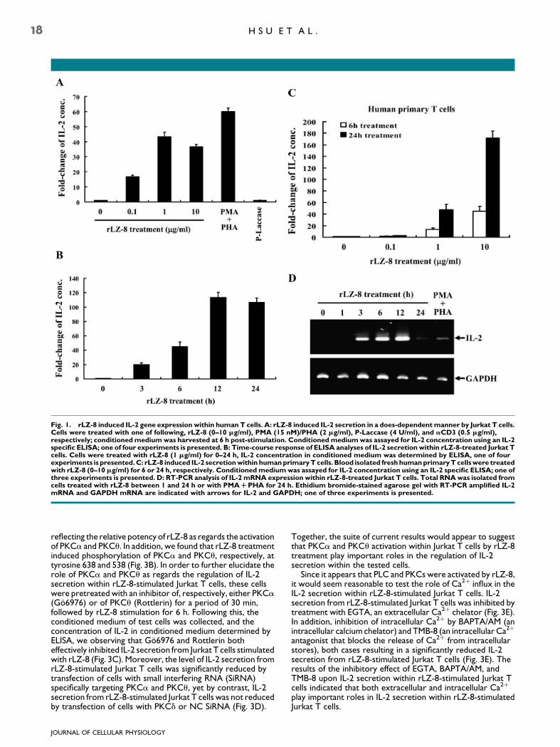
Fig. 1. rLZ-8 induced IL-2 gene expression within human T cells. A: rLZ-8 induced IL-2 secretion in a does-dependentmanner by Jurkat T cells.Cells were treated with one of following, rLZ-8 (0–10 mg/ml), PMA (15 nM)/PHA (2 mg/ml), P-Laccase (4 U/ml), and aCD3 (0.5 mg/ml),respectively; conditionedmediumwas harvested at 6 h post-stimulation. Conditionedmediumwas assayed for IL-2 concentration using an IL-2specific ELISA; one of four experiments is presented. B: Time-course response of ELISA analyses of IL-2 secretion within rLZ-8-treated Jurkat Tcells. Cells were treated with rLZ-8 (1 mg/ml) for 0–24 h, IL-2 concentration in conditioned medium was determined by ELISA, one of fourexperiments is presented.C: rLZ-8 induced IL-2 secretionwithin humanprimaryTcells. Blood isolated freshhumanprimaryTcellswere treatedwith rLZ-8 (0–10 mg/ml) for 6 or 24 h, respectively. Conditionedmediumwas assayed for IL-2 concentration using an IL-2 specific ELISA; one ofthree experiments is presented. D: RT-PCR analysis of IL-2 mRNA expression within rLZ-8-treated Jurkat T cells. Total RNAwas isolated fromcells treated with rLZ-8 between 1 and 24 h or with PMARPHA for 24 h. Ethidium bromide-stained agarose gel with RT-PCR amplified IL-2mRNA and GAPDH mRNA are indicated with arrows for IL-2 and GAPDH; one of three experiments is presented.
18 H S U E T A L .
reflecting the relative potency of rLZ-8 as regards the activationof PKCa and PKCu. In addition, we found that rLZ-8 treatmentinduced phosphorylation of PKCa and PKCu, respectively, attyrosine 638 and 538 (Fig. 3B). In order to further elucidate therole of PKCa and PKCu as regards the regulation of IL-2secretion within rLZ-8-stimulated Jurkat T cells, these cellswere pretreated with an inhibitor of, respectively, either PKCa(Go6976) or of PKCu (Rottlerin) for a period of 30 min,followed by rLZ-8 stimulation for 6 h. Following this, theconditioned medium of test cells was collected, and theconcentration of IL-2 in conditioned medium determined byELISA, we observing that Go6976 and Rottlerin botheffectively inhibited IL-2 secretion from Jurkat T cells stimulatedwith rLZ-8 (Fig. 3C). Moreover, the level of IL-2 secretion fromrLZ-8-stimulated Jurkat T cells was significantly reduced bytransfection of cells with small interfering RNA (SiRNA)specifically targeting PKCa and PKCu, yet by contrast, IL-2secretion from rLZ-8-stimulated Jurkat T cells was not reducedby transfection of cells with PKCd or NC SiRNA (Fig. 3D).
JOURNAL OF CELLULAR PHYSIOLOGY
Together, the suite of current results would appear to suggestthat PKCa and PKCu activation within Jurkat T cells by rLZ-8treatment play important roles in the regulation of IL-2secretion within the tested cells.
Since it appears that PLC and PKCs were activated by rLZ-8,it would seem reasonable to test the role of Ca2þ influx in theIL-2 secretion within rLZ-8-stimulated Jurkat T cells. IL-2secretion from rLZ-8-stimulated Jurkat T cells was inhibited bytreatment with EGTA, an extracellular Ca2þ chelator (Fig. 3E).In addition, inhibition of intracellular Ca2þ by BAPTA/AM (anintracellular calcium chelator) and TMB-8 (an intracellular Ca2þ
antagonist that blocks the release of Ca2þ from intracellularstores), both cases resulting in a significantly reduced IL-2secretion from rLZ-8-stimulated Jurkat T cells (Fig. 3E). Theresults of the inhibitory effect of EGTA, BAPTA/AM, andTMB-8 upon IL-2 secretion within rLZ-8-stimulated Jurkat Tcells indicated that both extracellular and intracellular Ca2þ
play important roles in IL-2 secretion within rLZ-8-stimulatedJurkat T cells.

Fig. 2. Role of Src-family kinases and PLC in the regulation of rLZ-8-induced IL-2 secretion. A: Jurkat T cells were treated with rLZ-8 (1mg/ml)for 0–60 min, cells were lysed and analyzed by Western blot with anti-phosphotyrosine monoclonal antibody (samples 1–6). The results of re-probed the blot with anti-Lck IgG or anti-PLC IgG were shown as samples 7 and 8, respectively. The indicated bars on the left side representmolecular weight (kDa). B: Src-family kinase inhibitors, PP1 and PP2, inhibited IL-2 secretion within rLZ-8-stimulated Jurkat T cells. Cells werepre-incubated, respectively,withPP1 (0.5, 1, or 3mM)orPP2 (0.5, 1, or 3mM) for30min, followedby rLZ-8 (1mg/ml) stimulation for additional 6h.Conditionedmediumwas assayed for IL-2 concentration using IL-2 specific ELISA; one of four experiments is presented. C: Src-family kinases lieupstream of ERK1/2 within rLZ-8-stimulated Jurkat T cells. Cells were pre-incubated, respectively, with PP1 (1 or 3 mM) or PP2 (1 or 3 mM) for30 min, followed by rLZ-8 (1 mg/ml) stimulation for additional 5 min. Phosphorylation of ERK1/2 was analyzed by Western blotting. D: JurkatT cells were pre-incubated with U73122 (2.5, 5, or 10 mM) for 30min, followed by rLZ-8 (1 mg/ml) stimulation for 6 h. Conditioned mediumwasassayed for IL-2 concentration using IL-2 specific ELISA; one of four experiments is presented.
r L Z - 8 I N D U C E S I L - 2 E X P R E S S I O N 19
MAPKs regulate IL-2 secretion withinrLZ-8-stimulated Jurkat T cells
In order to further examine rLZ-8-mediated signal transductionpathways as regards the regulation of IL-2 gene expression,we commenced by testing whether rLZ-8 stimulates thephosphorylation of MAPKs within cultured Jurkat T cells. Thelevel of ERK phosphorylation within Jurkat T cells appeared toincrease at around 5 min post-rLZ-8 stimulation, and extendedto 60 min post-rLZ-8 stimulation (Fig. 4A). In order to explorefor the presence of any additional rLZ-8-mediated signal-transduction molecules and pathways related to IL-2 geneexpression, we further examined whether the phosphorylationlevel of p38, another important stress-related MAPK familymember, increased in level within Jurkat T cells upon rLZ-8stimulation for 0–60 min. In this regard, we noted that the levelof p38 phosphorylation within cultured Jurkat T cells increasedsignificantly from basal levels at between 5 and 30 minsubsequent to rLZ-8 stimulation. Further, 60 min subsequent tosuch stimulation, the p38 phosphorylation level had graduallyreturned to around the basal level (Fig. 4A). In addition, rLZ-8
JOURNAL OF CELLULAR PHYSIOLOGY
treatment of Jurkat T cells also increased the phosphorylationlevel of JNK, another MAPK family member, within JurkatT cells (Fig. 4A).
Next, we examined whether the MAPK-related signalingconstituted one of the main downstream signaling cascades inthe regulation of IL-2 secretion within rLZ-8-stimulated JurkatT cells. Cells were exposed, separately, to one of pharmaceuticsprotein kinase inhibitor: PD98059, SP600125, and SB203580,such exposure inhibiting, respectively, MEK1, JNK1/2, and p38activity, this exposure then being followed by incubation withrLZ-8 for a period of 6 h. The results indicated that PD98059exposure at 50 mM completely inhibited of IL-2 secretion fromrLZ-8-stimulated Jurkat T cells (Fig. 4B), suggesting that theMEK1/ERK1/2 pathway is involved in the process of IL-2secretion within rLZ-8-stimulated Jurkat T cells. SB203580, ap38 inhibitor, was observed to only slightly inhibit IL-2 secretionfrom rLZ-8-stimulated Jurkat T cells, even at the relativelysubstantial concentration of 10 mM (Fig. 4B). Moreover, theJNK inhibitor, SP600125 appeared to exert no real effect uponIL-2 secretion from rLZ-8-stimulated Jurkat T cells at aconcentration of less than 10mM; however, SP600125 revealed

Fig. 3. Role of PKCs andCa2R in the regulation of rLZ-8-induced IL-2 secretion. A: PKCa and PKCumigrated to plasmamembrane upon rLZ-8stimulation. Jurkat T cells were incubatedwith rLZ-8 (1mg/ml) for 5 or 10min, respectively. After incubation,membrane fraction andwhole celllysates were subjected toWestern blotting analysis of PKCa and PKCu, respectively. CD45 acts as a loading control; one of three experiments ispresented. B: rLZ-8 induced phosphorylation of PKCa and PKCu. Jurkat T cells were incubated with rLZ-8 (1 mg/ml) for 0–60 min, thephosphorylation levels of PKCa (T638) and PKCu (T538) were analyzed byWestern blotting; one of three experiments is presented. C: JurkatT cells were pre-incubated with Go6976 (0.5, 1, or 2.5mM) or Rottlerin (1, 3, or 5mM) for 30min, followed by rLZ-8 (1mg/ml) stimulation for 6 h.Conditioned medium was assayed for IL-2 concentration using IL-2 specific ELISA; one of four experiments is presented. D: The level of IL-2secretion from rLZ-8-stimulated Jurkat T cells was significantly reduced by transfection of cells with small interfering RNA (SiRNA) specificallytargetingPKCaorPKCu, but notPKCdornegative control (NC)SiRNA.Theefficiencyof SiRNAwasverifiedbyWesternblotting as shown in thesmall figure. E:RoleofCa2RonrLZ-8-induced IL-2 secretionwithin JurkatTcells. rLZ-8-induced IL-2 secretion fromJurkatTcellswas inhibitedbyEGTA,TMB-8, andBAPTA-AM treatment. JurkatT cellswere pre-incubatedwith EGTA (100, 1,000 and 3,000mM), TMB-8 (25, 50, and 100mM),or BAPTA-AM (1, 3, and 10 mM), respectively, for 30min, followed by rLZ-8 (1 mg/ml) stimulation for 6 h. Conditionedmediumwas assayed forIL-2 concentration using IL-2 specific ELISA; one of four experiments is presented.
JOURNAL OF CELLULAR PHYSIOLOGY
20 H S U E T A L .
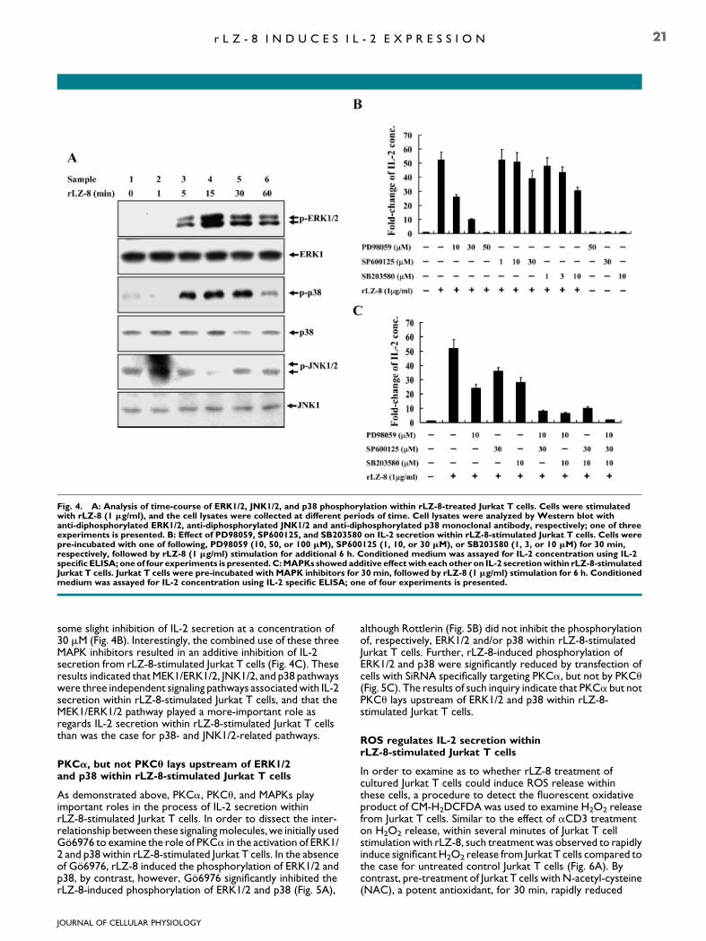
Fig. 4. A: Analysis of time-course of ERK1/2, JNK1/2, and p38 phosphorylation within rLZ-8-treated Jurkat T cells. Cells were stimulatedwith rLZ-8 (1 mg/ml), and the cell lysates were collected at different periods of time. Cell lysates were analyzed by Western blot withanti-diphosphorylated ERK1/2, anti-diphosphorylated JNK1/2 and anti-diphosphorylated p38 monoclonal antibody, respectively; one of threeexperiments is presented. B: Effect of PD98059, SP600125, and SB203580 on IL-2 secretion within rLZ-8-stimulated Jurkat T cells. Cells werepre-incubated with one of following, PD98059 (10, 50, or 100 mM), SP600125 (1, 10, or 30 mM), or SB203580 (1, 3, or 10 mM) for 30 min,respectively, followed by rLZ-8 (1 mg/ml) stimulation for additional 6 h. Conditioned medium was assayed for IL-2 concentration using IL-2specific ELISA; oneof four experiments is presented.C:MAPKs showed additive effectwith eachother on IL-2 secretionwithin rLZ-8-stimulatedJurkat T cells. Jurkat T cells were pre-incubated with MAPK inhibitors for 30 min, followed by rLZ-8 (1 mg/ml) stimulation for 6 h. Conditionedmedium was assayed for IL-2 concentration using IL-2 specific ELISA; one of four experiments is presented.
r L Z - 8 I N D U C E S I L - 2 E X P R E S S I O N 21
some slight inhibition of IL-2 secretion at a concentration of30 mM (Fig. 4B). Interestingly, the combined use of these threeMAPK inhibitors resulted in an additive inhibition of IL-2secretion from rLZ-8-stimulated Jurkat T cells (Fig. 4C). Theseresults indicated that MEK1/ERK1/2, JNK1/2, and p38 pathwayswere three independent signaling pathways associated with IL-2secretion within rLZ-8-stimulated Jurkat T cells, and that theMEK1/ERK1/2 pathway played a more-important role asregards IL-2 secretion within rLZ-8-stimulated Jurkat T cellsthan was the case for p38- and JNK1/2-related pathways.
PKCa, but not PKCu lays upstream of ERK1/2and p38 within rLZ-8-stimulated Jurkat T cells
As demonstrated above, PKCa, PKCu, and MAPKs playimportant roles in the process of IL-2 secretion withinrLZ-8-stimulated Jurkat T cells. In order to dissect the inter-relationship between these signaling molecules, we initially usedGo6976 to examine the role of PKCa in the activation of ERK1/2 and p38 within rLZ-8-stimulated Jurkat T cells. In the absenceof Go6976, rLZ-8 induced the phosphorylation of ERK1/2 andp38, by contrast, however, Go6976 significantly inhibited therLZ-8-induced phosphorylation of ERK1/2 and p38 (Fig. 5A),
JOURNAL OF CELLULAR PHYSIOLOGY
although Rottlerin (Fig. 5B) did not inhibit the phosphorylationof, respectively, ERK1/2 and/or p38 within rLZ-8-stimulatedJurkat T cells. Further, rLZ-8-induced phosphorylation ofERK1/2 and p38 were significantly reduced by transfection ofcells with SiRNA specifically targeting PKCa, but not by PKCu(Fig. 5C). The results of such inquiry indicate that PKCa but notPKCu lays upstream of ERK1/2 and p38 within rLZ-8-stimulated Jurkat T cells.
ROS regulates IL-2 secretion withinrLZ-8-stimulated Jurkat T cells
In order to examine as to whether rLZ-8 treatment ofcultured Jurkat T cells could induce ROS release withinthese cells, a procedure to detect the fluorescent oxidativeproduct of CM-H2DCFDA was used to examine H2O2 releasefrom Jurkat T cells. Similar to the effect of aCD3 treatmenton H2O2 release, within several minutes of Jurkat T cellstimulation with rLZ-8, such treatment was observed to rapidlyinduce significant H2O2 release from Jurkat T cells compared tothe case for untreated control Jurkat T cells (Fig. 6A). Bycontrast, pre-treatment of Jurkat T cells with N-acetyl-cysteine(NAC), a potent antioxidant, for 30 min, rapidly reduced
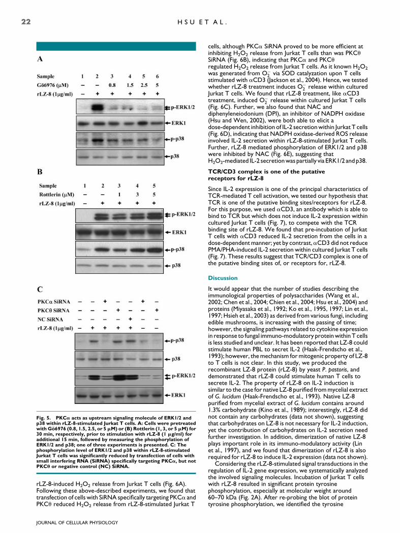
Fig. 5. PKCa acts as upstream signaling molecule of ERK1/2 andp38 within rLZ-8-stimulated Jurkat T cells. A: Cells were pretreatedwith Go6976 (0.8, 1.5, 2.5, or 5mM) or (B) Rottlerin (1, 3, or 5mM) for30 min, respectively, prior to stimulation with rLZ-8 (1 mg/ml) foradditional 15 min, followed by measuring the phosphorylation ofERK1/2 and p38; one of three experiments is presented. C: Thephosphorylation level of ERK1/2 and p38 within rLZ-8-stimulatedJurkat T cells was significantly reduced by transfection of cells withsmall interfering RNA (SiRNA) specifically targeting PKCa, but notPKCu or negative control (NC) SiRNA.
22 H S U E T A L .
rLZ-8-induced H2O2 release from Jurkat T cells (Fig. 6A).Following these above-described experiments, we found thattransfection of cells with SiRNA specifically targeting PKCa andPKCu reduced H2O2 release from rLZ-8-stimulated Jurkat T
JOURNAL OF CELLULAR PHYSIOLOGY
cells, although PKCa SiRNA proved to be more efficient atinhibiting H2O2 release from Jurkat T cells than was PKCuSiRNA (Fig. 6B), indicating that PKCa and PKCuregulated H2O2 release from Jurkat T cells. As it known H2O2
was generated from O�2 via SOD catalyzation upon T cells
stimulated with aCD3 (Jackson et al., 2004). Hence, we testedwhether rLZ-8 treatment induces O�
2 release within culturedJurkat T cells. We found that rLZ-8 treatment, like aCD3treatment, induced O�
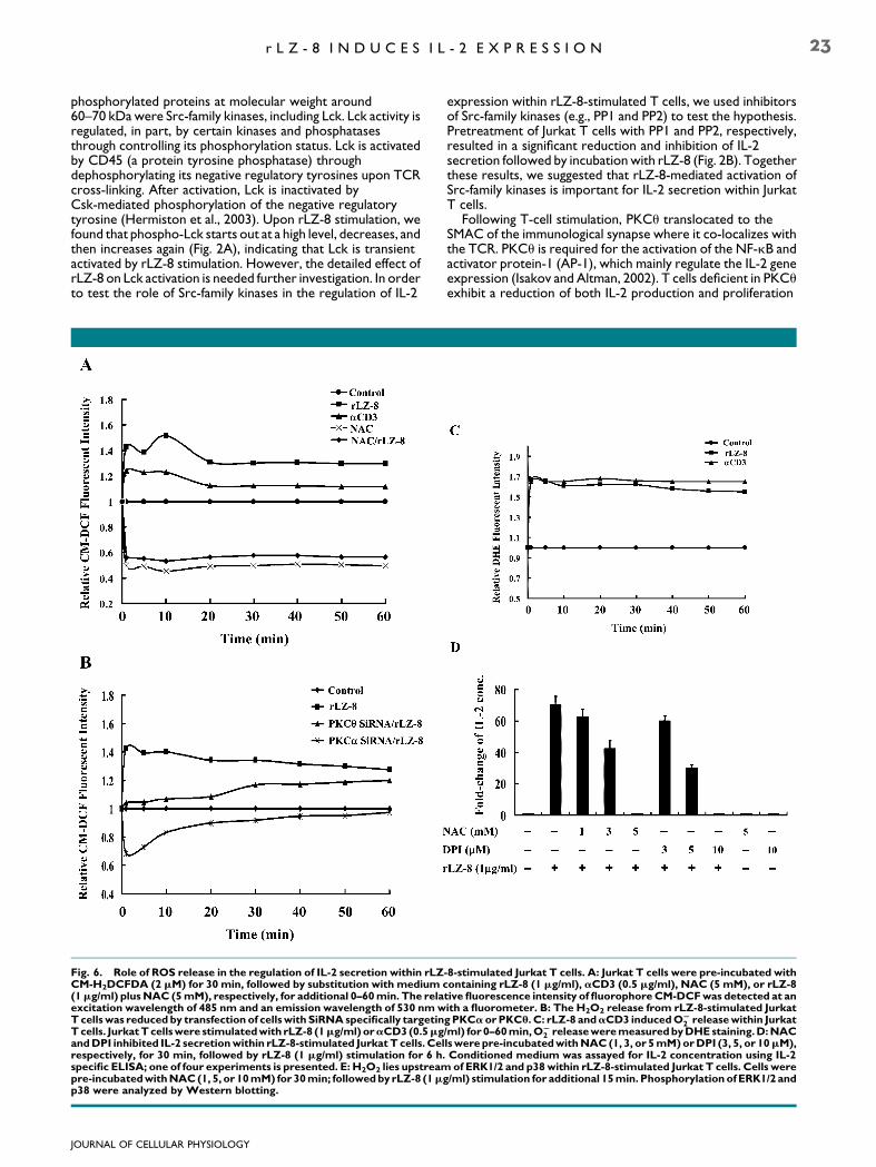
2 release within cultured Jurkat T cells(Fig. 6C). Further, we also found that NAC anddiphenyleneiodonium (DPI), an inhibitor of NADPH oxidase(Hsu and Wen, 2002), were both able to elicit adose-dependent inhibition of IL-2 secretion within Jurkat T cells(Fig. 6D), indicating that NADPH oxidase-derived ROS releaseinvolved IL-2 secretion within rLZ-8-stimulated Jurkat T cells.Further, rLZ-8 mediated phosphorylation of ERK1/2 and p38were inhibited by NAC (Fig. 6E), suggesting thatH2O2-mediated IL-2 secretion was partially via ERK1/2 andp38.
TCR/CD3 complex is one of the putativereceptors for rLZ-8
Since IL-2 expression is one of the principal characteristics ofTCR-mediated T cell activation, we tested our hypothesis thatTCR is one of the putative binding sites/receptors for rLZ-8.For this purpose, we used aCD3, an antibody which is able tobind to TCR but which does not induce IL-2 expression withincultured Jurkat T cells (Fig. 7), to compete with the TCRbinding site of rLZ-8. We found that pre-incubation of JurkatT cells with aCD3 reduced IL-2 secretion from the cells in adose-dependent manner; yet by contrast,aCD3 did not reducePMA/PHA-induced IL-2 secretion within cultured Jurkat T cells(Fig. 7). These results suggest that TCR/CD3 complex is one ofthe putative binding sites of, or receptors for, rLZ-8.
Discussion
It would appear that the number of studies describing theimmunological properties of polysaccharides (Wang et al.,2002; Chen et al., 2004; Chien et al., 2004; Hsu et al., 2004) andproteins (Miyasaka et al., 1992; Ko et al., 1995, 1997; Lin et al.,1997; Hsieh et al., 2003) as derived from various fungi, includingedible mushrooms, is increasing with the passing of time;however, the signaling pathways related to cytokine expressionin response to fungal immuno-modulatory protein within T cellsis less studied and unclear. It has been reported that LZ-8 couldstimulate human PBL to secret IL-2 (Haak-Frendscho et al.,1993); however, the mechanism for mitogenic property of LZ-8to T cells is not clear. In this study, we produced therecombinant LZ-8 protein (rLZ-8) by yeast P. pastoris, anddemonstrated that rLZ-8 could stimulate human T cells tosecrete IL-2. The property of rLZ-8 on IL-2 induction issimilar to the case for native LZ-8 purified from mycelial extractof G. lucidum (Haak-Frendscho et al., 1993). Native LZ-8purified from mycelial extract of G. lucidum contains around1.3% carbohydrate (Kino et al., 1989); interestingly, rLZ-8 didnot contain any carbohydrates (data not shown), suggestingthat carbohydrates on LZ-8 is not necessary for IL-2 induction,yet the contribution of carbohydrates on IL-2 secretion needfurther investigation. In addition, dimerization of native LZ-8plays important role in its immuno-modulatory activity (Linet al., 1997), and we found that dimerization of rLZ-8 is alsorequired for rLZ-8 to induce IL-2 expression (data not shown).
Considering the rLZ-8-stimulated signal transductions in theregulation of IL-2 gene expression, we systematically analyzedthe involved signaling molecules. Incubation of Jurkat T cellswith rLZ-8 resulted in significant protein tyrosinephosphorylation, especially at molecular weight around60–70 kDa (Fig. 2A). After re-probing the blot of proteintyrosine phosphorylation, we identified the tyrosine
r L Z - 8 I N D U C E S I L - 2 E X P R E S S I O N 23
phosphorylated proteins at molecular weight around60–70 kDa were Src-family kinases, including Lck. Lck activity isregulated, in part, by certain kinases and phosphatasesthrough controlling its phosphorylation status. Lck is activatedby CD45 (a protein tyrosine phosphatase) throughdephosphorylating its negative regulatory tyrosines upon TCRcross-linking. After activation, Lck is inactivated byCsk-mediated phosphorylation of the negative regulatorytyrosine (Hermiston et al., 2003). Upon rLZ-8 stimulation, wefound that phospho-Lck starts out at a high level, decreases, andthen increases again (Fig. 2A), indicating that Lck is transientactivated by rLZ-8 stimulation. However, the detailed effect ofrLZ-8 on Lck activation is needed further investigation. In orderto test the role of Src-family kinases in the regulation of IL-2
Fig. 6. Role of ROS release in the regulation of IL-2 secretion within rLZCM-H2DCFDA (2 mM) for 30 min, followed by substitution with medium c(1mg/ml) plusNAC (5mM), respectively, for additional 0–60min. The relaexcitation wavelength of 485 nm and an emission wavelength of 530 nmwTcellswas reduced by transfection of cellswith SiRNAspecifically targetinTcells. JurkatTcellswere stimulatedwith rLZ-8 (1mg/ml) oraCD3(0.5mgandDPI inhibited IL-2 secretionwithin rLZ-8-stimulated JurkatT cells. Celrespectively, for 30 min, followed by rLZ-8 (1 mg/ml) stimulation for 6 h.specific ELISA; one of four experiments is presented. E: H2O2 lies upstreampre-incubatedwithNAC(1, 5, or 10mM) for 30min; followedby rLZ-8 (1mp38 were analyzed by Western blotting.
JOURNAL OF CELLULAR PHYSIOLOGY
expression within rLZ-8-stimulated T cells, we used inhibitorsof Src-family kinases (e.g., PP1 and PP2) to test the hypothesis.Pretreatment of Jurkat T cells with PP1 and PP2, respectively,resulted in a significant reduction and inhibition of IL-2secretion followed by incubation with rLZ-8 (Fig. 2B). Togetherthese results, we suggested that rLZ-8-mediated activation ofSrc-family kinases is important for IL-2 secretion within JurkatT cells.
Following T-cell stimulation, PKCu translocated to theSMAC of the immunological synapse where it co-localizes withthe TCR. PKCu is required for the activation of the NF-kB andactivator protein-1 (AP-1), which mainly regulate the IL-2 geneexpression (Isakov and Altman, 2002). T cells deficient in PKCuexhibit a reduction of both IL-2 production and proliferation
-8-stimulated Jurkat T cells. A: Jurkat T cells were pre-incubated withontaining rLZ-8 (1 mg/ml), aCD3 (0.5 mg/ml), NAC (5 mM), or rLZ-8tive fluorescence intensity of fluorophoreCM-DCFwas detected at anith a fluorometer. B: The H2O2 release from rLZ-8-stimulated JurkatgPKCaorPKCu. C: rLZ-8 andaCD3 inducedOS
2 releasewithin Jurkat/ml) for 0–60min,OS
2 releaseweremeasuredbyDHEstaining.D:NAClswerepre-incubatedwithNAC (1, 3, or 5mM)orDPI (3, 5, or 10mM),Conditioned medium was assayed for IL-2 concentration using IL-2of ERK1/2 and p38within rLZ-8-stimulated Jurkat T cells. Cells were
g/ml) stimulation for additional 15min. Phosphorylation ofERK1/2 and
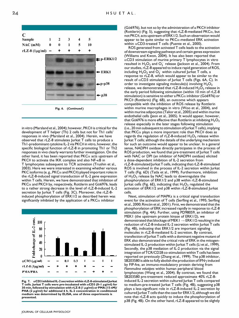
Fig. 6. (Continued )
24 H S U E T A L .
in vitro (Marsland et al., 2004); however, PKCu is critical for thedevelopment of T helper (Th) 2 cells but not for Th1 cells’responses in vivo (Marsland et al., 2004). Herein, we haveobserved that rLZ-8 stimulates Jurkat T cells to produce aTh1-predominant cytokine IL-2 via PKCu in vitro, however, thespecific biological function of rLZ-8 in promoting Th1 or Th2responses in vivo clearly warrants further investigation. On theother hand, it has been reported that PKCa acts upstream ofPKCu to activate the IKK complex and also NF-kB inT lymphocytes subsequent to TCR activation (Trushin et al.,2003). Here we were interested in examining whether variousPKC isoforms (e. g., PKCa and PKCu) played important roles inthe rLZ-8-induced signal transduction of IL-2 gene expressionwithin T cells. Herein, we have demonstrated that inhibition ofPKCa and PKCu by, respectively, Rottlerin and Go6976, leadsto a rather strong decrease in the level of rLZ-8-induced IL-2secretion by Jurkat T cells (Fig. 3C). Moreover, the rLZ-8-induced phosphorylation of ERK1/2 as described herein wassignificantly inhibited by the application of a PKCa inhibitor
Fig. 7. aCD3 inhibited IL-2 secretionwithin rLZ-8-stimulated JurkatT cells. Jurkat T cells were pre-incubated with aCD3 (0–1 mg/ml) for30min, followed by stimulationwith rLZ-8 (1mg/ml) or PMA (15 nM)/PHA (2 mg/ml) for additional 6 h. IL-2 concentration in conditionedmedium was determined by ELISA, one of three experiments ispresented.
JOURNAL OF CELLULAR PHYSIOLOGY
(Go6976), but not so by the administration of a PKCu inhibitor(Rottlerin) (Fig. 5), suggesting that rLZ-8-mediated PKCa, butnot PKCu, acts upstream of ERK1/2. Such an observation wouldappear to be quite similar to PKCa-mediated ERK activationwithin aCD3-treated T cells (Puente et al., 2000).
ROS generated from activated T cells leads to the activationof downstream signaling pathways and certain genes expression(Williams and Kwon, 2004). It has also been reported thataCD3 stimulation of murine primary T lymphocytes in vitroresulted in H2O2 and O�
2 release (Jackson et al., 2004). Fromour studies, rLZ-8 appeared to induce rapid generation of ROS,including H2O2 and O�
2 within cultured Jurkat T cells, aresponse to rLZ-8, which would appear to be similar to theresult of aCD3 stimulation of Jurkat T cells (Figs. 6A, C). Inorder to investigate signaling molecule(s) involving H2O2
release, we demonstrated that rLZ-8-induced H2O2 release inthe early period following stimulation (within 10 min of rLZ-8stimulation) is sensitive to either a PKCa inhibitor (Go6976) orPKCu (Rottlerin) (Fig. 6B), an outcome which appearscompatible with the inhibition of ROS release by Rottlerinwithin murine macrophages in vitro (Woo et al., 2004), andwithin murine adipocytes (Talior et al., 2005) and within murineendothelial cells (Jeon et al., 2005). It would appear, however,that Go6976 is more effective than Rottlerin at inhibiting H2O2
release especially in the later stages following stimulation(i.e., 20 min subsequent to stimulation of Jurkat T cells), implyingthat PKCa plays a more important role than PKCu does asregards the regulation of rLZ-8-induced H2O2 release withinJurkat T cells; although the details of the underlying mechanismfor such an outcome would appear to be unclear. In a generalsense, NADPH oxidase directly participates in the process ofROS production, we found that pre-treatment of Jurkat T cellswith NAC or DPI (an inhibitor of NADPH oxidase) eliciteda dose-dependent inhibition of IL-2 secretion fromrLZ-8-stimulated Jurkat T cells, indicating that rLZ-8-stimulatedROS is involved in the process of IL-2 secretion within JurkatT cells (Fig. 6D) (Tatla et al., 1999). Furthermore, inhibitionof H2O2 release by NAC leads to downregulate thephosphorylation of ERK1/2 and p38 within rLZ-8-stimulatedJurkat cells (Fig. 6E), indicating that H2O2 regulated theactivation of ERK1/2 and p38 within rLZ-8-stimulated Jurkatcells.
Next, stimulation of MAPKs is a critical signal transductionevent for the activation of T cells (Serfling et al., 1995; Serflinget al., 2000; Rincon et al., 2001). First, we demonstrated that thephosphorylation of ERK increased rapidly in response to rLZ-8stimulation (Fig. 4A). Further, using PD98059, an inhibitor ofMEK1 (the upstream protein kinase of ERK1/2), wedemonstrated that blockage of MEK1! ERK1/2 resulting in thereduction of rLZ-8-induced IL-2 secretion within Jurkat T cells(Fig. 4B), indicating that ERK1/2 are important signalingmolecules in rLZ-8-mediated IL-2 secretion. By contrast,transfection of Jurkat T cells with a dominant negative mutant ofERK also demonstrated the critical role of ERK in the mitogen-stimulated IL-2 production within Jurkat T cells (Li et al., 1999).Secondly, the p38 mediation of IL-2 production via the signalintegration of TCR/CD28 co-stimulation within T cells has beenreported on previously (Zhang et al., 1999). The p38 inhibitor,SB203580 is able to fully abolish the production of IFNg inducedby FIP-fve, an immuno-modulatory protein deriving fromFlammulina velutipes within human peripheral bloodlymphocytes (Wang et al., 2004). By contrast, we found thatSB203580 pre-treatment reduced approximate 40% rLZ-8-induced IL-2 secretion within cultured Jurkat T cells comparedto medium-pre-treated Jurkat T cells (Fig. 4B), suggesting p38plays a less-significant role in rLZ-8-induced IL-2 secretion bycultured Jurkat T cells than the case for ERK1/2, although we didnote that rLZ-8 acts quickly to induce the phosphorylation ofp38 (Fig. 4B). On the other hand, rLZ-8 appeared to be slightly
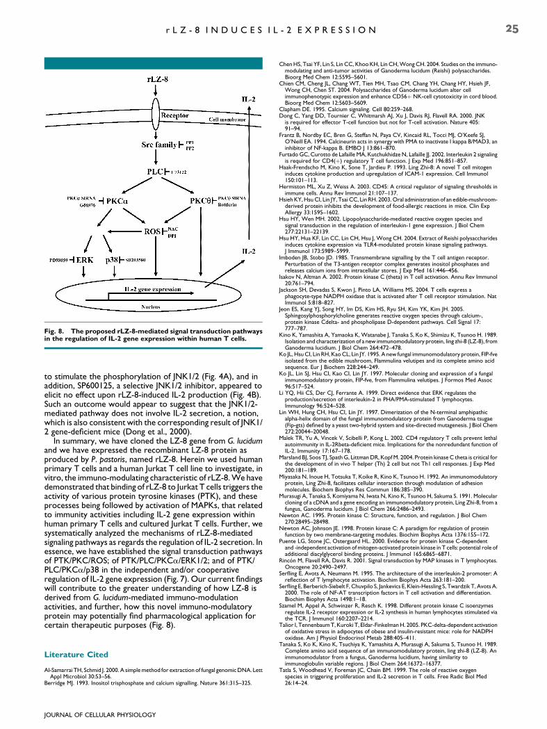
Fig. 8. The proposed rLZ-8-mediated signal transduction pathwaysin the regulation of IL-2 gene expression within human T cells.
r L Z - 8 I N D U C E S I L - 2 E X P R E S S I O N 25
to stimulate the phosphorylation of JNK1/2 (Fig. 4A), and inaddition, SP600125, a selective JNK1/2 inhibitor, appeared toelicit no effect upon rLZ-8-induced IL-2 production (Fig. 4B).Such an outcome would appear to suggest that the JNK1/2-mediated pathway does not involve IL-2 secretion, a notion,which is also consistent with the corresponding result of JNK1/2 gene-deficient mice (Dong et al., 2000).
In summary, we have cloned the LZ-8 gene from G. lucidumand we have expressed the recombinant LZ-8 protein asproduced by P. pastoris, named rLZ-8. Herein we used humanprimary T cells and a human Jurkat T cell line to investigate, invitro, the immuno-modulating characteristic of rLZ-8. We havedemonstrated that binding of rLZ-8 to Jurkat T cells triggers theactivity of various protein tyrosine kinases (PTK), and theseprocesses being followed by activation of MAPKs, that relatedto immunity activities including IL-2 gene expression withinhuman primary T cells and cultured Jurkat T cells. Further, wesystematically analyzed the mechanisms of rLZ-8-mediatedsignaling pathways as regards the regulation of IL-2 secretion. Inessence, we have established the signal transduction pathwaysof PTK/PKC/ROS; of PTK/PLC/PKCa/ERK1/2; and of PTK/PLC/PKCa/p38 in the independent and/or cooperativeregulation of IL-2 gene expression (Fig. 7). Our current findingswill contribute to the greater understanding of how LZ-8 isderived from G. lucidum-mediated immuno-modulationactivities, and further, how this novel immuno-modulatoryprotein may potentially find pharmacological application forcertain therapeutic purposes (Fig. 8).
Literature Cited
Al-Samarrai TH, Schmid J. 2000. A simple method for extraction of fungal genomic DNA. LettAppl Microbiol 30:53–56.
Berridge MJ. 1993. Inositol trisphosphate and calcium signalling. Nature 361:315–325.
JOURNAL OF CELLULAR PHYSIOLOGY
Chen HS, Tsai YF, Lin S, Lin CC, Khoo KH, Lin CH, Wong CH. 2004. Studies on the immuno-modulating and anti-tumor activities of Ganoderma lucidum (Reishi) polysaccharides.Bioorg Med Chem 12:5595–5601.
Chien CM, Cheng JL, Chang WT, Tien MH, Tsao CM, Chang YH, Chang HY, Hsieh JF,Wong CH, Chen ST. 2004. Polysaccharides of Ganoderma lucidum alter cellimmunophenotypic expression and enhance CD56þ NK-cell cytotoxicity in cord blood.Bioorg Med Chem 12:5603–5609.
Clapham DE. 1995. Calcium signaling. Cell 80:259–268.Dong C, Yang DD, Tournier C, Whitmarsh AJ, Xu J, Davis RJ, Flavell RA. 2000. JNK
is required for effector T-cell function but not for T-cell activation. Nature 405:91–94.
Frantz B, Nordby EC, Bren G, Steffan N, Paya CV, Kincaid RL, Tocci MJ, O’Keefe SJ,O’Neill EA. 1994. Calcineurin acts in synergy with PMA to inactivate I kappa B/MAD3, aninhibitor of NF-kappa B. EMBO J 13:861–870.
Furtado GC, Curotto de Lafaille MA, Kutchukhidze N, Lafaille JJ. 2002. Interleukin 2 signalingis required for CD4(þ) regulatory T cell function. J Exp Med 196:851–857.
Haak-Frendscho M, Kino K, Sone T, Jardieu P. 1993. Ling Zhi-8: A novel T cell mitogeninduces cytokine production and upregulation of ICAM-1 expression. Cell Immunol150:101–113.
Hermiston ML, Xu Z, Weiss A. 2003. CD45: A critical regulator of signaling thresholds inimmune cells. Annu Rev Immunol 21:107–137.
Hsieh KY, Hsu CI, Lin JY, Tsai CC, Lin RH. 2003. Oral administration of an edible-mushroom-derived protein inhibits the development of food-allergic reactions in mice. Clin ExpAllergy 33:1595–1602.
Hsu HY, Wen MH. 2002. Lipopolysaccharide-mediated reactive oxygen species andsignal transduction in the regulation of interleukin-1 gene expression. J Biol Chem277:22131–22139.
Hsu HY, Hua KF, Lin CC, Lin CH, Hsu J, Wong CH. 2004. Extract of Reishi polysaccharidesinduces cytokine expression via TLR4-modulated protein kinase signaling pathways.J Immunol 173:5989–5999.
Imboden JB, Stobo JD. 1985. Transmembrane signalling by the T cell antigen receptor.Perturbation of the T3-antigen receptor complex generates inositol phosphates andreleases calcium ions from intracellular stores. J Exp Med 161:446–456.
Isakov N, Altman A. 2002. Protein kinase C (theta) in T cell activation. Annu Rev Immunol20:761–794.
Jackson SH, Devadas S, Kwon J, Pinto LA, Williams MS. 2004. T cells express aphagocyte-type NADPH oxidase that is activated after T cell receptor stimulation. NatImmunol 5:818–827.
Jeon ES, Kang YJ, Song HY, Im DS, Kim HS, Ryu SH, Kim YK, Kim JH. 2005.Sphingosylphosphorylcholine generates reactive oxygen species through calcium-,protein kinase Cdelta- and phospholipase D-dependent pathways. Cell Signal 17:777–787.
Kino K, Yamashita A, Yamaoka K, Watanabe J, Tanaka S, Ko K, Shimizu K, Tsunoo H. 1989.Isolation and characterization of a new immunomodulatory protein, ling zhi-8 (LZ-8), fromGanoderma lucidium. J Biol Chem 264:472–478.
Ko JL, Hsu CI, Lin RH, Kao CL, Lin JY. 1995. A new fungal immunomodulatory protein, FIP-fveisolated from the edible mushroom, Flammulina velutipes and its complete amino acidsequence. Eur J Biochem 228:244–249.
Ko JL, Lin SJ, Hsu CI, Kao CI, Lin JY. 1997. Molecular cloning and expression of a fungalimmunomodulatory protein, FIP-fve, from Flammulina velutipes. J Formos Med Assoc96:517–524.
Li YQ, Hii CS, Der CJ, Ferrante A. 1999. Direct evidence that ERK regulates theproduction/secretion of interleukin-2 in PHA/PMA-stimulated T lymphocytes.Immunology 96:524–528.
Lin WH, Hung CH, Hsu CI, Lin JY. 1997. Dimerization of the N-terminal amphipathicalpha-helix domain of the fungal immunomodulatory protein from Ganoderma tsugae(Fip-gts) defined by a yeast two-hybrid system and site-directed mutagenesis. J Biol Chem272:20044–20048.
Malek TR, Yu A, Vincek V, Scibelli P, Kong L. 2002. CD4 regulatory T cells prevent lethalautoimmunity in IL-2Rbeta-deficient mice. Implications for the nonredundant function ofIL-2. Immunity 17:167–178.
Marsland BJ, Soos TJ, Spath G, Littman DR, Kopf M. 2004. Protein kinase C theta is critical forthe development of in vivo T helper (Th) 2 cell but not Th1 cell responses. J Exp Med200:181–189.
Miyasaka N, Inoue H, Totsuka T, Koike R, Kino K, Tsunoo H. 1992. An immunomodulatoryprotein, Ling Zhi-8, facilitates cellular interaction through modulation of adhesionmolecules. Biochem Biophys Res Commun 186:385–390.
Murasugi A, Tanaka S, Komiyama N, Iwata N, Kino K, Tsunoo H, Sakuma S. 1991. Molecularcloning of a cDNA and a gene encoding an immunomodulatory protein, Ling Zhi-8, from afungus, Ganoderma lucidum. J Biol Chem 266:2486–2493.
Newton AC. 1995. Protein kinase C: Structure, function, and regulation. J Biol Chem270:28495–28498.
Newton AC, Johnson JE. 1998. Protein kinase C: A paradigm for regulation of proteinfunction by two membrane-targeting modules. Biochim Biophys Acta 1376:155–172.
Puente LG, Stone JC, Ostergaard HL. 2000. Evidence for protein kinase C-dependentand -independent activation of mitogen-activated protein kinase in T cells: potential role ofadditional diacylglycerol binding proteins. J Immunol 165:6865–6871.
Rincon M, Flavell RA, Davis R. 2001. Signal transduction by MAP kinases in T lymphocytes.Oncogene 20:2490–2497.
Serfling E, Avots A, Neumann M. 1995. The architecture of the interleukin-2 promoter: Areflection of T lymphocyte activation. Biochim Biophys Acta 263:181–200.
Serfling E, Berberich-Siebelt F, Chuvpilo S, Jankevics E, Klein-Hessling S, Twardzik T, Avots A.2000. The role of NF-AT transcription factors in T cell activation and differentiation.Biochim Biophys Acta 1498:1–18.
Szamel M, Appel A, Schwinzer R, Resch K. 1998. Different protein kinase C isoenzymesregulate IL-2 receptor expression or IL-2 synthesis in human lymphocytes stimulated viathe TCR. J Immunol 160:2207–2214.
Talior I, Tennenbaum T, Kuroki T, Eldar-Finkelman H. 2005. PKC-delta-dependent activationof oxidative stress in adipocytes of obese and insulin-resistant mice: role for NADPHoxidase. Am J Physiol Endocrinol Metab 288:405–411.
Tanaka S, Ko K, Kino K, Tsuchiya K, Yamashita A, Murasugi A, Sakuma S, Tsunoo H. 1989.Complete amino acid sequence of an immunomodulatory protein, ling zhi-8 (LZ-8). Animmunomodulator from a fungus, Ganoderma lucidium, having similarity toimmunoglobulin variable regions. J Biol Chem 264:16372–16377.
Tatla S, Woodhead V, Foreman JC, Chain BM. 1999. The role of reactive oxygenspecies in triggering proliferation and IL-2 secretion in T cells. Free Radic Biol Med26:14–24.
26 H S U E T A L .
Trushin SA, Pennington KN, Carmona EM, Asin S, Savoy DN, Billadeau DD,Paya CV. 2003. Protein kinase Calpha (PKCalpha) acts upstream of PKCtheta toactivate IkappaB kinase and NF-kappaB in T lymphocytes. Mol Cell Biol 23:7068–7081.
Wang YY, Khoo KH, Chen ST, Lin CC, Wong CH, Lin CH. 2002. Studies on theimmuno-modulating and antitumor activities of Ganoderma lucidum (Reishi)polysaccharides: Functional and proteomic analyses of a fucose-containing glycoproteinfraction responsible for the activities. Bioorg Med Chem 10:1057–1062.
Wang PH, Hsu CI, Tang SC, Huang YL, Lin JY, Ko JL. 2004. Fungal immunomodulatoryprotein from Flammulina velutipes induces interferon-gamma production through p38mitogen-activated protein kinase signaling pathway. J Agric Food Chem 52:2721–2725.
JOURNAL OF CELLULAR PHYSIOLOGY
Williams MS, Kwon J. 2004. T cell receptor stimulation, reactive oxygen species, and cellsignaling. Free Radic Biol Med 37:1144–1151.
Woo CH, Lim JH, Kim JH. 2004. Lipopolysaccharide induces matrix metalloproteinase-9expression via a mitochondrial reactive oxygen species-p38 kinase-activator protein-1pathway in Raw 264.7 cells. J Immunol 173:6973–6980.
Yamamori T, Inanami O, Sumimoto H, Akasaki T, Nagahata H, Kuwabara M. 2002.Relationship between p38 mitogen-activated protein kinase and small GTPase Rac for theactivation of NADPH oxidase in bovine neutrophils. Biochem Biophys Res Commun293:1571–1578.
Zhang J, Salojin KV, Gao JX, Cameron MJ, Bergerot I, Delovitch TL. 1999. p38 mitogen-activated protein kinase mediates signal integration of TCR/CD28 costimulation in primarymurine T cells. J Immunol 162:3819–3829.