Embed Size (px)
Citation preview

Regulation of Muscular Contraction
Distribution of Actin Control and
Myosin Control in the Animal Kingdom
WILLIAM LEHMAN and ANDREW G. SZENT-GYORGYI
From the Department of Physiology, Boston University School of Medicine, Boston, Massa- chusetts 02118, the Department of Biology, Brandeis University, Waltham, Massachusetts 02154, and the Marine Biological Laboratory, Woods Hole, Massachusetts 02543
ABSTRACT The control systems regulating muscle contraction in approximately I00 organisms have been categorized. Both myosin control and actin control operate simultaneously in the majority of invertebrates tested. These include insects, chelicerates, most crustaceans, annelids, priapulids, nematodes, and some sipunculids. Single myosin control is present in the muscles of molluscs, brachiopods, echinoderms, echiuroids, and nemertine worms. Single actin control was found in the fast muscles of decapods, in mysidacea, in a single sip- unculid species, and in vertebrate striated muscles. Classification is based on functional tests that include measurements of the calcium dependence of the actomyosin ATPase activity in the presence and the absence of purified rabbit actin and myosin. In addition, isolated thin filaments and myosins were also analyzed. Molluscs lack actin control since troponin is not present in sufficient quantities. Even though the functional tests indicate the complete lack of myosin control in vertebrate striated muscle, it is difficult to exclude unambiguously the in vivo existence of this regulation. Both control systems have been found in animals from phyla which evolved early. We cannot ascribe any simple cor- relation between ATPase activity, muscle structure, and regulatory mechanisms.
I N T R O D U C T I O N
Two distinctly different control systems regulate the activity of various mus- cles. In vertebrate muscles, troponin and tropomyosin are apparently the only regulatory proteins and the control is therefore actinlinked (Ebashi and Endo, 1968; Weber and Murray, 1973). In molluscan muscles, a light chain of myosin acts as a regulatory subunit and the control is therefore myosin- linked (Kendrick-Jones et al., 1970, 1972; Szent-Gy6rgyi et al., 1973). In both types of regulation, contraction is triggered by small amounts of calcium. The resting state is maintained in both because actin and myosin are unable to interact in the absence of calcium, and this occurs by the blocking of sites
T H E JOURNAL OF GENERAL PHYSIOLOGY • VOLUME 66, 1975 • pages I - 3 o i
Dow
nloaded from http://rupress.org/jgp/article-pdf/66/1/1/1245936/1.pdf by guest on 03 D
ecember 2021

T H E J O U R N A L O F G E N E R A L P H Y S I O L O G Y - V O L U M I ~ 6 6 • x 9 7 5
either on actin or on myosin (Eisenberg and Kielley, 1970; Parker et al., 1970; Koretz et al., 1972; Lehman and Szent-Gy6rgyi, 1972; Szent-Gy6rgyi et al., 1973). Despite this overall similarity in function, the interaction between actin and myosin is prevented differently in the two regulatory systems, and the two systems contain different components (Lehman et al., 1972; Kendrick- Jones et al., 1972; Szent-Gy6rgyi et al., 1973). These components cannot be related to each other in any simple fashion, and, since common components are not found, it is very unlikely that one regulatory system could have evolved directly from the other. A comparative study may give insights into the way the two regulatory mechanisms evolved and also explain certain functional differences between various muscles.
In our previous study we presented a preliminary survey involving about two dozen species (Lehman et al., 1972). This initial investigation showed that the myosin control was not restricted to molluscs and was found in a number of invertebrate phyla. The results also led us to suggest that myosin control evolved before actin control, and showed that in a number of muscles both regulatory systems occur simultaneously. Furthermore, we described rapid methods which aided in establishing the presence of the different regulatory systems.
In the present study these observations have been extended to about 100 different animals. We show that myosin-linked regulation is wide-spread; however, the data are no longer consistent with our earlier view that actin control via troponin represents a relatively recent evolutionary development. In fact, we now find that muscles of many species are doubly regulated and contain both types of control, and that muscles having a single regulatory system are restricted mainly to vertebrates, some of the crustaceans and mol- luscs. We also describe in detail the methodology on which this survey is based.
Preparations
The actomyosin or myofibril preparations of all the species reported showed calcium-dependent ATPase activities (Table I). In general, actomyosin prepa- rations have a greater calcium sensitivity than the washed myofibrils, and therefore actomyosin was usually studied in greater detail. Our standard ap- proach was to determine whether a particular muscle contained a myosin- linked regulation or only an actin control by use of the competitive actin ac- tivation assay (Lehman et al., 1972). If a myosin-linked system was found, thin filaments were prepared and assayed to determine whether, in addition, a thin filament-linked system was also present in this muscle. The presence of an actin control was also explored by a competitive myosin-activation test, par- ticularly in cases when thin filaments were not prepared because of small tissue size.
Dow
nloaded from http://rupress.org/jgp/article-pdf/66/1/1/1245936/1.pdf by guest on 03 D
ecember 2021

W LEHMAN AND A. G. SZENT-GY6RGYI Regulation of Muscular Contraction 3
T A B L E I a
ATPASE ASSAYS ON ACTOMYOSIN AND THIN-FILAMENT PREPARATIONS: ANIMALS SHOWING ACTIN CONTROL
Species Common name Muaclea dissected
Calcium sensitivity
ATPage Calcium of thin activity in sensitivity filaments with
0.1 mM with rabbit rabbit CaCls actin* myosin~
Vertehrata Oryctolagus suniadus Mus musa~sH M#socrie#as aar~usn Gallus donu~Lieus Ig.ana igud=a Rams caL~beiana R ~ pipiots N , maus maadosus Carassims ~ t u s A ~ i l a a ~ i l a Raia ¢lamta Eptatr~s ~toutii
Protochordata BravcMostoma florida~ ¶
Arthropoda Crustacea
Mylidacea Mysis mixta Heteromysis formosa
Decapoda Crangon septonspinosus Paluomonass vulgaris Hippolyts ~ostca'cala
Homa'tus americanus Homarus amsricanus
Homarus vulgaris Cambarus sp. Libinia #raarginata Ca~tr irroratus Uca pugnax Uca pugilator
Callinactes sapidus Cardnus mosnas Pagums pollicaris Emsrita talpoida
Sipunculida Dendrostomura pyroides
pmol/mln/mg % %
Rabbit Back 0.5 0 Mome Leg 0.3 0 Hanater Leg 0.15 0 Chicken Pectoral 0 .3 0 Iguana Back 0.34 0 Bullfrog (tadpole) Leg O. 25 <5 Granfrog (adult) Leg 0.85 < I0 Mudpuppy Back 0.1 < 10 Goldfish Dorul O. 38 < 10 Eel Dorlal 0.18 <10 Skate Dorsal 0.2 0 Hagfish Dormal 0.08 < I 0
85 7,5 70 82
43 50
Amphinxus, lancelet Body 0.23 0 80
Opouum shrimp Tall 1.5 0 Opmaum shrimp Tail 0.25 0
Snapping shrimp Tail $.5 0 Prawn Tail 3.2 0 Shrimp Tail 1.5 0 Lobgtec Tail 0.7-1.1 0 Lobster Cutter claw ! . 0 0 Lobmter Fast abdominal O. 7-0.9 0
extenSOr Loblter Tail 1.1 0 Crayfish Tail 0.34 0 Spider crab Carapace 0.7 0 Mud crab Claw, carapace 1.2 0 Black fiddler crab Claw 1.0 0 Calico-back-fiddler Claw 0.45 0
crab Blue crab Claw O. 8 0 Green crab Claw, carapace 2.5 0 Hermit crab Claw 2.4 0 Sand crab Leg 2.3 0
56 96 95
80 84 86 77
Peanut worm Probmcis retractor 0.35 < 10 62
* Calcium senaitlvity (I00 -- (ATPaseEoTA)/(ATPase0at t) X 100) of mixtures containing equal weight* of actomyo- sins and added rabbit actln.
Highest sensitivity obtained, ttsuaily at weight ratim of 0.2-0.3 g thin filament* to l-g rabbit mymin. § In addition, competitive actin-binding a~ayl web's performed on washed myofiin'ila not exposed to high ionic strength solutionm and on actomymin extract* of unwashed muscles. No evidence for mymin control was found. [] Payne, M. R., unpublished data. ¶ After 24-h storage in cold. ** In addition, competitive actin-binding auay was performed on actomyosin extracted directly from unwashed muscle. No evidence for myosin control was found.
Dow
nloaded from http://rupress.org/jgp/article-pdf/66/1/1/1245936/1.pdf by guest on 03 D
ecember 2021
T H E J O U R N A L OF G E N E R A L P H Y S I O L O G Y • V O L U M E 66 i 9 7 5
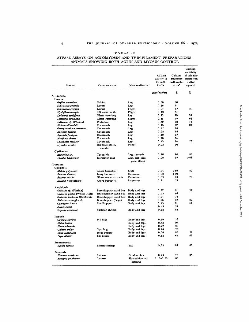
T A B L E ib
A T P A S E A S S A Y S O N A C T O M Y O S I N A N D T H I N - F I L A M E N T P R E P A R A T I O N S :
A N I M A L S S H O W I N G B O T H A C T I N A N D M Y O S I N C O N T R O L
Specie* Common name Muscles directed
CMcium sensitivity
ATPase Calcium of thin ilia- activity in semhivity meats with 0.1 mM with rabbit rabbit CaCh actin* mymin t
Arthropoda Insecta
Gryllus domesLicus Cricket Leg ScMstocsrca grsgarla Locust Leg Schiaoc#rta gr#garia Locust Flight Hyalophora ¢scropia Silkworm moth Flight Lahocsrus sordofanus Giant waterbug Leg Lethoccms eordofanus Giant waterbug Flight Lsthoeerus sp. (Florida) Waterbug Leg Blabsrus discoidalis Cockroach Leg Gromphadorltina portsntosa Cockroach Leg Eublabsr posacus Cockroach Leg Byrsotria fumi&ata Cockroach Leg Naupho#ts dnersa Cockroach Leg Leucophaea maderas Cockroach Leg Dynastss ~rcalss Hercules beetle, Flight
scarabs Chelicerata
EuryzOdma sp. Tarantula Liraulus polyphsmur Horseshoe crab
Leg, thoracic Leg, tail, cara-
pace, flexor Crnstacea
Cirrlpedia MitMla polymsms Goose barnacle Stalk Bdanus eburntus Ivory barnacle Depressor Balanus nubilis Giant acorn barnacle Depressor Balanus tlntinnabulum Acorn barnacle Depreaor
Amphipoda Orcheaia sp. (Florida) Beachhopper, sand flea Body and legs Ordustia grillus (Woods Hole) Bexchhoppe~, sand flea Body and legs OrdurLia trarkiana (California) Beachhopper, sand flea Body and legs Talord~stia longicornir Beachhopper (large) Body and legs Garnraarus locusta Sandhopper Body and legs dassa falcata Caprdla acutifrons Skeleton shrimp Body and legs
Isopoda Cirolana harfordi Pill bug Body and legs Idobm baRica Body and legs Idols eekotrnsis Body and legs Oais~us asdlus Sow bug Body and legs Ligia oeddeatalls Rock runner Body and legs Ligia olfetsii Sea roach Body and legs
Stomatopoda Squilla #mpusa Mantis shrimp Tail
Decapoda tlomarus amoicanus Lobmter Homam$ arasricanus Lobster
Crusher claw Slow abdominal
extensor
mnoUmin/mg % %
0.50 90 0.16 81 0.07 67 80 0.13 51 0.35 90 78 0.35 70 65 O. 30 88 78 O. 16 8 2 80 0.27 88 O. 23 89 0.19 87 0.28 64 O. 35 84 76 O. 25 90
O. I0 84 88 0.08 97 >95
O. 04 > 80 89 0.03 >80 0.09 83 72 0. I1 77
O.22 61 51 O. 23 60 0.24 72 O. 26 82 87 0.25 81 61 0.45 52 O. 32 84
O. 10 79 0 . 4 5 93 O. 22 90 0.14 78 O. 20 86 O. 15 68
0.25
0.25 O. 15-0.25
84
70 85
77 62
68
85
Dow
nloaded from http://rupress.org/jgp/article-pdf/66/1/1/1245936/1.pdf by guest on 03 D
ecember 2021
W. LEHMAN AND A. G. SZENT--GY~RGYI Regulation of Muscular Contraction
T A B L E I b - - - C o n t i n u e d
Species Common name Mmcles dissected
Calcium sensitivity
ATPMe Calcium of thin fda- activity in se~itivity ments with
0.1 mM with rabbit rabbit OaCls actlu* mymin$
#moI/min/mg % %
Annelida Lumbricus terrestris Earth worm Body wall 0 . 0 g 74 60 Nerds drrns Clam worm Body wall 0.12 93 80 Glyc4n, asp. Blood worm Body wall 0 .14 97 60 Eudyslilia polymorpha Featherdumter worm Body wall 0.55 85 67
Sipunculida Golflngia gouldi Acorn worm Proboscis retractor 0 .45 71 45
Priapulida Prlapulus eaudatus Body wall 0.32 > 9 5 70
Nematoda AscNis Isrnbrlcoidas§ Eel worm Longitudinal 0.01 55 67
* Calcium ~ i t i v i t y ( I00 -- (ATPaseEGT A ) / ( A T Pa se oa s+) X I00) of mixturez containing equal weights of acto- myoslm and added rabbit actln.
Highest aemitivity obtained, usually at weight ratios of 0.2--0.3 g thin filaments to 1 g rabbit myosin. § Myofibrils mlubilized in 0.6 M NaCI and 1 mM A T P .
It was difficult to obtain suitable experimental material from many animals. The problems included the small size of the animals, difficulty of isolating the muscles free from surrounding tissues, contamination with proteolytic enzymes, extraction of ATPases other than actomyosin, and resistance to homogeniza- tion. Of these problems, proteolysis was the most troublesome, and special care was taken to avoid or reduce the exposure of the muscle to intestinal con- tents. In some cases, proteolytic degradation was reduced by 10 -4 M phenyl- methylsulfonyl fluoride. Despite these precautions we were unable to obtain calcium-sensitive actomyosins from the sponge, Porifera sp., the jellyfish, Mnemiopsis leidyi, the sea anemones, Metridium senile and Haloclava producta, the acanthocephalid, Moniliformis dubius, the turbeUarian, Bdelloura candida, the planarian, Phagocata gracilis, a number of echinoderms, such as the starfish, Asterias forbsii, sea urchins, Arbacia punctulata, and Strongylocentrotus droebachien- s/s, the acorn worm, Saccoglossus kowalevskyi, and the tunicate, Ciona intestinalis.
WASHED MUSCLES Muscles were cut with scissors into 3- to 5-mm pieces and homogenized in a Sorvall Omnimixer (Dupont Instruments, Sorvall Operations, Newtown, Conn.) for 5-50 s in a solution containing 40 m M NaC1, 5 mM phosphate buffer (pH 7.0), 1 mM MgC12, and centrifuged and resus- pended several times with the same solution.
Whenever possible, the muscles were dissected from surrounding tissues (of. Table I). Dissection, however, was cumbersome in other instances. The
Dow
nloaded from http://rupress.org/jgp/article-pdf/66/1/1/1245936/1.pdf by guest on 03 D
ecember 2021
T H E J O U R N A L O F G E N E R A L P H Y S I O L O G Y • V O L U M E 6 6 • x 9 7 5
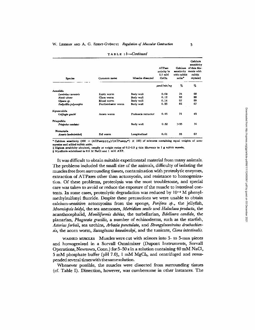
T A B L E I C
A T P A S E A S S A Y S O N A C T O M Y O S I N A N D T H I N - F I L A M E N T P R E P A R A T I O N S :
A N I M A L S S H O W I N G M Y O S I N C O N T R O L
Species Common name Muscles dissected
Calcium semitivity
ATPate Calcium of thin fila- activi W in seaaitlvity merits with
0.1 m M with rabbit rabbit CaCh actin* myolin:~
Echiuoderma Thyone brlareus Sea cucumber Lantern retractor Cucumariafrondosa Sea cucumber Lantern retractor
Mollusca Amphineura
Cryptochiton stelleri Sea bread Mant le
Gastropoda Acmea testudinalis Plate l impet Foot Pdinices duplieatus Shark eye Foot Lunatia heros Moon shell Foot Thais lapillus Dogwinkle Foot Busycon canaliculatum Whelk Foot
Pelecypoda Solemya velum Awning clam Foot Iroldia limatula File yoldia Adductor Aequipecten irradians Bay scallop Striated adductor AequipecUn irradians Bay scallop Smooth adductor Placapecten raagellardcus Deep sea scallop Striated adductor Placopecten magdlanicus Deep sea scallop Smooth adductor Pecten maxlmus Scallop Striated adductor Pecten maximue Scallop Smooth adductor Mytilus edulis Blue mussel Adductor, bymms re-
tractor Modiolus demissus Ribbed mussel Adductor Crassostrea ~irglniea Oyster T randucen t adductor Crassostrea drginica Oyster O p a q u e adductor Ensis direaus Razor clam Foot Ensis directus Razor clam Adductor Mya arenaria Soft shell clam Adductor Mercenaria mercenaria Quahog Pink adductor Mercenaria rasrcenaria Quahog White adductor Spisula solidissima Surf clam Adductor Anadara ovalis Bloodark Tramlucen t adductor Anadara ovalis Bloodark Opaque adductor Astarte cantanea Smooth aJtarte Adductor Laevieardium mortoni Egg cockle Foot ga¢oma tenla Teota macoma Foot Anomia simplex J ingle shell Adductor
Cephalopoda Leligo padd Squid Ventral pharynx re-
tractor Echiuroida
Urechis taupe Inn-keeper 's worm Body or sailor's penis
Braehiopoda Glottldia pyramidata Lampshell Pedunculus
Nemer t ina Cerebratulus lacteus Ribbon worm Ora l reglous of body Lineus longissimus Boodace worm Ora l regions of body
#md/m~/m& % %
0.025 90 0 0.028 59 0
0 .04 70 0
O. 15 76 O. 15 > 9 0 0 .07 91 0 0 .07 > 9 0
0 .14 > 9 0 0 ,27 > 9 0 0 . 4 8 97 0 O. 13 > 9 5 0 1 .2 97 0 0 .20 > 9 5 0 0 . 4 98 0 O. 12 > 9 5 0 0 .06 75 0
0 ,05 81 0 O. 35 77 0 .04 > 8 0 0 .20 >9O 0 0 .27 > 9 0 O. 13 88 O. 28 93 0 0 .05 > 9 0 O. 20 89 0 . 5 >95 0 0 .09 >90 0 .09 82 O. 2 92 0 0 .14 > 9 0 O. 19 84
O. 35 97 0
0 .09 76 0
0 .25 90 0
0 .15 75 0 0 .06 62 0
Dow
nloaded from http://rupress.org/jgp/article-pdf/66/1/1/1245936/1.pdf by guest on 03 D
ecember 2021

W. LEHMAN AND A. G. SZENT-GY6RGYI Regulation of Musadar Contraction
entire legs of the insects, tarantula and sandcrabs, were homogenized, and the cuticle or exoskeleton was removed by filtration through a single layer of gauze. The thoracic and abdominal regions of amphipods and isopods were isolated by removing the head and anal regions with the attached gonads and other internal organs under a dissecting microscope. Care was taken to avoid spilling of the intestinal contents into the thoracic and abdominal cavi- ties. The preparation was blended and the exoskeleton was removed by filtra- tion through a single layer of gauze. The body wall muscles of the annelids, Ureehis, Priapulus, and of the nemertine worms were obtained by cutting the body open and removing the internal organs. The body wall was rinsed and homogenized. Dissection was restricted to the anterior portion of the body, i.e. the region not containing the guts, in the nemertine worms and in Eudistylia. Special care was exercised not to disrupt the yellow soft tissues of Balanus nubilis during dissection since exposure of the muscles to their content led to a loss of myosin control. The sea cucumbers were anaesthetized in seawater con- taining 0.1% chloretone before the dissection of the lantern muscles.
ACTO~rVOSm Washed myofibrils were extracted with 0.6 M NaC1, 5 mM phosphate buffer (pH 7.0), and 1-2 mM ATP (pH 7.0). The insoluble ma- terial was removed by a short centrifugation of 10 min X 30,000 g, and the supernatant was tested for ATPase activity. In many cases, the actomyosin was also precipitated by reducing the ionic strength to 0.05 by dilution or by dialysis. The actomyosin precipitate was then washed with 40 mM NaC1 and 10 mM phosphate buffer (pH 7.0). The actomyosin preparations frequently contained significant amounts of paramyosin. Paramyosin contamination was reduced in some instances by extracting actomyosin at pH 6.0 with 0.4 M NaC1 (Szent-Gytrgyi et al., 1971). These preparations were tested immedi- ately, with the exception of actomyosin from Amphioxus which required over- night storage to show calcium sensitivity.
THIN mT.aMENTS Preparation essentially followed previous procedures (Szent-Gytrgyi et al., 1971 ; Kendrick-Jones et al., 1970; Lehman and Szent- Gytrgyi, 1972). 0.1 mM EDTA and 5 mM ATP (pH 6.0) was added to washed muscle preparations suspended in 40 mM NaC1, 1-5 mM MgCI~, 5 mM phos- phate buffer pH 6.0. The washed muscle was rehomogenized for a few seconds in a Sorvall Omnimixer. The suspension was centrifuged at 40,000-80,000 g for 30 min. Thin filaments were collected from the supernatant by a 2- to 3- h centrifugation at 80,000-100,000 g. The pellet was rinsed with 40 mM NaC1, 1 mM MgC12,5 mM phosphate buffer (pH 7.0) and resuspended with the aid of a Teflon-coated hand homogenizer. The thin-filament preparations were clarified by centrifugation at 30,000 g for 10 min. No attempt was made to further purify the thin filaments since we wished to retain all of the compo- nents of the thin filaments even if some additional impurities were not removed. The myosin or paramyosin impurities in the thin-filament preparations, how-
Dow
nloaded from http://rupress.org/jgp/article-pdf/66/1/1/1245936/1.pdf by guest on 03 D
ecember 2021

T H E J O U R N A L O F G E N E R A L P H Y S I O L O G Y • V O L U M E 66 • I 9 7 5
ever, were negligible. The preparations had no ATPase activity, and contained little or no material with chain weight greater than 80,000 daltons (Fig. 5). Calcium-sensitive Amphioxus thin filaments were prepared from actomyosin, precipitated at low ionic strength, since the preparations from muscle were not calcium sensitive.
MYOSIN A rapid procedure was used for myosin preparation since in many cases myosin from invertebrate muscles proved to be highly labile and lost ATPase activity quickly. To a reprecipitated actomyosin solution, 10 m M Mg-ATP (pH 7.0) was added, and then it was immediately centrifuged at 165,000-250,000 g for 3-4 h (cf. Weber, 1956). The upper half of the super- natant solution was dialyzed against 20 vol of 5 m M phosphate pH 6.5 for 3-5 tl. The myosin was then diluted with an equal volume of 5 m M phosphate solution, collected by centrifugation and resuspended and washed once in 40 m M NaCI, 5 m M pH 7.0 phosphate buffer and tested immediately for ATPase activity. This procedure removed most of the actin and the ATPase activity of the myosin preparations was activated 4- to 10-fold on addition of rabbit actin (Table I I I ; Figs. 3 and 4). No at tempt was made to remove the paramyo- sin impurity. Whereas this procedure yielded active myosin preparations from a number of invertebrate muscles, in many cases activity diminished signifi- cantly after a 1-day storage. Unfortunately, we have been unable to obtain active myosin from a number of insects, gastropods, and polychaete muscles, even though the actomyosin preparations from the same muscles were active and calcium sensitive for several days.
Purified calcium-sensitive myosin was prepared from Lirnulus muscle with a slight modification. Washed myofibrils were resuspended in a solution con- sisting of 0.6 M NaC1, 5 m M phosphate buffer (pH 7.0), 1 m M MgATP (pH 7.0) and sedimented for 4 h at 200,000 g. The top half of the super- natant was collected and dialyzed overnight against 40 mM NaCI, 1 m M MgCI~, 5 m M phosphate buffer (pH 7.0), and then diluted twofold with this solution. The precipitate was collected by centrifugation, dissolved in 0.6 M NaC1, 1 m M MgC12, 5 m M phosphate buffer (pH 7.0) and recentrifuged at 100,000 g for 3 h. The top half of the supernatant contained myosin and some paramyosin.
SCALLOP CALCIUM-BINDING PROTEIN A calcium-binding protein was ob- tained from scallop striated muscle. The initial low ionic strength extract of muscles containing soluble proteins was lyophilized and redissolved in ~ 0 vol of water. This solution was centrifuged for 2-3 h at 200,000 g. The supernatant was dialyzed against 40 m M NaC1, 5 m M phosphate buffer (pH 7.0), then brought to pH 4.3 by a dropwise addition of 0.5 M HC1 and the precipitated protein was removed by centrifugation. The supernatant was neutralized,
Dow
nloaded from http://rupress.org/jgp/article-pdf/66/1/1/1245936/1.pdf by guest on 03 D
ecember 2021

W. LEHMAN AND A. G. SZENT-GY6ROYI Regulation of Muscular Contraction 9
lyophilized, and then redissolved in a small volume of water and chromato- graphed on a Sephadex G-100 column (2.6 X 80 cm). A calcium-binding pro- tein comprised the last peak and showed only trace impurities on SDS acryla- mide gel electrophoresis (Fig. 6).
SOURCES OF MATERIAL Animals were obtained from the following sources: Marine Biological Laboratory, Woods Hole, Mass.; Gulf Specimen Corporation, Panacea, Fla.; Pacific Biomarine, Venice, Calif.; Peninsula Bio- logicals, Sand City, Calif.; Sheepscot Supply, West Southport, Me. ; Millport Marine Station, Scotland; Southwestern Supply Co., Tucson, Ariz.; Con- necticut Valley Biological Supply Co., Inc., Southampton, Mass., and various bait and pet shops. Cockroaches were given by Dr. L. Roth of the Natick Army Laboratories, Natick, Mass. Ascaris lumbri¢oides was a gift of Dr. D. Fairbairn, ZoolOgy Department , University of Massachusetts, Amherst, Mass. Lethocerus cordofanus was a gift of Mr. Richard Tregear, Depar tment of Zoology, Oxford University.
M E T H O D S
The Mg-activated actomyosin ATPase was measured in a pH stat at pH 7.5, 25 ° as previously described (Szent-Gy6rgyi et al., 1971). The assay solution consisted of 0.7 mM ATP, 1 mM MgC12, and 20-40 mM NaCI. Calcium sensitivity was measured by comparing the ATPase rates in the presence of 0.1 mM EGTA before and after the addition of 0.2 mM CaCh.
Isolated thin filaments were mixed with rabbit myosin in ratios of 0.3-0.5:1 (wt/wt) in 0.6 M NaCI and then diluted for the ATPase assays. When sufficient amounts of thin filaments were available, they were tested at several different weight ratios. Myo- sin preparations were assayed alone and mixed with rabbit aetin (2 : 1 wt/wt) in high salt, and the specific activities and calcium sensitivities were compared.
The calcium binding of muscle protein suspensions was determined as previously described (Kendrick-Jones et al., 1970), using a double-labeling technique. In this technique calcium binding is measured on sedimented protein. Correction is made for the void volume with the aid of a second label, [3H]glucose, which is not bound by muscle proteins. The proteins were washed twice with 4.0-8.0 ml [45Ca]EGTA buffer, containing labeled glucose, to ensure that the free calcium concentration was not significantly altered by the binding on the protein. Calcium binding of the scallop calcium-binding protein was measured by equilibrium dialysis. The protein was equilibrated twice for 24 h against 50-100 vol of 40 mM NaC1, 1 mM MgCI, 10 mM imidazole-HCl pH 7.0, containing 25 #M [45Ca]EGTA buffers. The dissociation con- stant of the ealeium-EGTA was taken as 1.9 >( 10 -7 M at pH 7.0 (Chaberek and Martell, 1959).
Protein concentrations were measured by the method of Lowry et al. (1951) stand- ardized by Kjeldahl nitrogen determinations of bovine serum albumin. Sodium dodecyl sulphate (SDS) polyacrylamide gel electrophoresis was performed using Coomassie Blue as a stain according to Weber and Osborn (1969).
Dow
nloaded from http://rupress.org/jgp/article-pdf/66/1/1/1245936/1.pdf by guest on 03 D
ecember 2021

I O T H E J O U R N A L O F G E N E R A L P H Y S I O L O G Y • V O L U M E 66 • i 9 7 5
R E S U L T S
Distribution of Regulatory Systems
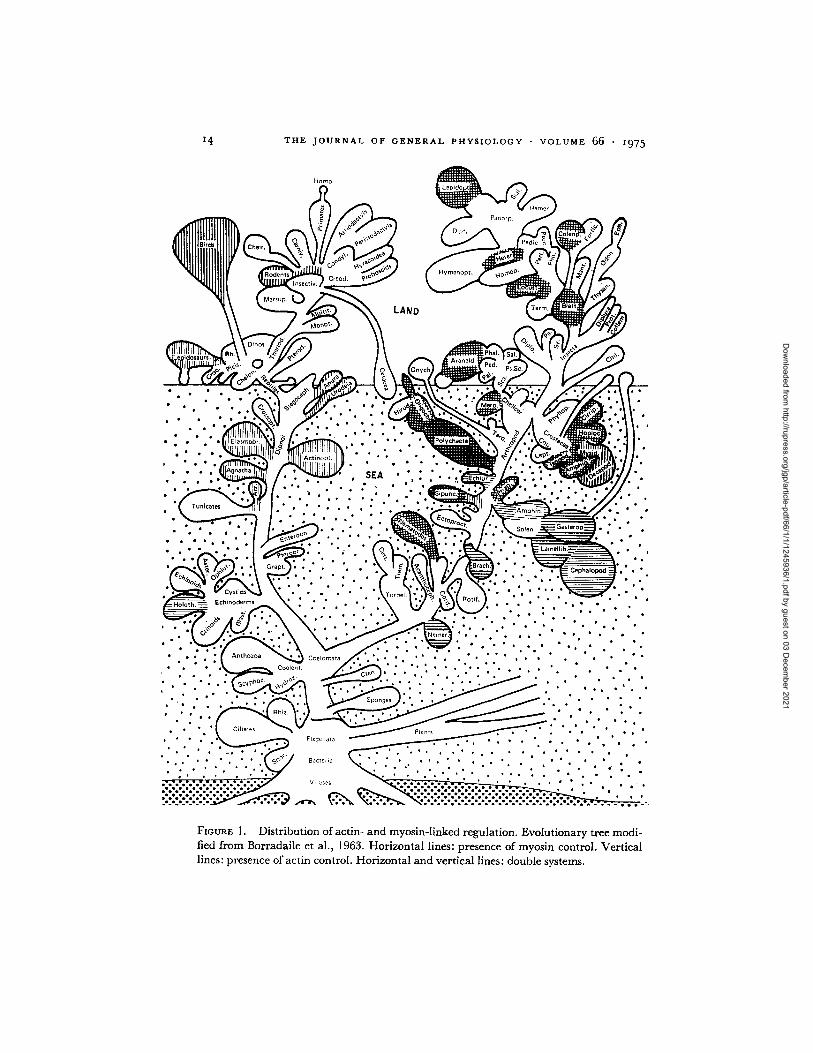
The control systems regulating the contraction of the muscles of approximately 100 species have been categorized (Table II ; Fig. 1). The single actin-linked regulation operates in the muscles of all the chordates tested (13 species), in- eluding the cephaloehordate, Amphioxus, which represents an early example of chordate evolution. Among invertebrates, the single actin control is found in most decapod muscles (14 species) and mysidacea (2 species). In addition, Dendrostomum pyroides, one of the three sipunculids tested, showed only an actin- linked regulation, although Dendrostomum myosin binds calcium (Table III).
The single myosin-linked regulation operates in all of the molluscs tested (23 species), in the two echinoderms, in the two nemertine worms, and in the single examples of echiuroids and brachiopods studied. Both the myosin- linked and the actin-linked regulations function together in the rest of the animals examined. Double regulation was demonstrated in all of the insects tested (12 species), in the two chelicerates (Limulus and Eurypelma), in the cir- ripeds (4 species), isopods (6 species), amphipods (7 species), and stomatopods (1 species). The slow crusher claw muscles and the slow superficial abdominal extensor muscles of the lobster also have both controls operating. Double regu- lation was found in all the annelids tested (four species), and in Golfingia gouldi, one of the three sipunculid worms examined. Myosin control is present also in Phascolosoma agassizi. The only nematode studied, Ascaris, has both regulations functioning.
Experimental Basis for Classification of Control Systems
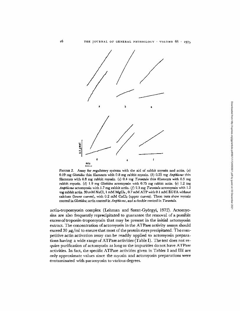
COMPETITIVE ACTIN-ACTIVATION ASSAY. This assay probes for the myosin control in actomyosin and in washed myofibrils by measuring the effect of excess pure rabbit actin on the ATPase activity in the absence of calcium (Lehman et al., 1972). If a myosin control operates, the myosin is unable to combine with pure actin, and the ATPase activity remains low until calcium is introduced (Fig. 2 d and / ) . In contrast, pure actin activates those prepara- tions that have an actin control, even in the absence of calcium. This activa- tion results from the ability of myosin to combine with pure actin in these sys- tems, a combination which is not influenced by the troponin- tropomyosin- containing thin filaments present in the preparation (Fig. 2 e).
Full activation of the ATPase by excess pure actin in the absence of calcium, i.e. the loss of calcium sensitivity, demonstrates that a particular muscle con- tains solely an actin-linked system and that myosin control is not functioning. The interpretation is straightforward and the identification is unambiguous. The lack of activation by pure actin demonstrates that the system contains a
Dow
nloaded from http://rupress.org/jgp/article-pdf/66/1/1/1245936/1.pdf by guest on 03 D
ecember 2021

W . LEHMAN AND A . G . SZENT-GY(JRGY| ReguZation of Muscular Contraction t I
myosin-linked regulatory system. This result, however, does not exclude the additional presence of an actin-linked regulation, and the competitive actin activation assay needs to be complemented by tests probing for actin control (cf. Fig. 2 a and c).
T A B L E I I
R E G U L A T I O N I N D I F F E R E N T A N I M A L S
Competi- Competltlve tive Ac My activa-
Ca ++ sensitivity activation tion test Ca ++ binding Troponin test (pros- (presence
on TF My + TF + ence of My of Ac Type oi Species My "IT SDS gels rabbit Ac rabbit My control) control) regulation
Vertebrata Or.jetolJgus curdadus Mus muSadus* M#sooicttuS am'ares* Gallus d~sticus$ lluana i&mma Rana ¢at#s&n'ana (tadpole) Rana ~pi.ns N¢¢turas maculosus Carasslgs aufalus An~ila anguila Raia davata Eptatr#tus stoutii
Protochordata Branchiostoma floridae
Echinoderma Tl~o~ briartus Cucumada frondosa
Arthropoda Inaecta
@rfllus domtstieus Shlstoc~rca grcgaria
leg flight
Hyalophora ¢~eropia (flight) Ltthoets~s ¢ordofaraw (flight)
leg Leclu~vus :p. (Florida) (leg) Blabous diseoidalis (leg) Gromphador~na portcntosa (leg) Eubla&r postims (leg) Byr$otria fumigata (leg) Naupho¢t¢ dner¢a Leucophaea maderae Dynastes hercul*s
Chelicerata Limulus polyphamus Eurypelma sp.
Crustacea Cirripedia
Mitella polymcrus Balanus eburneus Balanus nubilis Balanus tintlnnabulum
m
i
+ + - + + + + + + + + + - +
@ - + + + - +
+
+ + + +
+
+
+
+ ¶ + ¶ +
+ + + + + + + +
+
+ + +** +
m
m
m
+ +
+ +
+ +
+ + + + + + + + +
+ + + + + + + + + +
+
+
+
+
+ + + +
+ i
+
+ +
+
A ¢
A¢ Ac Ac Ac Ac Ac Ac Ac Ac Ac Ac
Ac
My My
My -}- AC My + (?)ll
My + Ac My + (?)~ My + Ac My + Ac My + Ac My + Ac My + (?)~ My + Ac My + t~)[[ ]My + (?) My + Ac My + Ac
My + Ac My + Ac
My + Ac My + Ac My + Ac My + Ac
Dow
nloaded from http://rupress.org/jgp/article-pdf/66/1/1/1245936/1.pdf by guest on 03 D
ecember 2021

I 2 T H E J O U R N A L OF G E N E R A L P H Y S I O L O G Y " V O L U M E 66 " I 9 7 5
T A B L E I I - - C o n t i n u e d
Species
Competi- Competitive rive Ae My aetiva-
Ca ++ semifivity activation tion test Ca ++ binding Troponin test (pres- (presence
onTF My + TF + eneeofMy of Ac My TF SDS gelg rabbit A rabbit My control) control)
Type of regulation
Amphipoda Orcb*stia sp. (Florida) Ordurtla £rillus (Woodt Hole) Orchestia trasldana (California) Talor¢t~rtla loagicornis Gammarus Iocusta Jassa falcata Caprdla acutifrons
+ + + My + My + + My
+ + + My + + + + My
+ My + + My
+ Ac + (?)~ + A¢ + Ac + Ac + Cr)ll + Ac
llopoda Cirdana ~rfordi Idoua balaza [dotea od~tensis Oni~us asdlur Ligla ocddratalis Li&ia olfirsii
Stomatopoda Squilla era#usa
+ +
+ +
+ + +
+ + My + Ac + + My + Ac + + My + Ae + + My + Ac + My + Ae +- My + Ac
+ My + Ac
Mysidacea Mysis mixta H#teromysis formosa
Decapoda Crangon septcmspinosus Palaeomonttss oulgaris H~olyt# ~oxtsricola Homarus am~ieanus (tail)
cntterelaw, fast abdominal ~tcl~or
crmher claw slow abdominal extensor
Homarus vulgarls Uambarus sp. Liblnia cmar&inata Cancer irro~tus U¢a lm~a# Uca pugitlaor CalllneaG¢ sapldus Cardrms moenas Pagurus polliewds Em~rlta talpddea
Annelida Lumbricus terresUis Nerds virens Glvc4rra sp. Eudystilia polymorpha
Molltmea Amphineura
Cryptochlton stdleri
Ac Ac
- - A c
- - A e
- - A c
+ * * - - + * * - - A c
- - Ae
+ + + + My + + My
Ac - - Ae - - A ¢
+ + -- Ac + + -- Ac + + -- Ac + + -- Ac + + -- Ac + + -- Ac + + -- Ac
-{- Ae + Ac
+ -- My
+ + + + + + + + + + + + +
-~- My + Ac + My + Ac + My + A¢ + My + Ac
Dow
nloaded from http://rupress.org/jgp/article-pdf/66/1/1/1245936/1.pdf by guest on 03 D
ecember 2021

W . L E H M A N A N D A . G . S Z E N T - G Y o R G Y I Regulation of Muscular Contraction 1 3
T A B L v. I i ~ C o n t i n u e d
Spec ies
Compet- Competitive itive Ae My aetiva-
Ca ++ sensitivity activation tlon test Ca ++ binding Troponin .test (pres- (presence
on TiC M y + T F + ¢ n c e o f M y of Ac My T I c SDS gel- rabbit Ac rabbit My control) control)
Type of regulation
Gastropoda Acmsa tesadanalis Polinic¢s da#licatus Lunaaa hsros Tlmit lapillus Busycon ¢analiculatum
Pelecypoda Solttr~a mlum Yoldia limatMa A#quip~ttn irmdia~ Placopeaen mqdlanicu,* Pectin maalmus Mytilus ¢dMis Modiola~ demissas Crassos~'m vlrglnica Ensis direaus Mya arsnaria Moc*naria ~ n a r i a $[dsmda solidlssima Anadara omlis Ast~,'~ mnLanea Laeoimrdium mortoni Maeoma to~a Anomia simplex
Cephalopoda Ldi&o p#ahi
Brachinpoda Glotddia pyramidata
Echiuroida Ureahis caupo
Sipuaculida Gol fingia gouldi Phascolosoma agassizi Dcndrostomum pyroldes
Priapulida Priapulus caudatus
Nematoda Ascaris lumbdcoidcs
Nemert ina Ctrebra~ulus lacUus Lintus lonsissimus
+ ~ My + -- My
+ ? + -- + -- My + - My
+ ? -- + -- My
+ I t - - + +QG - - + + H - - +
- +
+
+ I t - - +
B
m
B
m
+ ~ My + ~ My + ~ My + ~ My + - - My + M y + My + My + - - My + -- My + My -'F- -- My + My + - My + -- M y -'{- - My + -- My
+ - - + - -9 M y
+ -- -- + -- + My
+ ? + -- -1- My
--1.- -.[- -.[- + + My + My
+ + - + - Ac
q-- -{- -}'- + + My
+ + + + My
? -- + -- My - - + - - My
+ Ac + (?)§
+ Ac
+ Ac
* Payne, M., unpublfi~hed data. $ Ohmuki et aL (1971) and Hitchcock et al. (1973), isolated troponln from chicken muaclm. | Auayed on 2-day old actomymin preparat ion having a reduced ATPa te activity. I] Presence of actin control not totted. ¶ Lehman et al. (1974). ** Based on the finding of troponin in scarabs by Bullard et al. (1973). Baaed on the finding of troponin in lobster by Regenstcin and Szent-Gyorgyl (1975). ~$ Kendrlck-Jones et aL (1970). §§ Szent-Gyorgyi et aL (1973). Troponin-C has been prepared from the hake, M s r l ~ a s msdt~dus, the lizard, Saranus exanthemicas, and the python, Pytho sebae, by Demaille et al., 1974.
Dow
nloaded from http://rupress.org/jgp/article-pdf/66/1/1/1245936/1.pdf by guest on 03 D
ecember 2021
14 T H E J O U R N A L O F G E N E R A L P H Y S I O L O G Y • V O L U M E 66 • i975
H a m o
Ponorp.
' (
LAND
• , , • • .,(.~
"... ~
"Z • • • e •
Ech inoderms
• • °
J~° ""'" ': ' ~ ' ° "~" " " "° ~ " ~ ; ~ "If ~ :..:...:.:. ~ , , , / ~ ..
. .
, , , , , , : . ,, , , , , ~ _ , ~ , , ,
• ~ . . : . . :.il ~ - - " . . . . , , , . . . , , '"."..
• °
. . . . . . . , . . . . : . . . . ' : .
~ ~:.: ' . • . . . . : . , ~ " . . .
Coelomata . , . , 2~ * , * , . • * " *
c . . . . . . . " . ' . ' . ' . ' . : ' . : ' : ' . i ' . ' . " " . . . . . '
. . . . . . . . . . . . • : .: ~ ~ . . . . . . . o 0
. . . . . . ~ _ ~ : ~ : .~ > f ~ _ . ~ . ~ F / ~ . . . . . . .
' . : " { ......... ~ k Y ~ ~ ~ . . ' . . ' . . . . . , . . . , , ~ ~ ~,o~ ...... ~ . . , , . , , , , , ,
• , , . * , • ' . " " ¢ ~ ° ~ B a c t e r i a • " . ° , • , , " , . . * • • " , * * * .
• . ; . . . . . . * * ' i - T ' . . - . - v i , .ses - . - . - . - . - . - . . " " ' ' " • , • *' " • . ° e Q ~ ° I * j ° e ° o e B ° o l o ' I ' o o e o l o o • • • • • • • • • • • • • " • * • * J o o l e e e o o J e o t o a o o e m o * * o i I o g m l a o . " • . a
I ,* • • • • • • • * e o o J o e l o l e o e o m B o e a o e • * • • • • • • • • • • * •
FIGURE 1. Distribution of actin- and myosin-linked regulation. Evolutionary tree modi- fied from Borradaile et al., 1963. Horizontal lines: presence of myosin control. Vertical lines: presence of actin control. Horizontal and vertical lines: double systems.
Dow
nloaded from http://rupress.org/jgp/article-pdf/66/1/1/1245936/1.pdf by guest on 03 D
ecember 2021

W . LEHMAN AND A. G. SZENT-GY6RGYI Regulation of Muscular Contraction I5
T A B L E I I I
A T P A S E A C T I V I T Y , C A L C I U M S E N S I T I V I T Y , A N D C A L C I U M B I N D I N G O F M Y O S I N P R E P A R A T I O N S H A V I N G R E G U L A T O R Y
F U N C T I O N
ATPase activity in 0.I mM Ca ~q- Calcium eextaitivity
Myosin with with rabbit Calcium binding Myosin rabbit actin* acting: at 3 X 10⁻⁶ M
I.~mol/min/mg % I~mol/g
Locust§ 0.004 0.26[[ 58 Tarantula 0.001 0.10 75 1.7 Limulus 0.007 0.07][ 64 Eudistylia 0.05 0 .32 90 1.6 Urechis 0.04 0 .2 76 Golfingia 0.07 0 .42 62 2.1 Dendrostomum¶ 0.03 0 .22 5 2 .4 Lunatia 0.01 0 .07 82 Mya 0.03 0 .18 90 Mercenaria 0.05 0 .19 80 2 .2** 2Plytilus 0.02 0.1 89 Scallop 0.02 0 .25 80 2 . 8 , Squid 0.05 0 .37 92 3 .0 Glottidia 0.02 0 .34 77 2.1 Priapulus 0 .04 0 .18 95 Squilla empusa 0.03 0 .22 87
* 0.3--0.5 g actin to 1 g of myosin. :~ (1 -- ( A T P a s e E ~ T A ) / ( A T P a s e c ~ ) ) ) < 100. § Lehman et al . , 1974. [[ Tropomyosin also present in the ATPase tests with actin. ¶ Regulation is not linked to myosin. Specific activities between various preparations may vary by about 50%. ** Kendrick-Jones et al., 1970. :~: Szent-Gy6rgyi et al., 1973.
The competitive actin activation assay is an important one and has been employed with virtually all the muscles that we have examined (Table I). It has the advantage of being simple and requiring small amounts of material. Apart from its simplicity, this test is particularly significant since it is performed on contractile systems which have undergone minimal amounts of biochemi- cal manipulations. Thus, the contractile proteins are most likely to be unal- tered and present in their in situ molar ratios.
Although the competitive actin activation assay is a simple one, certain pre- cautions must be followed. Actin is mixed with actomyosin in 0.6 M NaG1 in various ratios (0.3-1.5 mg actin to 1 mg actomyosin) to ensure that actin is in excess and its combination with myosin is not sterically hindered. The myo- fibrils are also regularly solubilized with 0.6 M NaG1 and 1 mM ATP im- mediately before the addition of pure rabbit actin. The effect of pure tropomyo- sin should also be cbecked since some myosins are only fully activated by an
Dow
nloaded from http://rupress.org/jgp/article-pdf/66/1/1/1245936/1.pdf by guest on 03 D
ecember 2021
i6 T H E J O U R N A L O F G E N E R A L P H Y S I O L O G Y • V O L U M E 6 6 • i 9 7 b
J
/ I /
I
a b c
tl e
rain I t
!
FIGURE 2. Assay for regulatory systems with the aid of rabbit myosin and actin. (a) 0.19 mg Glotti&a thin filaments with 0.8 nag rabbit myosin. (b) 0.25 mg Amphioxus thin filaments with 0.8 mg rabbit myosin. (c) 0.4 mg Tarantula thin filaments with 0.5 mg rabbit myosin. (d) 1.9 mg Glottidea actomyosin with 0.75 mg rabbit actin. (e) 1.2 mg Amphioxus actomyosin with 1.7 nag rabbit actin. (f) 2.3 mg Tarantula actomyosin with 1.2 nag rabbit actin. 30 mM NaC1, 1 mM MgCI2,0.7 mM ATP with 0.1 mM EGTA without calcium (lower curves), with 0.2 mM CaCI~ (upper curves). These tests show myosin control in Glotti&a; actin control in Amphioxus, and a double control in Tarantula.
actin-tropomyosin complex (Lehman and Szent-Gy6rgyi, 1972). Actomyo- sins are also frequently reprecipitated to guarantee the removal of a possible excess oftroponin-tropomyosin that may be present in the initial actomyosin extract. The concentration of actomyosin in the ATPase activity assays should exceed 30 #g/ml to ensure that most of the protein stays precipitated. The com- petitive actin activation assay can be readily applied to actomyosin prepara- tions having a wide range of ATPase activities (Table I). The test does not re- quire purification of actomyosin as long as the impurities do not have ATPase activities. In fact, the specific ATPase activities given in Tables I and I I I are only approximate values since the myosin and actomyosin preparations were contaminated with paramyosin to various degrees.
Dow
nloaded from http://rupress.org/jgp/article-pdf/66/1/1/1245936/1.pdf by guest on 03 D
ecember 2021

W. LEHMAN AND A. G. SZENT-GY6RGY~ Regulation of Muscular Contraction 17
With proper precautions the actin activation test gave consistent results and could be applied to all muscles. This assay turned out to be the most reliable of the tests we have employed.
COMPETITIVE MYOSIN ACTIVATION ASSAY This test probes for the presence of an actin control in actomyosin or in myofibrils. The ATPase rates of an actomyosin are compared with and without calcium in the presence and in the absence of rabbit myosin (0.5-1.5 mg rabbit myosin mixed with 1 mg acto- myosin in 0.6 M NaC1). The differences in the ATPase activities give the rates for the complex formed from the rabbit myosin and from the actin originally present in the actomyosin. The calcium dependence of the incremental ATPase activity shows the presence of an actin control in the actomyosin; lack of cal- cium dependence indicates the absence of actin control. In practice, however, this assay cannot be employed with muscles having a high ATPase activity; because of the large background ATPase level it is difficult to evaluate the cal- cium sensitivity of the added rabbit myosin. Hence this assay was restricted to muscles whose specific ATPase activities are less than that of rabbit muscle. This assay was particularly useful with muscles from which thin filaments were not prepared (Table II), or where gel patterns of thin filaments indicated con- siderable losses of tropomyosin.
MYOSIN The myosin-linked regulation can be directly demonstrated using purified myosins. Preparations with greatly reduced actin content have been obtained from a number of different organisms (Figs. 3 and 4; Table III) . The magnesium-activated ATPase activity of these preparations in- creased in the presence of calcium 4- to 10-fold with added actin, further indi- cating that the actin content of the myosin preparation was low. A number of these myosin preparations required calcium for full ATPase activity and also bound calcium (Table III) . The presence of the myosin control was estab- lished with the aid of partially purified myosin preparations in a number of different molluscan muscles, including scallops, clams, snail, and the squid, in the brachiopod, Glottidia pyramidata, in the polychaete worm, Eudistylia polv- morpha, in tarantulas, Limulus, locusts, mantis shrimps, Priapulus, Golfingia, and Urechis.
We note, however, that the lack of calcium-regulated ATPase activity in certain myosin preparations may not necessarily exclude the existence of a myosin-linked regulatory system. Regulation in molluscs may be lost as a re- sult of experimental manipulations (cf. Szent-Gy6rgyi et al., 1973). The cal- cium response is lost from precipitated myosin preparations of locusts (Leh- man et al., 1974) and frequently from dilute samples of Limulus myosin.
THIN FILAMENTS The calcium sensitivity of the ATPase activation con- ferred by thin filaments directly demonstrates the presence of an actin control. Thin filaments were prepared from most of the muscles in which the competi-
Dow
nloaded from http://rupress.org/jgp/article-pdf/66/1/1/1245936/1.pdf by guest on 03 D
ecember 2021
~8 T H E . I O U I < N A I , O F G E N E R A l , P H Y S I O L O G Y • V O L U M E {56 - I ~ 7 5
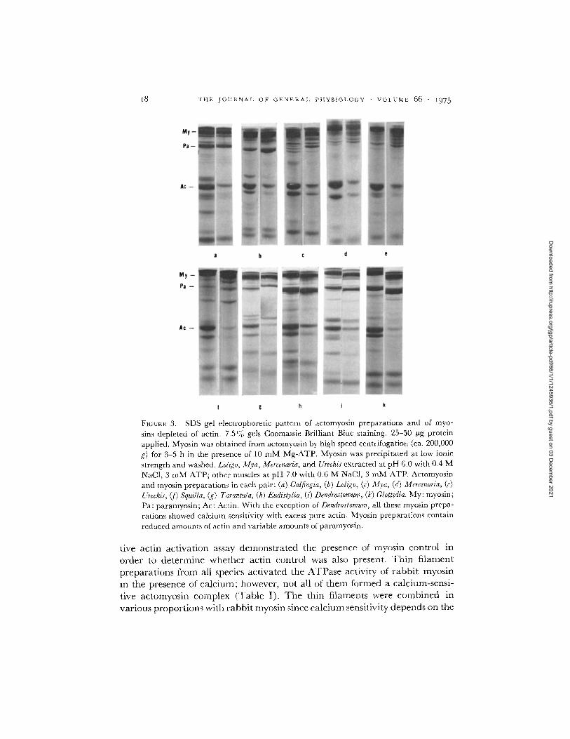
FICURE 3. SDS gel electrophoretic pattern of actomyosin preparations and of myo- sins depleted of actin. 7.5% gels Coomassie Brilliant Blue staining. 25-50 #g protein applied. Myosin was obtained from actomyosin by high speed centrifugation (ca. 200,000 g) for 3-5 h in the presence of 10 mM Mg-ATP. Myosin was precipitated at low ionic strength and washed. Lohgo, Mva, Mercenaria, and Urechis extracted at pH 6.0 with 0.4 M NaCI, 3 mM ATP; other muscles at pH 7.0 with 0.6 M NaCI, 3 mM ATP. Actomyosin and myosin preparations in each pair: (a) Gotfingia, (b) Loligo, (c) Mya, (d) Mercenaria, (e) Urechis, (f) SquiUa, (g) Tarantula, (h) Eudistylia, (i) Dendrostomum, (k) GIottidia. My: myosin; Pa: paramyosin; Ac: Actin. With the exception of Dendrostomum, all these myosin prepa- rations showed calcium sensitivity with excess pure actin. Myosin preparations contain reduced amounts of actin and variable amounts of paramyosin.
tive actin act ivat ion assay demons t ra ted the presence of myosin control in order to de termine whether actin control was also present. ' l 'h in f i lament preparat ions f rom all species act ivated the ATPase activity of rabbi t myosin
in the presence of ca lc ium; however, not all of them formed a calcium-sensi- tive ac tomyosin complex (rl'able I). T h e thin filaments were combined in
various propor t ions with rabbi t myosin since ca lc ium sensitivity depends on the
Dow
nloaded from http://rupress.org/jgp/article-pdf/66/1/1/1245936/1.pdf by guest on 03 D
ecember 2021

W. LEHMAN AND A. G. SZENT-C-Y()RGYI Regulation of Muscular Contraction 19
FIGURE 4. The components of Limulus actomyosin, myosin, and thin filaments. (a) 14 #g thin filaments; (b) 30 #g actomyosin; (c) 32 #g myosin. Pa: paramyosin; Ac: actin; TM: tropomyosin. 10% SDS acrylamide gels stained with Coomassie Brilliant Blue. Note that although both myosin and the thin filaments show calcium sensitivity, their com- ponents do not comigrate with the exception of the fastest component. Actomyosin con- tains the components of both the thin filaments and myosin.
relative proport ions of thin filaments to myosin. Frequent ly, ca lc ium sensi- tivity was greatest at a thin f i lament to myosin weight ratio of about 1 : 2-3.
For most thin filaments there is a good correlation between thin-f i lament composit ion and the functional tests, (Fig. 5), i.e., thin filaments conta ining regulated actin also have low molecular weight components tha t correspond in size to the subunits of invertebrate t roponin (Regenstein, 1972; Regenstein and Szent-Gy6rgyi, 1975; Bullard et al., 1973). Frequent ly , these th in fila- ments conta in three major components , in addi t ion to actin and tropomyosin, on SDS gels. Of these a componen t having a chain weight of about 25,000- 32,000 daltons, probably corresponding to rabbi t t roponin-I , is seen clearly on all th in-f i lament preparat ions showing control, rl'he band with a chain weight of about 15,000-20,000 daltons, probably corresponding to rabbi t t roponin-C, is less prominent and frequent ly stains poorly. Wi th the exception of annelids, a third componen t with a larger chain weight than actin is also present on invertebrate thin filaments. These thin filaments bind calcium (Lehman et al., 1972).
The thin filaments obta ined from muscles tha t have only myosin-l inked regulation, are, on the whole, relatively free of low molecular weight com- ponents and consist mostly of actin and t ropomyosin (Fig. 5). Minor bands, however, can be seen on the thin filaments of Busycon, Lunatia, Loligo, Glottidia, and Urechis, and can even be detected on Aequipecten (Fig. 6), Anadara, and
Dow
nloaded from http://rupress.org/jgp/article-pdf/66/1/1/1245936/1.pdf by guest on 03 D
ecember 2021

2 0 T H E J O U R N A L O F G E N E R A L P H Y S I O L O G Y • V O L U M E 6 6 . ~ 9 7 5
FIQURE 5. SDS acrylamide gel electrophoretic pattern of thin filaments. 10% gels stained with Coomassie Brilliant Blue. 8-16 tag proteins except Cryptochyton which was less than 5 #g. Note lack of myosin or paramyosin in significant quantities and the pres- ence of actin and tropomyosin in all preparations.
Dow
nloaded from http://rupress.org/jgp/article-pdf/66/1/1/1245936/1.pdf by guest on 03 D
ecember 2021

W. LEHMAN AND A. G. SZENT-GY6RGYI Regulation of Muscular Contraction 2I
FmtlRE 6. Minor components of scallop myofibrils. 10% SDS acrylamide gel electro- phoresis stained with Coomassie Brilliant Blue. (a) Isolated soluble calcium-binding com- ponent present in unwashed muscle. (b) Whole muscle. (c) Washed muscle. (d) Thin filaments. Preparations of muscles and thin filaments are overloaded (100 #g myofibrils and 30 #g thin filaments) to demonstrate the presence of minor components. Note that calcium-binding protein is not found in washed muscle. Three minor components are detectable on scallop thin filaments, and migrate similarly to invertebrate troponins. Thin filaments, however, do not sensitize rabbit myosin and the molar ratios of the minor components to tropomyosin are less than 1:5.
Ensis preparat ions . Low molecu la r weight componen ts are present in la rger amoun t s on the thin filaments of the r ibbon worm, Cerebratulus. Nevertheless, none of these thin filaments, including eight di f ferent p repara t ions f rom Cerebratulus, show ca lc ium regula t ion when mixed wi th r abb i t myosin. Myos in compet i t ion tests on ac tomyosin or on muscle suspensions of these animals fail to de tec t the presence of the actin control . Wi th the except ion of Busycon and Lunatia the molluscan thin fi laments do not b ind ca lc ium (Kendr ick-Jones et al., 1970).
All the muscles tested and all the thin fi laments p repa red conta in t ropo- myosin. Dens i tomet ry of ac ry lamide gels indicates tha t in m a n y th in- f i lament prepara t ions the weight ra t io of t ropomyos in to act in is abou t 1 : 3-4, suggest- ing tha t a mola r ra t io of abou t 6 actins to 1 t ropomyos in character izes in- ve r t eb ra t e thin filaments, and tha t there is little or no act in free of t ropo- myosin. T h e da t a suggest tha t act in is complexed wi th t ropomyos in even in muscles where t ropomyos in has no regu la to ry function. T ropomyos in , how- ever, m a y in some cases be lost f rom thin fi laments dur ing prepara t ion , and the t ropomyos in to act in ra t io in thin fi laments m a y fall below tha t found in
Dow
nloaded from http://rupress.org/jgp/article-pdf/66/1/1/1245936/1.pdf by guest on 03 D
ecember 2021

2 2 T H E J O U R N A L O F G E N E R A I , P t t Y S I O L O G Y • V O L U M E 6 6 • 1 9 7 5
muscle, especially in species which do not contain significant amounts of troponin. The losses are particularly great from Cucumaria, T@orm, and Cryptochflon thin filaments, where special precautions were necessary to retain even some of the tropomyosin. '['hese precautions included the use of high protein concentrations during preparations, higher magnesium concentra- tions (5 mM), and a pH of 6.0 at every step of the preparation. 'l'he competi- tive myosin-activation assay of these muscles indicating the lack of actin control is particularly important.
There appears to be a one-to-one molar ratio between the 25,000- to 32,000- dalton component, corresponding to troponin-I and tropolnyosin in a number of thin-filament preparations having a regulatory function (cf. Lehman et al., 1972). The lower chain weight component, corresponding to troponin-C, is less intensely stained, and the staining varies considerably. 'l'he molar ratio of one troponin to one tropomyosin to 5-7 actins is particularly relevant tbr our understanding of the double systems because the information argues against the presence of two populations of nmscles, one with only lnyosin- linked regulation and the other with an actin-linked regulation. The fact that some doubly regulated muscles retain fully their calcimn sensitivity in the presence of excess pure actin (Table I) indicates that these muscles contain predominantly a single population of regulated myosin.
The minor components of the thin filaments are distinct from the light chains of myosin. In some cases these components can be clearly identified in actomyosin preparations. For example, Limulus thin filaments contain four components in addition to actin and tropomyosin, while Limulus myosin has three different light chains. With the exception of components migrating at about 18,000 daltons which are present in both myosin and thin filaments, these low molecular weight components move differently in SDS acrylamide gel electrophoresis and the bands seen in actomyosin preparations may be easily traced either to myosin or to thin filaments (Fig. 4). ~l'he low molecular weight components of molluscan actomyosins can be largely accounted for by the light chains of myosin (Fig. 3).
High molecular weight components occasionally present on thin filaments may represent ol-actinin or other components of Z-line and dense body struc- tures. Significantly, little or no protein remains at the origin of the gels, indi- cating the absence of myosin and paramyosin.
The presence of both actin- and myosin-linked regulation can increase the fidelity of calcium control. ' lhe calcium sensitivity of regulated myosin to- gether with regulated thin filaments from Limulus or from locust is greater than the sensitivity of the individual colnponents tested with rabbit actin or myosin (Table IV).
Regulated invertebrate thin filaments bind fewer calcium ions than regu- lated vertebrate thin filaments (Lehman et al., 1972). Lobster troponin binds approximately one calcium for each mole of troponin (Regenstein and Szent-
Dow
nloaded from http://rupress.org/jgp/article-pdf/66/1/1/1245936/1.pdf by guest on 03 D
ecember 2021

W . L E H M A N A N D A . G . SZENT-GYORGYI Regulation of Muscular Contraction
T A B L E I V
COMBINED EFFECTS OF C A L C I U M - S E N S I T I V E T H I N F ILAMENTS AND MYOSINS
~3
A T P a s e ac t iv i ty C a l c i u m
0.1 m M E G T A 0.1 m M C a 2+ sens i t iv i ty*
IZmol/min/mg %
Limuhts thin filaments + r abb i t 0.12 0.6 80 myosin
Limulus myosin 4- r abb i t actin + 0.022 0.062 65 r abb i t tropomyosin
Limulus myosin + Limulus th in fila- 0.003 0.128 98 ments
Locust thin filaments + r a b b i t 0.1 0.5 80 myosin ~t
Locust myosin + r a b b i t act in + 0.068 0.27 75 r abb i t tropomyosin
Locust myosin + locust thin fila- 0.008 0.31 97 ments~
0.3-0.5 g thin f i lament or act in- t ropomyosin to 1 g myosin. * (1 - - (ATPaseEGT~)/(ATPaseca)) X 100.
Lehman et al., 1974.
Gy6rgyi, 1975) in contrast to the four calciums bound by a mole of rabbit troponin (Potter, 1974). Full ATPase activation by Limulus thin filaments re- quires large changes in calcium concentration (Fig. 7). This broad transition in the pCa curves reflects single noncooperative calcium-binding sites on the
10C , ~ . . . . 7 °z m= ...
/ . . . | '"
8C / , ..i" I e . "
~_~c ~..
4C
20 ¶," d t • "o / |...- " : / ~
pCo FICURE 7. Calcium dependence of ATPase activities. A scallop myofibrils, [] rabbit thin filaments with rabbit myosin, [] rabbit troponin-tropomyosin and rabbit actin with r abb i t myosin, • Limulus th in filaments with rabb i t myosin. The calcium dependence of the preparat ions was normal ized to 100 %. The actual sensitivity of scallop myofibrils amoun ted to 95%, the sensitivity of the r abb i t and Limulus th in fi laments to 85%, the sensitivity of the reconst i tuted rabb i t relaxing system to 70-80 %. The pCa values for 50% calcium sensitivity were also normal ized to the Limulus th in-f i lament values (1.4 M 10 -6 M Ca~+). The halfway point was reached at 0.5 N 10 -7 M Ca e+ in scallop and 1.5 X 10 -7 M in rabbi t preparat ions at neutra l pH.
Dow
nloaded from http://rupress.org/jgp/article-pdf/66/1/1/1245936/1.pdf by guest on 03 D
ecember 2021

24 T H E J O U R N A L O F G E N E R A L P H Y S I O L O G Y • V O L U M E 6 6 - ~ 9 7 5
thin filaments The pCa curves for vertebrate thin filaments, or the pCa dependence of molluscan muscles are sharp, indicating that more than one calcium is involved at each regulatory site (Fig. 7). Likewise, the transition for the doubly regulated Limulus myofibrils is abrupt. On the other hand, the calcium dependence of the thin-filament-regulated lobster tail myofibrils is broad.
D I S C U S S I O N
Interpretation of the Evidence
The presence of a particular regulation in a muscle can be established un- ambiguously by functional tests. It is more difficult, however, to interpret the evidence indicating the absence of a regulatory system since the lack of func- tion may on one hand represent in vivo conditions; alternatively, it could result from inactivation of the regulatory proteins due to experimental manipulations or other experimental artifacts. The fact that calcium-sensitive actomyosin preparations could not be obtained from a number of animals indicates some of the experimental difficulties. The organisms with double regulation thus may be underestimated; therefore, the evidence for single regulation has to be examined with particular caution.
In these studies we have tried to retain all components contributing to the function of myosin and thin filaments. Preparations were performed rapidly employing only a few steps to limit possible inactivation or loss of components. It was important, however, to reduce cross contamination of myosin in thin- filament preparation, and the components of the thin filaments in the myosin preparations. Other impurities may be and are present. Myosin preparations may contain considerable amounts of paramyosin; some of the minor bands in the thin-filament preparations may represent membrane fragments or other impurities.
The pattern of the distribution of the regulatory systems indicates a relative simplicity (Fig. 1). Single systems are not randomly distributed in the animal kingdom, and organisms within a major phylum or class tend to behave simi- larly. Single actin control is restricted to the chordates and, among the in- vertebrates, to the fast muscles of decapods and mysidacea. Single myosin control is restricted to molluscs, echinoderms, and several other minor phyla (brachiopod, echiuroid, and nemertine worms). The relative simplicity of the distribution of single regulatory systems is perhaps the most significant evolu- tionary aspect of these comparative studies. A similar consistency is seen among doubly regulated systems. All the insects and annelids tested behave similarly. Crustaceans, cirripeds, stomatopods, amphipods, and isopodes all show double control, although pure rabbit actin partially reduced the cal- cium sensitivity of amphipod actomyosins.
In lobster, however, the fast tail muscles show a single actin control, and
Dow
nloaded from http://rupress.org/jgp/article-pdf/66/1/1/1245936/1.pdf by guest on 03 D
ecember 2021

W. LIgI-IMAN AND A. G. SZENT-C-YtRGYI Regulation of Muscular Contraction 25
the slow muscles are doubly regulated. The sipunculids also seem to be an exception as regulation varies among the members of this phylum. Dendro- stomum myosin is, however, unusual by binding calcium without a demon- strable regulatory function. This may suggest a partial loss of regulatory function, or reflects an experimental artifact. Smooth muscles of chicken gizzard may also be an exception and evidence for myosin control has been reported (Bremel, 1974).
Evolutionary Aspects
The major evolutionary features which have emerged from this investigation are the wide occurrence of both regulatory systems and the relative simplicity of the distribution of single regulatory systems. When taken in the context of the differing properties of the components of actin- and myosin-linked regulation, these features are of particular interest. Troponin consists of three different subunits, two of these are considerably larger than the "regulatory" light chains. Troponin combines only with actin and tropomyosin but not with myosin. In contrast the "regulatory" light chain binds only to myosin. A common evolutionary origin for troponin-C, myosin light chains and par- valbumin has been proposed recently on the basis of similarities in amino acid sequences (Tufty and Kretsinger, 1975; Collins, 1974; Weeds and McLachlan , 1974); however, there is no functional overlap between troponin-C and the regulatory light chains. The two regulations act independently of each other, although their effect may be additive (Table IV).
Both the myosin-linked and the actin-linked regulations are found in phyla which appeared early in evolution, and at present there is no evidence for assuming that myosin-linked regulation evolved before actin-linked regula- tion, even though the myosin control requires only a single regulatory com- ponent, whereas actin control involves the interaction of a number of different regulatory components.
In our initial studies we speculated that myosin-linked regulation evolved first (Lehman et al., 1972). This hypothesis became untenable after finding both regulation systems in the nematode, Ascaris lumbricoides. Recently the presence of actin control was reported in the slime mold, Physarum polycepha- lum (Nachmias and Asch, 1974). I t is no longer necessary to assume a con- vergent evolution for the thin-filament-linked regulatory systems. The differ- ent calcium-binding properties of invertebrate and vertebrate troponins may have stemmed from an ancestral mutation.
Functional Aspects
In vivo regulation may be altered genetically in several different ways: the synthesis of normal regulatory components may be decreased; inactive regu- latory components may be produced; the binding sites on myosin or on actin for the regulatory proteins may be changed. Alternatively, mutations may
Dow
nloaded from http://rupress.org/jgp/article-pdf/66/1/1/1245936/1.pdf by guest on 03 D
ecember 2021

26 THE JOURNAL OF GENERAL PHYSIOLOGY • VOLUME 66 • i975
have made a regulatory system particularly sensitive to experimental manipu- lations and the apparent loss of regulation would not reflect the in vivo proper- ties of the muscle.
Absence of actin control in molluscan and brachiopod muscles has a simple explanation; regulatory proteins are not present in sufficient quanti ty to regulate actin (cf. Lehman et al., 1972). The reasons for the lack of significant amounts of troponin in these muscles are not known. Minor bands can be detected on overloaded SDS acrylamide gels of scallop thin filaments or washed muscles (Fig. 6). These bands may correspond in chain weight to the subunits of invertebrate troponin. However, these components are present only in small quantities; the molar ratio of the 25,000-chain weight peptide to tropomyosin is less than 0.2. Although we have not been able to demon- strate any actin control in molluscan muscles, and we have not been able to isolate a functional troponin from scallops, we cannot exclude that s o m e tro- ponin may be synthesized in the muscle. I t is also possible that a nonfunctional mutan t of troponin is synthesized that may have lost its ability to combine with actin and tropomyosin. If so, some of the troponin subunits may be found in the soluble protein fraction. We have isolated a protein from the soluble fraction of scallop striated muscle which consists of a single chain of about 22,000 daltons and binds about 1 mol of calcium at 3 × 10 -6 M Ca 2+ concentrations in the presence of 1 m M MgC12 (Fig. 6). This calcium-binding protein amounts to less than 0.5o-/0 of the muscle proteins, and does not com- plex with other soluble proteins or with the thin filaments. We have not as yet demonstrated that it has any regulatory function. This calcium-binding protein of scallop may be related to the parvalbumins, a group of calcium- binding proteins obtained from a number of vertebrate muscles. One notes, however, that the chain weight of the scallop calcium-binding protein exceeds the size range reported for parvalbumins (11,000-15,000 d altons) (Pechere et al., 1973), and it has a relatively high tryptophan and tyrosine content with an absorption peak at 280 nm and an extinction coefficient of about 1 . 4 0 D units (milligrams per milliliter per centimeter), in contrast to the parvalbu- rains that have few or no aromatic residues and show absorption maxima at around 260 nm.
The lack of myosin control in vertebrates and decapods is not due to the lack of "regula tory" light chains. Kendrick-Jones has shown that the DTNB- (5,5'-dithio-bis-(2-nitrobenzoic acid)) light chains of rabbit hybridize with a desensitized scallop myosin and the hybrid formed is regulated (1974). Similarly, "regulatory" light chains can be prepared from a number of other vertebrate myosins and also from lobster (Kendrick-Jones et al., to be pub- lished). In contrast, neither rabbit myosin nor any hybrid of the rabbit heavy chains is calcium sensitive. The lack of a myosin control is thus due either to an alteration in the heavy chain of myosin, such that it will not respond to regulatory light chains, or that the myosin control is particularly sensitive to
Dow
nloaded from http://rupress.org/jgp/article-pdf/66/1/1/1245936/1.pdf by guest on 03 D
ecember 2021
W. LEHMAN AND A. G. SZENT-GY6RGYI Regulation of Muscular Contraction 2 7
the relatively limited manipulations required even for the competitive actin- activati6n assay. At present, it is difficult to decide experimentally between these possibilities, and it will not be easy to detect alterations or mutations on a molecule the size of the myosin heavy chain. We have performed competi- tive actin-activation assays on unwashed mouse myofibrils not exposed to high ionic strength, and on actomyosin extracts from unwashed lobster mus- cles in order to avoid the possible loss of a myosin control during preparation. Nevertheless, we failed to detect the presence of a myosin-linked regulation in either case. I t is not obvious how to devise a more direct biochemical ap- proach which more faithfully approximates the in vivo conditions of a mus- cle, and the evidence strongly suggests that in at least some muscles myosin control is lacking. The importance of positive evidence, however, cannot be overstated.
The disappearance of the ordered cross-bridge lattice from frog sartorius upon stimulation of stretched muscle (Hazelgrove, 1972), the calcium- dependent fluorescent change of the DTNB light chain of rabbit myosin (Werber et al., 1972), and the calcium dependence of the viscosity and sedi- mentation properties of isolated and reconstituted thick filaments (Marimoto and Harrington, 1974) indicate that calcium may interact with vertebrate myosin. These observations, however, do not demonstrate directly that a myosin-linked control functions in vertebrate striated muscles. The ATPase activity of vertebrate myosins and actomyosins in the presence of pure actin is not stimulated by calcium ions, in fact, calcium may inhibit by about 15-200-/0 when magnesium is low (Weber and Murray, 1973).
The evidence at present that molluscan muscles are controlled by a single myosin-linked system is firm. In these muscles the lack of actin control is due to the lack of troponin. In vertebrates and in most decapod muscles, on the other hand, "regulatory" light chains are likely to be present although they do not seem to function in vitro, and it is difficult to establish with abso- lute certainty that in vitro results apply to in vivo conditions.
There is no obvious relationship between ATPase activity, the structure of the muscle, and a particular regulatory system. ATPase activities range widely irrespective of the nature of the control (Table I). Furthermore, both myosin- linked and actin-linked regulation are displayed by both striated and smooth muscles. Nevertheless, the studies reported here are relevant for interpreting some of the structural studies on muscle. The movement of myosin cross bridges in insect muscles upon addition of calcium, before contact with actin filaments is established (Miller and Tregear, 1971), i.e. the calcium-activated state, may be readily explained by the demonstration of myosin control in Lethocerus flight muscles. I t is also of interest that the increase in the intensity of the second layer line of actin during rigor in the byssus retractor muscle of Mytilus edulis indicates that tropomyosin may move in the absence of func- tioning troponin (Lowy and Vibert, 1972).
Dow
nloaded from http://rupress.org/jgp/article-pdf/66/1/1/1245936/1.pdf by guest on 03 D
ecember 2021

28 T H E J O U R N A L O F G E N E R A L P H Y S I O L O G Y • V O L U M E 6 6 • i975
The advantages of double regulation are obvious: the accuracy and pre- cision of the calcium control over rest and activity may be enhanced. Func- tional advantages of single regulation are less apparent. The evolutionary pressures for the development of single systems may be explained in the case of molluscs if one assumes that the presence of a regulation acting on the thin filaments is not compatible with the maintenance of "catch," a property im- portant for the survival of these animals (cf. Johnson, 1962). One may argue that the evolution of a troponin with multiple calcium-binding sites allowed for a sharp transition between rest and activity in vertebrates, and hence the importance of myosin control was reduced. There is no apparent advantage, however, for losing myosin-linked regulation in the crustacean decapods since these muscles would have additional requirements for rapid Ca++ removal if their muscles were regulated in vivo solely by invertebrate troponin.
The calcium dependence of tension measurements in skinned fibers of the carpopodite flexor muscle of the crayfish, Orconectes, is similar to that of frog muscles; b~th preparations are brought to full activity over a narrow calcium- concentration range (Brandt et al., 1972; Orentlicher et al., 1974). These crayfish muscles, however, are slow muscles with 8- to 10-#m long sarcomeres and are very likely doubly regulated.
The decapods are of particular interest because one species may contain both doubly regulated and singly regulated muscles. There are additional structural and biochemical differences. The muscles with single actin control in general have several-fold higher ATPase activities than the other doubly regulated crustacean muscles (Table I). The fast tail, cutter claw, and deep abdominal extensor muscles of the lobster that show a single actin control have shorter thick filaments and sarcomere lengths (2-3.5 ~m) compared to the slow crusher claw and slow superficial abdominal extensor muscles (6- to 9-~m sarcomere length) (Jahromi and Atwood, 1969, 1971). Paramyosin is found in the crusher claw muscle but not in the tail muscles of lobster (Weisel and Szent-Gy6rgyi, to be published). However, the two light chains of the myosins of both muscles migrate identically on SDS gel electrophoresis. It thus appears that the heavy chains of the two myosin types of the lobsters differ and that the lack of the myosin control in fast muscles is the result of alterations in the heavy chains of myosin. Similarly, a functional myosin control in vertebrate smooth muscles (Bremel, 1974) and the lack of such a control in vertebrate striated muscles may be simply explained by assuming differences in the heavy chains.
In summary, we suggest that the genorne of most, possibly of all, animals contains the information for both regulatory systems. This information may be expressed fully and all the components of both regulations are present in significant amounts in most animals with the exception of molluscs, brachio- pods, echinoderms, and echiuroids which lack in troponin. Regulation may
Dow
nloaded from http://rupress.org/jgp/article-pdf/66/1/1/1245936/1.pdf by guest on 03 D
ecember 2021

W. LEHMAN AND A. G. SZENT-GY6RGYI Regulation of Muscular Contraction 29
a l so b e lo s t as a r e s u l t o f c h a n g e s in t h e m y o s i n m o l e c u l e w i t h o u t a l t e r i n g o r
l o s i n g r e g u l a t o r y c o m p o n e n t s .
We thank Drs. Carolyn Cohen, Hugh E. Huxley, John Kendrick-Jones, Eva M. Szentkiralyi, Anne- marie Weber, Joe M. Regenstein, John Weisel, and Michael Payne for discussions. We also thank Drs. Carolyn Cohen, Eva M. Szentkiralyi, Peter Vibcrt, Annemarie Weber, John Weisel, and Michael Payne for criticizing the manuscript. We acknowledge the help of Dr. John Kendrick-Jones in some of the experiments. We thank Ms. Debbie Wygal for experiments on the calcium-binding protein of the scallop, Iris. Ruth Hoffman for expert assistance during most of this work, and Ms. Regina Niebieski for assistance at the later stages. We are grateful to the late Mr. Harold Williams for collecting Lethocerus sp. (Florida), and to the Supply Department at the Marine Biological Laboratory, Woods Hole, Mass., for collecting a large number of relatively rare specimens. Some of this work was performed at the Department of Zoology at Oxford University when one of us (W. L.) was on the A. R. C. staff. Some experimentation was done at the M. R. C. Laboratory of Molecular Biology at Cambridge when one of us (A. G. S. G.) was a visiting scientist. We thank Richard Tregear, Professor John Pringle, and Dr. Hugh E. Huxley for their hospitality. This work was supported by grants from the National Science Foundation (GB-40308) (W. L.) and from the Public Health Service (AM-17062) (W. L.) and (AM-15963) (A. G. S. G.).
Received for publication 14 January 1975.
R E F E R E N C E S
BRANDT, P. W., J. P. REtmEN, and H. GRUNDFEST. 1972. Regulation of tension in the skinned crayfish muscle fiber. II . Role of calcium. J. Gen. Physiol. 59:305.
BREMEL, R. D. 1974. Myosin-linked calcium regulation in vertebrate smooth muscle. Nature (Lord.). 252:405.
]~ORRADAILE, L. A., F. A. POTTS, L. E. S. EASTHA•, AND T. T. SANDERS. 1963. The Inver- tebrata: A manual for the use of students, revised by G. A. Kerkut. Cambridge Univer- sity Press, New York.
BULLARD, B., R. DA~ROWSKA, and L. B. WmKELMAN. 1973. The contractile and regulatory proteins of insect flight muscle. Biochem. J. 135:227.
CHAnER~K, S., and A. E. MARTELL. 1959. Organic sequestering agents. John Wiley & Sons, Inc., New York.
COLLINS, J. H. 1974. Homology of myosin light chains, troponin-C and parvalbumins deduced from comparison of their amino acid sequences. Biochem. Biophys. Res. Commun. 58:301.
DEMAILLE, J. , E. DUTRUGE, n. EISENBERG, J . O. CAPONY, a n d J . F. PSCHE~. 1974. Troponins C from reptile and fish muscles and their relation to muscular parvalbumins. FEBS (Fed. Fur. Biochem. Soc.) Lett. 42:173.
EBASHI, S., and M. ENDO. 1968. Calcium ions and muscle contraction. Prog. Biophys. Mol. Biol. 18:123.
EIS~NBERO, E., and W. W. KmLLEY. 1970. Native tropomyosin: effect on the interaction of actin with heavy meromyosin and sub-fragment-I. Biochem. Biophys. Res. Commun. 40:50.
HAZELGROV~, J . C. 1972. X-ray evidence for a conformational change in the actin-containing filaments of vertebrate striated muscle. Gold @ring Harbor Symp. Quant. Biol. 37:341.
HITCHOCK, S. E., H. E. HUXLEy, AND A. G. SZENT-GY~t~OYL 1973. Calcium sensitive bind- ing of troponin to actin-tropomyosin: A two-site model for troponin action. J. Mol. Biol. 80:825.
JAHROMI, S. S., and H.L. ATWOOD. 1969. Correlation of structure, speed of contraction, and total tension in fast and slow abdominal muscle fibers of the lobster (Homarus americanus). J . Exp. Zool. 171:25.
JAHROm, S. S., and H. L. AXWOOD. 1971. Structural and contractile properties of lobster leg- muscle fibers. J . Exp. Zool. 176:475.
JOHNSON, W. H. 1962. Tonic mechanisms in smooth muscles. Physiol. Rev. 42(Suppl. 5):113.
Dow
nloaded from http://rupress.org/jgp/article-pdf/66/1/1/1245936/1.pdf by guest on 03 D
ecember 2021

3 ° T H E J O U R N A L O F G E N E R A L P H Y S I O L O G Y • V O L U M E 66 • I975
K~NDRXCK-JoI, r~s, J. 1974. Role of myosin light chains in calcium regulation. Nature (Lond.). 249:631.
KENDRIC~-JoNEs, J., W. LBt-atAN, and A. G. SZENT-GY/SROyL 1970. Regulation in molluscan muscles. J. Mol. Biol. 54:313.
K~'.NDRICa~-JoNEs, J., E. M. SZEN~IaALYI, and A. G. SZENT-GY/SROYI. 1972. Myosin-linked regulatory systems: the role of the light chains. Cold Spring Harbor Symp. Quant. Biol. 37:47.
Ko~a~TZ, J. C., T. HUNT, and E. W. TAYLOR. 1972. Studies on the mechanism of myosin and actomyosin ATPase. Cold Spring Harbor Symp. Quant. Biol. 37:179.
l.~m~N, W., B. BULLAm~, and K. HAUatOND. 1974. Calcium-dependent myosin from insect flight muscles. J. Gen. Physiol. 63:553.
I-'~HMAN, W., J. KENDRICK-Jo~s, and A. G. SZENT-GY6ROyI. 1972. Myosin-linked regulatory systems: Comparative studies. Cold Spring Harbor Symp. Quant. Biol. 37:319.
L~HMAN, W., and A. G. SZE~T-GY6RG~. 1972. Activation of the adenosine triphosphatase of Limulus polyphemus aetomyosin by tropomyosin. J. Gen. Physiol. 59:375.
LowRY, O. H., N. J. ROSEBROUOH, A. L. FARR, andR. J. RANDALL. 1951. Protein measurement with the Folin phenol reagent. J. Biol. Chem. 193:265.
LowY, J., and P.J. VmERT. 1972. Studies of the low-angle X-ray pattern of a molluscan smooth muscle during tonic contraction and rigor. Cold Spring Harbor Symp. Quant. Biol. 37:353.
MAPa~OTO, K., and W. F. HAVa~aNOTON. 1974. Evidence for structural changes in verte- brate thick filaments induced by Ca ~-. J. Mol. Biol. 83:83.
MILLER, A., and R. T. Tm'.aEAR. 1971. X-ray studies on the structure and function of verte- brate and invertebrate muscle. In Contractility of Muscle Cells and Related Processes. R. J. Podolsky, editor. Prentice-Hall, Inc., Englewood Cliffs, N.J. 205.
NACHUIAS, V., and A. ASCH. 1974. Actin mediated calcium dependency of actomyosin in a myxomycete. Biochem. Biophys. Res. Commun. 60:656.
Ox-rrstmi, I., T. MASAKI, Y. NONOMURA, and S. EBASHI 1967. Periodic distribution of trop- onin along the thin filament. J. Biochem. 61:817.
Om~NTLICX-mR, M., J. P. R~traEN, and H. GRUNDF~.ST. 1974. Calcium binding and tension de- velopment in detergent-treated muscle fibers. J. Gen. Physiol. 63:168.
PARKER, L., H. Y. P v ~ , and D. J. HARTSHORNE. 1970. The inhibition of the adenosine tri- phosphatase activity of the subfragment-l-actin complex by troponin plus tropomyosin, troponin B plus tropomyosin and troponin B. Biochim. Biophys. Acts. 223:453.
PEcem~, J. F., J.P. CAVONY, and J. DEMAILL~. 1973. Evolutionary aspects of the structure of muscular parvalbumins. Syst. Zool. 22:533.
Poa-X~R, J. D. 1974. The content of troponin, tropomyosin, actin, and myosin in rabbit skeletal muscle myofibrils. Arch. Biochem. Biophys. 162:436.
REOE~STEXN, J. M. 1972, The regulatory proteins of lobster striated muscle. Ph.D. Thesis. Brandeis University, Waltham, Mass.
REGENST~IN, J. M., and A. G. SZENT-GY/SROYr. 1975. Regulatory proteins of lobster striated muscle. Biochemistry. 14:917.
SZENT-GY/SRGyI, A. G., C. COF~N, and J. KENDRIeK-JoNES. 1971. Paramyosin and the fila- ments of molluscan "catch" muscles. II. Native filaments: isolation and characterization. J. Mol. Biol. 56:239.
SZ~NT-GY6RGYI, A. G., E. M. SZENTKIRALYI, and J. KENDRteK-JosES. 1973. The light chains of scallop myosin as regulatory subunits. J. Mol. Biol. 74:179.
TuFty, R. M., and R. H. K~TStNO~-R. 1975. Troponin and parvalbumin calcium binding re- gions predicted in myosinlight chainsand the lysozymes. Science (Wash. D. C.). 187:167.
WV.BER, A. 1956. The ultracentrifugal separation of L-myosin and actin in an actomyosin sol under the influence of ATP. Biochim. Biophys. Acta. 19:345.
WEB~.R, A., and J. M. MURRAY. 1973. Molecular control mechanisms in muscle contraction. Physiol. Rev. 53:612.
WEBER, K., and M. OSBORN. 1969. The reliability of molecular weight determinations by do- decyl sulphate-polyacrylamide gel electrophoresis. J. Biol. Chem. 244:4406.
WEEDS, A. G., and A. D. MeLACHLA~. 1974. Structural homology of myosin alkali light chains, troponin C and carp calcium binding protein. Nature (Lond.). 252:646.
WERRER, M. M., S. L. GA~FI~, and A. OPLATKA. 1972. Physio-chernical studieson the fluores- cence of a light chain (LMP-II) from rabbit muscle. J. Mechanochem. Cell Motility. 1:91.
Dow
nloaded from http://rupress.org/jgp/article-pdf/66/1/1/1245936/1.pdf by guest on 03 D
ecember 2021