Embed Size (px)
Citation preview

REGULATION OF INFLAMMASOME AND NET FORMATION IN NEUTROPHILS BY PI3K SIGNALING
BY
NNEBUNNE GLORIA AKALUKA
A Thesis Submitted to the Faculty of Graduate Studies of University of Manitoba in Partial Fulfilment of the Requirements of the Degree of
MASTER OF SCIENCE
Department of Immunology
University of Manitoba
Winnipeg, Manitoba, Canada
Copyright © 2020 by Nnebunne Gloria Akaluka
I
TABLE OF CONTENT
ABSTRACT ________________________________________________________ V
ACKNOWLEDGEMENTS _____________________________________________ VII
DEDICATION ______________________________________________________ IX
LIST OF FIGURES ___________________________________________________ X
LIST OF TABLES ____________________________________________________ XI
LIST OF ABBREVIATIONS ___________________________________________ XII
CHAPTER 1: INTRODUCTION __________________________________________ 1
1.1 Inflammation --------------------------------------------------------------------------------------- 1
1.1.1 The inflammatory pathway ------------------------------------------------------------------- 1
1.1.2 Acute inflammation ---------------------------------------------------------------------------- 3
1.1.3 Chronic inflammation ------------------------------------------------------------------------- 4
1.1.4 Sepsis -------------------------------------------------------------------------------------------- 4
1.1.5 Regulation of inflammation ------------------------------------------------------------------- 6
1.2 Neutrophils ----------------------------------------------------------------------------------------- 7
1.2.1 Neutrophil development ----------------------------------------------------------------------- 8
1.2.2 Neutrophil recruitment ------------------------------------------------------------------------ 8
1.2.3 Neutrophil activation -------------------------------------------------------------------------- 9
1.2.4 Neutrophil functions ------------------------------------------------------------------------- 10
1.3 Inflammasomes ---------------------------------------------------------------------------------- 14
1.3.1 NLRP3 inflammasome ---------------------------------------------------------------------- 15

II
1.3.2 Activation of the NLRP3 inflammasome ------------------------------------------------- 15
1.3.3 Regulation of NLRP3 inflammasomes ---------------------------------------------------- 20
1.3.4 Cytokines produced by the inflammasome and their functions. ----------------------- 23
1.3.5 The role of the inflammasome in diseases ------------------------------------------------ 24
1.3.6 Therapeutics targeting the NLRP3 inflammasome -------------------------------------- 28
1.4 NET formation ----------------------------------------------------------------------------------- 30
1.4.1 Physiological functions of NET ------------------------------------------------------------ 31
1.4.2 Mechanisms of NET formation ------------------------------------------------------------ 31
1.4.3 NET – double edged sword: pathology and treatment ---------------------------------- 33
1.5.1 p110d in Leukocyte signalling and activation. --------------------------------------------- 38
1.5.2 p110d in Leukocyte migration. --------------------------------------------------------------- 39
1.5.3 p110d in Leukocyte cytokine production --------------------------------------------------- 39
1.6 Thesis overview --------------------------------------------------------------------------------- 40
1.6.1 Study rationale ---------------------------------------------------------------------------------- 40
1.6.2 Hypothesis --------------------------------------------------------------------------------------- 40
1.6.3 Study objectives -------------------------------------------------------------------------------- 41
CHAPTER 2: MATERIALS AND METHODS _______________________________ 42
2.1 MICE -------------------------------------------------------------------------------------------------- 42
2.2 BONE MARROW CELL ISOLATION ---------------------------------------------------------- 42
2.3 GENERATION OF MATURED MACROPHAGES FROM BONE MARROW CELLS 42
2.4 ISOLATION OF NEUTROPHILS FROM BONE MARROW CELLS --------------------- 43
2.5 INFLAMMASOME ACTIVATION -------------------------------------------------------------- 43

III
2.6 ELISA ------------------------------------------------------------------------------------------------- 44
2.7 WESTERN BLOT EXPERIMENTS ------------------------------------------------------------- 44
2.8 FLOW CYTOMETRY EXPERIMENTS -------------------------------------------------------- 45
2.9 CYTOSPIN ------------------------------------------------------------------------------------------- 46
2.10 NET INDUCTION --------------------------------------------------------------------------------- 46
2.11 IMMUNOFLUORESCENCE MICROSCOPY ------------------------------------------------ 47
2.12 RNA ISOLATION, CDNA SYNTHESIS AND RT PCR ------------------------------------ 47
2.13 TRANSWELL MIGRATION ASSAY --------------------------------------------------------- 48
2.14 SEPSIS INDUCTION ----------------------------------------------------------------------------- 49
2.15 PICOGREEN ASSAY ----------------------------------------------------------------------------- 49
2.17 IDELALISIB (CAL101) -------------------------------------------------------------------------- 50
2.18 STATISTICAL ANALYSIS --------------------------------------------------------------------- 50
2.19 ETHICAL STATEMENT ------------------------------------------------------------------------- 50
CHAPTER 3: RESULTS ______________________________________________ 51
3.1 WT and P110dD910A neutrophils have similar morphology, survival rate in vitro, and
surface markers expression. ----------------------------------------------------------------------------- 51
3.2 P110dD910A neutrophils have reduced chemokine receptor expression and impaired
migration compared to WT neutrophils. -------------------------------------------------------------- 53
3.3 CAL101 at different concentrations does not impair survival of neutrophils. --------------- 55
3.4 P110dD910A myeloid cells have a defect in cytokine and chemokine production post LPS
stimulation. ------------------------------------------------------------------------------------------------ 56
3.5 CAL101 treated macrophages and neutrophils are impaired in their ability to produce
proinflammatory cytokines like IL6 and TNFa ------------------------------------------------------ 58

IV
3.6 P110d isoform of PI3K is dispensable for NLRP3 inflammasome response in neutrophils
but not macrophages. ------------------------------------------------------------------------------------ 60
3.7 P110dD910A the expression of inflammasome genes in macrophages and neutrophils is
similar in WT macrophages and neutrophils from WT and P110dD910A mice. ------------------ 62
3.8 P110d isoform of PI3K regulates procaspase 1 production in macrophages but not in
neutrophils. ------------------------------------------------------------------------------------------------ 64
3.9 Berenil downregulates LPS induced proinflammatory cytokine production and
inflammasome response while upregulating anti-inflammatory cytokine production in bone
marrow derived macrophages. -------------------------------------------------------------------------- 67
3.10 Berenil downregulates LPS-induced proinflammatory cytokine production and
inflammasome response while upregulating anti-inflammatory cytokine production in bone
marrow-derived neutrophils. ---------------------------------------------------------------------------- 69
3.11 Qualitative evidence suggests that P110d is critical for NET formation. ------------------- 71
3.12 Quantitative evidence suggests that P110d is critical for NET formation. ----------------- 73
3.13 P110d signaling regulate NET formation in an animal model of sepsis. ------------------- 74
CHAPTER 4: DISCUSSION ____________________________________________ 76
4.1: GENERAL DISCUSSION ------------------------------------------------------------------------ 76
4.2 CONCLUSION -------------------------------------------------------------------------------------- 82
4.3 SIGNIFICANCE OF STUDY --------------------------------------------------------------------- 82
4.4 LIMITATION ---------------------------------------------------------------------------------------- 82
4.5 FUTURE DIRECTIONS --------------------------------------------------------------------------- 83
REFERENCES _____________________________________________________ 85

V
ABSTRACT
Introduction: The PI3K signalling pathway controls many physiological processes including
inflammation, chemotaxis, proliferation, phagocytosis and microbicidal activities in immune cells
such as macrophages and dendritic cells. However, it’s role in neutrophils especially in the
regulation of inflammasome activation and neutrophil extracellular traps (NET) formation is yet
to be understood. Here, I investigated the role of p110d isoform of the PI3K, which is uniquely
expressed in leukocytes, in neutrophilic inflammatory responses.
Methods: Bone marrow derived neutrophils (BMDN) were isolated from both WT and
P110dD910A mice. BMDN from both mice were challenged with LPS and Nigericin, the levels of
IL-6 and IL-1b were determined by ELISA. In some experiments, NET formation was assessed
using immunofluorescence microscopy and picogreen assay. Chemotaxis towards MIP1a and
CXCL1 was assessed using a transwell assay system. BMDN from WT mice were treated with a
pharmacological inhibitor of p110d (CAL101) to validate the above experiments. Western blot
was done to assess protein levels in pro-caspase1, ASC, NLRP3 (inflammasome proteins) and
pro-IL1b. Also, WT and P110dD910A mice were challenged with LPS and NET production was
measured.
Results: We observed significant impairment to core neutrophilic functions such as
proinflammatory cytokine IL-6 production, NET formation and migration by neutrophils from
p110dD910A mice compared to their WT counterparts. However, there was no significant
difference in IL-1b or pro-caspase1 production by WT and p110dD910A neutrophils. The same
result was observed in CAL101-treated BMDN. Sepsis induced P110dD910A mice were also
impaired in their ability to produce NET when compared to their WT counterparts.

VI
Conclusion: These findings suggest that signaling via the p110d isoform of PI3K plays an
important role in regulating neutrophil migration and NET formation but is dispensable for
inflammasome response.

VII
ACKNOWLEDGEMENTS
I would like to thank Dr. Jude Uzonna for the wonderful opportunity to be trained in his
lab. I am grateful for believing in me, for taking the chance to guide and mentor me and for
treating me as family. I would like to thank Dr. Yvonne Myal for recommending me to Dr. Jude
Uzonna. I am grateful to my committee members, Dr Sam Kung and Dr Kangmin Duan, for their
suggestions, support, critique and input in my research.
To all past and present members of Uzonna Lab, Dr. Gaurav Gupta, Dr Chukwunonso
Onyilagha, Dr. Shiby Kuriakose, Dr. Zhirong Mou, Ping Jia, Nnamdi Ikeogu, Enitan Salako,
Chidalu Arnold Edechi, Stella Onwah, your support, contributions and encouragement was
invaluable. To all faculty and students of the Department of Immunology, thank you for making
my stay worthwhile. To Edgard Mejia and Christine Zhang, Dr Susan Logue; thank you for
teaching me those necessary technical skills I needed for my research. I would like to
acknowledge Karen, Susan, Silvia, Bill and Mike for taking care of all the logistics involved in
my program.
I appreciate my friends Marshall Nkovadu, Enitan Salako, Nnamdi Ikeogu, Folayemi
Adefemi, Chidalu Edechi, Stella Onwah for finding ways to make this time less stressful. Thank
you for the jokes shared, late nights spent working hard, and for watching out for a sister.
I really want to appreciate my parents, Mr. George Akaluka and Mrs. Udobata Akaluka
for their encouragement, financial support and for instilling in me the passion for quality
education. To my siblings Osinachi, Chinenye and Chetachukwu, thank you for bearing with my
excesses and for praying for me.
To all members of More Than Conquerors Parish, my local church assembly, thank you
for encouraging and praying for me. To all the funding agencies Research Manitoba, NSERC,

VIII
Manitoba Medical Service Foundation, Mindel and Tom Olenik Scholarship, University of
Manitoba graduate fellowship; thank you for your contributions towards advancing my career in
science.
God bless you all.

IX
DEDICATION
I dedicate this thesis to the Almighty God, who alone gave me the life and ability to carry out
this wonderful project. Thank you, Lord, for being my banner and victory.

X
LIST OF FIGURES
Figure 1. Neutrophil crosstalk with other cells during inflammation ------------------------------- 13
Figure 2: Mechanisms of NLRP3 inflammasome activation ---------------------------------------- 20
Figure 3. Potential therapeutic targets in NET formation -------------------------------------------- 37
Figure 4. WT and P110dD910A neutrophils have similar morphology, survival rate in vitro, and
surface markers expression ------------------------------------------------------------------------------- 52
Figure 5: P110dD910A neutrophils have impaired migration and chemokine receptors compared to
WT neutrophils --------------------------------------------------------------------------------------------- 54
Figure 6. CAL101 at different concentrations does not impair survival of neutrophils ---------- 55
Figure 7: P110dD910A myeloid cells have a defect in cytokine and chemokine production
following LPS stimulation -------------------------------------------------------------------------------- 57
Figure 8. CAL101 treated macrophages and neutrophils are impaired in their ability to produce
proinflammatory cytokines like IL6 and TNFa -------------------------------------------------------- 59
Figure 9. Inflammasome activation in macrophages require P110d signaling but this pathway is
dispensable in neutrophils --------------------------------------------------------------------------------- 61
Figure 10. P110dD910A macrophages express inflammasome genes similarly to WT
macrophages; which is not necessarily the case with neutrophils ----------------------------------- 63
Figure 11. P110d isoform of PI3K regulates procaspase 1 production in macrophages but not in
neutrophils --------------------------------------------------------------------------------------------------- 66
Figure 12. Berenil downregulates inflammasome activity and other proinflammatory cytokines
but upregulates anti-inflammatory cytokine in macrophages ---------------------------------------- 68
Figure 13. Berenil downregulates inflammasome activity and other proinflammatory cytokines
but upregulates anti-inflammatory cytokine in Neutrophils ------------------------------------------ 70

XI
Figure 14. NET formation is reduced in P110dD910A neutrophils, CAL101 treatment reduced
NET formation, and LPS from P. aeruginosa is capable of inducing more NET than LPS from E.
coli ------------------------------------------------------------------------------------------------------------ 72
Figure 15. Quantitative evidence suggests that P110d is critical for NET formation ------------ 74
Figure 16. Absence of P110d signaling leads to decreased NET formation in vivo -------------- 75
LIST OF TABLES
Table 1. List of primers used in RT qPCR ------------------------------------------------------------- 48

XII
LIST OF ABBREVIATIONS
AAV - anti-neutrophil cytoplasmic antibody-associated vasculitis
AIM2 - absent in melanoma
ALR - AIM2 like receptors
APAP - acetaminophen
Apoe - Apolipoprotein E
APRIL - B cell-activating factor
ASC - apoptosis-associated speck-like protein containing a caspase recruitment
ATP - Adenosine triphosphate
BAFF - a proliferation-inducing ligand
BCR – B cell receptor
BRCC3 - BRCA1/BRCA2-Containing Complex Subunit 3
c-Raf - RAF proto-oncogene serine/threonine-protein kinase
C/EBP- β - CCAAT/enhancer-binding protein beta
Ca2+ - calcium ion
CAPS - cryopyrin- associated periodic syndromes
CARD - caspase recruitment domain
CCL3 - Chemokine (C-C motif) ligand 3
CD14 - cluster of differentiation 14
CD3 - cluster of differentiation 3
CD40 - cluster of differentiation 40
CD86- cluster of differentiation 86
CIA - collagen induced arthritis

XIII
Cl- - chloride ion
CLP - cecal ligation and puncture
COX-2 - Cyclooxygenase-2
CPG - cytosine–phosphate–guanine
CXCL12 - chemokine (C-X-C motif) ligand 12
CXCR4 - chemokine (C-X-C motif) ligand 4
DAG - diacyl glycerol
DAMPs - damage associated molecular patterns
DC - dendritic cells
DNA - Deoxyribonucleic acid
DSS-induced colitis
EAE - Experimental autoimmune encephalomyelitis
ECM - extracellular matrix
ER - endoplasmic reticulum
ERK1/2 - Extracellular regulated kinase 1/2
FasL - Fas ligand
FcgRs - Fc gamma receptor
fMLF - formyl-methionyl-leucyl-phenylalanine
G-CSF - granulocyte colony stimulating factor
GBP5 - guanylate binding protein 5
GLUT4 - Glucose transporter type 4
GMP - granulocyte monocyte progenitor
GPCRs - G-protein-coupled receptors

XIV
GROa - growth-regulated oncogene-alpha
H2O2 - hydrogen peroxide
HIV - human immunodeficiency virus
HOCl - hypochlorous acid
HPA - hypothalamic-pituitary-adrenal axis
HSC - Haematopoietic stem cells
Hsp90 - Heat shock protein 90
IAPP – a hormone released with insulin
ICAM - intercellular adhesion molecules
IFNg - interferon gamma
IgE - Immunoglobulin E
IgG - Immunoglobulin G
IgM - Immunoglobulin M
IkBa - Inhibitor of kappa B-alpha
IKK – IKB kinase
IL-1a - Interleukin-1 alpha
IL-12 – Interleukin 12
IL-15- Interleukin 15
IL-17 – Interleukin 17
IL-18 – Interleukin 18
IL-1b – Interleukin-1 beta
IL-6 – Interleukin 6
IL-8 – Interleukin 8

XV
iNOS - nitric oxide synthase
IRAK-1 - IL-1 receptor associated kinase 1
IRAK4 - IL-1 receptor associated kinase 4
JAK – Janus kinase
JNK – Jun N-terminal kinase
K+ - potassium ion
LMPP - lymphoid multipotent progenitor
LPS - lipopolysaccharide
MAPK - mitogen-activated protein kinases
MARK4 - Microtubule-affinity regulating kinase 4
MCP-1 – monocyte chemoattractant protein – 1
MEK – Mitogen-activated protein kinase kinase
mg – Microgram
MHC II – major histocompatibility complex – 1
MIF - Macrophage migration inhibitory factor
MIP1a - macrophage inflammatory protein – 1
MKP-1 – Mitogen-activated protein kinase phosphatase 1
ml - Millilitre
mM – Millimolar
MNS - Methylenedioxy-b-nitrostyrene
MPO - myeloperoxidase
MPP - multipotent progenitor
MS - multiple sclerosis

XVI
MSU - monosodium urate crystals
mtDNA – mitochondria Deoxyribonucleic acid
mTOR - mechanistic target of rapamycin
MyD88 – myeloid differentiation primary response 88
Na+ - sodium ion
NADPH - nicotinamide adenine dinucleotide phosphate
NAIP - NLR-family apoptosis inhibitory protein
NEK7 – NIMA-related kinase 7
NET - neutrophil extracellular traps
NFkB - nuclear factor kappa-light-chain-enhancer of activated B cells
ng – Nanogram
NK - natural killer
NKT - natural killer T cells
NLR - NOD like receptors
NLRC4 - NLR-family, caspase activation and recruitment domain (CARD)-containing 4
NLRP3 - NOD like receptors (NLR) family pyrin domain (PYD) - containing 3
nM – Nanomolar
NO - nitric oxide
O2- - superoxide
OVA - ovalbumin
p110g - Phosphatidylinositol three kinase gamma
p110δ - Phosphatidylinositol three kinase delta
PAD4 - protein-arginine deiminase type 4

XVII
PAMPS - pathogen associated molecular patterns
PBMC - peripheral blood mononuclear cell
PBS - Phosphate Buffered Saline
PCR - Polymerase Chain Reaction
pg – Picogram
PI3K - phosphoinositide 3 kinase
PKA - protein kinase A
PKR - Protein kinase RNA-activated
Poly I:C - Polyinosinic: polycytidylic acid
PRR - pathogen recognition receptors
PTPN22 - Protein tyrosine phosphatase, non-receptor type 22 (lymphoid)
PYD - pyrin domain
RA - rheumatoid arthritis
RNA - ribonucleic acid
ROS - Reactive oxygen species
RPMI - Roswell Park Memorial Institute Medium
S1PR1 - Sphingosine-1-phosphate receptor 1
SGT1 - suppressor of G2 allele of skp1
SLE - systemic lupus erythematosus
SOCS - Suppressor of cytokine signaling
STAT - signal transducer and activator of transcription
TCR – T cell receptor
TF - transcription factor
XVIII
TGFb - Transforming growth factor beta
Th1 - T-helper 1 cells
Th17 - T-helper 17 cells
Th2 - T-helper 2 cells
TIR - Toll/interleukin-1 receptor
TLR4 - Toll-like receptor-4
TNFa - Tumor necrosis factor alpha
TRAIL - TNF-related apoptosis-inducing ligand
TRIF - TIR-domain-containing adapter-inducing interferon-β
TRIM31 - Tripartite motif-containing protein 31
TRX – Thioredoxin
TXNIP - thioredoxin-interacting protein
USP47 - Ubiquitin Specific Peptidase 47
USP7 - Ubiquitin Specific Peptidase 7
VCAM1 - vascular cell adhesion molecule 1
VLA4 – Very late antigen 4
WHO – world health organization
WT - Wild Type

1
CHAPTER 1: INTRODUCTION 1.1 Inflammation Inflammation is a normal physiological process involving the body’s mechanism of restoring
homeostasis following an infection, tissue insult or internal stress(1). The microcirculation reacts
non-specifically by moving fluid and immune cells into the extravascular space where tissue
insult has occurred(1,2). The cardinal signs of inflammation include redness, heat, pain, swelling
and loss of function(2). There are three main stages of the inflammatory pathway and regulation
can occur at any of these stages. Inflammation has been classified into two - acute and chronic
responses; acute inflammation of which we are most familiar with(3).
1.1.1 The inflammatory pathway
Inducers. These are the signals that activate specialized sensors of the “danger”. They can be
exogenous or endogenous to the host. Exogenous inducers can be microbial products such as
pathogen associated molecular patterns (PAMPS) and virulent factors; or non-microbial like
allergens, irritants, foreign bodies or any toxic compounds(1,4). Endogenous inducers are cell,
tissue, plasma or extracellular matrix (ECM) derived and they are usually signals released from
malfunctioning, stressed or dead cells, damaged tissues, endogenous crystals or products of ECM
breakdown(1,5).
Sensors. Are usually pathogen recognition receptors (PRRs), plasma proteins and antibodies that
are capable of identifying inflammatory inducers(6). Inducers and sensors make up the initiation
stage of inflammation.
Mediators. When inflammation is induced, many inflammatory mediators are produced to
amplify the signal – this is the second stage of the inflammatory process. These mediators are
either plasma protein derived or secreted from cells and they have common effects on leukocyte

2
recruitment and the vasculature(3). There are seven groups of inflammatory mediators. First,
vasoactive amines which comprises of histamines and serotonin. They are pre-produced and
stored in mast cells and platelets and released upon degranulation. They can mediate vasodilation
or vasoconstriction depending on the context of inflammation(1,7). Second, vasoactive peptides
such as kinin and fibrin are derived when proteolysis occur by factor XII, plasmin or thrombin
and they function in vasodilation(1,7). Third, are complement proteins such as C3a, -4a, -5a
which function to promote leukocyte recruitment, degranulation of mast cells and consequently,
vasodilation(1,7). Fourth are lipid mediators which are phospholipid derived. These include
prostaglandins and leukotrienes which cause vasodilation and bronchoconstriction, respectively
thereby advancing the inflammatory response; and platelet activating factors that act to activate
platelets (7,8). Fifth, inflammatory cytokines such as IL-6, IL-1, TNFa, etc. which are produced
by both immune and structural cells act to boost the acute phase response and activate leukocytes
and the endothelium(1,7). Sixth are chemokines produced by numerous cells to increase
leukocyte migration towards affected site(1,7). Lastly, diverse proteolytic enzymes such as
cathepsin, matrix metalloproteinase, can function to degrade extracellular matrix thereby playing
very crucial roles in host defence, leukocyte migration and tissue repair(1,7). Most mediators can
in turn induce the production of other mediators or have direct effect on cells and tissues during
inflammation(1). In addition to causing vasodilation and leukocyte migration, these mediators
also function in metabolic and neuroendocrine activities (9).
Effectors. Cells and tissues are the most obvious effectors of inflammation. An example of how
this pathway can neatly come together can be seen when lipopolysaccharide (LPS) is sensed by
Toll-like receptor-4 (TLR4). This usually led to the generation of mediators such as IL-6, IL-1,
TNFa which will eventually exert its effects on leukocytes, hepatocytes, endothelium,

3
hypothalamus and other cells or tissues(10). Allergens can be sensed by IgE that are bound to the
IgE receptors on mast cells and basophils leading to the generation of histamines that would have
effect on smooth muscle and endothelial cells(11). Bacteria toxins or sodium crystals can be
sensed by the NOD like receptors (NLR) family pyrin domain (PYD) - containing 3 (NLRP3)
inflammasome. The inflammasome regulates the maturation of IL-1b which will have an effect
on leukocytes and other cells(12). Furthermore, the release of collagen from the ECM can be
sensed by factor XII which activate various cascades – Kallikrein-kinin, coagulation, fibrinolytic,
and complement cascade. This results in the production of mediators such as bradykinin and
complements, exerting an effect on smooth muscle cells and the endothelium(1).
1.1.2 Acute inflammation
Acute inflammation involves the influx of leukocytes and plasma to the site of tissue insult and it
lasts for a short duration. Initially, tissue resident macrophages and mast cells sense tissue insult
via their receptor network such as TLRs and NLRs(6). This recognition triggers the production
of mediators like histamines, cytokines, chemokines, etc. whose primary role is to cause
vasodilation, allow influx of leukocytes (mainly neutrophils) and activate them(1). Neutrophils
on the other hand attempt to resolve the “insult” by releasing toxic contents from its granules;
however, it is not so good at distinguishing between invaders and host targets, and this results in
damage to host tissues sometimes(13,14).
If acute inflammation is successful, it not only eliminates the insulting agent, it also resolves the
inflammatory pathway and repair wounded tissues(15,16). The resolution of inflammation is
marked by a flip in lipid mediators from pro-inflammatory prostaglandins to anti-inflammatory
lipoxins. Lipoxins are known to halt neutrophil recruitment and enhance monocyte recruitment
to encourage tissue remodelling and clearance of dead cells(15). TGFb produced by

4
macrophages, as well as resolvins and protectins which belong to another group of lipid
mediators, are also involved in acute inflammation resolution and tissue repair(16,17).
1.1.3 Chronic inflammation
Chronic inflammation results when acute inflammation fails to eliminate the inflammatory agent.
It is usually caused by the persistence of a foreign body, toxic agent, infection or the
development of autoimmunity(18). There are also changes in histological features as well.
Neutrophils are replaced with macrophages, inflammatory monocytes, T cells, and plasma
cells(3,18). The action of inflammatory cells leads to tissue destruction and granuloma tissue is
observed which is characterized by fibrosis and angiogenesis in an attempt to heal(1).
1.1.4 Sepsis
An example of an inflammatory dysfunction is sepsis. The term sepsis is used to define a
systemic response to infection (mostly bacterial infections) characterized by persistent and
dysregulated inflammation. It is mainly characterized by a cytokine storm; and if untreated, may
result in organ failure and even death (19). According to the WHO, about 30 million people are
affected by sepsis worldwide every year and about 6 million deaths are recorded due to sepsis. It
is one of the leading causes of neonate and maternal mortality and morbidity around the world
(20). Recent studies have implicated gram- negative bacteria such as Escherichia coli,
Pseudomonas aeruginosa and Klebsiella species as the leading cause of sepsis (21,22).
Therefore, sepsis has become one of the models used to study the dynamics of inflammation in
mice. Some factors that have been associated with increased risk for sepsis include chronic
illness like HIV infection and cancer and genetic variations (23) According to Wang et al., the
presence of single nucleotide polymorphisms in CD14 and TLR4 genes were linked to increased
risk for sepsis in the Chinese population (24).

5
Within one hour of onset of sepsis, six steps are taken to ensure its management. This
include the administration of oxygen to maintain oxygen saturation at greater than 94%, collect
blood culture to confirm infection and determine the causative agent, administer appropriate
antibiotics to control causative pathogen, consider intravenous fluid resuscitation towards lactate
clearance, check serial lactates and commence hourly urine output measurement(23). Further
clinical management might employ the use of vasopressors to increase blood pressure and
inotropes to increase cardiac output(23).
There has been a need to improve the available therapy for sepsis. Corticosteroids were
one of the first therapies for sepsis. However, whether corticosteroid therapy for sepsis patients is
useful is still an ongoing debate. In murine model of septic shock, both low and high doses of
corticosteroid have been reported beneficial (24). In contrast, it showed no benefit when used for
all patients with septic shock in a clinical study(23). In 2001, a recombinant human activated
protein C called drotrecogin alfa was approved for sepsis treatment. As the first biological agent
approved in the united states of America for the treatment of severe sepsis, this was a perceived
breakthrough. It began showing signs of decreased death risk; however, it later resulted in greater
risk of severe bleeding, as activated protein C is an anticoagulant protein (25,26). This drug was
therefore of no use to patients with low death risk. Another class of drugs that have been tried are
statins. Statins are inhibitors of the enzyme 3-hydroxy-3-methyl-glutaryl-CoA reductase (HMG-
CoA reductase) which are critical for cholesterol synthesis. Statins being effective in the
treatment of cardiovascular diseases was not as effective in sepsis. Kruger et al. did not find a
difference in plasma IL-6 concentrations between placebo and statin groups and only patients
who used statin prior to sepsis had better outcome (27).

6
Numerous efforts aimed at targeting the pro-inflammatory cytokine network to manage
inflammatory mediated disease such as sepsis have been largely unsuccessful. Therefore, novel
treatment strategies are required, and this requires a clear understanding of the pathophysiology
of inflammation, including the cells that orchestrate the process.
1.1.5 Regulation of inflammation
Inflammation can be regulated by the nervous system, steroids, post translational modifications,
receptors, signalling molecules like Suppressor of cytokine signalling (SOCS), other immune
cells, vagus nerve activity and external stimuli like Berenil. Several studies show that steroids
can regulate inflammation. Glucocorticoids, a mediator of the hypothalamic-pituitary-adrenal
(HPA) axis has been shown to regulate the expression of adhesion molecules and inflammatory
mediators such as L-selectin, CD18 integrins, prostaglandins and leukotrienes on activated
neutrophils(28). Post translational modifications such as deubiquitination has been shown to
decrease inflammation in recent studies. Douglas et al. demonstrates that OTULIN, which is a
deubiquitinase, can dismantle ubiquitin chains assembled by LUBAC (a potent ubiquitin
assemble complex) and this results in decreased NFkB activation, decreased apoptosis and
reduced necrosis(29). The immune system may make use of decoy receptors which are receptors
without a signalling domain to mop up excess cytokines. Most cytokines have decoy receptors
including IL-1RII (IL-1b), DcR1/2 (TRAIL) and DcR3 (FasL) (30). SOCS proteins inhibit TLR-
NFkB signalling by binding MyD88 and other signalling motifs; inhibits JAK-STAT pathway;
regulate T cell selection, maturation and differentiation; and regulate mitogen activated protein
kinases(31). By so doing, SOCS proteins act as powerful inhibitors of inflammation. In addition
to these various regulators, immune cells function to regulate themselves via autocrine and
paracrine cytokine stimulation(16,32). In addition, Niijima et al. reported that efferent vagus

7
signalling to the thymus is affected by afferent vagus nerve activity in response to the
administration of IL-1b or LPS in rats (33).
External molecules have been shown to regulate inflammation. One such agents is
diminazene aceturate (Berenil). Generally used as a trypanolytic agent, the mechanism of Berenil
has recently began to unfold(34). Some studies propose the alteration of DNA conformation by
interfering with DNA topoisomerase binding, heterochromatin unfolding and the binding of
kinetoplast DNA to result in akinetoplast parasites as its mechanisms of action (35–37).
Recently, Arowolo et al. demonstrate that Berenil has the ability to block responses induced by
histamine and exerts other anti-inflammatory effects apart from its trypanocidal activity (38).
Also, Kuriakose et al. strongly affirms that Berenil suppressed IL-6, IL-12 and TNF production
after LPS, CPG and Poly I:C stimulation in murine bone marrow derived macrophages without
interfering with TLR expression. Their result also showed that proinflammatory cytokines were
suppressed due to the suppression of NF-кB p65 activity, mitogen-activated protein kinases (p38
and JNK), STAT proteins (STAT1 and STAT3) by berenil both in vitro and in vivo (39). This
suggest a potent role of Berenil in regulating inflammation; however, its role in anti-
inflammatory cytokines and the inflammasome is yet to be studied.
1.2 Neutrophils
Neutrophils are polymorphonuclear leukocytes produced by the bone marrow in substantial
amounts daily, about 1011 cells daily. Human neutrophils make up about 50-70% of all
leukocytes in circulation(40). These cells are usually the first line of immunity against various
bacteria, fungi and protozoa and are a major hallmark of the innate immune response. They
exhibit a short lifespan of about 8-12 hours in circulation and up to 2 days in tissues(40,41). It
was previously thought that neutrophils are effective only during inflammation’s acute phase;

8
however, neutrophils have been shown to influence the immune response by communicating
with other innate immune cells and adaptive immune cells in both an autocrine and paracrine
manner(40).
1.2.1 Neutrophil development
Neutrophils are generated from haematopoietic stem cells in the bone marrow(42).
Haematopoietic stem cells (HSC) which are self-renewable differentiates into multipotent
progenitor (MPP) that do not self-renew. MPPs further differentiate into a lymphoid multipotent
progenitor (LMPP) which later become a granulocyte monocyte progenitor (GMP) when
exposed to the right environment(42). According to Vietinghoff and Ley 2008, granulocyte
colony stimulating factor (G-CSF) is necessary for GMPs to commit to the neutrophil fate (43).
During microbial challenge, C/EBP- β is the key transcription factor (TF) driven by G-CSF and
GM-CSF to induce granulopoiesis. However, C/EBP-α TF is crucial for steady state neutrophil
production(44). The developing neutrophil’s nucleus evolves from a rounded to a lobated shape.
Also, since the stromal cells of the bone marrow express CXCL12 and VCAM1 which are
ligands for CXCR4 and VLA4, respectively, found on progenitor cells to restrict them to the
bone marrow, mature neutrophils downregulate CXCR4 and VLA4 and upregulate CXCR2
which help them leave the bone marrow(45). In addition, matured neutrophils comprise of
secretory vesicles and granules used to store antimicrobials such as myeloperoxidase (MPO),
elastase, defensins, matrix metalloproteinases and cathelicidins(46).
1.2.2 Neutrophil recruitment
Neutrophil migration from bone marrow into the blood involves an interplay between receptors
expressed on the neutrophils, ligands expressed on bone marrow stromal cells and other ligands
expressed on other cells outside the bone marrow. Mature bone marrow neutrophils

9
downregulate CXCR4 while bone marrow stromal cells downregulate CXCL12 - a major ligand
for CXCR4, allowing those neutrophils the freedom to leave the bone marrow as they upregulate
CXCR2 that can sense CXCR2 ligands such as CXCL1, 2, 5 and 8 (in humans) on
megakaryocytes and endothelial cells in the blood (47,48). The mobilization of neutrophils from
the blood to the site of inflammation is known as leukocyte adhesion cascade. Blood vessel
endothelial cells near the site of inflammation becomes activated and upregulate E-, and P-
selectin on their surfaces which bind neutrophil’s glycoprotein ligands and slow them down
causing them to roll on the endothelium. Next, host chemokines induce a conformational change
on neutrophils and cause them to express b2 integrins, which enable firm binding to ICAM1 and
ICAM2 on inflamed endothelial cells thereby arresting the neutrophils (49,50). Neutrophils then
transmigrate into peripheral tissues via endothelial cell-cell junctions where ICAM expression is
highest, this of course is dictated by chemokine gradient. Once neutrophils are at the site of
inflammation, they are led to complete their function by following chemokine gradients such as
C5a and formyl-methionyl-leucyl-phenylalanine (fMLF) (51,52).
1.2.3 Neutrophil activation
During the transition of neutrophils from the bone marrow to the blood and then tissues;
neutrophil activation is a multistep process that has already began during this journey. However,
full activation is obtained by proinflammatory stimuli response at the site of inflammation. Full
activation of neutrophils is characterized by phagocytic capabilities, Neutrophil extracellular
traps (NET) production and the release of granular proteins(40). Proinflammatory neutrophils are
identified by an upregulation of CD11b, Gr1/Ly6G and a downregulation of CXCR4(52).
Therefore, CD11b and Ly6G have become the key markers for identifying mouse neutrophils by
flow cytometry. Infact, neutrophils have been identified accurately using Ly6G than Gr1(53).

10
Neutrophils are activated when PAMPs or damage associated molecular patterns (DAMPs)
interact with their PRRs like TLRs, NODs, C-type lectin receptors, complement receptors,
chemokine/cytokine receptors or Fc receptors(40,52,54). Also, during the activation process,
neutrophils undergo priming. Here, exposure to one stimulus (e.g. LPS, chemokine, TNF,
adhesion molecules or growth factors) can increase the subsequent activation response to a
second stimulus(55).
1.2.4 Neutrophil functions
Cytokine and chemokine synthesis. Neutrophils can produce several cytokines including IL-6,
TNFa, IL-1b, GM-CSF, IL-17, IFNg, BAFF, APRIL, IL-10, TGFb; and chemokines such as
CXCL8/IL-8, CCL3/MIP1a, CXCL1/GROa, CCL2/MCP-1, CCL2, CCL20 and many
others(56). Cytokines and chemokines are important for recruitment of different immune cell
populations to the site of injury, as well as their activation and regulation of their functions. For
instance, neutrophils can facilitate macrophage recruitment and activation by producing TNFa
and MIP1a, which are crucial for macrophage function(57). Neutrophils can produce IL-8 at the
site of inflammation, which is a chemoattractant for the recruitment of more leukocytes(57). In
addition, IL-17 derived from neutrophils have been shown in recent studies to regulate the
activation of natural killer T cells (NKT), infiltration of neutrophils, production of IFNg,
inflammation and tissue injury in a mouse model of kidney ischemia-reperfusion injury(58).
These observations suggest that neutrophils are a major producer and regulator of the cytokine
milieu.
Phagocytosis. Phagocytosis or “cell eating” is dependent on the engagement of opsonized
bodies to receptors such as C-type lectin or FcgRs(59). This allows the phagocyte to enclose the

11
opsonized body in a vesicle known as phagosome. Next, the phagolysosome is formed, a process
where the phagosome fuses with the lysosome containing preformed granules. Killing
mechanisms are then initiated by hydrolytic enzymes and NADPH oxidases contained in these
granules(59). Phagocytosis in neutrophils is very rapid and this is a major advantage in immune
defense against pathogens(60).
Reactive oxygen species (ROS) species generation. The activation of the NADPH oxidase leads
to the generation of reactive oxygen species and this is associated with phagocytosis, pathogen
killing and particle binding(61). Oxygen is catalyzed into superoxide (O2-) and hydrogen
peroxide (H2O2) during respiratory burst. Hydrogen peroxide fuses with chloride ion to form
hypochlorous acid (HOCl) in a reaction catalyzed by MPO enzyme(61). The killing of bacteria,
fungi and other pathogens is then carried out by these oxygen derivatives. Also, upon neutrophil
priming or bacterial infection, inducible nitric oxide synthase (iNOS) produces nitric oxide (NO)
to complement neutrophil ROS production(62,63). A study demonstrated that mice were
susceptible to spontaneous infection caused by commensal flora when lacking both iNOS and
NADPH oxidase, but mice deficient in one of these was fine(64).
Degranulation. The granules found in neutrophils are antimicrobial peptides and proteinases.
These granules fuses with phagosome to mediate killing. Apart from the usual MPO and elastase
which are the prototypic antimicrobials, neutrophils possess many cationic antimicrobials like
defensins and cathelicidins(46) that function through interacting with pathogen’s negatively
charged components. This leads to formation of membrane pores, inhibition of DNA/RNA
synthesis via direct binding and biofilm disruption in bacteria(65). LL37, which are the best
studies cathelicidin has been shown to function not only as an antimicrobial peptide but also as

12
an immunoregulatory agent (66). Therefore, it is evident that neutrophil antimicrobial agents
have enormously contributed to host defense.
NET formation. In the last few years, NET formation has been a hot spot in neutrophil functions.
According to Zychlinsky et al., neutrophils can externalize their DNA, histones and other
components of its azurophilic granules such as elastase and MPO with the aim of trapping
pathogens for killing(67). Although NET formation is a mechanism adopted by neutrophils for
host defense, it can become detrimental to the host and cause tissue injury when not properly
regulated(68). NET is fully discussed in Section 1.4.
Immunomodulation of other immune cells by neutrophils. Neutrophils can communicate with
other immune cells such as dendritic cells (DC), T and B lymphocytes, natural killer (NK) cells,
monocytes, macrophages and even endothelial cells. This is due to their ability to express a
variety of surface molecules, receptors and cytokines that can directly or indirectly interact with
other cells(13). For instance, in in vitro studies utilizing human neutrophils and monocyte
derived DC, DC activation requires direct contact between neutrophils and DCs. In these
experimental systems, DCs upregulate their MHC II, CD40, CD86 expression and IL-12
production when exposed to live neutrophils; translating to better antigen presentation and T cell
response(69). In another study, neutrophils incubated with OVA peptide was able to directly
present these peptides to OVA specific T cells via MHC II. Therefore, they can act as antigen
presenting cells and can induce Th1 and Th17 antigen specific immune response(70).
Neutrophils are also known to produce cytokines such as BAFF and APRIL required for B cell
activation, proliferation and survival (71). Cytokine production in NK cells can be increased by
neutrophils. During Legionella infection in mice, IL-18 derived from neutrophils is required for

13
the production of IFNg by NK cells(72). The interactions between macrophages and neutrophils
are very critical in the inflammation process. Recruited neutrophils at inflammatory sites can
attract monocytes by secreting chemokines and antimicrobial peptides and increase
macrophage’s antimicrobial abilities(73). ROS production by neutrophils and the interaction of
neutrophil integrins with adhesion molecules of the endothelium lead to decreased endothelial
barrier integrity and encourages neutrophil recruitment(74). These findings help us appreciate the
function of neutrophils in modulating the entire immune system, in inflammation and infection.
Figure 1. Neutrophil crosstalk with other cells during inflammation (13). When tissue insult occurs, neutrophils leaves the circulation and communicate with both immune and non-immune cells to modulate their functions.
14
1.3 Inflammasomes
Inflammasomes are multiprotein complexes that are assembled in the cystol by intracellular
PRRs when PAMPs or DAMPs are sensed(75). These intracellular PRRs, usually nucleotide-
binding domain, leucine-rich repeats containing proteins (NLRs or NOD like receptors) and
absent in melanoma 2-like receptors (AIM2-like receptors), once activated, form oligomers with
apoptosis-associated speck-like protein containing a caspase recruitment domain (ASC or
adaptor molecule) and recruit Pro-caspase 1 resulting in the cleavage of matured caspase 1
enzyme necessary for the maturation of proinflammatory cytokines such as IL1b and
IL18(75)(76). The inflammasome is also responsible for pyroptosis, an inflammatory form of
cell death. Here, gasdermin D form pores in the plasma membrane and releases IL1b, IL18 and
caspase 1 into the extracellular environment(76).
Inflammasomes are critical in innate immune function and have a final aim of inducing
inflammation, causing pyroptosis and modulating the resulting adaptive immune response
(77)(78). Basically, inflammasomes have three components – a sensor, an adaptor (mostly ASC)
and an effector (mostly caspase 1). Names are given to inflammasome complexes based on their
sensors or intracellular receptors and there are two major receptor classifications namely, NLR
and AIM2 like receptors (ALR). Some fully recognized inflammasomes include the NLRP1,
NLR-family apoptosis inhibitory protein (NAIP), NLR-family, caspase activation and
recruitment domain (CARD)-containing 4 (NLRC4), AIM2 and NLRP3 (79–81). Other NLRs
such as NLRP2, NLRP6, NLRP7, NLRP9, NLRP12 have shown an ability to serve as sensors
and assemble the inflammasome complex but their biological function has not been well
characterized and their role in inflammation and diseases are not well described (82–86).

15
1.3.1 NLRP3 inflammasome
The NLRP3 inflammasome is the best characterized inflammasome so far because of its range of
activators. Although it have been shown to be important for defending the host against viral,
bacterial and fungal infections, it has also been implicated in the pathology of many diseases
(79,87–89). The NLRP3 gene encodes for a central nucleotide binding and oligomerization
domain, a N-terminal pyrin domain and a c-terminal leucine rich repeat domain (90). Also, the
NLRP3 does not possess a caspase recruitment domain (CARD); therefore, it requires an adaptor
molecule like ASC which possesses a CARD domain to recruit pro-caspase 1 (87). Here, there is
pyrin – pyrin interactions between NLRP3 and ASC to allow inflammasome formation. Myeloid
cells like macrophages, neutrophils, monocytes, conventional dendritic cells express robust
NLRP3 and this is negligible in lymphoid cells, plasma dendritic cells, and eosinophils. NLRP3
is usually upregulated when PAMPs such as TLRs and DAMPs are stimulated(91).
1.3.2 Activation of the NLRP3 inflammasome
The activation of the NLRP3 inflammasome has been described by a two-signal model. An
interaction between PAMP and PRR on the cell surface usually provides the first signal, while
the second signal comes from extracellular stress.
1.3.2.1 Signal 1: Priming the NLRP3 inflammasome
Studies have shown that NLRP3 activators alone were not sufficient to induce the
inflammasome; therefore, a priming stimulant was required to activate inflammasomes in many
cells including macrophages(92). Cytokine receptors, TLRs or NLRs must first be exposed to
their ligands. This interaction acts to prime the cell by phosphorylating NF-kB, a transcription
factor responsible for the upregulation of NLRP3 and pro-IL-1b gene activities. At resting states,
NLRP3 expression is insufficient for the induction of inflammasomes(1,2). On the other hand,

16
ASC and procaspase-1 expression are not affected by priming. In response to ligation of TLRs,
MYD88 and TRIF signalling molecules associated with NF-kB signalling pathway are
responsible for inducing NLRP3 and pro-IL-1 (92).
NLRP3 induction can be dispensable for NLRP3 inflammasome activation in a fast
priming process where LPS was used as a stimulant for 10 minutes(94,95). This accelerated
transcription independent priming is mediated by a signalling molecule downstream of TLRs and
MyD88 known as IRAK-1 (IL-1 receptor associated kinase 1)(96,97). Phosphorylation of IRAK-
1 encourages inflammasome activation to occur independent of IKK signalling complex;
suggesting that the role of IRAK-1 in enhancing inflammasome activation does not require NF-
kB signalling(98). The priming signal is responsible for initiating NLRP3 phosphorylation
mediated by JNK1 and initiate mitochondrial DNA synthesis by activating the transcription
factor IRF1, both of which are essential for the activation of NLRP3(99,100). Therefore, the
priming signal uses both transcriptional dependent and independent pathways to activate NLRP3
inflammasome.
1.3.2.2 Signal 2: Activating the NLRP3 inflammasome
A wide range of stimuli can activate the NLRP3 inflammasomes after priming. They include
ATP, K+ ionophores (101), pathogen associated RNA(102), bacterial and fungal components like
nigericin, hyphae and other toxins (103), particulate matter(12,104,105) and heme(106).
Although NLRP3 does not directly interact with these stimuli; these stimuli go on to induce
many signalling events that have been shown to activate NLRP3 inflammasome like ionic flux,
reactive oxygen species production, mitochondrial malfunction and lysosomal damage.

17
Ionic flux. Ionic flux events such as K+ efflux, Cl- efflux, Ca2+ mobilization and Na+ influx is
induced by NLRP3 stimuli and are involved in activating NLRP3 inflammasome in treated cells.
▪ K+ efflux: This usually occur through toxin-induced plasma membrane pores or activated
ATP gated P2X7 ion channel(80). In studies preceding the discovery of inflammasomes,
the depletion of cytosolic K+ has been shown to mediate the maturation and release of IL-
1b from macrophages and monocytes following ATP or nigericin treatment (107). It has
also been shown that K+ efflux alone is sufficient to activate NLRP3, and the activation
of NLRP3 but not AIM2 or NLRC4 inflammasome is blocked when the extracellular
concentration of K+ is high. Therefore, the most common trigger for NLRP3
inflammasomes is considered to be a decrease in intracellular K+(108,109).
▪ Na+ influx and Cl- efflux: These ionic events are also involved in activating the NLRP3
inflammasomes. Blocking Na+ influx has been shown to raise the threshold of K+ efflux
for NLRP3 activation. Also, monosodium urate crystals (MSU) have been reported to
increase intracellular Na+, promoting water influx, and this translated to decreased
intracellular K+(110). If NLRP3 inflammasome is not activated by the influx of Na+ alone
by ionophores; then, the role of Na+ influx in activating the NLRP3 inflammasome is
possibly due to its ability to regulate K+ efflux(108). The first study to suggest the role of
Cl- efflux in inflammasome activation noted that ATP induced IL-1b maturation and
secretion was enhanced when extracellular Cl- was decreased. On the other hand, the
release of IL-1b is inhibited when extracellular Cl- was increased(111,112). Other studies
suggest that Cl- efflux can lead to ASC speck formation but not the activation of NLRP3
inflammasomes(113). This suggest that Cl- efflux might be cooperating with other ionic
events such as K+ efflux to trigger inflammasome activation.

18
▪ Ca2+ mobilization: Although Ca2+ mobilization is common in many signalling pathways;
its role is very controversial in NLRP3 inflammasome activation. Studies have shown
that changes in intracellular Ca2+ can be triggered by many NLRP3 stimuli such as
nigericin, ATP and particulate matter. This increase in intracellular Ca2+ can be mediated
by phospholipase C which is activated upon NLRP3 stimulation of G-protein-coupled
receptors (GPCRs), leading to Ca2+ efflux from the endoplasmic reticulum (ER) to the
cytosol(114). Also, plasma membrane Ca2+ channels such as P2RX7 and lysosomes are
other sources of intracellular Ca2+ that may trigger NLRP3 inflammasome
activation(111,114,115). How intracellular Ca2+ can enhance the activation of NLRP3
inflammasome is still not certain; however, some studies suggest that Ca2+ overload of
the mitochondria following increase in intracellular Ca2+. This enhances mitochondrial
dysfunction and leads to activation of NLRP3 inflammasome(114).
Reactive oxygen species (ROS) and mitochondrial dysfunction. Most NLRP3 stimuli can induce
ROS in treated cells; therefore, ROS was thought to be the predominant signal for NLRP3
inflammasome activation with the lysosomal NADPH oxidase as the source of ROS(105,116).
However, studies have shown that both pharmacological and genetic inhibition of NADPH
oxidase does not affect the activation of NLRP3 inflammasomes in both human and mouse
cells(104,117,118). The mitochondria have been implicated with inflammasome activation
through their ability to produce ROS when dysfunctional and mitochondrial ROS has been
shown to be essential for LPS & ATP-induced inflammasome activation(119). Studies have
recently shown that oxidized mitochondrial DNA play a crucial role in NLRP3 inflammasome
activation and mitochondria colocalizes with NLRP3 inflammasome(100,120). These results

19
suggest that ROS, mtDNA, and mitochondrial dysfunction play a role in NLRP3 inflammasome
activation.
Lysosomal damage. NLRP3 inflammasome activation in macrophages can be induced by
particulate matter such as alum, MSU, cholesterol crystals, asbestos, silica and calcium
crystals(12,104,105). A critical step for activating the NLRP3 inflammasomes by particulate
matter is lysosomal disruption which leads to loss of lysosomal material into the cytosol. When
the lysosome is directly damaged by L-leucyl-L-leucine methyl ester (leu-leu-OMe) – a
lysosomal damaging agent, inflammasome activation is triggered(104). Although the link
between inflammasome activation and lysosomal damage is not so clear, some studies suggest
that the acidic nature of lysosomes can result in decreased intracellular K+ by triggering Na+ and
water influx(110). Others propose that active lysosomal enzymes such as cathepsin B are
released to the cytosol after phagocytosis of particulate matter to induce NLRP3 inflammasome
activation(104).

20
Figure 2: Mechanisms of NLRP3 inflammasome activation (75). LPS priming induces the upregulation of NLRP3 and IL-1b through NF-kb activation. NLRP3 is licensed by deubiquitination after priming. Likewise, ASC needs to be phosphorylated and ubiquitinated linearly before inflammasome assembly can occur. Relocalization of NLRP3 to the mitochondria, release of mitochondrial products into the cytosol, potassium efflux and the release of cathepsin following lysosomal damage are the most common activating stimulus for NLRP3. 1.3.3 Regulation of NLRP3 inflammasomes
Adequate host defense is achieved with the help of inflammasomes; however, dysregulated
NLRP3 inflammasome has been shown to be involved in the pathogenesis of many diseases.
Hence, there is a need to properly regulate the NLRP3 inflammasome to ensure integrity of the
immune system without compromising host protection. Mechanisms that have been identified to
regulate NLRP3 inflammasome activation includes post translational modifications, NLRP3
interacting partners, amongst others.

21
Post translational modifications of NLRP3. Ubiquitination and phosphorylation are the most
studied post translational modifications regulating the NLRP3 inflammasome. According to Py
et al. (2013), the activation of NLRP3 inflammasomes is suppressed by a deubiquination
inhibitor G5. They further established that the deubiquitination of NLRP3 during priming was
carried out by BRCC36 (human)/BRCC3 (mouse) enzymes. In addition, cleavage by BRCC3 is
specific to K63 linked polyubiquitin chains(121). The above research proposes that NLRP3
ubiquitination plays an inhibitory role in NLRP3 inflammasome activation. In contrast to the
inhibitory role of NLRP3 ubiquitination, Pellino2, an E3 ubiquitin ligase, enhances NLRP3
inflammasome activation at some stage in the priming phase by inducing the K63-linked
ubiquitination of NLRP3(122). TRIM31, a E3 ubiquitin ligase, can interact with NLRP3 and
promote both K48-related ubiquitination and proteasomal degradation(123). ASC
oligomerization and speck formation can also be promoted when deubiquitinases USP7 and
USP47 positively regulate NLRP3 inflammasome(124). Depending on the type of ubiquitination
and the ubiquitin ligase involved, NLRP3 ubiquitination can either play a positive or negative
role in the regulation of the NLRP3 inflammasome.
Protein phosphorylation is a typical regulatory mechanism for many signalling pathways,
the NLRP3 inflammasome inclusive. Recent studies have reported the phosphorylation of human
NLRP3 at Ser295 (Ser291 in mouse) by protein kinase A (PKA) and this phosphorylation
adversely regulates NLRP3 inflammasome activation by inhibiting NLRP3 ATPase
activity(125). Another study by Zhang et al. suggest that NLRP3 stimuli can cause accumulation
of diacyl glycerol (DAG) in the Golgi membrane leading to protein kinase D activation,
subsequently phosphorylating Ser295 in human NLRP3 and promoting NLRP3 inflammasome

22
complex assembly(126). Also, priming signals can lead to NLRP3 phosphorylation at Ser194
mediated by JNK1, which is needed for NLRP3 activation and deubiquitination(99).
Dephosphorylation of NLRP3 at Tyr861 by PTPN22, a phosphatase, has been shown to be
required for NLRP3 inflammasome activation(127). Therefore, kinases and phosphatases are
important for the regulation of NLRP3 inflammasomes.
Other post transcriptional modification of NLRP3 includes Nitrosylation and sumoylation
of NLRP3 which can suppress its activity; ADP-ribosylation of NLRP3 can enhance NLRP3
inflammasome assembly(128–130).
NLRP3 interacting partners. Other than ASC, there are many other molecules that can interact
and regulate NLRP3. Hsp90 and its co-chaperone protein SGT1 are necessary to protect NLRP3
from degradation by autophagy and proteosomes. This is achieved when Hsp90 forms a complex
by recruiting SGT1 to NLPR3. This complex stabilizes NLRP3 in a state capable of signalling
but inactive. Pharmacologically inhibiting Hsp90 or depleting SGT1 results in an inhibition of
NLRP3 inflammasome activation(131). TXNIP, an oxidative sensor, binds TRX in resting
reducing conditions; this binding is destabilized by NLRP3 inflammasome stimuli such as ATP,
MSU and other uric acid crystals (due to release of ROS and its ability to oxidize TRX) resulting
in TXNIP binding to NLRP3 and inflammasome activation(117). In response to ATP, nigericin
and pathogenic bacteria; guanylate binding protein 5 (GBP5), a gene inducible by LPS or IFN-g
has been shown to activate NLRP3 inflammasome but not in response to particulate matter.
Tetrameric GBP5 is capable of binding NLRP3’s pyrin domain to enhance ASC
oligomerization(132). Depletion of PKR, an RNA dependent kinase leads to reduced NLRP3
inflammasome activation and NF-kB signalling in osteoblasts(133). Macrophage migration
23
inhibitory factor (MIF) and Microtubule-affinity regulating kinase 4 (MARK4) are capable of
interacting with NLRP3 to enhance NLRP3 inflammasome activation and spatial
arrangement(134,135). NEK7 which has been involved in mitosis progression, response to DNA
damage and embryo development has shown a critical role in regulating NLRP3 inflammasomes
but not NLRC4 or AIM2. NEK7 uses its catalytic domain to interact with NLRP3 at its NOD and
LRR domains; leading to enhanced ASC speck formation, caspase-1 activation and NLRP3
oligomerization(136,137). During NLRP3 inflammasome activation, CARD8 interacts with
NLRP3 leading to an inhibition of IL-1b secretion. In diseases such as cryopytin – associated
periodic syndrome (CAPS), NLRP3 is mutated and disrupts CARD8 binding, preventing the
regulation of NLRP3 in the pathogenesis of CAPS(138). Overall, other molecules can interact
with NLRP3 to positively or negatively regulate its inflammasome activation.
1.3.4 Cytokines produced by the inflammasome and their functions.
The assembly of inflammasomes leads to the cleavage of two major proinflammatory cytokines -
IL-1b and IL-18, which are both being members of the IL-1 cytokine family. These cytokines
have been implicated in numerous functions in immunity and inflammation. IL-1b and IL-18 are
able to bind IL-1R1 and recruit co-receptor IL-1RAcP to induce signal transduction. MYD88 and
IRAK4 are two important signalling proteins assembled by the TIR domain of IL1R1. The
corresponding downstream signalling events lead to NF-kB, P38 MAPK and JNK signalling
leading to IL-1b target genes induction. IL-1b and IL-18 are known to upregulate other
proinflammatory genes such as IL-6, IL-8, MCP-1, COX-2, IkBa, MKP-1, IL-1a in an autocrine
or paracrine manner and even act as a positive feedback to amplify IL-1b and IL-18
response(139). The cytosolic TIR domain of IL-1R1 is very similar to those found in TLRs.
Hence, it is not surprising that IL-1b and IL-18 – more vigorously IL-1b - would induce the

24
production of several chemokines and cytokines, synthesis of nitric oxide, adhesion molecules,
neutrophil chemotaxis and many other proinflammatory functions common to TLR ligands(140).
The roles of IL-1b and IL-18 do not necessarily always overlap. IL-1b is known to induce fever
in both mice and humans, but this property was not attributed to IL-18 when injected
intraperitoneally in B6 mice(141). Secondly, IL-1b but not IL-18 has been shown to produce
prostaglandin E2 by inducing cyclooxygenase-2 in human PBMCs and macrophages leading to
Th17 differentiation(142). In addition, IL-1b can induce T lymphocyte proliferation via
enhancing IL-2 secretion(143). On the other hand, IL-18 is commonly known as IFN-g inducing
factor. IL-18 in collaboration with IL-12 or IL-15 is necessary for IFN-g induction and is an
active participant of the Th1 paradigm. Here, IL-12 and IL-15 upregulate IL-18Rb necessary for
IL-18 signalling in T, B, NK cells and macrophages to trigger IFN-g secretion(144). This also
contributes to host protection against mycobacteria, C. neoformans, L. major and herpes simplex
virus; as the absence of IL-18 in infected mice lead to severe infection than in the wild type
mice(145–148). However, in the absence of IL-12 and IL-15, IL-18 tilts naïve T cells towards
Th2 subset. IL-18 has also been shown to enhance FasL mediated killing and Perforin dependent
cytotoxic activity of NK and T cells but not the expression of TRAIL(144).
1.3.5 The role of the inflammasome in diseases
The inflammasome and metabolic disorders. Inflammation has been identified as a key predictor
of metabolic disorders such as obesity, type 2 diabetes mellitus and atherosclerosis. For instance,
cytokine upregulation and inflammatory cytokine pathways have been associated with obesity
and type 2 diabetes mellitus, while atherosclerotic plaques are characterized by immune cells
accumulation(149). Amongst other cytokines, IL-1b has been implicated in the severity of
metabolic disorders. Recent clinical studies suggest that a single dose of anti- IL-1b antibody

25
can improve secretion of insulin, suggesting a role of IL-1b in insulin signalling
impairment(150)(146–149). Continuous IL-1b treatment results in decreased insulin-stimulated
glucose uptake and fat formation as GLUT4 expression is reduced and glucose transport to the
plasma membrane in adipocytes is impaired(151). Also, IL-1b deficiency leads to lesser
atherosclerosis lesions in Apoe-/- mice that can spontaneously develop atherosclerosis(152). Two
studies have implicated a high level of glucose and IAPP – a hormone released with insulin – in
the mechanism of metabolic stress induced IL-1b. Tschopp et al. proposes the induction of ROS
by glucose to directly activate NLRP3 inflammasome, while O’Neill et al. suggest that NLRP3
inflammasome activation is induced by IAPP(153,154).
The inflammasome and intestinal inflammation. Inflammatory bowel diseases such as Crohn’s
disease and ulcerative colitis are linked to massive proinflammatory cytokine production
including IL-1b(155). Recent studies have shown that NLRP3 signalling, ASC and Caspase-1
protect mice against DSS-induced colitis; however, their absence caused susceptibility to
colitis(156). Similarly, Saleh et al. show increased intestinal inflammation, NF-kB activation
and impaired tissue repair in caspase-1 deficient mice following DSS treatment(157). Trinchieri
et al. hypothesized that IL-18 and not IL-1b provides protection against intestinal inflammation
because IL-18 is required for intestinal homeostasis while IL-1b signalling induces other
proinflammatory cytokines. True to their hypothesis, they found that mice deficient in IL-18 was
susceptible to intestinal inflammation and tissue damage following DSS induced colitis(158).
The inflammasome and liver injury. NLRP3 inflammasome has been shown to amplify the
immune response to acetaminophen (APAP)- induced liver injury and aggravate liver damage.

26
APAP causes hepatotoxicity resulting in immune cell recruitment, proinflammatory cytokine
production, continuous sterile injury and dysfunctional liver. Here, mice deficient in caspase-1,
ASC or NLRP3 showed reduced liver injury and reduced mortality(159).
The inflammasome and joint. The NLRP3 inflammasome is important in regulating collagen
induced arthritis (CIA) in mice. This is largely because it can lead to joint destruction by
mediating Th17 differentiation and IL-17 production. IL-1b inhibition has shown better results
than TNFa inhibition as it can reverse established disease in addition to inhibiting disease
progression(160). Increased levels of IL-18 is also present in CIA joints and blood of rheumatoid
arthritis patients(161). Gout’s causative crystal – uric acid- has been implicated in inflammasome
activation and IL-1b secretion(12). These results suggest a role of inflammasome in joint
inflammation.
The inflammasome and the skin. Inflammasomes have been shown to mediate multiple skin
diseases including cryopyrin- associated periodic syndromes (CAPS) which occurs as a result of
NLRP3 gene mutation. A major hallmark of CAPS is an overproduction of IL-1b by
chondrocytes, monocytes and macrophages(162). IL-1b and IL-18 are also major mediators of
Inflammation is a normal physiological process involving the body’s mechanism of restoring
homeostasis following an infection, tissue insult or internal stress(1). The microcirculation reacts
non-specifically by moving fluid and immune cells into the extravascular space where tissue
insult has occurred(1,2). The cardinal signs of inflammation include redness, heat, pain, swelling
and loss of function(2). There are three main stages of the inflammatory pathway and regulation
can occur at any of these stages. Inflammation has been classified into two - acute and chronic

27
responses; acute inflammation of which we are psoriasis as these patients have increased IL-1b
and IL-18 in their skin lesions. Polymorphisms in CARD8, negative regulators of caspase-1 or
NLRP3 genes have been shown to correlate with psoriasis susceptibility(163). Patients with
atopic dermatitis have increased IL-18 serum levels and the genetic deletion or blockage of IL-18
protects atopic dermatitis mice model against dermatitis(164).
The inflammasome and the brain. The inflammasome can also mediate brain inflammations such
as multiple sclerosis (MS). MS is a severe demyelination disease with Th1 and Th17 cells being
the predominant mediators. Given that IL-18 and IL-1b function to activate Th1 & Th17 cells
and increase vascular permeability of the blood brain vessels respectively; mice deficient in IL-
1R1 does not develop EAE – the experimental model for MS(165).
The inflammasome and the lungs. Asthma patients express increased IL-1b in alveolar
macrophages. IL-1b has also been implicated in lungs eosinophil infiltration, increased airway
hyperresponsiveness, increased production of allergen specific IgG and IgE and increased goblet
cell hyperplasia – full disease development(166). On the other hand, IL-18 confers protection in
acute allergen challenge but causes lung pathology in chronic allergen challenge in mice
model(167). This suggest a critical role for inflammasomes in pulmonary inflammation.
The inflammasome and cancer. The inflammasome have been implicated in several cancers due
to their ability to modulate the immune system, cytokine milieu, cell apoptosis and
differentiation(168). It has been reported that NLRP3 inflammasome enhances metastasis and
proliferation in lung cancer as studied in human alveolar epithelial adenocarcinoma cell line. It
28
does this by increasing AKT, ERK1/2 and CERB phosphorylation, increasing Snail expression
and decreasing E-Cadherin expression(169). Upregulation of IL-1b has been observed in breast
cancer which is the number one leading cause of cancer in the world(170). Inflammasome
activation, IL-1b and S1PR1 signalling in tumor infiltrating cells promote a conducive
environment for breast cancer development(171). Some genetic variations in NLRP3 resulting in
increased IL-1b levels have been shown to enhance susceptibility to colorectal cancer and
melanoma(172).
The inflammasome and sepsis. Recent studies suggest that the inhibition of NLRP3
inflammasome could prevent or control sepsis related inflammation. Myocardial injury related to
sepsis have been shown to decrease when NLRP3 is inhibited by cortistatin – an
immunomodulatory factor and neuropeptide(173). Another study proposes the activation of
NLRP3 in platelets of septic mice and links it to endothelial permeability, inflammation and
multi organ injury observed(174). Also, the silencing of NLRP3 gene 48h before sepsis was
induced, lessened bile acid concentrations, reduced cytokine levels macrophage pyrocytosis and
neutrophil infiltration in hepatic tissue(175). In addition to the above studies, sepsis patients also
have breathing difficulties due to the presence of elevated IL-1b and IL-18, neutrophil
infiltration and lung edema in the lung tissues(176). Therefore, NLRP3 inflammasome activation
can confer susceptibility to various diseases.
1.3.6 Therapeutics targeting the NLRP3 inflammasome
The inhibition of inflammasome activation has become a concern given the range of diseases it is
associated with. Fortunately, the numerous signalling cascades leading to the assembly of

29
inflammasome can be targeted at different stages to block it. NLRP3 can be blocked directly,
indirectly or blocking constituents of the NLRP3 inflammasome.
Direct inhibitors of NLRP3. One potent way to target the NLRP3 inflammasome would be to use
an antagonist that can bind NLRP3 directly. MCC950, a potent and selective inhibitor of NLRP3
inflammasome directly binds to ATPase domain of NLRP3 protein to prevent ATP hydrolysis
and inflammasome formation(177). In vivo mice studies report that MCC950 can lower
pulmonary and skin inflammation and reduce EAE severity(178). Other selective inhibitors that
directly bind NLRP3 and blocks it’s ATPase activity include Methylenedioxy-b-nitrostyrene
(MNS) which inhibits NLRP3 ATPase by modifying its cysteine residues; Tranilast, an analog of
tryptophan which prevent NLRP3-ASC and NLRP3-NLRP3 interactions by binding NLRP3
NACHT domain; Oridonin, an anti-inflammatory component of Rabdosia rubescens – a Chinese
herbal plant – can directly bind NLRP3 NACHT domain to block NLRP3-NEK7 interactions,
consequently inhibiting NLRP3 inflammasome activation(179–181).
Indirect inhibitors of NLRP3. Arglabin and resveratrol have been shown to inhibit the NLRP3
inflammasome by autophagy induction and mitochondrial damage suppression in
macrophages(182,183). A sulfonylurea drug used for type 2 diabetes treatment called glyburide
has shown potential in inhibiting the NLRP3 inflammasome by inhibiting ATP-sensitive K+
channels, caspase-1 activation and IL-1b release from macrophages and pancreatic b cells. It is
suggested to work downstream of P2X7 but upstream NLRP3(184). JC124, a structural
optimized version of glyburide has demonstrated the ability to decrease NLRP3, caspase-1, pro-
IL-1b, ASC, TNFa, iNOS expression following treatment of traumatic brain injury(185). In

30
addition, a small synthetic molecule known as FC11A-2 can prevent the proteolytic cleavage of
procaspase-1 independent of NF-kB activation(186). These drugs take advantage of the complex
signalling pathways leading the NLRP3 inflammasome formation to indirectly block it.
Inhibiting constituents of the NLRP3 inflammasomes. Other molecules try to target the
components of the inflammasomes. For instance, b-Hydroxybutyrate- a ketone metabolite-
blocks NLRP3 inflammasome activation by inhibiting K+ efflux and reducing ASC
oligomerization(187). Parthenolide, known to exhibit anti-inflammatory properties, can inhibit
the activation of caspase-1 by alkylating its cysteine residues; while Bay11-7082 blocks IKKb
kinase activity by alkylating its cysteine residue, leading to NF-kB signalling inhibition and
ultimately the inhibition of NLRP3 inflammasome(188).
Targeting inflammasome products have been shown to be successful as well.
Canakinumab IL-1b antibody, IL-18 binding protein, recombinant IL-1RA anakinra and decoy
soluble receptor rilonacept recorded improvement in the treatment of autoinflammatory diseases
such as CAPS(189).
1.4 NET formation
The formation of neutrophil extracellular traps is another strategy devised by neutrophils to limit
and clear pathogens. NET formation is a term used to describe the process where neutrophils
externalize their cellular contents like chromatin DNA, histones and proteins in a web-like
structure (67). Although the nucleus contributes to the majority of DNA seen in NET structures,
NET also consist of mitochondrial DNA(190). Neutrophils undergoing NET formation do not
have the “eat me” signal on their cell surfaces; hence they are distinct from apoptotic cells.

31
Therefore, the remnant chromatin from NETting neutrophils are cleared by nucleases, not
phagocytes(191).
1.4.1 Physiological functions of NET
The most important function of NET is to defend the host against pathogens such as bacteria,
viruses and fungi(192). They are useful in immobilizing and killing pathogens due to their
association with antimicrobial peptides, histones and proteases(192). Some proteins associated
with NET include the serine protease – neutrophil elastase, histones, MPO, cathelicidins,
defensins, calprotectin and actin(193). Another rising function of NETs is their ability to regulate
inflammation. A recent study claimed that aggregate NET structures degrade cytokines and
chemokines including IL-1b, IL-6, TNF-a, IL-10, MCP-1 and MIP1 due to the activity of NET-
associated proteases(194). Also, neutrophil swarming might be associated with NET formation.
Neutrophil swarming is a highly coordinated movement of neutrophils accompanied by
neutrophil accumulation regulated by chemokines and adhesion molecules during sterile
inflammation or infection. Neutrophil swarming is proposed to be amplified by NETs observed
in surrounding inflamed tissue or within neutrophil clusters and this encourages immune
activation(195). Furthermore, NETs are able to wall-off necrotic tissues or lumps of material
with immunostimulatory activity(196). This limits the reactivity of the immune system to sterile
insult. Bilyy et al. clearly show the separation of necrotic areas from surrounding healthy tissues
in pancreatitis and peritonitis patients(196).
1.4.2 Mechanisms of NET formation
Different stimulants can induce different signalling leading to NET formation(197). The first
described pathway of NET formation is triggered by stimuli such as PMA, microbial
components, cholesterol, antibodies, mitogens and concanavalin. These stimuli in turn induce

32
PKC, ERK, MEK, c-Raf and AKT signalling, activating NADPH oxidase 2 (NOX2) and
subsequently, neutrophil death(198).
Synergy between neutrophil elastase and MPO are necessary for chromatin
decondensation at the later stages of NET formation and they do so by degrading histones(199).
NE and MPO are housed in azurophilic granules and are released by NOX2-derieved ROS.
However, recent studies proposed some form of NET formation can happen without NOX2 and
MPO activity(197).
Histone citrullination regulated by protein-arginine deiminase type 4(PAD4) enzyme is
the most common chromatin modification implicated in NET induced chromatin decondensation.
PAD4 is known to convert arginine residues to citrulline, eliminating the positive charge on
histones tails and weakening their binding to negatively charged DNA phosphate backbone(200).
PAD4 deficient mice fails to induce histone citrullination, chromatin decondensation, NET
formation, kill bacteria and are susceptible to S. flexneri infection(201). Inhibition of PAD4 by
pharmacological inhibitors was sufficient to prevent NET formation in mouse and humans(202).
Autophagy has been implicated in NET formation in recent years. According to Remijsen
et al. adequate NET formation induced by PMA requires a combination of autophagy and ROS
production in human neutrophils(203). Also, neutrophils gotten from acute gout arthritic patients
induce spontaneous NET release mediated by autophagy(204). Furthermore, the bacteria-derived
peptide FMLP upregulated both autophagosome formation, histone citrullination and NET
formation following pharmacological inhibition of the mTOR pathway(205). These studies show
that autophagy is involved in regulating NET formation, although the mechanism is not yet
defined.

33
Other NET inducers that do not require NOX2 and MPO pathway are calcium chelator
A23187, a product of streptomyces chartreusensis which is depends on calcium flux to induce
NET formation(197);and the potassium ionophore nigericin, which is a derivative of the bacteria
Streptomyces hygroscopicus.
1.4.3 NET – double edged sword: pathology and treatment
Although NET formation plays a beneficial role in regulating the immune response to pathogens,
they can also be harmful as they have been implicated in the pathogenesis of many diseases.
Amongst these are sepsis, rheumatoid arthritis (RA), systemic lupus erythematosus (SLE),
thrombosis and cancer.
Sepsis is characterized by multi-organ dysfunction as a result of dysregulated immune
response to infection(19). Neutrophils from patients who yielded to sepsis produce less NETs
invitro compared to neutrophils from patients who survived sepsis(206). This suggest a role of
NET in enhancing sepsis survival in early infection; maybe, due to the antimicrobial role of
NETs. Similarly, when mice were treated with DNase, the onset of sepsis was accelerated(207).
In contrast, NET can damage the lungs and liver as the disease progresses by causing epithelial
and endothelial cell death(208). When sepsis was induced in C57BL/6 mice by cecal ligation and
puncture(CLP), the serum concentration of NETs increased significantly(209). Also, LPS-
induced endotoxic shock resulted in heart, lung and liver organ damage and NET deposition in
kidney tissues in C57BL/6 mice and sepsis patients(209). The administration of DNase improved
survival and decreased organ failure only when DNase is given in a well-established sepsis
condition(209). These results summarize the dual role of NET in early and late stages of sepsis.
It provides a good knowledge of when to administer treatment.

34
NET formation has been implicated in autoimmune diseases. SLE, RA and anti-
neutrophil cytoplasmic antibody-associated vasculitis (AAV) patients produce autoantibodies
against NET components such as citrullinated proteins(210), double-stranded DNA(211), MPO,
neutrophil elastase(212) amongst many. These antibodies may develop due to the host’s inability
to degrade NET or as a result of continuous NET exposure(213). As shown by Yasutomo et al.,
genetic mutations in DNase are observed in SLE patients(214). Internalization of NET by
antigen presenting cells accelerates autoimmunity in RA patients as fibroblasts containing NET
enhance the production of anti-citrullinated peptide antibody(215). In SLE and AAV,
glomerulonephritis is a common consequence of SLE and AAV(216). As SLE and AAV
progresses, immune complexes of NET components such as MPO contribute to
glomerulonephritis in human(216). A deficiency of NOX2 which is necessary for alleviating
disease was observed in K/BxN mice which spontaneously induce arthritis and MRL.Faslpr mice
which spontaneously develops SLE(217,218). Taken together, these results summarize the
contribution of NET to autoimmunity.
Thrombosis is the clotting of blood on the wall of a blood vessel and NET has been
shown to provide a scaffold and the required stimuli for thrombus formation(219). They do this
by coagulating factors that are involved in blood clotting, promoting platelet and RBC adhesion
to blood vessels and concentrating NET effector proteins(219–221). Citrullinated histone H3
expressing neutrophils are found on venous thrombus in mice(221). Thrombus associated
neutrophils in humans are critical for deep vein thrombosis (DVT) due to their ability to form
NETs that bind factor XII, the intrinsic coagulation pathway initiator(219). Heparin have been
shown to remove histones from NETs leading to their disassembly, as NET formation was
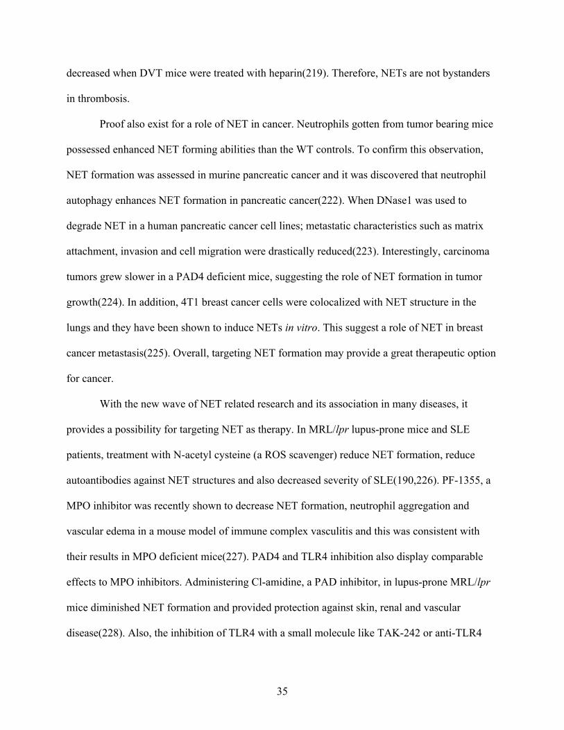
35
decreased when DVT mice were treated with heparin(219). Therefore, NETs are not bystanders
in thrombosis.
Proof also exist for a role of NET in cancer. Neutrophils gotten from tumor bearing mice
possessed enhanced NET forming abilities than the WT controls. To confirm this observation,
NET formation was assessed in murine pancreatic cancer and it was discovered that neutrophil
autophagy enhances NET formation in pancreatic cancer(222). When DNase1 was used to
degrade NET in a human pancreatic cancer cell lines; metastatic characteristics such as matrix
attachment, invasion and cell migration were drastically reduced(223). Interestingly, carcinoma
tumors grew slower in a PAD4 deficient mice, suggesting the role of NET formation in tumor
growth(224). In addition, 4T1 breast cancer cells were colocalized with NET structure in the
lungs and they have been shown to induce NETs in vitro. This suggest a role of NET in breast
cancer metastasis(225). Overall, targeting NET formation may provide a great therapeutic option
for cancer.
With the new wave of NET related research and its association in many diseases, it
provides a possibility for targeting NET as therapy. In MRL/lpr lupus-prone mice and SLE
patients, treatment with N-acetyl cysteine (a ROS scavenger) reduce NET formation, reduce
autoantibodies against NET structures and also decreased severity of SLE(190,226). PF-1355, a
MPO inhibitor was recently shown to decrease NET formation, neutrophil aggregation and
vascular edema in a mouse model of immune complex vasculitis and this was consistent with
their results in MPO deficient mice(227). PAD4 and TLR4 inhibition also display comparable
effects to MPO inhibitors. Administering Cl-amidine, a PAD inhibitor, in lupus-prone MRL/lpr
mice diminished NET formation and provided protection against skin, renal and vascular
disease(228). Also, the inhibition of TLR4 with a small molecule like TAK-242 or anti-TLR4

36
antibody has demonstrated the ability to prevent NET formation and ROS production in vitro
with human neutrophils(229). Another potent way to target NETs is by disrupting their
architecture with DNase and promoting clearance. When DNase 1 was administered intranasally
in patients with primary graft disruption and in a corresponding mice model after lung transplant,
NET formation in the lung was significantly reduced, and improved oxygenation was
observed(230). Recombinant DNase can also decrease autoantibody production and delay
mortality in lupus-prone MRL/lpr mice(231).

37
Figure 3. Potential therapeutic targets in NET formation (68). Targeting pathways, cells, receptors and proteins that lead to NET formation such as TLR, complement proteins and their receptors, MPO, autoreactive B-cells, peptidylarginine deiminase (PAD)4, NADPH oxidase complex and/or mitochondrial activation; and the use of degradation strategies such as DNases can potentially lead to an effective treatment of diseases characterized by dysregulated NET formation.

38
1.5 PI3K signalling A critical early event preceding immune cells activation that affects its overall physiologic
responses including inflammation, phagocytosis, microbicidal activities and chemotaxis is the
activation of the phosphoinositide 3 kinase (PI3K) pathway(232). The PI3K are family of enzymes
that phosphorylate the 3’-hydroxyl group of phosphatidylinositol(233). These enzymes are
associated with numerous cell functions such as cell growth, differentiation, proliferation, motility,
metabolism, trafficking and survival. Numerous studies have demonstrated that PI3K signalling is
involved in the activation and function of B cells, T cells, macrophages, mast cells, DCs,
neutrophils and NK cells. Class IA PI3Ks are heterodimers that consist of a p85 regulatory subunit
and one of the p110 catalytic subunits (p110a, p110b, p110d). The first two isoforms (p110a and
p110b) are expressed in all cells while p110d is primarily expressed in leukocytes including
neutrophils(234,235). Neutrophils possess all forms of class1A PI3K catalytic subunits(236).
Unlike other catalytic subunits which are globally expressed in all cells, the p110d is expressed in
leukocytes primarily (234,235), suggesting that it may have a uunique role and/or function in the
physiology of these cells. Understanding the functions of p110d could explain how it can regulate
immunity.
1.5.1 p110d in Leukocyte signalling and activation. Studies have shown that following BCR and TCR crosslinking with anti-IgM and anti-CD3
antibodies respectively, signalling proteins such as p-ERK, Akt and Ca2+ flux were decreased in B
and T cells of p110dD910A mice (a knock-in mice expressing p110d protein that is catalytically
inactive)(237). In contrast, other studies have shown that TLR and NF-kB signalling are negatively
regulated by p110d activation in macrophages(238). These murine bone marrow-derived
macrophages (BMDM) from p110dD910A mice also exhibit an enhanced MAPK activation(239).

39
Furthermore, TLR4 internalization is controlled by p110d in dendritic cells(240). These
observations suggest that p110d plays a role in cell signalling and activation and this role can differ
depending on the cell type and stimulant.
1.5.2 p110d in Leukocyte migration. Migration of leukocytes was thought to be originally regulated by p110g alone due to their
GPCR association (usually downstream chemokine receptors); however, p110d signalling has
also been implicated in leukocyte migration recently(241). Some studies have shown that
neutrophil migration can be regulated by endothelial p110d signalling as the selective inhibition
of p110d by IC87114 in endothelial cells but not neutrophils decreased flow cell attachment
(241,242). However, the role of p110d in bone marrow derived neutrophil as regards to
migration has not been directly established. Others reported that NK cell migration to SIP and
CXCL10 is regulated by p110d and p110g(243). B cells deficient in p110d fail to migrate to
CXCL13(241). Likewise, T cells show a reduced ability to migrate towards CCL19, CCL21 &
CXCL12 chemokines and nonlymphoid antigenic tissues such as skin & gut(244). These results
show the importance of p110d signalling in migration.
1.5.3 p110d in Leukocyte cytokine production p110d signalling regulates cytokine production in many immune cells. For instance, p110d
deficient NK cells are defective in their ability to produce IFN-g, TNFa and GM-CSF cytokines
following NK1.1, CD16, Ly49D and NKG2D dependent stimulation(245). Likewise, the
production of IL-8 from neutrophils and IL-6 & TNFa from mast cells were impaired in p110d
deficient mice in an allergic murine model(246). The activation of AKT enhanced the production
of IL-10, an anti-inflammatory cytokine, from macrophages(238). In a murine model of sepsis,

40
p110dD910A mice upregulated the production of IL-1b and IL-10 in the lungs simultaneously
following LPS challenge(247). However, another murine sepsis study reported an increase in
TNFa, IL-6, IL-1b and a decrease in IL-10 concentration in the serum following LPS challenge
in p110d deficient mice (240). From the above studies, p110d signalling is also critical for
cytokine production.
1.6 Thesis overview
1.6.1 Study rationale With the numerous data available on the role of neutrophils in inflammation, there still exist some
gaps to be filled. For example, the pathways that regulate inflammasome and NET formation in
neutrophils are not completely understood. In particular, the role of PI3K signaling in orchestrating
neutrophilic inflammatory responses and net formation is poorly defined. ATP‐mediated ROS
production has been shown to stimulate the PI3K pathway, and pharmacological inhibition of PI3K
inhibits ATP‐mediated caspase‐1 activation in macrophages suggesting that PI3K may be involved
in inflammasome activation(248). Moreover, phosphoinositide 3-kinase (PI3K) and AKT have
been linked to NETosis in response to PMA and Leishmania(249); however, the particular
catalytic subunit involved still remains unclear. Previous studies have focused mainly on the role
of p110g in NET formation, inflammation and neutrophil migration as a positive regulator and has
largely neglected the role of p110d in these processes. Hence, little is known about the role of
p110d in the context of inflammasome activation and NET formation. If PI3K signalling occurs in
neutrophils, and neutrophil activity is usually associated with inflammation; then p110δ signaling
like p110g might regulate inflammation and NET formation in neutrophils.
1.6.2 Hypothesis

41
Signaling via the p110d subunit of PI3K regulates inflammasome and NET formation in
neutrophils.
1.6.3 Study objectives
1. Characterize neutrophils from WT and p110dD910A mice - morphology, apoptosis,
migration and migratory receptors, cytokine and chemokine response.
2. Assess inflammasome response in WT and p110dD910A mice.
3. Investigate the contribution of p110dD910A signalling in NET formation.
4. Assess the effect of a pharmacological inhibitor of p110d (CAL101) on inflammasome and
NET formation in Neutrophils.

42
CHAPTER 2: MATERIALS AND METHODS 2.1 MICE Female WT and P110dD910A mice on BALB/C background aged six to eight weeks were
purchased from the University of Manitoba Central Animal care Services (CACS) breeding
facility. The guidelines stipulated by the Canadian Council for Animal Care were followed for
mice usage. The CACS provided specific-pathogen free environment, 12 hours light-dark rhythm
and standard water and chow ad libitum for mice maintenance and survival.
2.2 BONE MARROW CELL ISOLATION Mice were sacrificed by cervical dislocation. Lower limbs were exposed by removing fur. Hind
limbs were dislocated from the hip bone, muscles and tissue were removed to expose tibia and
femur. Tibia was separated from femur and ends of each bone was cut open. With a 10mL
syringe, 25g needle and RPMI medium; the bone marrow was flushed until transparent. Cells
were transferred to a conical tube and gently pipetted until single cell suspension was reached.
Cells were centrifuged for 5 mins at 1200rpm, 1mL ACK lysis buffer (150 mM NH4Cl, 10 mM
KHCO3, 0.1 mM Na2EDTA, pH 7.2-7.4) was used to lyse red blood cells for 5mins, washed,
resuspend in 10mL RPMI and a hemocytometer was used to count cells using trypan blue dye.
Cells are ready to use.
2.3 GENERATION OF MATURED MACROPHAGES FROM BONE MARROW CELLS
Bone marrow cells were resuspended at 4´106/ml. Conditional macrophage media was prepared
by augmenting complete RPMI (RPMI supplemented with 10% heat inactivated FBS, 2 mM
glutamine, 100 U/ml penicillin, and 100 μg/ml streptomycin) with 30% L929 supernatant. In
each 15´100 mm petri dish, 1mL of 4´106 cells were added to 10ml conditional macrophage

43
media. Cells were incubated in 5% CO2 at 37°C. 3 days after incubation, 10mL conditional
macrophage media was used to top up media. Mature macrophages are ready for use after 6 days
and cells are detached with the help of a cell scraper.
2.4 ISOLATION OF NEUTROPHILS FROM BONE MARROW CELLS
1× HBSS (ca2+/mg2+ free HBSS – supplemented with 20mM Na-HEPES & 0.5% FBS) was
prepared (recommended neutrophil medium). Different concentrations of percoll (GE Healthcare
Life Sciences, Mississauga, Ontario, Canada) solutions (45%, 50%, 55%, 62%, 81%) were also
prepared using 1× HBSS while 100% percoll was prepared by mixing 90% percoll with 10%
10× HBSS. Bone marrow cells were resuspended in 3mL of 45% percoll. In a 15mL tube, from
bottom to top, different percoll densities were gently loaded: 3mL-81%, 2mL-62%, 2mL-55%,
2mL-50%, 3ml-45% percoll with cells. This was followed by a centrifuge at 2700rpm for 30mins
without the brakes. Cells from 62%-81% interface were collected as mature PMN. Cells were
briefly washed before use. A second way of isolating neutrophils is the use of a kit. EasySepTM
Mouse neutrophil enrichment kit (19762 – STEMCELL technologies) which uses a negative
selection method was also used for most neutrophil isolation steps. Percoll method of neutrophil
isolation yielded £ 80% neutrophils while the STEMCELL kit yielded ~92% neutrophil purity
from bone marrow cells.
2.5 INFLAMMASOME ACTIVATION
1µg/mL LPS was used to prime cells and incubated for 6 hours (macrophages) and 4 hours
(neutrophils) at 37°C. 5µM nigericin or 5mM ATP was used as a second signal to activate the
inflammasome and incubated for 45mins(macrophages) and 4 hours (neutrophils) at 37°C.

44
Supernatant was collected and stored for ELISA. Cells were collected and used for western
blotting.
2.6 ELISA
Sandwich ELISA method was used to quantify IL-1b (ELISA max standard set mouse IL-1b;
Cat# 432601 by biolegend), IL-6, TNF-a(ELISA max standard set mouse TNF-a; Cat# 430901
by biolegend), CXCL1(Mouse CXCL1/KC DuoSet ELISA; cat# DY453-05 by R&D systems),
IL-12p40(ELISA max standard set mouse IL-12/IL-23 (p40); Cat# 431601 by biolegend), IL-
10(ELISA max standard set mouse IL-10; Cat# 431411 by biolegend). Procedure was performed
according to manufacturer’s suggested protocols. Briefly, purified coating antibody was used to
coat plates (50µL/well) in an ELISA coating buffer and incubated at 4ºC overnight. Plate was
washed 4-5 times in 1´PBS + 0.05% TWEEN-20. 100µL of assay diluent (5%FBS in 1´PBS)
was used to block the each well for 1 hour and washed. 50µL of standard or sample was
added/well. Standards and samples were added, and serial dilution was done in assay diluent.
This was also incubated overnight at 4ºC. After washing, 50µL of Biotin Antibody was added
per well and incubated for 2hours at 37ºC. After washing, 50µL of Avidin HRP was added per
well and incubated for 40mins at 37ºC. After washing, 50µL of ABTS peroxidase substrate or
TMB substrate was added for 15mins at room temperature away from light. The reaction was
stopped using 50µL of 2N H2SO4 per well. After the desired color development, plate was
acquired at 450nm (Spectra Max).
2.7 WESTERN BLOT EXPERIMENTS BMDMs and BMDNs whose inflammasome were activated were collected. 200µL of 1×
laemmli sample buffer was used to directly lyse 106 cells - make a solution of 1× laemmli

45
sample buffer by mixing 2× laemmli sample buffer (Sigma Aldrich – S3401), ddH2O and
protease inhibitors (Sigma Aldrich – 11836170001). Mixture was transferred to a 1.5mL
Eppendorf tube and lysed for 30 mins while vortexing every 10 mins. This was immediately
followed by centrifugation for 10mins at 10,000rpm at 4 ºC. Clear lysate without cell pellet was
removed to a clean Eppendorf tube and was boiled at 95ºC for 10 mins. 10µL clear lysate was
loaded and resolved on SDS-PAGE polyacrylamide (4561104 – BIORAD) under denaturing
conditions at 100v for 1 hour and was wet transferred to a methanol charged PVDF membrane
(Amersham Pharmacia Biotech) at 100v for 1 hour. Block membrane with 5% BSA in Tris-
Buffered Saline and 0.1% Tween 20 (TBST) at room temperature for 1 hour. Add primary
antibody of Rabbit anti - mouse CARD8 (Abcam), Rabbit anti - mouse NLRP3, Rabbit anti -
mouse Caspase1, Rabbit anti - mouse IL-1b, Rabbit anti - mouse ASC, Rabbit anti - mouse B-
actin (Cell Signaling Technology, MA, U.S.A) in 2% BSA in TBST and incubate overnight in 4
ºC. Membrane was washed 4× for 5 mins with TBST the next day and secondary antibody (Goat
anti – rabbit IgG – Cell Signaling Technology) was added in 2% BSA in TBST and incubate for
1 hour. Wash step was repeated and ECL substrate (RPN2232 - Amersham Pharmacia Biotech)
was used to image. Membrane was stripped for 15 mins using a stripping buffer (46430 –
Thermofisher Scientific) for loading controls. BIORAD image lab software on Chemidoc MP
imaging system was used to measure band intensity.
2.8 FLOW CYTOMETRY EXPERIMENTS Extracellular and intracellular flow cytometry staining used Fluorochrome - conjugated
monoclonal antibodies according to the manufacturer’s protocol. To summarize, FACS buffer
(PBS containing 0.01% sodium azide and 5% FBS) was used to wash cells. Fc-block

46
(0.25µg/tube; eBiosciences) was added to cells for 10 mins on ice and washed. Fluorochrome
conjugated antibodies against CD11b, Ly6G, CCR1, and CXCR2 (eBiosciences) was used to
stain the surface of the cells. 2% - 4% paraformaldehyde (Sigma Aldrich) was used to fix cells
after surface staining for 10 mins on ice; cells can be washed and analyzed or permeabilized with
0.1% saponin (Sigma Aldrich) in staining buffer if continuing onto intracellular staining.
Predetermined concentration of fluorochrome-conjugated mAbs against MPO (Abcam) diluted
in saponin buffer, was added to permeabilized cells. Azide-free Flow buffer was used for MPO
staining. Annexin V and 7AAD (eBiosciences) were used to determine cell viability. BD FACS
Canto II (BD Bioscience, Mississauga, ON, Canada) was used to acquire cells and Flowjo
software to analyze results (Tree Star Inc, Ashland, OR).
2.9 CYTOSPIN
Isolated neutrophils (~105 cells) were deposited on microscope slides by cytospin centrifugation
(ThermoShandon, Pittsburgh, PA). Hematoxylin and Eosin (H&E) solutions were then used to
stain the slides and dried. Zeiss Primostar iLED microscope (Carl Zeiss, Ontario, Canada) was
used to capture neutrophils.
2.10 NET INDUCTION Separate poly-L-lysine coated glass coverslips (NEU-GG-12-PLL; Neuvitro) and place each one
into each well of a sterile 12-well cell culture plate. Onto the center of the poly-L lysine
coverslip, gently pipette about 100µL (106 cells/µL) of resuspended BMDN stimulated with
100nM PMA and/or LPS 1ug/ml (from E. coli [O111:B4, LPS25 – Sigma Aldrich] or P.
aeruginosa[L9143 – Sigma Aldrich]). Cover the culture plate and incubate for 3-5 hours at 37°C
in an incubator to allow to proper attachment and stimulation.

47
2.11 IMMUNOFLUORESCENCE MICROSCOPY
Following NET induction, cells that did not attach to poly-L-lysine coated glass coverslips were
washed with PBS 3 or more times and aspirated with a pipette. 2% - 4% paraformaldehyde
(Sigma Aldrich) was used to fix cells for 15 mins at room temperature. Wash PFA away and
store at 4°C until ready to stain cells or continue immediately with the staining process. Ice cold
methanol was used to permeabilize attached cells in a -20°C freezer for 10 mins, cells were
washed with PBS and blocked immediately with 3% BSA at room temperature for 1 hour. Rabbit
anti-mouse Neutrophil elastase (ab68672) and rat anti-mouse Histone H3(ab10543) primary
antibodies (Abcam) were used to stain adherent cells at a dilution of 1:200 in 3% BSA at room
temperature for 2 hours (or incubate overnight in a humified chamber at 4°C. Goat anti-rabbit
IgG Alexa546 (A-11010 Molecular Probes) and goat anti-rat IgG FITC (CLCC40001 –
cedarlane) secondary antibodies were used to stain adherent cells at a dilution of 1:1000 in 3%
BSA at room temperature for 1 hours. DAPI was used to counterstain nucleus at a dilution of
1:5000 (5mg/mL) for 10 mins at room temperature. A small drop of fluorescent mounting media
was used to mount coverslips unto slides, and they were visualized via a Zeiss CSU-X1M 5000
spinning Disc Confocal Microscope (Carl Zeiss, Ontario, Canada) with a 63x oil immersion
objective.
2.12 RNA ISOLATION, CDNA SYNTHESIS AND RT PCR
After inflammasome activation invitro, trizol reagent was used to isolate BMDM and BMDN’s
RNA. About 300µl of trizol reagent was added to 106 cells and homogenized by pipetting
immediately after treatment without washing cells. The mixture I transferred to a 1.5ml
Eppendorf tube and left to lyse for 5 minutes. 0.2ml of chloroform per 1ml trizol used was added
to the same Eppendorf tube and mixed. This was incubated for 2-3 minutes and centrifuged at

48
12000xg at 4°C for 15 minutes. The top aqueous phase containing the RNA was transferred into
a new tube and 0.5ml isopropanol per 1ml trizol used was added to precipitate the RNA. This
was incubated for 10 minutes and centrifuged for 10 minutes at 12000xg at 4°C. The supernatant
was decarded and 1ml of 75% alcohol per 1ml of trizol used to added to wash the RNA pellet.
Mixture was centrifuged for 7500xg at 4°C for 5 minutes, supernatant removed, air dried, RNA
pellet solubilized in 20µl RNase-free water and heated for 10 minutes in a water bath set at 55°C.
RNA can be stored at -80°C or immediately converted to cDNA. cDNA conversion was done by
a first strand cDNA synthesis kit (E6300L – New England Biolabs) according to manufacturer’s
protocols and PCR was performed using Luna universal qPCR master mix (M3003X - New
England Biolabs) also according to manufacturer’s protocols.
No. Name Sequence 1. Procaspase 1 F - ACAAGGCACGGGACCTATG
R - TCCCAGTCAGTCCTGGAAATG 2. ProIL1b F - GCAACTGTTCCTGAACTCAACT
R- ATCTTTTGGGGTCCGTCAACT 3. NLRP3 F – ATTACCCGCCCGAGAAAGG
R - TCGCAGCAAAGATCCACACAG 4. ASC (PYCARD) F – CTTGTCAGGGGATGAACTCAAAA
R - GCCATACGACTCCAGATAGTAGC 5. GAPDH F - AGGTCGGTGTGAACGGATTTG
R - TGTAGACCATGTAGTTGAGGTCA Table 1. List of primers used in RT qPCR.
2.13 TRANSWELL MIGRATION ASSAY BMDN were tested for their ability to migrate towards CXCL1 and MIP1a chemokines. Here,
106 neutrophil cells in 100µl were placed on the top chamber containing a permeable membrane
with a 3µm pore size and the chemokine prepared in 500µl complete RPMI was placed in the

49
bottom chamber. After 1,2 and 3 hours; cells that migrated towards the chemokines were counted
using a hemocytometer.
2.14 SEPSIS INDUCTION This was done by injecting LPS from E. coli (O111:B4, LPS25 – Sigma Aldrich) into WT and
P110dD910A mice intraperitoneally.
2.15 PICOGREEN ASSAY
Following NET induction invitro or collection of peritoneal wash & serum from LPS treated
mice; Quanti-iT PicoGreen dsDNA assay kit (P11496 - Invitrogen) was used to quantify NET
formation and this was done according to the manufacturer’s protocol. To summarize, a working
concentration of Quanti-iT PicoGreen reagent and DNA stock solution (standard) in 1x TE
buffer was prepared and the standard dilutions was made according to manufacturer’s
instructions. 100µl of standards and samples were first added into the 96 well plate. A working
concentration of 100µl Quanti-iT PicoGreen reagent was added to each well. This was mixed by
light shaking and incubated for 2-5 minutes at room temperature. A synergy Neo2 multi-mode
reader was used to determine the fluorescence following incubation using a monochromator-
based method with an excitation wavelength of 480nm and an emission wavelength of 520nm.
The data was collected from the top with 10 reads per well. Fluorescence value of blank was
subtracted from those of the samples and the resulting data was used to generate a standard curve
where the DNA concentration could be calculated from the resulting fluorescent values.

50
2.16 DIMINAZENE ACETURATE (BERENIL)
BMDN and BMDM from WT mice were treated with 10µg/ml Berenil for 4 hours before
inflammasome activation to assess the function of Berenil treatment of inflammasome formation
and cytokine production.
2.17 IDELALISIB (CAL101) BMDN and BMDN from WT mice were treated with 10µm CAL101/Idelalisib which is a
pharmacological inhibitor of P110d for 4 hours prior to inflammasome activation to assess its
effect on inflammasomes, NET formation and cytokine production.
2.18 STATISTICAL ANALYSIS Most results were represented in bar graphs and were presented as the mean ± SEM. One way or
two-way analysis of variance (One-way or Two-way ANOVA) was used for comparison of
cytokine & chemokine production, neutrophil migration, chemokine receptor expression and
inflammasome complex protein expression between the different mice groups using Prism
program (GraphPad Software Inc., CA, USA). If p ≤ 0.05, the result is considered to be
significant.
2.19 ETHICAL STATEMENT The University of Manitoba Human and Animal Ethics Review Boards approved and reviewed
all animal studies that supported this work.

51
CHAPTER 3: RESULTS 3.1 WT and P110dD910A neutrophils have similar morphology, survival rate in vitro, and
surface markers expression.
PI3K has been shown to regulate many cell functions including survival and signalling(234). I
sought to find out whether P110d deficiency in neutrophils would affect basic structure and
functions such as survival and expression of surface markers. To address this, I isolated bone
marrow-derived neutrophils (BMDN) from WT and P110dD910A mice. About 105 cells were
immobilized to slides by cytospin and stained with Giemsa solution to characterize neutrophil
morphology. Next, WT and P110dD910A BMDN were then assessed for their ability to express
CD11b and Ly6G; which are two surface markers that characterize neutrophils. WT and
P110dD910A BMDN were treated with LPS at different concentrations (0,100,1000) ng/mL for 48
hours and later stained with Annexin V and 7AAD to assess their ability to survive in vitro in
LPS. I observed similar numbers and morphological structure of neutrophil in both WT and
P110dD910A BMDNs (Fig. 4A). In addition, both WT and p110dD910A BMDN express the similar
levels of CD11b and Ly6G molecules on their surfaces (Fig. 4B). Also, WT and P110dD910A
BMDN have similar survival rates irrespective of the presence or absence of LPS or an increase
in its concentration (Fig. 4C). Collectively these results show that WT and P110dD910A BMDNs
do not differ morphologically and their response to LPS stimulation were identical, suggesting
that any difference in inflammasome or NET formation between WT and P110dD910A BMDN
would not be due to differences in any of these basic neutrophil characteristics.

52
Figure 4. WT and P110dD910A neutrophils have similar morphology, survival rate in vitro, and surface markers expression. Neutrophils from wild type (WT) and P110dD910A mice were isolated from bone marrow and stained with Giemsa stain to assess morphology (A). Some cells were stained with anti-Ly6G and CD11b antibodies (B), or stimulated with or without different concentrations of LPS, and stained with 7AAD and Annexin V dyes to assess cell viability (C) by flow cytometry
C

53
3.2 P110dD910A neutrophils have reduced chemokine receptor expression and impaired
migration compared to WT neutrophils.
Neutrophils are one of the first responders to the site of an inflammation response in tissues. In
both in vitro and in vivo models of inflammation, a reduction but not total abolition in migratory
abilities was observed in neutrophils and macrophages from P110γ −/− mice (ref). This indicates
that other PI3K subunits might have a role to play in cell migration(250,251). Therefore, I
wanted to assess the effect of P110dD910A mutation on neutrophil migration. A transwell
migration assay was used to achieve this. BMDN (top chamber) was exposed to two chemokines
(CXCL1 and MIP1a) and the number of neutrophils they attracted over time was counted. I
observed that P110d deficient neutrophils were defective in their ability to migrate towards the
chemokines, especially at the early time points (Fig. 5A). Next, I wanted to assess if this
observed defect in migration was due to reduced expression of their corresponding chemokine
receptors on P110dD910A neutrophils. To carry out this study, I exposed BMDN to LPS and
stained them for expression of CXCR2 and CCR1, which are receptors for CXCL1 and MIP1a,
respectively. Following flow cytometry analysis, I observed that P110dD910A neutrophils
expressed less CXCR2 and CCR1 when compared to their WT counterparts even without LPS
stimulation (Fig. 5B, C). Therefore, P110dD910A neutrophils have impaired migration due to their
inability to sense chemokines via its chemokine receptors.

54
Figure 5: P110dD910A neutrophils have impaired migration and chemokine receptors compared to WT neutrophils. Bone marrow-derived neutrophils from wild type (WT) and P110dD910A mice were assessed for migration towards CXCL1 and MIP1a (A) gradients using a transwell migration assay. The expression of CXCR2 and CCR1 receptors by neutrophils from WT and P110dD910A mice were also assessed by flow cytometry. Shown are histograms (B) and mean fluorescent intensity (MFI, C) after stimulation with or without LPS. ns = not significant; *, p<0.05; **, p<0.01; ***, p<0.001.

55
3.3 CAL101 at different concentrations does not impair survival of neutrophils.
Given that we do not normally knock-in genes in humans in our attempt to regulate
inflammation, using a pharmacological inhibitor of P110d called CAL101 or Idelalisib is ideal.
This would be the best way to translate my result to real situations in humans. Before that, I
wanted to test CAL101 at different concentrations to make sure it does not impair the survival of
neutrophils. BMDN were isolated from both WT and P110dD910A mice, treated with CAL101 at
different concentrations (0,0.1,1,10) µM for 4 hours and later stained with Annexin V and 7AAD
to assess their ability to survive in vitro in CAL101. WT and P110dD910A BMDN have similar
survival rates irrespective of the presence or absence of CAL101 or an increase in its
concentration (Fig. 6A).
Figure 6. CAL101 at different concentrations does not impair survival of neutrophils. Neutrophils from wild type (WT) and P110dD910A mice were isolated from bone marrow, treated with CAL101 at different concentrations (0,0.1,1,10) µM for 4 hours and stained 7AAD and Annexin V dyes to assess and cell viability by flow cytometry.

56
3.4 P110dD910A myeloid cells have a defect in cytokine and chemokine production post LPS
stimulation.
Next, I wanted to assess the implication of P110d mutation on proinflammatory response in
neutrophils. Before that, it was wise to first assess proinflammatory response in macrophages as
they are easier to work with, and many studies have used macrophages to assess
proinflammatory cytokine response(84,95,248). Therefore, I used these cells to optimize
efficiency of my system. Bone marrow-derived macrophages (BMDMs) were isolated from both
WT and P110dD910A mice and stimulated with LPS for 6 hours and the culture supernatant fluid
was collected and assayed for IL-6 and TNF-a protein levels by ELISA. I observed that
P110dD910A macrophages were defective in both IL-6 and TNF-a production when compared to
their WT counterparts (Fig. 7A, B). Now that I know my system is working, I went on to assess
the effect of P110d mutation on cytokine and chemokine production in neutrophils. BMDNs
were stimulated with LPS at different concentrations (0,10,100,1000 ng/mL) for 4 hours, and the
culture supernatant fluids were assayed for IL-6, TNF-a and CXCL1 protein levels by ELISA. I
noticed that BMDNs from P110dD910A produced decreased amounts of IL-6, TNF-a and CXCL1
(Fig. 7C-E) compared to their WT counterparts.

57
Figure 7: P110dD910A myeloid cells have a defect in cytokine and chemokine production following LPS stimulation. Bone marrow derived macrophages (BMDM; A, B) or neutrophils (BMDN; C, D, E) from wild type (WT) and P110dD910A mice were stimulated with LPS. The levels of IL-6 (A, C), TNF-a (B, D) and CXCL1 (E) in the culture supernatant fluids were determined by ELISA. **, p<0.01; ****, p<0.0001.

58
3.5 CAL101 treated macrophages and neutrophils are impaired in their ability to produce
proinflammatory cytokines like IL6 and TNFa
Next, I wanted to investigate the effect of CAL101 treatment on proinflammatory cytokine
production in macrophages and neutrophils given the results seen in section 3.4. I isolated and
grew BMDM and BMDN, treated them with CAL101 at different concentrations (1,10) µM for 4
hours before stimulating them with LPS from E. coli or P. aeruginosa and nigericin. Supernatant
was collected; IL-6 and TNFa cytokine levels were measured by ELISA. BMDM (Fig. 8A, B)
and BMDN (Fig. 8C, D) that were treated with CAL101 had a defect in TNFa and IL-6
production when compared to their WT counterparts irrespective of the LPS and the
concentration of CAL101 used. This is consistent with the result gotten from the p110dD910A
macrophages and neutrophils when compared to WT in Figure 7.

59
Figure 8. CAL101 treated macrophages and neutrophils are impaired in their ability to produce proinflammatory cytokines like IL6 and TNFa BMDM and BMDN from wild type (WT) mice were treated with varying concentrations of CAL101(1,10) µM for 4 hours before stimulation with LPS from E. coli and P. aeruginosa (A, B). The levels of IL-6 (A, C) and TNF-a (B, D) in the culture supernatant fluids were determined by ELISA. ****, p<0.0001.
C D

60
3.6 P110d isoform of PI3K is dispensable for NLRP3 inflammasome response in
neutrophils but not macrophages.
The inflammasome facilitate the maturation of pro-inflammatory cytokines such as IL-1b.
Figures 4, 5 shows that P110d is important for pro-inflammatory response not requiring the
inflammasome in both macrophages and neutrophils. Next, I wanted to investigate if P110d is
critical for inflammasome response in macrophages and neutrophils. BMDMs and BMDNs were
isolated from both WT and P110dD910A mice, treated with LPS and nigericin as described in the
materials and methods section and the supernatant was collected and used for ELISA. I observed
that p110d deficient macrophages were defective in their ability to produce IL-1b (Fig. 9A) as
observed for IL-6 and TNF-a(Fig. 7, 8 (A, B)) when compared to their WT counterparts.
Surprisingly, P110d deficient neutrophils produce similar amount of IL-1b as WT neutrophils
and this was very different from what was observed for other proinflammatory cytokines
including IL-6 and TNF-a(Fig. 7,8 (C, D)). To validate this, I used CAL101 to selectively inhibit
P110d in WT BMDM (Fig. 9C) and BMDN (Fig. 9C, D). This was then followed by LPS and
Nigericin or ATP treatment to activate the inflammasome. This led me to conclude that
inflammasome signalling in macrophages requires p110d signaling but this pathway is
dispensable in neutrophils.
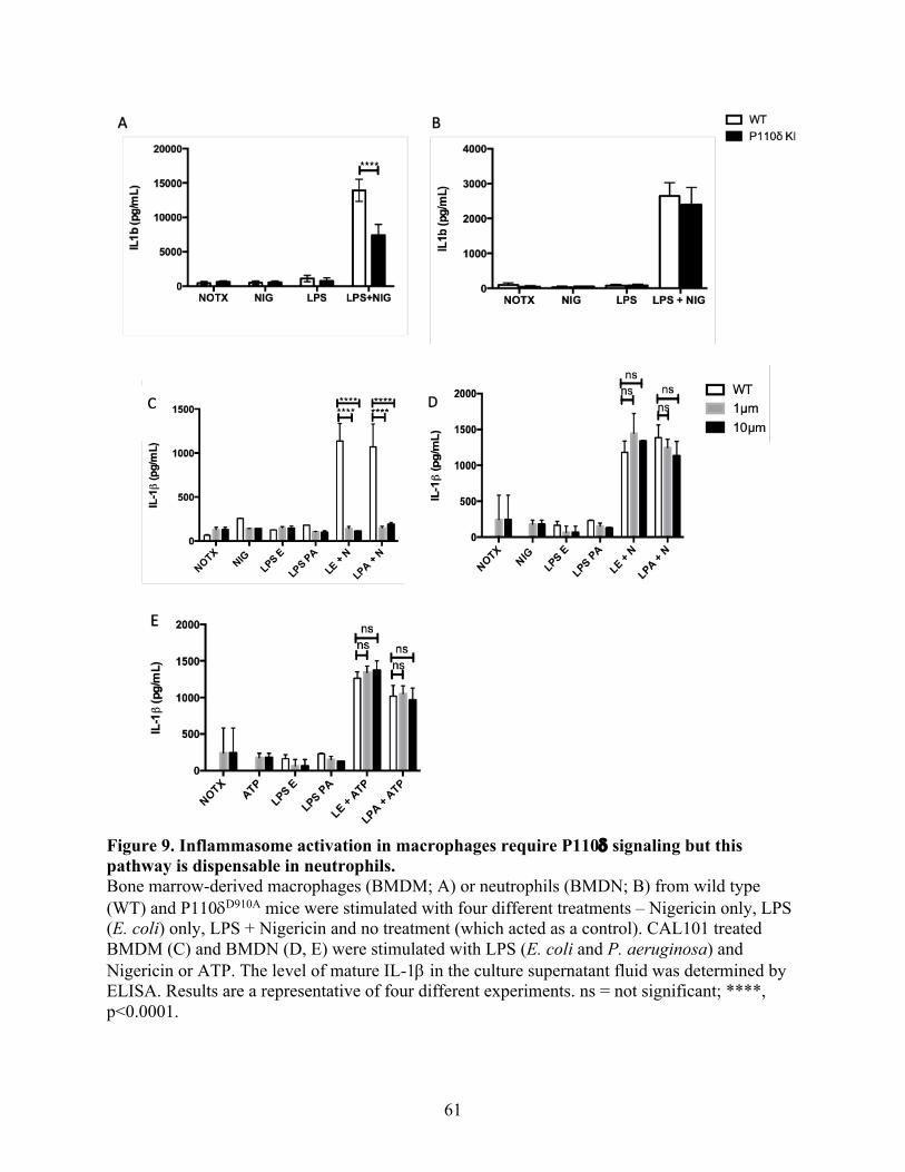
61
Figure 9. Inflammasome activation in macrophages require P110d signaling but this pathway is dispensable in neutrophils. Bone marrow-derived macrophages (BMDM; A) or neutrophils (BMDN; B) from wild type (WT) and P110dD910A mice were stimulated with four different treatments – Nigericin only, LPS (E. coli) only, LPS + Nigericin and no treatment (which acted as a control). CAL101 treated BMDM (C) and BMDN (D, E) were stimulated with LPS (E. coli and P. aeruginosa) and Nigericin or ATP. The level of mature IL-1b in the culture supernatant fluid was determined by ELISA. Results are a representative of four different experiments. ns = not significant; ****, p<0.0001.

62
3.7 P110dD910A the expression of inflammasome genes in macrophages and neutrophils is
similar in WT macrophages and neutrophils from WT and P110dD910A mice.
I observed a significant difference in IL-1b production by macrophages and not neutrophils from
WT and P110dD910A mice (Fig 9), so I sought to investigate if P110d deficient macrophages and
neutrophils were expressing the inflammasome complex genes such as pro- IL-1b, pro-caspase1,
NLRP3, ASC differently. BMDMs and BMDNs were stimulated with LPS and nigericin as
described in the material and methods section and mRNA was extracted, converted into cDNA
and used for real-time PCR analysis. Consistent with the IL-1b ELISA result, there was no
significant difference between the expression of pro- IL-1b, pro-caspase1, NLRP3, ASC mRNA
in P110dD910A and WT macrophages (Fig. 10A). In contrast, P110dD910A neutrophils seem to
express slightly more procaspase1 and NLRP3 mRNA levels than WT neutrophils (Fig. 10B).
However, there was no significant difference in the expression of ASC and pro- IL-1b mRNA by
WT and P110dD910A neutrophils Fig. 10B).

63
Figure 10. P110dD910A macrophages express inflammasome genes similarly to WT macrophages, which is not necessarily the case with neutrophils. Bone marrow derived macrophages (BMDM; A) or neutrophils (BMDN; B) from wild type (WT) and P110dD910A mice were stimulated with four different treatments – Nigericin only, LPS only, LPS + Nigericin and no treatment which acted as a control. This was followed by RNA isolation and cDNA preparation which was later used for quantifying the level of pro-IL1b, pro-caspase1, NLRP3, ASC gene expression by qPCR and was expressed as Fold change. ns = not significant; *, p<0.05.

64
3.8 P110d isoform of PI3K regulates procaspase 1 production in macrophages but not in
neutrophils.
Although there appear to be little or no differences in the expression of inflammasome mRNA in
macrophages and neutrophils from WT and P110d deficient mice, it is conceivable that
differences may exist in the protein levels because of differences in translational regulation.
Therefore, I sought to investigate whether P110d deficient macrophages and neutrophils were
expressing different levels of the inflammasome complex proteins (such as ASC, NLRP3, pro-
IL-1b, pro-caspase1) and its negative regulators (such as CARD8). BMDM and BMDN were
stimulated with LPS and nigericin and cell lysates were prepared and used for western blot. I
observed that P110d deficient macrophages showed a significant decrease in the expression of
pro-caspase1 (the enzyme that cleaves pro-IL-1b into mature IL-1b) and increased expression of
pro-IL-1b when compared to WT macrophages (Fig. 11A, B). However, there was no significant
change in ASC, NLRP3 and CARD8 expression (Fig. 11A, B). In contrast to macrophages,
P110d deficient neutrophils maintained similar expression of inflammasome related proteins
similar to their WT counterparts (Fig. 11C, D). The decrease observed in the expression of pro-
caspase1 in P110d deficient macrophages would most likely translate to less caspase1 enzyme
available. This would be the rate limiting step as less caspase 1 expression would lead to less
mature IL-1b cleaved, which would explain the observed effect of P110d signaling in
macrophages. In contrast, since the expression of pro-caspase1 in P110d deficient neutrophils
doesn’t differ from that observed in WT neutrophils, IL-1b expression would remain the same.
In conclusion, P110d isoform of PI3K regulates procaspase 1 production in macrophages but not
in neutrophils.
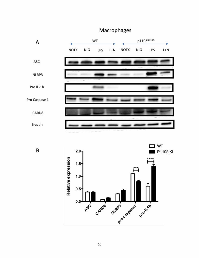
65

66
Figure 11. P110d isoform of PI3K regulates procaspase 1 production in macrophages but not in neutrophils. Bone marrow derived macrophages (BMDM; A and B) or neutrophils (BMDN; C and D) from wild type (WT) and P110dD910A mice were stimulated with with or without LPS in the presence or absence of Nigericin. The cell lysate was prepared and used for western blot to probe for the expression of ASC, NLRP3, pro- IL-1b, pro-caspase1, CARD8 proteins. ***, p<0.001, ****, p<0.0001.

67
3.9 Berenil downregulates LPS induced proinflammatory cytokine production and
inflammasome response while upregulating anti-inflammatory cytokine production in bone
marrow derived macrophages.
Berenil can function as an immunoregulatory agent. According to Kuriakose 2014, Berenil can
downregulate proinflammatory cytokine production in macrophages by downregulating the
phosphorylation of MAPKs, STATS, NFKb and upregulating SOCS phosphorylation(39).
However, it’s role in inflammasome is yet to be determined. To investigate this, I isolated
BMDMs and treated them with Berenil prior to stimulation with LPS and nigericin. I observed
that Berenil treatment downregulated IL-1b production, which is regulated by the inflammasome
response (Fig. 12A). In addition, and consistent with previous reports (39), Berenil pre-treatment
also downregulated the production of IL-6, TNF-a, and IL-12p40 (Fig. 12B, C, E), cytokines
that do not require the inflammasome for maturation. In contrast, IL-10, which is an anti-
inflammatory cytokine, was upregulated by Berenil treatment (Fig. 12D). Collectively, these
findings indicate that Berenil may be targeting the inflammasome response in a specific manner
that is distinct from its effect on production of proinflammatory cytokines.

68
Figure 12. Berenil downregulates inflammasome activity and other proinflammatory cytokines but upregulates anti-inflammatory cytokine in macrophages. WT Bone marrow derived macrophages (BMDM) were treated with Berenil (10µg/mL) for four hours prior to LPS and nigericin stimulation, then collected their supernatant for IL-1b, IL-6, TNF-a, IL-12p40 and IL-10 ELISA.

69
3.10 Berenil downregulates LPS-induced proinflammatory cytokine production and
inflammasome response while upregulating anti-inflammatory cytokine production in bone
marrow-derived neutrophils.
The preceding result showed that Berenil downregulates LPS-induced cytokine production and
inflammasome response in macrophages while upregulating anti-inflammatory response. Next, I
wanted to investigate if Berenil has a similar effect on neutrophils. I isolated BMDN and treated
them with Berenil prior to LPS and nigericin stimulation, then collected their supernatant for
ELISA. As with macrophages, I observed that Berenil treatment downregulated IL-1b
production (which is regulated by the inflammasome) (Fig. 13A). Berenil treatment also
downregulated IL-6 and TNF-a production (Fig. 13B, C) which do not require the
inflammasome for production. As with macrophages, IL-10 which is an anti-inflammatory
cytokine, was upregulated by Berenil treatment (Fig. 13D). IL-12p40 was undetectable in bone
marrow-derived neutrophils. This observation indicates that Berenil affects the inflammasome
and proinflammatory cytokine responses in neutrophils and macrophages similarly.

70
Figure 13. Berenil downregulates inflammasome activity and other proinflammatory cytokines but upregulates anti-inflammatory cytokine in Neutrophils. WT Bone marrow derived neutrophils (BMDN) were treated with Berenil (10 µg/mL) for four hours prior to LPS and nigericin stimulation. The culture supernatant fluids were collected and assayed for IL-1b, IL-6, TNF-a, IL-12p40 and IL-10 ELISA.

71
3.11 Qualitative evidence suggests that P110d is critical for NET formation.
P110g signaling has been shown to be critical for NET formation following Leishmania
infection(249), but little is known about the role of P110d in NET formation. Therefore, to
investigate the role of P110d in NET formation, BMDN were isolated from both WT and
P110dD910A mice. In addition, BMDN were isolated from WT and treated with the P110d selective
inhibitor, CAL101. NET formation was induced with PMA and/or LPS (from E. coli or P.
aeruginosa). Immunofluorescence staining was performed for elastase (orange) and histone H3
(green). DAPI dye (blue) was used to stain the nuclei. Result was visualized by confocal
microscopy. I observed reduced NET formation in P110dD910A neutrophils compared to WT
neutrophils. I also observed reduced NET formation in CAL101 treated neutrophils compared to
WT neutrophils. This indicates that P110d is critical for NET formation following PMA and/or
LPS stimulation. In addition, LPS from P. aeruginosa produced more NETs than LPS from E.
coli. This implied that the virulence of the bacteria LPS can be influence NET formation in the
neutrophils.

72
Figure 14. NET formation is reduced in P110dD910A neutrophils, CAL101 treatment reduced NET formation, and LPS from P. aeruginosa is capable of inducing more NET than LPS from E. coli. Bone marrow derived Neutrophils were incubated on Poly-L-Lysine coated coverslips for 5 hours with or without CAL101(1µM) in the presence of PMA (100 nM) and LPS (1mg/mL) from E. coli or P. aeruginosa. NET formation was assessed by staining with anti-Histone H3 (green) and anti-Elastase (orange) antibodies and the images were assessed by confocal microscopy. DAPI (blue) staining was included to visualize cell nuclei.

73
3.12 Quantitative evidence suggests that P110d is critical for NET formation.
Following my observation that P110d is critical for NET formation where NET formation was
assessed via immunofluorescent microscopy (Fig 14), I went ahead to verify this using a
quantitative measure of dsDNA known as picogreen assay. To achieve this, BMDN were
isolated from both WT and P110dD910A mice. In addition, BMDN were isolated from WT and
treated with the P110d selective inhibitor, CAL101. NET formation was induced with PMA
and/or LPS (from E. coli or P. aeruginosa). The cell culture was used to perform the picogreen
assay according to section 2.15 and a fluorescent microplate reader was used to determine the
amount of dsDNA fluorescence present. As it was with the confocal microscopy result in section
3.11, I observed reduced NET formation in P110dD910A neutrophils compared to WT neutrophils.
I also observed reduced NET formation in CAL101 treated neutrophils compared to WT
neutrophils. This indicates that P110d is critical for NET formation following PMA and/or LPS
stimulation.
74
Figure 15. Quantitative evidence suggests that P110d is critical for NET formation. Bone marrow derived Neutrophils were treated with or without CAL101(1µM) in the presence of PMA (100 nM) and LPS (1mg/mL) from E. coli or P. aeruginosa. Net formation was assessed by picogreen assay and the resulting amount of dsDNA fluorescence present was determined with a fluorescent microplate reader. ****, p<0.0001. 3.13 P110d signaling regulate NET formation in an animal model of sepsis.
Finally, I wanted to know if P110d specifically regulates NET formation in an animal model of
sepsis, which is a form of dysregulated inflammation. To induce Sepsis, WT and P110dD910A
mice were challenged with 2.5mg/kg LPS from E. coli for 6 hours. This was immediately
followed by peritoneal wash (PW) and blood collection. Immunofluorescence staining was
performed for elastase (orange) and histone H3 (green), while DAPI dye (blue) was used to stain
the nuclei. Result was visualized by confocal microscopy. Concurrently, picogreen assay was
performed according to section 2.15 and a fluorescent microplate reader was used to determine
the amount of dsDNA fluorescence present. I observed that P110dD910A mice is able to decrease
the production of NET in both PW and blood sites following LPS challenge. This was observed
both qualitatively with confocal microscopy (Fig. 16A) and quantitatively with picogreen assay
NOTXLPS E
LPS E + PMA
LPS PA
LPS PA + PMA
0
200
400
600
800D
NA
(ng/
ml)
WTKI1µm CAL10110µm CAL101
****
****
****
****

75
(Fig. 16B, C). These results confirm the in vitro data seen in Figure 14 and 15, suggesting that
the absence of P110d signaling leads to decreased Net formation in vivo.
Figure 16. Absence of P110d signalling leads to decreased NET formation in vivo. WT and p110dD910A mice were challenged with 2.5mg/kg LPS from E. coli for 6 hours. This was immediately followed by peritoneal wash (PW) and blood collection. NET formation was assessed by staining with anti-Histone H3 (green) and anti-Elastase (orange) antibodies and the images were assessed by confocal microscopy. DAPI (blue) staining was included to visualize cell nuclei (A). Alternatively, picogreen assay was used to quantify NET and the resulting amount of dsDNA fluorescence present was determined with a fluorescent microplate reader (B, C). **, p<0.01; ****, p<0.0001.

76
CHAPTER 4: DISCUSSION 4.1: GENERAL DISCUSSION
In recent years, Phosphatidylinositol 3-kinases has been recognized as an important
protein responsible for influencing various activities of immune and non-immune cells, including
proliferation, differentiation, and effector activities. Different studies are beginning to decipher
the distinct cell specific roles for different Phosphatidylinositol 3-kinase subunits in immunity
and inflammation (252).
The findings from this study clearly highlights the role played by the P110d subunit of
PI3K play in neutrophil migration, inflammasome activation and NET formation. To overcome
limitations that arise when only one study approach is used and hence validate the observations
reported here, I used both genetic and pharmacological inhibition of P110d subunit of PI3K to
investigate its role in neutrophil functions. My findings suggest that signaling via the P110d
isoform of PI3K plays an important role in regulating migration and NET formation but is
dispensable for inflammasome response in neutrophils.
According to Lieschke, Keightley and Manley (2018), the physiological state of a
neutrophil cell can affect its plasticity, migratory properties and maturation(253). Accelerated
apoptosis can decrease neutrophil’s lifespan and downregulate its function while delayed
apoptosis can upregulate neutrophil’s function and increase its lifespan(254). Neutrophils
possess a variety of cell surface molecules such as CD11b and Ly6G for pathogen recognition,
migration and differentiation(255,256). Therefore, my discovery that P110d subunit of PI3K
does not affect neutrophil’s morphology, survival rate or surface molecule expression was
critically important because any other differences observed in this study could not be attributed
to structural differences, apoptosis or surface marker expression on neutrophils.

77
The P110d isoform of PI3K have been shown to regulate the migration of NK, B and T
cells(241,243,244). Likewise, studies have shown that other class 1A PI3Ks such as P110g are
involved in neutrophil migration(241,249). Although P110d have been shown to play an
important role in neutrophil functions, their direct involvement in neutrophil migration has not
been established. Following exposure of bone marrow-derived neutrophils (BMDNs) to
chemokines such as MIP1a and CXCL1, I observed that neutrophils from P110d deficient mice
exhibited impaired ability to migrate towards these chemokines. This was later found to be due
to the low expression of chemokine receptors such as CXCR2 and CCR1, suggesting that p110d
controls the expression of these receptors on neutrophils. This result correlated with the literature
and is in line with my hypothesis as there are two main reasons cells would not migrate: either
the cell’s morphology has been compromised or the cell has lost its ability to respond to these
chemokines(257,258). Collectively, these observations show that the P110d isoform of PI3K is
necessary for neutrophils to respond to chemokines and migrate towards them accordingly.
The production of proinflammatory cytokines such as IL-6, IL-1b, TNFa, etc. is a great
indication that the immune system is able to boost the acute phase response and activate the
endothelium(1,7). However, an excessive and unregulated production of these proinflammatory
cytokines result in many inflammatory diseases (18). I observed that P110d deficient
macrophages as well as CAL101 treated WT macrophages were defective in their ability to
produce IL-1b, a key proinflammatory cytokine who production is influenced by inflammasome
activation. This was also the case with IL-6 and TNF-a, other proinflammatory cytokines not
influenced by inflammasome activity. Surprisingly, P110d deficient neutrophils produce similar
amount of IL-1b as WT neutrophils and this was very different from what was observed with IL-
6 and TNF-a both in the P110d KI neutrophils and those treated with CAL101. This confirms

78
that indeed, the regulation of IL-1b by P110d signaling is different from other pro-inflammatory
cytokines in neutrophils. These findings suggest that signalling pathways can function differently
in different immune cells, probably based on the cell’s function, and time of stimulation. I
noticed that neutrophils do not release IL-1b until after 2 - 4 hours of nigericin stimulation
whereas macrophages can release IL-1b after 30 – 45 mins of nigericin stimulation. This might
mean that IL-1b produced by macrophages and neutrophils might be needed at different times to
exert different functions in the immune system.
According to Mulero et al. (2021), neutrophils prefer the use of serine proteases such as
proteinase 3 and cathepsin to cleave matured IL-1b while macrophages prefer the use of
inflammasome dependent pathways (259). My result show that P110d signaling affects
procaspase-1 which is the effector enzyme in macrophage inflammasome but does not affect
procaspase-1 in neutrophils. These findings together suggest that P110d signaling may not affect
procaspase-1 expression in neutrophils because of their preferential use of serine proteases (and
not the regular inflammasome pathway) to cleave IL-1b. Although not tested in this study, it is
conceivable that P110d signaling might not be affecting proteinase 3 and cathepsin expression.
Notwithstanding, these speculations need to be further investigated.
I focused on NLRP3 inflammasome to study the impact of P110d signaling on
inflammasome activation inflammasome because NLRP3 is the best characterized
inflammasome so far because it has a wide range of activators (260). I found that blocking P110d
signaling in macrophages decreased NLRP3 inflamamsome activation. Other studies have shown
that P110d signaling play a similar role in NLRP3 inflammasome in other cell types. Blocking
P110d signaling in epithelial cells improved fungal induced allergic inflammation by modulating
NLRP3 inflammasome-associated IL-1b (261). However, this was not reflected in neutrophils.

79
Furthermore, my findings suggest that P110d signalling uses a feedback loop to control
inflammasome activation. In the P110d deficient macrophages, I observed more production of
proIL-1b, less production of caspase-1 and less production of matured IL-1b than in WT
macrophages. These observations suggest that the low production of mature IL-1b feeds back to
the macrophage to produce more proIL-1b. However due to reduced caspase-1 production, the
produced proIL-1b cannot be cleaved leading to its accumulation in the cell. Therefore, caspase-
1 is the rate limiting step. In contrast, WT macrophages can cleave its proIL-1b to matured IL-1b
due to abundance of caspase-1, and this feedbacks to the cells to decrease the production of
proIL-1b. I did not see these changes in neutrophils, confirming that P110d signalling does not
affect inflammasome activation in neutrophils.
Next, I investigated at what stage of inflammasome activation that P110d signalling was
acting. Procaspase-1 and proIL-1b protein expressions are affected after priming BMDM with
LPS but were not affected in BMDN after LPS priming. Collectively, these findings suggest that
P110d signalling is acting at the priming stage of inflammasome activation.
Although there appear to be little or no differences in the expression of inflammasome
mRNA in macrophages and neutrophils from WT and p110d deficient mice, it is conceivable
that differences may exist in the protein levels because of differences in translational regulation.
Inflammasome protein expression is more impactful than its mRNA expression because many
post-translational regulations such as NLRP3 ubiquitynation deubiquitylation, NLRP3
phosphorylation and dephosphorylation, Nitrosylation and sumoylation can occur before the
protein is expressed(262).
Recently, Arowolo et al. demonstrate that Berenil has the ability to block responses induced
by histamine and exerts other anti-inflammatory effects apart from its trypanocidal activity (38).

80
My findings suggest that Berenil treatment downregulated IL-1b production and other
proinflammatory cytokines such as IL-6, TNF-a, IL-12p40 which do not require the
inflammasome activation for their production. In contrast, IL-10 which is an anti-inflammatory
cytokine, was upregulated by Berenil treatment in both neutrophils and macrophages. This
finding confirms that Berenil is targeting the inflammasome response in a specific manner.
Therefore, its impact on decreasing proinflammatory cytokines production was not due to
nonspecific toxicity of the drug on the cells. My findings suggest that Berenil is acting at the
inflammasome priming stage as well, since it is able to influence other cytokines that do not
require the inflammasome for maturation. Thus, Berenil is a plausible therapy for inflammation
and diseases associated with dysregulated inflammasome as it does not just decrease pro-
inflammatory cytokines, but it enhances anti-inflammatory cytokines such as IL-10, making it an
immunoregulatory agent. Previous reports by Kuriakose et al. showed that Berenil suppresses IL-
6, IL-12 and TNF production in BMDM by regulating NF-кB p65 activity, mitogen-activated
protein kinases (p38 and JNK), signal transducer and activator of transcription (STAT) proteins
(STAT1 and STAT3) activities(39). Therefore, Berenil might be suppressing the inflammasome
activity and upregulate IL-10 production via similar mechanisms; however, that would have to
be validated with proper experimental studies.
Earlier studies found that P110g have been linked to the regulation of ROS dependent
NETosis in response to Leishmania infection by activating ERK (249). Although the mechanism
is yet to be delineated, my findings suggest that NET formation is regulated by P110d signalling
both in vitro and in vivo in LPS-induced animal model of sepsis. This finding supports existing
literature on the role of PI3K in regulating NET formation and NETosis (249). NET formation in
the context of sepsis has been shown to be both protective and detrimental to the host depending

81
on the stage of sepsis. Some studies show that when mice were treated with DNase (which
breaks down NETs), the onset of sepsis was accelerated(207). This suggests that NET formation
is protective during sepsis and could explain why P110d deficient mice which have impaired
NETs formation show increased mortality following LPS-induced sepsis compared to their WT
counterparts(247). In contrast, NET can damage the lungs and liver as the disease progresses by
causing epithelial and endothelial cell death(208). However, the administration of DNase
improved survival and decreased organ failure in a well-established sepsis condition(209). The
regulation of NET by P110d signalling can be useful based on the stage of sepsis according to
these studies. The use of CAL101 as a drug therapy to treat sepsis can be administered in
increasing dosage as the disease progresses to control NET formation and subsequently sepsis
condition. However, more research has to be done to confirm these speculations.
One potential assumption is that the reduction in proinflammatory cytokine production,
inflammasome response and NET formation observed in P110d deficient mice could be as a
result of reduced ability of their neutrophils to migrate appropriately to the site of inflammation
or tissue insult. However, my in vitro studies have provided some evidence that even when you
have equal number of neutrophils in culture; reduced IL-6 and TNF-a cytokines and NET
formation is observed in P110d deficient neutrophils while IL-1b remains the same. Therefore,
the reduction in proinflammatory cytokine production, inflammasome response and NET
formation observed in P110d deficient mice would occur even when cell migration is not a
variable. However, while impaired migration is most likely a factor, I have no sufficient
evidence to unequivocally prove that cell migration defects does not worsen these observations.

82
4.2 CONCLUSION
In conclusion, my findings suggest that signaling via the p110d isoform of PI3K plays an
important role in regulating neutrophil migration and NET formation but is dispensable for
NLRP3 inflammasome response in neutrophils, though signaling via the p110d isoform of PI3K
regulate NLRP3 inflammasome response in other myeloid cells such as macrophages.
4.3 SIGNIFICANCE OF STUDY
This study unravels the role of the p110d isoform of PI3K signaling in NET formation, the
induction of proinflammatory cytokine production, and inflammasome response in neutrophils.
This study also proposes novel strategies for controlling inflammatory diseases. P110d isoform
of PI3K signaling could be a potential drug target for controlling inflammatory diseases such as
sepsis, since CAL101 has shown reduced inflammatory response in vitro.
4.4 LIMITATION
1. Due to species differences, the findings in mouse models are not always translatable in
humans. Studies should be conducted using human neutrophils to determine whether this
isoform of PI3K plays similar role in inflammasome and net formation in neutrophils.
2. Another limitation is the use of dsDNA assays to quantify NET formation. Apart from
NET formation, the active metabolic secretion of DNA from cells and other forms of cell
death such as necrosis can result in the release of circulation cell free DNA(263). To
make sure NET is properly assessed in vivo, NETosis markers such as NE, MPO, and
citrullinated histone H3 can be tagged with GFP, YFP or RFP and used to track how
much NET is formed outside the cell.

83
4.5 FUTURE DIRECTIONS
Use live bacteria to study inflammatory response in sepsis model
I have previously used LPS found on gram negative bacteria’s cell wall to simulate what would
have naturally occurred with a bacterial infection in all my findings. The use of live bacteria to
study the inflammatory response in a sepsis model in both WT and p110dD910A mice would
further emphasize the role of p110d in disease progression as it relates to NET formation,
cytokine profile and the inflammatory phenotype in different tissues due to the fact that it would
simulate exactly the infection process – as infectious agents such as bacteria is more likely to
invade animals rather than LPS.
Check the impact of treating sepsis induced mice with CAL101
My finding shows that CAL101 – the specific inhibitor of P110d subunit of PI3K signaling –
was able to reduce inflammatory response in vitro. Administration of CAL101 drug in a dose
dependent manner to a WT mouse that has been induced with sepsis would give us some insight
into whether drug therapy targeting P110d signalling can impact survival, NET formation and
inflammasome activity in vivo.
Delineate the mechanism of P110d regulated NET formation
Other studies have linked ERK, PKC, Ca2+, ROS, NADPH oxidase, PAD4, autophagy and others
to the mechanism of NET formation in different systems(198,202,203,249). To know
definitively the mechanism of P110d regulated NET formation in neutrophils, loss or gain of
function studies can be done on ERK, PKC, Ca2+, ROS, NADPH oxidase, PAD4, autophagy and

84
others to identify those that are responsible. Also, their individual inhibitors can be employed to
validate this.
Discover other molecules associated with sepsis induced NET formation
I found that P110d signalling regulates LPS-induced sepsis in mice. It would be interesting to
study the cytokines, granular and nuclear molecules associated with sepsis-induced NET
formation at different sites. More so, studying the impact of the absence of P110d signaling on
the production of these cytokines and molecules would give us more insight into the regulation
of NET formation by P110d signalling.
Understand the systemic effects of sepsis induced NET formation
When sepsis was induced intra-peritoneally by LPS, I found that there was a defect in NET
formation in both peritoneal wash and peripheral blood of mice deficient in P110d signalling. I
am interested in investigating whether P110d signalling can affect NET formation in a distal site
of inflammation such as the lungs. This would provide more insight into the systemic influence
of P110d signalling on the inflammatory response.
Determine the impact of P110d signaling on other inflammasomes
The impact of P110d signaling on inflammasome activation was studied in the NLRP3
inflammasome because it is the best characterized inflammasome so far as a result of its range of
activators (260). Further studies would focus on other inflammasome types to determine whether
they are affected by P110d signalling.

85
REFERENCES 1. Majno G, Joris I. Cells, Tissues, and Disease: Principles of General Pathology. Oxford
University Press; 2004. 1037 p.
2. Tracy RP. The Five Cardinal Signs of Inflammation: Calor, Dolor, Rubor, Tumor … and Penuria (Apologies to Aulus Cornelius Celsus, De medicina, c. A.D. 25). J Gerontol Ser A. 2006 Oct 1;61(10):1051–2.
3. Medzhitov R. Origin and physiological roles of inflammation. Nature. 2008 Jul;454(7203):428–35.
4. Medzhitov R, Janeway CA. Innate Immunity: The Virtues of a Nonclonal System of Recognition. Cell. 1997 Oct 31;91(3):295–8.
5. Pober JS, Sessa WC. Evolving functions of endothelial cells in inflammation. Nat Rev Immunol. 2007 Oct;7(10):803–15.
6. Barton GM. A calculated response: control of inflammation by the innate immune system. J Clin Invest. 2008 Feb 1;118(2):413–20.
7. Kumar V, Abbas AK, Aster JC. Robbins Basic Pathology E-Book. Elsevier Health Sciences; 2017. 955 p.
8. Higgs GA, Moncada S, Vane JR. Eicosanoids in inflammation. Ann Clin Res. 1984;16(5–6):287–99.
9. Turner V. A., River L. C. Regulation of the Hypothalamic-Pituitary-Adrenal Axis by Cytokines: Actions and Mechanisms of Action | Physiological Reviews [Internet]. [cited 2020 Jul 15]. Available from: https://journals-physiology-org.uml.idm.oclc.org/doi/full/10.1152/physrev.1999.79.1.1
10. Lu Y-C, Yeh W-C, Ohashi PS. LPS/TLR4 signal transduction pathway. Cytokine. 2008 May 1;42(2):145–51.
11. Kawakami T, Kashiwakura J, Kawakami Y. Histamine-Releasing Factor and Immunoglobulins in Asthma and Allergy. Allergy Asthma Immunol Res. 2014 Jan;6(1):6–12.
12. Martinon F, Pétrilli V, Mayor A, Tardivel A, Tschopp J. Gout-associated uric acid crystals activate the NALP3 inflammasome. Nature. 2006 Mar;440(7081):237–41.
13. Mantovani A, Cassatella MA, Costantini C, Jaillon S. Neutrophils in the activation and regulation of innate and adaptive immunity. Nat Rev Immunol. 2011 Aug;11(8):519–31.
14. Nathan C. Points of control in inflammation. Nature. 2002 Dec;420(6917):846–52.
15. Serhan CN, Savill J. Resolution of inflammation: the beginning programs the end. Nat Immunol. 2005 Dec;6(12):1191–7.

86
16. Headland Sarah E., Norling Lucy V. The resolution of inflammation: Principles and challenges. Semin Immunol. 2015 May 1;27(3):149–60.
17. Serhan CN. Resolution Phase of Inflammation: Novel Endogenous Anti-Inflammatory and Proresolving Lipid Mediators and Pathways. Annu Rev Immunol. 2007 Mar 21;25(1):101–37.
18. Fullerton JN, Gilroy DW. Resolution of inflammation: a new therapeutic frontier. Nat Rev Drug Discov. 2016 Aug;15(8):551–67.
19. Levy MM, Fink MP, Marshall JC, Abraham E, Angus D, Cook D, et al. 2001 SCCM/ESICM/ACCP/ATS/SIS International Sepsis Definitions Conference. Crit Care Med. 2003 Apr;31(4):1250–6.
20. WHO | Sepsis [Internet]. WHO. World Health Organization; [cited 2020 Jul 12]. Available from: http://www.who.int/sepsis/en/
21. Vincent J-L, Rello J, Marshall J, Silva E, Anzueto A, Martin CD, et al. International study of the prevalence and outcomes of infection in intensive care units. JAMA. 2009 Dec 2;302(21):2323–9.
22. Angus DC, van der Poll T. Severe Sepsis and Septic Shock. N Engl J Med. 2013 Aug 29;369(9):840–51.
23. Keeley A, Hine P, Nsutebu E. The recognition and management of sepsis and septic shock: a guide for non-intensivists. Postgrad Med J. 2017 Oct 1;93(1104):626–34.
24. Soleimanpour H, Safari S, Rahmani F, Nejabatian A, Alavian SM. Hepatic Shock Differential Diagnosis and Risk Factors: A Review Article. Hepat Mon [Internet]. 2015 Oct 10 [cited 2020 Jul 12];15(10). Available from: https://www.ncbi.nlm.nih.gov/pmc/articles/PMC4644574/
25. Levy MM, Dellinger RP, Townsend SR, Linde-Zwirble WT, Marshall JC, Bion J, et al. The Surviving Sepsis Campaign: results of an international guideline-based performance improvement program targeting severe sepsis. Crit Care Med. 2010 Feb;38(2):367–74.
26. Bernard GR, Vincent JL, Laterre PF, LaRosa SP, Dhainaut JF, Lopez-Rodriguez A, et al. Efficacy and safety of recombinant human activated protein C for severe sepsis. N Engl J Med. 2001 Mar 8;344(10):699–709.
27. Kruger P, Bailey M, Bellomo R, Cooper DJ, Harward M, Higgins A, et al. A multicenter randomized trial of atorvastatin therapy in intensive care patients with severe sepsis. Am J Respir Crit Care Med. 2013 Apr 1;187(7):743–50.
28. Filep János G., Delalandre Aline, Payette Yves, Földes-Filep Éva. Glucocorticoid Receptor Regulates Expression of L-Selectin and CD11/CD18 on Human Neutrophils. Circulation. 1997 Jul 1;96(1):295–301.

87
29. Douglas T, Saleh M. Post-translational Modification of OTULIN Regulates Ubiquitin Dynamics and Cell Death. Cell Rep. 2019 Dec 10;29(11):3652-3663.e5.
30. Mantovani A, Locati M, Vecchi A, Sozzani S, Allavena P. Decoy receptors: a strategy to regulate inflammatory cytokines and chemokines. Trends Immunol. 2001 Jun 1;22(6):328–36.
31. Yoshimura A, Naka T, Kubo M. SOCS proteins, cytokine signalling and immune regulation. Nat Rev Immunol. 2007 Jun;7(6):454–65.
32. Liu J, Cao X. Cellular and molecular regulation of innate inflammatory responses. Cell Mol Immunol. 2016 Nov;13(6):711–21.
33. Niijima A, Hori T, Aou S, Oomura Y. The effects of interleukin-1β on the activity of adrenal, splenic and renal sympathetic nerves in the rat. J Auton Nerv Syst. 1991 Dec 1;36(3):183–92.
34. Kuriakose S, Uzonna JE. Diminazene aceturate (Berenil), a new use for an old compound? Int Immunopharmacol. 2014 Aug 1;21(2):342–5.
35. Brack C, Delain E, Riou G, Festy B. Molecular organization of the kinetoplast DNA of Trypanosoma cruzi treated with berenil, a DNA interacting drug. J Ultrastruct Res. 1972 Jun;39(5):568–79.
36. Haaf T, Feichtinger W, Guttenbach M, Sanchez L, Müller CR, Schmid M. Berenil-induced undercondensation in human heterochromatin. Cytogenet Genome Res. 1989;50(1):27–33.
37. Poot M, Kausch K, Köhler J, Haaf T, Hoehn H. The Minor-Groove Binding DNA-Ligands Netropsin, Distamycin A and Berenil Cause Polyploidisation via Impairment of the G2 Phase of the Cell Cycle. Cell Struct Funct. 1990;15(3):151–7.
38. Arowolo RO, Adepolu FO. The interaction of Berenil with histamine on isolated mammalian tissues. Trans R Soc Trop Med Hyg. 1981 Jan 1;75(2):302–3.
39. Kuriakose S, Muleme H, Onyilagha C, Okeke E, Uzonna JE. Diminazene aceturate (Berenil) modulates LPS induced pro-inflammatory cytokine production by inhibiting phosphorylation of MAPKs and STAT proteins. Innate Immun. 2014 Oct 1;20(7):760–73.
40. Mayadas TN, Cullere X, Lowell CA. The Multifaceted Functions of Neutrophils. Annu Rev Pathol. 2014;9:181–218.
41. Rosales C. Neutrophil: A Cell with Many Roles in Inflammation or Several Cell Types? Front Physiol [Internet]. 2018 [cited 2020 Feb 26];9. Available from: https://www.frontiersin.org/articles/10.3389/fphys.2018.00113/full#B172

88
42. Görgens A, Radtke S, Möllmann M, Cross M, Dürig J, Horn PA, et al. Revision of the Human Hematopoietic Tree: Granulocyte Subtypes Derive from Distinct Hematopoietic Lineages. Cell Rep. 2013 May 30;3(5):1539–52.
43. Vietinghoff S von, Ley K. IL-17A Controls IL-17F Production and Maintains Blood Neutrophil Counts in Mice. J Immunol. 2009 Jul 15;183(2):865–73.
44. Hirai H, Zhang P, Dayaram T, Hetherington CJ, Mizuno S, Imanishi J, et al. C/EBPβ is required for “emergency” granulopoiesis. Nat Immunol. 2006 Jul;7(7):732–9.
45. Bachelerie F, Ben-Baruch A, Burkhardt AM, Combadiere C, Farber JM, Graham GJ, et al. International Union of Basic and Clinical Pharmacology. LXXXIX. Update on the Extended Family of Chemokine Receptors and Introducing a New Nomenclature for Atypical Chemokine Receptors. Pharmacol Rev. 2014 Jan 1;66(1):1–79.
46. Häger M, Cowland JB, Borregaard N. Neutrophil granules in health and disease. J Intern Med. 2010;268(1):25–34.
47. Eash KJ, Means JM, White DW, Link DC. CXCR4 is a key regulator of neutrophil release from the bone marrow under basal and stress granulopoiesis conditions. Blood. 2009 May 7;113(19):4711–9.
48. Eash KJ, Greenbaum AM, Gopalan PK, Link DC. CXCR2 and CXCR4 antagonistically regulate neutrophil trafficking from murine bone marrow. J Clin Invest. 2010 Jul 1;120(7):2423–31.
49. Abram CL, Lowell CA. The Ins and Outs of Leukocyte Integrin Signaling. Annu Rev Immunol. 2009;27:339–62.
50. Hajishengallis G, Chavakis T. Endogenous modulators of inflammatory cell recruitment. Trends Immunol. 2013 Jan 1;34(1):1–6.
51. Sumagin R, Sarelius IH. ICAM-1 enrichment near tri-cellular endothelial junctions is preferentially associated with leukocyte transmigration, and signals for reorganization of these junctions to accommodate leukocyte passage. J Immunol Baltim Md 1950. 2010 May 1;184(9):5242–52.
52. Kolaczkowska E, Kubes P. Neutrophil recruitment and function in health and inflammation. Nat Rev Immunol. 2013 Mar;13(3):159–75.
53. Rose S, Misharin A, Perlman H. A novel Ly6C/Ly6G-based strategy to analyze the mouse splenic myeloid compartment. Cytom Part J Int Soc Anal Cytol. 2012 Apr;81(4):343–50.
54. Hayashi F, Means TK, Luster AD. Toll-like receptors stimulate human neutrophil function. Blood. 2003 Oct 1;102(7):2660–9.

89
55. Doerfler ME, Danner RL, Shelhamer JH, Parrillo JE. Bacterial lipopolysaccharides prime human neutrophils for enhanced production of leukotriene B4. J Clin Invest. 1989 Mar;83(3):970–7.
56. Tecchio C, Micheletti A, Cassatella MA. Neutrophil-Derived Cytokines: Facts Beyond Expression. Front Immunol [Internet]. 2014 Oct 21 [cited 2019 Oct 20];5. Available from: https://www.ncbi.nlm.nih.gov/pmc/articles/PMC4204637/
57. Cassatella MA. The production of cytokines by polymorphonuclear neutrophils. Immunol Today. 1995 Jan 1;16(1):21–6.
58. Li Li, Liping Huang, Amy L. Vergis, Hong Ye, Amandeep Bajwa, Vivek Narayan, et al. IL-17 produced by neutrophils regulates IFN-γ–mediated neutrophil migration in mouse kidney ischemia-reperfusion injury [Internet]. 2010 [cited 2020 Jul 7]. Available from: https://www-ncbi-nlm-nih-gov.uml.idm.oclc.org/pmc/articles/PMC2798679/
59. Nordenfelt P, Tapper H. Phagosome dynamics during phagocytosis by neutrophils. J Leukoc Biol. 2011;90(2):271–84.
60. Anthony W. Segal, Jack Dorling, Stephen Coade. Kinetics of fusion of the cytoplasmic granules with phagocytic vacuoles in human polymorphonuclear leukocytes. Biochemical and morphological studies. J Cell Biol. 1980 Apr 1;85(1):42–59.
61. Lambeth JD. NOX enzymes and the biology of reactive oxygen. Nat Rev Immunol. 2004 Mar;4(3):181–9.
62. Evans TJ, Buttery LD, Carpenter A, Springall DR, Polak JM, Cohen J. Cytokine-treated human neutrophils contain inducible nitric oxide synthase that produces nitration of ingested bacteria. Proc Natl Acad Sci U S A. 1996 Sep 3;93(18):9553–8.
63. Wheeler MA, Smith SD, García-Cardeña G, Nathan CF, Weiss RM, Sessa WC. Bacterial infection induces nitric oxide synthase in human neutrophils. J Clin Invest. 1997 Jan 1;99(1):110–6.
64. Shiloh MU, MacMicking JD, Nicholson S, Brause JE, Potter S, Marino M, et al. Phenotype of Mice and Macrophages Deficient in Both Phagocyte Oxidase and Inducible Nitric Oxide Synthase. Immunity. 1999 Jan 1;10(1):29–38.
65. Choi K-Y, Chow LNY, Mookherjee N. Cationic Host Defence Peptides: Multifaceted Role in Immune Modulation and Inflammation. J Innate Immun. 2012 Jun;4(4):361–70.
66. Lande R, Gregorio J, Facchinetti V, Chatterjee B, Wang Y-H, Homey B, et al. Plasmacytoid dendritic cells sense self-DNA coupled with antimicrobial peptide. Nature. 2007 Oct;449(7162):564–9.
67. Brinkmann V, Reichard U, Goosmann C, Fauler B, Uhlemann Y, Weiss DS, et al. Neutrophil Extracellular Traps Kill Bacteria. Science. 2004;303(5663):1532–5.

90
68. Gupta S, Kaplan MJ. The role of neutrophils and NETosis in autoimmune and renal diseases. Nat Rev Nephrol. 2016 Jul;12(7):402–13.
69. Boudaly S. Activation of dendritic cells by polymorphonuclear neutrophils. Front Biosci. 2009;Volume(14):1589.
70. Abi Abdallah DS, Egan CE, Butcher BA, Denkers EY. Mouse neutrophils are professional antigen-presenting cells programmed to instruct Th1 and Th17 T-cell differentiation. Int Immunol. 2011 May 1;23(5):317–26.
71. Scapini P, Bazzoni F, Cassatella MA. Regulation of B-cell-activating factor (BAFF)/B lymphocyte stimulator (BLyS) expression in human neutrophils. Immunol Lett. 2008 Feb 15;116(1):1–6.
72. Spörri R, Joller N, Hilbi H, Oxenius A. A Novel Role for Neutrophils As Critical Activators of NK Cells. J Immunol. 2008 Nov 15;181(10):7121–30.
73. Soehnlein O, Weber C, Lindbom L. Neutrophil granule proteins tune monocytic cell function. Trends Immunol. 2009 Nov 1;30(11):538–46.
74. DiStasi MR, Ley K. Opening the flood-gates: how neutrophil-endothelial interactions regulate permeability. Trends Immunol. 2009 Nov 1;30(11):547–56.
75. Guo H, Callaway JB, Ting JP-Y. Inflammasomes: Mechanism of Action, Role in Disease, and Therapeutics. Nat Med. 2015 Jul;21(7):677–87.
76. Swacha P, Gekara NO, Erttmann SF. Chapter Seventeen - Biochemical and microscopic analysis of inflammasome complex formation. In: Sohn J, editor. Methods in Enzymology [Internet]. Academic Press; 2019 [cited 2020 Apr 14]. p. 287–98. (DNA Sensors and Inflammasomes; vol. 625). Available from: http://www.sciencedirect.com/science/article/pii/S0076687919301739
77. Davis BK, Wen H, Ting JP-Y. The Inflammasome NLRs in Immunity, Inflammation, and Associated Diseases. Annu Rev Immunol. 2011;29:707–35.
78. Greten FR, Grivennikov SI. Inflammation and Cancer: Triggers, Mechanisms, and Consequences. Immunity. 2019 Jul 16;51(1):27–41.
79. de Sá DC, Festa Neto C. Inflammasomes and dermatology. An Bras Dermatol. 2016;91(5):566–78.
80. Schroder K, Tschopp J. The Inflammasomes. Cell. 2010 Mar 19;140(6):821–32.
81. Christgen S, Place DE, Kanneganti T-D. Toward targeting inflammasomes: insights into their regulation and activation. Cell Res. 2020 Apr;30(4):315–27.
82. Minkiewicz J, Vaccari JP de R, Keane RW. Human astrocytes express a novel NLRP2 inflammasome. Glia. 2013;61(7):1113–21.

91
83. Levy M, Thaiss CA, Zeevi D, Dohnalová L, Zilberman-Schapira G, Mahdi JA, et al. Microbiota-Modulated Metabolites Shape the Intestinal Microenvironment by Regulating NLRP6 Inflammasome Signaling. Cell. 2015 Dec 3;163(6):1428–43.
84. Khare S, Dorfleutner A, Bryan NB, Yun C, Radian AD, de Almeida L, et al. An NLRP7-Containing Inflammasome Mediates Recognition of Microbial Lipopeptides in Human Macrophages. Immunity. 2012 Mar 23;36(3):464–76.
85. Zhu S, Ding S, Wang P, Wei Z, Pan W, Palm NW, et al. Nlrp9b inflammasome restricts rotavirus infection in intestinal epithelial cells. Nature. 2017 Jun;546(7660):667–70.
86. Vladimer GI, Weng D, Paquette SWM, Vanaja SK, Rathinam VAK, Aune MH, et al. The NLRP12 Inflammasome Recognizes Yersinia pestis. Immunity. 2012 Jul 27;37(1):96–107.
87. Allen IC, Scull MA, Moore CB, Holl EK, McElvania-TeKippe E, Taxman DJ, et al. The NLRP3 Inflammasome Mediates in vivo Innate Immunity to Influenza A Virus through Recognition of Viral RNA. Immunity. 2009 Apr 17;30(4):556–65.
88. Gross O, Poeck H, Bscheider M, Dostert C, Hannesschläger N, Endres S, et al. Syk kinase signalling couples to the Nlrp3 inflammasome for anti-fungal host defence. Nature. 2009 May;459(7245):433–6.
89. Miao EA, Leaf IA, Treuting PM, Mao DP, Dors M, Sarkar A, et al. Caspase-1-induced pyroptosis is an innate immune effector mechanism against intracellular bacteria. Nat Immunol. 2010 Dec;11(12):1136–42.
90. Hoffman HM, Mueller JL, Broide DH, Wanderer AA, Kolodner RD. Mutation of a new gene encoding a putative pyrin-like protein causes familial cold autoinflammatory syndrome and Muckle–Wells syndrome. Nat Genet. 2001 Nov;29(3):301–5.
91. Guarda G, Zenger M, Yazdi AS, Schroder K, Ferrero I, Menu P, et al. Differential Expression of NLRP3 among Hematopoietic Cells. J Immunol. 2011 Feb 15;186(4):2529–34.
92. Bauernfeind F, Horvath G, Stutz A, Alnemri ES, MacDonald K, Speert D, et al. NF-kB activating pattern recognition and cytokine receptors license NLRP3 inflammasome activation by regulating NLRP3 expression. J Immunol Baltim Md 1950. 2009 Jul 15;183(2):787–91.
93. Franchi L, Eigenbrod T, Núñez G. TNF-α Mediate Sensitization to ATP and Silica via the NLRP3 Inflammasome in the Absence of Microbial Stimulation. J Immunol Baltim Md 1950. 2009 Jul 15;183(2):792–6.
94. Juliana C, Fernandes-Alnemri T, Kang S, Farias A, Qin F, Alnemri ES. Non-transcriptional Priming and Deubiquitination Regulate NLRP3 Inflammasome Activation. J Biol Chem. 2012 Oct 19;287(43):36617–22.

92
95. Schroder K, Sagulenko V, Zamoshnikova A, Richards AA, Cridland JA, Irvine KM, et al. Acute lipopolysaccharide priming boosts inflammasome activation independently of inflammasome sensor induction. Immunobiology. 2012 Dec 1;217(12):1325–9.
96. Lin K-M, Hu W, Troutman TD, Jennings M, Brewer T, Li X, et al. IRAK-1 bypasses priming and directly links TLRs to rapid NLRP3 inflammasome activation. Proc Natl Acad Sci U S A. 2014 Jan 14;111(2):775–80.
97. Fernandes-Alnemri T, Kang S, Anderson C, Sagara J, Fitzgerald KA, Alnemri ES. Toll-Like Receptor Signaling Licenses IRAK1 For Rapid Activation Of The NLRP3 Inflammasome. J Immunol Baltim Md 1950. 2013 Oct 15;191(8):3995–9.
98. Kim S-J, Cha J-Y, Kang HS, Lee J-H, Lee JY, Park J-H, et al. Corosolic acid ameliorates acute inflammation through inhibition of IRAK-1 phosphorylation in macrophages. BMB Rep. 2016 May 31;49(5):276–81.
99. Song N, Liu Z-S, Xue W, Bai Z-F, Wang Q-Y, Dai J, et al. NLRP3 Phosphorylation Is an Essential Priming Event for Inflammasome Activation. Mol Cell. 2017 Oct 5;68(1):185-197.e6.
100. Zhong Z, Liang S, Sanchez-Lopez E, He F, Shalapour S, Lin X, et al. New mitochondrial DNA synthesis enables NLRP3 inflammasome activation. Nature. 2018 Aug;560(7717):198–203.
101. Mariathasan S, Weiss DS, Newton K, McBride J, O’Rourke K, Roose-Girma M, et al. Cryopyrin activates the inflammasome in response to toxins and ATP. Nature. 2006 Mar;440(7081):228–32.
102. Eigenbrod T, Dalpke AH. Bacterial RNA: An Underestimated Stimulus for Innate Immune Responses. J Immunol. 2015 Jul 15;195(2):411–8.
103. Greaney AJ, Leppla SH, Moayeri M. Bacterial Exotoxins and the Inflammasome. Front Immunol [Internet]. 2015 Nov 10 [cited 2020 Jun 13];6. Available from: https://www.ncbi.nlm.nih.gov/pmc/articles/PMC4639612/
104. Hornung V, Bauernfeind F, Halle A, Samstad EO, Kono H, Rock KL, et al. Silica crystals and aluminum salts mediate NALP-3 inflammasome activation via phagosomal destabilization. Nat Immunol. 2008 Aug;9(8):847–56.
105. Dostert C, Pétrilli V, Van Bruggen R, Steele C, Mossman BT, Tschopp J. Innate Immune Activation Through Nalp3 Inflammasome Sensing of Asbestos and Silica. Science. 2008 May 2;320(5876):674–7.
106. Erdei J, Tóth A, Balogh E, Nyakundi BB, Bányai E, Ryffel B, et al. Induction of NLRP3 Inflammasome Activation by Heme in Human Endothelial Cells. Oxid Med Cell Longev [Internet]. 2018 Mar 20 [cited 2020 Jun 13];2018. Available from: https://www.ncbi.nlm.nih.gov/pmc/articles/PMC5883980/

93
107. Perregaux D, Gabel CA. Interleukin-1 beta maturation and release in response to ATP and nigericin. Evidence that potassium depletion mediated by these agents is a necessary and common feature of their activity. J Biol Chem. 1994 May 27;269(21):15195–203.
108. K+ efflux is the Common Trigger of NLRP3 inflammasome Activation by Bacterial Toxins and Particulate Matter [Internet]. [cited 2020 Jun 13]. Available from: https://www-ncbi-nlm-nih-gov.uml.idm.oclc.org/pmc/articles/PMC3730833/
109. Pétrilli V, Papin S, Dostert C, Mayor A, Martinon F, Tschopp J. Activation of the NALP3 inflammasome is triggered by low intracellular potassium concentration. Cell Death Differ. 2007 Sep;14(9):1583–9.
110. Schorn C, Frey B, Lauber K, Janko C, Strysio M, Keppeler H, et al. Sodium Overload and Water Influx Activate the NALP3 Inflammasome. J Biol Chem. 2011 Jan 7;286(1):35–41.
111. Verhoef PA, Kertesy SB, Lundberg K, Kahlenberg JM, Dubyak GR. Inhibitory Effects of Chloride on the Activation of Caspase-1, IL-1β Secretion, and Cytolysis by the P2X7 Receptor. J Immunol. 2005 Dec 1;175(11):7623–34.
112. Perregaux DG, Laliberte RE, Gabel CA. Human Monocyte Interleukin-1β Posttranslational Processing EVIDENCE OF A VOLUME-REGULATED RESPONSE. J Biol Chem. 1996 Nov 22;271(47):29830–8.
113. Green JP, Yu S, Martín-Sánchez F, Pelegrin P, Lopez-Castejon G, Lawrence CB, et al. Chloride regulates dynamic NLRP3-dependent ASC oligomerization and inflammasome priming. Proc Natl Acad Sci U S A. 2018 Oct 2;115(40):E9371–80.
114. Murakami T, Ockinger J, Yu J, Byles V, McColl A, Hofer AM, et al. Critical role for calcium mobilization in activation of the NLRP3 inflammasome. Proc Natl Acad Sci U S A. 2012 Jul 10;109(28):11282–7.
115. Weber K, Schilling JD. Lysosomes Integrate Metabolic-Inflammatory Cross-talk in Primary Macrophage Inflammasome Activation. J Biol Chem. 2014 Mar 28;289(13):9158–71.
116. ATP Activates a Reactive Oxygen Species-dependent Oxidative Stress Response and Secretion of Proinflammatory Cytokines in Macrophages [Internet]. [cited 2020 Jun 13]. Available from: https://www-ncbi-nlm-nih-gov.uml.idm.oclc.org/pmc/articles/PMC2693903/
117. Zhong Z, Zhai Y, Liang S, Mori Y, Han R, Sutterwala FS, et al. TRPM2 links oxidative stress to the NLRP3 inflammasome activation. Nat Commun. 2013;4:1611.
118. van Bruggen R, Köker MY, Jansen M, van Houdt M, Roos D, Kuijpers TW, et al. Human NLRP3 inflammasome activation is Nox1-4 independent. Blood. 2010 Jul 1;115(26):5398–400.

94
119. Autophagy proteins regulate innate immune response by inhibiting NALP3 inflammasome-mediated mitochondrial DNA release [Internet]. [cited 2020 Jun 13]. Available from: https://www-ncbi-nlm-nih-gov.uml.idm.oclc.org/pmc/articles/PMC3079381/
120. Park S, Juliana C, Hong S, Datta P, Hwang I, Fernandes-Alnemri T, et al. The mitochondrial anti-viral protein MAVS associates with NLRP3 and regulates its inflammasome activity. J Immunol Baltim Md 1950 [Internet]. 2013 Oct 15 [cited 2020 Jun 13];191(8). Available from: https://www.ncbi.nlm.nih.gov/pmc/articles/PMC3848201/
121. Py BF, Kim M-S, Vakifahmetoglu-Norberg H, Yuan J. Deubiquitination of NLRP3 by BRCC3 Critically Regulates Inflammasome Activity. Mol Cell. 2013 Jan 24;49(2):331–8.
122. The E3 ubiquitin ligase Pellino2 mediates priming of the NLRP3 inflammasome [Internet]. [cited 2020 Jun 15]. Available from: https://www-ncbi-nlm-nih-gov.uml.idm.oclc.org/pmc/articles/PMC5908787/
123. Song H, Liu B, Huai W, Yu Z, Wang W, Zhao J, et al. The E3 ubiquitin ligase TRIM31 attenuates NLRP3 inflammasome activation by promoting proteasomal degradation of NLRP3. Nat Commun [Internet]. 2016 Dec 8 [cited 2020 Jun 15];7. Available from: https://www.ncbi.nlm.nih.gov/pmc/articles/PMC5155141/
124. USP7 and USP47 deubiquitinases regulate NLRP3 inflammasome activation [Internet]. [cited 2020 Jun 15]. Available from: https://www-ncbi-nlm-nih-gov.uml.idm.oclc.org/pmc/articles/PMC6172458/
125. NLRP3 inflammasome inhibition is disrupted in a group of auto-inflammatory disease CAPS mutations | Nature Immunology [Internet]. [cited 2020 Jun 15]. Available from: https://www-nature-com.uml.idm.oclc.org/articles/ni.3538
126. Zhang Z, Meszaros G, He W, Xu Y, de Fatima Magliarelli H, Mailly L, et al. Protein kinase D at the Golgi controls NLRP3 inflammasome activation. J Exp Med. 2017 Sep 4;214(9):2671–93.
127. Spalinger MR, Kasper S, Gottier C, Lang S, Atrott K, Vavricka SR, et al. NLRP3 tyrosine phosphorylation is controlled by protein tyrosine phosphatase PTPN22. J Clin Invest. 126(5):1783–800.
128. Mishra BB, Rathinam VAK, Martens GW, Martinot AJ, Kornfeld H, Fitzgerald KA, et al. Nitric oxide controls tuberculosis immunopathology by inhibiting NLRP3 inflammasome-dependent IL-1β processing. Nat Immunol. 2013 Jan;14(1):52–60.
129. Barry R, John SW, Liccardi G, Tenev T, Jaco I, Chen C-H, et al. SUMO-mediated regulation of NLRP3 modulates inflammasome activity. Nat Commun [Internet]. 2018 Aug 1 [cited 2020 Jun 15];9. Available from: https://www.ncbi.nlm.nih.gov/pmc/articles/PMC6070540/

95
130. Bose S, Segovia JA, Somarajan SR, Chang T-H, Kannan TR, Baseman JB. ADP-Ribosylation of NLRP3 by Mycoplasma pneumoniae CARDS Toxin Regulates Inflammasome Activity. mBio [Internet]. 2014 Dec 23 [cited 2020 Jun 15];5(6). Available from: https://www.ncbi.nlm.nih.gov/pmc/articles/PMC4278538/
131. Mayor A, Martinon F, De Smedt T, Pétrilli V, Tschopp J. A crucial function of SGT1 and HSP90 in inflammasome activity links mammalian and plant innate immune responses. Nat Immunol. 2007 May;8(5):497–503.
132. GBP5 Promotes NLRP3 Inflammasome Assembly and Immunity in Mammals | Science [Internet]. [cited 2020 Jun 15]. Available from: https://science-sciencemag-org.uml.idm.oclc.org/content/336/6080/481.long
133. Yoshida K, Okamura H, Hiroshima Y, Abe K, Kido J, Shinohara Y, et al. PKR induces the expression of NLRP3 by regulating the NF-κB pathway in Porphyromonas gingivalis-infected osteoblasts. Exp Cell Res. 2017 May 1;354(1):57–64.
134. Li X, Thome S, Ma X, Amrute-Nayak M, Finigan A, Kitt L, et al. MARK4 regulates NLRP3 positioning and inflammasome activation through a microtubule-dependent mechanism. Nat Commun [Internet]. 2017 Jun 28 [cited 2020 Jun 15];8. Available from: https://www.ncbi.nlm.nih.gov/pmc/articles/PMC5493753/
135. Lang T, Lee JPW, Elgass K, Pinar AA, Tate MD, Aitken EH, et al. Macrophage migration inhibitory factor is required for NLRP3 inflammasome activation. Nat Commun [Internet]. 2018 Jun 8 [cited 2020 Jun 15];9. Available from: https://www.ncbi.nlm.nih.gov/pmc/articles/PMC5993818/
136. Nek7 is an essential mediator of NLRP3 activation downstream of potassium efflux [Internet]. [cited 2020 Jun 15]. Available from: https://www-ncbi-nlm-nih-gov.uml.idm.oclc.org/pmc/articles/PMC4810788/
137. Salem H, Rachmin I, Yissachar N, Cohen S, Amiel A, Haffner R, et al. Nek7 kinase targeting leads to early mortality, cytokinesis disturbance and polyploidy. Oncogene. 2010 Jul;29(28):4046–57.
138. Ito S, Hara Y, Kubota T. CARD8 is a negative regulator for NLRP3 inflammasome, but mutant NLRP3 in cryopyrin-associated periodic syndromes escapes the restriction. Arthritis Res Ther. 2014;16(1):R52.
139. Weber A, Wasiliew P, Kracht M. Interleukin-1 (IL-1) Pathway. Sci Signal. 2010 Jan 19;3(105):cm1–cm1.
140. Dinarello CA. Overview of the IL-1 family in innate inflammation and acquired immunity. Immunol Rev. 2018 Jan;281(1):8–27.
141. Gatti S, Beck J, Fantuzzi G, Bartfai T, Dinarello CA. Effect of interleukin-18 on mouse core body temperature. Am J Physiol-Regul Integr Comp Physiol. 2002 Mar 1;282(3):R702–9.

96
142. Lee J-K, Kim S-H, Lewis EC, Azam T, Reznikov LL, Dinarello CA. Differences in signaling pathways by IL-1β and IL-18. Proc Natl Acad Sci U S A. 2004 Jun 8;101(23):8815–20.
143. Lichtman AH, Chin J, Schmidt JA, Abbas AK. Role of interleukin 1 in the activation of T lymphocytes. Proc Natl Acad Sci U S A. 1988 Dec;85(24):9699–703.
144. Nakanishi K, Yoshimoto T, Tsutsui H, Okamura H. Interleukin-18 is a unique cytokine that stimulates both Th1 and Th2 responses depending on its cytokine milieu. Cytokine Growth Factor Rev. 2001 Mar 1;12(1):53–72.
145. Kobayashi K, Nakata N, Kai M, Kasama T, Hanyuda Y, Hatano Y. Decreased Expression of Cytokines That Induce Type 1 Helper T Cell/Interferon-γ Responses in Genetically Susceptible Mice Infected withMycobacterium avium. Clin Immunol Immunopathol. 1997 Oct 1;85(1):112–6.
146. Fujioka N, Akazawa R, Ohashi K, Fujii M, Ikeda M, Kurimoto M. Interleukin-18 Protects Mice against Acute Herpes Simplex Virus Type 1 Infection. J Virol. 1999 Mar 1;73(3):2401–9.
147. Kawakami K, Qureshi MH, Zhang T, Okamura H, Saito A. IL-18 protects mice against pulmonary and disseminated infection with Cryptococcus neoformans by inducing IFN-gamma production. J Immunol. :8.
148. Monteforte GM, Takeda K, Rodriguez-Sosa M, Akira S, David JR, Satoskar AR. Genetically Resistant Mice Lacking IL-18 Gene Develop Th1 Response and Control Cutaneous Leishmania major Infection. J Immunol. 2000 Jun 1;164(11):5890–3.
149. Hotamisligil GS. Inflammation and metabolic disorders. Nature. 2006 Dec;444(7121):860–7.
150. Mandrup-Poulsen T, Pickersgill L, Donath MY. Blockade of interleukin 1 in type 1 diabetes mellitus. Nat Rev Endocrinol. 2010 Mar;6(3):158–66.
151. Lagathu C, Yvan-Charvet L, Bastard J-P, Maachi M, Quignard-Boulangé A, Capeau J, et al. Long-term treatment with interleukin-1β induces insulin resistance in murine and human adipocytes. Diabetologia. 2006 Sep 1;49(9):2162–73.
152. Lack of Interleukin-1β Decreases the Severity of Atherosclerosis in ApoE-Deficient Mice | Arteriosclerosis, Thrombosis, and Vascular Biology [Internet]. [cited 2020 Jun 18]. Available from: https://www.ahajournals.org/doi/full/10.1161/01.ATV.0000064374.15232.C3
153. Thioredoxin-interacting protein links oxidative stress to inflammasome activation | Nature Immunology [Internet]. [cited 2020 Jun 15]. Available from: https://www-nature-com.uml.idm.oclc.org/articles/ni.1831

97
154. Activation of the NLRP3 inflammasome by islet amyloid polypeptide provides a mechanism for enhanced IL-1β in type 2 diabetes. - Abstract - Europe PMC [Internet]. [cited 2020 Jun 18]. Available from: https://europepmc.org/article/PMC/3103663
155. Karin M, Greten FR. NF-κB: linking inflammation and immunity to cancer development and progression. Nat Rev Immunol. 2005 Oct;5(10):749–59.
156. Allen IC, TeKippe EM, Woodford R-MT, Uronis JM, Holl EK, Rogers AB, et al. The NLRP3 inflammasome functions as a negative regulator of tumorigenesis during colitis-associated cancer. J Exp Med. 2010 May 10;207(5):1045–56.
157. Dupaul-Chicoine J, Yeretssian G, Doiron K, Bergstrom KSB, McIntire CR, LeBlanc PM, et al. Control of Intestinal Homeostasis, Colitis, and Colitis-Associated Colorectal Cancer by the Inflammatory Caspases. Immunity. 2010 Mar 26;32(3):367–78.
158. MyD88-mediated signaling prevents development of adenocarcinomas of the colon: role of interleukin 18. - Abstract - Europe PMC [Internet]. [cited 2020 Jun 18]. Available from: https://europepmc.org/article/PMC/2916129
159. Acetaminophen-induced hepatotoxicity in mice is dependent on Tlr9 and the Nalp3 inflammasome. - Abstract - Europe PMC [Internet]. [cited 2020 Jun 18]. Available from: https://europepmc.org/article/PMC/2631294
160. Joosten LAB, Helsen MMA, Saxne T, Loo FAJ van de, Heinegård D, Berg WB van den. IL-1αβ Blockade Prevents Cartilage and Bone Destruction in Murine Type II Collagen-Induced Arthritis, Whereas TNF-α Blockade Only Ameliorates Joint Inflammation. J Immunol. 1999 Nov 1;163(9):5049–55.
161. Ahmad H, Singh SV, Srivastava SK, Awasthi YC. Glutathione S-transferases of bovine iris and ciliary body: characterization of isoenzymes. Curr Eye Res. 1989 Jan 1;8(2):175–84.
162. Miyamae T. Cryopyrin-Associated Periodic Syndromes. Pediatr Drugs. 2012 Apr 1;14(2):109–17.
163. Carlström M, Ekman A-K, Petersson S, Söderkvist P, Enerbäck C. Genetic support for the role of the NLRP3 inflammasome in psoriasis susceptibility. Exp Dermatol. 2012 Dec;21(12):932–7.
164. Terada M, Tsutsui H, Imai Y, Yasuda K, Mizutani H, Yamanishi K, et al. Contribution of IL-18 to atopic-dermatitis-like skin inflammation induced by Staphylococcus aureus product in mice. Proc Natl Acad Sci. 2006 Jun 6;103(23):8816–21.
165. Matsuki T, Nakae S, Sudo K, Horai R, Iwakura Y. Abnormal T cell activation caused by the imbalance of the IL-1/IL-1R antagonist system is responsible for the development of experimental autoimmune encephalomyelitis. Int Immunol. 2006 Feb 1;18(2):399–407.

98
166. Wang C-C, Fu C-L, Yang Y-H, Lo Y-C, Wang L-C, Chuang Y-H, et al. Adenovirus expressing interleukin-1 receptor antagonist alleviates allergic airway inflammation in a murine model of asthma. Gene Ther. 2006 Oct;13(19):1414–21.
167. Yamagata S, Tomita K, Sato R, Niwa A, Higashino H, Tohda Y. Interleukin-18-deficient mice exhibit diminished chronic inflammation and airway remodelling in ovalbumin-induced asthma model. Clin Exp Immunol. 2008;154(3):295–304.
168. Virgilio FD. The Therapeutic Potential of Modifying Inflammasomes and NOD-Like Receptors. Pharmacol Rev. 2013 Jul 1;65(3):872–905.
169. Wang Y, Kong H, Zeng X, Liu W, Wang Z, Yan X, et al. Activation of NLRP3 inflammasome enhances the proliferation and migration of A549 lung cancer cells. Oncol Rep. 2016 Apr;35(4):2053–64.
170. Cancer [Internet]. [cited 2020 Jun 18]. Available from: https://www.who.int/news-room/fact-sheets/detail/cancer
171. Weichand B, Popp R, Dziumbla S, Mora J, Strack E, Elwakeel E, et al. S1PR1 on tumor-associated macrophages promotes lymphangiogenesis and metastasis via NLRP3/IL-1β. J Exp Med. 2017 Sep 4;214(9):2695–713.
172. Karki R, Man SM, Kanneganti T-D. Inflammasomes and Cancer. Cancer Immunol Res. 2017;5(2):94–9.
173. Zhang B, Liu Y, Sui Y-B, Cai H-Q, Liu W-X, Zhu M, et al. Cortistatin Inhibits NLRP3 Inflammasome Activation of Cardiac Fibroblasts During Sepsis. J Card Fail. 2015 May;21(5):426–33.
174. Cornelius DC, Baik CH, Travis OK, White DL, Young CM, Pierce WA, et al. NLRP3 inflammasome activation in platelets in response to sepsis. Physiol Rep. 2019;7(9):e14073.
175. Wu Y, Ren J, Zhou B, Ding C, Chen J, Wang G, et al. Gene silencing of non-obese diabetic receptor family (NLRP3) protects against the sepsis-induced hyper-bile acidaemia in a rat model. Clin Exp Immunol. 2015 Feb;179(2):277–93.
176. Luo Y, Jiang L, Kang K, Fei D, Meng X, Nan C, et al. Hemin inhibits NLRP3 inflammasome activation in sepsis-induced acute lung injury, involving heme oxygenase-1. Int Immunopharmacol. 2014 May;20(1):24–32.
177. Coll RC, Hill JR, Day CJ, Zamoshnikova A, Boucher D, Massey NL, et al. MCC950 directly targets the NLRP3 ATP-hydrolysis motif for inflammasome inhibition. Nat Chem Biol. 2019 Jun;15(6):556–9.
178. Zahid A, Li B, Kombe AJK, Jin T, Tao J. Pharmacological Inhibitors of the NLRP3 Inflammasome. Front Immunol [Internet]. 2019 [cited 2020 Jun 19];10. Available from: https://www.frontiersin.org/articles/10.3389/fimmu.2019.02538/full#B68

99
179. He H, Jiang H, Chen Y, Ye J, Wang A, Wang C, et al. Oridonin is a covalent NLRP3 inhibitor with strong anti-inflammasome activity. Nat Commun. 2018 Jun 29;9(1):2550.
180. He Y, Varadarajan S, Muñoz-Planillo R, Burberry A, Nakamura Y, Núñez G. 3,4-Methylenedioxy-β-nitrostyrene Inhibits NLRP3 Inflammasome Activation by Blocking Assembly of the Inflammasome. J Biol Chem. 2014 Oct 1;289(2):1142–50.
181. Jiang H, He H, Chen Y, Huang W, Cheng J, Ye J, et al. Identification of a selective and direct NLRP3 inhibitor to treat inflammatory disorders. J Exp Med. 2017 Nov 6;214(11):3219–38.
182. Chang Y-P, Ka S-M, Hsu W-H, Chen A, Chao LK, Lin C-C, et al. Resveratrol inhibits NLRP3 inflammasome activation by preserving mitochondrial integrity and augmenting autophagy. J Cell Physiol. 2015;230(7):1567–79.
183. Abderrazak Amna, Couchie Dominique, Mahmood Dler Faieeq Darweesh, Elhage Rima, Vindis Cécile, Laffargue Muriel, et al. Anti-Inflammatory and Antiatherogenic Effects of the NLRP3 Inflammasome Inhibitor Arglabin in ApoE2.Ki Mice Fed a High-Fat Diet. Circulation. 2015 Mar 24;131(12):1061–70.
184. Lamkanfi M, Mueller JL, Vitari AC, Misaghi S, Fedorova A, Deshayes K, et al. Glyburide inhibits the Cryopyrin/Nalp3 inflammasome. J Cell Biol. 2009 Oct 5;187(1):61–70.
185. Kuwar R, Rolfe A, Di L, Xu H, He L, Jiang Y, et al. A novel small molecular NLRP3 inflammasome inhibitor alleviates neuroinflammatory response following traumatic brain injury. J Neuroinflammation. 2019 Apr 11;16(1):81.
186. Liu W, Guo W, Wu J, Luo Q, Tao F, Gu Y, et al. A novel benzo[d]imidazole derivate prevents the development of dextran sulfate sodium-induced murine experimental colitis via inhibition of NLRP3 inflammasome. Biochem Pharmacol. 2013 May 15;85(10):1504–12.
187. Youm Y-H, Nguyen KY, Grant RW, Goldberg EL, Bodogai M, Kim D, et al. The ketone metabolite β-hydroxybutyrate blocks NLRP3 inflammasome–mediated inflammatory disease. Nat Med. 2015 Mar;21(3):263–9.
188. Juliana C, Fernandes-Alnemri T, Wu J, Datta P, Solorzano L, Yu J-W, et al. Anti-inflammatory Compounds Parthenolide and Bay 11-7082 Are Direct Inhibitors of the Inflammasome. J Biol Chem. 2010 Mar 26;285(13):9792–802.
189. Jesus AA, Goldbach-Mansky R. IL-1 Blockade in Autoinflammatory Syndromes. Annu Rev Med. 2014;65:223–44.
190. Lood C, Blanco LP, Purmalek MM, Carmona-Rivera C, De Ravin SS, Smith CK, et al. Neutrophil extracellular traps enriched in oxidized mitochondrial DNA are interferogenic and contribute to lupus-like disease. Nat Med. 2016 Feb;22(2):146–53.

100
191. Fuchs TA, Abed U, Goosmann C, Hurwitz R, Schulze I, Wahn V, et al. Novel cell death program leads to neutrophil extracellular traps. J Cell Biol. 2007 Jan 15;176(2):231–41.
192. Menegazzi R, Decleva E, Dri P. Killing by neutrophil extracellular traps: fact or folklore? Blood. 2012 Feb 2;119(5):1214–6.
193. Urban CF, Ermert D, Schmid M, Abu-Abed U, Goosmann C, Nacken W, et al. Neutrophil Extracellular Traps Contain Calprotectin, a Cytosolic Protein Complex Involved in Host Defense against Candida albicans. PLoS Pathog [Internet]. 2009 Oct 30 [cited 2020 Jun 22];5(10). Available from: https://www.ncbi.nlm.nih.gov/pmc/articles/PMC2763347/
194. Schauer C, Janko C, Munoz LE, Zhao Y, Kienhöfer D, Frey B, et al. Aggregated neutrophil extracellular traps limit inflammation by degrading cytokines and chemokines. Nat Med. 2014 May;20(5):511–7.
195. Stephen J, Scales HE, Benson RA, Erben D, Garside P, Brewer JM. Neutrophil swarming and extracellular trap formation play a significant role in Alum adjuvant activity. Npj Vaccines. 2017 Jan 23;2(1):1–9.
196. Bilyy R, Fedorov V, Vovk V, Leppkes M, Dumych T, Chopyak V, et al. Neutrophil Extracellular Traps Form a Barrier between Necrotic and Viable Areas in Acute Abdominal Inflammation. Front Immunol [Internet]. 2016 [cited 2020 Jun 22];7. Available from: https://www.frontiersin.org/articles/10.3389/fimmu.2016.00424/full
197. Kenny EF, Herzig A, Krüger R, Muth A, Mondal S, Thompson PR, et al. Diverse stimuli engage different neutrophil extracellular trap pathways. eLife [Internet]. 2017 [cited 2020 Jun 23];6. Available from: https://www.ncbi.nlm.nih.gov/pmc/articles/PMC5496738/
198. Boeltz S, Amini P, Anders H-J, Andrade F, Bilyy R, Chatfield S, et al. To NET or not to NET:current opinions and state of the science regarding the formation of neutrophil extracellular traps. Cell Death Differ. 2019 Mar;26(3):395–408.
199. Papayannopoulos V, Metzler KD, Hakkim A, Zychlinsky A. Neutrophil elastase and myeloperoxidase regulate the formation of neutrophil extracellular traps. J Cell Biol. 2010 Nov 1;191(3):677–91.
200. Kaplan MJ, Radic M. Neutrophil extracellular traps (NETs): Double-edged swords of innate immunity. J Immunol Baltim Md 1950. 2012 Sep 15;189(6):2689–95.
201. Li P, Li M, Lindberg MR, Kennett MJ, Xiong N, Wang Y. PAD4 is essential for antibacterial innate immunity mediated by neutrophil extracellular traps. J Exp Med. 2010 Aug 30;207(9):1853–62.
202. Lewis HD, Liddle J, Coote JE, Atkinson SJ, Barker MD, Bax B D, et al. Inhibition of PAD4 activity is sufficient to disrupt mouse and human NET formation. Nat Chem Biol. 2015 Mar;11(3):189–91.

101
203. Remijsen Q, Berghe TV, Wirawan E, Asselbergh B, Parthoens E, De Rycke R, et al. Neutrophil extracellular trap cell death requires both autophagy and superoxide generation. Cell Res. 2011 Feb;21(2):290–304.
204. Mitroulis I, Kambas K, Chrysanthopoulou A, Skendros P, Apostolidou E, Kourtzelis I, et al. Neutrophil Extracellular Trap Formation Is Associated with IL-1β and Autophagy-Related Signaling in Gout. PLoS ONE [Internet]. 2011 Dec 16 [cited 2020 Jun 23];6(12). Available from: https://www.ncbi.nlm.nih.gov/pmc/articles/PMC3241704/
205. Itakura A, McCarty OJT. Pivotal role for the mTOR pathway in the formation of neutrophil extracellular traps via regulation of autophagy. Am J Physiol-Cell Physiol. 2013 May 29;305(3):C348–54.
206. Park SY, Shrestha S, Youn Y-J, Kim J-K, Kim S-Y, Kim HJ, et al. Autophagy Primes Neutrophils for Neutrophil Extracellular Trap Formation during Sepsis. Am J Respir Crit Care Med. 2017 Mar 30;196(5):577–89.
207. Meng W, Paunel-Görgülü A, Flohé S, Hoffmann A, Witte I, MacKenzie C, et al. Depletion of neutrophil extracellular traps in vivo results in hypersusceptibility to polymicrobial sepsis in mice. Crit Care. 2012 Jul 26;16(4):R137.
208. Saffarzadeh M, Juenemann C, Queisser MA, Lochnit G, Barreto G, Galuska SP, et al. Neutrophil Extracellular Traps Directly Induce Epithelial and Endothelial Cell Death: A Predominant Role of Histones. PLOS ONE. 2012 Feb 28;7(2):e32366.
209. Czaikoski PG, Mota JMSC, Nascimento DC, Sônego F, Castanheira FV e S, Melo PH, et al. Neutrophil Extracellular Traps Induce Organ Damage during Experimental and Clinical Sepsis. PLOS ONE. 2016 Feb 5;11(2):e0148142.
210. Pratesi F, Dioni I, Tommasi C, Alcaro MC, Paolini I, Barbetti F, et al. Antibodies from patients with rheumatoid arthritis target citrullinated histone 4 contained in neutrophils extracellular traps. Ann Rheum Dis. 2014 Jul 1;73(7):1414–22.
211. Wu J-F, Yang Y-H, Wang L-C, Lee J-H, Shen E-Y, Chiang B-L. Antinucleosome antibodies correlate with the disease severity in children with systemic lupus erythematosus. J Autoimmun. 2006 Sep 1;27(2):119–24.
212. Falk RJ, Jennette JC. Anti-Neutrophil Cytoplasmic Autoantibodies with Specificity for Myeloperoxidase in Patients with Systemic Vasculitis and Idiopathic Necrotizing and Crescentic Glomerulonephritis. N Engl J Med. 1988 Jun 23;318(25):1651–7.
213. Hakkim A, Fürnrohr BG, Amann K, Laube B, Abed UA, Brinkmann V, et al. Impairment of neutrophil extracellular trap degradation is associated with lupus nephritis. Proc Natl Acad Sci. 2010 May 25;107(21):9813–8.
214. Yasutomo K, Horiuchi T, Kagami S, Tsukamoto H, Hashimura C, Urushihara M, et al. Mutation of DNASE1 in people with systemic lupus erythematosus. Nat Genet. 2001 Aug;28(4):313–4.

102
215. Carmona-Rivera C, Carlucci PM, Moore E, Lingampalli N, Uchtenhagen H, James E, et al. Synovial fibroblast-neutrophil interactions promote pathogenic adaptive immunity in rheumatoid arthritis. Sci Immunol [Internet]. 2017 Apr [cited 2020 Jun 23];2(10). Available from: https://www.ncbi.nlm.nih.gov/pmc/articles/PMC5479641/
216. O’Sullivan KM, Lo CY, Summers SA, Elgass KD, McMillan PJ, Longano A, et al. Renal participation of myeloperoxidase in antineutrophil cytoplasmic antibody (ANCA)-associated glomerulonephritis. Kidney Int. 2015 Nov 1;88(5):1030–46.
217. Maicas N, Ferrándiz ML, Brines R, Ibáñez L, Cuadrado A, Koenders MI, et al. Deficiency of Nrf2 Accelerates the Effector Phase of Arthritis and Aggravates Joint Disease. Antioxid Redox Signal. 2011 Aug 15;15(4):889–901.
218. Campbell AM, Kashgarian M, Shlomchik MJ. NADPH Oxidase Inhibits the Pathogenesis of Systemic Lupus Erythematosus. Sci Transl Med. 2012 Oct 24;4(157):157ra141-157ra141.
219. von Brühl M-L, Stark K, Steinhart A, Chandraratne S, Konrad I, Lorenz M, et al. Monocytes, neutrophils, and platelets cooperate to initiate and propagate venous thrombosis in mice in vivo. J Exp Med. 2012 Apr 9;209(4):819–35.
220. Clark SR, Ma AC, Tavener SA, McDonald B, Goodarzi Z, Kelly MM, et al. Platelet TLR4 activates neutrophil extracellular traps to ensnare bacteria in septic blood. Nat Med. 2007 Apr;13(4):463–9.
221. Brill A, Fuchs TA, Savchenko AS, Thomas GM, Martinod K, De Meyer SF, et al. Neutrophil extracellular traps promote deep vein thrombosis in mice. J Thromb Haemost. 2012 Jan;10(1):136–44.
222. Boone BA, Orlichenko L, Schapiro NE, Loughran P, Gianfrate GC, Ellis JT, et al. The Receptor for Advanced Glycation End Products (RAGE) Enhances Autophagy and Neutrophil Extracellular Traps in Pancreatic Cancer. Cancer Gene Ther. 2015 Jun;22(6):326–34.
223. Wen F, Shen A, Choi A, Gerner EW, Shi J. “Extracellular DNA in pancreatic cancer promotes cell invasion and metastasis.” Cancer Res. 2013 Jul 15;73(14):4256–66.
224. Demers M, Wong SL, Martinod K, Gallant M, Cabral JE, Wang Y, et al. Priming of neutrophils toward NETosis promotes tumor growth. Oncoimmunology [Internet]. 2016 Feb 18 [cited 2020 Jun 23];5(5). Available from: https://www.ncbi.nlm.nih.gov/pmc/articles/PMC4910712/
225. Park J, Wysocki RW, Amoozgar Z, Maiorino L, Fein MR, Jorns J, et al. Cancer cells induce metastasis-supporting neutrophil extracellular DNA traps. Sci Transl Med. 2016 Oct 19;8(361):361ra138-361ra138.
226. Garcia RJ, Francis L, Dawood M, Lai Z, Faraone SV, Perl A. Attention Deficit and Hyperactivity Disorder Scores Are Elevated and Respond to N-Acetylcysteine Treatment

103
in Patients With Systemic Lupus Erythematosus. Arthritis Rheum. 2013 May;65(5):1313–8.
227. Zheng W, Warner R, Ruggeri R, Su C, Cortes C, Skoura A, et al. PF-1355, a Mechanism-Based Myeloperoxidase Inhibitor, Prevents Immune Complex Vasculitis and Anti–Glomerular Basement Membrane Glomerulonephritis. J Pharmacol Exp Ther. 2015 May 1;353(2):288–98.
228. Knight JS, Subramanian V, O’Dell AA, Yalavarthi S, Zhao W, Smith CK, et al. Peptidylarginine deiminase inhibition disrupts NET formation and protects against kidney, skin and vascular disease in lupus-prone MRL/lpr mice. Ann Rheum Dis. 2015 Dec;74(12):2199–206.
229. Yalavarthi S, Gould TJ, Rao AN, Mazza LF, Morris AE, Núñez‐Álvarez C, et al. Release of Neutrophil Extracellular Traps by Neutrophils Stimulated With Antiphospholipid Antibodies: A Newly Identified Mechanism of Thrombosis in the Antiphospholipid Syndrome. Arthritis Rheumatol. 2015;67(11):2990–3003.
230. Sayah DM, Mallavia B, Liu F, Ortiz-Muñoz G, Caudrillier A, DerHovanessian A, et al. Neutrophil Extracellular Traps Are Pathogenic in Primary Graft Dysfunction after Lung Transplantation. Am J Respir Crit Care Med. 2015 Feb 15;191(4):455–63.
231. MACANOVIC M, SINICROPI D, SHAK S, BAUGHMAN S, THIRU S, LACHMANN PJ. The treatment of systemic lupus erythematosus (SLE) in NZB/W F1 hybrid mice; studies with recombinant murine DNase and with dexamethasone. Clin Exp Immunol. 1996 Nov;106(2):243–52.
232. Bonnans C, Fukunaga K, Keledjian R, Petasis NA, Levy BD. Regulation of phosphatidylinositol 3–kinase by polyisoprenyl phosphates in neutrophil-mediated tissue injury. J Exp Med. 2006 Apr 17;203(4):857–63.
233. Cantley LC. The Phosphoinositide 3-Kinase Pathway. Science. 2002 May 31;296(5573):1655–7.
234. Engelman JA, Luo J, Cantley LC. The evolution of phosphatidylinositol 3-kinases as regulators of growth and metabolism. Nat Rev Genet. 2006 Aug;7(8):606–19.
235. Whitman M, Downes CP, Keeler M, Keller T, Cantley L. Type I phosphatidylinositol kinase makes a novel inositol phospholipid, phosphatidylinositol-3-phosphate. Nature. 1988 Apr;332(6165):644–6.
236. Hawkins PT, Stephens LR, Suire S, Wilson M. PI3K Signaling in Neutrophils. In: Rommel C, Vanhaesebroeck B, Vogt PK, editors. Phosphoinositide 3-kinase in Health and Disease: Volume 1 [Internet]. Berlin, Heidelberg: Springer; 2011 [cited 2020 Jun 25]. p. 183–202. (Current Topics in Microbiology and Immunology). Available from: https://doi.org/10.1007/82_2010_40

104
237. Okkenhaug K, Bilancio A, Farjot G, Priddle H, Sancho S, Peskett E, et al. Impaired B and T Cell Antigen Receptor Signaling in p110δ PI 3-Kinase Mutant Mice. Science. 2002 Aug 9;297(5583):1031–4.
238. Vergadi E, Ieronymaki E, Lyroni K, Vaporidi K, Tsatsanis C. Akt Signaling Pathway in Macrophage Activation and M1/M2 Polarization. J Immunol. 2017 Feb 1;198(3):1006–14.
239. Uno JK, Rao KN, Matsuoka K, Sheikh SZ, Kobayashi T, Li F, et al. Altered Macrophage Function Contributes to Colitis in Mice Defective in the Phosphoinositide-3 Kinase Subunit p110δ. Gastroenterology. 2010 Nov;139(5):1642-1653.e6.
240. Aksoy E, Taboubi S, Torres D, Delbauve S, Hachani A, Whitehead MA, et al. The p110δ isoform of the kinase PI(3)K controls the subcellular compartmentalization of TLR4 signaling and protects from endotoxic shock. Nat Immunol. 2012 Nov;13(11):1045–54.
241. Saudemont A, Colucci F. PI3K signalling in lymphocyte migration. Cell Cycle. 2009 Oct 15;8(20):3307–10.
242. Puri KD, Doggett TA, Douangpanya J, Hou Y, Tino WT, Wilson T, et al. Mechanisms and implications of phosphoinositide 3-kinase δ in promoting neutrophil trafficking into inflamed tissue. Blood. 2004 May 1;103(9):3448–56.
243. Saudemont A, Garçon F, Yadi H, Roche-Molina M, Kim N, Segonds-Pichon A, et al. p110γ and p110δ isoforms of phosphoinositide 3-kinase differentially regulate natural killer cell migration in health and disease. Proc Natl Acad Sci. 2009 Apr 7;106(14):5795–800.
244. Zhang J, Vanhaesebroeck B, Rittenhouse SE. Human platelets contain p110δ phosphoinositide 3-kinase. Biochem Biophys Res Commun. 2002 Aug 9;296(1):178–81.
245. Kim N, Saudemont A, Webb L, Camps M, Ruckle T, Hirsch E, et al. The p110delta catalytic isoform of PI3K is a key player in NK-cell development and cytokine secretion. Blood. 2007 Nov 1;110(9):3202–8.
246. Ali K, Bilancio A, Thomas M. Essential role for the p110delta phosphoinositide 3-kinase in the allergic response. Nature. 2004;431(7011):1007–11.
247. Okeke EB, Mou Z, Onyilagha N, Jia P, Gounni AS, Uzonna JE. Deficiency of Phosphatidylinositol 3-Kinase δ Signaling Leads to Diminished Numbers of Regulatory T Cells and Increased Neutrophil Activity Resulting in Mortality Due to Endotoxic Shock. J Immunol. 2017 Aug 1;199(3):1086–95.
248. Cruz CM, Rinna A, Forman HJ, Ventura ALM, Persechini PM, Ojcius DM. ATP Activates a Reactive Oxygen Species-dependent Oxidative Stress Response and Secretion of Proinflammatory Cytokines in Macrophages. J Biol Chem. 2007 Feb 2;282(5):2871–9.

105
249. DeSouza-Vieira T, Guimarães-Costa A, Rochael NC, Lira MN, Nascimento MT, Lima-Gomez P de S, et al. Neutrophil extracellular traps release induced by Leishmania: role of PI3Kγ, ERK, PI3Kσ, PKC, and [Ca2+]. J Leukoc Biol. 2016 Oct;100(4):801–10.
250. Vanhaesebroeck B, Leevers SJ, Ahmadi K, Timms J, Katso R, Driscoll PC, et al. Synthesis and Function of 3-Phosphorylated Inositol Lipids. Annu Rev Biochem. 2001 Jun;70(1):535–602.
251. Hannigan M, Zhan L, Li Z, Ai Y, Wu D, Huang C-K. Neutrophils lacking phosphoinositide 3-kinase γ show loss of directionality during N-formyl-Met-Leu-Phe-induced chemotaxis. Proc Natl Acad Sci U S A. 2002 Mar 19;99(6):3603–8.
252. Okkenhaug K. Rules of engagement: distinct functions for the four class I PI3K catalytic isoforms in immunity. Ann N Y Acad Sci. 2013 Mar;1280:24–6.
253. Manley HR, Keightley MC, Lieschke GJ. The Neutrophil Nucleus: An Important Influence on Neutrophil Migration and Function. Front Immunol [Internet]. 2018 Dec 4 [cited 2020 Oct 24];9. Available from: https://www.ncbi.nlm.nih.gov/pmc/articles/PMC6288403/
254. Gasmi L, McLennan AG, Edwards SW. The diadenosine polyphosphates Ap3A and Ap4A and adenosine triphosphate interact with granulocyte-macrophage colony-stimulating factor to delay neutrophil apoptosis: implications for neutrophil: platelet interactions during inflammation. Blood. 1996 Apr 15;87(8):3442–9.
255. Futosi K, Fodor S, Mócsai A. Neutrophil cell surface receptors and their intracellular signal transduction pathways. Int Immunopharmacol. 2013 Nov 1;17(3):638–50.
256. Daley JM, Thomay AA, Connolly MD, Reichner JS, Albina JE. Use of Ly6G-specific monoclonal antibody to deplete neutrophils in mice. J Leukoc Biol. 2008;83(1):64–70.
257. Hiraiwa T, Yamada TG, Miki N, Funahashi A, Hiroi N. Activation of cell migration via morphological changes in focal adhesions depends on shear stress in MYCN-amplified neuroblastoma cells. J R Soc Interface. 2019 Mar 29;16(152):20180934.
258. Hughes CE, Nibbs RJB. A guide to chemokines and their receptors. Febs J. 2018 Aug;285(16):2944–71.
259. Mulero, Victoriano, candel, Sergio, Tyrkalska, Sylwia D. The neutrophil inflammasome. Dev Comp Immunol. 2021 Feb 1;115:103874.
260. Strowig T, Henao-Mejia J, Elinav E, Flavell R. Inflammasomes in health and disease. Nature. 2012 Jan;481(7381):278–86.
261. Kim SR, Park HJ, Lee KB, Kim HJ, Jeong JS, Cho SH, et al. Epithelial PI3K-δ Promotes House Dust Mite-Induced Allergic Asthma in NLRP3 Inflammasome-Dependent and -Independent Manners. Allergy Asthma Immunol Res. 2020 Jan 7;12(2):338–58.

106
262. Kelley N, Jeltema D, Duan Y, He Y. The NLRP3 Inflammasome: An Overview of Mechanisms of Activation and Regulation. Int J Mol Sci [Internet]. 2019 Jul 6 [cited 2020 Jun 12];20(13). Available from: https://www.ncbi.nlm.nih.gov/pmc/articles/PMC6651423/
263. Hendy OM, Motalib TA, El Shafie MA, Khalaf FA, Kotb SE, Khalil A, et al. Circulating cell free DNA as a predictor of systemic lupus erythematosus severity and monitoring of therapy. Egypt J Med Hum Genet. 2016 Jan 1;17(1):79–85.