Embed Size (px)
Citation preview

17/2005 17/2005
LAU
RA
SEPPÄ R
egulation of Heat Shock R
esponse in Yeast and Mam
malian C
ells
Regulation of Heat Shock Response inYeast and Mammalian Cells
Dissertationes bioscientiarum molecularium Universitatis Helsingiensis in Viikki
LAURA SEPPÄ
Institute of Biotechnology andDepartment of Biological and Environmental Sciences
Division of BiochemistryFaculty of Biosciences and
Helsinki Graduate School in Biotechnology andMolecular Biology
University of Helsinki
Recent Publications in this Series:
34/2004 Johanna FuruhjelmRab8 and R-Ras as Modulators of Cell Shape35/2004 Reetta KariolaMolecular Pathogenesis in Hereditary Non-Polyposis Colorectal Cancer (HNPCC) Syndrome36/2004 Pauliina LankinenLigninolytic Enzymes of the Basidiomycetous Fungi Agaricus bisporus and Phlebia radiata on Lignocellulose-Containing Media37/2004 Katri JuutiSurface Protein Pls of Methicillin-Resistant Staphylococcus aureus - Role in Adhesion, Invasion andPathogenesis, and Evolutionary Aspects38/2004 Martin BonkeThe Roles of WOL and APL in Phloem Development in Arabidopsis thaliana Roots1/2005 Timo TakalaNisin Immunity and Food-Grade Transformation in Lactic Acid Bacteria2/2005 Anna von Bonsdorff-NikanderStudies on a Cholesterol-Lowering Microcrystalline Phytosterol Suspension in Oil3/2005 Nina KatajavuoriVangittu tieto vapaaksi— Asiantuntijuus ja sen kehittyminen farmasiassa4/2005 Ras TrokovicFibroblast Growth Factor Receptor 1 Signaling in the Early Development of the Midbrain- Hindbrain andPharyngeal Region5/2005 Nina TrokovicFibroblast Growth Factor 1 in Craniofacial and Midbrain-Hindbrain Development6/2005 Sanna EdelmanMucosa-Adherent Lactobacilli: Commensal and Pathogenic Characteristics7/2005 Leena KarhinenGlycosylation and Sorting Of Secretory Proteins in the Endoplasmic Reticulum of the Yeast Saccharomycescerevisiae8/2005 Saurabh SenFunctional Studies on alpha2-Adrenergic Receptor Subtypes9/2005 Tiina E. RaevaaraFunctional Significance of Minor MLH1 Germline Alterations Found in Colon Cancer Patients10/2005 Katja PihlainenLiquid Chromatography and Atmospheric Pressure Ionisation Mass Spectrometry in Analysing Drug Seizures11/2005 Pietri PuustinenPosttranslational Modifications of Potato Virus A Movement Related Proteins CP and VPg12/2005 Irmgard SuominenPaenibacillus and Bacillus Related to Paper and Spruce Tree13/2005 Heidi HyytiäinenRegulatory Networks Controlling Virulence in the Plant Pathogen Erwinia Carotovora Ssp. Carotovora14/2005 Sanna JanhunenDifferent Responses of the Nigrostriatal and Mesolimbic Dopaminergic Pathways to Nicotinic Receptor Agonists15/2005 Denis KainovPackaging Motors of Cystoviruses16/2005 Ivan PavlovHeparin-Binding Growth-Associated Molecule (HB-GAM) in Activity-Dependent Neuronal Plasticity inHippocampus
Helsinki 2005 ISSN 1795-7079 ISBN 952-10-2644-8

Dissertationes bioscientiarum molecularium Universitatis Helsingiensis in Viikki17/2005
Regulation of heat shock responsein yeast and mammalian cells
Laura Seppä
Institute of Biotechnology and
Faculty of BiosciencesDepartment of Biological and Environmental Sciences
Division of Biochemistry and
Helsinki Graduate School in Biotechnology and Molecular Biology
University of HelsinkiFinland
ACADEMIC DISSERTATIONTo be presented for public examination, with the permission of the Faculty of Biosciencesof the University of Helsinki, in auditorium 1041, Viikki Biocenter 2 (Viikinkaari 5),Helsinki, on September 30th, 2005, at 12 noon.
Supervised byProfessor Marja MakarowInstitute of Biotechnology andDepartment of Applied Chemistry and MicrobiologyUniversity of Helsinki
Reviewed byDocent Sirkka KeränenInstitute of BiotechnologyUniversity of Helsinki
Docent Jukka WestermarckTurku Centre for BiotechnologyUniversity of Turku
OpponentDoctor Carmen GarridoInstitut National de la Santé et de la Recherche Medicale U-517Faculty of MedicineDijon UniversityFrance
KustosProfessor Carl G. GahmbergDepartment of Biological and Environmental SciencesDivision of BiochemistryUniversity of Helsinki
© Laura Seppä 2005ISBN 952-10-2644-8 (print)ISBN 952-10-2645-6 (PDF; http://ethesis.helsinki.fi)ISSN 1795-7079
Gummerus Kirjapaino OySaarijärvi 2005

Nothing shocks me. I’m a scientist.
Harrison Ford (1942-), as Indiana Jones


SUMMARY
Seppä, L., 2005. Regulation of heat shock response in yeast and mammalian cells.Dissertationes bioscientiarum molecularium Universitatis Helsingiensis in Viikki, 17/2005,59 pp.ISBN 952-10-2644-8 (print) ISBN 952-10-2645-6 (ethesis, PDF) ISSN 1795-7079
Cells of both unicellular and multicellularorganisms experience conditions threateningthe integrity of their proteome.Circumstances affecting protein foldinginclude environmental stress, chemical stress,or pathophysiological states of multicellularmetazoans. This study characterizes theregulation of components of the heat shockresponse, a cellular stress defensemechanism present in all eukaryotes. Thehallmark of the heat shock response is anincrease in heat shock protein expression,which results from transcriptional activationaccomplished by heat shock transcriptionfactors.The human K562 erythroleukemia cell linecan be induced to differentiate along eitherthe erythroid or the megakaryocytic lineage.In the present study, differentiation lineage-specific expression patterns of human heatshock transcription factor 2 (HSF2) weredetected. During hemin-induceddifferentiation of K562 cells, HSF2 isupregulated, accompanied by activation ofDNA-binding. This upshift was detected atthe protein and at the transcriptional level,and was shown to be due to mRNAstabilization in addition to transcriptionalinduction. In contrast, megakaryocyticdifferentiation of K562 cells induced with12-O-tetradecanoyl-phorbol-13-acetate ledto downregulation of HSF2 expression andDNA-binding. This downregulation occurredvia the HSF2 promoter.
Using Saccharomyces cerevisiae, a novelregulatory mechanism of yeast chaperoneswas identified. When yeast cells, grown at24°C, were preconditioned at 37°C, exposedto a brief thermal insult at 50°C andthereafter returned to 24°C to recover,chaperone expression was induced severalhours after the thermal insult, although thecells were maintained at 24°C. This novelregulatory mechanism was designatedDelayed Upregulation (DUR). The heat shockproteins Hsp104 (cytosol), BiP/Kar2p andLhs1p (endoplasmic reticulum), as well asHsp78 (mitochondria), were subject to DUR,showing that it occurs in variouscompartments of the yeast cell. For bothHsp104 and BiP/Kar2p, the heat shockpromoter element (HSE) was necessary andsufficient for DUR. In the case of Hsp104,the MAP kinase Hog1p and the transcriptionfactors Msn2/4p were required for DUR.Hog1p was also necessary for removingcytosolic heat-aggregated proteins duringrecovery. The biological functions of BiP/Kar2p, translocation of polypeptides into theendoplasmic reticulum and their folding tosecretion-competent forms, were abolishedby the thermal insult. However, translocationrecovered concomitantly with DUR of BiP/Kar2p, followed by resumption of exit fromthe endoplasmic reticulum and secretion.

2
Summary ................................................................................................1
Table of contents ...................................................................................2
Abbreviations .........................................................................................4
List of original publications ..................................................................5
Review of the literature ........................................................................61 Regulation of eukaryotic gene expression ............................................................ 6
1.1 Transcriptional regulation ..................................................................................... 71.2 Regulation of mRNA splicing ................................................................................ 81.3 mRNA half-life ...................................................................................................... 91.4 Translational control ........................................................................................... 101.5 Regulation of protein activity and function ......................................................... 11
2 Protein folding and functions of molecular chaperones .................................... 122.1 Folding of newly synthesized polypeptides in eukaryotic cells ............................. 122.2 Molecular chaperones and translocation into organelles .................................... 132.3 Chaperone function and changing of protein subunits ....................................... 13
3 Stress responses .................................................................................................... 143.1 Unfolded protein response ................................................................................. 143.2 Heat shock response .......................................................................................... 16
3.2.1 Heat shock proteins .................................................................................. 163.2.2 Heat shock elements ................................................................................. 203.2.3 Heat shock factors .................................................................................... 20
4 K562 cells as a model for hematopoietic differentiation ................................... 215 Yeast as a model organism for studies of stress responses ............................... 22
Aims of the study ................................................................................ 24
Materials and methods ....................................................................... 25
Results .................................................................................................. 281 Differentiation lineage-dependent regulation of human heat shock
transcription factor 2 in K562 erythroleukemia cells (I) ..................................... 281.1 HSF2 is activated and upregulated in K562 cells specifically within
erythroid differentiation ..................................................................................... 281.2 Upregulation of HSF2 is due to transcriptional activation and
stabilization of HSF2 mRNA in hemin-treated K562 cells .................................... 291.3 TPA-induced downregulation of HSF2 is mediated via the HSF2 promoter .......... 291.4 The differentiation lineage-dependent expression patterns of
HSF2 are specific for K562 cells .......................................................................... 292 Yeast chaperones upregulated during recovery from thermal insult (II, III) ..... 30
2.1 Hsp104 is upregulated during recovery fromthermal insult (II and unpublished) ..................................................................... 30

3
2.2 Delayed upregulation of Hsp104 at the mRNA level (II and unpublished) ............ 312.3 Chaperones of the endoplasmic reticulum and mitochondria
are also subject to DUR (III) ................................................................................. 312.4 The promoter element required for DUR of HSP104 and KAR2
after thermal insult (II, III and unpublished) ......................................................... 322.5 Delayed upregulation of Hsp104 after thermal insult requires the
transcription factors Msn2p and Msn4p (II) ........................................................ 352.6 The MAP kinase Hog1p is required for DUR of Hsp104 (II) .................................. 362.7 Biological functions of Kar2p after thermal insult (III) .......................................... 36
Discussion ............................................................................................. 381 Differentiation lineage-dependent expression of human HSF2 ........................ 38
1.1. The role of HSF2 in developmental processes and heat shock response ............. 381.2 Regulation of HSF2 activation in hemin-induced K562 cells ................................ 381.3 Expression and function of HSF2 ........................................................................ 39
2 Novel regulation mechanism of yeast chaperones ............................................. 402.1 Delayed upregulation of yeast chaperones during recovery from
thermal insult ..................................................................................................... 402.2 The promoter element and transcription factors involved in DUR ....................... 412.3 DUR is different from upregulation at 37°C ........................................................ 412.4 DUR and signaling pathways .............................................................................. 42
2.4.1 The high-osmolarity glycerol pathway ....................................................... 422.4.2 The unfolded protein response pathway ................................................... 43
2.5 The biological function of DUR ........................................................................... 432.5.1 Biological functions of BiP/Kar2p during recovery from thermal insult ....... 44
Concluding remarks ............................................................................. 45
Acknowledgements ............................................................................. 46
References ............................................................................................ 48
Original publications
4
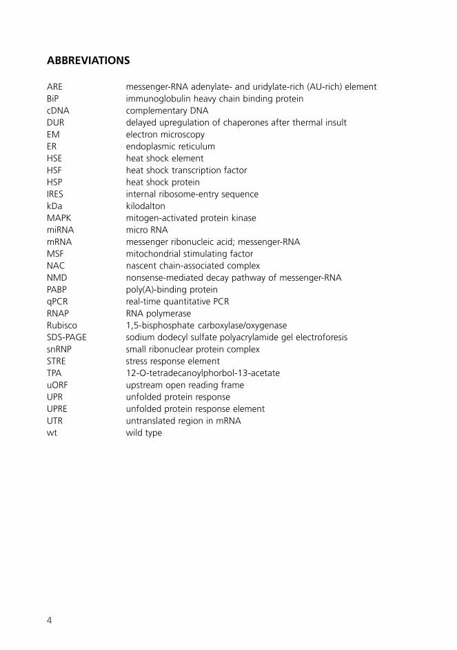
ARE messenger-RNA adenylate- and uridylate-rich (AU-rich) elementBiP immunoglobulin heavy chain binding proteincDNA complementary DNADUR delayed upregulation of chaperones after thermal insultEM electron microscopyER endoplasmic reticulumHSE heat shock elementHSF heat shock transcription factorHSP heat shock proteinIRES internal ribosome-entry sequencekDa kilodaltonMAPK mitogen-activated protein kinasemiRNA micro RNAmRNA messenger ribonucleic acid; messenger-RNAMSF mitochondrial stimulating factorNAC nascent chain-associated complexNMD nonsense-mediated decay pathway of messenger-RNAPABP poly(A)-binding proteinqPCR real-time quantitative PCRRNAP RNA polymeraseRubisco 1,5-bisphosphate carboxylase/oxygenaseSDS-PAGE sodium dodecyl sulfate polyacrylamide gel electroforesissnRNP small ribonuclear protein complexSTRE stress response elementTPA 12-O-tetradecanoylphorbol-13-acetateuORF upstream open reading frameUPR unfolded protein responseUPRE unfolded protein response elementUTR untranslated region in mRNAwt wild type
ABBREVIATIONS
5
ORIGINAL PUBLICATIONS
This thesis is based on the following original articles, which are referred to in the text bytheir Roman numerals, and on unpublished results presented in the text.
I Pirkkala, L., Alastalo, T.-P., Nykänen, P., Seppä, L., and Sistonen, L. 1999. Differentiationlineage-specific expression of human heat shock transcription factor 2. FASEB J. 13:1089-1098.
II Seppä, L., Hänninen, A. L., and Makarow, M. 2004. Upregulation of the Hsp104chaperone at physiological temperature during recovery from thermal insult. Mol.Microbiol. 52: 217-225.
III Seppä, L. and Makarow, M. 2005. Regulation and recovery of functions of the yeastchaperone BiP/Kar2p after thermal insult. Manuscript.
6
All cells in a multicellular organism containthe same set of genes. Yet the structure andfunction of different cell types vary greatly.This is because only a fraction of the genesis expressed within any given cell at a giventime. The pattern of gene expression is whatdistinguishes a skin cell from a liver cell, anda cancerous cell from a normal cell. The termgene expression refers to all processes thatare needed to convert the geneticinformation contained in a gene to producea functional protein (Fig. 1).Regulation of gene expression in aneukaryotic cell is a series of complex control
REVIEW OF THE LITERATURE
1 Regulation of eukaryotic gene expression
�������
�������
������������
���������������������
� �������� �� �����!�����
!���!�"������#�� ��
����!������#�� �������������
�����!������!������!
������!��##�������������!#$!�#���#��������
��������������#���������%�����
�
�
� �
�
�
�
�
������������
mechanism, starting with the activation andnuclear transport of the transcription factors,transcription in the nucleus, followed bymodification and splicing of the transcript,export of the mRNA from the nucleus to thecytoplasm, cytoplasmic distribution of themRNA, and finally, translation to protein(Fig. 1). Subsequently the activity, multi-merization and degradation of the functionalprotein are likewise tightly regulated. Thefollowing sections review the steps that aresubject to regulation for controllingexpression of a protein.
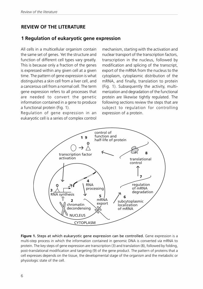
Figure 1. Steps at which eukaryotic gene expression can be controlled. Gene expression is amulti-step process in which the information contained in genomic DNA is converted via mRNA toprotein. The key steps of gene expression are transcription (3) and translation (8), followed by folding,post-translational modification and targeting (9) of the gene product. The pattern of proteins that acell expresses depends on the tissue, the developmental stage of the organism and the metabolic orphysiologic state of the cell.
Review of the literature

7
1.1 Transcriptional regulationIn transcription, a single-stranded RNAmolecule is produced, using genomic DNAas a template, in a reaction catalyzed by RNApolymerases (RNAP). In eukaryotes, there arethree types of RNA polymerases, RNAPI toIII, each of which transcribes its own subsetof RNAs. This chapter focuses on expressionof genes transcribed by RNAPII, which isresponsible for the expression of protein-coding genes.Transcription is accomplished by a proteincomplex called the general transcriptionmachinery, which, in addition to RNAPIIcomplex, consists of a series of additionalfactors called general transcription factors.They recognize specific promoter sequencesand perform the steps required fortranscription initiation. The generaltranscription machinery binds to a specificregion upstream of a gene, referred to asthe core promoter, located typically 40 to+50 relative to the initiation site (+1). DNA-binding is followed by RNA synthesisperformed in three steps, transcriptioninitiation, transcript elongation andtermination.Before the general transcription machinerycan act, however, it must be activated bytrans-acting regulatory proteins calledtranscription factors (Fig. 1, steps 1 and 3).They bind to DNA regulatory sequencesupstream of the core promoter andmodulate the activity of the generaltranscription machinery. Transcription factorsplay a key role in regulation of geneexpression; the fact that 5% of human genesencode transcription factors reflects theirimportance (Tupler et al ., 2001).Transcription factors allow tight regulationof transcription in response to nutritional,environmental or hormonal stimuli, and in amanner specific for tissue type anddevelopmental stage.
The transcription-regulating, cis-actingsequences fall into two different classes. Thefirst class, promoters, are usually locatedwithin 100-400 basepairs upstream from thetranscriptional start site. They are composedof various combinations of short, conservedDNA sequences called promoter elementsspecifically recognized by transcriptionfactors. Examples of promoter elements areheat shock elements (HSEs), which arepresent in the promoters of numerous stress-induced genes, e.g. HSP104 and KAR2 inthe yeast Saccharomyces cerevisiae, andwhich are recognized by the yeast heat shocktranscription factor 1 (Hsf1p).The second class of cis-acting regulatorysequences, enhancers, consists likewise ofmodular elements recognized bytranscription factors. However, unlikepromoters, enhancers can be locatedkilobases away from the promoter and theyare orientation- and position-independent.Enhancers may exist upstream, downstream,or within a gene.Promoters of most eukaryotic genes containbinding sites for several transcription factors,allowing the gene to be activated inresponse to a range of stimuli. For example,the above-mentioned KAR2 gene promotercontains HSEs, binding sites for Hsf1p,allowing the gene to be induced upon heatstress. Unfolded protein response elements,UPREs, which are binding sites for thetranscription factor Hac1p, allow the KAR2gene to be induced upon accumulation ofunfolded proteins inside the endoplasmicreticulum (ER; Kohno et al., 1993). On theother hand, one transcription factorfrequently activates several genes, allowingconcerted expression of all genes needed inresponse to a specific stimulus. Such is thecase for Hsf1p and Hac1p, which bothactivate numerous genes.Most transcription factors contain at leasttwo modular domains, a DNA-binding
Review of the literature

8
domain and a regulatory domain. Whenbound to their target promoter via the DNA-binding domain, transcription factors induceor increase transcription by attracting,positioning and modifying the generaltranscription factors, chromatin modifyingproteins and RNAPII at the promoter so thattranscription can begin. Examples of thespecific mechanism of transcription factoraction are described in chapter 3.2.3concerning heat shock transcription factors1 and 2.For transcription to occur, the transcriptionfactors must be present in the nucleus andactivated, but in addition, the appropriatechromosome segments must be madeaccessible. Regulation of chromatin structure(Fig. 1, step 2) plays an important role ingene expression by altering DNA packagingand nuclear localization of the chromosomalDNA, and hence DNA accessibility.Untranscribed regions of the genome occurin a highly-condensed state called“heterochromatin”, whereas activelytranscribed segments are packaged into“euchromatin”. Each cell and tissue type hasits own pattern of DNA packaging intoheterochromatin and euchromatin, and thispattern is inherited in cell division. Regulationof chromatin structure is a complex anddynamic network of regulatory mechanisms.Chromatin decondensation frequentlyoccurs jointly with binding of sequence-specific transcription factors to the genepromoter. In a widely accepted modelchromatin remodeling proteins are recruitedto the promoter by the transcription factors(reviewed in Narlikar et al., 2002).
1.2 Regulation of mRNA splicingAfter and during transcription, the RNAtranscript emerging from the transcriptionapparatus is subject to extensivemodifications (Fig. 1, step 4), such ascleavage, adding of a polyadenine tail to the
3’ end, adding of a cap structure to the 5’end, and splicing of introns. One of the mostcrucial steps in RNA processing is RNAsplicing, where the non-coding introns arecut from the transcript and protein-codingexons are ligated to form a functional mRNA.RNA splicing takes place in the nucleuswithin the spliceosome, a large complexconsisting of 145 different proteins and fivesmall nuclear RNA molecules, which formsmall ribonuclear protein complexes (snRNP;reviewed in Jurica and Moore, 2003).An average human gene is 28 000nucleotides long, whereas an average mRNAis 960 nucleotides long (Lander et al., 2001).This means that the exons are small and existwithin extensive intron sequences. mRNAsplicing is typical for multicellular eukaryotes.S. cerevisiae, in contrast, has introns in only~3% of its genes (Barrass and Beggs, 2003).Unlike the human introns, which arethousands of nucleotides long, the yeastintrons are in average only 270 nucleotideslong (Barrass and Beggs, 2003).Furthermore, usually the spliced yeast genesonly contain one intron, whereas theaverage human gene contains 7-8 introns(Lander et al., 2001).One major factor contributing to humanproteome complexity is alternative splicing,which results in production of different formsof a protein from the same mRNA. 35-65%of human genes are assumed to bealternatively spliced, which explains howapproximately 30 000 human protein-coding genes can give rise to more than90 000 different proteins (Mironov et al.,1999; Modrek and Lee, 2002). The mostusual type of alternative splicing isperformed by skipping an exon, resulting ina shorter and a longer isoform of the sameprotein (reviewed in Ast, 2004). This is thecase for human and mouse heat shocktranscription factors 1 and 2, which are bothexpressed as a longer α isoform and a shorter
Review of the literature

9
β isoform (Goodson and Sarge, 1995;Goodson et al., 1995). Alternative splicingis regulated in a cell type- and developmentalstage-specific manner, which results inspecific expression of the isoforms indifferent tissues.Transcription efficiency affects splicingefficiency, and surprisingly, the same is truein the opposite direction. Splicing occursfrequently cotranscriptionally. For instance,the human gene for dystrophin is 2 400 000nucleotides long. It would take 16 h totranscribe this gene, and it is obvious thatthe splicing of the dozens of dystrophinintrons would be mechanically difficult fromthe huge pre-mRNA. Dystrophin is, indeed,spliced cotranscriptionally (Tennyson et al.,1995). Coupling of transcription and pre-mRNA processing is thought to depend onthe ability of RNA polymerase II to recruitsome of the pre-mRNA processing factorsin a complex forming a “mRNA factory” (Duand Warren, 1997; McCracken et al., 1997a;McCracken et al., 1997b; Hirose and Manley,1998). At least 30 out of the 145spliceosomal proteins are anticipated to beinvolved in coupling between splicing andtranscription (Zhou et al., 2002). However,it is not only that transcription affectssplicing. Introducing experimentally an intronimmediately downstream from a promoterenhances transcription both in mammalianand yeast genes (Furger et al., 2002).Furthermore, spliceosomal factors, byinteracting with the human transcriptionelongation factor TAT-SF1, stimulatepolymerase II elongation (Fong and Zhou,2001), suggesting a strong connectionbetween transcription and splicing.
1.3 mRNA half-lifeAfter synthesis and processing, the newly-made mRNA is exported from the nucleus(Fig. 1, step 5) and distributed to its correctsubcytoplasmic localization (step 6). The
abundance of a given mRNA species is thesum of its production and decay, which inturn is determined by the stability of themessage (Fig. 1, step 7). In many cases, theregulation of protein production is principallyperformed by controlling mRNA levels. Thehalf-life of different eukaryotic mRNAs variesbetween some minutes and 24 hours(reviewed in Tourrière et al., 2002). OftenmRNAs for constantly expressed,“housekeeping” proteins are very stable, likethe β-globin mRNA in mammalian cells andthe PGK1 mRNA for 3-phosphoglyceratekinase in yeast cells (Muhlrad et al., 1995).The long-lasting message provides the cella prolonged time window for translation. Incontrast, mRNAs for proteins that aretransiently expressed in response to a specificstimulus such as developmental, nutritional,hormonal, or environmental input, are oftenunstable, which enables the cell todownregulate translation rapidly. Examplesof such unstable transcripts are mRNAs forgrowth factors, proto-oncogenes, cytokines,and lymphokines (Schiavi et al., 1994; Statonand Leedman, 1998). Apart from regulatingthe expression of an individual protein,mRNA turnover is a major controlmechanism to synchronize gene expression.Recent studies suggest that mRNAs involvedin similar mechanisms decay at similar rates(Wang et al., 2002; Yang et al., 2003), andthat the cell achieves this synchronizationof decay by concentrating mRNAdegradation to discrete cytoplasmic locations(van Dijk et al., 2002; Ingelfinger et al., 2002;Sheth and Parker, 2003; Cougot et al.,2004).Sequence elements regulating mRNAstability are present throughout the mRNAsequence. mRNA decay is interplay of thesecis-acting elements with trans-acting factorsthat recognize and bind to them. In theuntranslated regions of eukaryotictranscripts, the cap structure at the 5’ end,
Review of the literature

10
the 5’ untranslated region (UTR), thepolyadenylated (poly(A)) tail at the 3’ endand the 3’ UTR are determinants of RNAstability. In addition, sequences in the proteincoding region of the mRNA have beensuggested to act as instability elements, aswell (Parker and Jacobson, 1990; Wisdomand Lee, 1991; Ito and Jacobs-Lorena, 2001).The 3’ poly(A) tail, which occurs on mosteukaryotic mRNAs, stabilizes the message inwhich it is present. The highly expressedpoly(A)-binding protein (PABP) binds to thepoly(A) with high affinity and protects themRNA from ribonuclease attack (Gorlach etal., 1994; for review, see Shim and Karin,2002). In addition to protecting the poly(A)tail from deadenylases and 3’-5’exonucleases, PABP also enhances themessage by increasing the rate of translation(Otero et al., 1999). The 5' cap, in turn,likewise protects the new transcript fromattack by nucleases, and serves as a bindingsite for proteins involved in export of themature mRNA into the cytoplasm and itstranslation into protein.Many unstable mRNAs contain destabilizingelements in their 3’ UTR region. These cis-acting elements can be quite variable inlength and sequence, but some of themcontain adenylate- and uridylate-rich (AU-rich) elements (AREs). AREs are typical formRNAs that are upregulated during cellgrowth and differentiation (reviewed inTourrière et al., 2002). The first step in ARE-mediated decay is thought to bedeadenylation of the mRNA (Brewer andRoss, 1988; Wilson and Treisman, 1988;Shyu et al., 1991; Chen and Shyu, 1994).The presence of AREs is a very widespreadand efficient determinant of RNA instability,and their effect is in part due to their potentstimulatory effect on decapping anddeacetylation processes (Xu et al., 1997; Gaoet al., 2001).
In addition to the pathways that stabilize ordegrade mRNAs specifically, there is apathway that functions independently of theindividual nature of the mRNAs. Thisnonsense-mediated decay pathway (NMD)targets aberrant or truncated mRNAs fordegradation (reviewed in Alonso, 2005).NMD targets incorrectly spliced transcriptsand mRNAs with nonsense codons for rapiddegradation to prevent accumulation ofdefective and non-functional proteins (He etal., 1993; Pulak and Anderson, 1993).
1.4 Translational controlProtein synthesis (Fig. 1, step 8) is one ofthe key points in regulation of geneexpression. Translational control enables thecell to rapidly manipulate protein productionwithout new RNA synthesis, processing andexport. It is especially important, whentranscription is silenced, like in oocytes ofmany species during meiotic maturation, orwhen a concentration gradient of a proteinis created by local translation in a polarizedcell.Eukaryotic cells employ two general modesof translational control. In global control,translation of most cellular mRNAs isregulated as a whole. mRNA-specific control,in turn, is applied to modulate the translationof a defined group of mRNAs withoutaffecting overall translation in the cell(reviewed in Gebauer and Hentze, 2004).Global control is performed by regulatinggeneral translation-initiation factors,whereas mRNA-specific control involvesregulatory protein complexes that recognizeparticular elements present in the 5’ or 3’untranslated regions (UTR) of the targetmRNAs. Furthermore, mRNA-specific controlcan also be regulated by small micro RNAs(miRNAs) that hybridize to specific sequencesfound in the 3’ UTR of their target mRNAs(Ambros et al., 2003; Stark et al., 2003;Brennecke et al., 2005).
Review of the literature

11
Recently, Iborra and co-workers (2001)reported that mRNA translation also takesplace in the nucleus. Nuclear translation hasbeen suggested to represent an additionalmechanism of translational control and tobe involved in nonsense-mediated RNAdecay. Thus, the purpose of nucleartranslation seems not to be the productionof proteins, but scanning of aberrant mRNAsby nuclear ribosomes (Wilkinson and Shyu,2002; Iborra et al., 2004).In the mRNA molecule itself, there are severaldifferent structural features and regulatorysequences which control translation of themRNA on which they exist. The cap structureat the 5’ end and the poly(A) tail in the 3’end of the mRNA have a strong enhancingeffect on translational initiation, whereasupstream open reading frames (uORFs)reduce translation from the main ORF.Secondary and tertiary structures found inthe 5’ end of the mRNA, in turn, commonlyblock initiation of translation, while internalribosome-entry sequences (IRESs) mediatecap-independent translation by bypassingthe usual translation initiation process (forreview, see Dever, 2002). All these regulatorymotifs found in the mRNA UTRs base theiractivity on specific interactions with differentRNA-binding proteins, in particular thetranslation initiation complex of ribosomes.Upstream open reading frames (uORFs)regulating translation are found both ineukaryotes and prokaryotes. Commonly,they impair translation from the downstreamORF, probably by “misleading” the ribosomal43S preinitiation complex, which scans alongthe 5’ UTR until it reaches and identifies aninitiation codon (for review, see Dever, 2002).One of the best-studied examples ofregulatory uORFs is the S. cerevisiae GCN4transcript, which contains four uORFs(Mueller and Hinnebusch, 1986). TheseuORFs regulate GCN4 translation by
derepression in response to amino acidlimitation. Gcn4p is a transcriptionalactivator that induces at least 40 genesencoding amino acid biosynthetic enzymes(reviewed in Hinnebusch, 1997).
1.5 Regulation of protein half-live andfunctionAs the final steps in regulation of geneexpression, the functionality of the proteinsis controlled in a variety of ways (Fig. 1, step9). Some proteins, such as hormones, areproduced to function only for short times,whereas others, such as myosin chains inmuscles are meant to persist. Protein half-life is tightly regulated by several differentmechanisms. One of them is ubiquitin-mediated proteasomal degradation, whichplays a central role in regulating destructionof both aberrant and normal proteins. Upto 30% of the newly-synthesizedpolypeptides in a cell are selected for rapiddegradation in the proteasome because theydo not pass the quality control system of thecell. In addition to degradation of abnormalproteins, the ubiquitin system plays animportant role in a broad array of basiccellular processes. Among these areregulation of cell cycle, modulation of theimmune and inflammatory responses,control of signal transduction pathways,development and differentiation (Hershko etal., 2000).Proteins are functional only in interactionwith appropriate partners and modifyingfactors, localized in the correct cellularcompartment, and activated appropriately.Human heat shock factor 2 is an example ofall these processes. When inactive, it islocalized in the cytoplasm as dimers. Uponactivation, it forms trimers, localizes to thenucleus and is additionally activated bysumoylation (Sistonen et al., 1994; Goodsonet al., 2001).
Review of the literature
12
2 Protein folding and functions of molecular chaperones
A living cell synthesizes approximately100 000 different proteins. Virtually everycellular task, on which our lives depend, isperformed by proteins. Every protein has itsunique, native three-dimensional structure,which is essential for its function. Proteinfolding is the process, where the linearinformation in the amino acid sequence ofa polypeptide chain is interpreted to a well-defined three-dimensional structure of afunctional protein.Molecular chaperones are proteins that assistthe folding of other proteins. Molecularchaperones are found in every organism,every cell and in nearly every compartmentof the cells. They are conserved in evolutionand essential for all life (for review, see Hartl,1996). The term molecular chaperone wascoined by Laskey and co-workers (1978)when describing the histone-assemblingfunction of nucleoplasmin. More recently,the definition of molecular chaperones hasexpanded to describe a variety of proteins.The common feature for all these proteinsis that they prevent improper interactionsbetween their immature clients and favormore appropriate contacts. Furthermore,molecular chaperones are not part of thefinal protein structures. Chaperones do notthemselves contain the information forfolding (Anfinsen, 1973) and do notcovalently modify their target proteins, buthelp the self-assembly of proteins bypreventing alternative folding pathwaysleading to nonfunctional products. At thesame time, they prevent aggregation ofexposed hydrophobic regions in the protein.The binding mechanisms of chaperones arerather unspecific, because they recognizesuch a wide variety of targets. In a study,where random synthetic peptides wherepresented to BiP/Kar2p, which resides in theER, the chaperone was shown to
preferentially bind to 4-7 amino-acidpeptides with a high content of hydrophobicresidues (Flynn et al., 1991). The Escherichiacoli chaperone SecB preferentially binds topositively charged regions of polypeptidechains (Randall, 1992). The current view isthat chaperones bind to reactive surfaces ofpolypeptides, and by doing so, stabilize anotherwise unstable conformation of theirtargets, and thereby promote their correctfate: folding, transport, assembly, orswitching between active/inactiveconformations. The roles of molecularchaperones in these processes are describedin the following sections.
2.1 Folding of newly synthesized poly-peptides in eukaryotic cellsTo be functional, a protein has to assume itscorrect three-dimensional structure and tofind its correct location in the cell. All theinformation required for proper proteinfolding is contained in the primary aminoacid sequence of the polypeptide chain(Anfinsen, 1973). When a polypeptideemerges from the ribosome, it cannot foldcorrectly until an entire domain is completed.Translation takes seconds to minutes,whereas folding intermediates build inmilliseconds (for review, see Hendrick andHartl, 1993). Consequently, actively growingcells contain large numbers of aggregation-prone, unfolded polypeptides. A polypeptideemerging from the ribosome into the cytosolis first bound by a complex termed thenascent chain-associated complex (NAC),which protects the first 30 C-terminalresidues (Wang et al., 1995). Whendisassociated from the ribosome in thecytoplasm, folding of the newly synthesizedchain is assisted by Hsp70 (DnaK, Ssb),Hsp40 (DnaJ), and Hsp60 (CCT, GroEL) typeof chaperones (Bukau et al., 2000). These
Review of the literature

13
chaperones promote the properconformation of the newly-synthesizedpolypeptide through repeated cycles ofbinding, folding and releasing the substrate.
2.2 Molecular chaperones and trans-location into organellesThe majority of proteins functioning in differentcompartments of the cell is produced in thecytosol from nuclear genes. Thus, they haveto be specifically recognized, transported tothe surface of the organelle, and vectoriallytranslocated into the organelle lumen.Molecular chaperones are involved in virtuallyall of these transport processes.Cytosolic Hsp70 family chaperones bind to thetarget polypeptide and assist its transport tothe appropriate organelle membrane. In theorganelle lumen, Hsp70 family chaperonesstimulate the import of proteins. This is alsothe case for ER (Chirico et al., 1988). Insidethe ER lumen, the Hsp70 chaperone BiP/Kar2p,binds transiently to the J-domain of the ERtransmembrane Hsp40 protein Sec63p(Misselwitz et al., 1999). Both BiP/Kar2p andSec63p are required for both cotranslationaland posttranslational translocation ofpolypeptides (Brodsky et al., 1995).Although mitochondria contain their owngenome, most mitochondrial proteins aresynthesized in the cytosolic ribosomes.Import into mitochondria may be describedin analogous terms as translocation into theER. Cytosolic molecular chaperonesincluding Hsp70s and the mitochondrialstimulating factor (MSF) target the precursorproteins to the mitochondrial membrane(Hachiya et al., 1994). MSF is a cytosolicchaperone, which hydrolyzes ATP wheninteracting with precursors of mitochondrialproteins (Hachiya et al., 1994). In themitochondrial matrix, heat shock protein 70(mtHsp70) cooperates with its fourcochaperones Mge1, Tim44, Pam18, and
Pam16 at the mitochondrial innermembrane (Frazier et al., 2004).
2.3 Chaperone function and changing ofprotein subunitsWhen protein subunits are rearranged,hydrophobic patches in the protein aretransiently exposed. Therefore chaperonesare needed to prevent aggregation andincorrect associations between these reactivesurfaces. Some cytoskeletal components arehighly dynamic, i.e. they are constantlyrearranged. Cytoskeletal microtubules arehollow cylinders constructed of tubulin,which is a heterodimeric protein consistingof α- and β-tubulin (for review, see Liangand MacRae, 1997). There is evidence thatthe cytosolic chaperone Hsp70 regulatestubulin assembly/disassembly. MammalianHsp70 binds to polymerized tubulin and mayprevent microtubule formation by inhibitingthe binding of microtubule-associatedproteins, which are able to promote tubulinassembly and stabilize microtubules(Sánchez et al., 1994).Chaperone activity is crucial for the assemblyof the chloroplast enzyme 1,5-bisphosphatecarboxylase/oxygenase (Rubisco), as well.Rubisco, responsible for CO2 fixation duringphotosynthesis, exists in vascular plants andgreen algae as a 500-kDa holoenzyme. Eightof its subunits are of chloroplast origin andeight are synthesized in the cytosol (forreview, see Gutteridge and Gatenby, 1995).The chloroplast chaperone cpn60 binds tonewly synthesized or imported subunits ofRubisco, which are very prone toaggregation. cpn60 is related to the bacterialchaperonin GroEL (Hemmingsen et al., 1988)and like GroEL, it functions by stabilizingfolding intermediates and therebypreventing their aggregation. The release ofRubisco subunits from cpn60 requires thecochaperonin cpn10 (Schmidt et al., 1994).
Review of the literature
14
3 Stress responses
Cells of both unicellular and multicellularorganisms experience every day conditionsthreatening the integrity of their proteome(Fig. 2). Circumstances affecting proteinfolding include environmental stress, suchas fluctuations in temperature, hydrationand nutrient balance, chemical stress causedby oxygen-free radicals, transition heavymetals, or pathophysiological states ofmulticellular metazoans such as ischemia,viral or bacterial infections or tissue injury.To cope with these stress conditions,organisms have evolved a variety ofstrategies operating on very different levels,from molecular responses to physiologicaland behavioural adaptation. Two examplesof stress response mechanisms at themolecular level, both aiming at protection
of proper protein conformation, are theunfolded protein response (UPR) and theheat shock response.
3.1 Unfolded protein responseThe unfolded protein response (UPR) is anintracellular signaling pathway that controlstranscription of genes encoding ER-residentchaperones, proteins involved inphospholipid biosynthesis, secretorypathway and ER-associated degradation(Travers et al., 2000). UPR is induced inresponse to accumulation of unfolded andmisfolded proteins inside the ER lumen,following exposure to ER stressors such astunicamycin, ditiothreitol, glucose starvation,disturbance of Ca2+ ion stores or geneticmutations. Activation of UPR is accompanied
��%���������!������������������ ������������ �������!�
�������������"����
&��
������
&��������������
��!���!������������
���� !���������
��#�!���� �����������
� ����������
����%��������
����%��������
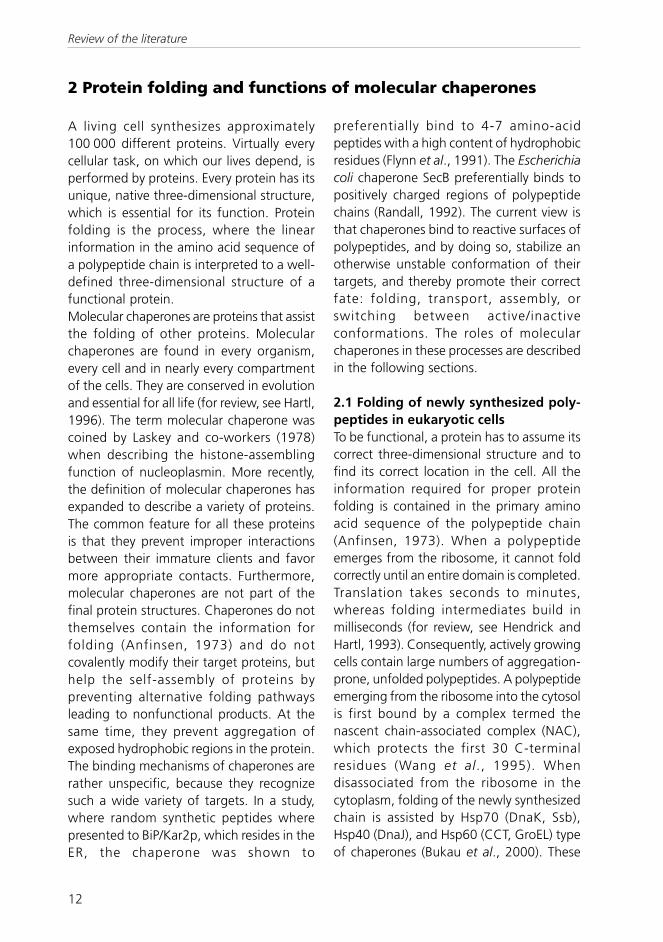
Figure 2. Cellular defense mechanisms against genotoxic and proteotoxic stresses. Misfoldedand disabled proteins can occur as a result of alterations in the genomic DNA, direct damage to thecellular proteins by various stresses, or as a part of normal physiology. Antioxidant enzymes preventoxidative damage of cellular components, DNA repair systems restore DNA integrity, and molecularchaperones refold damaged proteins or direct them to degradation. Modified from Ohtsuka andHata, 2000.
Review of the literature
15
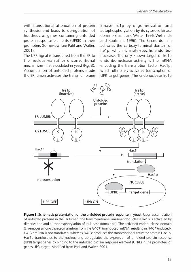
with translational attenuation of proteinsynthesis, and leads to upregulation ofhundreds of genes containing unfoldedprotein response elements (UPRE) in theirpromoters (for review, see Patil and Walter,2001).The UPR signal is transferred from the ER tothe nucleus via rather unconventionalmechanisms, first elucidated in yeast (Fig. 3).Accumulation of unfolded proteins insidethe ER lumen activates the transmembrane
kinase Ire1p by oligomerization andautophosphorylation by its cytosolic kinasedomain (Shamu and Walter, 1996; Welihindaand Kaufman, 1996). The kinase domainactivates the carboxy-terminal domain ofIre1p, which is a site-specific endoribo-nuclease. The only known target of Ire1pendoribonuclease activity is the mRNAencoding the transcription factor Hac1p,which ultimately activates transcription ofUPR target genes. The endonuclease Ire1p
' '
� �
� ������
�����
��#�!�����������
(��)�*������%�+
(��)�*����%�+
�
��������!�����
,��)�
,��)�
� �-- � ��
������������ �
�������
�����!�����
,��)�
' '
� �
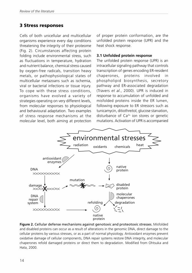
Figure 3. Schematic presentation of the unfolded protein response in yeast. Upon accumulationof unfolded proteins in the ER lumen, the transmembrane kinase-endonuclease Ire1p is activated bydimerization and autophosphorylation of its kinase domain (K). The activated endonuclease domain(E) removes a non-spliceosomal intron from the HAC1u (uninduced) mRNA, resulting in HAC1i (induced).HAC1u mRNA is not translated, whereas HAC1i produces the transcriptional activator protein Hac1p.Hac1p translocates to the nucleus and upregulates the expression of unfolded protein response(UPR) target genes by binding to the unfolded protein response element (UPRE) in the promoters ofgenes UPR target. Modified from Patil and Walter, 2001.
Review of the literature

16
cuts HAC1 mRNA at two sites, removing anon-spliceosomal intron (Sidrauski andWalter, 1997), producing HAC1i mRNA(induced). HAC1u mRNA (uninduced) isconstitutively produced, but not translatedinto Hac1p (Chapman and Walter, 1997;Kawahara et al., 1997). The HAC1i mRNA,free of the inhibitory intron, is efficientlytranslated to produce the transcription factorHac1p, which then translocates to thenucleus and binds to UPR-specific upstreamactivating sequences, unfolded proteinresponse elements (UPREs), on its targetgenes. Genes activated by UPR include theER-resident chaperones KAR2, PDI1 andFKB2 (Mori et al., 1998), and the UPRE intheir promoters is necessary and sufficientto activate transcription in response to UPR(Mori et al., 1992; Kohno et al., 1993).UPR occurs in all eukaryotes, and most ofthe molecular mechanisms are conserved inyeast and mammals, among those Ire1phomologues, the function of their lumenaldomains and dependence of UPR-inducedBiP on Ire1 (for review, see Patil and Walter,2001).
3.2 Heat shock responseOne highly conserved stress defensemechanism in all eukaryotes is the heatshock response, whose hallmark is a strongincrease in heat shock protein (hsp)expression. The induction of heat shockproteins by temperature shock was firstreported in Drosophila salivary glands(Ritossa, 1962; Tissières et al., 1974). Lateron, the expression was found to occur alsoin a wide variety of other stress conditions,such as exposure to cadmium sulfate, theamino acid analogue L-azetidine-2-carboxylic acid, puromycin, proteasomeinhibitors, by mutations in the proteolyticpathways, by expressing mutant actin, or bysimply injecting denatured proteins intoliving cells (reviewed in Georgopoulos and
Welch, 1993). The common nominator forall these conditions is the accumulation ofabnormally folded proteins. Furthermore,protein conformation-stabilizing agents suchas glycerol reduce the heat shock response(Georgopoulos and Welch, 1993).Remarkable features of the heat shockresponse are the rapidity of its induction andthe sensitivity of attenuation. This is achievedby tightly regulating the heat shock proteinexpression, described below.Cells survive exposure to otherwise lethallyhigh temperatures, if they are firstpreconditioned in a temperature moderatelyabove their physiological temperature(Lindquist and Kim, 1996). This phenomenoncalled thermotolerance is due to productionof heat shock proteins, among othercytoprotective agents such as trehalose inyeast cells (Iwahashi et al., 1997). Certainheat shock proteins play important roles inacquisition of thermotolerance, or toleranceto other forms of stress. The yeast chaperoneHsp104 is required for acquisition ofthermotolerance (Sanchez and Lindquist,1990). It also protects the cells againstharmful effects of ethanol, sodium arseniteand long-term storage at low temperatures(Sanchez et al., 1992).The phenomenon of cross-tolerance islikewise based on the increased productionof heat shock proteins induced by exposureto one form of stress. The elevated amountof hsps in the cell thus protects the cell fromdamage by another form of stress.
3.2.1 Heat shock proteinsHeat shock proteins are needed both instress conditions and in physiologicalconditions, where they accomplish similartasks. Some heat shock proteins areinducible, some are constitutively expressed.Heat shock proteins comprise the largestclass of molecular chaperones, but not allchaperones are heat shock proteins, or vice
Review of the literature
17
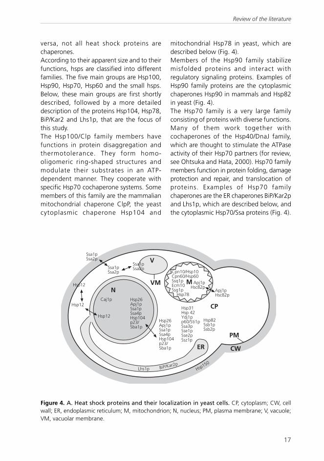
versa, not all heat shock proteins arechaperones.According to their apparent size and to theirfunctions, hsps are classified into differentfamilies. The five main groups are Hsp100,Hsp90, Hsp70, Hsp60 and the small hsps.Below, these main groups are first shortlydescribed, followed by a more detaileddescription of the proteins Hsp104, Hsp78,BiP/Kar2 and Lhs1p, that are the focus ofthis study.The Hsp100/Clp family members havefunctions in protein disaggregation andthermotolerance. They form homo-oligomeric ring-shaped structures andmodulate their substrates in an ATP-dependent manner. They cooperate withspecific Hsp70 cochaperone systems. Somemembers of this family are the mammalianmitochondrial chaperone ClpP, the yeastcytoplasmic chaperone Hsp104 and
mitochondrial Hsp78 in yeast, which aredescribed below (Fig. 4).Members of the Hsp90 family stabilizemisfolded proteins and interact withregulatory signaling proteins. Examples ofHsp90 family proteins are the cytoplasmicchaperones Hsp90 in mammals and Hsp82in yeast (Fig. 4).The Hsp70 family is a very large familyconsisting of proteins with diverse functions.Many of them work together withcochaperones of the Hsp40/DnaJ family,which are thought to stimulate the ATPaseactivity of their Hsp70 partners (for review,see Ohtsuka and Hata, 2000). Hsp70 familymembers function in protein folding, damageprotection and repair, and translocation ofproteins. Examples of Hsp70 familychaperones are the ER chaperones BiP/Kar2pand Lhs1p, which are described below, andthe cytoplasmic Hsp70/Ssa proteins (Fig. 4).
�� ��
��
�����
�����
�����
����� ���������������������� ����������
���
�����������
����������
����������
���������������������� ����������
����������������� ��������������������������
���������������
���� ����� ��� ���� ��������� ��������������
����������
�����������
����
��
!"��� #�$�%�&��
Figure 4. A. Heat shock proteins and their localization in yeast cells. CP, cytoplasm; CW, cellwall; ER, endoplasmic reticulum; M, mitochondrion; N, nucleus; PM, plasma membrane; V, vacuole;VM, vacuolar membrane.
Review of the literature

18
Figure 4. B. Heat shock proteins and their localization in mammalian cells. CP, cytoplasm; ER,endoplasmic reticulum; M, mitochondrion; N, nucleus; PM, plasma membrane; P, peroxisome.
��
��
���� �����
�
���� ������ �'�$
�
#�$
��
���� �����
���� ����
���� ��(�"&)���''�������*+)������������,�$(����- ��(�
����� �.���
/$$���/01���$#�(���$#�(�����
Hsp60 family proteins are often calledchaperonins. They share some homologywith the bacterial chaperonin GroEL.Chaperonins function in assembly ofmultimeric protein complexes and folding ofcertain newly synthesized proteins. Membersof the chaperonin family are e.g. thecytoplasmic chaperonin-containing t-complex polypeptide CCT, also called TriC,and mitochondrial Hsp60/Cpn60 (Fig. 4).Hsp40 proteins are homologues of bacterialDnaJ proteins. They interact with Hsp70family members and are as abundant as theirpartners: tens of Hsp40/DnaJ homologueshave been identified both in yeast and inmammals (for review, see Ohtsuka and Hata,2000).The small hsps are the least conserved ofthe major hsp families and have divergentstructures. They vary between 15 and 40 kDain size and are targeted to the eukaryoticcytosol and are especially abundant in plants.Small hsps have been reported to functionin stabilization of misfolded proteins and
thermotolerance (for review, see Parsell andLindquist, 1993). The most studiedrepresentatives of this family are the α/β-crystallins, which are structural proteins inthe vertebrate eye lens, and additionally,function there in a chaperone-like manner(Fig. 4).
Hsp104The cytoplasmic yeast chaperone Hsp104 isessential for acquired thermotolerance(Sanchez and Lindquist, 1990) and itfunctions in refolding of aggregated proteinsin cooperation with the Hsp70/Hsp40cochaperone system (Parsell et al., 1994).Prior to the reactivation of damaged proteinsby the Hsp104/Hsp70/Hsp40 chaperonecomplex, Hsp26 sequesters the aggregatedproteins and makes them accessible forrefolding (Cashikar et al., 2005). Hsp104 isneeded for refolding of heat-damagedproteins in the cytosol (Parsell et al., 1994)and unexpectedly, in the ER lumen, as well(Hänninen et al., 1999). The stress-protective
Review of the literature

19
yeast disaccharide trehalose assists Hsp104in refolding of heat-damaged proteins in thecytosol (Singer and Lindquist, 1998) andinside the ER lumen (Simola et al., 2000).
Hsp78In the yeast mitochondria, the Hsp100/ClBhomologue Hsp78 is crucial for themaintenance of respiratory competence andresumption of mitochondrial proteinsynthesis after thermal insult at 50°C(Schmitt et al., 1996). Hsp78 functions as acomponent of the mitochondrial proteolysissystem and is required for efficientdegradation of substrate proteins in thematrix (Röttgers et al., 2002). Similar toHsp104 in the cytosol, Hsp78 cooperateswith the mitochondrial Hsp70 machinery inreactivation of denaturated mitochondrialproteins (Krzewska et al., 2001); however,Hsp78 does not affect acquisition of cellularthermotolerance (Schmitt et al., 1996).
BiP/Kar2pBiP/Kar2p is a member of the 70-kDa heatshock protein (Hsp70) family, resident in theER. Yeast BiP/Kar2p is an essential protein(Normington et al., 1989) with multiple roles.It functions in translocation of newlysynthesized polypeptides through the Sec61translocon into the ER (Corsi and Schekman,1997; McClellan et al., 1998). BiP/Kar2pinteracts with the lumenal J-domain of theco-chaperone Sec63p, a member of theHsp40 family, and provides the driving forcefor translocation by hydrolyzing ATP (Brodskyand Schekman, 1993; Scidmore et al., 1993;Cyr et al., 1994). Like other Hsp70 proteins,BiP/Kar2p consists of two domains, an N-terminal domain harbouring an ATPasecatalytic site and a C-terminal domainresponsible for substrate binding (Flynn etal., 1989; McKay, 1993; Blond-Elguindi etal., 1993). Two models have been proposedfor the function of BiP/Kar2p in protein
translocation (for review, see Jensen andJohnson, 1999). In the ratchet or trappingmodel, BiP/Kar2p binds to the polypeptideemerging from the translocon preventing itfrom sliding backwards (Matlack et al.,1999). In the translocation motor model, BiP/Kar2p binds to the emerging polypeptide,and an ATP hydrolysis-dependentconformational change in BiP/Kar2p pulls theprotein through the translocon (Glick, 1995).Moreover, BiP/Kar2p assists folding ofpolypeptides in the ER by shieldinghydrophobic amino acid side chains from theaqueous environment of the ER lumen(Simons et al., 1995). BiP/Kar2p functionsin quality control by binding to misfoldedproteins and preventing their exit from theER (Gething et al., 1986; Hurtley et al.,1989).
Lhs1pLhs1p is an Hsp70-related chaperonelocalized in the yeast ER lumen, member ofthe GRP170 subfamily of Hsp70 chaperoneswith homologues in mammals (Chen et al.,1996; Kuwabara et al., 1996). Unlike BiP/Kar2p, Lhs1p is not essential for viability, butcells carrying an lhs1 null mutation exhibit adefect in post-translational translocation(Baxter et al., 1996; Craven et al., 1996;Hamilton and Flynn, 1996) and areconstitutively induced in the unfoldedprotein response UPR (Baxter et al., 1996;Craven et al., 1996). Lhs1p is required forrefolding of heat-damaged proteins insidethe ER lumen and for acquisition ofthermotolerance (Saris et al., 1997; Saris andMakarow, 1998). Recently, Lhs1p was foundto interact with BiP/Kar2p. The ATPase cyclesof Lhs1p and BiP/Kar2p are coupled: Lhs1pstimulates the nucleotide exchange phaseof the BiP/Kar2p ATPase cycle, andreciprocally, BiP/Kar2p stimulates thehydrolysis phase of Lhs1p ATPase cycle (Steelet al., 2004).
Review of the literature

20
3.2.2 Heat shock elementsHeat shock protein expression is controlledby specific sequences called heat shockelements (HSEs) located in the upstreamregions of heat shock protein genes.Transcription of hsp genes is activated bybinding of heat shock transcription factors(HSFs) to HSEs in their target genes. The HSEconsensus sequence is defined by arepeating array of the 5-bp sequence nGAAnarranged in alternating orientations (Aminet al., 1988; Xiao and Lis, 1988). The numberof nGAAn repeats in a functional HSE varies,but usually ranges from three to six.Furthermore, the number of HSEs and thedistance between them in a heat shock genepromoter varies. The binding activity of HSFtrimers increases as the number of nGAAnrepeats in the promoter increases (Topol etal., 1985; Xiao et al., 1991). However, thesequence of HSE has been conserved inevolution among eukaryotes as diverse asyeasts, ciliated protozoa, insects, nematodes,amphibians, and mammals (for review, seeFernandes et al., 1994).
3.2.3 Heat shock factorsHeat shock transcription factors (HSFs)activate the transcription of heat shockprotein genes by binding to HSEs in theirtarget genes. HSFs from different organismscomprise a large protein family, and differenteukaryotes harbor different numbers ofHSFs. The HSF gene was originally identifiedin yeast as an essential gene for survival andthe only HSF in yeast (Sorger and Pelham,1988; Wiederrecht et al., 1988). Fruit fly wasshown to have one HSF, as well (Westwoodet al., 1991), required under normal growthconditions for oogenesis and early larvaldevelopment (Jedlicka et al., 1997). Incontrast, several members of HSF family havebeen found in vertebrates and plants.Human and mouse HSF1 and HSF2 werecloned almost simultaneously (Rabindran et
al., 1991; Sarge et al., 1991; Schuetz et al.,1991), Subsequently, two more HSFs, HSF3and HSF4, have been identified in vertebrates(Nakai and Morimoto, 1993; Nakai et al.,1997), of which HSF3 seems to be an avian-specific factor. Plants, in turn, harbornumerous HSFs; tomato more than 16 andwall cress (Arabidopsis) 21 putative HSFs(Nover et al., 2001).All members of the heat shock factor familyshare a similar structure, comprising of anamino-terminal helix-turn-helix DNA-bindingdomain, an adjacent coiled-coil trimerizationdomain and with the exception of HSF4, asecond coiled-coil domain, located towardthe carboxyl-terminus of the protein(reviewed in Pirkkala et al., 2001).Despite their similar structure, vertebrateHSFs exhibit different functions, targettissues and activation patterns. Thevertebrate HSF1 and its yeast homologueHsf1p can be considered as typical metazoanstress-inducible heat shock factors. Underphysiological conditions, human HSF1 occursas inactive monomers in the cytoplasm.Upon stress stimuli, HSF1 trimerizes,translocates into the nucleus, binds to targetstress protein gene promoters and activatestheir transcription (reviewed in Voellmy,2004). Yeast Hsf1p, in turn, is constitutivelytrimerized and bound to DNA (Sorger et al.,1987; Jakobsen and Pelham, 1988) and itsDNA-binding is enhanced upon stress stimuli(Giardina and Lis, 1995). Phosphorylation ofhuman HSF1 on serine residue 230 promotesits transcriptional activity (Holmberg et al.,2001), whereas phosphorylation on serineresidues 303, 307 and 363 is involvedattenuation of HSF1 activity subsequent tostress (Knauf et al., 1996; Kline andMorimoto, 1997). The amount of HSF1varies greatly in different tissues (reviewedin Pirkkala et al., 2001). The most studiedfunction of HSF1 is to activate hsp expressionupon various stress stimuli that cause protein
Review of the literature

21
misfolding, as described above. However,studies with HSF1 null mice suggest a rolefor HSF1 also in spermatogenesis, in earlymammalian development and in apoptosisprotection (McMillan et al., 1998; Xiao etal., 1999; Christians et al., 2000).Functions of HSF1 and HSF2 seem to bedistinct, but they display different specifitiesfor different HSEs. Results from severalstudies on HSF2 suggest that it is a nonstress-responsive member of the HSF family,controlling hsp expression in developmentand differentiation (reviewed in Pirkkala etal., 2001). The functions of HSF2 are largelyunknown. HSF2 is not activated by heatshock or stress stimuli, but during hemin-induced differentiation of K562 cells(Sistonen et al., 1992; Sistonen et al., 1994);see chapter below. Whereas HSF1 isactivated within minutes upon stress, hemin-induced activation of HSF2, in turn, rangesfrom hours to days (Sistonen et al., 1992;Sistonen et al., 1994). In contrast to HSF1,activation of HSF2 seems not to be regulatedby phosphorylation, but by an increase inprotein levels (Sistonen et al., 1994) and bymodification by SUMO-1, an ubiquitin-related protein (Goodson et al., 2001). InK562 cells, the inactive, non-DNA-bindingform of HSF2 exists in the cytoplasm asdimers. Upon activation, HSF2 is trimerizedand transported into the nucleus (Sistonenet al., 1994). Like HSF1, HSF2 also exists astwo isoforms, HSF2-α and HSF2-β.Additional splicing of the hsf2 transcriptresults in the smaller HSF2-β isoform, whichlacks 18 amino acids present in the longerHSF2-α isoform (Goodson et al., 1995).
Traditionally, HSF2 has been viewed as anonstress-activated heat shock factor,involved mainly in developmental processes,such as embryogenesis, organogenesis andspermatogenesis (Eriksson et al., 2000; Minet al., 2000). HSF2 is expressed in a cell-typedependent manner in the testis (Sarge et al.,1994; Alastalo et al., 1998) and the HSF2-αand HSF2-β isoforms are developmentallyregulated in a stage-dependent manner(Alastalo et al., 1998).Three groups reported simultaneously thegeneration of mice deficient in hsf2, withsomewhat different results. All of the threeknock-out mouse strains were viable. Thehsf2-deficiency did not affect the expressionpattern of hsps, but it was equivalent inwildtype and hsf2-deficient embryos bothin normal conditions and after heat shock(McMillan et al., 2002; Wang et al., 2003).Two of the studies resulted in mice thatexhibit several defects in meiosis, braindevelopment and female hormone response(Kallio et al., 2002; Wang et al., 2003),whereas one group reported that their hsf2-deficient mice were normal in terms offertility, brain development and cognitiveand psychomotor function (McMillan et al.,2002). In contrast, disruption of both hsf1and hsf2 leads to arrest in spermatogenesisand thus male infertility (Wang et al., 2004).The differencies between these mousemodels remain enigmatic, but they can bespeculated to partially depend on thedifferent technical manners and geneticbackgrounds, in which the hsf2 knock-outswere made.
4 K562 cells as a model for hematopoietic differentiation
The K562 cell line was established in 1971from a patient with chronic myeloidleukemia in the acute phase (Lozzio andLozzio, 1975). K562 is a widely used model
for studying gene expression duringhematopoiesis, because these cells can beinduced to differentiate along severallineages. Erythroid differentiation of K562
Review of the literature

22
cells can be induced by a variety of agents,including hemin and sodium butyrate(reviewed in Tsiftsoglou et al., 2003).Treatment of K562 cells with the tumorpromoter 12-O-tetradecanoyl-phorbol-13-acetate (TPA) induces them to differentiatealong megakaryocytic lineage, in turn(reviewed in Alitalo, 1990).Hemin, the synthetic chloride form of hemeand a natural regulator of erythropoiesis,binds to hemin-binding proteins inside thecell and modulates the transcription factorsbinding to Gγ-globin gene promoter(reviewed in Tsiftsoglou et al., 2003). Wheninduced towards the erythroid pathway ofdifferentiation, K562 cells start the synthesisof red-cell specific proteins, including globins(Benz, Jr. et al., 1980) and glycophorin A(Gahmberg et al., 1979). Globin productionoccurs by transcriptional activation of theembryonic α- and β-like globin genes, ε andζ, respectively, as well as the fetal γ-globinand adult α-globin genes (Charnay andManiatis, 1983). Erythroid differentiation ofK562 cells induced by hemin treatment doesnot, however, lead to terminal maturation(Dean et al., 1981).During hemin-induced erythroiddifferentiation, K562 cells upregulate theexpression of Hsp70, which is due toactivation of HSF2 binding to the hsp70 gene
promoter (Theodorakis et al., 1989; Sistonenet al., 1992). In addition, the expression ofthioredoxin is induced in K562 cells inresponse to hemin in a HSF2-dependentmanner (Leppä et al., 1997a). The existenceof two HSF2 isoforms (Goodson et al., 1995)adds complexity to the regulatory functionsof HSF2 in response to hemin. HSF2-α is thepredominantly expressed HSF2 isoform inK562 cells (Leppä et al., 1997b). Further-more, the molar ratio of the isoforms α andβ regulates HSF2 activity in hemin-treatedK562 cells; overexpression of HSF2-β inhibitsHSF2 activation and hemin-inducederythroid differentiation of K562 cells (Leppäet al., 1997b).Megakaryoblastoid differentiation of K562cells induced by TPA is characterized by lossof the erythroid properties and synthesis ofseveral megakaryoblastoid markers such asplatelet-derived growth factor, glycoproteinIIIa and TGF-β. In addition, TPA treatmentenhances the expression of tromboxan A2
receptors, normally found on platelets, onthe surface of K562 cells (reviewed in Alitalo,1990). TPA exerts its effect through PKC, aphospholipid/calcium-dependent serine-threonine kinase, which mediates activationof genes containing TPA-responsiveelements in their upstream regulatorysequences (reviewed in Alitalo, 1990).
5 Yeast as a model organism for studies of stress responses
In addition to K562 cells, baker’s yeastSaccharomyces cerevisiae was used as modelin this study. This unicellular eukaryote is anoutstanding experimental model to dissectand understand the biochemicalmechanisms by which cells sense andrespond to stress. Firstly, growing yeast cellsin laboratory is fast, safe and non-expensive.Secondly, stable manipulation of the haploidgenome of laboratory yeast strains is oftenfairly uncomplicated and does not pose
ethical problems. Furthermore, S. cerevisiaegenome was the first eukaryotic genome tobe completely sequenced (Goffeau et al.,1996), and the availability of the yeastgenome sequence has provided aremarkable resource for yeast biologists.Finally, as unicellular organisms live in directcontact with their environments, exposingyeast cells to highly defined conditions forstudying stress responses is uncomplicated.Consequently, yeast stress responses for
Review of the literature

23
instance to starvation, heat, pro-oxidants,heavy metals and changes in osmolarity orionic balance are well-characterized(Hohmann and Mager, 1997). Moreover,yeast cells are known to carry out a widerange of physiological processes with highmechanistic similarity to human cells(Bassett, Jr. et al., 1996).
Despite the above mentioned benefits ofyeast as a model organism, as a unicellularorganism it is unsuitable for studying mecha-nisms that involve cell-to-cell interactions.Thus, for obvious reasons, processes that arebased on specialized tissues such as organo-genesis, neuromuscular functions ordevelopment of metastases cannot bestudied using yeast.
Review of the literature

24
AIMS OF THE STUDY
The aim of the present study was to characterize the regulation of different components ofthe heat stress response using two different model systems, human K562 erythroleukemiacell line and Saccharomyces cerevisiae yeast cells. The focus of the study was, however, inprocesses different from the classical heat shock response, namely differentiation in thecase of human HSF2 and recovery from thermal insult in the case of yeast heat shockproteins.
Specific aims:1) to characterize the regulation of human HSF2 in hemin-induced differentiation of
K562 cells2) to study the regulation of yeast chaperones, specifically Hsp104 and BiP/Kar2p, during
recovery after thermal insult
Aims of the Study

25
Method Publication Described in Analysis of bulk protein synthesis II II Antibody supershift analysis and competition experiment
I
I, Mosser et al., 1988
Bacterial transformation II, III Standard methods -galactosidase assay III III
Gel mobility shift analysis
I
I, Garner and Revzin, 1981; Mosser et al., 1988
Glucose consumption assay II II Human cell culture I I Northern analysis I, II I, II, Russo et al., 1993 Nuclear run-on analysis I I, Banerji et al., 1984 Plasmid construction II, III Standard methods Pulse-labelling and immunoprecipitation of proteins
II, III
Saris et al., 1997
Real-time quantitative PCR III III SDS-PAGE analysis I-III Standard methods Site-directed mutagenesis by PCR Thermotolerance assay
II, III II
II, III II
Transmission electron microscopy II Simola et al., 2000 Western analysis I I Yeast cell culture II, III II, III Yeast cell transformation II, III Standard methods
MATERIALS AND METHODS
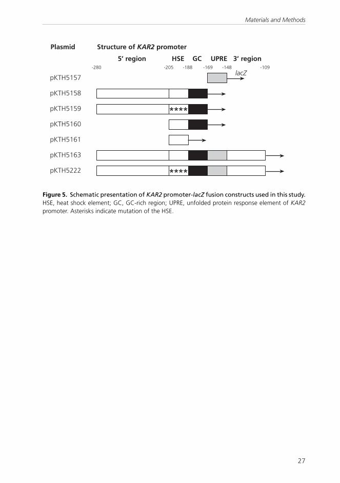
Experimental methods used in this work are summarized in Table 1. Detailed descriptionsof the methods are given in the original publications or references therein. The S. cerevisiaeyeast strains are listed in Table 2 and the KAR2 promoter-lacZ fusion constructs used instudy III are schematically presented in Figure 5.
Table 1. Methods used in this study.
Materials and Methods

26
Strain
Relevant mutation
Fusion construct
Publication
Source or reference
H1 none II R. Schekman H4 sec18-1 II R. Schekman H7 none III J. Knowles H245
none
II, III
Thomas and Rothstein, 1989
H335
none
HSP150 --lactamase
III
Simonen et al., 1994
H454
hsp104
II
Sanchez and Lindquist, 1990
H720 ire1 III This study H960 hog1 II Schüller et al., 1994 H1095 msn2 msn4 II Treger et al., 1998 H1097 none II Treger et al., 1998 H1138 STREI in HSP104 promoter1 II This study H1139 STREII in HSP104 promoter1 II This study H1140 STREIII in HSP104 promoter1 II This study H1166 STREI-III in HSP104 promoter1 II This study H1189 STREI,II in HSP104 promoter1 II This study H1190 STREI,III in HSP104 promoter1 II This study H1377 HSE in HSP104 promoter1 II This study H1378
HSE+STREI-III in HSP104 promoter1
II
This study
H1388 hog1 HOG1+ II This study H1765 none pKTH51572 III This study H1766 none pKTH51582 III This study H1767 none pKTH51592 III This study H1768 none pKTH51602 III This study H1769 none pKTH51612 III This study H1777 hac1 III Euroscarf H1806 none pKTH51632 III This study H1807 ire1 pKTH51632 III This study H1808 hac1 pKTH51632 III This study H2004 none pKTH52222 III This study
Table 2. S. cerevisiae strains used in this study.
1 The heat shock element (HSE) and the three stress response elements (STREs) in HSP104 promoterwere mutated individually and in combinations. The HSP104 cDNAs with the different promoter
variants were integrated into the genome of a deletion strain lacking HSP104 (∆hsp104; strain H454).2 KAR2 promoter-lacZ fusion constructs, for schematic presentation see Figure 5.
Materials and Methods
27
�%,� � �
(�� (� (��� (�- (��� (� -
�� �������������������
��������� �� �� ���� ���������
������
����
����
�%,� � �
�%,� � -
�%,� �
�%,� ��
�%,� ��
�%,� ��� ����
Figure 5. Schematic presentation of KAR2 promoter-lacZ fusion constructs used in this study.HSE, heat shock element; GC, GC-rich region; UPRE, unfolded protein response element of KAR2promoter. Asterisks indicate mutation of the HSE.
Materials and Methods

28
RESULTS
1 Differentiation lineage-dependent regulation of human heatshock transcription factor 2 in K562 erythroleukemia cells (I)
1.1 HSF2 is activated and upregulatedin K562 cells specifically within erythroiddifferentiationThe K562 erythroleukemia cell line haspreviously been shown be induced todifferentiate along the erythroid lineage withhemin, and along the megakaryocyticlineage with 12-O-tetradecanoyl-phorbol-13-acetate (TPA; Alitalo, 1990; Tsiftsoglouet al., 2003). Erythroid differentiation ofK562 cells is characterized by expression ofseveral erythroid markers, such as globins(Benz, Jr. et al., 1980) and glycophorin A(Gahmberg et al., 1979). K562 cells inducedto differentiate along the megakaryocyticlineage, in turn, lose their erythroidproperties and instead, synthesize severalmegakaryoblastoid markers, such asplatelet-derived growth factor (PDGF),glycoprotein IIIa and transforming growthfactor β (TGF-β; Alitalo, 1990).Transcriptional activation of genes encodingheat shock proteins is regulated by a familyof heat shock transcription factors bybinding to heat shock elements in the targetgene promoters. HSF2 is one of the threehuman HSFs identified so far, suggested tofunction as a developmental regulator,expressed and active during mouseembryogenesis and spermatogenesis, and inembryonal carcinoma cells (Sarge et al.,1994; Murphy et al., 1994; Mezger et al.,1994a; Rallu et al., 1997; Alastalo et al.,1998).Abundant expression of Hsp70 in K562 cellsundergoing hemin-mediated erythroiddifferentiation (Theodorakis et al., 1989) waspreviously reported to be due to the HSE-binding activity of HSF2 (Sistonen et al.,1992; Sistonen et al., 1994). However, it was
not known, if the activation of HSF2 wasspecific for K562 cells differentiating alongthe erythroid lineage. That is why we wantedto investigate the expression of HSF2 in K562cells induced to differentiate along themegakaryocytic lineage.Western and Northern analyses confirmedthat hemin-treated K562 cells expressed theerythroid marker γ-globin, but not mRNA forthe megakaryoblastoid marker PDGF-B (I,Fig. 1A), in accordance with earlier studies(Dean et al., 1981; Mäkelä et al., 1987). Incontrast, TPA-treated cells expressed PDGF-B, but not γ-globin mRNA. Thus, K562 cellsappeared to be able to differentiate alongthe erythroid and megakaryocytic lineagesby hemin and TPA, respectively.Activation of DNA-binding of HSF2 wasinhibited in TPA-treated K562 cells, and,consistent with earlier results (Sistonen etal., 1992; Sistonen et al., 1994), activatedin hemin-treated cells (I, Fig. 1B). Hemintreatment following TPA pretreatment couldnot induce DNA-binding of HSF2 (I, Fig. 1B).Likewise, TPA treatment following heminpretreatment did not abolish activation ofHSF2, suggesting that commitment of K562cells to erythroid differentiation is irreversible.Western analysis showed that HSF2expression increased in response to hemin-induced erythroid differentiation of K562cells (I, Fig. 1C). In contrast, brief TPA-treatment led to a decrease in, andprolonged treatment to a complete loss ofHSF2 protein expression. The increase inHSF2 levels induced by hemin pretreatmentcould not be reversed by TPA, and thedecrease in HSF2 levels induced by TPApretreatment could not be reversed bysubsequent hemin treatment (I, Fig. 1C),
Results
29
suggesting that the differentiation lineagecommitment of K562 cells is irreversible.
1.2 Upregulation of HSF2 is due totranscriptional activation andstabilization of HSF2 mRNA in hemin-treated K562 cellsNorthern analysis of HSF2 mRNA levelsconfirmed the increase of HSF2 upon hemintreatment and decrease upon TPA treatment(I, Fig. 2). Nuclear run-on assay similarlyshowed transcriptional induction of the HSF2gene (I, Fig. 3A). However, the transcriptionalinduction was 1.5- to 2-fold, whereas theincrease in the mRNA level was 6-fold. Thissuggests that the modest transcriptionalinduction could not alone give rise to theprominent increase in steady-state mRNAlevels. That is why the effect of hemin onthe half-life of HSF2 was examined bytreating the cells with actinomycin D, whichprevents de novo transcription, andsubsequently analyzing the HSF2 mRNAlevels with Northern blotting. The resultsdemonstrated that the half-life of HSF2 waslonger in hemin-treated cells than in controlcells, showing a marked stabilization of HSF2mRNA by hemin (I, Fig. 2B). In addition,treatment of K562 cells with the proteinsynthesis inhibitor cycloheximide, incombination and without hemin,demonstrated that stabilization of HSF2mRNA was independent of de novo proteinsynthesis (not shown).
1.3 TPA-induced downregulation ofHSF2 is mediated via the HSF2 promoterMegakaryoblast differentiation of K562 cellsinduced by TPA led to a decrease in HSF2mRNA levels (I, Fig. 2B). By using stablytransfected K562 cell clones overexpressingmouse HSF2-α or HSF2-β isoforms under thecontrol of the human β-actin promoter, wewanted to investigate, whether the decreasein HSF2 mRNA in response to TPA occurred
at the promoter level. Upon hemintreatment, the increase in HSF2 protein levelswas observed in HSFα-overexpressing, butnot in HSF2-β-overexpressing cells (I, Fig. 4),in agreement with earlier work (Leppä et al.,1997b). Upon TPA treatment, the levels ofendogenous human HSF2 protein decreasedin both K562 cells and in the transfectedcell clones, as expected. On the contrary, theprotein levels of mouse HSF2 isoformsexpressed under the human β-actinpromoter remained unaffected by TPA (I, Fig.4). Thus, the downregulation of HSF2 inK562 cells induced to megakaryoblastoiddifferentiation with TPA appears to beregulated via the endogenous HSF2promoter.
1.4 The differentiation lineage-dependent expression patterns of HSF2are specific for K562 cellsHSF2 expression appears to be strictlydifferentiation lineage-dependent in K562cells. The human cell lines Raji (Burkitt’slymphoma), Molt-4 (T-lymphoblastleukemia), and HeLa (cervical carcinoma)were treated with hemin or TPA, and HSF2expression was analyzed with Westernblotting. HSF2 protein expression occurredin all studied cell lines, but in varyingamounts, Raji cells containing the highestand HeLa cells the lowest levels (I, Fig. 5).The increase in HSF2 expression in responseto hemin, or the loss of HSF protein inresponse to TPA were, however, detectedonly in K562 cells. In the other cell lines, HSF2protein expression remained unchangedafter hemin or TPA treatments, suggestingthat the differentiation lineage-dependentregulation of HSF2 is specific for K562 cells.In conclusion, during hemin-induceddifferentiation of K562 cells, HSF2 isupregulated, accompanied by activation ofDNA-binding. This upshift was detected atthe protein and at the transcriptional level,
Results

30
and was shown to be due to mRNAstabilization, in addition to transcriptionalinduction. In contrast, megakaryocyticdifferentiation of K562 cells induced with
TPA led to downregulation of HSF2expression and DNA-binding activity. Thisdownregulation occurred via the HSF2promoter.
2 Yeast chaperones upregulated during recovery from thermalinsult (II, III)
After investigating the regulation of HSF2in human cells, the focus of the study wasshifted to regulation of heat shock proteinexpression in another model organism, theyeast Saccharomyces cerevisiae. HSEsbinding the yeast heat shock factor Hsf1pare found, among others, in the promotersof genes encoding for the yeast chaperonesHsp104 and BiP/Kar2p.
2.1 Hsp104 is upregulated duringrecovery from thermal insult (II andunpublished)Hsp104 is a cytoplasmic chaperone of theHsp100/ClpB family, required for refoldingof damaged cytoplasmic and ER proteins(Parsell et al., 1994; Hänninen et al., 1999)and for acquisition of thermotolerance(Sanchez and Lindquist, 1990). A novel typeof regulation of Hsp104 expression wasidentified in the present work. Theexperimental set-up was as follows. S.cerevisiae cells were grown at 24°C,preconditioned for an hour at 37°C, thenshifted to 50°C for 20 min (thermal insult)and then back to physiological temperature24°C (recovery; see II, Fig. 1A). The cells werelabelled with 35S-methionine-cysteine duringsuccessive 1 h time periods (indicated by barsin Fig. 1A), and thereafter lysed andimmunoprecipitated with anti-Hsp104antibody, followed by SDS-PAGE analysis (II,Fig. 1B) and quantitation by phosphorimager(Fig 1C).In all experiments involving the above-described heat treatment scheme (both thework in this thesis and previous work in our
laboratory), the basal expression level at24°C (II, Fig. 1, lane a) is defined as the valueto which all the other time points arecompared to. This strategy was chosenbecause at 24°C, the cells have notundergone any treatments and this samplecan thus be thought to reflect genuine basalexpression.Fig. 1 of publication II shows that upon shiftto 37°C, Hsp104 was induced approximately3-fold, in agreement with previous studies(Sanchez and Lindquist, 1990; Lindquist andKim, 1996). After thermal insult at 50°C andshift back to 24°C, Hsp104 expression wasnegligible for some hours. Thereafter itincreased to levels higher than after shiftfrom 24°C to 37°C. The maximal expressionlevel, 4- to 5-fold, was reached atapproximately 3 h of recovery at 24°C (II,Fig. 1B-C).Western analysis with Hsp104 antibody (Fig.6 of this thesis) showed that, upon shift to37°C, the levels of Hsp104 increased severalfold, in agreement with earlier work(Sanchez and Lindquist, 1990; Lindquist andKim, 1996). Unlike the de novo proteinsynthesis, which was shut off after thermalinsult, the elevated level of Hsp104 persistedafter thermal insult and increased further at3 h of recovery at 24°C (Fig. 6 of this thesis).The upregulation of Hsp104 during recoveryafter thermal insult was not due to an overallupregulation of protein synthesis, but wasspecific for Hsp104. Cells were exposed toheat treatments and metabolic labelling asbefore, but total protein was precipitatedfrom the cell lysates with trichloroacetic acid
Results

31
(II, Fig. 1D). After thermal insult at 50°C andshift back to 24°C, protein synthesis was atfirst negligible (lane c). Then, it graduallyrecovered, reaching the physiological level5-6 h after thermal insult (II, Fig. 1D). Whenthe cells were only preconditioned at 37°Cand shifted back to 24°C, omitting thethermal insult at 50°C, no delayedupregulation of Hsp104 occurred (II, Fig. 1E).In conclusion, thermal insult at 50°C resultedin a slow upregulation of Hsp104 expression,although the cells were maintained at 24°C.This novel type of regulation was designateddelayed upregulation, or DUR, for short.
2.2 Delayed upregulation of Hsp104 atthe mRNA level (II and unpublished)Northern blot hybridization experimentsconfirmed the upregulation of HSP104mRNA during recovery from thermal insult(II, Fig. 2). The increase in mRNA precededthe increase of de novo protein synthesis.Because the kinetics of DUR of Hsp104 wasremarkably slow, the induction of HSP104mRNA at 37°C was analyzed for reference.Fig. 7 of this thesis shows that at 24°C, the
HSP104 mRNA level was barely detectable,in agreement with earlier reports (Sanchezand Lindquist, 1990). At 37°C, HSP104expression was strongly induced after 5minutes and reached its maximal level after15 minutes. After subsequent thermal insultat 50°C and shift back to 24°C, the HSP104mRNA expression started to increase after1 hour of recovery at 24°C. These resultsshow that upregulation of HSP104 mRNAoccurs slowlier during recovery from thermalinsult than at 37°C.
2.3 Chaperones of the endoplasmicreticulum and mitochondria are alsosubject to DUR (III)The next subject of study was to investigate,whether DUR is specific for Hsp104 only. BiP/Kar2p is a member of the 70-kDa heat shockprotein (Hsp70) family, resident in the ER.To study the expression of BiP/Kar2p afterthermal insult, the metabolic labellingexperiment, described above for Hsp104,was performed for BiP/Kar2p (III, Fig. 1). BiP/Kar2p expression was negligible for 1.5hours after shift back to 24°C, but increased
��2�
��2�
2�
��"
��"
� ����
�
� �
�
.
� � � �
&��*3�&)�4"5
�
�
Figure 6. The steady-state level of Hsp104 remains elevated after thermal insult. S. cerevisiaecells, grown at 24°C, were preconditioned for 1 hour at 37°C, exposed to thermal insult at 50°C for20 minutes, and thereafter allowed to recover at 24°C. Cells were collected at the indicated timepoints (panel A), and the cell lysates were resolved in SDS-PAGE, followed by Western blotting withHsp104 antibody (panel B).
Results
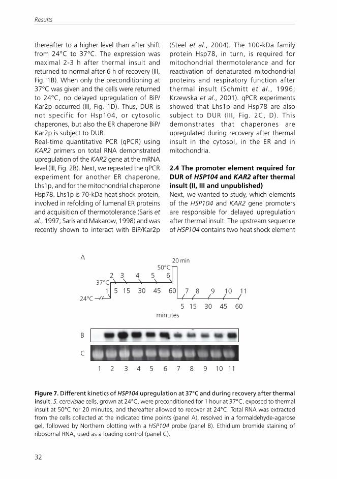
32
thereafter to a higher level than after shiftfrom 24°C to 37°C. The expression wasmaximal 2-3 h after thermal insult andreturned to normal after 6 h of recovery (III,Fig. 1B). When only the preconditioning at37°C was given and the cells were returnedto 24°C, no delayed upregulation of BiP/Kar2p occurred (III, Fig. 1D). Thus, DUR isnot specific for Hsp104, or cytosolicchaperones, but also the ER chaperone BiP/Kar2p is subject to DUR.Real-time quantitative PCR (qPCR) usingKAR2 primers on total RNA demonstratedupregulation of the KAR2 gene at the mRNAlevel (III, Fig. 2B). Next, we repeated the qPCRexperiment for another ER chaperone,Lhs1p, and for the mitochondrial chaperoneHsp78. Lhs1p is 70-kDa heat shock protein,involved in refolding of lumenal ER proteinsand acquisition of thermotolerance (Saris etal., 1997; Saris and Makarow, 1998) and wasrecently shown to interact with BiP/Kar2p
(Steel et al., 2004). The 100-kDa familyprotein Hsp78, in turn, is required formitochondrial thermotolerance and forreactivation of denaturated mitochondrialproteins and respiratory function afterthermal insult (Schmitt et al ., 1996;Krzewska et al., 2001). qPCR experimentsshowed that Lhs1p and Hsp78 are alsosubject to DUR (II I, Fig. 2C, D). Thisdemonstrates that chaperones areupregulated during recovery after thermalinsult in the cytosol, in the ER and inmitochondria.
2.4 The promoter element required forDUR of HSP104 and KAR2 after thermalinsult (II, III and unpublished)Next, we wanted to study, which elementsof the HSP104 and KAR2 gene promotersare responsible for delayed upregulationafter thermal insult. The upstream sequenceof HSP104 contains two heat shock element
��2�
��2�
2�� ����
�
�
�
�
-
�
�
���
�
�
�
�
�
�
���6���
#
�
� � � � � � - � ��
Figure 7. Different kinetics of HSP104 upregulation at 37°C and during recovery after thermalinsult. S. cerevisiae cells, grown at 24°C, were preconditioned for 1 hour at 37°C, exposed to thermalinsult at 50°C for 20 minutes, and thereafter allowed to recover at 24°C. Total RNA was extractedfrom the cells collected at the indicated time points (panel A), resolved in a formaldehyde-agarosegel, followed by Northern blotting with a HSP104 probe (panel B). Ethidium bromide staining ofribosomal RNA, used as a loading control (panel C).
Results
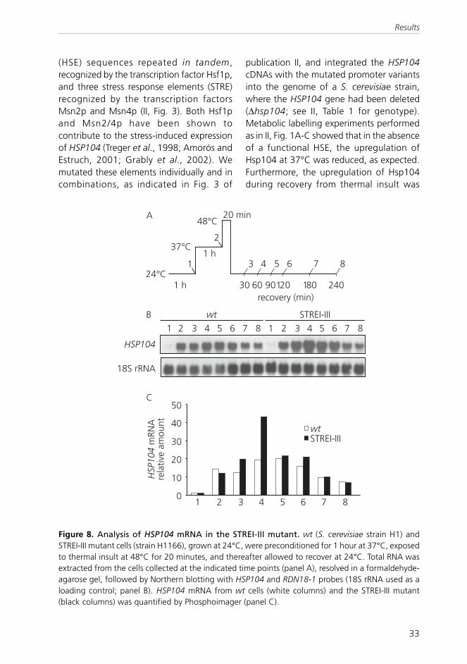
33
#
&��*3�&)�4���5
��2�
��2�
��2�
� ��"
��"
� ����
��
� �
�
�
-
��
�
��
�
����
�����&.7
� � � � � � � � � � � ��� �,.�0(000
�
�
�
�
���,.�0(000
�
���
� �.7
&�'���3����
*6��
� � � � � �
(HSE) sequences repeated in tandem,recognized by the transcription factor Hsf1p,and three stress response elements (STRE)recognized by the transcription factorsMsn2p and Msn4p (II, Fig. 3). Both Hsf1pand Msn2/4p have been shown tocontribute to the stress-induced expressionof HSP104 (Treger et al., 1998; Amorós andEstruch, 2001; Grably et al., 2002). Wemutated these elements individually and incombinations, as indicated in Fig. 3 of
publication II, and integrated the HSP104cDNAs with the mutated promoter variantsinto the genome of a S. cerevisiae strain,where the HSP104 gene had been deleted(∆hsp104; see II, Table 1 for genotype).Metabolic labelling experiments performedas in II, Fig. 1A-C showed that in the absenceof a functional HSE, the upregulation ofHsp104 at 37°C was reduced, as expected.Furthermore, the upregulation of Hsp104during recovery from thermal insult was
Figure 8. Analysis of HSP104 mRNA in the STREI-III mutant. wt (S. cerevisiae strain H1) andSTREI-III mutant cells (strain H1166), grown at 24°C, were preconditioned for 1 hour at 37°C, exposedto thermal insult at 48°C for 20 minutes, and thereafter allowed to recover at 24°C. Total RNA wasextracted from the cells collected at the indicated time points (panel A), resolved in a formaldehyde-agarose gel, followed by Northern blotting with HSP104 and RDN18-1 probes (18S rRNA used as aloading control; panel B). HSP104 mRNA from wt cells (white columns) and the STREI-III mutant(black columns) was quantified by Phosphoimager (panel C).
Results

34
abolished, showing that HSE was requiredfor DUR of Hsp104 (II, Fig. 4A). The HSEmutant was able to acquirethermotolerance; it formed colonies at 24°Cafter thermal insult as efficiently as controlcells (II, Fig. 5A). Furthermore, the HSEmutant strain remained metabolically active,consuming glucose (not shown) andsynthesizing proteins (II, Fig. 4C) as efficientlyas control cells during the 6-h recoveryperiod after thermal insult. Thus, the residualexpression of Hsp104 in the HSE mutant wassufficient to ensure survival of the strain.Furthermore, the absence of DUR of Hsp104in the HSE mutant was not due to animpaired survival of the strain.Mutagenization of the three STREs of theHSP104 promoter individually or incombination had little or no effect on DUR.In the STREI-III mutant, DUR of Hsp104 wasin average fourfold (II, Fig. 4), suggestingthat the STREs were dispensable. Themetabolic labeling experiment with theSTREI-III mutant was performed twice, ofwhich the average is shown in II, Fig. 4. TheDUR of Hsp104 was 3.7- and 4.4-fold,respectively (not shown). In turn, themetabolic labeling experiment with the wtstrain H1 was repeated five times. In theseexperiments, DUR of Hsp104 was in average5.2-fold, ranging from 2.5- to 8.7-fold (notshown). Due to the variation between singlemetabolic labeling experiments, weconfirmed the result from the STREI-IIImutant using Northern blot hybridisation.Fig. 8 of this thesis shows that DUR ofHSP104 mRNA was 43-fold in the STREI-IIImutant, whereas in the wt strain, DUR was20-fold. This Northern experiment wasperformed twice with similar results. Inconclusion, mutagenisation of the STREs inthe HSP104 promoter did not decrease DURof Hsp104.In contrast, mutagenization of all three STREsand the HSE in combination abolished
Hsp104 expression completely, leading toseverely impaired ability to form coloniesafter thermal insult (II, Fig. 5A) and to resumeprotein synthesis (II, Fig. 4C) after thermalinsult.The KAR2 promoter contains one heat shockelement (HSE), an unfolded protein responseelement (UPRE), recognized by Hac1p, anda GC-rich region between them. KAR2 is anessential gene and disturbance of itsupstream regions is potentially lethal. Thatis why we used ∆ICL1-lacZ fusion constructs,where the E.coli β-galactosidase gene wasplaced under different KAR2 promotervariants (III, Fig. 3; Zimmer et al., 1999).Strain H1806 contains the KAR2 promoterfragment sufficient for KAR2 expression,nucleotides -280 to -109 (Mori et al., 1992),directly upstream of the lacZ reporter(Zimmer et al., 1999). H1806 cells wereexposed to the heat treatments andmetabolic labelling, and β-galactosidaseexpression was monitored byimmunoprecipitation (III, Fig. 3). Enzymaticassays were used only for measuring basalactivity at 24°C due to thermal inactivationof the enzyme by the heat treatments. Underthe control of this KAR2 promoter variant,β-galactosidase expression was regulatedsimilarly after thermal insult as that ofauthentic genomic BiP/Kar2p (III, Fig. 3B).Thus, this assay system was feasible forinvestigating KAR2 promoter elements inDUR.Next, the four consecutive GAA repeats inthe HSE of the KAR2 promoter weremutated (strain H2004; II I, Table 1).Metabolic labelling experiments showed thatin strain H2004, the upregulation of β-galactosidase at 37°C (III, Fig. 3, column HS)was reduced, as expected (Mori et al., 1992;Kohno et al., 1993) but that DUR occurredsimilarly as in the reference strain H1806 (III,Fig. 3D).
Results

35
The GC-rich region has been shown toregulate basal expression of KAR2 (Mori etal., 1992). Next, we investigated a strainharbouring a construct, where the 5' regionand the GC-rich region were deleted inaddition to the HSE (strain H1765; III, Fig.3E). In this strain, the induction at 37°C wasabolished, and DUR dramatically reduced (III,Fig. 3E). This suggests that the 5' and theGC-rich regions backed up the HSE whenthe latter was absent, because mutation inthe HSE abolished DUR of KAR2 only incombination with deletion of the 5' and GC-rich regions.The next question was, whether the GC-richregion could be responsible for DUR in theabscence of the HSE. In strain H1767,harbouring a construct where the 5' and theGC-rich regions were the only functionalKAR2 promoter elements, β-galactosidaseexpression was undetectable in bothimmunoprecipitation and enzymatic assays(III, Fig. 3I). Thus, the 5' and GC-rich regionsfailed to drive DUR alone. This result is inagreement with the work of Zimmer and co-workers (1999), who reported lack of β-galactosidase activity from the sameconstruct under ER stress conditionsproduced by artificially misfoldedcytochrome P450. Finally, results from strainH1769 showed that HSE alone was sufficientfor evoking DUR (III, Fig. 3H).Kar2p is a target of the unfolded proteinresponse (UPR), activated via the unfoldedprotein response element (UPRE) in itspromoter, when unfolded proteinsaccumulate in the ER (Mori et al., 1992;Kohno et al., 1993). The role of the UPRE inDUR was studied using a strain harboring aconstruct, where the KAR2 promoter lackedthe UPRE (III, Fig. 3F; strain 1766). In thisstrain, both induction at 37°C and DUR wereunaffected, showing that the unfoldedprotein response element UPRE wasdispensable for DUR. This was likewise the case
for strains H1768 (III, Fig. 3G) and H1769 (III,Fig. 3H), harboring shorter variants of theUPRE-deleted KAR2 promoter.The UPR signal is transmitted from the ER tothe cytosol by the transmembrane kinaseIre1p, which causes splicing of HAC1 mRNA(Fig. 3 of this thesis). Hac1p is thetranscription factor, which activates the UPRtarget genes (Fig. 3 of this thesis). Toinvestigate the role of these UPR key playersin DUR of KAR2, strains lacking the IRE1 orHAC1 genes were transformed with the lacZreporter construct, resulting in strains H1807and H1808, respectively (III, Fig. 3J-K). Bothdeletions prevent the activation of UPRpathway (Cox et al., 1993; Mori et al., 1996).In these strains, DUR occurred as in thereference strain H1806 (II I, Fig. 3C),demonstrating that the UPR pathway wasdispensable for DUR (III, Fig. 3J-K). Thus, ourstudies have revealed a novel regulatorymechanism for the KAR2 gene, which isunrelated to UPR.In conclusion, DUR occurred via the HSE ofHSP104 and KAR2 promoters, and the otherstress-responsive elements in these genepromoters were dispensable for DUR.
2.5 Delayed upregulation of Hsp104after thermal insult requires thetranscription factors Msn2p and Msn4p(II)The transcription factors Msn2p and Msn4pactivate stress-dependent expression ofSTRE-containing target genes. Their role inDUR of Hsp104 was examined by using astrain lacking the genes for MSN2 and MSN4(∆msn2/4; II, Table 1). In this strain, only aslight upregulation of Hsp104 expressionduring recovery was detected (II, Fig. 6).Because the STREs in the HSP104 promoterwere dispensable for DUR, it appears thatsome other target genes of the transcriptionfactors Msn2p and Msn4p are required toinduce DUR of Hsp104. These unknown
Results

36
proteins also contribute to acquisition ofthermotolerance, because it was impairedin the ∆msn2/4 strain (II, Fig. 5B). However,total protein synthesis was normal in thisstrain (II, Fig. 6C).
2.6 The MAP kinase Hog1p is requiredfor DUR of Hsp104 (II)Hog1p is a MAP kinase implicated inupstream regulation of transcription factorsMsn2p and Msn4p upon osmotic stress(Alepuz et al., 2001) and shown to beactivated also upon heat stress at 39°C(Winkler et al., 2002). In a strain lacking theHOG1 gene (∆hog1; II, Table 1), delayedupregulation of Hsp104 after thermal insultwas abolished, whereas the 24°C-to-37°Cinduction persisted (II, Fig. 6). The ∆hog1strain displayed normal bulk proteinsynthesis (II, Fig. 6) and glucose consumption(not shown) at least for 6 hours of recoveryat 24°C, but its capability of acquiringthermotolerance was impaired (II, Fig. 5).Because Hog1p was required forthermotolerance, we wanted to study theeffect of Hog1p on another biologicalfunction of Hsp104, solubilization of heat-aggregated proteins (Parsell et al., 1994).Transmission electron microscopy showedthat after growth at 24°C, the morphologyof ∆hog1 cells was normal (II, Fig. 7A).However, after preconditioning at 37°C andthermal insult for 20 min at 50°C (II, Fig.7B), the cytosol of ∆hog1 cells was full ofaggregate-like structures that persisted evenafter 6 hours of recovery (II, Fig. 7C). Innormal cells treated the same way, the heat-induced aggregates dissolved after 6 hoursof recovery at 24°C (Parsell et al., 1994;Simola et al., 2000). In conclusion, Hog1pwas required for DUR of Hsp104, for efficientacquisition of thermotolerance, and forsolubilization of heat-induced cytosolicaggregates.
2.7 Biological functions of BiP/Kar2pafter thermal insult (III)Kar2p functions in the translocation ofpolypeptides into the ER, in facilitating ofprotein folding, and in retention of misfoldedproteins in the ER (for review, see Gething,1999). BiP/Kar2p synthesis was abolished bythe thermal insult and resumed only aftertwo hours of recovery (III, Fig. 1). To studythe biological functions of BiP/Kar2p, thetranslocation kinetics of a secretory reporterprotein, Hsp150∆-β-lactamase, wasfollowed. The 150∆ fragment, a signal-peptide-containing N-terminal fragment ofthe natural yeast secretory glycoproteinHsp150 (Russo et al., 1993) assists the β-lactamase portion to fold properly in theyeast ER (Simonen et al., 1994). The Hsp150signal peptide confers post-translationaltranslocation (Paunola et al., 1998). Thecytosolic, ER and mature forms of thereporter can be distinguished by theirdifferent extent of O-glycosylation (Paunolaet al., 1998). In SDS-PAGE, theunglycosylated cytoplasmic form ofHsp150∆-β-lactamase migrates like a 66-kDa protein, the ER-resident form like a 110-kDa protein, and the fully glycosylated,mature form like a 145-kDa protein (Paunolaet al., 1998; Fatal et al., 2004). Hsp150∆-β-lactamase was expressed under the heat-activated HSP150 promoter (Russo et al.,1993). Six parallel samples undergoing thethermal treatments described above werelabelled with 35S methionine/cysteine (III, Fig.4A; bars) followed by chase for differentperiods (III, Fig. 4B). The growth media wereseparated from the cells, andimmunoprecipitation with β-lactamaseantiserum was performed for the cell lysatesand the media, followed by SDS-PAGE andautoradiography.After the pulse at 24°C (III, Fig. 4B, graph a),60% of β-lactamase was found in thecytoplasmic form (triangles), 13% in the ER
Results

37
form (squares), and 26% in the fullyglycosylated, mature form (circles). Thefraction of mature molecules increased withincreasing chase periods. The fraction of ER-resident β-lactamase (Fig. 4B; graph a;squares) remained relatively constant atroughly 10% for the first time points, whichprobably reflects equal flow of translocationand of ER exit. Finally, after 30 minutes ofchase, practically all of the labelled reporterprotein was in the mature form (circles),meaning it had exited the ER and becomeglycosylated in the Golgi. Afterpreconditioning at 37°C and thermal insultat 50°C, a 5-minutes pulse failed to label β-lactamase molecules (not shown), due tooverall halt of protein synthesis (II, Fig. 1D;Jämsä et al., 1995). After 1 hour of recoveryat 24°C (Fig. 4B; graph b), a 5-minutes pulsefollowed by different chase times showedthat a sufficient amount of the reporterprotein was produced that the experimentcould be performed. However, the reporterremained in the cytoplasm and had thusfailed to translocate even after 40 minutesof chase (triangles). This was the case alsoafter 2 hours of recovery at 24°C (graph c).
Only after 3 hours of recovery the newlysynthesized reporter molecules started totranslocate. The cytoplasmic pool decreasedfrom 90% to 40% (graph d; triangles), whilethe ER-resident pool increased from 5% to20% (squares). However, the mature poolonly increased from 3% to 35% (circles),suggesting that the reporter did notefficiently exit the ER. After 4 hours ofrecovery, the translocation velocity oflabelled β-lactamase molecules was onlyslightly slower than before the thermal insult(graph e). Finally, when the reporter waspulse-labelled after 6 hours of recovery,practically all reporter molecules were inmature form after 40 minutes of chase(graph f; circles), showing that thetranslocation kinetics had recovered to thesame level as at 24°C before the heattreatments. In conclusion, ER translocationresumed concomitantly with enhancedsynthesis of BiP/Kar2p after 3 hours ofrecovery, whereafter ER exit and proteinsecretion also were resumed.
Results

38
DISCUSSION
1 Differentiation lineage-dependent expression of human HSF2
Eukaryotic heat shock gene expression ismediated by a family of heat shocktranscription factors, which bind to heatshock promoter elements in their targetgene promoters. In vertebrates, fourmembers of this family, HSF1-4, have beenidentified (Rabindran et al., 1991; Sarge etal., 1991; Schuetz et al., 1991; Nakai andMorimoto, 1993; Nakai et al., 1997). Theexistence of multiple HSFs raises the questionabout their specific or overlapping functions,and possible divergent or convergentsignaling pathways leading to theiractivation.
1.1. The role of HSF2 in developmentalprocesses and heat shock responseIt has been known previously that humanHSF2 is upregulated during hemin-induceddifferentiation of human K562erythroleukemia cells. In this work, thisupshift was detected at the protein level andat the transcriptional level, and was shownto be due to mRNA stabilization, in additionto transcriptional induction. Theupregulation of HSF2 was accompanied byactivation of DNA-binding of HSF2.Furthermore, the results of the present studyindicate that HSF2 expression and activationwere specific for K562 cells induced todifferentiate along the erythroid lineage. Incontrast, in cells undergoing megakaryocyticdifferentiation, HSF2 expression and DNA-binding was abolished in a HSF2 promoter-dependent manner.HSF2 has traditionally been offered adevelopmental role among the vertebrateheat shock factors. Earlier work has shownthat HSF2 is activated in mouse embryoniccarcinoma cells, during the blastocyst stageof mouse embryogenesis, during spermato-
genesis and during heart development(Sarge et al., 1994; Murphy et al., 1994;Mezger et al., 1994a; Rallu et al., 1997;Alastalo et al., 1998; Eriksson et al., 2000).However, recent reports expand the pictureby suggesting a role for HSF2 in heat stress,as well. HSF2 was shown to interactphysically with HSF1 in specialized sub-nuclear structures called stress granules, andthereby to influence the localization of HSF1,which is a regulatory mechanism of HSF1-mediated heat shock response (Jolly et al.,1999; Alastalo et al., 2003). Furthermore,HSF2 was also suggested to form hetero-complexes with HSF1 both upon heat shockand hemin treatment (He et al., 2003;Trinklein et al., 2004). Moreover, heat stressenhanced activation of the hsp70 promoterby these hybrid trimers (He et al., 2003).Further studies are needed to elucidate thepossible interaction of HSF1 and 2 indevelopmental processes and heat shockresponse.
1.2 Regulation of HSF2 activation inhemin-induced K562 cellsIn agreement with earlier work, an increasein HSF2 expression and DNA-binding wasdetected in hemin-treated K562 cells(Theodorakis et al., 1989; Sistonen et al.,1992; Sistonen et al., 1994). In contrast, incells induced to differentiate along themegakaryocytic lineage with TPA, HSF2expression and DNA-binding were abolished.Furthermore, the expression pattern of HSF2in K562 cells committed to one of theselineages could not be reversed.The signals leading to up- or downregulationof HSF2 in K562 cells are still unknown.Hemin has been shown to activate theexpression of globins via heme response
Discussion

39
promoter elements, which resemble theconsensus binding sites for the erythroid-specific transcription factor NF-E2 (Ney et al.,1990; Solomon et al., 1993; Palma et al.,1994; Inamdar et al., 1996). These bindingsites are identical to the antioxidant responseelement and the heme-responsive element(Johnsen et al., 1998). An analysis of humanHSF2 promoter showed the presence ofputative binding sites for NF-E2, suggestingan explanation for the hemin-inducedupregulation of HSF2 mRNA (Nykänen et al.,2001). No HSEs are present in the HSF2promoter, indicating that HSF2 does notregulate its own transcription (Nykänen etal., 2001). Future studies will reveal, whetherthe heme responsive element-like sequencesof HSF2 promoter are functional. So far, thepossible role HSF2 in erythropoiesis remainselusive.We showed here that the increase in HSF2protein seen previously in hemin-treatedK562 cells (Theodorakis et al., 1989;Sistonen et al., 1992; Sistonen et al., 1994)was due to stabilization of HSF2 mRNA. Thetranscriptional induction observed for HSF2was only 1.5- to 2-fold, whereas the increasein steady-state mRNA was 6-fold. Whenhemin-treated cells were treated with thetranscription inhibitor actinomycin D, HSF2mRNA levels remained higher in than incontrol cells, indicating a prolonged half-lifeof the mRNA molecules. In another study,luciferase reporter protein expressioncontrolled by a 950-bp HSF2 promoter didnot increase upon hemin treatment, furthersuggesting that the augmentation in HSF2upon hemin occurs posttrancriptionally(Nykänen et al., 2001).In K562 cells, the inactive, non-DNA-bindingform of HSF2 exists in the cytoplasm asdimers. Upon activation, HSF2 is trimerizedand transported into the nucleus (Sistonenet al., 1994). In contrast, constitutive HSE-binding activity of HSF2 has been reported
in various cell types (Mezger et al., 1989;Murphy et al., 1994; Mezger et al., 1994a;Mezger et al., 1994b). In addition totrimerization, the DNA-binding activity ofHSF2 has been shown to be enhanced byattachment of SUMO-1, an ubiquitin-relatedprotein with a number of different functions(Goodson et al ., 2001), and byheterotrimerization with HSF1 (He et al.,2003). Furthermore, the HSF2-α isoform istranscriptionally more active than the βisoform (Goodson et al., 1995; Leppä et al.,1997b). Consistent with earlier work, ourresults showed that hemin-inducedaccumulation of HSF2 protein occurred inHSF2-α- but not in HSF2-β-overexpressingcells (Leppä et al., 1997b). The molar ratiobetween the isoforms seems to modify thetranscriptional activity of HSF2, becauseoverexpression of HSF2-β was shown torepress the hemin-mediated increase ofHSP70 expression in K562 cells (Leppä et al.,1997b). Furthermore, the ratio of HSF2-αand HSF2-β isoforms varies significantlybetween different mouse tissues, furtherimplicating that regulation of isoformexpression could represent a mechanism ofregulating HSF2 activity (Goodson et al.,1995).
1.3 Expression and function of HSF2Cell differentiation involves a complexexpression program of spatially andtemporally organized factors. The results ofthis study suggest that DNA-binding of HSF2increases upon hemin-induceddifferentiation of K562 cells. This result wasin agreement with some previous reports(Sistonen et al., 1992; Sistonen et al., 1994),but inconsistent with another (Yoshima etal., 1998). The latter implicates that HSF1,instead of HSF2, is the primary factor bindingto HSE in the HSP70 promoter and activatingits transcription in hemin-treated K562 cells.One explanation to this apparent
Discussion

40
contradiction might be the heterogeneity ofK562 cells, which exist in several subtypespresumably exhibiting different HSFactivation patterns. Another explanationcould be the affinity differences in antibodiesraised against mouse, human and chickenHSFs, used in different laboratories to detecthuman HSF-HSE complexes (Pirkkala andSistonen, 1999).HSF2 activity was shown to be required forthe erythroid differentiation of K562 cells(Leppä et al., 1997b). The cellular role andthe target genes of HSF2 in hemin-inducedK562 cells remain, however, still unclear.Trinklein and co-workers recently reportedabout 9 genes, whose expression wasinduced 10-fold or more by hemintreatment, and about 6 genes, whosepromoters were occupied by HSF2 uponhemin treatment. However, the extent ofHSF2 occupation did not correlate with therange of induction of these genes by hemintreatment (Trinklein et al., 2004), suggestingthat DNA-binding of HSF2 and target geneactivation may not be coupled. Consistentlywith this work, during mouse embryogenesisand rat spermatogenesis, induced heat
shock gene expression was shown to beindependent of HSF2 expression and DNA-binding activity (Rallu et al., 1997; Alastaloet al., 1998; Eriksson et al., 2000).In conclusion, human HSF2 was upregulatedduring hemin-induced differentiation ofhuman K562 erythroleukemia cells. Thisupshift was detected at the protein level andat the transcriptional level, and was shownto be due to mRNA stabilization, in additionto transcriptional induction. Theupregulation of HSF2 was accompanied byactivation of DNA-binding of HSF2. Incontrast, megakaryocytic differentiation ofK562 cells induced with 12-O-tetradecanoyl-phorbol-13-acetate led to downregulationof HSF2 expression and DNA-binding activity.This downregulation occurred via the HSF2promoter.These results support a role for HSF2 indevelopment and differentiation,underlining the divergent functions of themultiple HSFs in vertebrates. Thedifferentiation lineage-specificity of HSF2could possibly represent an early marker fordifferentiating pathways of multipotenthematopoietic progenitor cells.
2 Novel regulation mechanism of yeast chaperones
2.1 Delayed upregulation of yeastchaperones during recovery fromthermal insultIn this study, a novel regulatory mechanismof yeast chaperones was discovered. WhenS. cerevisiae cells grown at 24°C, werepreconditioned at 37°C, exposed to a briefthermal insult at 50°C and thereafterreturned to 24°C, chaperone expression wasupregulated 3 hours after the thermal insult,although the cells were maintained at 24°C.We designated this novel regulationmechanism Delayed Upregulation (DUR). Theheat shock proteins Hsp104 (cytosol), BiP/Kar2p and Lhs1p (ER), and Hsp78
(mitochondria), were subject to DUR. Thisshows that several chaperone genes aresubject to DUR and DUR occurs in variouscompartments of the yeast cell. For bothHsp104 and BiP/Kar2p, the heat shockpromoter element (HSE) was necessary andsufficient for DUR. In the case of Hsp104,the stress response elements (STRE) weredispensable. The STRE-specific transcriptionfactors Msn2p and Msn4p appeared toaffect DUR of Hsp104 via other unknowntarget proteins. The MAP kinase Hog1p wasalso required for DUR of Hsp104 and forremoving cytosolic heat-aggregated proteinsafter thermal insult. In the case of BiP/Kar2p,
Discussion
41
the unfolded protein response element(UPRE) and the unfolded protein responsepathway were dispensable for DUR. Thermalinsult abolished the biological activities ofBiP/Kar2p. They were resumed afterexcessive amounts of new BiP/Kar2pmolecules were synthesized due to DUR.
2.2 The promoter element andtranscription factors involved in DURWe identified the KAR2 and HSP104promoter element responsible for DUR. Inboth cases, the HSE was essential andsufficient for DUR. In contrast to the HSEs,the results show for both genes that theother types of stress-inducible promoterelements, the STREs and the UPRE, inHSP104 and KAR2 promoter, respectively,were dispensable for DUR. DUR appears tobe a regulatory mechanism, where thetranscription factor of HSE, Hsf1p, has aninstrumental role, as no other transcriptionfactor is known to operate via the HSE.Future studies should address the activationof Hsf1p after thermal insult.Hsp104 is required for acquisition ofthermotolerance of yeast cells (Sanchez andLindquist, 1990). Although mutation of theHSE in the HSP104 promoter abolished DURof Hsp104, a basal level of Hsp104expression was still present in the HSEmutant after thermal insult. The HSE mutantacquired thermotolerance, demonstratingthat even the residual amount of Hsp104present in this strain was adequate toguarantee thermoprotection. It is knownfrom previous studies that the HSP104promoter elements are capable of backingeach other up (Amorós and Estruch, 2001;Grably et al., 2002). In contrast to the HSE,all the three STREs in the HSP104 promotercould be destroyed without decreasing DURor the ability of the strain to acquirethermotolerance. The mutations fromAGGGG to ACGAG that we introduced to
the STREs in HSP104 promoter, have beenreported to abolish stress-induced expressionthrough STRE, which confirms that ourmutated elements truly are unfunctional(Marchler et al., 1993). However, deletionof the HSE in combination with all the threeSTREs resulted in the same phenotype as thatcaused by deletion of HSP104, namely lackof Hsp104 expression and failure to survivethe thermal insult. Surprisingly, although theSTREs of the HSP104 promoter weredispensable for DUR, deletion of the genesfor the STRE-specific transcription factorsMsn2/4p led to a decreased DUR of Hsp104and an impaired ability to acquirethermotolerance. This apparentcontradiction suggests that some otherSTRE-regulated genes are involved in DURof Hsp104. One candidate could be the bZIPtranscription factor Yap4, whose inductionunder osmotic and oxidative stress has beenshown to require Msn2p (Nevitt et al.,2004a; Nevitt et al., 2004b). YAP4 mRNA isupregulated under various stress conditions,including heat shock at 37°C, oxidative andosmotic stress (Posas et al., 2000; Rep et al.,2000; Gasch et al., 2000; Nevitt et al.,2004b). A promoter search for putative Yap4recognition sites in the HSP104 promoterwould imply, whether Msn2p and Yap4could build a transcription factor activationchain contributing to DUR of Hsp104.
2.3 DUR is different from upregulationat 37°CThe results from this work demonstratedthat DUR occurs via the HSEs in the HSP104and KAR2 promoters. The HSE has beenpreviously thought to be responsible for thewell-known upregulation of all hsps uponshift of the cells from physiologicaltemperature to 37°C. However, DUR differsfrom upregulation upon shift to 37°C inseveral regards. Firstly, upregulation of bothHsp104 and BiP/Kar2p at 37°C occurs within
Discussion
42
minutes (Fig. 7 of this thesis; Kohno et al.,1993; Gasch et al., 2000), whereas maximalDUR within hours. Secondly, the Hsf1p/HSEregulon of the HSP104 promoter is notessential for activation at 37°C (Treger et al.,1998; Amorós and Estruch, 2001; Grably etal., 2002), but essential and sufficient forDUR. Thirdly, activation at 37°C can be drivenvia HSP104 STREs alone (Grably et al., 2002),whereas the STREs had no role in DUR.Fourthly, Mns2/4p-regulated proteins haveno significant role in activation at 37°C (Boy-Marcotte et al., 1999), whereas they docontribute to DUR. And finally, Hog1p isrequired only for DUR of Hsp104 but notfor the upregulation of Hsp104 at 37°C.Thus, DUR is a novel regulatory mechanismdifferent from upregulation of hsps uponshift to 37°C.What could be the molecular mechanismsleading to the remarkable slowness of DUR?Slow induction of stress proteins has beenreported for other severe stresses. Uponsevere osmotic shock (1.4 M NaCl), nuclearaccumulation and phosphorylation of theMAP kinase Hog1p is delayed comparedwith mild stress (0.4 M NaCl; VanWuytswinkel et al., 2000). Severe osmoticshock renders the active and nuclear-localized Hog1p transiently unable to inducetranscription of osmotic stress-responsivegenes. Furthermore, there is reasonableevidence that many different stressconditions, even when mild, transientlyarrest the yeast cell cycle. Moderate heatshock and experimental accumulation ofmisfolded proteins cause G1 arrest via acommon Hsf1p-dependent mechanism(Bedard et al., 1980; Johnston and Singer,1980; Shin et al., 1987; Trotter et al., 2001).Likewise, moderate osmotic and oxidativeshocks cause a cell cycle delay, as well (Leeet al., 1996; Bellí et al., 2001). Thus, theremarkable slowness of heat shock proteininduction during recovery from thermal
insult may be caused by delay in nucleartransport and phosphorylation of somecrucial factor(s), and/or delay in progressionof the cell cycle.
2.4 DUR and signaling pathways
2.4.1 The high-osmolarity glycerolpathwayThe high-osmolarity glycerol (HOG) MAPkinase pathway is one of five MAPKpathways regulating a variety of biologicalresponses in yeast. Upon osmotic stress, theMAP kinase Hog1p is phosphorylated andtransported to the nucleus (Reiser et al.,1999), where it has been shown to berecruited by Msn2/4p to the CTT1 and HSP12promoters (Reiser et al., 1999; Alepuz et al.,2001). Hog1p mediates its effect throughat least five different transcription regulators,amongst them Msn2p and Msn4p. Theresults of this study showed that DUR ofHsp104 was dependent on the MAP kinaseHog1p. In a ∆hog1 deletion strain, bulkprotein synthesis and the rate of glucoseconsumption were normal for at least sixhours of recovery from thermal insult,whereas DUR and acquisition ofthermotolerance were abolished. Moreover,the ∆hog1 strain was incapable of removingcytosolic aggregates induced by thermalinsult. In normal cells, the heat-inducedaggregates dissolved during recovery fromthermal insult. The precise involvement ofHog1p in DUR remains, however, elusive.There are some studies that suggest aconnection between the HOG pathway andcellular responses to thermal stress. Winklerand co-workers (2002) showed that, inaddition to osmotic stress, also heat stress(shift from 23 to 39°C) increased the kinaseactivity of Hog1p. Moreover, Hog1p was notessential at 39°C, but the recovery from heatstress of a ∆hog1 strain was, however, slowerthan that of normal cells (Winkler et al.,
Discussion

43
2002). Furthermore, the Hog1p homologueof fission yeasts, Spc1/Sty1, is known to beactivated in response to heat and oxidativestress, in addition to osmotic stress (Degolset al ., 1996). In another study,osmosensitivity of a ∆hog1 strain wasreduced when grown at 37°C, furtherimplicating that the HOG pathway plays arole in thermoprotection (Siderius et al.,2000). More studies are needed to elucidatethe role of the HOG pathway in thermalstress responses.
2.4.2 The unfolded protein responsepathwayInduction of BiP/Kar2p expression inresponse to perturbation of protein foldingin the ER lumen activates the unfoldedprotein response pathway UPR and occursvia the UPRE in the KAR2 promoter. The UPRsignal is transferred from the ER lumen tothe cytosol by the transmembrane kinaseIre1p (Fig. 3). Ire1p causes splicing of HAC1mRNA in the cytosol. Hac1p is thetranscription factor that recognizes the UPREin the target genes (Fig. 3). In the presentstudy, experiments with strains lacking thegenes for Hac1p or Ire1p showed that thesemediators of UPR are not involved in DURof BiP/Kar2p. These results demonstrate thatDUR is a regulatory mechanism differentfrom UPR.
2.5 The biological function of DURIn this work it was demonstrated thatexpression of chaperones is upregulatedafter thermal insult in the cytosol, in the ERand in the mitochondria. After thermalinsult, aggregated proteins accumulate inthe cell. Could the accumulation of heat-denatured aggregates elicit a chain ofregulatory events leading to DUR? Wepropose that the increased chaperoneamount in the cell serves to repair the heat-induced damage to cellular proteins. Earlier
it was shown in our laboratory that proteins,denatured by thermal insult and aggregatedinside the ER, were renatured duringrecovery at physiological temperature by anATP-dependent mechanism (Jämsä et al.,1995). Both Lhs1p and BiP/Kar2p werefound to be associated with the aggregates(Jämsä et al., 1995; Saris et al., 1997). Inthe present work, an upregulation of yeastchaperone expression after thermal insultwas observed, although the cells weremaintained at physiological temperature.Accumulation of misfolded proteinscontaining the amino acid analogueazetidine 2-carboxylic acid in place of prolinehas been shown to activate Hsf1p andinduce expression of heat shock factor-regulated genes in the absence oftemperature changes (Trotter et al., 2001;Trotter et al., 2002). Under ER stressconditions, overexpression of BiP/Kar2p hasbeen reported to restore a cell growth defectcaused by overproduction of ∆pro, amutated fungal secretory proteinase, whichaccumulates as misfolded aggregates Theextra BiP/Kar2p associated directly with theaggregates (Umebayashi et al., 1999).Yeast as a unicellular organism cannot relyon its neighbouring cells or sacrificedamaged cells by apoptosis. In their naturalhabitat, on the surface of fruit, yeast cellsare exposed to greatly varying conditions intemperature, hydration and nutrientbalance. In the Mediterranean climate zone,the origins of most wine yeast strains,temperatures ranging to 40°C are notunusual. Although the temperature changesin nature are not as sudden as the heattreatments applied in this study, on asummer day the local temperature on thesurface of a wine grape could be 24°C inthe morning, 37°C during the midday, and45°C and even above in the afternoon sun.These extreme temperatures challenge theproper conformation of cellular proteins of
Discussion

44
yeast cells living on the surface of this grape.In the evening, when the temperature sinksto 24°C, yeast cells may need an augmentedexpression of hsps to repair the cellulardamage caused by the heat stress during theday. In conclusion, we propose that DURrepresents a mechanism to ensure survivalafter severe heat stress by enhancingconformational repair of heat-denaturedproteins.
2.5.1 Biological functions of BiP/Kar2pduring recovery from thermal insultBiP/Kar2p is involved in the translocation ofpolypeptides into the ER, in facilitating ofprotein folding, and in retention of misfoldedproteins in the ER (for review, see Gething,1999). The results of this study showed thatafter thermal insult, newly synthesizedreporter proteins failed to translocate, asthey were detected in the cytosolicunglycosylated form. At the same time afterthermal insult, BiP/Kar2p synthesis wasabolished. Thus, BiP/Kar2p molecules thatexisted prior to thermal insult were not ableto perform the translocation function, or
other components required for ERtranslocation were inactivated at 50°C. After4 hours of recovery at 24°C, the reporterprotein was translocated and glycosylated,it exited the ER and was secreted to themedium. Earlier, it was found thatintracellular transport of vacuolarcarboxypeptidase Y and cell wall invertasealso resumed after 3-4 h of recovery (Sariset al., 1997; Saris and Makarow, 1998).Concomitantly, BiP/Kar2p expression wasupregulated due to DUR after 3 hours ofrecovery. After 6 hours of recovery,translocation, ER exit and secretion hadrecovered to normal, meaning that functionsof Bip/Kar2p were resumed. Resumption ofthe functions of BiP/Kar2p during therecovery period may have resulted fromnewly synthesized BiP/Kar2p due to DUR,repair of BiP/Kar2p molecules which existedprior to thermal insult and were damagedat 50°C, or both. Enhanced synthesis of BiP/Kar2p molecules due to DUR may serve toensure resumption of translocation and ERexit functions.
Discussion

45
CONCLUDING REMARKS
Heat shock proteins (hsps) are needed bothin stress conditions and in physiologicalconditions. They exist in all organismsstudied so far, and in all cellular compart-ments. This study characterized theregulation of different components of theheat stress response, specifically the humanheat shock factor HSF2 and the yeastchaperones Hsp104 and BiP/Kar2p. Thefocus of the study was, however, inprocesses different from the classical heatshock response, namely differentiation in thecase of human HSF2 and recovery fromthermal insult in the case of yeast hsps.The results concerning the regulation ofHSF2 in differentiating K562 humanerythroleukemia cells suggest an erythroid-specific role for HSF2. For themegakaryocytic differentiation of K562 cells,HSF2 appeared to be dispensable. However,the molecular mechanisms behind thelineage-dependent regulation of HSF2 areunknown. During hemin-induceddifferentiation of K562 cells, HSF2 wasshown to be regulated transcriptionally andby mRNA stabilization. Despite the relativelynumerous studies on HSF2 in recent years,HSF2 stil l remains rather mysterious.Identification of HSF2 target genes andfactors regulating HSF2 expression willhopefully reveal the biological role of HSF2.
Concluding Remarks
When studying recovery of yeast S. cerevisiaefrom thermal insult, a novel regulatorymechanism of yeast heat shock proteins wasidentified. This regulatory mechanism, whichwe designated Delayed Upregulation (DUR),turned out to concern several chaperonesfunctioning in different compartments of theyeast cell. The results from this study showthat DUR occurs exclusively via the heatshock element (HSE) in heat shock genepromoters. However, DUR is regulateddifferently than the well-known upshift ofyeast hsps upon shift of the cells fromphysiological temperature to 37°C. Detailedunderstanding of the precise biological roleof DUR still requires further studies. Twolarge areas of future research needed aredetermination DUR target genes and thesignaling pathways leading to its activation.Yeasts are unicellular organisms living inhighly dynamic environments in nature.Yeast cells cannot rely on apoptosis tosacrifice damaged cells, but need elaboraterepair machineries to refold vital proteins.We propose that after thermal insult, whichdenatures proteins in all compartments ofthe cell, enhanced production of chaperonesis a mechanism which ensures survival.

46
ACKNOWLEDGEMENTS
This work was carried out at the Institute ofBiotechnology, and Division of Biochemistry,Department of Biological and EnvironmentalSciences, University of Helsinki (1999-2005)and at the Turku Centre for Biotechnology,Åbo Akademi (1997-1998). Financialsupport was provided by Helsinki GraduateSchool in Biotechnology and MolecularBiology and Sigrid Jusélius foundation.I am deeply grateful to my supervisor,Professor Marja Makarow for her excellentscientific guidance and encouragement overthese years. I could always trust on her helpin finding solutions to problems in myresearch, and her enormous effectivenessnever stops astonishing me.I wish to express my gratitude to my formersupervisor, Professor Lea Sistonen. Herpassionate enthusiasm towards research andwarm encouragement were extremelyvaluable during my first steps into the worldof science. As a member of my follow-upgroup, she continued to support my studieswith helpful advice. In this context, I alsowish to thank Professor Leevi Kääriäinen, theother member of my follow-up group, forstimulating discussions and for setting upthe time-table for this dissertation.I am grateful to Professor Mart Saarma,Director of the Institute of Biotechnology forproviding excellent facilities. Professor CarlG. Gahmberg, the head of the Division ofBiochemistry, is thanked for his positiveattitude towards my studies. Professor HeikkiRauvala and Docent Pekka Lappalainen, theformer and present heads of HelsinkiGraduate School in Biotechnology andMolecular Biology, are thanked for theopportunity to conduct this research withinthe graduate school system. Special thanksbelong to docents Sirkka Keränen and JukkaWestermarck for valuable comments andsuggestions to improve this thesis.
Acknowledgements
I wish to thank all the past and presentmembers of the yeast group for creating awarm and friendly spirit in the lab. I warmlythank my friends and co-workers Ansku,Anna Liisa, Anton, Eeva, Eija, Hanna, Juha,Leena, Mari, Monica, Netta, Taina, Qiao,Ricardo, Serguei and Vijay for their adviceand all the joyful moments. In particular, Iwish to thank Ansku for helping me to getinstalled when I first started in the lab andfor teaching me everything I needed to knowabout cloning. Special thanks to Taina forher help in all possible (and impossible)scientific and practical questions in the lab,and to Anna Liisa for her skillful and patienttechnical assistance.I wish to thank all members of Lea Sistonen’sand John Eriksson’s labs at the Turku Centrefor Biotechnology for their friendship andhelp through the years. Special thanksbelong to the “first generation”; Andrey,Ann-Sofi, Carina, Cecilia, Diana, Helena, Iina,Lila, Minna, Pia, Päivi, Sirpa, Stefanie, Tao,Tero-Pekka, Tim and Ville for guiding me intothe world of molecular biology during myM.Sc. year. I have been fortunate to be ableto follow the progress of your research (andto hear the latest gossips) in the Nordic StressNetwork and other meetings.I wish to thank all the personnel in ViikkiBiocenter and in the Ph.D. students in thegraduate schools for companionship,collegiality and fun Christmas parties. I alsothank Erkki Raulo, Anita Tienhaara and EevaSievi at the graduate schools’ coordinationoffice for help with all practical anddissertation-related issues. Timo Päivärintais thanked for excellent graphical assistancein preparation of this thesis, as well as thepublications.I would like to thank Biobio Society i.e.Societas biochemica, biophysica etmicrobiologica Fenniae, whose secretary I

47
Acknowledgements
have been, for the educational experiencein administrative tasks, for financial support,as well as for the opportunity to meet manyinteresting people. I especially thank theformer and present presidents ProfessorDennis Bamford and Docent Marc Baumannfor pleasant collaboration.I wish to thank all my friends outside the labfor their unconditional friendship. My friendssince primary school Tikki, Tessa and Outiare thanked for the past 22 years together.Susanna, Petri, Jan, my cousin Anna,Beatrice; I am very fortunate in life to havefriends like you. Thank you for friendship,support and for bringing so much joy in mylife.
My warmest thanks go to my parents Liisaand Anders for their love and advice, and tomy sisters Hanna and Eeva and to my brotherEero for always being there and for creatingsuch a strong feeling of togetherness. Thewarm support of my family-in-law is heartilyacknowledged, as well.Finally, I wish to express my dearest gratitudeto my husband Ove. His encouragement,understanding and confidence in me havebeen essential for the completion of thisthesis. I thank our children Oskar and Ameliefor filling my days with joy and happinessand showing me, what is important in life.
Helsinki, September 2005
48
REFERENCESAlastalo, T.P., Hellesuo, M., Sandqvist, A.,Hietakangas, V., Kallio, M., and Sistonen, L.(2003) Formation of nuclear stress granulesinvolves HSF2 and coincides with the nucleolarlocalization of Hsp70. J Cell Sci 116: 3557-3570.
Alastalo, T.P., Lönnström, M., Leppä, S.,Kaarniranta, K., Pelto-Huikko, M., Sistonen, L.,and Parvinen, M. (1998) Stage-specific expressionand cellular localization of the heat shock factor2 isoforms in the rat seminiferous epithelium. ExpCell Res 240: 16-27.
Alepuz, P.M., Jovanovic, A., Reiser, V., andAmmerer, G. (2001) Stress-induced map kinaseHog1 is part of transcription activationcomplexes. Mol Cell 7: 767-777.
Alitalo, R. (1990) Induced differentiation of K562leukemia cells: a model for studies of geneexpression in early megakaryoblasts. Leuk Res 14:501-514.
Alonso, C.R. (2005) Nonsense-mediated RNAdecay: a molecular system micromanagingindividual gene activities and suppressinggenomic noise. Bioessays 27: 463-466.
Ambros, V., Lee, R.C., Lavanway, A., Williams,P.T., and Jewell, D. (2003) MicroRNAs and othertiny endogenous RNAs in C. elegans. Curr Biol13: 807-818.
Amin, J., Ananthan, J., and Voellmy, R. (1988)Key features of heat shock regulatory elements.Mol Cell Biol 8: 3761-3769.
Amorós, M. and Estruch, F. (2001) Hsf1p andMsn2/4p cooperate in the expression ofSaccharomyces cerevisiae genes HSP26 andHSP104 in a gene- and stress type-dependentmanner. Mol Microbiol 39: 1523-1532.
Anfinsen, C.B. (1973) Principles that govern thefolding of protein chains. Science 181: 223-230.
Ast, G. (2004) How did alternative splicingevolve? Nat Rev Genet 5: 773-782.
Banerji, S.S., Theodorakis, N.G., and Morimoto,R.I. (1984) Heat shock-induced translationalcontrol of HSP70 and globin synthesis in chickenreticulocytes. Mol Cell Biol 4: 2437-2448.
Barrass, J.D. and Beggs, J.D. (2003) Splicing goesglobal. Trends Genet 19: 295-298.
Bassett, D.E., Jr., Boguski, M.S., and Hieter, P.(1996) Yeast genes and human disease. Nature379: 589-590.
Baxter, B.K., James, P., Evans, T., and Craig, E.A.(1996) SSI1 encodes a novel Hsp70 of theSaccharomyces cerevisiae endoplasmic reticulum.Mol Cell Biol 16: 6444-6456.
Bedard, D.P., Singer, R.A., and Johnston, G.C.(1980) Transient cell cycle arrest of Saccharomycescerevisiae by amino acid analog beta-2-DL-thienylalanine. J Bacteriol 141: 100-105.
Bellí, G., Garí, E., Aldea, M., and Herrero, E.(2001) Osmotic stress causes a G1 cell cycle delayand downregulation of Cln3/Cdc28 activity inSaccharomyces cerevisiae. Mol Microbiol 39:1022-1035.
Benz, E.J., Jr., Murnane, M.J., Tonkonow, B.L.,Berman, B.W., Mazur, E.M., Cavallesco, C., Jenko,T., Snyder, E.L., Forget, B.G., and Hoffman, R.(1980) Embryonic-fetal erythroid characteristicsof a human leukemic cell line. Proc Natl Acad SciU S A 77: 3509-3513.
Blond-Elguindi, S., Cwirla, S.E., Dower, W.J.,Lipshutz, R.J., Sprang, S.R., Sambrook, J.F., andGething, M.J. (1993) Affinity panning of a libraryof peptides displayed on bacteriophages revealsthe binding specificity of BiP. Cell 75: 717-728.
Boy-Marcotte, E., Lagniel, G., Perrot, M.,Bussereau, F., Boudsocq, A., Jacquet, M., andLabarre, J. (1999) The heat shock response inyeast: differential regulations and contributionsof the Msn2p/Msn4p and Hsf1p regulons. MolMicrobiol 33: 274-283.
Brennecke, J., Stark, A., Russell, R.B., and Cohen,S.M. (2005) Principles of microRNA-targetrecognition. PLoS Biol 3: e85
Brewer, G. and Ross, J. (1988) Poly(A) shorteningand degradation of the 3' A+U-rich sequencesof human c-myc mRNA in a cell-free system. MolCell Biol 8: 1697-1708.
Brodsky, J.L., Goeckeler, J., and Schekman, R.(1995) BiP and Sec63p are required for both co-and posttranslational protein translocation intothe yeast endoplasmic reticulum. Proc Natl AcadSci U S A 92: 9643-9646.
References
49
Brodsky, J.L. and Schekman, R. (1993) A Sec63p-BiP complex from yeast is required for proteintranslocation in a reconstituted proteoliposome.J Cell Biol 123: 1355-1363.
Bukau, B., Deuerling, E., Pfund, C., and Craig,E.A. (2000) Getting newly synthesized proteinsinto shape. Cell 101: 119-122.
Cashikar, A.G., Duennwald, M.L., and Lindquist,S.L. (2005) A chaperone pathway in proteindisaggregation: Hsp26 alters the nature of proteinaggregates to facilitate reactivation by Hsp104.J Biol Chem 280: 23869-23875.
Chapman, R.E. and Walter, P. (1997) Translationalattenuation mediated by an mRNA intron. CurrBiol 7: 850-859.
Charnay, P. and Maniatis, T. (1983) Transcriptionalregulation of globin gene expression in thehuman erythroid cell line K562. Science 220:1281-1283.
Chen, C.Y. and Shyu, A.B. (1994) Selectivedegradation of early-response-gene mRNAs:functional analyses of sequence features of theAU-rich elements. Mol Cell Biol 14: 8471-8482.
Chen, X., Easton, D., Oh, H.J., Lee-Yoon, D.S.,Liu, X., and Subjeck, J. (1996) The 170 kDaglucose regulated stress protein is a large HSP70-, HSP110-like protein of the endoplasmicreticulum. FEBS Lett 380: 68-72.
Chirico, W.J., Waters, M.G., and Blobel, G. (1988)70K heat shock related proteins stimulate proteintranslocation into microsomes. Nature 332: 805-810.
Christians, E., Davis, A.A., Thomas, S.D., andBenjamin, I.J. (2000) Maternal effect of Hsf1 onreproductive success. Nature 407: 693-694.
Corsi, A.K. and Schekman, R. (1997) The lumenaldomain of Sec63p stimulates the ATPase activityof BiP and mediates BiP recruitment to thetranslocon in Saccharomyces cerevisiae. J Cell Biol137: 1483-1493.
Cougot, N., Babajko, S., and Seraphin, B. (2004)Cytoplasmic foci are sites of mRNA decay inhuman cells. J Cell Biol 165: 31-40.
Cox, J.S., Shamu, C.E., and Walter, P. (1993)Transcriptional induction of genes encodingendoplasmic reticulum resident proteins requiresa transmembrane protein kinase. Cell 73: 1197-1206.
Craven, R.A., Egerton, M., and Stirling, C.J.(1996) A novel Hsp70 of the yeast ER lumen isrequired for the efficient translocation of anumber of protein precursors. EMBO J 15: 2640-2650.
Cyr, D.M., Langer, T., and Douglas, M.G. (1994)DnaJ-like proteins: molecular chaperones andspecific regulators of Hsp70. Trends Biochem Sci19: 176-181.
Dean, A., Erard, F., Schneider, A.P., and Schechter,A.N. (1981) Induction of hemoglobinaccumulation in human K562 cells by hemin isreversible. Science 212: 459-461.
Degols, G., Shiozaki, K., and Russell, P. (1996)Activation and regulation of the Spc1 stress-activated protein kinase in Schizosaccharomycespombe. Mol Cell Biol 16: 2870-2877.
Dever, T.E. (2002) Gene-specific regulation bygeneral translation factors. Cell 108: 545-556.
Du, L. and Warren, S.L. (1997) A functionalinteraction between the carboxy-terminal domainof RNA polymerase II and pre-mRNA splicing. JCell Biol 136: 5-18.
Eriksson, M., Jokinen, E., Sistonen, L., and Leppä,S. (2000) Heat shock factor 2 is activated duringmouse heart development. Int J Dev Biol 44: 471-477.
Fatal, N., Karhinen, L., Jokitalo, E., and Makarow,M. (2004) Active and specific recruitment of asoluble cargo protein for endoplasmic reticulumexit in the absence of functional COPII componentSec24p. J Cell Sci 117: 1665-1673.
Fernandes, M., O’Brien, T., and Lis, J. T. (1994)Structure and regulation of heat shock genepromoters. In Morimoto, R.I., Tissières, A. andGeorgopoulos, C. (eds), The Biology of HeatShock Proteins and Molecular Chaperones. ColdSpring Harbor Press, Cold Spring Harbor, NY, pp.375-393.
Flynn, G.C., Chappell, T.G., and Rothman, J.E.(1989) Peptide binding and release by proteinsimplicated as catalysts of protein assembly.Science 245: 385-390.
Flynn, G.C., Pohl, J., Flocco, M.T., and Rothman,J.E. (1991) Peptide-binding specificity of themolecular chaperone BiP. Nature 353: 726-730.
References

50
Fong, Y.W. and Zhou, Q. (2001) Stimulatory effectof splicing factors on transcriptional elongation.Nature 414: 929-933.
Frazier, A.E., Dudek, J., Guiard, B., Voos, W., Li,Y., Lind, M., Meisinger, C., Geissler, A., Sickmann,A., Meyer, H.E., Bilanchone, V., Cumsky, M.G.,Truscott, K.N., Pfanner, N., and Rehling, P. (2004)Pam16 has an essential role in the mitochondrialprotein import motor. Nat Struct Mol Biol 11: 226-233.
Furger, A., O’Sullivan, J.M., Binnie, A., Lee, B.A.,and Proudfoot, N.J. (2002) Promoter proximalsplice sites enhance transcription. Genes Dev 16:2792-2799.
Gahmberg, C.G., Jokinen, M., and Andersson,L.C. (1979) Expression of the major red cellsialoglycoprotein, glycophorin A, in the humanleukemic cell line K562. J Biol Chem 254: 7442-7448.
Gao, M., Wilusz, C.J., Peltz, S.W., and Wilusz, J.(2001) A novel mRNA-decapping activity in HeLacytoplasmic extracts is regulated by AU-richelements. EMBO J 20: 1134-1143.
Garner, M.M. and Revzin, A. (1981) A gelelectrophoresis method for quantifying thebinding of proteins to specific DNA regions:application to components of the Escherichia colilactose operon regulatory system. Nucleic AcidsRes 9: 3047-3060.
Gasch, A.P., Spellman, P.T., Kao, C.M., Carmel-Harel, O., Eisen, M.B., Storz, G., Botstein, D., andBrown, P.O. (2000) Genomic expression programsin the response of yeast cells to environmentalchanges. Mol Biol Cell 11: 4241-4257.
Gebauer, F. and Hentze, M.W. (2004) Molecularmechanisms of translational control. Nat Rev MolCell Biol 5: 827-835.
Georgopoulos, C. and Welch, W.J. (1993) Roleof the major heat shock proteins as molecularchaperones. Annu Rev Cell Biol 9: 601-634.
Gething, M.J. (1999) Role and regulation of theER chaperone BiP. Semin Cell Dev Biol 10: 465-472.
Gething, M.J., McCammon, K., and Sambrook,J. (1986) Expression of wild-type and mutantforms of influenza hemagglutinin: the role offolding in intracellular transport. Cell 46: 939-950.
Giardina, C. and Lis, J.T. (1995) Dynamic protein-DNA architecture of a yeast heat shock promoter.Mol Cell Biol 15: 2737-2744.
Glick, B.S. (1995) Can Hsp70 proteins act asforce-generating motors? Cell 80: 11-14.
Goffeau, A., Barrell, B.G., Bussey, H., Davis, R.W.,Dujon, B., Feldmann, H., Galibert, F., Hoheisel,J.D., Jacq, C., Johnston, M., Louis, E.J., Mewes,H.W., Murakami, Y., Philippsen, P., Tettelin, H.,and Oliver, S.G. (1996) Life with 6000 genes.Science 274: 546, 563-546, 567.
Goodson, M.L., Hong, Y., Rogers, R., Matunis,M.J., Park-Sarge, O.K., and Sarge, K.D. (2001)Sumo-1 modification regulates the DNA bindingactivity of heat shock transcription factor 2, apromyelocytic leukemia nuclear body associatedtranscription factor. J Biol Chem 276: 18513-18518.
Goodson, M.L., Park-Sarge, O.K., and Sarge, K.D.(1995) Tissue-dependent expression of heatshock factor 2 isoforms with distincttranscriptional activities. Mol Cell Biol 15: 5288-5293.
Goodson, M.L. and Sarge, K.D. (1995) RegulatedExpression of Heat Shock Factor 1 Isoforms withDistinct Leucine Zipper Arrays via Tissue-Dependent Alternative Splicing. Biochem BiophysRes Commun 211: 943-949.
Gorlach, M., Burd, C.G., and Dreyfuss, G. (1994)The mRNA poly(A)-binding protein: localization,abundance, and RNA-binding specificity. Exp CellRes 211: 400-407.
Grably, M.R., Stanhill, A., Tell, O., and Engelberg,D. (2002) HSF and Msn2/4p can exclusively orcooperatively activate the yeast HSP104 gene.Mol Microbiol 44: 21-35.
Gutteridge, S. and Gatenby, A.A. (1995) RubiscoSynthesis, Assembly, Mechanism, and Regulation.Plant Cell 7: 809-819.
Hachiya, N., Komiya, T., Alam, R., Iwahashi, J.,Sakaguchi, M., Omura, T., and Mihara, K. (1994)MSF, a novel cytoplasmic chaperone whichfunctions in precursor targeting to mitochondria.EMBO J 13: 5146-5154.
Hamilton, T.G. and Flynn, G.C. (1996) Cer1p, anovel Hsp70-related protein required forposttranslational endoplasmic reticulumtranslocation in yeast. J Biol Chem 271: 30610-30613.
References

51
Hänninen, A.L., Simola, M., Saris, N., andMakarow, M. (1999) The cytoplasmic chaperoneHsp104 is required for conformational repair ofheat-denatured proteins in the yeast endoplasmicreticulum. Mol Biol Cell 10: 3623-3632.
Hartl, F.U. (1996) Molecular chaperones in cellularprotein folding. Nature 381: 571-579.
He, F., Peltz, S.W., Donahue, J.L., Rosbash, M.,and Jacobson, A. (1993) Stabilization andribosome association of unspliced pre-mRNAs ina yeast upf1- mutant. Proc Natl Acad Sci U S A90: 7034-7038.
He, H., Soncin, F., Grammatikakis, N., Li, Y.,Siganou, A., Gong, J., Brown, S.A., Kingston, R.E.,and Calderwood, S.K. (2003) Elevated expressionof heat shock factor (HSF) 2A stimulates HSF1-induced transcription during stress. J Biol Chem278: 35465-35475.
Hemmingsen, S.M., Woolford, C., van der Vies,S.M., Tilly, K., Dennis, D.T., Georgopoulos, C.P.,Hendrix, R.W., and Ellis, R.J. (1988) Homologousplant and bacterial proteins chaperone oligomericprotein assembly. Nature 333: 330-334.
Hendrick, J.P. and Hartl, F.U. (1993) Molecularchaperone functions of heat-shock proteins.Annu Rev Biochem 62: 349-384.
Hershko, A., Ciechanover, A., and Varshavsky, A.(2000) Basic Medical Research Award. Theubiquitin system. Nat Med 6: 1073-1081.
Hinnebusch, A.G. (1997) Translational regulationof yeast GCN4. A window on factors that controlinitiator-trna binding to the ribosome. J Biol Chem272: 21661-21664.
Hirose, Y. and Manley, J.L. (1998) RNA polymeraseII is an essential mRNA polyadenylation factor.Nature 395: 93-96.
Hohmann, S. and Mager, W H. (eds) (1997) Yeaststress responses. R.G. Landes Company, Austin,TX.
Holmberg, C.I., Hietakangas, V., Mikhailov, A.,Rantanen, J.O., Kallio, M., Meinander, A.,Hellman, J., Morrice, N., MacKintosh, C.,Morimoto, R.I., Eriksson, J.E., and Sistonen, L.(2001) Phosphorylation of serine 230 promotesinducible transcriptional activity of heat shockfactor 1. EMBO J 20: 3800-3810.
Hurtley, S.M., Bole, D.G., Hoover-Litty, H.,Helenius, A., and Copeland, C.S. (1989)Interactions of misfolded influenza virushemagglutinin with binding protein (BiP). J CellBiol 108: 2117-2126.
Iborra, F.J., Escargueil, A.E., Kwek, K.Y.,Akoulitchev, A., and Cook, P.R. (2004) Molecularcross-talk between the transcription, translation,and nonsense-mediated decay machineries. J CellSci 117: 899-906.
Iborra, F.J., Jackson, D.A., and Cook, P.R. (2001)Coupled transcription and translation withinnuclei of mammalian cells. Science 293: 1139-1142.
Inamdar, N.M., Ahn, Y.I., and Alam, J. (1996) Theheme-responsive element of the mouse hemeoxygenase-1 gene is an extended AP-1 bindingsite that resembles the recognition sequences forMAF and NF-E2 transcription factors. BiochemBiophys Res Commun 221: 570-576.
Ingelfinger, D., Arndt-Jovin, D.J., Luhrmann, R.,and Achsel, T. (2002) The human LSm1-7 proteinscolocalize with the mRNA-degrading enzymesDcp1/2 and Xrnl in distinct cytoplasmic foci. RNA8: 1489-1501.
Ito, J. and Jacobs-Lorena, M. (2001) Functionalmapping of destabilizing elements in the protein-coding region of the Drosophila fushi tarazumRNA. J Biol Chem 276: 23525-23530.
Iwahashi, H., Obuchi, K., Fujii, S., and Komatsu,Y. (1997) Barotolerance is dependent on bothtrehalose and heat shock protein 104 but isessentially different from thermotolerance inSaccharomyces cerevisiae. Letters in AppliedMicrobiology 25: 43-47.
Jakobsen, B.K. and Pelham, H.R. (1988)Constitutive binding of yeast heat shock factorto DNA in vivo. Mol Cell Biol 8: 5040-5042.
Jämsä, E., Vakula, N., Arffman, A., Kilpeläinen,I., and Makarow, M. (1995) In vivo reactivationof heat-denatured protein in the endoplasmicreticulum of yeast. EMBO J 14: 6028-6033.
Jedlicka, P., Mortin, M.A., and Wu, C. (1997)Multiple functions of Drosophila heat shocktranscription factor in vivo. EMBO J 16: 2452-2462.
References
52
Jensen, R.E. and Johnson, A.E. (1999) Proteintranslocation: is Hsp70 pulling my chain? CurrBiol 9: R779-R782
Johnsen, Ø., Murphy, P., Prydz, H., and Kolstø,A.B. (1998) Interaction of the CNC-bZIP factorTCF11/LCR-F1/Nrf1 with MafG: binding-siteselection and regulation of transcription. NucleicAcids Res 26: 512-520.
Johnston, G.C. and Singer, R.A. (1980) Ribosomalprecursor RNA metabolism and cell division in theyeast Saccharomyces cerevisiae. Mol Gen Genet178: 357-360.
Jolly, C., Usson, Y., and Morimoto, R.I. (1999)Rapid and reversible relocalization of heat shockfactor 1 within seconds to nuclear stress granules.Proc Natl Acad Sci U S A 96: 6769-6774.
Jurica, M.S. and Moore, M.J. (2003) Pre-mRNAsplicing: awash in a sea of proteins. Mol Cell 12:5-14.
Kallio, M., Chang, Y., Manuel, M., Alastalo, T.P.,Rallu, M., Gitton, Y., Pirkkala, L., Loones, M.T.,Paslaru, L., Larney, S., Hiard, S., Morange, M.,Sistonen, L., and Mezger, V. (2002) Brainabnormalities, defective meiotic chromosomesynapsis and female subfertility in HSF2 null mice.EMBO J 21: 2591-2601.
Kawahara, T., Yanagi, H., Yura, T., and Mori, K.(1997) Endoplasmic reticulum stress-inducedmRNA splicing permits synthesis of transcriptionfactor Hac1p/Ern4p that activates the unfoldedprotein response. Mol Biol Cell 8: 1845-1862.
Kline, M.P. and Morimoto, R.I. (1997) Repressionof the heat shock factor 1 transcriptionalactivation domain is modulated by constitutivephosphorylation. Mol Cell Biol 17: 2107-2115.
Knauf, U., Newton, E.M., Kyriakis, J., andKingston, R.E. (1996) Repression of human heatshock factor 1 activity at control temperature byphosphorylation. Genes Dev 10: 2782-2793.
Kohno, K., Normington, K., Sambrook, J.,Gething, M.J., and Mori, K. (1993) The promoterregion of the yeast KAR2 (BiP) gene contains aregulatory domain that responds to the presenceof unfolded proteins in the endoplasmicreticulum. Mol Cell Biol 13: 877-890.
Krzewska, J., Langer, T., and Liberek, K. (2001)Mitochondrial Hsp78, a member of the Clp/
Hsp100 family in Saccharomyces cerevisiae,cooperates with Hsp70 in protein refolding. FEBSLett 489: 92-96.
Kuwabara, K., Matsumoto, M., Ikeda, J., Hori,O., Ogawa, S., Maeda, Y., Kitagawa, K., Imuta,N., Kinoshita, T., Stern, D.M., Yanagi, H., andKamada, T. (1996) Purification andcharacterization of a novel stress protein, the 150-kDa oxygen-regulated protein (ORP150), fromcultured rat astrocytes and its expression inischemic mouse brain. J Biol Chem 271: 5025-5032.
Lander, E.S., Linton, L.M., Birren, B., Nusbaum,C., Zody, M.C., Baldwin, J., Devon, K., Dewar,K., Doyle, M., FitzHugh, W., Funke, R., Gage, D.,Harris, K., Heaford, A., Howland, J., Kann, L.,Lehoczky, J., LeVine, R., McEwan, P., McKernan,K., Meldrim, J., Mesirov, J.P., Miranda, C., Morris,W., Naylor, J., Raymond, C., Rosetti, M., Santos,R., Sheridan, A., Sougnez, C., Stange-Thomann,N., Stojanovic, N., Subramanian, A., Wyman, D.,Rogers, J., Sulston, J., Ainscough, R., Beck, S.,Bentley, D., Burton, J., Clee, C., Carter, N.,Coulson, A., Deadman, R., Deloukas, P., Dunham,A., Dunham, I., Durbin, R., French, L., Grafham,D., Gregory, S., Hubbard, T., Humphray, S., Hunt,A., Jones, M., Lloyd, C., McMurray, A., Matthews,L., Mercer, S., Milne, S., Mullikin, J.C., Mungall,A., Plumb, R., Ross, M., Shownkeen, R., Sims, S.,Waterston, R.H., Wilson, R.K., Hillier, L.W.,McPherson, J.D., Marra, M.A., Mardis, E.R.,Fulton, L.A., Chinwalla, A.T., Pepin, K.H., Gish,W.R., Chissoe, S.L., Wendl, M.C., Delehaunty,K.D., Miner, T.L., Delehaunty, A., Kramer, J.B.,Cook, L.L., Fulton, R.S., Johnson, D.L., Minx, P.J.,Clifton, S.W., Hawkins, T., Branscomb, E., Predki,P., Richardson, P., Wenning, S., Slezak, T.,Doggett, N., Cheng, J.F., Olsen, A., Lucas, S.,Elkin, C., Uberbacher, E., Frazier, M., Gibbs, R.A.,Muzny, D.M., Scherer, S.E., Bouck, J.B.,Sodergren, E.J., Worley, K.C., Rives, C.M., Gorrell,J.H., Metzker, M.L., Naylor, S.L., Kucherlapati,R.S., Nelson, D.L., Weinstock, G.M., Sakaki, Y.,Fujiyama, A., Hattori, M., Yada, T., Toyoda, A.,Itoh, T., Kawagoe, C., Watanabe, H., Totoki, Y.,Taylor, T., Weissenbach, J., Heilig, R., Saurin, W.,Artiguenave, F., Brottier, P., Bruls, T., Pelletier, E.,Robert, C., Wincker, P., Smith, D.R., Doucette-Stamm, L., Rubenfield, M., Weinstock, K., Lee,H.M., Dubois, J., Rosenthal, A., Platzer, M.,Nyakatura, G., Taudien, S., Rump, A., Yang, H.,Yu, J., Wang, J., Huang, G., Gu, J., Hood, L.,
References

53
Rowen, L., Madan, A., Qin, S., Davis, R.W.,Federspiel, N.A., Abola, A.P., Proctor, M.J., Myers,R.M., Schmutz, J., Dickson, M., Grimwood, J.,Cox, D.R., Olson, M.V., Kaul, R., Raymond, C.,Shimizu, N., Kawasaki, K., Minoshima, S., Evans,G.A., Athanasiou, M., Schultz, R., Roe, B.A.,Chen, F., Pan, H., Ramser, J., Lehrach, H.,Reinhardt, R., McCombie, W.R., de la, B.M.,Dedhia, N., Blocker, H., Hornischer, K., Nordsiek,G., Agarwala, R., Aravind, L., Bailey, J.A.,Bateman, A., Batzoglou, S., Birney, E., Bork, P.,Brown, D.G., Burge, C.B., Cerutti, L., Chen, H.C.,Church, D., Clamp, M., Copley, R.R., Doerks, T.,Eddy, S.R., Eichler, E.E., Furey, T.S., Galagan, J.,Gilbert, J.G., Harmon, C., Hayashizaki, Y.,Haussler, D., Hermjakob, H., Hokamp, K., Jang,W., Johnson, L.S., Jones, T.A., Kasif, S., Kaspryzk,A., Kennedy, S., Kent, W.J., Kitts, P., Koonin, E.V.,Korf, I., Kulp, D., Lancet, D., Lowe, T.M.,McLysaght, A., Mikkelsen, T., Moran, J.V., Mulder,N., Pollara, V.J., Ponting, C.P., Schuler, G., Schultz,J., Slater, G., Smit, A.F., Stupka, E., Szustakowski,J., Thierry-Mieg, D., Thierry-Mieg, J., Wagner, L.,Wallis, J., Wheeler, R., Williams, A., Wolf, Y.I.,Wolfe, K.H., Yang, S.P., Yeh, R.F., Collins, F., Guyer,M.S., Peterson, J., Felsenfeld, A., Wetterstrand,K.A., Patrinos, A., Morgan, M.J., de Jong, P.,Catanese, J.J., Osoegawa, K., Shizuya, H., Choi,S., and Chen, Y.J. (2001) Initial sequencing andanalysis of the human genome. Nature 409: 860-921.
Laskey, R.A., Honda, B.M., Mills, A.D., and Finch,J.T. (1978) Nucleosomes are assembled by anacidic protein which binds histones and transfersthem to DNA. Nature 275: 416-420.
Lee, J., Romeo, A., and Kosman, D.J. (1996)Transcriptional remodeling and G1 arrest indioxygen stress in Saccharomyces cerevisiae. J BiolChem 271: 24885-24893.
Leppä, S., Pirkkala, L., Chow, S.C., Eriksson, J.E.,and Sistonen, L. (1997a) Thioredoxin istranscriptionally induced upon activation of heatshock factor 2. J Biol Chem 272: 30400-30404.
Leppä, S., Pirkkala, L., Saarento, H., Sarge, K.D.,and Sistonen, L. (1997b) Overexpression of HSF2-beta inhibits hemin-induced heat shock geneexpression and erythroid differentiation in K562cells. J Biol Chem 272: 15293-15298.
Liang, P. and MacRae, T.H. (1997) Molecularchaperones and the cytoskeleton. J Cell Sci 110 (Pt 13): 1431-1440.
Lindquist, S. and Kim, G. (1996) Heat-shockprotein 104 expression is sufficient forthermotolerance in yeast. Proc Natl Acad Sci U SA 93: 5301-5306.
Lozzio, C.B. and Lozzio, B.B. (1975) Humanchronic myelogenous leukemia cell-line withpositive Philadelphia chromosome. Blood 45:321-334.
Mäkelä, T.P., Alitalo, R., Paulsson, Y., Westermark,B., Heldin, C.H., and Alitalo, K. (1987) Regulationof platelet-derived growth factor gene expressionby transforming growth factor beta and phorbolester in human leukemia cell lines. Mol Cell Biol7: 3656-3662.
Marchler, G., Schüller, C., Adam, G., and Ruis,H. (1993) A Saccharomyces cerevisiae UASelement controlled by protein kinase A activatestranscription in response to a variety of stressconditions. EMBO J 12: 1997-2003.
Matlack, K.E., Misselwitz, B., Plath, K., andRapoport, T.A. (1999) BiP acts as a molecularratchet during posttranslational transport ofprepro-alpha factor across the ER membrane. Cell97: 553-564.
McClellan, A.J., Endres, J.B., Vogel, J.P., Palazzi,D., Rose, M.D., and Brodsky, J.L. (1998) Specificmolecular chaperone interactions and an ATP-dependent conformational change are requiredduring posttranslational protein translocation intothe yeast ER. Mol Biol Cell 9: 3533-3545.
McCracken, S., Fong, N., Rosonina, E., Yankulov,K., Brothers, G., Siderovski, D., Hessel, A., Foster,S., Shuman, S., and Bentley, D.L. (1997a) 5'-Capping enzymes are targeted to pre-mRNA bybinding to the phosphorylated carboxy-terminaldomain of RNA polymerase II. Genes Dev 11:3306-3318.
McCracken, S., Fong, N., Yankulov, K., Ballantyne,S., Pan, G., Greenblatt, J., Patterson, S.D.,Wickens, M., and Bentley, D.L. (1997b) The C-terminal domain of RNA polymerase II couplesmRNA processing to transcription. Nature 385:357-361.
References
54
McKay, D.B. (1993) Structure and mechanism of70-kDa heat-shock-related proteins. Adv ProteinChem 44: 67-98.
McMillan, D.R., Christians, E., Forster, M., Xiao,X., Connell, P., Plumier, J.C., Zuo, X., Richardson,J., Morgan, S., and Benjamin, I.J. (2002) Heatshock transcription factor 2 is not essential forembryonic development, fertility, or adultcognitive and psychomotor function in mice. MolCell Biol 22: 8005-8014.
McMillan, D.R., Xiao, X., Shao, L., Graves, K., andBenjamin, I.J. (1998) Targeted disruption of heatshock transcription factor 1 abolishesthermotolerance and protection against heat-inducible apoptosis. J Biol Chem 273: 7523-7528.
Mezger, V., Bensaude, O., and Morange, M.(1989) Unusual levels of heat shock element-binding activity in embryonal carcinoma cells. MolCell Biol 9: 3888-3896.
Mezger, V., Rallu, M., Morimoto, R.I., Morange,M., and Renard, J.P. (1994a) Heat shock factor2-like activity in mouse blastocysts. Dev Biol 166:819-822.
Mezger, V., Renard, J.P., Christians, E., andMorange, M. (1994b) Detection of heat shockelement-binding activities by gel shift assay duringmouse preimplantation development. Dev Biol165: 627-638.
Min, J.N., Han, M.Y., Lee, S.S., Kim, K.J., and Park,Y.M. (2000) Regulation of rat heat shock factor2 expression during the early organogenic phaseof embryogenesis. Biochim Biophys Acta 1494:256-262.
Mironov, A.A., Fickett, J.W., and Gelfand, M.S.(1999) Frequent alternative splicing of humangenes. Genome Res 9: 1288-1293.
Misselwitz, B., Staeck, O., Matlack, K.E., andRapoport, T.A. (1999) Interaction of BiP with theJ-domain of the Sec63p component of theendoplasmic reticulum protein translocationcomplex. J Biol Chem 274: 20110-20115.
Modrek, B. and Lee, C. (2002) A genomic viewof alternative splicing. Nat Genet 30: 13-19.
Mori, K., Kawahara, T., Yoshida, H., Yanagi, H.,and Yura, T. (1996) Signalling from endoplasmicreticulum to nucleus: transcription factor with abasic-leucine zipper motif is required for theunfolded protein-response pathway. Genes Cells1: 803-817.
Mori, K., Ogawa, N., Kawahara, T., Yanagi, H.,and Yura, T. (1998) Palindrome with spacer ofone nucleotide is characteristic of the cis-actingunfolded protein response element inSaccharomyces cerevisiae. J Biol Chem 273: 9912-9920.
Mori, K., Sant, A., Kohno, K., Normington, K.,Gething, M.J., and Sambrook, J.F. (1992) A 22bp cis-acting element is necessary and sufficientfor the induction of the yeast KAR2 (BiP) gene byunfolded proteins. EMBO J 11: 2583-2593.
Mosser, D.D., Theodorakis, N.G., and Morimoto,R.I. (1988) Coordinate changes in heat shockelement-binding activity and HSP70 genetranscription rates in human cells. Mol Cell Biol8: 4736-4744.
Mueller, P.P. and Hinnebusch, A.G. (1986)Multiple upstream AUG codons mediatetranslational control of GCN4. Cell 45: 201-207.
Muhlrad, D., Decker, C.J., and Parker, R. (1995)Turnover mechanisms of the stable yeast PGK1mRNA. Mol Cell Biol 15: 2145-2156.
Murphy, S.P., Gorzowski, J.J., Sarge, K.D., andPhillips, B. (1994) Characterization of constitutiveHSF2 DNA-binding activity in mouse embryonalcarcinoma cells. Mol Cell Biol 14: 5309-5317.
Nakai, A. and Morimoto, R.I. (1993)Characterization of a novel chicken heat shocktranscription factor, heat shock factor 3, suggestsa new regulatory pathway. Mol Cell Biol 13: 1983-1997.
Nakai, A., Tanabe, M., Kawazoe, Y., Inazawa, J.,Morimoto, R.I., and Nagata, K. (1997) HSF4, anew member of the human heat shock factorfamily which lacks properties of a transcriptionalactivator. Mol Cell Biol 17: 469-481.
Narlikar, G.J., Fan, H.Y., and Kingston, R.E. (2002)Cooperation between complexes that regulatechromatin structure and transcription. Cell 108:475-487.
Nevitt, T., Pereira, J., Azevedo, D., Guerreiro, P.,and Rodrigues-Pousada, C. (2004a) Expressionof YAP4 in Saccharomyces cerevisiae underosmotic stress. Biochem J 379: 367-374.
Nevitt, T., Pereira, J., and Rodrigues-Pousada, C.(2004b) YAP4 gene expression is induced inresponse to several forms of stress inSaccharomyces cerevisiae. Yeast 21: 1365-1374.
References

55
Ney, P.A., Sorrentino, B.P., McDonagh, K.T., andNienhuis, A.W. (1990) Tandem AP-1-binding siteswithin the human beta-globin dominant controlregion function as an inducible enhancer inerythroid cells. Genes Dev 4: 993-1006.
Normington, K., Kohno, K., Kozutsumi, Y.,Gething, M.J., and Sambrook, J. (1989) S.cerevisiae encodes an essential proteinhomologous in sequence and function tomammalian BiP. Cell 57: 1223-1236.
Nover, L., Bharti, K., Doring, P., Mishra, S.K.,Ganguli, A., and Scharf, K.D. (2001) Arabidopsisand the heat stress transcription factor world:how many heat stress transcription factors do weneed? Cell Stress Chaperon 6: 177-189.
Nykänen, P., Alastalo, T.P., Ahlskog, J., Horelli-Kuitunen, N., Pirkkala, L., and Sistonen, L. (2001)Genomic organization and promoter analysis ofthe human heat shock factor 2 gene. Cell StressChaperon 6: 377-385.
Ohtsuka, K. and Hata, M. (2000) Molecularchaperone function of mammalian Hsp70 andHsp40—a review. Int J Hyperthermia 16: 231-245.
Otero, L.J., Ashe, M.P., and Sachs, A.B. (1999)The yeast poly(A)-binding protein Pab1pstimulates in vitro poly(A)-dependent and cap-dependent translation by distinct mechanisms.EMBO J 18: 3153-3163.
Palma, J.F., Gao, X., Lin, C.H., Wu, S., andSolomon, W.B. (1994) Iron protoporphyrin IX(hemin) but not tin or zinc protoporphyrin IX canstimulate gene expression in K562 cells fromenhancer elements containing binding sites forNF-E2. Blood 84: 1288-1297.
Parker, R. and Jacobson, A. (1990) Translation anda 42-nucleotide segment within the codingregion of the mRNA encoded by the MAT alpha1 gene are involved in promoting rapid mRNAdecay in yeast. Proc Natl Acad Sci U S A 87: 2780-2784.
Parsell, D.A., Kowal, A.S., Singer, M.A., andLindquist, S. (1994) Protein disaggregationmediated by heat-shock protein Hsp104. Nature372: 475-478.
Parsell, D.A. and Lindquist, S. (1993) The functionof heat-shock proteins in stress tolerance:degradation and reactivation of damagedproteins. Annu Rev Genet 27: 437-496.
Patil, C. and Walter, P. (2001) Intracellularsignaling from the endoplasmic reticulum to thenucleus: the unfolded protein response in yeastand mammals. Curr Opin Cell Biol 13: 349-355.
Paunola, E., Suntio, T., Jämsä, E., and Makarow,M. (1998) Folding of active beta-lactamase in theyeast cytoplasm before translocation into theendoplasmic reticulum. Mol Biol Cell 9: 817-827.
Pirkkala, L., Nykänen, P., and Sistonen, L. (2001)Roles of the heat shock transcription factors inregulation of the heat shock response andbeyond. FASEB J 15: 1118-1131.
Pirkkala, L. and Sistonen, L. (1999) Antibodysupershift assay is inadequate for determiningHSF stoichiometry in HSE complexes. Cell StressChaperones 4: 259-261.
Posas, F., Chambers, J.R., Heyman, J.A., Hoeffler,J.P., de Nadal, E., and Arino, J. (2000) Thetranscriptional response of yeast to saline stress.J Biol Chem 275: 17249-17255.
Pulak, R. and Anderson, P. (1993) mRNAsurveillance by the Caenorhabditis elegans smggenes. Genes Dev 7: 1885-1897.
Rabindran, S.K., Giorgi, G., Clos, J., and Wu, C.(1991) Molecular cloning and expression of ahuman heat shock factor, HSF1. Proc Natl AcadSci U S A 88: 6906-6910.
Rallu, M., Loones, M., Lallemand, Y., Morimoto,R., Morange, M., and Mezger, V. (1997) Functionand regulation of heat shock factor 2 duringmouse embryogenesis. Proc Natl Acad Sci U S A94: 2392-2397.
Randall, L.L. (1992) Peptide binding by chaperoneSecB: implications for recognition of nonnativestructure. Science 257: 241-245.
Reiser, V., Ruis, H., and Ammerer, G. (1999) Kinaseactivity-dependent nuclear export opposes stress-induced nuclear accumulation and retention ofHog1 mitogen-activated protein kinase in thebudding yeast Saccharomyces cerevisiae. Mol BiolCell 10: 1147-1161.
Rep, M., Krantz, M., Thevelein, J.M., andHohmann, S. (2000) The transcriptional responseof Saccharomyces cerevisiae to osmotic shock.Hot1p and Msn2p/Msn4p are required for theinduction of subsets of high osmolarity glycerolpathway-dependent genes. J Biol Chem 275:8290-8300.
References

56
Ritossa, F.M. (1962) A new puffing patterninduced by a temperature shock and DNP inDrosophila. Experientia 18: 571-573.
Röttgers, K., Zufall, N., Guiard, B., and Voos, W.(2002) The ClpB homolog Hsp78 is required forthe efficient degradation of proteins in themitochondrial matrix. J Biol Chem 277: 45829-45837.
Russo, P., Simonen, M., Uimari, A., Teesalu, T.,and Makarow, M. (1993) Dual regulation by heatand nutrient stress of the yeast HSP150 geneencoding a secretory glycoprotein. Mol GenGenet 239: 273-280.
Sánchez, C., Padilla, R., Paciucci, R., Zabala, J.C.,and Avila, J. (1994) Binding of heat-shock protein70 (hsp70) to tubulin. Arch Biochem Biophys 310:428-432.
Sanchez, Y. and Lindquist, S.L. (1990) HSP104required for induced thermotolerance. Science248: 1112-1115.
Sanchez, Y., Taulien, J., Borkovich, K.A., andLindquist, S. (1992) Hsp104 is required fortolerance to many forms of stress. EMBO J 11:2357-2364.
Sarge, K.D., Park-Sarge, O.K., Kirby, J.D., Mayo,K.E., and Morimoto, R.I. (1994) Expression of heatshock factor 2 in mouse testis: potential role as aregulator of heat-shock protein gene expressionduring spermatogenesis. Biol Reprod 50: 1334-1343.
Sarge, K.D., Zimarino, V., Holm, K., Wu, C., andMorimoto, R.I. (1991) Cloning andcharacterization of two mouse heat shock factorswith distinct inducible and constitutive DNA-binding ability. Genes Dev 5: 1902-1911.
Saris, N., Holkeri, H., Craven, R.A., Stirling, C.J.,and Makarow, M. (1997) The Hsp70 homologueLhs1p is involved in a novel function of the yeastendoplasmic reticulum, refolding and stabilizationof heat-denatured protein aggregates. J Cell Biol137: 813-824.
Saris, N. and Makarow, M. (1998) Transient ERretention as stress response: conformationalrepair of heat-damaged proteins to secretion-competent structures. J Cell Sci 111: 1575-1582.
Schiavi, S.C., Wellington, C.L., Shyu, A.B., Chen,C.Y., Greenberg, M.E., and Belasco, J.G. (1994)Multiple elements in the c-fos protein-coding
region facilitate mRNA deadenylation and decayby a mechanism coupled to translation. J BiolChem 269: 3441-3448.
Schmidt, M., Buchner, J., Todd, M.J., Lorimer,G.H., and Viitanen, P.V. (1994) On the role ofgroES in the chaperonin-assisted folding reaction.Three case studies. J Biol Chem 269: 10304-10311.
Schmitt, M., Neupert, W., and Langer, T. (1996)The molecular chaperone Hsp78 conferscompartment-specific thermotolerance tomitochondria. J Cell Biol 134: 1375-1386.
Schuetz, T.J., Gallo, G.J., Sheldon, L., Tempst, P.,and Kingston, R.E. (1991) Isolation of a cDNAfor HSF2: evidence for two heat shock factorgenes in humans. Proc Natl Acad Sci U S A 88:6911-6915.
Schüller, C., Brewster, J.L., Alexander, M.R.,Gustin, M.C., and Ruis, H. (1994) The HOGpathway controls osmotic regulation oftranscription via the stress response element(STRE) of the Saccharomyces cerevisiae CTT1gene. EMBO J 13: 4382-4389.
Scidmore, M.A., Okamura, H.H., and Rose, M.D.(1993) Genetic interactions between KAR2 andSEC63, encoding eukaryotic homologues ofDnaK and DnaJ in the endoplasmic reticulum. MolBiol Cell 4: 1145-1159.
Shamu, C.E. and Walter, P. (1996) Oligomerizationand phosphorylation of the Ire1p kinase duringintracellular signaling from the endoplasmicreticulum to the nucleus. EMBO J 15: 3028-3039.
Sheth, U. and Parker, R. (2003) Decapping anddecay of messenger RNA occur in cytoplasmicprocessing bodies. Science 300: 805-808.
Shim, J. and Karin, M. (2002) The control ofmRNA stability in response to extracellular stimuli.Mol Cells 14: 323-331.
Shin, D.Y., Matsumoto, K., Iida, H., Uno, I., andIshikawa, T. (1987) Heat shock response ofSaccharomyces cerevisiae mutants altered in cyclicAMP-dependent protein phosphorylation. MolCell Biol 7: 244-250.
Shyu, A.B., Belasco, J.G., and Greenberg, M.E.(1991) Two distinct destabilizing elements in thec-fos message trigger deadenylation as a first stepin rapid mRNA decay. Genes Dev 5: 221-231.
References
57
Siderius, M., Van Wuytswinkel, O., Reijenga, K.A.,Kelders, M., and Mager, W.H. (2000) The controlof intracellular glycerol in Saccharomycescerevisiae influences osmotic stress response andresistance to increased temperature. MolMicrobiol 36: 1381-1390.
Sidrauski, C. and Walter, P. (1997) Thetransmembrane kinase Ire1p is a site-specificendonuclease that initiates mRNA splicing in theunfolded protein response. Cell 90: 1031-1039.
Simola, M., Hänninen, A.L., Stranius, S.M., andMakarow, M. (2000) Trehalose is required forconformational repair of heat-denatured proteinsin the yeast endoplasmic reticulum but not formaintenance of membrane traffic functions aftersevere heat stress. Mol Microbiol 37: 42-53.
Simonen, M., Jämsä, E., and Makarow, M. (1994)The role of the carrier protein and disulfideformation in the folding of beta-lactamase fusionproteins in the endoplasmic reticulum of yeast. JBiol Chem 269: 13887-13892.
Simons, J.F., Ferro-Novick, S., Rose, M.D., andHelenius, A. (1995) BiP/Kar2p serves as amolecular chaperone during carboxypeptidase Yfolding in yeast. J Cell Biol 130: 41-49.
Singer, M.A. and Lindquist, S. (1998) Multipleeffects of trehalose on protein folding in vitroand in vivo. Mol Cell 1: 639-648.
Sistonen, L., Sarge, K.D., and Morimoto, R.I.(1994) Human heat shock factors 1 and 2 aredifferentially activated and can synergisticallyinduce hsp70 gene transcription. Mol Cell Biol14: 2087-2099.
Sistonen, L., Sarge, K.D., Phillips, B., Abravaya,K., and Morimoto, R.I. (1992) Activation of heatshock factor 2 during hemin-induceddifferentiation of human erythroleukemia cells.Mol Cell Biol 12: 4104-4111.
Solomon, W.B., Lin, C.H., Palma, J., Gao, X.Y.,and Wu, S. (1993) Suppression of a cellulardifferentiation program by phorbol esterscoincides with inhibition of binding of a cell-specific transcription factor (NF-E2) to anenhancer element required for expression of anerythroid-specific gene. J Biol Chem 268: 5089-5096.
Sorger, P.K., Lewis, M.J., and Pelham, H.R. (1987)Heat shock factor is regulated differently in yeastand HeLa cells. Nature 329: 81-84.
Sorger, P.K. and Pelham, H.R. (1988) Yeast heatshock factor is an essential DNA-binding proteinthat exhibits temperature-dependentphosphorylation. Cell 54: 855-864.
Stark, A., Brennecke, J., Russell, R.B., and Cohen,S.M. (2003) Identification of DrosophilaMicroRNA targets. PLoS Biol 1: E60
Staton, J.M. and Leedman, P.J. (1998)Posttranscriptional regulation of thyrotropin beta-subunit messenger ribonucleic acid by thyroidhormone in murine thyrotrope tumor cells: aconserved mechanism across species.Endocrinology 139: 1093-1100.
Steel, G.J., Fullerton, D.M., Tyson, J.R., andStirling, C.J. (2004) Coordinated activation ofHsp70 chaperones. Science 303: 98-101.
Tennyson, C.N., Klamut, H.J., and Worton, R.G.(1995) The human dystrophin gene requires 16hours to be transcribed and is cotranscriptionallyspliced. Nat Genet 9: 184-190.
Theodorakis, N.G., Zand, D.J., Kotzbauer, P.T.,Williams, G.T., and Morimoto, R.I. (1989) Hemin-induced transcriptional activation of the HSP70gene during erythroid maturation in K562 cellsis due to a heat shock factor-mediated stressresponse. Mol Cell Biol 9: 3166-3173.
Thomas, B.J. and Rothstein, R. (1989) Elevatedrecombination rates in transcriptionally activeDNA. Cell 56: 619-630.
Tissières, A., Mitchell, H.K., and Tracy, U.M.(1974) Protein synthesis in salivary glands ofDrosophila melanogaster: relation tochromosome puffs. J Mol Biol 84: 389-398.
Topol, J., Ruden, D.M., and Parker, C.S. (1985)Sequences required for in vitro transcriptionalactivation of a Drosophila hsp 70 gene. Cell 42:527-537.
Tourrière, H., Chebli, K., and Tazi, J. (2002) mRNAdegradation machines in eukaryotic cells.Biochimie 84: 821-837.
Travers, K.J., Patil, C.K., Wodicka, L., Lockhart,D.J., Weissman, J.S., and Walter, P. (2000)Functional and genomic analyses reveal anessential coordination between the unfolded
References

58
protein response and ER-associated degradation.Cell 101: 249-258.
Treger, J.M., Schmitt, A.P., Simon, J.R., andMcEntee, K. (1998) Transcriptional factormutations reveal regulatory complexities of heatshock and newly identified stress genes inSaccharomyces cerevisiae. J Biol Chem 273:26875-26879.
Trinklein, N.D., Chen, W.C., Kingston, R.E., andMyers, R.M. (2004) Transcriptional regulation andbinding of heat shock factor 1 and heat shockfactor 2 to 32 human heat shock genes duringthermal stress and differentiation. Cell StressChaperones 9: 21-28.
Trotter, E.W., Berenfeld, L., Krause, S.A., Petsko,G.A., and Gray, J.V. (2001) Protein misfolding andtemperature up-shift cause G1 arrest via acommon mechanism dependent on heat shockfactor in Saccharomycescerevisiae. Proc Natl AcadSci U S A 98: 7313-7318.
Trotter, E.W., Kao, C.M., Berenfeld, L., Botstein,D., Petsko, G.A., and Gray, J.V. (2002) Misfoldedproteins are competent to mediate a subset ofthe responses to heat shock in Saccharomycescerevisiae. J Biol Chem 277: 44817-44825.
Tsiftsoglou, A.S., Pappas, I.S., and Vizirianakis,I.S. (2003) Mechanisms involved in the induceddifferentiation of leukemia cells. Pharmacol Ther100: 257-290.
Tupler, R., Perini, G., and Green, M.R. (2001)Expressing the human genome. Nature 409: 832-833.
Umebayashi, K., Hirata, A., Horiuchi, H., Ohta,A., and Takagi, M. (1999) Unfolded proteinresponse-induced BiP/Kar2p production protectscell growth against accumulation of misfoldedprotein aggregates in the yeast endoplasmicreticulum. Eur J Cell Biol 78: 726-738.
van Dijk, E., Cougot, N., Meyer, S., Babajko, S.,Wahle, E., and Seraphin, B. (2002) Human Dcp2:a catalytically active mRNA decapping enzymelocated in specific cytoplasmic structures. EMBOJ 21: 6915-6924.
Van Wuytswinkel, O., Reiser, V., Siderius, M.,Kelders, M.C., Ammerer, G., Ruis, H., and Mager,W.H. (2000) Response of Saccharomycescerevisiae to severe osmotic stress: evidence fora novel activation mechanism of the HOG MAPkinase pathway. Mol Microbiol 37: 382-397.
Voellmy, R. (2004) On mechanisms that controlheat shock transcription factor activity inmetazoan cells. Cell Stress Chaperon 9: 122-133.
Wang, G., Ying, Z., Jin, X., Tu, N., Zhang, Y.,Phillips, M., Moskophidis, D., and Mivechi, N.F.(2004) Essential requirement for both hsf1 andhsf2 transcriptional activity in spermatogenesisand male fertility. Genesis 38: 66-80.
Wang, G., Zhang, J., Moskophidis, D., andMivechi, N.F. (2003) Targeted disruption of theheat shock transcription factor (hsf)-2 gene resultsin increased embryonic lethality, neuronal defects,and reduced spermatogenesis. Genesis 36: 48-61.
Wang, S., Sakai, H., and Wiedmann, M. (1995)NAC covers ribosome-associated nascent chainsthereby forming a protective environment forregions of nascent chains just emerging from thepeptidyl transferase center. J Cell Biol 130: 519-528.
Wang, Y., Liu, C.L., Storey, J.D., Tibshirani, R.J.,Herschlag, D., and Brown, P.O. (2002) Precisionand functional specificity in mRNA decay. ProcNatl Acad Sci U S A 99: 5860-5865.
Welihinda, A.A. and Kaufman, R.J. (1996) Theunfolded protein response pathway inSaccharomyces cerevisiae. Oligomerization andtrans-phosphorylation of Ire1p (Ern1p) arerequired for kinase activation. J Biol Chem 271:18181-18187.
Westwood, J.T., Clos, J., and Wu, C. (1991) Stress-induced oligomerization and chromosomalrelocalization of heat-shock factor. Nature 353:822-827.
Wiederrecht, G., Seto, D., and Parker, C.S. (1988)Isolation of the gene encoding the S. cerevisiaeheat shock transcription factor. Cell 54: 841-853.
Wilkinson, M.F. and Shyu, A.B. (2002) RNAsurveillance by nuclear scanning? Nat Cell Biol 4:E144-E147
Wilson, T. and Treisman, R. (1988) Removal ofpoly(A) and consequent degradation of c-fosmRNA facilitated by 3' AU-rich sequences. Nature336: 396-399.
Winkler, A., Arkind, C., Mattison, C., Burkholder,A., Knoche, K., and Ota, I. (2002) Heat stressactivates the yeast high-osmolarity glycerolmitogen-activated protein kinase pathway, and
References

59
protein tyrosine phosphatases are essential underheat stress. Euk Cell 1: 163-173.
Wisdom, R. and Lee, W. (1991) The protein-coding region of c-myc mRNA contains asequence that specifies rapid mRNA turnover andinduction by protein synthesis inhibitors. GenesDev 5: 232-243.
Xiao, H. and Lis, J.T. (1988) Germlinetransformation used to define key features ofheat-shock response elements. Science 239:1139-1142.
Xiao, H., Perisic, O., and Lis, J.T. (1991)Cooperative binding of Drosophila heat shockfactor to arrays of a conserved 5 bp unit. Cell 64:585-593.
Xiao, X., Zuo, X., Davis, A.A., McMillan, D.R.,Curry, B.B., Richardson, J.A., and Benjamin, I.J.(1999) HSF1 is required for extra-embryonicdevelopment, postnatal growth and protectionduring inflammatory responses in mice. EMBO J18: 5943-5952.
Xu, N., Chen, C.Y., and Shyu, A.B. (1997)Modulation of the fate of cytoplasmic mRNA byAU-rich elements: key sequence featurescontrolling mRNA deadenylation and decay. MolCell Biol 17: 4611-4621.
Yang, E., van Nimwegen, E., Zavolan, M.,Rajewsky, N., Schroeder, M., Magnasco, M., andDarnell, J.E., Jr. (2003) Decay rates of humanmRNAs: correlation with functional characteristicsand sequence attributes. Genome Res 13: 1863-1872.
Yoshima, T., Yura, T., and Yanagi, H. (1998) Heatshock factor 1 mediates hemin-induced hsp70gene transcription in K562 erythroleukemia cells.J Biol Chem 273: 25466-25471.
Zhou, Z., Licklider, L.J., Gygi, S.P., and Reed, R.(2002) Comprehensive proteomic analysis of thehuman spliceosome. Nature 419: 182-185.
Zimmer, T., Ogura, A., Ohta, A., and Takagi, M.(1999) Misfolded membrane-bound cytochromeP450 activates KAR2 induction through twodistinct mechanisms. J Biochem 126: 1080-1089.
References