Embed Size (px)
Citation preview

Regulation of endocytosis and secretion by Rab
GTPase activating proteins
Dissertation
zur Erlangung des Doktorgrades der Naturwissenschaften
(Dr. rer. nat.)
der Fakultät für Biologie
der Ludwigs-Maximilians-Universität München
vorgelegt von
Alexander Haas
München 2008

1. Gutachter: PD Dr. Angelika Böttger
2. Gutachter: Prof. Dr. Michael Schleicher
Tag der mündlichen Prüfung: 17.06.2008

Ehrenwörtliche Versicherung und Erklärung über frühere
Promotionsversuche
Hiermit versichere ich, Alexander Haas, ehrenwörtlich, dass die vorgelegte Dissertation von
mir selbständig und ohne unerlaubte Hilfe angefertigt ist. Des Weiteren erkläre ich, dass die
vorgelegte Dissertation weder ganz noch in wesentlichen Teilen einer anderen
Prüfungskommission vorgelegt wurde. Ich habe mich anderweitig keiner Doktorprüfung
unterzogen.
München, 13. Februar 2008
__________________________
Alexander Haas

“All there is to thinking (…) is seeing something noticeable which makes you see something
you weren’t noticing which makes you see something that isn’t even visible.”
Norman Maclean

I
Zusammenfassung Vesikulärer Transport ist ein hochgradig regulierter Prozess. Eine Schlüsselfunktion
übernehmen hierbei Proteine der Rab Familie, die mit 60 Mitgliedern die größte Gruppe
innerhalb der Ras Superfamilie kleiner monomerer GTPasen darstellt. Rabs rekrutieren als
Effektoren bezeichnete Proteine zur Membran von Organellen, deren Identität sie hierdurch
definieren. Rabs können sowohl in einer aktiven, GTP gebundenen, als auch in einer
inaktiven Gestalt, gebunden an GDP, auftreten. Eine Schlüsselfunktion in ihrer Regulation
kommt RabGAPs zu, die katalytisch die Hydrolyse gebundenen GTPs beschleunigen.
Durch Datenbankanalyse wurden im humanen Genom 40 dieser GAPs, die durch
eine TBC Domäne charakterisiert sind, identifiziert. Um spezifische Rab-RabGAP Paare
erkennen zu können, wurde ein neuartiges Hefe-Hybrid Verfahren entwickelt. Mittels dieses
Verfahrens wurde ein RabGAP-5 benanntes GAP identifiziert, das spezifisch die GTP
Hydrolyse durch Rab5 stimulierte. Die Expression von RabGAP-5 führte zur Inaktivierung
von Rab5 und dem Verlust des Rab5 Effektors EEA1 von Endosomen. RabGAP-5 war des
Weiteren in der Lage, die von Rab5 abhängige Aufnahme von EGF und Transferrin zu
blockieren. Die Depletion von RabGAP-5 führte durch die erhöhte Menge von GTP
gebundenem Rab5 zu vergrößerten Endosomen und blockierte den Transport von EGF.
Um im Weiteren an der Regulation der Sekretion beteiligte Rabs und ihre GAPs zu
identifizieren, wurde eine neue Methode etabliert, die es, basierend auf der Inaktivierung von
endogenen Rabs durch Expression ihrer GAPs, ermöglicht, beide Partner zugleich zu
erkennen.
Mittels dieses Verfahrens wurde der Einfluss von RabGAPs auf den Golgi Apparat,
das ERGIC und die Sekretion untersucht. Dies führte zur Identifikation von TBC1D20 als
alleinigem gemeinsamem Regulator dieser Prozesse und Organellen, einem hochgradig
konservierten Protein. Dieses Protein stimulierte die GTP Hydrolyse sowohl durch Rab1 als
auch durch Rab2, und reguliert in vivo in erster Linie Rab1. Als einziges RabGAP besitzt
TBC1D20 eine Transmembran-Domäne, durch die es im ER verankert wird. Hier interagiert
TBC1D20 mit RTN-1, welches seine Aktivität moduliert.
Diese Ergebnisse zeigen eine bisher unbekannt Funktion von Rab1 bei der
Regulation von Prozessen auf der Ebene des ER auf. Des Weiteren wird das klassische Bild
von RabGAPs als Regulatoren der Lebensspanne aktiver, GTP gebundener Rab GTPasen,
durch diese Ergebnisse erweitert.

II
Abstract Vesicle traffic in eukaryotic cells is a tightly organized process involving a multitude of
regulatory proteins. Key regulators of this traffic are small GTPases called Rabs. With about
60 members in the human genome, they constitute the largest subgroup in the superfamily of
Ras like monomeric GTPases. They recruit effector proteins to specific membranes and thus
define the identity of organelles. Rabs switch between an active, GTP bound state and an
inactive GDP bound state. Key regulators of this conversion are RabGAPs, which accelerate
the hydrolysis of bound GTP. All RabGAPs are characterized by the presence of a TBC
domain.
In the human genome 40 RabGAPs were identified, most of which had not been
studied so far. To assign them to their specific Rab proteins, a novel reverse yeast two-
hybrid screening method was developed. This identified a GAP for Rab5 termed RabGAP-5.
RabGAP-5 stimulated the GTPase activity of Rab5. Its expression inactivated Rab5 and
redistributed the Rab5 effector EEA1 from early endosomes to the cytoplasm. RabGAP-5
also blocked the Rab5 dependent uptake of EGF and transferrin from the plasma membrane.
When RabGAP-5 was depleted, the size of endosomes was increased, indicating elevated
Rab5-GTP levels. Endocytosed EGF was unable to exit the endosome, indicating that
trafficking through endosomes was also blocked.
To identify GAPs and Rabs implicated in the regulation of early secretory events
simultaneously, a second novel screening method was established. It involved the analysis of
phenotypes caused by the inactivation of endogenous target Rabs via the overexpression of
RabGAPs.
Changes in Golgi morphology, ERGIC organisation and the proceeding of secretion
were only observed with one candidate RabGAP, the highly conserved protein TBC1D20.
TBC1D20 showed activity towards Rab1 and Rab2 in vitro, and acted primarily on Rab1 in
vivo. In contrast to all other RabGAPs it has a transmembrane domain, which localises it to
the ER. TBC1D20 interacts with RTN-1 on ER membranes. This interaction modulates the
activity of TBC1D20.
These data indicate a novel function for Rab1 in regulating ER exit, and thus extend the
classical view of RabGAPs as regulators of active Rab lifetime.

Index
III
Zusammenfassung_________________________________________________________ I
Abstract _________________________________________________________________II
1 Introduction __________________________________________________________ 6 1.1 Intracellular membrane trafficking ________________________________________ 6
1.1.1 Organelles and vesicle trafficking ________________________________________________ 6 1.1.2 Trafficking pathways__________________________________________________________ 6 1.1.3 Vesicle trafficking has defined stages _____________________________________________ 8
1.2 Small GTPases and membrane traffic _____________________________________ 10 1.2.1 The G domain ______________________________________________________________ 10 1.2.2 GTPases function as molecular switches__________________________________________ 11 1.2.3 GTPases in vesicle trafficking__________________________________________________ 12 1.2.4 Rab GTPases _______________________________________________________________ 12 1.2.5 Rabs define membrane compartments____________________________________________ 14 1.2.6 Rabs modulate multiple steps in vesicular trafficking________________________________ 15 1.2.7 The classic Rab Cycle ________________________________________________________ 16
1.3 RabGAPs regulate GTP hydrolysis by Rabs ________________________________ 18 1.3.1 The TBC domain____________________________________________________________ 18 1.3.2 The GTP hydrolysis reaction___________________________________________________ 19 1.3.3 Open questions about Rab and their GAPs ________________________________________ 21
2 Regulation of endocytosis by RabGAP-5 __________________________________ 22 2.1 Aim of this Work ______________________________________________________ 22 2.2 Results _______________________________________________________________ 22
2.2.1 Bioinformatic identification and characterisation of human RabGAPs __________________ 22 2.2.2 Cloning of human RabGAPs___________________________________________________ 24 2.2.3 Yeast two-hybrid screening to identify RabGAPs regulating endocytosis ________________ 24 2.2.4 RabGAP-5 specifically activates GTP hydrolysis by Rab5____________________________ 27 2.2.5 RN-tre is a specific Rab43 GAP ________________________________________________ 28 2.2.6 RabGAP-5 redistributes Rab5 effectors in vivo_____________________________________ 30 2.2.7 RabGAP-5 causes redistribution of Rab5 _________________________________________ 32 2.2.8 RN-tre does not function as a GAP for Rab5 in vivo_________________________________ 34 2.2.9 Expression of RabGAP-5 blocks the endocytosis of EGF ____________________________ 35 2.2.10 RabGAP-5 blocks transferrin receptor trafficking ________________________________ 37 2.2.11 RabGAP-5 is an essential regulator of Rab5 ____________________________________ 39 2.2.12 Elevated levels of Rab5 cause a phenotype resembling RabGAP-5 depletion ___________ 42 2.2.13 Depletion of RabGAP-5 blocks trafficking through early endosomes _________________ 42
2.3 Summary _____________________________________________________________ 45 3 Regulation of secretion by TBC1D20 _____________________________________ 46
3.1 Aim of this Work ______________________________________________________ 46 3.2 Results _______________________________________________________________ 46
3.2.1 Rab inactivation screening ____________________________________________________ 46 3.2.2 A Rab inactivation screen for Golgi fragmentation__________________________________ 47 3.2.3 A Rab inactivation screen for changes in ERGIC morphology_________________________ 50 3.2.4 A Rab inactivation screen for a block of VSV-G trafficking __________________________ 53 3.2.5 TBC1D20 is a highly conserved TBC domain protein _______________________________ 55 3.2.6 Over-expression of TBC1D20 causes a unique “loss of Golgi” phenotype _______________ 57 3.2.7 TBC1D20 blocks exit of VSV-G from the ER _____________________________________ 62 3.2.8 TBC1D20 expression causes scattering of COPII___________________________________ 65 3.2.9 TBC1D20 expression does not interfere with COPII dynamics ________________________ 67 3.2.10 The cargo receptor p24 reveals a sorting defect caused by TBC1D20 _________________ 68 3.2.11 TBC1D20 is a GAP for Rab1 and Rab2 in vitro _________________________________ 71 3.2.12 Dominant negative Rab1N121I mimics the TBC1D20 phenotype _____________________ 72 3.2.13 Depletion of Rab1 causes Golgi fragmentation __________________________________ 75

Index
IV
3.2.14 Depletion of Rab1 but not Rab2 blocks VSV-G transport __________________________ 77 3.2.15 Dominant negative Rab1N121I blocks VSV-G transport ____________________________ 79 3.2.16 TBC1D20 depletion causes increased p115 clustering_____________________________ 81 3.2.17 The depletion of TBC1D20 does not block VSV-G trafficking ______________________ 84 3.2.18 TBC1D20 is a RabGAP localising to the ER ____________________________________ 86 3.2.19 TBC1D20 localises to the ER via a C-terminal TMD _____________________________ 87 3.2.20 The TMD of TBC1D20 is required for its ER localisation__________________________ 89 3.2.21 TBC1D20 interacts with Reticulon 1 variant 2___________________________________ 91 3.2.22 RTN-1 modulates the activity of TBC1D20 in vivo _______________________________ 94
3.3 Summary _____________________________________________________________ 98 4 Discussion __________________________________________________________ 99
4.1 RabGAPs are a highly diverse protein family _______________________________ 99 4.2 Novel methods to study the interactions of Rabs and their GAPs _______________ 99
4.2.1 A novel yeast two-hybrid system to identify Rab-GAP pairs __________________________ 99 4.2.2 Rab inactivation screening as a novel method to analyse trafficking ___________________ 101
4.3 RabGAP-5 is a specific Rab5 GAP _______________________________________ 103 4.3.1 RabGAP-5 links signalling to membrane traffic ___________________________________ 104 4.3.2 RabGAP-5 and Merlin_______________________________________________________ 106
4.4 RN-tre is a GAP for Rab43 _____________________________________________ 107 4.5 TBC1D20 regulates secretion by inactivating Rab1 _________________________ 108
4.5.1 The Rab1 GAP TBC1D20 regulates Golgi morphology and ER exit ___________________ 108 4.5.2 RTN-1 is an ER associated TBC1D20 interactor that modulates its activity _____________ 110 4.5.3 Rab1 is the conserved key regulator of the early secretory pathway____________________ 112 4.5.4 TBC1D20 and the Rab cascade________________________________________________ 113 4.5.5 Rab1 and Golgi biogenesis ___________________________________________________ 114
4.6 An additional model of GAP mediated Rab regulation ______________________ 115 4.7 The specificity of Rab-GAP interactions __________________________________ 118
5 Material and Methods ________________________________________________ 120 5.1 Materials ____________________________________________________________ 120
5.1.1 Reagents _________________________________________________________________ 120 5.1.2 Equipment ________________________________________________________________ 120 5.1.3 Solutions _________________________________________________________________ 121 5.1.4 PCR primer _______________________________________________________________ 122 5.1.5 siRNA oligonucleotides _____________________________________________________ 124 5.1.6 Antibodies ________________________________________________________________ 126
5.1.6.1 Primary antibodies _____________________________________________________ 126 5.1.6.2 Secondary antibodies ___________________________________________________ 127
5.2 Bacterial methods _____________________________________________________ 127 5.2.1 Growth and maintenance of E. coli _____________________________________________ 127 5.2.2 Bacterial strains ____________________________________________________________ 127 5.2.3 Preparation and transformation of chemically competent bacteria _____________________ 128 5.2.4 Preparation and transformation of electrocompetent bacteria _________________________ 129 5.2.5 Plasmid DNA preparation from bacteria _________________________________________ 129 5.2.6 Purification of 6xHis-tagged proteins from bacteria ________________________________ 130
5.3 DNA methods ________________________________________________________ 131 5.3.1 "Shortway" cloning strategy __________________________________________________ 131 5.3.2 Compatible vectors _________________________________________________________ 131 5.3.3 Restriction digests and agarose gel electrophoresis of DNA__________________________ 132 5.3.4 Cloning digested DNA fragments ______________________________________________ 132 5.3.5 cDNA synthesis____________________________________________________________ 132 5.3.6 PCR and cloning of PCR products _____________________________________________ 133 5.3.7 Site-directed mutagenesis ____________________________________________________ 134

Index
V
5.3.8 DNA sequencing ___________________________________________________________ 134 5.4 Protein methods ______________________________________________________ 135
5.4.1 SDS-PAGE and Coomassie staining ____________________________________________ 135 5.4.2 Western blotting ___________________________________________________________ 136 5.4.3 Determination of protein concentration__________________________________________ 136 5.4.4 Protein precipitation with TCA ________________________________________________ 136 5.4.5 Antibody generation and purification ___________________________________________ 137
5.5 Yeast methods ________________________________________________________ 138 5.5.1 Strains, media and growth ____________________________________________________ 138 5.5.2 Yeast transformation (frozen cell method) _______________________________________ 138 5.5.3 Plasmid DNA minipreps from yeast cells ________________________________________ 139 5.5.4 Yeast two-hybrid screening___________________________________________________ 139
5.6 Mammalian cell culture ________________________________________________ 140 5.6.1 Cell culture _______________________________________________________________ 140 5.6.2 Transient transfection of mammalian cells _______________________________________ 140 5.6.3 RNA interference __________________________________________________________ 140
5.7 Mammalian cell methods _______________________________________________ 141 5.7.1 Immunofluorescence ________________________________________________________ 141 5.7.2 Cell extracts_______________________________________________________________ 142 5.7.3 Immunoprecipitation ________________________________________________________ 142 5.7.4 Cell fractionation___________________________________________________________ 143 5.7.5 Carbonate extraction ________________________________________________________ 143 5.7.6 Proteinase K digestion_______________________________________________________ 144
5.8 Cellular and biochemical assays _________________________________________ 144 5.8.1 EGF uptake assay __________________________________________________________ 144 5.8.2 VSV-G transport assay ______________________________________________________ 145 5.8.3 GTP-hydrolysis assay _______________________________________________________ 145
Abbreviations ___________________________________________________________ 147
References _____________________________________________________________ 149
Publications ____________________________________________________________ 161
Presentations ___________________________________________________________ 162
Acknowledgements ______________________________________________________ 163
Curriculum Vitae ________________________________________________________ 164

Introduction
6
1 Introduction
1.1 Intracellular membrane trafficking
1.1.1 Organelles and vesicle trafficking
Eukaryotic cells are compartmentalised into membrane bound organelles that establish and
maintain functionally discrete environments. Organelles therefore differ in their protein and
lipid composition as well as in their luminal content.
To equip the organelles with their unique protein and lipid composition as well as to
exchange material between them, membrane trafficking is required. Membrane traffic
describes a series of steps by which proteins and lipids are exchanged between
compartments in the form of small membrane bound carriers termed vesicles. This exchange
is a highly organised and tightly regulated process generating and maintaining the different
properties of each organelle.
To organise the various steps along the route of a vesicle from one to the other
compartment, many regulatory and accessory proteins are required at each step. Even though
the understanding of these proteins and the processes needed for proper vesicle mediated
transport has made great progress in recent years (Jahn and Scheller, 2006; van Vliet et al.,
2003), many questions still remain to be answered.
1.1.2 Trafficking pathways
Membrane trafficking can be divided into two general pathways (Figure 1-1). Material taken
up from the plasma membrane and transported towards the inside of the cell follows the
endocytic pathway. Proteins and lipids produced by the cell that traffic to destinations within
the cell or become secreted, follow the secretory pathway.
Proteins trafficking through the secretory pathway are synthesised at the
endoplasmatic reticulum (ER) by ER attached ribosomes. They leave the ER at ER exit sites
(ERES) in vesicles coated with the Coat Protein Complex II (COPII) (Tang et al., 2005).
These vesicles fuse with each other and form the ER-Golgi intermediate compartment
(ERGIC) (Appenzeller-Herzog and Hauri, 2006). The next stage is the Golgi apparatus
where cargo is modified and sorted. ER resident proteins recycle back to the ER in COPI
vesicles. The Golgi is divided in functionally discrete sub-compartments termed cis-, medial-
and trans-Golgi. The final compartment of the Golgi is called the trans-Golgi network
(TGN) (Griffiths and Simons, 1986). Here cargo is sorted into clathrin-coated vesicles

Introduction
7
(McNiven and Thompson, 2006), which traffic to the plasma membrane (PM) or other sub-
cellular compartments (Gu et al., 2001). The TGN also receives vesicles from organelles of
the endocytic pathway (Bonifacino and Rojas, 2006), and thus functions as an integrator of
both the secretory and the endocytic pathway.
Figure 1-1 Mammalian membrane compartments and trafficking pathways. Left hand side: The secretory pathway is initiated at the endoplasmatic reticulum (ER). Cargo proteins (*) are produced by ER associated ribosomes. Cargo leaves the ER in COPII (orange coat) vesicles at ER exit sites (ERES). Cargo traffics via the ER-Golgi intermediate compartment (ERGIC) to the cis- medial- and trans-Golgi (CG, MG, TG). ER resident proteins are recycled in COPI (green coat) vesicles. The final stage of the Golgi is the trans-Golgi network (TGN) that functions as an integrator of secretory and endocytic traffic. Cargo can undergo regulated secretion (RS) and traffic to the plasma membrane (PM) via secretory granules (SG) or directly traffic to the PM via the constitutive secretory pathway (CS). Right hand side: The endocytic pathway is initiated at the PM. Activated receptors (T) are endocytosed via clathrin-coated (blue coat) pits (CCP) and clathrin-coated vesicles (CCV) to early endosomes (EE). Cargo can be recycled via recycling endosomes (RE). Cargo destined for degradation traffics via late endosomes or multi vesiculated bodies (LE/MVB) to lysosomes (LY). Drawing is not to scale.
Multiple parallel endocytic pathways are initiated at the PM (Mayor and Pagano,
2007). In clathrin dependent endocytosis, activated receptors accumulate in clathrin-coated
pits (CCP), which are pinched off and form clathrin-coated vesicles (CCV) (McNiven and

Introduction
8
Thompson, 2006). These vesicles fuse with early endosomes (EEs). Cargo designated to be
recycled traffics through the recycling endosome (RE) back to the PM (Maxfield and
McGraw, 2004). Cargo designated for degradation remains in the EE, which matures into the
late endosome (LE) or multivesicular body (MVB). Internal vesicles are generated at the
MVB that remove the active receptors from the cytoplasm and stop signalling events
(Katzmann et al., 2002). MVBs finally fuse with lysosomes (LY) and degrade their content
(Futter et al., 1996).
1.1.3 Vesicle trafficking has defined stages
Vesicle trafficking is initiated from a donor compartment at specific vesicle formation sites
that are enriched in cargo (Figure 1-2A). Cytosolic coat components are recruited to these
sites and promote vesicle formation. Coats are supra-molecular assemblies of proteins that
cover vesicles. Three main coat complexes can be distinguished in mammalian cells: the
clathrin coats, associated with trafficking between the PM, the TGN and endosomal
compartments; the COPI coat which functions in intra-Golgi and Golgi to ER traffic; and the
COPII coat that is involved in anterograde trafficking from the ER to the Golgi.
As coats or coat associated proteins recognize sorting signals, their association leads
to further concentration of cargo at the vesicle formation site. The addition of further coat
components leads to the polymerisation of the coat into a regulatory lattice. This
polymerisation deforms flat membrane patches into buds (Figure 1-2B) and ultimately
vesicles (Bonifacino and Lippincott-Schwartz, 2003). The neck still connecting the budding
vesicle to the donor membrane (Figure 1-2C) is then severed either directly by the action of
the coat (Matsuoka et al., 1998) or by accessory proteins like dynamin (McNiven and
Thompson, 2006). After abscission from the donor membrane, the newly formed vesicles
un-coat (Figure 1-2D). The coat is lost from vesicles either by the direct activity of the coat
(Bi et al., 2002) or accessory proteins triggering uncoating (Lafer, 2002). The coat proteins
then recycle to the vesicle formation site and participate in the formation of further vesicles.
Motor proteins are recruited to the vesicle and mediate its transport along
cytoskeletal structures such as actin fibres or microtubules (Figure 1-2E). This ensures long-
distance movement, and may not be essential for short distance traffic.
Long distance docking and target recognition of the vesicle (Figure 1-2F) requires
cytosolic proteins called tethers (Sztul and Lupashin, 2006). Tethers are proteins or protein
complexes that usually form elongated rod like structures and bind to proteins on both the

Introduction
9
vesicle and the target. The proximity generated by tethers is thought to enhance the
efficiency of vesicle transport and facilitate the subsequent fusion (Pfeffer, 1999).
The docking (Figure 1-2G) of vesicles to their acceptor membranes requires specific
soluble NSF attachment protein receptors (SNAREs) (Jahn and Scheller, 2006). Specific
SNAREs are found on both the vesicle and the donor membrane. All these SNAREs have a
SNARE motif. Four SNARE motifs associate into a complex of four intertwined parallel α-
helices, in with each helix is provided by a different SNARE motif. This complex of
extraordinary stability is called a SNAREpin (Weber et al., 1998). The SNAREpin assembly
is thought to exert mechanical force on membranes, and thus causes fusion (Figure 1-2H).
The fusion leads to the release of the cargo into the lumen of the acceptor compartment. The
SNAREpin is then unwound by the combined action of the NEM-sensitive factor (NSF)
(Block et al., 1988) and the soluble NSF attachment protein (SNAP) (Clary et al., 1990). The
SNAREs and cargo receptor molecules are then recycled (Figure 1-2I) in vesicles to the
donor membrane.
Figure 1-2 Overview of vesicle transport. (A) Initiation of coat assembly at the donor membrane. (B) Budding and (C) abscission of the newly formed vesicle. (D) Uncoating of the vesicle and recycling of coat proteins. (E) Movement of the vesicle along cytoskeletal structures. (F) Tethering of the vesicle to the target membrane. (G) Docking and trans-SNARE complex formation. (H) SNAREpin formation driven membrane fusion and cargo release. (I) SNAREs and cargo receptors recycle to the donor compartment. Drawing is not to scale.

Introduction
10
1.2 Small GTPases and membrane traffic
1.2.1 The G domain
Small monomeric GTPases belong to the so-called Ras superfamily. The family members,
usually about 25 kDa in size, make one of the largest protein families in the human genome.
Sequence analysis has revealed about 200 members (Wennerberg et al., 2005). This
superfamily is divided into the five mayor groups Ras, Rho, Ran, Arf and Rab.
The members of this family are characterised by a G domain that binds to guanine
nucleotides. This domain is built by six β-strands surrounded by five α-helices (Figure 1-3),
a typical conformation for nucleotide-binding domains. The nucleotide-interacting portion of
the G domain, the G box, consists of five sequence elements, G1 to G5 (Bourne et al., 1991).
Nucleotide binding is mediated by interactions of both the nucleotide base with an N/TKxD
motif in G4 and the β- and γ-phosphates with the G1 phosphate-binding loop (P-loop), a
GxxxxGKS/T motif. The G domain also coordinates a magnesium ion that is required for
binding nucleotides. The DxxGQ motif in G3 is involved in GTP hydrolysis. Specificity for
guanine nucleotides is due to an aspartate that forms hydrogen bonds with the guanine ring
and hinders binding of adenine by the GTPase (Vetter and Wittinghofer, 2001).
Figure 1-3 The G-domain. Protein in ribbon representation bound to GTP in ball and stick representation. The switch-I region is in green, the switch-II in lavender. Magnesium is represented as a blue sphere. Adapted from Vetter, 2001.

Introduction
11
1.2.2 GTPases function as molecular switches
A conserved characteristic of these GTPases is their ability to form stable complexes with
both GTP and GDP. Two regions of the G-domain called switch-I and switch-II (Figure 1-3)
undergo conformational changes dependent on the state of the bound nucleotide. The ability
of small GTPases to exist in two conformations makes them molecular switches (Figure
1-4). In the active, GTP-bound form, two hydrogen bonds from the γ-phosphate are formed
to invariant threonine and glycine residues in switch-I and switch-II. These hydrogen bonds
are lost when hydrolysis from GTP to GDP occurs and the switch regions relax into the
inactive GDP bound conformation. The conformational change therefore works like a loaded
spring (Vetter and Wittinghofer, 2001).
To activate the GTPases, GTP is exchanged to GDP by guanine nucleotide exchange
factors (GEFs). Even though different domains catalyse nucleotide exchange, they use
similar mechanisms. GEFs open the nucleotide-binding cleft by destabilising it. This
destabilisation reduces the nucleotide affinity and leads to dissociation of GDP (Vetter and
Wittinghofer, 2001). There is no preference which nucleotide will be inserted by the GEF
reaction. As the cellular GTP concentration is higher than the concentration of GDP and the
interaction of the active GTPases their with downstream interacting proteins sequesters
them, the equilibrium is shifted into the GDP-to-GTP direction (Cherfils and Chardin, 1999).
GTPase activating proteins (GAPs) stimulate the weak basal GTP hydrolysis activity
of the GTPases and thus inactivate them. The mechanism of GAP accelerated GTP
hydrolysis will be discussed in more detail later.
Figure 1-4 The GTPase cycle. GTPases cycle between an active conformation bound to GTP (red) and a inactive conformation (green) bound to GDP. Hydrolysis of GTP to GDP is stimulated by GAPs. The exchange of GDP to GTP is catalysed by GEFs.

Introduction
12
The downstream interacting proteins of small GTPases, so-called effectors have
much higher affinity to the GTPases in their active GTP bound conformation (Vetter and
Wittinghofer, 2001). They bind to surfaces of the GTPases including the switch regions. The
switch regions also contribute to the specificity of this interaction (Pereira-Leal and Seabra,
2000). When the switch regions change their conformation upon hydrolysis from GTP to
GDP, the interaction of the effector with the GTPase is lost.
1.2.3 GTPases in vesicle trafficking
Most tethers and coat subunits are recruited to membranes by the action of small GTPases
(Munro, 2002). COPII requires Sar1 to associate with membranes and initiate the coat
formation. Sar1 also functions as the timer for coat release, as a subunit of the coat also
functions as a GAP for Sar1 (Bi et al., 2002). COPI on the other hands requires the small
GTPase ARF (Serafini et al., 1991) that is closely related to Sar1. One group of Golgi
associated tethers and structural elements termed Golgins (Short et al., 2005) have a so-
called GRIP domain (Munro and Nichols, 1999) that specifically binds to small GTPases of
the Arl family (Panic et al., 2003). The related GRAB domain present in another group of
golgins binds Arf1 (Gillingham et al., 2004). The exocyst, a huge multi subunit complex
involved in exocytosis (Short and Barr, 2002), requires Rho and CDC42 for its proper spatial
regulation (Guo et al., 2001; Zhang et al., 2001).
1.2.4 Rab GTPases
Many proteins or protein complexes involved in membrane trafficking require a member of
the Rab family of small GTPases to be recruited to specific membranes. Rab GTPases were
first identified as Ras-like from brain, (Gallwitz et al., 1983) and soon after shown to be
involved in trafficking (Salminen and Novick, 1987).
While coat proteins and SNARE machineries have only diversified modestly in the
course of eukaryotic evolution, the Rab GTPase family expanded substantially during the
specialization of the endomembrane system (Gurkan et al., 2007). With more than 60
members (Wennerberg et al., 2005) in the human genome, the Rab family is the largest
group of the Ras superfamily. S.cerevisiae has only eleven Rabs (Lazar et al., 1997) called
Ypts (yeast protein transport). However, alignments of Rab and Ypt proteins show a high
degree of conservation, and human and yeast Rab proteins can be grouped according to their
segregation pattern in a phylogenetic tree (Figure 1-5). These groups that reflect similarity of
sequence also represent shared ancestry (Pereira-Leal and Seabra, 2001).

Introduction
13
Most Rabs that co-segregate in the phylogenetic tree share similar sub-cellular
localisation and function. Ypt51/52/53 regulates endocytic events in yeast (Lazar et al.,
1997). It falls into one group with the mammalian Rabs Rab5a,b,c, Rab22a and Rab31
(Figure 1-5). These Rabs regulate endocytosis in mammalian cells (Zerial and McBride,
2001). Other Rabs of this group like Rab17, which are less closely related to Ypt51/52/53,
are also involved in regulation of endosome associated trafficking (Zacchi et al., 1998). This
suggests that specialised Rabs for different sorting events arose early in evolution. The
increase in number of regulatory proteins reflects the increased complexity of higher
eukaryotic cells. On the other hand, yeast Ypt1 is in a group with only two almost identical
isoforms, Rab1a and Rab1b. This suggests that ER to Golgi trafficking, which is regulated
by these Rabs was not subject to diversification throughout evolution.
Some groups in this phylogenetic tree, like the Rab3/Rab27 group that is involved in
regulated secretion (Zerial and McBride, 2001) do not have a yeast member. This suggests
that these Rabs regulate trafficking events that are specific for higher eukaryotic cells.
Figure 1-5 A phylogenetic tree of human and S.cerevisiae Rabs. S.cerevisiae Ypt proteins are depicted in red for easier discrimination. A blue background highlights one group of Rabs regulating endocytosis that are related to Ypt51/52/53. Length of lines represents similarity of amino acid sequence.

Introduction
14
Many Rabs have isoforms with almost identical sequences. The differences between
these isoforms are so far poorly studied. Most probably they regulate the same process
redundantly or are tissue or developmental stage specific in their expression. Without
isoforms, approximately 40 independent Rab proteins with a distinct function are found in
the human genome.
1.2.5 Rabs define membrane compartments
Rabs localise to specific sub-cellular compartments (Figure 1-6) (Zerial and McBride, 2001).
Every membrane compartment and trafficking step necessary to make a functioning cell is
identified by a specific set of Rabs.
Rabs tightly associate to membranes with two highly hydrophobic geranyl-geranly
moieties that are covalently linked to cysteine residues at their very C-terminus. Active Rabs
recruit effectors to specific membranes (Grosshans et al., 2006). Rab effectors can be
integral and peripheral membrane proteins as well as cytosolic proteins and complexes. Rabs
usually have multiple effectors but most effectors are specific for one Rab. Rabs provide
identity to the membrane they are localising to by specifically concentrating these effector
molecules (Pfeffer, 2001). This mechanism is not limited to entire organelles, as Rabs also
define sub-domains (Pfeffer, 2003). Recycling endosomes for example are characterised by
both Rab4 and Rab11 (Figure 1-6).
Rabs define membrane compartments in combination with their effectors. Rab9 for
example generates a functional sub-domain on late endosomes (Pfeffer, 2001). Rab9 is
involved in the transport of MPRs from late endosomes to the TGN. The tail interacting
protein of 47 kDa (TIP47) is a Rab9 effector that binds to MPRs. TIP47 preferentially binds
to MPRs in the presence of Rab9. Its interaction with Rab9 enhances the affinity of TIP47
for MPRs (Carroll et al., 2001). Active Rab9 therefore generates membrane domain enriched
in MPRs and TIP47 designated to traffic to the TGN.
Rabs can act in combination with specific lipids, mostly phosphoinositides.
Phosphoinositides are generated by phosphorylation of PtdIns on specific inositol ring
positions by PtdIns-kinases (Behnia and Munro, 2005). The Rab5 effector EEA1 for
example requires both PtdIns(3)P and Rab5 to be recruited to early endosomes (Simonsen et
al., 1998).
Hypervariable sequences at the C-terminus of Rabs were previously thought to
confer the membrane specificity (Chavrier et al., 1991). More recent findings challenge this
view and show that a cooperative mode of Rabs and their effectors (Aivazian et al., 2006)

Introduction
15
ensures proper membrane localisation. This suggests a model in which effectors need Rabs
as much as Rabs need their effectors for specific localization.
Figure 1-6 Rab proteins provide membrane identity. A selection of human Rab proteins linked to processes and organelles they regulate is depicted. The figure layout is based on Figure 1-1. Drawings are not to scale.
1.2.6 Rabs modulate multiple steps in vesicular trafficking
Rabs are involved in the regulation of multiple steps of vesicle trafficking. Their effectors
exert various functions at these steps. In general, five distinct levels of trafficking have been
shown to require Rabs (Segev, 2001).
First, Rabs are involved at the step of vesicles formation and cargo or SNARE
recruitment. The Rab1 effector p115 programs budding COPII vesicles by the incorporation
of SNAREs necessary for their subsequent docking and fusion (Allan et al., 2000). Rab5 as
another example is required for sequestration of activated receptors into CCPs prior to CCVs
abscission (McLauchlan et al., 1998).
Second, a role for Rabs in vesicle motility is suggested by several findings (Hammer
and Wu, 2002). Rab6 interacts with the dynein/ dynactin complex and thus links vesicles to
microtubules (Short et al., 2002). Melanosome localised Rab27 binds Melanophilin, which
in turns binds myosin Va. This generates a tripartite protein complex that is required for
melanosome motility along the actin skeleton (Fukuda et al., 2002).

Introduction
16
Third, Rabs are proposed to be involved in active membrane remodelling processes.
Rab5 and its effector EEA1 are proposed to interact with PI(3)-kinase, which generates
PtdIns(3)P (Christoforidis et al., 1999b; Simonsen et al., 1998). The Rab5 effector
Rabenosyn-5 binds to the Rab5 GEF Rabex-5 and is recruited by PtdIns(3)P as well (Nielsen
et al., 2000). Thus a positive feedback loop is formed that generates more active Rab5 and a
local increase of PtdIns(3)P.
Fourth, Rabs are involved in long range docking of vesicles to their target by
tethering. The tether p115 is recruited onto ER derived vesicles by Rab1 and interacts with
the Golgi associated proteins GM130 and Giantin (Short et al., 2005). Furthermore, p115 is
necessary for clustering of COPII vesicles to form the ERGIC (Alvarez et al., 1999). The
Rab5 effector EEA1 is involved in homotypic early endosome docking and fusion
(Christoforidis et al., 1999a; Mills et al., 1998; Simonsen et al., 1998). It is also required for
the heterotypic fusion of early endosomes with CCVs (Rubino et al., 2000).
Fifth, evidence is pointing to a role for Rabs in the event of membrane fusion by
regulating the SNAREpin formation. Rabs themselves appear not to be directly involved in
the regulation of SNARE function. However, in numerous cases, for example in the case of
Rab1 (Allan et al., 2000) or Rab5 (McBride et al., 1999; Simonsen et al., 1999), their
effectors interact with and modulate the activity of SNAREs.
1.2.7 The classic Rab Cycle
The regulation of Rabs is a process that involves multiple accessory and regulatory proteins.
It is called the Rab cycle (Goody et al., 2005; Seabra and Wasmeier, 2004).
Due to their highly hydrophobic geranyl-geranyl moieties, Rabs need a special
chaperone called GDP dissociation inhibitor (GDI) (Sasaki et al., 1990) to be extracted from
membranes and shuttle through the cytoplasm (Figure 1-7). GDI only associates with Rabs
that are both prenylated and bound to GDP (Rak et al., 2003). This ensures that only inactive
Rabs are extracted from membranes. GDI binds to the Rab and also provides a binding
platform and a cavity to shield both prenyl groups from the cytoplasm (Pylypenko et al.,
2006).
To release Rabs from GDI and insert them into membranes additional factors called
GDI displacement factors (GDF) are required (Figure 1-7) (Wu et al., 2007). Only few of
these are characterised so far, called either Ypt interacting proteins (YIP) (Sivars et al.,
2003) or prenylated Rab acceptors (PRA) (Hutt et al., 2000). These transmembrane proteins
probably form a pore that facilitates the insertion of Rabs into the membrane. The kinetics of

Introduction
17
the insertion and extraction of prenylated Rabs from membranes by GDI are
thermodynamically similar (Pylypenko et al., 2006). To prevent recurrent membrane
extraction the Rab must therefore become bound to GTP (Soldati et al., 1994).
This exchange of GDP to GTP is mediated by GEFs (Figure 1-7). The substrate
specificity of GEFs is crucial for the fidelity of membrane association of Rabs. GEFs are so
far only poorly characterised. In contrast to other regulatory proteins GEFs do not share a
common domain. Proteins with a VPS9 domain mediate nucleotide exchange in Rab5 related
Rabs (Delprato et al., 2004). Sec2, which is the GEF for Sec4, catalyses the exchange
reaction with a coiled coil (Dong et al., 2007). The transport protein particle complex I
(TRAPPI) (Sacher et al., 1998) consists of multiple sub-units and has GEF activity towards
YPT1. The addition of three further subunits changes both its localization and properties; the
complex is then called TRAPPII and shows GEF activity towards YPT31 (Jones et al., 2000;
Wang et al., 2000).
The affinity of the Rab for its effector molecules rises by several orders of magnitude
when it is in to the active conformation bound to GTP. The active Rab therefore recruits
specific effector molecules to the membrane (Figure 1-7), which fulfil their downstream
functions at the membrane specified by the Rab. The Rab-effector complex is dynamically
regulated. When a vesicle fuses with its target membrane, the Rab has to be inactivated. As
active Rabs define the identity of a membrane, this termination of their active state is
mandatory to maintain the identity of the acceptor membrane after fusion with vesicles of
different identity.
To inactivate Rabs GAPs are needed (Figure 1-7). When the Rab is inactivated by the
GAP, its affinity for its effectors is strongly reduced. The effectors relocate to the cytoplasm
and recycle to the pool of GTP bound Rab. The inactive Rab is then extracted from the
membrane by GDI. GDI shuttles the prenylated Rab through the cytoplasm and the Rab is
reinserted in the donor membrane (Figure 1-7).

Introduction
18
Figure 1-7 The classic Rab cycle. The small red sphere labelled GTP is active Rab bound to GTP, a small green square labelled GDP indicates inactive Rab bound to GDP. Rab interacting molecules are identified by common abbreviations. Sheets of grey spheres indicated lipid head groups. Drawing not to scale.
1.3 RabGAPs regulate GTP hydrolysis by Rabs
1.3.1 The TBC domain
GAPs are key regulators of this classic Rab cycle. They regulate the lifetime of GTP bound
Rab, and therefore regulate and maintain the identity of membranes conferred by Rabs.
RabGAPs were first identified in S. cerevisiae (Strom et al., 1993), a decade after the first
discovery of Rab GTPases (Gallwitz et al., 1983). A biochemical activity, which accelerated
the GTP hydrolysis by Ypt6, was found in extracts generated by multi-copy plasmid based
overexpression. Analysis of the plasmid revealed a gene named GAP for Ypt6 (GYP6) that
encodes a 458 aa protein. After the identification of further Ypt GAPs (Albert and Gallwitz,
1999; Vollmer and Gallwitz, 1995; Vollmer et al., 1999) it was noted that these proteins

Introduction
19
share a common protein domain. Due to its sequence similarity with the human oncogene
Tre2, with S.cerevisiae Bub2, and S.pombe Cdc16 (Neuwald, 1997) it was called a TBC
domain. An arginine in the TBC domain is required to catalyse GTP hydrolysis similar to
RasGAP (Albert et al., 1999). Human RabGAPs are TBC domain proteins as well (Cuif et
al., 1999; Lanzetti et al., 2000).
Bioinformatic analysis showed that the TBC domain is comprised of six sequence
motifs termed A to F (Neuwald, 1997). Three of these motifs contain invariant so-called
signature sequences: RxxxW in motif A; IxxDxxR in motif B; and YxQ in motif C. The
sequence motifs A to F are part of the catalytically active region of the Ypt GAPs (Albert et
al., 1999). The arginine required for hydrolysis is found in the signature sequence in motif B.
The overall structure of the TBC domain is fully α-helical, and adopts the shape of the letter
“V” (Rak et al., 2000). Invariant hydrophobic amino acids, which are contributed by
multiple α-helices, form the core of the structure. The sequence motifs B and C are located
in a rectangular groove inside the V. Co-crystallisation of yeast Gyp1 with human Rab33b
showed that the Rab is bound in this rectangular groove (Pan et al., 2006).
The TBC domain makes contact with the Rab via multiple α-helices. They mainly
interact with both switch regions and the P-loop. This explains the nucleotide specificity and
the substrate selectivity of RabGAPs. Mutations of residues contributing to this interaction
decrease GAP activity. The specific recognition and interaction with target Rabs is therefore
mandatory for specific acceleration of GTP hydrolysis. The interaction with the GAP is
needed to position the Rab correctly relation to the GAP as the B and C motifs of the TBC
domain form a loop that extends into the nucleotide-binding cleft of the Rab.
1.3.2 The GTP hydrolysis reaction
The GTP hydrolysis reaction cleaves the high-energy phosphoanhydride bond between the γ-
and the β-phosphate. This leads to the conversion of GTP to GDP and inorganic phosphate
(Pi) (Wittinghofer, 2006). After this reaction the two switch regions can no longer form
hydrogen bonds to the γ-phosphate and thus change their conformation.
The mechanism by which TBC domain proteins accelerate the GTP hydrolysis by
Rabs was believed to be similar to the mechanism described for Ras. Here an arginine finger
provided in trans by the GAP and a conserved glutamine provided by Ras in cis mediate
GTP hydrolysis (Wittinghofer et al., 1997). However, recent co-crystallisation of a TBC
domain with a Rab showed that a variation of this mechanism is used by RabGAPs (Pan et

Introduction
20
al., 2006). Both the arginine and the glutamine are provided by the GAP in trans (Figure
1-8). The conserved glutamine provided by the Rab mediates interaction with the backbone
carbonyl of a tyrosine and the amino group of the glutamine provided by the TBC domain.
This interaction is crucial to position the glutamine of the GAP properly.
This trans glutamine of the TBC domain coordinates a water molecule for a
nucleophilic attack on the γ- phosphate (Feuerstein et al., 1989). It is therefore is equivalent
to the cis glutamine in other GTPases (Vetter and Wittinghofer, 2001). The nucleophilic
attack leads to a shift of negative charge from the γ- to the β-phosphate (Figure 1-8). This
charge distribution is closer to GDP than to GTP (Allin et al., 2001). The accumulating
negative charge is compensated by the positively charged arginine provided by the GAP in
trans (Figure 1-8). It was shown that this charge compensation by the arginine reduces the
activation energy for the β - γ bond cleavage (Kotting et al., 2006). This arginine therefore
helps to destabilise the bond between the γ- and the β-phosphate. It also promotes the
formation of a dissociative transition state with a penta-coordinated phosphate group
(Scheffzek et al., 1998). The Pi released during GTP hydrolysis can either fuse back to form
GTP again or become the leaving group. Therefore the release of Pi is the rate-limiting step
of the GTP hydrolysis reaction (Allin et al., 2001). Structures of Ras and its GAP show that
the GAP covers the leaving group. The GAP is therefore thought to blocks Pi release
(Scheffzek et al., 1997; Scheffzek et al., 1998). These structures suggest that the GAP acts as
the rate-limiting factor in GAP activated GTP hydrolysis.
Figure 1-8 Rab-GAP mediated GTP hydrolysis. This schematic depicts the probable transition state in TBC domain activated GTP hydrolysis. The amino acids involved directly or indirectly in catalysis are shown in orange. They are labelled with the common three letter abbreviations. The lavender sphere indicates the magnesium ion required for nucleotide coordination by the Rab. The

Introduction
21
letter “G” indicates the guanine nucleotide. Attacking water molecule in red. Solid lines indicate bonds dashed lines indicate hydrogen bonds or ionic interactions. Drawing is not to scale.
1.3.3 Open questions about Rab and their GAPs
Many of the steps and factors involved in vesicle trafficking have been analysed in great
detail. Both the vesicle formation by coats (Matsuoka et al., 1998) and SNARE mediated
fusion (Weber et al., 1998) have been reconstituted in vitro. Much less is known about the
other steps in vesicle trafficking. Rab GTPases and their effectors regulate most of these
steps. The sheer amount of Rabs and their effectors has made the understanding of these
steps so difficult. A better understanding of their regulation will help to gain further insight
into this complex network.
GAPs for Ras and Rho as prototypes for the Ras superfamily of monomeric GTPases
have been studied in great detail (Bos et al., 2007). On the other hand, little is known about
the GAPs of Rabs. Multiple GAPs for Rab GTPases have been identified by database
research (Bernards, 2003). The attempt to match pairs of Rabs and their GAPs was only
made in S. cerevisiae. In the human system so far only very few TBC domain proteins have
been described or assigned to the Rabs they regulate.
Work on RabGAPs in yeast led to the idea that these proteins are promiscuous in
recognizing their substrate (Albert and Gallwitz, 1999). This is a very puzzling idea since
GAPs bind the switch regions contributing to the specificity of effector binding (Pereira-Leal
and Seabra, 2000). They furthermore regulate Rabs that regulate specific events in
membrane trafficking.
This work establishes the tools necessary to match pairs of human Rabs and GAPs and
investigate the specificity of RabGAP substrate recognition. It also provides new insights
into the trafficking processes they regulate.

Results
22
2 Regulation of endocytosis by RabGAP-5
2.1 Aim of this Work
Endocytosis is a highly controlled process that involves many conserved families of proteins.
One of the key players in this tightly orchestrated network of proteins is the family of Rab
GTPases (Miaczynska and Zerial, 2002; Zerial and McBride, 2001). Rabs coordinate many
steps of endocytic transport (Markgraf et al., 2007). One of the key Rabs regulating
endocytosis and trafficking events at early endosomes is Rab5 (Zerial and McBride, 2001).
GEFs and GAPs regulate the state of activity of Rabs. To find RabGAPs involved in
the regulation of endocytosis, and for Rab5 in particular, the complete GAP family first had
to be identified in the human genome. In a second step a system to identify the human GAP
for Rab5 had to be established. Finally, this regulation had to be verified in cells by studying
endocytic trafficking.
2.2 Results
2.2.1 Bioinformatic identification and characterisation of human RabGAPs
It was known from work in yeast that RabGAPs contain a TBC domain (Albert et al., 1999;
Strom et al., 1993; Vollmer et al., 1999). Human RabGAPs were known to contain TBC
domains as well (Cuif et al., 1999; Lanzetti et al., 2004) but only very few members of this
protein family were identified so far. The human genome was therefore searched for proteins
containing a TBC domain using the online Basic Local Alignment Search Tool (BLAST)
(http://www.ncbi.nlm.nih.gov/blast/). After eliminating splicing variants 40 proteins
containing a TBC domain remained. The sequences of these proteins were aligned using the
Vector NTI® software and further analysed (Figure 2-1).
Even though these proteins showed enough similarity in their TBC domains for the
algorithms to identify their relatedness, the TBC domain and the signature sequence
IxxDxxR (T/S) in motif B are mutable. The arginine in the signature sequence that is
involved in GTP hydrolysis is highly conserved, but not invariant. In some proteins, for
example in USP6, part of the human tre oncogene, the arginine is shifted one position
towards the N-terminus. Others like the TBC1D3 group showed no arginine in the catalytic
motif. Some proteins like TBC1D7 have a TBC domain that lacks the signature sequence in
motif B.

Results
23
Figure 2-1 Sequence alignment of human TBC domain containing proteins. Only a part of the B motif with the signature sequence (IxxDxxR) containing the catalytic arginine is shown here. S.Cerevisiae BUB2 is added as a founding member of the TBC domain. In the consensus sequence the arginine involved in GTP hydrolysis is depicted in red.
Most human TBC domain proteins are multi-domain proteins. The smallest proteins
of this family only contain a TBC domain and have molecular weights between 30 and 40
kDa like TBC1D20. The largest TBC domain proteins reach up to 160 kDa (e.g. USP6). The
TBC domain can be situated at any position in the protein (Figure 2-2). Many different
domains were found in RabGAPs, including phosphotyrosine-binding PTB domains; lipid
binding PH domains; GRAM domains that are found in membrane associated phosphatases;
ubiquitin ligase domains; SH3 domains involved in receptor tyrosine kinase signalling. Five
examples of these diverse domain structures are shown in Figure 2-2.

Results
24
Figure 2-2 Schematic representation of the domain structure of selected TBC domain proteins. Domains are indicated by the common abbreviations. Drawings are not to scale.
2.2.2 Cloning of human RabGAPs
After their bioinformatic identification, the RabGAPs were amplified from human cDNA
libraries using PCR technology. The libraries used were either commercially available
libraries generated from whole Foetus, Testis, Liver or Kidney, or were generated from
mRNA purified from HeLa L cells.
As a general strategy a mixture of these libraries was used as a source. PCR reactions
were carried out using a nested PCR method described in (5.3.6). After excision of bands of
the appropriate size from agarose gels, the DNA was integrated into a parental pCRIITOPO
Vector. All inserts were verified by DNA sequencing and subsequently sub-cloned into
mammalian and yeast expression vectors.
2.2.3 Yeast two-hybrid screening to identify RabGAPs regulating endocytosis
To identify a GAP for Rab5 the yeast two-hybrid technique was used. In a reverse screening
system all TBC-domain proteins were tested against all human Rabs. All Rabs were sub-
cloned into the pGBT9 vector, which carries the GAL4 DNA binding domain. The TBC
domain proteins were sub-cloned into the pACT2 vector, which contributes the GAL4
activation domain to the synthetic transcription factor. Competent yeast cells were

Results
25
transfected with both plasmids and plated onto selective media to select for co-transfection.
After three days 5 independent colonies were transferred to quadruple dropout medium
(QDO) to select for growth on medium lacking histidine and adenine as a readout of
potential interactions.
When the GAPs were tested against wild-type Rabs no specific signals were
obtained. This was due to the fact that the interaction of a GAP and its target GTPase is
transient (Allin et al., 2001) and is lost after GTP hydrolysis has occurred (Albert et al.,
1999).
A conserved glutamine of all Rabs was therefore mutated to leucine to generate
hydrolysis deficient Rabs that are restricted to their GTP bound conformation (Scheffzek et
al., 1997). Each TBC domain protein was then tested against these Q-L Rabs. This approach
identified several Rab- RabGAP interactions. Out of these only the RUN- and TBC-domain
containing protein 3 interacted with Rab5Q79L. It also interacted with Rab22aQ64L and
Rab31Q65L (Figure 2-3A) in this assay. The phylogenetic tree of Rabs (Figure 1-5) shows that
these Rabs form a subfamily. This suggests that some, but not full specificity was gained
using this approach. It led to the speculation that the introduction of the hydrophobic leucine
in the Rabs prevented the correct insertion of the arginine finger of the GAP into the active
site. This steric hindrance probably allowed the interaction of the GAP with several closely
related Rabs.
To overcome this steric hindrance, the arginine in the TBC domain was replaced by
alanine. Each R-A mutant GAP was then tested against all Q-L Rabs. Again, several Rab-
RabGAP pairs were identified. Out of all RabGAPs tested, only the RUN- and TBC-domain
containing protein 3R165A interacted with Rab5Q79L (Figure 2-3B). The interaction was
stronger than in the previous experiments. In this assay the RUN- and TBC-domain
containing protein 3 no longer showed an interaction with Rab22aQ64L and Rab31Q65L.
Therefore it was referred to as RabGAP-5 to conform to the naming of the Rab5 exchange
factor Rabex-5 (Horiuchi et al., 1997).

Results
26
Figure 2-3 A system with two mutations is used for screening GAPs against Rabs. Five independent colonies were tested on SC-LW and QDO. Growth on QDO indicates an interaction between the two proteins. (A) Wild-type RabGAP-5 was tested against all human Rabs locked in their GTP bound state by introduction of Q-L point mutations. On selective QDO medium growth with Rab5Q79L, Rab22aQ64L and Rab31Q65L was observed. (B) RabGAP-5R165A was tested against a library of human Rabs locked in their GTP bound state. In this double mutant approach, RabGAP-5R165A only interacted with Rab5Q79L.
Conformation specific Rab5 mutants were then used to confirm that the interaction
between RabGAP-5 and Rab5 is dependent on the nucleotide state of Rab5 (Figure 2-4).
When RabGAP-5 was tested against the Rab5 mutants the interaction was strongest
with Rab5Q79L and RabGAP-5R165A. The interaction was weaker with Rab5WT and RabGAP-
5R165A. When both proteins where used as wild-type, no interaction was observed. Further
demonstrating specificity, RabGAP-5 did not show any interaction with GDP-locked
Rab5S20N.
As a control for the nucleotide state of the mutant Rabs, the Rab5 exchange factor
Rabex-5 (Horiuchi et al., 1997) and the Rab5 effector EEA1 (Simonsen et al., 1998) were
used. Rabex-5 exclusively interacted with GDP-locked Rab5S20N whereas EEA1 only
interacted with the GTP restricted Rab5Q79L.

Results
27
Figure 2-4 Conformation specific mutants of Rab5 identify GEFs, effector molecules and GAPs. Directed two-hybrid tests of 3 different conformations of Rab5 against Rabex-5, EEA1, wild-type RabGAP-5, and RabGAP-5R165A. Five independent colonies were tested on SC-LW and QDO. Growth on QDO indicates an interaction between the two proteins.
Like most other TBC domain proteins, RabGAP-5 is a multi-domain protein (Figure
2-2). The TBC domain is localised at its N-terminus. RabGAP-5 contains an Src-homology-
3 (SH3) domain C-terminal of the TBC domain. This domain recognizes proline-rich
sequences (Mayer, 2001) and is mostly found in proteins involved in signal transduction.
The third domain of RabGAP-5 is a RPIP8, UNC-14 and NESCA (RUN) domain (Callebaut
et al., 2001) at its very C-terminus. The RUN domain is present in several proteins that are
linked to GTPases of both the Rab and the Rap families. However, its molecular function is
not known.
2.2.4 RabGAP-5 specifically activates GTP hydrolysis by Rab5
Next, the ability of RabGAP-5 to accelerate GTP-hydrolysis by Rab5 was tested using
purified proteins. To address the question of specificity other Rabs related to Rab5 or
reported to be involved in the regulation of endocytosis were tested as well. The Golgi
associated Rab6 was used as a negative control.
Rab proteins were generated as 6xHis and GST-tagged recombinant proteins in
E.coli. They were purified using the 6xHis tag on Ni-NTA columns and dialysed into TBS
containing 2mM DTT. RabGAP-5 was purified from E.coli tagged with maltose-binding
protein (MBP) on the N-terminus. The C-terminus of the protein was tagged with a 6xHis
tag. Recombinant RabGAP-5 was purified using the 6xHis tag on Ni-NTA agarose to ensure
purification of full-length protein only. It was dialysed into TBS containing 2mM DTT.
RabGAP-5 accelerated GTP-hydrolysis only by the three Rab5 isoforms Rab5a, 5b,
and 5c (Figure 2-5A). None of the other Rabs tested showed significant increase of GTP
hydrolysis upon addition of RabGAP-5 (Figure 2-5A). Also Rab22a and Rab31, which
interacted with RabGAP-5 in the first yeast two-hybrid screen, were not stimulated by the
addition of RabGAP-5. The N-terminal TBC-domain including aa 1-451 was purified in the

Results
28
same way as the full-length protein. When tested under the same conditions, it activated
GTP hydrolysis by Rab5 equivalent to the full-length protein (Figure 2-5B). This confirms
the idea that the TBC domain acts as a RabGAP domain. The mutation of arginine165 in the
catalytic site to alanine strongly reduced the ability of RabGAP-5 to stimulate GTP-
hydrolysis by Rab5 (Figure 2-5C). The constitutive active Rab5Q79L mutant did not
hydrolyze GTP and was not stimulated upon addition of wild-type RabGAP-5 (Figure 2-5C).
Taken together, these results indicate that RabGAP-5 is a GAP acting specifically on
Rab5. They also support the data obtained by yeast two-hybrid screening using R-A mutant
GAPs and Q-L Rabs.
Figure 2-5 GTP- hydrolysis by Rab5 is accelerated by RabGAP-5. Gap assays were carried out using 100pmoles of the Rabs indicated. 10pmoles of RabGAP-5 indicated by filled bars or buffer as indicated by open bars were added. (A) Rabs were tested for their ability to hydrolyse GTP in the presence of buffer or RabGAP-5. (B) The TBC-domain of RabGAP-5 shows similar activity to the full-length protein. (C) RabGAP-5R165A did not stimulate GTP hydrolysis by Rab5. Rab5Q79L is hydrolysis deficient.
2.2.5 RN-tre is a specific Rab43 GAP
Another TBC-domain protein called RN-tre (related to the N-terminus of tre) has previously
been reported to have GAP activity towards Rab5 (Lanzetti et al., 2004; Lanzetti et al.,
2000). RN-tre was therefore analysed using the reverse yeast two-hybrid screening method.
When wild-type RN-tre was tested against a library of GTP-restricted Rabs no
growth of yeast on selective medium was observed. When RN-TreR150A was tested against a
library of GTP-locked Rabs it interacted with Rab43Q77L and Rab30Q68L (Figure 2-6). These
two Rabs form a group in the phylogenetic tree of human Rabs (Figure 1-5). While a weak

Results
29
background signal was seen with Rab4Q72L, no interaction of RN-tre with Rab5Q79L was
observed.
Rab30 is known to associate with the Golgi apparatus (de Leeuw et al., 1998) but not
with endosomal compartments. Rab43, formerly known as Rab41, was previously only
described as a cDNA clone (Guo et al., 2003).
Figure 2-6 RN-tre interacts with Rab30 and Rab43. A weak background signal was observed for Rab4. Five independent colonies were tested on SC-LW and QDO. Growth on QDO indicates an interaction between the two proteins.
To further investigate the interaction of RN-tre with Rab43, recombinant RN-tre was
produced as a MBP and 6xHis tagged protein as described for RabGAP-5. In an in vitro
assay under the same conditions used for RabGAP-5, RN-Tre activated GTP-hydrolysis by
Rab43 100-fold over the level observed in the absence of the GAP (Figure 2-7 A). RN-tre
also weakly activated GTP-hydrolysis by Rab5, but to a much lesser extent. GTP Hydrolysis
by Rab30 was stimulated by RN-tre to a minor extent. Based on the close relationship of
Rab43 and Rab30 such a background activity in vitro is not surprising. When the catalytic
arginine150 of RN-tre was mutated to alanine, the activity of RN-Tre towards Rab43 was lost
(Figure 2-7B). It was therefore concluded that RN-tre acts as a GAP specific for Rab43 and
not for Rab5.

Results
30
Figure 2-7 RN-tre specifically activates GTP hydrolysis by Rab43. GAP assays were carried out using 100pmoles of the Rabs indicated. 10pmoles of RN-tre indicated by filled bars or buffer as indicated by open bars were added. (A) Rabs were tested for their ability to hydrolyse GTP in the presence of buffer or RN-tre. (B) RN-treR150A was not able to stimulate hydrolysis by Rab43.
2.2.6 RabGAP-5 redistributes Rab5 effectors in vivo
If RabGAP-5 acts as a GAP for Rab5 in vivo, then overexpression of the protein should
redistribute Rab5 effectors such as EEA1 (Mu et al., 1995) to the cytosol.
To localise to the early endosome, EEA1 requires both active Rab5 and PtdIns(3)P
(Lawe et al., 2000; Simonsen et al., 1998). Neither factor in isolation is sufficient to localise
EEA1 to early endosomes. If RabGAP-5 functions as a Rab5 specific GAP in vivo and
inactivates Rab5, EEA1 would be lost from early endosomes.
Consistent with this prediction, the expression of full-length myc–epitope tagged
RabGAP-5 in HeLa cells resulted in the loss of EEA1 from punctate endosomal structures
(Figure 2-8). The same was true for expression of the N-terminal TBC domain ranging from
aa 1-451 (Figure 2-8). A faint staining of EEA1 throughout the cytoplasm was observed in
both cases. Expression of the C-terminal SH3 and RUN domain containing part of RabGAP-
5 did not redistribute EEA1 (Figure 2-8). This indicates that EEA1 is lost from early
endosomes as a result of the expression of the TBC domain of RabGAP-5. Expression of
full-length inactive RabGAP-5R165A did not have an effect on the distribution of EEA1
(Figure 2-8). This suggests that the redistribution of EEA1 depends on the catalytic
inactivation of Rab5 by RabGAP-5. These data show that RabGAP-5 acts as a GAP for
Rab5 in vivo.

Results
31
Figure 2-8 RabGAP-5 redistributes EEA1. 24 hours after transfection with myc-epitope tagged full-length wild-type RabGAP-5, truncation constructs or RabGAP-5R165A HeLa cells were fixed and stained with antibodies against the Myc epitope (red) and EEA1 (green). The bar indicates 10µm.
If RabGAP-5 is a specific GAP for Rab5, then organelles independent of Rab5 for
their formation and function will not be affected by expression of RabGAP-5.
No effect of RabGAP-5 expression was observed on lysosomes (Figure 2-9A) stained
with antibodies against the lysosomal protein LAMP1 (Rohrer et al., 1996). The Golgi
apparatus stained with antibodies against the cis-Golgi marker GM130 (Nakamura et al.,
1995) was not affected by expression of RabGAP-5 as well (Figure 2-9B).
This suggests that EEA1 is lost from early endosomes due to the specific inactivation
of Rab5 by RabGAP-5. Non-specific side effects of RabGAP-5 expression on vesicle
trafficking in general or inactivation of other Rab GTPases appear unlikely, as Rab5
independent organelles like lysosomes and the Golgi remained unaffected.

Results
32
Figure 2-9 RabGAP-5 expression does not affect Rab5 independent organelles. HeLa cells were transfected with myc-epitope tagged RabGAP-5 for 24h. The cells were then fixed and stained with for the Myc epitope (red) and (A) LAMP1 (green) or (B) GM130 (green) respectively. Bars indicate 10µm.
2.2.7 RabGAP-5 causes redistribution of Rab5
To investigate the inactivation of Rab5 by RabGAP-5, the fate of GFP-tagged Rab5 was
followed.
Wild-type Rab5 localized to early endosomes stained with EEA1 (Figure 2-10A).
Expression of constitutive active Rab5Q79L caused the formation of enlarged endosomes
(Figure 2-10A) as previously reported (Stenmark et al., 1994). Surprisingly, the GDP
restricted Rab5S34N predominantly localized to the Golgi apparatus (Figure 2-10A). Co-
expression of myc-epitope tagged wild-type RabGAP-5 with GFP-tagged wild-type Rab5
relocated Rab5 to the Golgi apparatus (Figure 2-10B) similar to the inactive Rab5S34N mutant
(Figure 2-10A). This effect was due to the inactivation of Rab5 by RabGAP-5, as
catalytically inactive RabGAP-5R165A did not redistribute wild-type Rab5 to the Golgi
(Figure 2-10B).
These findings indicate that Rab5 is inactivated by the catalytic activity of RabGAP-
5. RabGAP-5 therefore functions as a specific Rab5 GAP in vivo.

Results
33
Figure 2-10 RabGAP-5 inactivates Rab5. (A) HeLa cells were transfected with GFP-tagged wild-type and mutant Rab5 for 24h. The cells were then fixed and stained for EEA1 (red) or GM130 (red) respectively. (B) HeLa cells were co-transfected with myc-epitope tagged wild-type and inactive RabGAP-5R165A together with GFP-tagged wild-type Rab5 for 24h. The cells were then fixed with PFA and stained with for the myc-epitope tag (red). Bars indicate 10µm.

Results
34
2.2.8 RN-tre does not function as a GAP for Rab5 in vivo
Even though RN-tre was previously reported to act as a GAP for Rab5 it predominantly
acted on Rab43 in vitro (Figure 2-7).
To investigate the activity of RN-tre towards Rab5 in vivo, its influence on the
distribution of EEA1 and Rab5 was analysed. In contrast to RabGAP-5, RN-Tre did not
redistribute EEA1 (Figure 2-11A). The localisation of GFP-tagged wild-type Rab5 was also
not affected by expression of RN-tre (Figure 2-11C). Rab43 localized to the Golgi (Figure
2-11B) and is therefore unlikely to play a direct role in the regulation of endocytosis.
These results suggest that RN-tre does not function as a specific Rab5 GAP in vivo. It
rather appears to be involved in the regulation of Golgi associated trafficking.
Figure 2-11 RN-tre does not function as a Rab5 GAP in vivo. (A) HeLa cells were transfected with myc-tagged RN-tre for 24 hours. The cells were then fixed and stained for the myc-epitope (red) and

Results
35
EEA1 (green). (B) HeLa cells were transfected with GFP-tagged wild-type Rab43 for 24 hour. The cells were the fixed with PFA and stained for GM130 (red). (C) HeLa cells were co-transfected with myc-epitope tagged wild-type or inactive RN-treR150A together with GFP-tagged wild-type Rab5 24 hours. The cells were then fixed with PFA and stained for the myc-epitope (red). Bars indicate 10µm.
2.2.9 Expression of RabGAP-5 blocks the endocytosis of EGF
Rab5 and its effector EEA1 are required on early endosomes for the regulation of several
trafficking events (Christoforidis et al., 1999a; Rubino et al., 2000). Therefore, it was
possible that RabGAP-5 overexpression might lead to a defect in early endosomal
trafficking. To test this hypothesis, the endocytosis of the EGF receptor was investigated.
After binding to its ligand the EGF receptor dimerises, auto-phosphorylates and
signalling cascades are initiated (Carpenter, 2000). The EGF receptor is subsequently
endocytosed into CCVs in a process that requires Rab5 (McLauchlan et al., 1998). CCVs
fuse with each other and EEs in a Rab5 dependent process (Ceresa, 2006; Christoforidis et
al., 1999a). Receptor signalling is propagated and amplified at EEs (Miaczynska et al.,
2004). After inactivation of Rab5 and activation of Rab7 (Feng et al., 1995; Rink et al.,
2005) the EGF receptor is sorted to MVBs and degraded (Stahl and Barbieri, 2002).
When RabGAP-5 expression inactivates Rab5, trafficking of the EGF receptor from
the cell surface through the early endocytic compartment should be blocked. This was
analysed by following the fate of fluorescently labelled EGF. HeLa L cells were transfected
with either wild-type or inactive RabGAP-5R165A 24 hours prior to the incubation with
labelled EGF. Non-expressing neighbouring cells were used as control. The cells were
incubated at 4°C in the presence of labelled EGF to allow binding to the receptor and then
shifted to 37°C to initiate endocytosis of the activated receptor.
Comparable amounts of EGF were bound to the surface of both RabGAP-5
expressing and non-expressing cells (Figure 2-12A,B). After 10 minutes at 37°C EGF was
found in small punctate structures, most likely clathrin coated vesicles, in non-expressing
cells. In adjacent cells expressing RabGAP-5 EGF was still present at the plasma membrane
(Figure 2-12A). After 30 minutes EGF was present in larger punctate structures resembling
early endosomes in control cells. EGF was essentially lost from the PM of RabGAP-5
expressing cells at this time point (Figure 2-12A). This loss was probably due to competition
of excess unlabelled EGF in the growth medium. In cells expressing the inactive RabGAP-
5R165A EGF behaved exactly as in control cells was at any time point (Figure 2-12B).
These data show that the expression of RabGAP-5 blocks the endocytosis of the
EGF-receptor by the inactivation of Rab5.

Results
36
Figure 2-12 Trafficking of EGF is blocked by RabGAP-5. HeLa cells were transfected with myc-tagged (A) wild-type or (B) catalytically inactive RabGAP-5R165A for 24 hours. The cells were shifted

Results
37
to 4°C and incubated with fluorescent EGF (green). Endocytosis of EGF was initiated by shifting of the cells to 37°C. The cells were fixed at the time points indicated, and stained for the myc-epitope tag (red). Bars indicate 10µm.
2.2.10 RabGAP-5 blocks transferrin receptor trafficking
After studying the fate of EGF receptor, a receptor undergoing degradation after endocytosis
(Carpenter, 2000), a receptor recycling to the PM was analysed.
The transferrin receptor recycles between endocytic compartments and the PM. EGF
receptor and transferrin receptor are both endocytosed by the same pathway, but they are
separated before EGF receptor is sorted into MVBs (Futter et al., 1996). The transferrin
receptor is involved in iron homeostasis (Dautry-Varsat, 1986). Transferrin, a glycoprotein
of the blood, transports iron. The iron-free form, apotransferrin, binds iron and forms
ferrotransferrin. The transferrin receptor binds to ferrotransferrin and is endocytosed by a
Rab5 dependent process into EEs. At the low pH of endosomes iron is released and
apotransferrin is formed. Apotransferrin remains bound to the receptor. The receptor then
recycles via REs in a Rab11 dependent process back to the PM (Sheff et al., 1999). At the
neutral pH of the interstitial fluid apotransferrin is released from the transferrin receptor.
To examine the effect of the expression of RabGAP-5 the fate of fluorescently
labelled transferrin was followed. HeLa L cells were transfected with either wild-type or
mutant RabGAP-5R165A 24 hours prior to the incubation with labelled transferrin.
Neighbouring non-expressing cells were used as control. The cells were incubated with
fluorescent transferrin at 4°C to allow binding to the receptor and subsequently shifted to
37°C to initiate receptor endocytosis.
Comparable amounts of transferrin were bound to the surface of both expressing and
non-expressing cells (Figure 2-13A, B). After 5 minutes at 37°C, transferrin was internalised
into small punctate structures in control cells. In adjacent RabGAP-5 expressing cells
transferrin was still present as a diffuse haze characteristic of the plasma membrane (Figure
2-13A). After 7.5 minutes transferrin was mainly seen in punctate structures in control cells.
After this time point the fluorescence signal faded due to the recycling process. In cells
expressing RabGAP-5 transferrin was still present at the plasma membrane at this time point
(Figure 2-13A). When the catalytically inactive RabGAP-5R165A mutant was used, no
difference in the trafficking of transferrin was observed between transfected and adjacent
non-transfected control cells at any time-point (Figure 2-13B).
These data show that endocytosis of the transferrin receptor is blocked in cells over-
expressing RabGAP-5 dependent on its ability to inactivate Rab5.

Results
38
Figure 2-13 RabGAP-5 blocks endocytosis of the transferrin receptor. HeLa cells were transfected with myc-tagged (A) wild-type or (B) catalytically inactive RabGAP-5R165A for 24 hours. The cells were shifted to 4°C and incubated with fluorescent transferrin (green). Endocytosis of transferrin was initiated by shifting the cells to 37°C. The Cells were fixed at the time points indicated and stained for the myc-epitope tag (red). Bars indicate 10µm.

Results
39
Next, the steady state distribution of the transferrin receptor was analysed in cells
over-expressing RabGAP-5. The steady state distribution of this recycling receptor depends
on various trafficking routes, at least one of which is dependent on the activity of Rab5.
Various pools of the transferrin receptor can be observed in HeLa cells (Figure 2-14).
One pool is on the cell surface to bind transferrin. This pool consists of recycled and newly
synthesized receptors. The second pool is found in punctate structures inside the cell and
corresponds to early endosomes. A third pool accumulates in the perinuclear region and
corresponds to recycling endosomes. This normal distribution of the transferrin receptor was
changed in cells expressing wild-type RabGAP-5 (Figure 2-14). The pool of transferrin
receptor at the plasma membrane appeared unchanged, but the pools of transferrin receptor
inside the cell were reduced. This effect was not observed in cells expressing RabGAP-
5R165A (Figure 2-14).
Taken together, these findings show that RabGAP-5 interferes with trafficking of
recycling proteins such as the transferrin receptor. The steady state of recycling proteins
dependent on the activity of Rab5 is altered by RabGAP-5 expression as well. Therefore
these data indicate that RabGAP-5 functions as a GAP for Rab5 in vivo.
Figure 2-14 RabGAP-5 changes the steady state distribution of the transferrin receptor. HeLa cells were transfected with myc-tagged wild-type or catalytically inactive RabGAP-5R165A for 24 hours. The cells were then fixed and stained with antibodies against the myc-epitope (red) and transferrin receptor (green). The bar indicates 10µm.
2.2.11 RabGAP-5 is an essential regulator of Rab5
As RabGAP-5 is a GAP for Rab5 both in vitro and in vivo, the question arose of whether or
not endogenous RabGAP-5 is essential for the regulation of Rab5.

Results
40
To address this question small interfering RNA (siRNA) oligonucleotides were used.
Antibodies against RabGAP-were generated to show the depletion of the endogenous
protein. The antibodies were raised in rabbits against RabGAP-5 aa 1-451 tagged with MBP.
MBP specific antibodies were removed by adsorption over an Affigel-15 column with 1mg
MBP coupled per ml of gel. Subsequently, RabGAP-5 antibodies were affinity purified
using a RabGAP-51-451 column. This purified antibody was used at 1 µg/ml for western blot
and staining of cells. The antibody did not stain endogenous RabGAP-5 by
immunofluorescence independent of the fixation method. However, the antibody recognized
a band of the appropriate molecular weight on western blot. This binding was competed by
pre-incubation of the purified antibody with recombinant RabGAP-5 (Figure 2-15). This
shows that the antibody specifically binds to RabGAP-5.
Figure 2-15 Antigen competition of the RabGAP-5 antibody. Western blot strips loaded with either 25 µg cell extracts (CE) or 1ng recombinant MBP-RabGAP-51-451 (RP) were probed with the α-RabGAP-5 antibody. The Antibody was pre-incubated with 10µ/ml recombinant RabGAP-5 for 1 hour (+) or left untreated (-).
HeLa cells were treated with RabGAP-5 specific siRNA duplexes for 72 hours.
Western blots of extracts generated from these cells revealed that RabGAP-5 was efficiently
depleted by specific siRNA oligonucleotides (Figure 2-16A, marked by an arrowhead). The
levels of α-tubulin as a loading control as well as the levels of EEA1 and LAMP1 were not
affected by depletion of RabGAP-5 (Figure 2-16A).
In cells depleted of RabGAP-5 the staining of EEA1 was more intense and present on
larger clustered structures than in control cells where EEA1 was found on smaller dispersed
structures typical of early endosomes (Figure 2-16B). This distribution of EEA1 in RabGAP-

Results
41
5 depleted cells is similar to cells expressing GTP restricted Rab5Q79L (Lawe et al., 2002;
Stenmark et al., 1994). The staining for the lysosomal marker LAMP1 was strongly reduced
in cells depleted of RabGAP-5 (Figure 2-16C). When these cells were stained without
detergent extraction of the plasma membrane, LAMP1 was found on the cell surface (Figure
2-16C). This finding argues that endosomal trafficking is defective in cells depleted of
RabGAP-5, since newly synthesized LAMP1 is transported from the TGN via early
endosomes to lysosomes (Cook et al., 2004; Rohrer et al., 1996).
Figure 2-16 RabGAP-5 depletion causes enlarged endosomes and sorting defects. HeLa cells treated with control Lamin A or RabGAP-5 siRNA duplexes for 72 hours were (A) analysed by western blot with antibodies against RabGAP-5 (marked by an arrowhead), α-tubulin, or EEA1 and LAMP1 (note that EEA1 runs at 175 kDa while LAMP1 is a smeared band in the 83 kDa region). (B) Cells treated equally were fixed and then stained for EEA1 (red), or (C) fixed and then stained for LAMP1 (red) either with (Internal) or without detergent extraction (Cell surface). Bars indicate 10 µm.

Results
42
2.2.12 Elevated levels of Rab5 cause a phenotype resembling RabGAP-5 depletion
If the enlarged endosomes caused by depletion of RabGAP-5 are due to elevated levels of
endogenous active Rab5, then an elevation of the levels of GTP Rab5 by other means should
cause the same phenotype.
The size of endosomes was increased in cells expressing the GTP restricted mutant
Rab5Q79L (Figure 2-17). Cells expressing Rabex-5 (Figure 2-17), the GEF for Rab5
(Horiuchi et al., 1997), also showed enlarged endosomes.
These results indicate that elevated levels of active Rab5 cause the phenotype
observed after depletion of RabGAP-5.
Figure 2-17 Elevated Rab5 GTP levels cause enlarged endosomes. HeLa cells were transfected with GFP-tagged Rab5Q79L or GFP-tagged Rabex-5 for 24h. The Cells were fixed and stained for EEA1 (red). The bar indicates 10µm.
2.2.13 Depletion of RabGAP-5 blocks trafficking through early endosomes
The depletion of RabGAP-5 probably led to miss-localisation of LAMP1 because of
defective endosomal trafficking (Figure 2-16C). To further investigate this defect, the
trafficking of EGF was analysed in cells depleted of RabGAP-5. Again, fluorescent EGF
was followed as readout for receptor trafficking. HeLa L cells were treated with control or
RabGAP-5 specific siRNA duplexes for 72 hours. Then they were incubated at 4°C in the
presence of labelled EGF to allow binding to the EGF receptor. The cells were then shifted
to 37°C to initiate endocytosis of the activated receptor and fixed at the indicated time
points.

Results
43
Both binding of EGF to the receptor at the PM and internalisation of EGF after 10
minutes into EEA1 positive structures were not altered by depletion of RabGAP-5 in
comparison to control siRNA (Figure 2-18). This indicates that the uptake of EGF into
clathrin-coated vesicles and their trafficking to early endosomes is not affected by depletion
of RabGAP-5.
Figure 2-18 Depletion of RabGAP-5 doesn’t block the uptake of EGF. Uptake of EGF (green) was followed in HeLa cells treated with control Lamin A or RabGAP-5 specific siRNA oligonucleotides for 72 hours. After 0 and 10 minutes of uptake, cells were fixed and stained for EEA1 (red). Bars indicate 10 µm.
EGF started to separate from EEA1 positive structures 30 minutes after
internalisation in cells treated with control siRNA. After 60 minutes the EGF signal was lost
from early endosomes in these cells (Figure 2-19). The reduction of the red EGF signal
suggests that the EGF receptor was properly sorted into the degradative pathway. In cells

Results
44
depleted of RabGAP-5 EGF accumulated in enlarged early endosomes after 30 minutes. It
was still retained in these structures after 60 min (Figure 2-19) and did not enter the
degradative pathway. This is clearly seen in the enlarged merged images where the yellow
colour indicates an overlap of EGF and EEA1.
Trafficking through early endosomes, which depends on Rab5, is therefore blocked
in the absence of RabGAP-5 and endocytosed material is not sorted for lysosomal
degradation. This shows that endogenous RabGAP-5 is an important regulator of Rab5
dependent traffic.
Figure 2-19 Depletion of RabGAP-5 blocks trafficking of EGF. Trafficking of EGF (green) was followed in HeLa cells treated with control Lamin A or RabGAP-5 specific siRNA oligonucleotides for 72 hours. The cells were fixed after 30 and 60 minutes and stained for EEA1 (red). An enlargement of the merged images is shown in the rightmost panel. Bars indicate 10 µm.

Results
45
2.3 Summary
To identify GAPs regulating Rabs involved in endocytosis the human genome database was
searched for proteins containing a TBC domain. These proteins are known from work in S.
cerevisiae to function as RabGAPs. Forty proteins were identified, amplified by PCR from
cDNA libraries and sub-cloned.
To identify pairs of Rabs and their regulatory GAPs, a reverse yeast two-hybrid
screening system was established. In this system all TBC domain proteins were tested
against a all human Rabs for their ability to interact. To gain the required specificity, this
system required the introduction of two mutations: a conserved glutamine of the Rabs was
mutated to alanine to restrict them in the GTP bound form; a conserved arginine of the GAPs
was replaced by alanine to ensure correct substrate recognition. Using this screening method,
a GAP for Rab5 termed RabGAP-5 was identified. This novel RabGAP stimulated GTP
hydrolysis by Rab5 in vitro dependent on its catalytic arginine165. The TBC domain was
mapped to aa 1-451. Expression of RabGAP-5 in human cells redistributed the Rab5 effector
EEA1 to the cytoplasm. Co-transfected Rab5 was relocated to the Golgi apparatus by the
expression of RabGAP-5. Structures independent of Rab5 such as the Golgi apparatus were
not affected. Expression of RabGAP-5 blocked the uptake of both EGF and transferrin. It
also altered the steady state distribution of the endogenous transferrin receptor.
When RabGAP-5 was depleted by siRNA the size of endosomes was increased.
Similar phenotypes were observed in cells with elevated levels of GTP bound Rab5. In cells
depleted of RabGAP-5 trafficking through endosomes was blocked. This was shown by
distribution of lysosomal LAMP1 to the plasma membrane and the inability of endocytosed
EGF to exit from endosome into the degradative pathway.
Taken together, these data strongly suggest that RabGAP-5 is a novel and specific
GAP for Rab5, and an essential regulator of endocytosis.
RN-tre, which was previously reported to act as a Rab5 GAP interacted with the
closely related Rab43 and Rab30 in the yeast two-hybrid screen but failed to interact with
Rab5. When tested in vitro, RN-tre strongly stimulated GTP hydrolysis by Rab43. Rab43
was found to localise to the Golgi apparatus. RN-tre furthermore failed to cause Rab5
dependent phenotypes. This suggests that RN-tre does not act as a GAP for Rab5 in vivo,
and that it is a novel Rab43 GAP.

Results
46
3 Regulation of secretion by TBC1D20
3.1 Aim of this Work
Like endocytosis, secretion is a highly regulated process (Bonifacino and Glick, 2004).
Many steps in the secretory pathway involve Rab GTPases (Zerial and McBride, 2001). The
early secretory pathway from the ER to the Golgi apparatus (Duden, 2003) is regulated by
the sequential activities of Rab1 (Allan et al., 2000; Segev, 1991) and Rab2 (Short et al.,
2001; Tisdale and Balch, 1996).
Despite the fact that numerous Rabs are involved in the control of the secretory
pathway, little is known about their regulation. It is also not known which Rabs are essential
for these processes. To identify both GAPs and Rabs involved in the regulation of early
secretory trafficking at the same time, a novel screening method had to be established. Using
this method not only regulatory GAPs, but also the Rabs essential for secretion should be
identified.
3.2 Results
3.2.1 Rab inactivation screening
When a Rab is inactivated by its GAP it no longer recruits its effectors to the membrane.
Trafficking processes dependent on these effectors are therefore blocked. The expression of
RabGAP-5 redistributed the Rab5 effector EEA1 to the cytoplasm (Figure 2-8) but did not
affect Rab5 independent organelles. RabGAP-5 also blocked the Rab5 dependent trafficking
of EGF (Figure 2-12). All phenotypes caused by RabGAP-5 expression were dependent on
its catalytic activity.
Based on these initial studies on Rab5 it was proposed that the expression of a library
of human RabGAPs and the analysis of the resulting phenotypes could be used to identify
the GAPs involved in the regulation of a specific trafficking step. This screening technique
uses three steps to define specificity: First, the catalytically inactive GAPs must not give rise
to the same phenotype as the wild-type proteins. This ensures that the phenotypes observed
are due to catalytic Rab inactivation. Second, the GAPs that cause activity dependent
phenotypes have to be tested for their ability to promote hydrolysis against all Rabs. This
ensures full specificity in the identification of the target Rab. Third, siRNA mediated
depletion and expression of dominant negative putative target Rabs should recapitulate the
phenotypes of GAP expression.
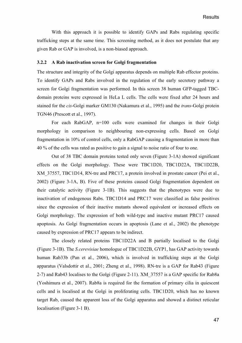
Results
47
With this approach it is possible to identify GAPs and Rabs regulating specific
trafficking steps at the same time. This screening method, as it does not postulate that any
given Rab or GAP is involved, is a non-biased approach.
3.2.2 A Rab inactivation screen for Golgi fragmentation
The structure and integrity of the Golgi apparatus depends on multiple Rab effector proteins.
To identify GAPs and Rabs involved in the regulation of the early secretory pathway a
screen for Golgi fragmentation was performed. In this screen 38 human GFP-tagged TBC-
domain proteins were expressed in HeLa L cells. The cells were fixed after 24 hours and
stained for the cis-Golgi marker GM130 (Nakamura et al., 1995) and the trans-Golgi protein
TGN46 (Prescott et al., 1997).
For each RabGAP, n=100 cells were examined for changes in their Golgi
morphology in comparison to neighbouring non-expressing cells. Based on Golgi
fragmentation in 10% of control cells, only a RabGAP causing a fragmentation in more than
40 % of the cells was rated as positive to gain a signal to noise ratio of four to one.
Out of 38 TBC domain proteins tested only seven (Figure 3-1A) showed significant
effects on the Golgi morphology. These were TBC1D20, TBC1D22A, TBC1D22B,
XM_37557, TBC1D14, RN-tre and PRC17, a protein involved in prostate cancer (Pei et al.,
2002) (Figure 3-1A, B). Five of these proteins caused Golgi fragmentation dependent on
their catalytic activity (Figure 3-1B). This suggests that the phenotypes were due to
inactivation of endogenous Rabs. TBC1D14 and PRC17 were classified as false positives
since the expression of their inactive mutants showed equivalent or increased effects on
Golgi morphology. The expression of both wild-type and inactive mutant PRC17 caused
apoptosis. As Golgi fragmentation occurs in apoptosis (Lane et al., 2002) the phenotype
caused by expression of PRC17 appears to be indirect.
The closely related proteins TBC1D22A and B partially localised to the Golgi
(Figure 3-1B). The S.cerevisiae homologue of TBC1D22B, GYP1, has GAP activity towards
human Rab33b (Pan et al., 2006), which is involved in trafficking steps at the Golgi
apparatus (Valsdottir et al., 2001; Zheng et al., 1998). RN-tre is a GAP for Rab43 (Figure
2-7) and Rab43 localises to the Golgi (Figure 2-11). XM_37557 is a GAP specific for Rab8a
(Yoshimura et al., 2007). Rab8a is required for the formation of primary cilia in quiescent
cells and is localised at the Golgi in proliferating cells. TBC1D20, which has no known
target Rab, caused the apparent loss of the Golgi apparatus and showed a distinct reticular
localisation (Figure 3-1 B).

Results
48
Figure 3-1 Golgi fragmentation in HeLa cells. HeLa cells were transfected with GFP-tagged TBC domain proteins for 24 hours. The cells were then fixed and stained with antibodies against GM130 and TGN46. (A) n=100 cells for each TBC domain protein were analysed and their Golgi was rated as fragmented or not fragmented compared to neighbouring non-expressing control cells. (B) GFP tagged TBC domain proteins scoring Golgi fragmentation in more than 40% of the cells are depicted as both wild-type and inactive (RA) mutants. Bars indicate 10µm.

Results
49
Next, the same screen was carried out in telomerase immortalized human retinal
epithelial (hTERT-RPE1) cells to identify ubiquitously expressed essential Rabs and GAPs
and to eliminate cell-type specific proteins. This second screen also helps to validate the
screen in HeLa cells.
In hTRET-RPE1 cells only three TBC domain-containing proteins caused significant
alterations of the Golgi morphology: TBC1D20, RN-tre and TBC1D14 (Figure 3-2A). In the
case of TBC1D14, the phenotype again did not depend on the catalytic activity (Figure
3-2A) of the protein.
Figure 3-2 Golgi fragmentation in hTERT-RPE1 cells. The cells were transfected with GFP-tagged TBC domain proteins for 24 hours. The cells were then fixed and stained with antibodies against GM130 and TGN46. (A) n=100 cells for each TBC domain protein were analysed and their Golgi morphology was rated as fragmented or not fragmented compared to non-expressing neighbouring cells. (B) GFP-tagged TBC domain proteins causing Golgi fragmentation in more than 40% of the cells are depicted both as wild-type and inactive (RA) mutants. Bars indicate 10µm.
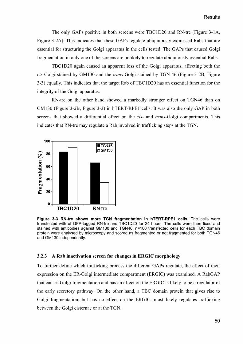
Results
50
The only GAPs positive in both screens were TBC1D20 and RN-tre (Figure 3-1A,
Figure 3-2A). This indicates that these GAPs regulate ubiquitously expressed Rabs that are
essential for structuring the Golgi apparatus in the cells tested. The GAPs that caused Golgi
fragmentation in only one of the screens are unlikely to regulate ubiquitously essential Rabs.
TBC1D20 again caused an apparent loss of the Golgi apparatus, affecting both the
cis-Golgi stained by GM130 and the trans-Golgi stained by TGN-46 (Figure 3-2B, Figure
3-3) equally. This indicates that the target Rab of TBC1D20 has an essential function for the
integrity of the Golgi apparatus.
RN-tre on the other hand showed a markedly stronger effect on TGN46 than on
GM130 (Figure 3-2B, Figure 3-3) in hTERT-RPE1 cells. It was also the only GAP in both
screens that showed a differential effect on the cis- and trans-Golgi compartments. This
indicates that RN-tre may regulate a Rab involved in trafficking steps at the TGN.
Figure 3-3 RN-tre shows more TGN fragmentation in hTERT-RPE1 cells. The cells were transfected with of GFP-tagged RN-tre and TBC1D20 for 24 hours. The cells were then fixed and stained with antibodies against GM130 and TGN46. n=100 transfected cells for each TBC domain protein were analysed by microscopy and scored as fragmented or not fragmented for both TGN46 and GM130 independently.
3.2.3 A Rab inactivation screen for changes in ERGIC morphology
To further define which trafficking process the different GAPs regulate, the effect of their
expression on the ER-Golgi intermediate compartment (ERGIC) was examined. A RabGAP
that causes Golgi fragmentation and has an effect on the ERGIC is likely to be a regulator of
the early secretory pathway. On the other hand, a TBC domain protein that gives rise to
Golgi fragmentation, but has no effect on the ERGIC, most likely regulates trafficking
between the Golgi cisternae or at the TGN.

Results
51
ERGIC-53, a mannose-specific transmembrane lectin was used as a marker for this
screen. This protein predominantly localises to the ERGIC, but a small fraction is found in
the ER and on ERES (Hauri et al., 2000). It directly recycles between the ERGIC and the
ER, and only little ERGIC-53 recycles from the Golgi to the ER (Klumperman et al., 1998).
HeLa cells were transfected with 38 GFP-tagged TBC domain proteins
independently and fixed after 24 hours. The cells were stained with an antibody against
ERGIC-53. They were analysed and the morphology of their ERGIC was scored as either
normal or altered in comparison to neighbouring non-expressing cells. Based on an altered
ERGIC morphology in 10% of control cells, a cut-off of 40% was chosen.
TBC1D20 fragmented the ERGIC (Figure 3-4A) dependent on its catalytic activity
(Figure 3-4B). Interestingly, ERGIC-53 did not stain small punctate structures in the
periphery of the cell, most likely ERES, in cells expressing TBC1D20 (Figure 3-4B).
TBC1D22B, but not TBC1D22A, disrupted the ERGIC dependent on its catalytic activity
(Figure 3-4A). The perinuclear pool of ERGIC-53 was distributed into fragments in cells
expressing TBC1D22B, but in contrast to TBC1D20 expressing cells the small peripheral
structures were still present (Figure 3-4B). RN-tre and XM_037557 only caused subtle
changes to the ERGIC (Figure 3-4A, B).
Figure 3-4 A screen for TBC domain proteins altering ERGIC structure. HeLa cells were transfected with GFP-tagged TBC domain proteins for 24 hours. The cells were then fixed and stained for ERGIC-53 (red). (A) n=100 cells for each TBC domain protein were analysed by microscopy and scored as fragmented or not fragmented. (B, see next page) GFP-tagged TBC domain proteins that caused ERGIC fragmentation in more than 40% of expressing cells are depicted both as wild-type (WT) and inactive (RA) mutants. Bars indicate 10µm.

Results
52

Results
53
3.2.4 A Rab inactivation screen for a block of VSV-G trafficking
To further analyse which of the human GAPs regulates secretion, the trafficking of a protein
from the ER through the secretory pathway to the plasma membrane was followed in cells
expressing these GAPs. As a model cargo a temperature sensitive mutant of a type-I
transmembrane glycoprotein of the vesicular stomatitis virus (VSV), ts-O45-G (VSV-G) was
used. VSV-G misfolds and accumulates in the ER at the non-permissive temperature of
39.5°C. It refolds at 4°C and is then transported to the cell surface at 31.5°C (Presley et al.,
1997; Scales et al., 1997). A GFP tag is fused to the C-terminus of VSV-G that faces the
cytoplasm. A specific antibody detects the luminal domain of VSV-G. When used on cells
without prior detergent extraction of the plasma membrane, this antibody only stains VSV-G
at the cell surface and therefore provides readout for successful trafficking.
Similar to the previous screens (see 3.2.2 and 3.2.3), 38 myc-epitope-tagged human
TBC domain proteins were transfected in parallel into HeLa cells. A VSV-G encoding
plasmid was co-transfected, and the cells were incubated for 2 hours at 37°C. Then the cells
were incubated for 12 hours to 39.5°C to accumulate misfolded VSV-G in the ER. The cells
were shifted to 4°C for 30 minutes to stop trafficking and promote proper folding of VSV-G.
After these 30 minutes a sample, which corresponds to the t=0 time point, was immediately
fixed. The cells were then incubated at 31.5°C to allow pulse secretion of VSV-G. After 60
minutes the cells were fixed without detergent extraction of the plasma membrane to stain
VSV-G at the plasma membrane. Afterwards the cells were permeabilised and stained for
the myc-epitope-tag to identify GAP expressing cells. Co-transfected cells were identified by
microscopy and dependent on the presence of VSV-G at the plasma membrane cells that
block transport were identified. As in the previous screens for Golgi fragmentation or
changes in the ERGIC morphology, a cut-off of 40% was chosen.
Strikingly, out of all TBC domain proteins only TBC1D20 was able to prevent the
appearance of the VSV-G at the cell surface after 60 minutes (Figure 3-5A). This block of
transport was dependent on the catalytic arginine105 of TBC1D20 (Figure 3-5B). This
indicates that this effect on VSV-G trafficking was due to specific inactivation of a Rab. The
other RabGAPs that fragmented the Golgi or affected the morphology of the ERGIC did not
block VSV-G transport (Figure 3-5A). They also didn’t show any significant difference in
VSV-G trafficking between wild-type and catalytically inactive mutants (Figure 3-5B). This
shows that out of all human RabGAPs only TBC1D20 inactivates a Rab that is essential for
the secretion of VSV-G.

Results
54
Figure 3-5 TBC1D20 is the only TBC domain protein that blocks VSV-G trafficking. VSV-G transport assays were performed for a total time of 15 hours in HeLa cells expressing myc-epitope-tagged TBC domain proteins (A) HeLa cells co-transfected with VSV-G and wild-type TBC domain proteins were stained for surface VSV-G and the myc-epitope tag. n=100 cells were analysed by microscopy for each GAP and were scored as either block or no block of trafficking. (B) HeLa cells were co-transfected with VSV-G and wild-type (WT) or catalytically inactive (RA) mutant TBC domain proteins that fragment the Golgi. They were stained for surface VSV-G and the myc-epitope tag. n=100 cells were analysed for each GAP and were scored as either block or no block of trafficking.
The over-expression of TBC1D20 caused a strong effect on Golgi morphology in all
cell lines analysed. It also perturbed the organisation of the ERGIC in a unique way. Out of
all TBC domain proteins it was the only one that blocked the secretion of VSV-G. TBC1D20
is therefore the most likely candidate for a GAP that regulates an essential Rab involved in
the regulation of the early secretory pathway. This TBC domain protein and the phenotypes
caused by its over-expression were therefore studied in more detail.

Results
55
3.2.5 TBC1D20 is a highly conserved TBC domain protein
To investigate the evolutionary conservation of TBC1D20 the genomes of several
vertebrates and yeast were searched. TBC1D20 homologues were identified using online
database search engines at BLAST (http://www.ncbi.nlm.nih.gov/blast/).in macaque, mouse,
rat, dog, cow, zebrafish, African clawed toad, and the three yeasts Saccharomyces
cerevisiae, Schizosaccharomyces pombe, and Kluveromyces lactis. Partial sequences of
homologue proteins were also found in other species but omitted for simplicity. The protein
sequences of the homologue proteins were aligned to the human TBC1D20 protein sequence
using the Vector NTI® software (Figure 3-6).
This alignment showed that TBC1D20 is a highly conserved protein. It is almost
identical in all mammals, and the similarity is very striking even to lower vertebrate
sequences. The similarity to the yeast proteins appears less pronounced. But even in yeast,
the consensus sequences that identify the TBC domain (1.3.1) and the catalytically relevant
residues arginine105 and glutamine144 (Pan et al., 2006) are conserved.
TBC1D20 is a rather small member of the family of TBC domain containing proteins
with 403 aa for the human protein. In contrast to most other RabGAPs (Figure 2-2) it does
not contain additional conserved protein domains. The only other component identified in
the sequence of all homologue proteins is a hydrophobic stretch close to the C-terminus of
TBC1D20 (Figure 3-6). Further analysis with the PSORTII prediction algorithms
(http://psort.nibb.ac.jp/form2.html) suggested that this hydrophobic stretch might form a
transmembrane domain (TMD) or membrane anchor. This indicates that TBC1D20 might be
localised to membranes via its C-terminus.

Results
56
Figure 3-6 Alignment of TBC1D20 sequences. Proteins were identified in human (hs), macaque (ma), mouse (mm), rat (rn), dog (cf), cow (bt), zebrafish (dr), African clawed toad (xl), Saccharomyces cerevisiae (scGyp8), Schizosaccharomyces pombe (spGyp10), and Kluveromyces lactis (klTBC1D20). Only full length sequences were included. Identical amino acids are coloured in red on a yellow background. Dark blue letters on a light blue background indicate a high degree of conservation. Dark blue letters on a green background show similar amino acids. Red arrowheads indicate the conserved arginine and glutamine residues required for GTP hydrolysis.

Results
57
3.2.6 Over-expression of TBC1D20 causes a unique “loss of Golgi” phenotype
TBC1D20 was unique amongst the GAPs that caused Golgi fragmentation since it triggered
a complete loss of defined Golgi structures. To further investigate this “loss of Golgi”
phenotype, markers for different Golgi sub-compartments were analysed in cells over-
expressing TBC1D20: the Rab1 effector p115 (Alvarez et al., 1999) that localises to the cis-
Golgi and ERGIC; the cis-Golgi transmembrane protein Golgin84 (Bascom et al., 1999;
Diao et al., 2003); and Golgin97, which localises to the TGN via its ARF1 interacting GRIP
domain (Barr, 1999; Griffith et al., 1997; Lu and Hong, 2003).
The expression of TBC1D20 resulted in the loss of recognizable Golgi or punctate
vesicular staining for all of these markers (Figure 3-7A). The specific staining was barely
detectable by immunofluorescence and appeared as a haze distributed throughout the
cytoplasm. On the other hand, the expression of TBC1D20 did not alter the staining of the
endosomal Rab5 effector EEA1 (Figure 3-7A). The morphology of the Golgi apparatus was
not affected by the expression of catalytically inactive TBC1D20R105A (Figure 3-7B)
although a similar expression level and distribution was observed.
These data show that TBC1D20 specifically perturbs all compartments of the Golgi,
but doesn’t have an effect on other sub-cellular compartments like early endosomes. Taken
together with the observation that catalytically inactive TBC1D20 did not have an effect on
the morphology of the Golgi apparatus, these data suggest that the “loss of Golgi” phenotype
is due to inactivation of a Rab involved in regulation of trafficking events at the Golgi.

Results
58
Figure 3-7 TBC1D20 causes a “loss of Golgi” phenotype. HeLa cells were transfected with myc epitope-tagged (A) wild type or (B) catalytically inactive TBC1D20R105A for 24 hours. The cells were then fixed and stained with antibodies against the myc epitope tag (red), p115 (green), Golgin84 (green), Golgin97 (green) or EEA1 (green). Bars indicate 10µm.

Results
59
The “loss of Golgi” phenotype in cells expressing TBC1D20 raised the question of
the fate of the Golgi proteins. Various fates are conceivable: integral membrane proteins
such as Golgi enzymes might be redistributed to other membrane compartments or become
degraded by missorting to lysosomes; peripheral membrane proteins such as GM130 or p115
might be degraded by the proteasome or might be relocated to the cytosol; the Golgi proteins
might also reside on or in membrane structures that are too small to be resolved by
immunofluorescence. To test these possibilities a series of experiments was performed.
First, the fate of a Golgi-enzyme was studied. N-acetylglucosaminyltransferase-I
(NAGT-I) is a protein of the medial- and trans-Golgi (Nilsson et al., 1993). It is specifically
retained in these Golgi cisternae by its transmembrane domain and stalk region (Nilsson et
al., 1996) and initiates the conversion of N-linked oligosaccharides to the complex type
(Dunphy et al., 1985).
HeLa cells were transfected with GFP-tagged NAGT-I. The cells were then
incubated for 10 hours to start the expression of NAGT-I and to localise it to the medial- and
trans-Golgi. After 10 hours the cells were transfected with myc-epitope-tagged wild-type or
catalytically inactive TBC1D20R105A. After a total time of 22 hours the cells were fixed and
stained with the antibodies indicated.
NAGT-I localised to the Golgi in cells that only expressed the GFP-tagged protein
(Figure 3-8). In cells co-expressing wild-type TBC1D20 NAGT-I failed to co-localise with
the Golgi marker GM130 (Figure 3-8), which was diffusely dispersed throughout the
cytoplasm. Instead, it was found to localise to a reticular structure. When cells were stained
for the ER marker calnexin, NAGT-I was found to co-localise with calnexin (Figure 3-8) in
cells expressing wild-type TBC1D20. This shows that NAGT was redistributed to the ER. In
cells expressing catalytically inactive TBC1D20R105A, NAGT-I was found in the Golgi and
did not redistribute to the ER (Figure 3-8). The redistribution of NAGT-I was therefore due
to the specific inactivation of a Rab GTPase by TBC1D20.
These data show that Golgi enzymes such as NAGT-I are redistributed to the ER in
cells over-expressing TBC1D20.

Results
60
Figure 3-8 Expression of TBC1D20 redistributes Golgi enzymes to the ER. HeLa cells were transfected with GFP-tagged NAGT-I (green) for 10 hours. Then they were transfected with Myc-tagged wild-type or catalytically inactive TBC1D20R105A for further 12 hours. The cells were then fixed and stained for GM130 (red) or Calnexin (red) and the myc-epitope tag (blue). The bar indicates 10µm.
To test whether or not peripheral Golgi proteins are degraded in cells over-expressing
TBC1D20 several western blot experiments were carried out. HeLa cells grown on 10 cm
cell culture dishes were transfected for 24 hours with myc-epitope-tagged wild-type or
catalytically inactive TBC1D20R105A. The cells were then harvested, lysed in mammalian
lysis buffer, and the lysate was cleared by centrifugation, and 20µg of these extracts were
western blotted. The blots were probed with antibodies against the myc-epitope-tag, p115,
GM130, and to tubulin as a loading control. Noticeably, the expression of wild-type and
catalytically inactive TBC1D20R105A reached similar levels (Figure 3-9A). Despite the high
transfection efficiency, over 50% estimated by immunofluorescence, no change was
observed in the levels of either GM130 or p115 in cells expressing TBC1D20 (Figure 3-9A).
This finding shows that these Golgi proteins were not degraded in cells over-expressing
wild-type TBC1D20.
To analyse whether or not the Golgi proteins were still membrane bound, a set of
membrane fractionation experiments were carried out. For these experiments a 10 cm dish of
HeLa cells was transfected with myc-epitope-tagged wild-type TBC1D20 for 24 hours. To

Results
61
compare the TBC1D20 phenotype to other factors that cause Golgi fragmentation, another
dish of HeLa cells was transfected with constitutive active Sar1H79G. Expression of Sar1H79G
prevents un-coating of COPII and causes a phenotype similar to TBC1D20. (Ward et al.,
2001; Yoshimura et al., 2004). Another dish of cells was not transfected but incubated with
Brefeldin A (BFA) for 30 minutes prior to harvesting. BFA treatment also fragments the
Golgi and blocks secretion (Lippincott-Schwartz et al., 1989). As a control a dish of non-
transfected and non-treated cells was used.
The cells were harvested and broken open by passing them 40 times through a 27G
needle. Nuclei and cell debris were removed by centrifugation for 5 min at 1000x g and 4°C.
This post-nuclear supernatant was split into two equal halves. One of them was kept on ice
and represents the total material. The other half was spun for 30 min at 100,000x g. The
supernatant of this centrifugation contains all the cytosolic proteins. Membranes and
membrane bound proteins are found in the pellet fraction. The distribution of proteins
between membranes and the cytoplasm can be analysed by western blotting these fractions.
The peripheral membrane protein GM130 and the integral membrane protein
Golgin84 remained associated with the membranes to the same extent in control cells and in
cells expressing wild type TBC1D20 (Figure 3-9B). The same was true in cells expressing
Sar1H79G or treated with BFA (Figure 3-9B). Under the same conditions, the membrane-
associated pool of the Rab1 effector p115 was lost in cells expressing TBC1D20 (Figure
3-9B). This loss was not due to Golgi fragmentation, as BFA treatment and Sar1H79G
expression did not have an effect on the distribution of p115 (Figure 3-9B). The fractionation
experiments also showed that TBC1D20 is associated with membranes (Figure 3-9B)
Taken together, these results show that Golgi proteins have different fates in cells
expressing TBC1D20. Golgi enzymes are redistributed to the ER. Golgi matrix proteins such
as GM130 of Golgin84 remain associated with membranes. They appear as a haze in
immunofluorescence and are therefore likely to be associated with vesicles that are too small
to be resolved by the light microscope. This suggests that the Golgi apparatus is vesiculated
in the “loss of Golgi” phenotype. The Rab1 effector p115 was lost from the membrane
fraction in TBC1D20 expressing cells. This indicates that the phenotypes observed in cells
over-expressing TBC1D20 might be due to defects in the Rab1-p115 dependent COPII
vesicle-tethering pathway. It also suggests that TBC1D20 may act as a GAP for Rab1, as
p115 requires active Rab1 for its membrane association (Allan et al., 2000).

Results
62
Figure 3-9 TBC1D20 displaces p115 from membranes. (A) HeLa cells were transfected with either wild-type (WT) or catalytically inactive TBC1D20R105A (RA) for 24 hours. 20µg of cell extracts were western blotted and probed for GM130 and p115. The myc-epitope shows the expression of TBC1D20, and α-tubulin acts as a loading control. The asterisk indicates a GM130 breakdown product. (B) HeLa cells left untreated, expressing myc-tagged TBC1D20 or Sar1H79G for 24 hours, or treated with BFA for 30 minutes were harvested and fractionated. Equivalent amounts of the total lysate (T), the membrane pellet (P), or the soluble cytosolic fraction (S) were western blotted and probed for p115, GM130, Golgin84 and the myc-epitope-tag, respectively.
3.2.7 TBC1D20 blocks exit of VSV-G from the ER
Next, the block of VSV-G trafficking in cells over-expressing TBC1D20 was studied in
more detail. Wild-type or catalytically inactive TBC1D20R105A were co-transfected with a
plasmid encoding VSV-G into HeLa cells. The experimental setup was as described in 3.2.4.
COPII positive structures were stained with antibodies against Sec31 and the Golgi was
stained for GM130. After 60 minutes cells were fixed without detergent extraction to stain
VSV-G at the plasma membrane.
At t=0 VSV-G accumulated in the ER (Figure 3-10) independent of TBC1D20. The
amount of VSV-G was the same in cells transfected with either wild-type or catalytically
inactive TBC1D20R105A as judged by immunofluorescence. This indicates that the absence of

Results
63
VSV-G at the cell surface (Figure 3-5A, B) was not simply due to the lack of synthesised
viral protein. VSV-G did not co-localise with COPII positive structures at this time point.
After 10 minutes VSV-G started to leave the ER in cells expressing TBC1D20R105A
as well as in cells expressing only VSV-G (Figure 3-10). The GFP-tagged VSV-G co-
localised with COPII positive structures at this time point. This shows that a wave of folded
VSV-G started to exit the ER. In cells expressing wild type TBC1D20 however, the viral
protein was retained in the ER (Figure 3-10).
After 30 minutes of transport, the majority of VSV-G was found at the Golgi in cells
expressing only VSV-G or both VSV-G and TBC1D20R105A (Figure 3-10). In cells
expressing wild-type TBC1D20 it was still retained in the ER (Figure 3-10).
At t=60 VSV-G reached the cell surface in cells expressing TBC1D20R105A (Figure
3-10). The same was true for cells only expressing VSV-G. Even after 60 minutes VSV-G
did not exit the ER in wild-type TBC1D20 expressing cells (Figure 3-10). VSV-G did not
accumulate at any structures at the ER. This shows that VSV-G was not recruited to ERES in
cells expressing wild-type TBC1D20.
These data show that TBC1D20 blocks trafficking of VSV-G dependent on its
catalytic activity. Importantly, VSV-G is retained in the ER and does not accumulate in
COPII positive structures or at ERES. These data suggest a role for TBC1D20 and the Rab it
regulates at the level of the ER.

Results
64

Results
65
Figure 3-10 TBC1D20 blocks VSV-G trafficking at the ER. VSV-G transport assays were performed for a total time of 15 hours in HeLa cells expressing myc-epitope-tagged wild-type (WT) or catalytically inactive TBC1D20R105A (RA). The cells were fixed at the time points indicated and stained for the myc-epitope-tag (blue), Sec31 (red at t=0, t=10), GM130 (red at t=30) or surface VSV-G (red at t=60). GFP shows total VSV-G. The bars indicate 10 µm.
3.2.8 TBC1D20 expression causes scattering of COPII
Budding of vesicles from the ER and trafficking between the ER and the Golgi depends on
the COPII coat. The block of ER to Golgi transport in cells expressing TBC1D20 might
therefore be linked to defective COPII function.
COPII vesicles are formed at ERES (Hammond and Glick, 2000). The activation of
the small GTPase Sar1 leads to the consecutive stepwise recruitment of the heterodimeric
COPII subunits Sec23/24 and Sec13/31 (Barlowe et al., 1994). The curvature of the sub-
complexes deforms the membrane and leads to abscission of the vesicles (Bonifacino and
Glick, 2004). COPII vesicles form clusters, and consequently the ERGIC, dependent on the
Rab1-p115 tethering pathway (Allan et al., 2000).
HeLa cells were transfected with myc-epitope-tagged wild-type or inactive
TBC1D20R105A for 24 hours to study COPII in more detail. The cells were then fixed and
stained for Sec31 or Sec24C, respectively. In control cells COPII positive structures
accumulated in the perinuclear region (Figure 3-11 A). In cells expressing wild-type
TBC1D20 no perinuclear accumulation of COPII positive structures was observed (Figure
3-11 A) in cells were stained for Sec31 or Sec24 (Figure 3-11 B). The peripheral COPII
structures on the other hand were not affected. This indicates that COPII clustering but not
COPII vesicle formation is defective in cells over-expressing wild-type TBC1D20. In cells
expressing catalytically inactive TBC1D20R105A no scattering of COPII positive structures
was observed (Figure 3-11A, B). The COPII positive structures observed in these cells were
comparable to non-expressing control cells. This shows that the phenotype is due to the
specific inactivation of a target Rab.
TBC1D20 therefore interferes with the clustering of COPII vesicles that depends on
the Rab1 effector p115. This finding fits with the observation that p115 is lost from
membranes in cells over-expressing wild-type TBC1D20.

Results
66
Figure 3-11 TBC1D20 expression causes COPII scattering. HeLa cells were transfected with myc-epitope-tagged wild-type (WT) or catalytically inactive TBC1D20R105A (R105A) for 24 hours before fixation. (A) The cells were stained for the myc-epitope (green) and Sec31 (red). Bar corresponds to 10 µm. (B) n=100 cells stained for Sec31 or Sec24C were analysed and the COPII staining was scored as either scattered or perinuclear as compared to neighbouring non-expressing cells.

Results
67
3.2.9 TBC1D20 expression does not interfere with COPII dynamics
To analyse if over-expression of TBC1D20 blocks transport of cargo in the ER by
interfering with the assembly of the COPII vesicle coat, the dynamics of COPII vesicle
formation were analysed.
Fluorescence recovery after photobleaching (FRAP) was used in collaboration with
Dr. David Stephens in the University of Bristol to study the dynamics of COPII vesicle
formation in cells over-expressing TBC1D20. FRAP is a widely used technique (Presley,
2005; Reits and Neefjes, 2001) that exploits the fact that chromophores can be bleached in a
defined area by a laser pulse of an appropriate wavelength. The fluorescence of the bleached
area is then measured over time. By measuring the time needed to regain the fluorescence
intensity after the laser pulse, the dynamics of a system can be studied.
As the COPII coat component Sec23 has GAP activity towards the small GTPase
Sar1 required for COPII coat formation, it causes destabilisation and dissociation of the coat
(Bickford et al., 2004). Therefore the COPII coat undergoes a cycle of disassembly and
reassembly until the vesicle buds of the ERES. If TBC1D20 blocks ER exit by interfering
with COPII dynamics, the FRAP kinetics of COPII should be different in cells over-
expressing TBC1D20. The fluorescence of the COPII component Sec16 (Connerly et al.,
2005; Watson et al., 2006) in cells expressing TBC1D20 was regained 50 seconds after
bleaching (Figure 3-12A). Compared to the turnover of COPII components in control cells
no change in the fluorescence recovery kinetics was observed (Figure 3-12B, C). This shows
that TBC1D20 does not interfere with the dynamics of COPII vesicle formation.
This finding indicates that the block of ER exit and the altered distribution of COPII
positive structures in cells expressing TBC1D20 cannot be explained by alteration in the
dynamics of COPII coat assembly. These effects are therefore likely to be caused by the
inactivation of a Rab required for both COPII vesicle clustering or cargo exit from the ER.

Results
68
Figure 3-12 COPII dynamics are not affected by TBC1D20 expression. FRAP was performed on cells expressing (A) myc-epitope-tagged TBC1D20 and Venus- Sec16 or (B) in control cells only expressing Venus- Sec16. A Leica TCS SP3 AOBS scanning confocal microscope with a pinhole size of 2 Airy units, eightfold line averaging, was used at 1 frame per second, and region-of-interest bleaching with 10 iterations of the 514 nm laser at 100% AOTF power. Cells used for photobleaching were confirmed to express myc-epitope-tagged TBC1D20 by subsequent immunofluorescence; 95% of all cell transfected with Venus-Sec16 were found to be transfected with myc-epitope tagged TBC1D20. Plots are of the average intensity of individual structures over time; error bars indicate the standard deviation (five cells per experiment, n=3).
3.2.10 The cargo receptor p24 reveals a sorting defect caused by TBC1D20
As the expression of TBC1D20 did not alter COPII dynamics, defective cargo selection as a
reason for the block of ER exit was investigated.
Both ERGIC-53, which recycles between the ERGIC and the ER, and p24, which
recycles between the ER, the ERGIC and the Golgi were analysed. p24, a protein of a family
of type-I membrane proteins, binds both COPI (Sohn et al., 1996) and COPII (Dominguez et
al., 1998) to mediate its cycling. p24 proteins form oligomeric complexes (Denzel et al.,
2000) and function as receptors for various cargo molecules (Fiedler et al., 1996), which are
proposed to be enriched in budding vesicles by p24 (Schimmoller et al., 1995).
To analyse the effect of TBC1D20 over-expression on these molecules, HeLa cells
were transfected with GFP-tagged p24 for 6 hours. Then the cells were transfected with

Results
69
myc-epitope-tagged wild-type or catalytically inactive TBC1D20R105A for a further 12 hours.
The cells were then fixed and stained for ERGIC-53 or Sec31, respectively.
GFP-p24 localised to the ER, to ERES, the ERGIC and the Golgi apparatus in
control cells. GFP-tagged p24 largely co-localised with ERGIC-53 (Figure 3-13B) and did
not alter the distribution of ERGIC-53 positive structures. Their co-localisation on small
punctate structures, most likely ERES, is shown more clearly in the enlargement. In cells
expressing wild-type TBC1D20 GFP-p24 localised to large punctate structures (Figure
3-13A). When cells expressing both wild-type TBC1D20 and GFP-tagged p24 were stained
for ERGIC-53, both p24 and ERGIC-53 localised to the same large punctate structures
(Figure 3-13B). Notably, the small punctate structures resembling ERES were no longer
observed under these conditions. This can be seen more clearly in the enlargements.
When cells expressing GFP-tagged p24 were stained for Sec31 as a COPII marker, a
large extent of co-localisation was observed, especially on small punctate ERES (Figure
3-13C). This is seen more clearly in the enlargement. In cells over-expressing wild-type
TBC1D20 (Figure 3-13C), GFP-p24 localised to large structures whereas Sec31 was
scattered throughout the cell as previously observed (Figure 3-11). Sec31 was also found on
small punctate structures resembling ERES under these conditions. GFP-p24 did not co-
localise with Sec31 in these structures in cells expressing wild-type TBC1D20. This is more
clearly seen in the enlargement that shows red punctate structures stained for Sec31, which
lack green GFP-p24 signal (Figure 3-13C). The expression of TBC1D20R105A did not result
in changes in the distribution of p24, ERGIC-53 or Sec31 in comparison to control cells
(Figure 3-13A, B, C). This shows that the phenotypes observed are due to the catalytic
activity of TBC1D20.
These data show that cargo receptors such as ERGIC-53 and p24 are absent from
ERES in cells over-expressing TBC1D20. This finding might provide an explanation for the
defect in trafficking of cargo from the ER under these conditions. These findings also show
that the ERGIC is a functionally separate compartment that reacts distinct from the Golgi
and COPII positive structures to the over-expression of TBC1D20.
Figure 3-13 Recycling ERGIC markers are lost from ERES in TBC1D20 expressing cells. See next page. HeLa cells were transfected with GFP-tagged p24 for 6 hours. Then they were transfected with wild-type (WT) or catalytically inactive (RA) myc-epitope-tagged TBC1D20 (blue). The cells were fixed after further 18 hours and stained for (A) the myc-epitope-tag (red), (B) ERGIC-53 (Red) and the myc-epitope-tag (blue) or (C) Sec31 (red) and the myc-epitope-tag (blue). GFP was visualized (A, B, C) directly (green). The enlargements correspond to 10x10 µm and only show red and green colour. The bars indicate 10µm.

Results
70

Results
71
3.2.11 TBC1D20 is a GAP for Rab1 and Rab2 in vitro
To identify the target Rab of TBC1D20, a series of in vitro GTP-hydrolysis assays similar to
the ones used to identify Rab5 as the target of RabGAP-5 was performed.
TBC1D20 could not be produced as a full-length recombinant protein in the same
vector used for the production of RabGAP-5, probably due to the hydrophobic stretch
(Figure 3-6). Without this stretch TBC1D20 was successfully produced in E. coli as a
recombinant protein with a N-terminal MBP-tag and a C-terminal 6x His tag. Recombinant
TBC1D20 was then tested for its ability to stimulate GTP hydrolysis by all human Rabs. For
simplicity some Rab isoforms were omitted. The basal GTPase activity of the Rabs was
subtracted from the activity stimulated by TBC1D20, so that only GAP mediated hydrolysis
is shown.
TBC1D20 strongly stimulated GTP hydrolysis by Rab1 and Rab2 (Figure 3-14A).
Some additional activity of TBC1D20 was observed towards Rab8A, Rab13, Rab18, and
Rab35 (Figure 3-14A). All these Rabs are closely related members of the Rab1/Sec4 group
(Figure 1-5). This might explain why TBC1D20 can activate them in vitro. TBC1D20 did
not display GAP activity towards Sar1 and ARF1 (Figure 3-14 A). Both these non-Rab
GTPases also belong to the Ras superfamily and are required for protein trafficking and the
maintenance of normal Golgi structure (Bonifacino and Glick, 2004).
The minimal predicted TBC domain TBC1D20 aa 1-317 stimulated GTP hydrolysis
by Rab1 to a similar extent as TBC1D20 aa 1-364 (Figure 3-14B). The catalytic activity of
both the longer version of the protein and the minimal TBC domain was abolished when the
catalytic arginine 105 of the TBC-domain was mutated to alanine (Figure 3-14B). When the
glutamine 67 of Rab1 was mutated to leucine to restrict it in the GTP bound state the basal
hydrolysis rate was strongly reduced (Figure 3-14B). TBC1D20 did not stimulate GTP
hydrolysis by Rab1Q67L (Figure 3-14B).
To investigate if TBC1D20 is the only GAP for Rab1 or Rab2, the five TBC domain
proteins, which were able to disrupt the Golgi (see Figure 3-1 and Figure 3-2) or alter the
structure of the ERGIC (see Figure 3-4), were produced in the same way as TBC1D20 and
tested for their ability to stimulate GTP hydrolysis by Rab1 or Rab2. These assays showed
that only TBC1D20, but none of the other GAPs was able to stimulate GTP-hydrolysis by
Rab1 or Rab2 to a significant extent (Figure 3-14C). This finding is consistent with
publications showing that the TBC1D22 family acts on Rab33 (Pan et al., 2006), and that

Results
72
XM_037557 is a GAP for Rab8a (Yoshimura et al., 2007). RN-tre was already shown to be a
GAP for Rab43 (Figure 2-11).
These experiments show that TBC1D20 is a GAP for Rab1 and Rab2 in vitro. They
also show that TBC1D20 is the only GAP that causes Golgi fragmentation or changes in the
ERGIC morphology that acts on Rab1 or Rab2.
Figure 3-14 TBC1D20 is a GAP for Rab1 and Rab2 in vitro. All reactions were carried out for 60 minutes. (A) To determine the specific activity towards a wide range of GTPases, 0.5 pmoles TBC1D20 aa1-364 were tested against 100 pmoles of the GTPases indicated. The basal GTP hydrolysis with a buffer control was subtracted from TBC1D20 stimulated hydrolysis in each case. (B) 5 pmoles of wild-type and TBC1D20R105A aa1-364 or the minimal TBC-domain aa 1-317 were tested against 100 pmoles of either wild-type Rab1 or Rab1Q67L. Basal GTP hydrolysis with buffer is plotted as open bars and GAP-stimulated GTP hydrolysis as grey bars. (C) Buffer (control) or 0.5 pmoles of the GAP indicated were tested against 100 pmoles of Rab1 or Rab2. Basal GTP hydrolysis with buffer is plotted as open bars and GAP-stimulated GTP hydrolysis as grey bars.
3.2.12 Dominant negative Rab1N121I mimics the TBC1D20 phenotype
To narrow down the target of TBC1D20 in vivo, the phenotypes caused by expression of
dominant-negative Rabs were compared to the phenotypes caused by the over-expression of

Results
73
TBC1D20. Dominant negative Rabs are generated by mutations that either keep the Rab in a
nucleotide free state or prevent GEFs from reloading them with GTP. HeLa cells were
transfected with dominant negative GFP-tagged Rabs for 24 hours. The cells were then
fixed, and stained for the Golgi marker GM130 or the COPII marker Sec31, respectively.
Dominant-negative Rab43T32N was used as a positive control (Fuchs et al., 2007).
When n=100 cells were analysed only Rab43T32N and Rab1N121I fragmented the Golgi
and scattered COPII positive structures (Figure 3-15A). None of the other Rabs that were
activated by TBC1D20 in vitro caused significant changes to either structure.
In cells expressing Rab43T32N small COPII clusters were observed that probably
localised to the Golgi fragments that were observed under these conditions (Figure 3-15B).
Dominant negative Rab1N121I on the other hand caused a “loss of Golgi” phenotype and
scattered COPII into evenly sized structures throughout the cell (Figure 3-15B). These
phenotypes mimic the phenotypes observed in cells over-expressing wild-type TBC1D20
(Figure 3-7, Figure 3-11). The expression of neither wild-type nor constitutive active
Rab1Q67L had an effect on the Golgi morphology or the distribution of COPII (Figure 3-15A,
B). This shows that the phenotypes observed were not simply due to elevated Rab1 levels.
These findings indicate that TBC1D20 acts as a specific Rab1 GAP in vivo.
Figure 3-15 Dominant negative Rab1 causes a “loss of Golgi” phenotype. HeLa cells were transfected with GFP-tagged Rabs for 24 hours. The cells were then fixed and (B, see next page) stained for GM130 (red) or Sec31 (red), respectively. (A) In n=100 cells the Golgi morphology was scored as either normal or fragmented in comparison to non-expressing neighbouring cells. The COPII distribution was scored as either perinuclear or scattered in comparison to neighbouring non-expressing cells. The bar indicates 10µm.

Results
74

Results
75
3.2.13 Depletion of Rab1 causes Golgi fragmentation
To further narrow down the in vivo target of TBC1D20, both Rab1 and Rab2 were depleted
by siRNA. The expression of a dominant negative Rab or the specific GAP diminishes the
amount of active Rab and thus inhibits processes dependent on active Rab. The same should
be achievable by diminishing the endogenous Rab by siRNA.
To deplete HeLa cells of endogenous Rab1 or Rab2, respectively, cells were treated
with specific siRNA duplexes or control siRNA for 72 hours. To check mRNA degradation,
the cells were transfected with GFP-tagged wild-type Rabs for the last 24 hours. The cells
were then fixed and stained for the Golgi marker GM130. Cell extracts were generated and
10µg were analysed by western blot and probed for the GFP tag. This method is proven to be
an effective readout for mRNA depletion in the case of other Rabs (Fuchs et al., 2007).
Western blotting showed the depletion of the messages for Rab1 and Rab2 (Figure
3-16A). No GFP signal was observed by immunofluorescence for Rab1 and Rab2 as
compared to control siRNA (Figure 3-16B,C).
Depletion of Rab2 caused scattering of the Golgi apparatus (Figure 3-16B).
Remarkably, these Golgi fragments remained clustered in the perinuclear region. On the
other hand, in cells depleted of Rab1 the Golgi apparatus was massively fragmented and
vesiculated (Figure 3-16C). These fragments, in contrast to cells depleted of Rab2, were
evenly distributed throughout the cytoplasm.
In contrast to expression of dominant negative Rab1N121I or TBC1D20, in cells
depleted of Rab1 the Golgi was only fragmented and no “loss of Golgi” phenotype was
observed. The phenotype of Rab1 depletion, however, still resembles the TBC1D20
phenotype better than depletion of Rab2. This again suggests that Rab1 is the primary target
of TBC1D20 in vivo.

Results
76
Figure 3-16 Rab1 depletion causes Golgi fragmentation. HeLa cells were treated with control, Rab1 or Rab2 siRNA duplexes for 72 hours were transfected for 24 hours with GFP-tagged Rab1 or Rab2, respectively. The cells were lysed and (A) analysed by western blot with antibodies against the GFP-tag to show mRNA depletion and α-tubulin as a loading control. (B) Cells treated equally with Rab2 specific or control siRNA duplexes were fixed and stained for GM130 (red). (c) Cells treated equally with Rab1 specific or control siRNA duplexes were fixed and stained for GM130 (red). The bars indicate 10 µm.

Results
77
3.2.14 Depletion of Rab1 but not Rab2 blocks VSV-G transport
To further narrow down the in vivo target Rab of TBC1D20, the trafficking of VSV-G was
analysed in cells depleted of Rab1 or Rab2. The effects of depleting the Rab1 key effector
p115 and the Rab1 and p115 interacting golgin GM130 on VSV-G trafficking were
investigated as well. After 60 hours of depletion, HeLa cells were transfected with VSV-G
and treated as previously described (see 3.2.7).
The depletion of Rab1 resulted in a block of VSV-G trafficking at the level of the ER
(Figure 3-17B). The phenotype observed was similar to expression of TBC1D20 (Figure
3-10). Depletion of Rab2 on the other hand did not block anterograde transport of VSV-G
from the ER to the cell surface, even though the Golgi was fragmented (Figure 3-17B). This
shows that Rab1 but not Rab2 is essential for VSV-G trafficking.
The effects of depleting the Rab1 effector p115 and GM130 on VSV-G transport
were then analysed. Western blotting showed that these proteins were specifically depleted
by siRNA (Figure 3-17A). In cells depleted of p115 VSV-G left the ER and accumulated in
a collapsed punctate structure. VSV-G didn’t reach the cell surface after 60 minutes (Figure
3-17B). This finding demonstrates the importance of p115 in the regulation of trafficking
steps between the Golgi and the ER. Even though GM130 is required for normal Golgi-
ribbon and COPII vesicle organization (Puthenveedu et al., 2006), it was not required for
VSV-G transport (Figure 3-17B). The Golgi was disrupted in cells depleted of GM130, but
VSV-G was transported the plasma membrane after 60 minutes.
These data show that the block of VSV-G trafficking observed in cells over-
expressing wild-type TBC1D20 is due to the inactivation of endogenous Rab1. They also
show that Rab1 it the primary target of TBC1D20 in vivo. Although Rab1 binding proteins,
such as its effector p115 and GM130 may contribute to the phenotypes caused by TBC1D20
overexpression, they can’t explain why secretory cargo is blocked in the ER. This indicates
that TBC1D20 and Rab1 have a function independent of these Rab1 binding proteins at the
level of the ER.
Results
78
Figure 3-17 Depletion of Rab1 blocks VSV-G trafficking at the ER. VSV-G transport assays were performed for a total time of 15 hours in HeLa cells depleted of Rab1, Rab2, p115 or GM130. (A) GM130 and p115 were specifically depleted as shown by western blot. (B) Cells were fixed after 60 minutes and stained for surface VSV-G (red). GFP shows total VSV-G. The bar indicates 10 µm.
Results
79
3.2.15 Dominant negative Rab1N121I blocks VSV-G transport
To consolidate that the block of VSV-G trafficking seen in cells expressing TBC1D20 is due
to inactivation of Rab1, the effect of various Rab1 mutants on the trafficking of VSV-G was
investigated.
HeLa cells were transfected with myc-epitope-tagged wild-type Rab1, constitutive
active Rab1Q67L or dominant negative Rab1N121I and incubated at 37°C for 10 hours.
Subsequently, the cells were transfected with a plasmid encoding VSV-G and treated as
previously described (see 3.2.4).
For each Rab1 variant n=100 cells were analysed and dependent on the presence of
VSV-G at the plasma membrane after 60 minutes they were scored as either block or
transport. Only the expression of Rab1N121 caused a block of VSV-G trafficking (Figure
3-18A). It blocked the exit of VSV-G from the ER (Figure 3-18B), similar to the phenotype
in cells depleted of Rab1 or over-expressing wild-type TBC1D20. Cells expressing wild-
type Rab1 did not show significant differences in transport when compared to non-
expressing control cells (Figure 3-18A, B). The hydrolysis deficient (Figure 3-14B)
Rab1Q67L mutant did not cause a block of VSV-G transport to the plasma membrane (Figure
3-18A,B).
These findings support the idea that the ER exit of VSV-G depends on active Rab1.
The depletion of endogenous Rab1 by siRNA or the over-expression of TBC1D20 showed
the same phenotype as the expression of dominant negative Rab1N121I. Surprisingly, the
hydrolysis deficient Rab1Q67L did not affect the transport of VSV-G as compared to control
cells. According to the standard model, in which the GAP regulates the lifetime of active
Rab, VSV-G should accumulate in COPII positive structures under these conditions, similar
to EGF accumulating in early endosomes in Rab5Q79L expressing cells (Fuchs et al., 2007).
This suggests that in the case of Rab1 another regulatory mechanism is used.

Results
80
Figure 3-18 Dominant negative Rab1 blocks ER exit of VSV-G. VSV-G transport assays were performed for a total of 15 hours in HeLa cells expressing myc-epitope-tagged wild-type (WT) constitutive active (Q67L) or dominant negative (N121I) Rab1 (A) HeLa cells co-transfected with VSV-G and Rab1 were stained for surface VSV-G and the myc-epitope tag. n=100 cells were scored as either block or no block of trafficking. (B) Cells treated equally were fixed after 60 minutes and stained for surface VSV-G (red) and the myc-epitope-tag (blue). GFP indicates total VSV-G. The bar indicates 10 µm.

Results
81
3.2.16 TBC1D20 depletion causes increased p115 clustering
To test whether or not endogenous TBC1D20 is required to regulate Rab1 in HeLa and
hTERT-RPE1 cells it was depleted using specific siRNA oligonucleotides.
Various attempts with recombinant protein or synthetic peptides were made to
generate an antibody against TBC1D20. None of these antibodies had high enough affinity
to detect endogenous TBC1D20. To check the depletion of TBC1D20 therefore an approach
similar to the one used for determining the depletion of Rabs (Figure 3-16) was used. HeLa
cells were treated with specific siRNA duplexes to target TBC1D20 in parallel with control
siRNA duplexes for 72 hours. For the last 24 hours, the cells were transfected with GFP-
tagged TBC1D20R105A. The inactive mutant was used to avoid the phenotype of TBC1D20
over-expression to interfere with the analysis of the siRNA-mediated depletion. The cells
were fixed and stained for various markers of the early secretory pathway. Cells treated
equally were lysed and 10µg were analysed by western blot.
Probing the western blot for with antibodies against GFP showed that TBC1D20 was
efficiently silenced (Figure 3-19B). The intensity of the p115 staining was increased in cells
depleted of TBC1D20 when compared to control cells (Figure 3-19A). The Golgi stained by
p115 was also more compact. The staining pattern of GM130 was more compact in cells
depleted of TBC1D20 as well, but it did not increase in its intensity (Figure 3-19A). COPII,
stained with an antibody against Sec31, partially lost its perinuclear organization and was
scattered in cells depleted of TBC1D20 (Figure 3-19A). More and smaller clusters compared
to control cells were observed under these conditions.
These data show that endogenous TBC1D20 contributes to the organization of COPII
vesicles, the formation of VTCs and the morphology of the Golgi complex. They
furthermore ague that TBC1D20 functions as a Rab1 GAP in vivo as the observed
phenotypes can be explained by elevated levels of active Rab1. More active Rab1 recruits
more of its effector p115 to membranes. Rab1 binds GM130 (Moyer et al., 2001) but does
not recruit it to Golgi membranes. The compaction observed is consistent with the idea that
p115 is required for the clustering of pre- Golgi compartments. The formation of COPII
clusters also depends on p115; therefore an increase in active Rab1 might cause the
formation of too many independent structures.

Results
82
Figure 3-19 Depletion of TBC1D20 increases p115 staining and changes COPII distribution. HeLa cells treated with control (Control) or specific TBC1D20 siRNA duplexes (TBC1D20) for 72 hours were transfected with GFP-tagged TBC1D20R105A for 24 hours and (B) analysed by western blotting with antibodies against the GFP-tag to show depletion and α-tubulin as a loading control. (A) Cells treated equally were fixed and stained for p115, GM130 or Sec31 (all red). Bars indicate 10 µm.

Results
83
Next, TBC1D20 was depleted in hTERT-RPE1 cells in the same way as in HeLa
cells. The cells were stained for COPII using Sec31 as a marker.
In control hTERT-RPE1 cells the staining of COPII positive structures did not
accumulate in the perinuclear region like in HeLa cells. Instead, the COPII positive
structures were evenly distributed (Figure 3-20). In cells depleted of TBC1D20, an
accumulation of these structures in the perinuclear region was observed (Figure 3-20). This
clustering depends on the Rab1 effector p115. It indicates that there is more active Rab1 in
cells depleted of TBC1D20, which recruits more p115 and thus causes increases clustering
of COPII positive structures.
The data obtained by depleting TBC1D20 in different cell lines strongly suggest that
it acts as the GAP for Rab1 in vivo. Its depletion led to increased p115 staining and
amplified p115 dependent COPII clustering. Both of these effects are most likely due to
elevated levels of active endogenous Rab1.
Figure 3-20 Depletion of TBC1D20 causes increased clustering of COPII. hTERT-RPE1 cells treated with control or specific TBC1D20 siRNA duplexes for 72 hours were fixed and stained with antibodies against Sec31 (red). The bar indicates 10 µm.

Results
84
3.2.17 The depletion of TBC1D20 does not block VSV-G trafficking
Surprisingly, the expression of Rab1Q67L did not block VSV-G transport. Therefore the effect
of the depletion of TBC1D20 on VSV-G trafficking was analysed. Both expression of
Rab1Q67L and depletion of TBC1D20 increase the amount of active Rab1 and should
therefore cause the same phenotype.
TBC1D20 was depleted for 72 hours as previously (see 3.2.16). 14 hours prior to the
fixation the cells were transfected with a plasmid encoding VSV-G and treated as described
previously (see 3.2.4).
At the t=0 time point, equal amounts of VSV-G were found at the ER in both control
and TBC1D20 depleted cells (Figure 3-21) as judged by microscopy. This shows that
depletion of TBC1D20 did not affect the expression of VSV-G. After 5 minutes VSV-G was
predominantly localised in COPII positive structures and after 30 minutes in the Golgi
(Figure 3-21). Neither the disorganisation of COPII nor the compaction of the Golgi
apparatus in cells depleted of TBC1D20 (see Figure 3-20) caused changes in VSV-G
trafficking. In both the control and TBC1D20 depleted cells VSV-G was trafficking with
similar kinetics. When the cell surface was stained for VSV-G after 60 minutes, in both
control and TBC1D20 depleted cells VSV-G reached its destination.
These data show that depletion of TBC1D20 does not cause a block of the trafficking
of VSV-G. This observation fits with the finding that Rab1Q67L does not block VSV-G
trafficking and is in contrast to the observations on EGF trafficking in the case of RabGAP-5
depletion (Figure 2-19) and Rab5Q79L expression (Fuchs et al., 2007). Therefore, the
regulation of Rab1 must differ from the regulation of Rab5 and does not fit the classical Rab
cycle.

Results
85

Results
86
Figure 3-21 Depletion of TBC1D20 does not block VSV-G trafficking. HeLa cells were treated with TBC1D20 specific siRNA duplexes (TBC1D20) or control siRNA duplexes (Control) for 72 hours. VSV-G transport assays were performed for a total time of 15 hours. The cells were fixed at the time points indicated and stained for GM130 (blue, t=0,5,30) and Sec31 (red t=0,5,30) or surface VSV-G (red t=60). GFP indicates total VSV-G. The bars indicate 10 µm.
3.2.18 TBC1D20 is a RabGAP localising to the ER
The results presented so far show that TBC1D20 controls the activity of Rab1. Inactivation
of Rab1 blocked the exit of cargo molecules from the ER, but the depletion of the Rab1
effector p115 did not block ER exit. Therefore the question arose whether TBC1D20 and
Rab1 have a function on the ER independent of p115.
HeLa cells were transfected with wild-type or catalytically inactive TBC1D20R105A.
After 24 hours the cells were fixed and stained for the ER marker Calnexin. TBC1D20 co-
localised with calnexin independent of its catalytic activity (Figure 3-22). It localised to the
tubulated portions of the ER and also to the ring like structure characteristic of the nuclear
envelope. Even though expression of TBC1D20 causes a “loss of Golgi” phenotype and
blocks trafficking at the ER, no change of the ER morphology was observed.
These data show that TBC1D20 is an ER localised RabGAP. As TBC1D20 regulates
Rab1, they furthermore suggest the presence of Rab1 dependent processes on the ER.
Figure 3-22 TBC1D20 localises to the ER. HeLa cells were transfected with myc-epitope-tagged wild-type (WT) or catalytically inactive TBC1D20R105A (RA) for 24 hours and then fixed with PFA. The cells were stained with antibodies against the myc-epitope-tag (red) and Calnexin (green). The bar indicates 10µm.

Results
87
3.2.19 TBC1D20 localises to the ER via a C-terminal TMD
Next, the ER localisation and the membrane association (Figure 3-9) of TBC1D20 were
analysed in more detail. TBC1D20 has a hydrophobic stretch at its C-terminus (Figure 3-6).
To analyse whether or not TBC1D20 is an integral membrane protein a series of
carbonate extraction experiments was carried out (Fujiki et al., 1982). These experiments use
Na2CO3 to break open membrane bound compartments. This releases their content and
removes peripheral membrane proteins. Integral membrane proteins on the other hand
remain associated with membranes. A 15 cm dish of HeLa cells was transfected with GFP-
tagged TBC1D20 for 24 hours. The cells were then harvested and broken open by passing
them 40 times through a 27G needle. Nuclei and cell debris were removed by centrifugation
for 5 min at 1000x g and 4°C to generate a post-nuclear supernatant (PNS). An aliquot of the
PNS was split in two halves. One half was kept on ice as the total material (T), while the
other was spun at 100,000-x g to generate a membrane pellet. The membrane pellet was
resuspended in 100 mM Na2CO3 and incubated on ice for 30 min. To recover the
membranes, the sample was spun at 100,000-x g again. The supernatant was precipitated
with TCA and the pellet was resuspended in sample buffer. Equal amounts of the total (T),
the carbonate extracted supernatant (SN) and the membrane pellet (P) were analysed by
western blot.
TBC1D20 was found in the in the membrane fraction, but was absent from the
supernatant (Figure 3-23A). The type-II transmembrane protein Golgin-84 (Bascom et al.,
1999) behaved equally (Figure 3-23A). The peripheral membrane protein GM130 on the
other hand was not found in the membrane fraction, but was precipitated from the
supernatant (Figure 3-23A). These data show that TBC1D20 behaves like an integral
membrane protein.
To analyse the topology of TBC1D20 in the membrane a series of proteinase K
digestion experiments (Tarone et al., 1982) was carried out. Proteinase K digests proteins,
but cannot penetrate through intact membranes. Therefore portions of proteins inside the
lumen of organelles are only accessible to proteinase K digestion after detergent treatment.
A 15 cm dish of HeLa cells was transfected with N-terminally GFP-tagged TBC1D20 for 24
hours. The cells were harvested and broken open. The cell suspension was cleared by
centrifugation. The PNS was split into three equal aliquots: one was left untreated on ice;
one was incubated in the presence of proteinase K on ice; the third was incubated in the

Results
88
presence of both proteinase K and the detergent TritonX-100. All three aliquots were
precipitated in TCA, and equal amounts were analysed by western blot.
These experiments showed that TBC1D20 was degraded by proteinase K
independent of the presence of detergent (Figure 3-23B). Therefore, the N-terminus of
TBC1D20 tagged with GFP faces the cytoplasm. The type-II transmembrane protein Golgin-
84, which has a large cytosolic domain behaved like TBC1D20 (Figure 3-23B). TGN46 on
the other hand, which has a large heavily glycosylated luminal domain, was only accessible
to proteinase K digestion in the presence of detergent (Figure 3-23B).
These findings show that TBC1D20 is an ER associated type-II transmembrane
protein and that its N-terminal TBC domain is oriented towards the cytoplasm (Figure
3-23C).
Figure 3-23 TBC1D20 is a type-II-transmembrane protein. (A) Carbonate extraction experiments were performed to investigate the membrane association of GFP-tagged TBC1D20. Golgin84 and GM130 were taken as representative integral and peripheral membrane proteins, respectively. Total (T) refers to the input, Pellet (P) indicates the membrane bound fraction and the supernatant (SN) shows the membrane free fraction. (B) The topology of TBC1D20 was analysed by proteinase K (PK) digestion experiments in the presence (+) or absence (-) of Triton X-100 (TX). TGN46 and Golgin84 were taken as representative proteins with large luminal or cytosolic domains, respectively. As TGN46 is glycosylated, a smear is observed. (C) A schematic shows the topology of TBC1D20. Lumen refers to ER lumen.

Results
89
3.2.20 The TMD of TBC1D20 is required for its ER localisation
To further analyse the targeting of TBC1D20, a series of truncations were generated. HeLa
cells were transfected with these truncations for 24 hours. The cells were then fixed and
stained for the Golgi marker GM130.
Myc epitope tagged C-terminal truncations of wild-type TBC1D20 were used to
determine the minimal TBC domain. Full length wild-type TBC1D20 localised to the ER
and caused the “loss of Golgi” phenotype (Figure 3-24). TBC1D20 aa 1-337 and aa 1-317
were catalytically active (Figure 3-24) but did not localise to the ER. The “loss of Golgi”
phenotype was not observed in cells expressing TBC1D20 aa 1-300 (Figure 3-24). These
data are consistent with the biochemical data (Figure 3-14B) and show that the ER
localisation of TBC1D20 is dispensable for its activity.
Figure 3-24 The activity of TBC1D20 is independent of its ER localisation. HeLa cells were transfected with wild-type myc-epitope-tagged TBC1D20 full length and truncation constructs. The cells were fixed after 24 hours and stained for the myc-epitope-tag (red) and GM130 (green). The enlargements to clarify TBC1D20 localisation correspond to 10x10 µm. The bar indicates 10µm.
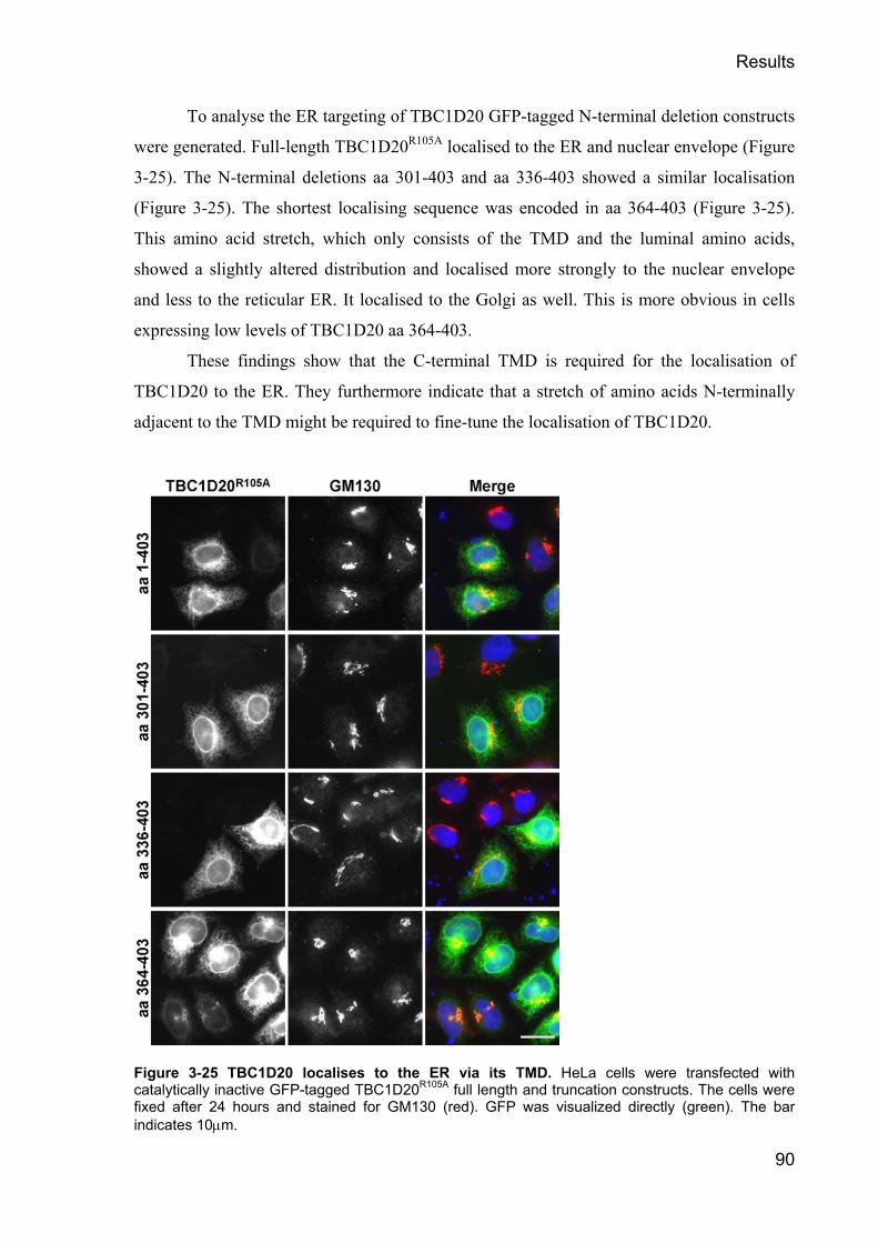
Results
90
To analyse the ER targeting of TBC1D20 GFP-tagged N-terminal deletion constructs
were generated. Full-length TBC1D20R105A localised to the ER and nuclear envelope (Figure
3-25). The N-terminal deletions aa 301-403 and aa 336-403 showed a similar localisation
(Figure 3-25). The shortest localising sequence was encoded in aa 364-403 (Figure 3-25).
This amino acid stretch, which only consists of the TMD and the luminal amino acids,
showed a slightly altered distribution and localised more strongly to the nuclear envelope
and less to the reticular ER. It localised to the Golgi as well. This is more obvious in cells
expressing low levels of TBC1D20 aa 364-403.
These findings show that the C-terminal TMD is required for the localisation of
TBC1D20 to the ER. They furthermore indicate that a stretch of amino acids N-terminally
adjacent to the TMD might be required to fine-tune the localisation of TBC1D20.
Figure 3-25 TBC1D20 localises to the ER via its TMD. HeLa cells were transfected with catalytically inactive GFP-tagged TBC1D20R105A full length and truncation constructs. The cells were fixed after 24 hours and stained for GM130 (red). GFP was visualized directly (green). The bar indicates 10µm.

Results
91
A schematic representation of these experiments is added (Figure 3-26) for clarity.
The minimal TBC domain is encoded by aa 1-317, consistent with the data obtained by in
vitro GAP assays. The localisation of TBC1D20 depends on the TMD on its C-terminus, and
is encoded by the aa 364-403.
Figure 3-26 Schematic representation of TBC1D20 mapping experiments. The red of the TBC domain represents myc-epitope-tagged constructs; GFP-tagged constructs are indicated by the green colour of the TMD. Drawings are not to scale.
3.2.21 TBC1D20 interacts with Reticulon 1 variant 2
The localisation of TBC1D20 to the ER raised the question of whether this targeting is
mediated by the interaction of TBC1D20 with ER associated proteins.
To identify interactors of TBC1D20 that could help its localisation to the ER, two
yeast two-hybrid screens were performed. Full-length TBC1D20 and TBC1D20 aa 364-403
was used as bait, respectively. A cDNA library generated from human testis was chosen as
prey for these screens. The screen of TBC1D20 aa 364-403 covered 875,000 clones but
failed to identify interactors. This indicates that this part of TBC1D20 might not be sufficient
to interact with other proteins. The screen with full length TBC1D20 however, which
covered 1,780,00 clones, revealed two interactors.

Results
92
One of these interactors was a full-length clone of an ER resident protein called
BAP31. This protein is involved in the regulation of ER exit and the quality control of MHC
molecules (Ladasky et al., 2006) and IgD (Schamel et al., 2003). This protein is very
interesting in the aspect of the role TBC1D20 plays in the regulation of ER exit. The second
positive clone was the C-terminus of Reticulon 1 variant 2 (RTN-1). Reticulons are a group
of 4 proteins, each of which has multiple isotypes (Yan et al., 2006). They are associated
with the ER by four C-terminal hydrophobic regions (Iwahashi et al., 2007). RTNs were
shown to be involved in ER-Golgi trafficking (Wakana et al., 2005), bind to SNAREs
(Steiner et al., 2004) and shape sub-domains of the ER (Voeltz et al., 2006). A S. cerevisiae
RTN homologue has been show to interact with Ypt1, the yeast Rab1 (Geng et al., 2005). As
TBC1D20 functions as a GAP for Rab1, the interaction of RTN-1 and TBC1D20 was
analysed in more detail.
First, a series of directed yeast two-hybrid assays was performed. These showed that
the TMD of TBC1D20 is necessary but not sufficient for the interaction with RTN-1 (Figure
3-27A). The minimal RTN-1 interacting region included the TMD and an adjacent
cytoplasmic stretch. This is the shortest fragment that showed the same localisation as full-
length TBC1D20 when expressed in cells (Figure 3-25). This suggests that the interaction
with RTN-1 might be required for the proper localisation of TBC1D20.
To confirm these results at the protein level, immunoprecipitation experiments were
performed. The cells were lysed in digitonin as this mild detergent was previously shown to
preserve RTN membrane complexes (Voeltz et al., 2006). 10 cm dishes of HeLa cells were
co-transfected with one of the myc-epitope-tagged TBC1D20 constructs indicated and GFP-
tagged RTN-1 for 24 hours. The cells were then harvested, lysed and cleared by
centrifugation. 1 µg of purified anti-GFP antibody and protein-G sepharose were added and
the slurry was incubated for 2 hours. The slurry was then washed to remove non-bound
proteins and boiled in SDS sample buffer. These experiments showed that RTN-1 interacts
with full-length TBC1D20 irrespective of its catalytic activity (Figure 3-27B). The first 337
amino acids of TBC1D20, which lack the ER-targeting region that interacted with RTN-1 in
direct yeast two-hybrid, did not show any significant binding to RTN-1 under the same
conditions (Figure 3-27B). These findings show that TBC1D20 forms a complex with
RTN1.

Results
93
Figure 3-27 TBC1D20 interacts with RTN-1. (A) A schematic of RTN-1 showing the two hydrophobic regions (HR). The minimal TBC1D20 binding fragment identified by yeast two-hybrid (Y2H) screening is shown in blue. Drawing is not to scale. Directed yeast two-hybrid assays were performed using RTN-1 as prey and the TBC1D20 truncations indicated. Growth on selective medium (QDO) indicates interaction. (B) GFP-immunoprecipitations were performed from HeLa cells co-transfected with myc-epitope-tagged RTN-1 and GFP-tagged TBC1D20 as indicated. The immunoprecipitates were probed with antibodies against GFP or Myc to detect TBC1D20 and RTN-1, respectively. Total (T) corresponds to the input, supernatant (S) to the non-bound and (IP) to precipitated material. (C) HeLa cells were transfected with GFP-tagged RTN-1 for 24 hours, fixed and stained for GM130 (red) or Calnexin (red). GFP was visualized directly (green). The bar indicates 10µm.

Results
94
HeLa cells were then transfected with RTN-1 to analyse its localisation. The cells
were fixed after 24 hours and stained for GM130 or calnexin, respectively. RTN-1 co-
localised with calnexin (Figure 3-27C) but not with GM130 (Figure 3-27C). Notably, the
morphology of the Golgi apparatus was not affected by the over-expression of RTN-1.
Taken together, these data show that TBC1D20 contains a specific ER-targeting
motif that interacts with a member of a family of known ER proteins. This interaction
appears to be required for the proper ER localisation of TBC1D20. The interaction requires
the TMD of TBC1D20 together with an adjacent cytoplasmic stretch and the hydrophobic
regions of RTN-1. This suggests that these proteins form a complex in the ER membrane.
3.2.22 RTN-1 modulates the activity of TBC1D20 in vivo
The interaction of TBC1D20 with RTN-1 suggests that RTN-1 might be important to control
the activity of TBC1D20. As over-expression of TBC1D20 causes a “loss of Golgi”, this
phenotype was used as readout of the activity of TBC1D20. HeLa cells were either
transfected with myc-epitope-tagged TBC1D20 alone, or co-transfected with GFP-tagged
RTN-1 for 24 hours. The cells were then fixed and stained for the Golgi marker GM130.
The over-expression of wild-type TBC1D20 caused a loss of the Golgi (Figure 3-28).
This effect was antagonized by the co-expression of RTN-1, as judged by the recovery of
GM130 staining into a fragmented perinuclear ribbon structure in cells expressing both wild-
type TBC1D20 and RTN-1 (Figure 3-28). The “loss of Golgi” phenotype was therefore
reduced in these cells to a Golgi fragmentation phenotype. The expression of TBC1D20R105A
did not cause a “loss of Golgi” phenotype (Figure 3-28). When co-expressed, RTN-1 and
TBC1D20R105A co-localised in the ER, and the Golgi appeared normal as compared to non-
expressing control cells (Figure 3-28). The expression of TBC1D20 aa 1-337 caused a “loss
of Golgi” phenotype. If the antagonising effect of RTN-1 on TBC1D20 is specific, then it
should require the RTN-1 interacting part of TBC1D20. As predicted, TBC1D20 aa 1-337,
which does not interact with RTN-1 (Figure 3-27) caused the “loss of Golgi” phenotype
independent of the presence of RTN-1 (Figure 3-28).
These findings show that the interaction of RTN-1 with TBC1D20 not only serves to
localise TBC1D20 to the ER, but also modulates its activity. This modulation might be
linked to the properties of RTNs to define sub-compartments of the ER and thus restrict the
distribution of TBC1D20 in the ER.

Results
95
Figure 3-28 RTN-1 expression antagonises the “loss of Golgi” phenotype. HeLa cells were transfected with myc-epitope-tagged wild-type, catalytically inactive TBC1D20R105A, or TBC1D20 aa 1-337 in the absence or presence of GFP-tagged RTN-1 (Reticulon) for 24 hours. The cells were fixed and stained for GM130 and the myc epitope. In single transfection panels DNA was stained with DAPI. The Bar indicates 10µm.

Results
96
To show the distribution of these phenotypes the Golgi morphology in n=100 cells
was analysed for each condition. The Golgi morphology was scored as intact, when it was
comparable to neighbouring non-expressing cells. It was scored as fragmented, when
GM130 stained a fragmented perinuclear ribbon structure. The cells were scored to have a
“loss of Golgi” phenotype when these structures were not observed.
This analysis showed that that the co-expression of RTN-1 with wild-type full length
TBC1D20 strongly reduced the percentage of cells with a “loss of Golgi” phenotype while
the percentage of cells with fragmented Golgi was equivalently increased (Figure 3-29). In
the case of TBC1D20 aa 1-337, the co-expression of RTN-1 did not significantly alter the
distribution of the phenotypes (Figure 3-29). Expression of TBC1D20R105A showed the same
distribution of phenotypes as control cells independent of the presence of RTN-1 (Figure
3-29). The over-expression of RTN-1 alone did not alter the distribution of phenotypes
compared to non-expressing control cells (Figure 3-29).
Figure 3-29 Distribution of phenotypes in cells expressing RTN-1 and TBC1D20. HeLa cells were transfected with myc-epitope-tagged wild-type, catalytically inactive TBC1D20R105A, or TBC1D20 aa 1-337 in the absence (-) or presence (+) of GFP-tagged RTN-1 (Reticulon) for 24 hours. The cells were fixed and stained for GM130 and the myc-epitope tag. n=100 cells were analysed for each condition and the Golgi was scored as normal, fragmented or lost in comparison to non-expressing control cells.
To exclude that the modulatory effect of RTN-1 on the activity of TBC1D20 is due
to altered expression levels a series of western blots was performed. HeLa cells were
transfected with myc-epitope-tagged TBC1D20 constructs alone or co-transfected with GFP-
tagged RTN-1 for 24 hours. The cells were then lysed and equal amounts were analysed by
western blot. The blots were probed with antibodies against GFP and the myc-epitope-tag to

Results
97
show RTN-1 and TBC1D20, respectively. These experiments showed that the co-expression
of RTN-1 did not alter the expression levels of TBC1D20 (Figure 3-30), independent of their
physical interaction. The modulatory effect of RTN-1 is therefore not due to decreased
expression levels of TBC1D20.
Taken together, these findings indicate that the interaction of RTN-1 with TBC1D20
not only ensures the proper localisation of TBC1D20 to the ER, but also serves as a way to
regulate the activity of TBC1D20. They furthermore suggest that RTN-1 helps to
compartmentalize the ER not only by shaping ER sub-domains (Voeltz et al., 2006), but also
by controlling the activity of TBC1D20, which is probably mediated by restricting
TBC1D20 to specific ER sub-domains.
Figure 3-30 Expression of RTN-1 does not alter TBC1D20 expression levels. HeLa cells were transfected with myc-epitope-tagged wild-type, catalytically inactive TBC1D20R105A, or TBC1D20 aa 1-337 in the absence (-) or presence (+) of GFP-tagged RTN-1 (Reticulon) for 24 hours. The cells were harvested and lysed. Western blots were probed with antibodies against GFP to show RTN-1 and against the myc-epitope-tag to show TBC1D20. The blots were probed with α-tubulin antibodies as a loading control.

Results
98
3.3 Summary
To identify GAPs and Rabs involved in the regulation of early secretory traffic a novel
screening method was established. This method uses the over-expression of RabGAPs to
inactivate endogenous target Rabs. This inactivation causes phenotypes that help to identify
which Rabs and GAPs are involved in the regulation of the process of interest. Screening for
changes in the morphology of both the Golgi apparatus and the ERGIC as well as the ability
of RabGAPs to block secretion resulted in only one candidate among all 38 RabGAPs tested,
the highly conserved protein TBC1D20.
The over-expression of the ER localised TBC1D20 caused a unique “loss of Golgi”
phenotype. When the fate of Golgi proteins was analysed by western blot, they were found
to remain associated to membranes, with the exception of the Rab1 effector p115. Golgi
enzymes redistributed to the ER. The exit of VSV-G from the ER was blocked in cells
expressing TBC1D20. When TBC1D20 was tested in a biochemical assay against a broad
selection of Rabs, it stimulated GTP hydrolysis by Rab1 and Rab2. Subsequent analysis of
dominant negative mutant Rabs and specific siRNA showed that Rab1 is the primary in vivo
target of TBC1D20. The expression of TBC1D20 scattered COPII positive structures, but
did not interfere with the dynamics of the COPII coat. Under these circumstances cargo
receptors such as p24 were absent from ERES, which possibly explains the block of
secretion. Using truncations of TBC1D20 the minimal TBC domain was mapped to aa 1-
317, consistent with biochemical data. Truncations and biochemical analysis also showed
that the ER localisation of TBC1D20 depends on the presence of a TMD on its C-terminus.
These experiments showed that TBC1D20 a type-II transmembrane protein.
The localisation of TBC1D20 depends on RTN-1, an interactor identified by a yeast
two-hybrid screen. RTN-1 modulates the activity of TBC1D20 dependent on the physical
interaction of both proteins. This suggests a model where TBC1D20 is localised to ER sub-
domains by RTN-1.
These findings indicate a novel function of Rab1 at the level of the ER. They
furthermore show that the activity of Rab1 is controlled at the level of the ER. Furthermore,
the classical view of RabGAPs as the limiting factors of the lifetime of active Rab is
challenged as both the depletion of TBC1D20 and the expression of constitutive active Rab1
failed to block trafficking at the level of COPII vesicles. This suggests a novel mode of Rab
regulation at least in the case of Rab1.

Discussion
99
4 Discussion
4.1 RabGAPs are a highly diverse protein family
Work in yeast has shown that RabGAPs contain a TBC domain (Albert et al., 1999; Strom et
al., 1993; Vollmer et al., 1999). However, only very few members of this protein family
were identified in the human genome prior to this work. A search of the human genome
identified 40 proteins containing a TBC domain, some of which also give rise to splicing
variants. Sequence analysis revealed that the TBC domain and the catalytic site signature
sequence IxxDxxR (T/S) in motif B are variable. The arginine in this signature sequence,
which is involved in GTP hydrolysis, is highly conserved, but not invariant. In some of the
TBC domain proteins the arginine is shifted, in others the arginine is replaced by other
amino acids. These findings might reflect differences in the Rabs that these GAPs regulate.
Rab20, Rab24 and Rab25 for example do not have a conserved glutamine in the DxxGQ
motif in G3 (1.2.1), and are therefore unlikely to hydrolyse GTP in the same way as the rest
of the Rab family (Pan et al., 2006). Variations in the catalytic mechanism to hydrolyse GTP
are reported for other members of the Ras superfamily. For example, RanGAP has an
asparagine instead of an arginine, and the small GTPases Sar1 contributes as histidine
instead of a glutamine to the reaction (Bos et al., 2007; Daumke et al., 2004; Seewald et al.,
2002). It is therefore possible that in the largest group of the Ras super-family not only one
catalytic mechanism is used to hydrolyse GTP.
Most human TBC domain proteins are multi-domain proteins. Many different domains
are found in these proteins, for example: PTB domains, PH domains, GRAM domains,
ubiquitin ligase domains and SH3 domains to name only a few. The GAPs of other small
GTPases of the Ras super-family are also multi-domain proteins (Bos et al., 2007). All these
GAPs function as integrators of signals onto the activity of a GTPase. Their composition of
multiple domains therefore appears to be critical for their function.
4.2 Novel methods to study the interactions of Rabs and their GAPs
4.2.1 A novel yeast two-hybrid system to identify Rab-GAP pairs
To match the GAPs identified using a bioinformatic approach to the Rabs they regulate, a
series of novel methods had to be established. First, a yeast two-hybrid based screen was
established. Initial tests showed that wild-type GAPs do not interact with wild-type Rabs.

Discussion
100
This finding is consistent with data that show that the interaction of a GAP and its target
GTPase is only transient (Allin et al., 2001). After GTP hydrolysis has occurred, the GAP no
longer interacts with its target and is released. It is also consistent with previous studies of
yeast showing that the affinity of TBC domain proteins for their substrates is very low
(Albert et al., 1999). To strengthen the interaction between the GAP and its target Rab a
conserved glutamine of the Rabs was mutated to leucine to restrict them in their GTP bound
active conformation. When wild-type GAPs were tested against these mutant Rabs
interactions were observed but only partial specificity was achieved. It is likely that the
introduction of the bulky hydrophobic leucine into the nucleotide-binding cleft of the Rab
prevents the insertion of the arginine of the GAP into this cleft. This steric hindrance might
explain why the GAPs showed reduced substrate specificity and interacted with groups of
related Rabs under these circumstances. The arginine of the B motif of the GAPs was
therefore replaced by alanine. When the mutant GAPs were tested against the constitutive
active Rabs, specific interactions were observed. As shown for RabGAP-5 and RN-tre, the
Rabs that interacted with the GAPs in this double mutant screen are their biochemical target.
When RN-tre and RabGAP-5 were later tested against all human Rab GTPases, it was shown
that these GAPs only activated GTP hydrolysis by the Rabs that they interacted with in the
double mutant screen (Fuchs et al., 2007). These observations therefore validate the double
mutant yeast two-hybrid approach.
The yeast two-hybrid data are also consistent with the recently solved structure of a
Rab bound to a GAP (Pan et al., 2006). The glutamine of the Rab forms hydrogen bonds to
the polypeptide backbone of a loop of the GAP that is inserted into the nucleotide-binding
cleft. It is likely that this loop of the GAP cannot be inserted into the cleft if the glutamine of
the Rab is mutated to leucine. As the GAP also interacts with the switch regions of the Rab,
some interaction is still maintained, but might not be sufficient to ensure full specificity.
When the positively charged arginine of the GAP is replaced by alanine, the truncated loop
might fit into the nucleotide-binding cleft and the GAP can bind to its specific target.
Therefore, this double mutant screen turns the GAP into an effector of the Rab and generates
a stable interaction.
The data presented in this work also shed new light on the GTP restriction of
constitutive active Rab mutants. The introduction of mutations in the conserved glutamine of
Rabs was previously thought to prevent the glutamine from coordinating a water molecule
for a nucleophilic attack at the γ-phosphate and thus keeps the Rabs GTP bound. According

Discussion
101
to the recently solved structure discussed above (Pan et al., 2006) this glutamine interacts
with a loop contributed by the GAP and is not directly involved in the catalysis of GTP
hydrolysis. The findings that wild-type GAPs do not interact, or have reduced specificity for
GTP restricted Rab mutants, suggests that the restriction is due to steric hindrance. This is
illustrated by the finding that wild-type RN-tre does not interact with GTP restricted Rab43,
and that RabGAP-5R165A interacts more strongly with Rab5Q79L than wild-type RabGAP-5
does. It was shown in the case of Rab1 and Rab5 that the GTP restricted mutants indeed
have a reduced basal GTP hydrolysis rate, but are not completely hydrolysis deficient. This
reduced basal hydrolysis rate however was resistant to RabGAP-5 or TBC1D20 mediated
stimulation of GTP hydrolysis. These findings suggest that constitutive active Rabs are kept
in their active conformation because they fail to properly interact with their cognate GAPs.
4.2.2 Rab inactivation screening as a novel method to analyse trafficking
The yeast two-hybrid screening method established in this work is a potent tool to identify
pairs of Rabs and their regulatory GAPs. This method however, has numerous limitations.
First, this method might not work in all cases, due to the structural differences between the
various GAPs and their target Rabs and thus generate false negative results. Second, and
most important, the organelles and trafficking processes regulated by particular Rabs and
their GAPs are largely unknown. To gain more information about both partners and the
processes they regulate another novel screening approach was established. Based on the fact
that an inactivated Rab does not recruit effector molecules onto the membrane it provides
identity to, a novel method called Rab inactivation screening was established. It was
proposed that the expression of all human GAPs and analysis of the resulting phenotypes
should identify GAPs involved in the regulation of a specific trafficking step.
To ensure that the phenotypes observed in the screen are due to the catalytic
inactivation of a Rab, the catalytically inactive mutants of GAPs have to be examined in a
next step. If a GAP causes a phenotype due to catalytic Rab inactivation, the inactive mutant
GAP should not cause such an effect. RabGAP-5R165A and RN-treR150A interacted with their
specific target in yeast two-hybrid, which shows that the secondary structure of these
proteins was maintained. The R-A mutation however efficiently abolished their activity to
stimulate GTP hydrolysis by their target Rabs in vitro. These findings show that the arginine
to alanine mutation specifically inactivates the TBC domain proteins without effects on their
secondary structure. Therefore these R-A mutant GAPs are valid controls to show the
specificity of a phenotype that is caused by the inactivation of an endogenous Rab.

Discussion
102
In a consequent step, the candidate GAPs have to be tested against all Rabs for their
ability to promote hydrolysis to identify their target Rab. To consolidate that the Rab
identified by in vitro experiments is the in vivo target of the GAP, the expression of the
dominant negative mutant of the Rab and its depletion should mimic the phenotypes caused
by the over-expression of the GAP.
Proving the generality of this screening procedure, other researchers in the group
used this method and successfully identified Rabs and their GAPs (Figure 4-1) involved in
the formation of primary cilia and the regulation of clathrin independent endocytosis (Fuchs
et al., 2007; Yoshimura et al., 2007). This technique was also used to prove that RabGAP-5
is the only GAP that regulates clathrin dependent endocytosis of EGF (Fuchs et al., 2007).
Not only Rabs, but also the other small GTPases of the Ras superfamily are
controlled by GAPs and GEFs in their activity. The GAP mediated inactivation approach
might therefore be useful to study the processes regulated by these GTPases as well.
Figure 4-1 Rab inactivation screening results. Rab inactivation screening is a proven method to identify Rabs involved in a specific trafficking pathway and their regulatory GAPs. Figure layout is based on Figure 1-1.

Discussion
103
4.3 RabGAP-5 is a specific Rab5 GAP
Many evolutionary conserved proteins tightly control endocytosis. One of the key Rabs
involved in the regulation of clathrin dependent endocytosis is Rab5 (Zerial and McBride,
2001). In order to identify a Rab5 specific GAP out of forty human TBC domain proteins,
the double mutant yeast two-hybrid screen was used. Only one out of all human TBC
domain proteins was found to interact with Rab5, a protein later called RabGAP-5.
Like many other TBC domain proteins, RabGAP-5 is a multi domain protein.
According to own observations and data from a later publication (Lan et al., 2005), the
minimal TBC domain of RabGAP-5 ranges from aa 13 to aa 411 and is followed by a short
coiled- coil stretch. It in the case of the TBC domain protein GAPCenA, a coiled-coil C-
terminally of the TBC domain contributes to specific Rab binding (Fuchs et al., 2007). This
coiled coil of RabGAP-5 might therefore contribute to specific substrate recognition as well.
The TBC domain tested in the in vitro GAP assays and used in further experiments therefore
contains this coiled-coil and ranges from aa 1 to aa 451. RabGAP-5 contains an Src-
homology-3 (SH3) domain C-terminal of the TBC domain. This domain recognizes proline-
rich sequences (Mayer, 2001) and is mostly found in proteins involved in signal
transduction. This is an interesting finding for a regulator of Rab5 since this Rab is involved
in the regulation of endocytosis of activated growth-factor receptors that follow the clathrin
dependent pathway (Carpenter, 2000). The third domain of RabGAP-5 is a RUN domain at
its C-terminus. The RUN domain was named after RPIP8, UNC-14 and NESCA (Callebaut
et al., 2001). It is present in several proteins that are linked to GTPases of both the Rab and
the Rap families. Its actual molecular function is not known. The C-terminus of RabGAP-5
including the RUN domain has been previously shown to be required for the interaction of
RabGAP-5 with other molecules, which will be discussed later in more detail (Ichioka et al.,
2005; Lee et al., 2004).
When the ability of RabGAP-5 to stimulate GTP hydrolysis by Rabs was analysed in
vitro, it was found to specifically activate GTP hydrolysis by the three Rab5 isoforms. It was
shown later (Fuchs et al., 2007), when RabGAP-5 was tested against all human Rabs, that it
only stimulates GTP hydrolysis by the same three Rab5 isoforms.
To confirm that RabGAP-5 is a GAP for Rab5, the effects of over-expressing it in
cells were analysed. RabGAP-5 over-expression redistributed the Rab5 effector EEA1 to the
cytoplasm and inactivated co-expressed Rab5. Other sub-cellular organelles remained
unaffected by the expression of RabGAP-5, which shows the specificity of these phenotypes.

Discussion
104
These observations are consistent with previous publications that show that active Rab5 is
required to recruit EEA1 to early endosomes (Christoforidis et al., 1999a; Lawe et al., 2000;
McBride et al., 1999). EGF trafficking, which depends on Rab5 (Carpenter, 2000), was
blocked by the over-expression of RabGAP-5. The recycling of the transferrin receptor was
blocked under these circumstances as well. Remarkably, when all three isoforms of Rab5
were depleted by siRNA (Fuchs et al., 2007), the phenotypes observed resembled the
phenotypes observed in cells over-expressing RabGAP-5. This shows that diminishing the
endogenous pool of active Rab5 caused these phenotypes.
To analyse whether RabGAP-5 is an essential regulator of endocytic traffic, it was
depleted by specific siRNA duplexes. The size of early endosomes was increased in cells
depleted of RabGAP-5. This phenotype is also observed in cells that have elevated levels of
active Rab5 (Stenmark et al., 1994). This shows that RabGAP-5 is required to control the
activity of endogenous Rab5 and functions as a Rab5 specific GAP. When the trafficking of
EGF was analysed in cells depleted of RabGAP-5, it was found that EGF is trapped in early
endosomes and was not trafficked to lysosomes for degradation. This shows that RabGAP-5
is required to inactivate Rab5 on early endosomes in order to allow the maturation events
required for the late stages of the endocytic pathway. This observation is consistent with
another study, which showed that Rab7 replaces Rab5 on early endosomes as they mature
(Rink et al., 2005). It furthermore shows the essential role for RabGAP-5 in the control of
trafficking through early endocytic compartments.
Taken together, these data show that RabGAP-5 has all the characteristics expected
of a GAP specific for Rab5. Evidence that RabGAP-5 in fact is the only human Rab5 GAP
comes from a subsequent study (Fuchs et al., 2007), which showed that out of all human
TBC domain proteins only RabGAP-5 blocks the Rab5 dependent endocytosis of EGF.
4.3.1 RabGAP-5 links signalling to membrane traffic
As RabGAP-5 is essential for sorting events at the early endosome and for the regulation of
Rab5, the question raises of how RabGAP-5 might be controlled.
RabGAP-5 has been shown to interact with the tumour-suppressor Merlin (Lee et al.,
2004) also known as Schwannomin, the gene product of NF2 (Okada et al., 2007). Merlin is
an interaction partner of Hrs, which is an early endosome-associated adaptor molecule
required for the sorting of growth-factor-receptors to late endosomes and lysosomes (Clague
and Urbe, 2001). Hrs is recruited to the endosome by a complex of Eps15 (Komada and
Kitamura, 2001) and the signal transducing adaptor molecule (STAM) (Row et al., 2005).

Discussion
105
This complex is recruited to the endosome by activated growth factor receptors (Clague and
Urbe, 2001). Therefore, RabGAP-5 may provide a link between growth-factor-receptor
signalling and the regulation of endosomal trafficking by modulating the activity of Rab5.
Based on these findings it can be proposed that activated growth-factor-receptors
control their own degradation and the termination of their signalling in a feedback loop
(Figure 4-2). Activated growth-factor-receptors are endocytosed and traffic to Rab5 positive
early endosomes where they recruit adaptor molecules like Hrs. Hrs and its binding partner
Merlin could the recruit RabGAP-5, which inactivates Rab5. Recent findings show that the
inactivation of Rab5 is required to mature Rab5 positive endosomes into Rab7 positive late
endosomes to enter the degradative pathway (Rink et al., 2005). It was also proposed that the
probable Rab7 GEF C-VPS/HOPS or CORVET (Peplowska et al., 2007; Wurmser et al.,
2000) is a Rab5 effector. Rab5 might therefore indirectly activate Rab7. The inactivation of
Rab5 by RabGAP-5 can thus promote the maturation of early endosomes into late
endosomes and MVBs, and thus stimulate the degradation of signalling molecules.
Interestingly, another RabGAP-5-interacting protein (Ichioka et al., 2005) called Alix is
required for formation of the multivesicular body and endosome organization (Cabezas et
al., 2005; Matsuo et al., 2004).
Consistent with the proposed model, the depletion of RabGAP-5 did not have an
effect on the Rab5 dependent uptake of EGF, but trapped EGF in early endosomes and
prevented its degradation. RabGAP-5 therefore seems to be required to terminate the activity
of Rab5 to allow the initiation of consecutive trafficking steps. On the other hand, Rab5
activity is also required for CCV formation (McLauchlan et al., 1998). According to this role
of Rab5, the expression of RabGAP-5 and the depletion of Rab5 (Fuchs et al., 2007) blocked
EGF trafficking at this early stage.
A recent publication shows that RabGAP-5 binds directly to TrkA and TrkB (Liu et
al., 2007) and suggests a different model for the regulation of endosomal signalling. These
authors propose that early recruitment of RabGAP-5 by TrkA/B prevents Rab5 from being
activated and thereby results in long-lived signalling endosomes. Activation of Rab7 would
therefore be prevented and the signalling molecules would therefore not enter the
degradative pathway.

Discussion
106
Figure 4-2 RabGAP-5 provides a link between signalling and Rab5 regulation. Activated growth factor receptors recruit protein complexes onto the early endosome that recruit RabGAP-5. RabGAP-5 inactivates Rab5 and the early endosome matures into a Rab7 positive late endosome. The number 5 indicates Rab5, Rab7 is indicated by the number 7. Active, GTP bound Rab is symbolised by a red sphere, inactive Rab by a green square.
4.3.2 RabGAP-5 and Merlin
Of special interest for future research into RabGAP-5 function is its interaction with the
tumor suppressor Merlin (Lee et al., 2004). Merlin switches between an active and an
inactive conformation dependent on the phosphorylation of its C-terminus by the p21-
activated kinase (PAK). Merlin accumulates at adherence junctions (Lallemand et al., 2003)
and is essential for cell-cell contact mediated cell cycle arrest (Lallemand et al., 2003;
Morrison et al., 2001; Okada et al., 2005). The ability of PAK to release cells from contact
inhibition is blocked by a form of Merlin that lacks the PAK phosphorylation site, which
suggests that PAK promotes cell proliferation by phosphorylating and thus inactivating
Merlin (Lallemand et al., 2003; Okada et al., 2005).
Recent findings show that Merlin inhibits cell proliferation by stabilising adherence
junctions and negatively regulates EGF receptor signalling and internalization (Curto et al.,
2007). The block of EGF endocytosis in cells over-expressing RabGAP-5 suggests an
interesting link to these observations. Merlin might recruit RabGAP-5 to block Rab5
dependent EGF uptake and thus stop cell proliferation. Further support for this idea comes
from the finding that Connexin43, a component of gap junctions that interacts with cadherins
(Giepmans, 2006), binds RabGAP-5 (Lan et al., 2005). RabGAP-5 might therefore be

Discussion
107
recruited to cell-cell contacts by the combined action of Merlin and Connexin43 and block
Rab5 dependent endocytosis at these sites.
This model implies a role for RabGAP-5 not only in terminating Rab5 activity on the
early endosome but also points to a role of RabGAP-5 in the regulation of cell proliferation
and cell polarity.
4.4 RN-tre is a GAP for Rab43
The TBC domain protein RN-tre was reported to act as a GAP for Rab5 (Lanzetti et al.,
2004; Lanzetti et al., 2000). The data presented in this work show that RN-Tre acts as a
weak Rab5 GAP in vitro, and that RN-tre is in fact a GAP for Rab43. The weak activity of
RN-tre towards Rab5 is unlikely to be physiologically relevant for multiple reasons.
RN-Tre was found to bind to Rab43 and Rab30 but not Rab5 in the yeast two-hybrid
screen. These interactions were only observed when both the Rabs and RN-tre were mutated.
This indicates that this interaction is due to a GAP-Rab interaction and not due to the fact
that RN-tre as a Rab5 GAP is an effector for Rab43 or Rab30.
RN-tre strongly activated GTP hydrolysis by Rab43 in vitro, and only weakly by
Rab5. When RN-tre was later tested against all human Rab GTPases it was shown to have
specific activity towards Rab43 (Fuchs et al., 2007). This indicates that the activation of
Rab5 by RN-tre is most likely a consequence of using purified proteins at high
concentrations in vitro. Remarkably, despite its high degree of similarity to Rab43, no
significant activation of GTP hydrolysis by Rab30 was observed in the presence of RN-tre.
This shows that the yeast two-hybrid system can identify interacting Rabs, but final
specificity can only be provided by biochemical in vitro GAP assays with recombinant
proteins.
In contrast to RabGAP-5, the over-expression of RN-Tre failed to redistribute the
Rab5 effector EEA1 or Rab5 in vivo. When tested in hTERT-RPE1 cells, the TGN marked
by TGN46 was more affected by the expression of RN-tre than the cis-Golgi marked by
GM130. Consistent with this, the target of RN-tre is present at the Golgi apparatus. This
observation is consistent with the finding that RN-tre and Rab43 are regulators of retrograde
transport from the PM to the TGN by a clathrin independent trafficking route exploited by
Shiga toxin (Fuchs et al., 2007).
The observation that RN-tre regulates an endocytic retrograde trafficking step helps to
explain why it was previously found to interfere with EGF uptake (Lanzetti et al., 2000).

Discussion
108
With low doses of EGF, the EGF receptor is almost entirely transported via the clathrin
dependent pathway. At higher or saturating doses it spills over into the clathrin independent
pathway (Sigismund et al., 2005). With saturating amounts of EGF, RN-tre would prevent
transport of the pool of EGF receptor entering via the clathrin-independent pathway, and
thus show a reduction in EGF uptake. It will therefore be interesting to investigate the role of
the RN-tre and RabGAP-5 regulated pathways under true physiological conditions in tissues
where EGF signalling is critical for normal growth and development.
4.5 TBC1D20 regulates secretion by inactivating Rab1
4.5.1 The Rab1 GAP TBC1D20 regulates Golgi morphology and ER exit
Out of 38 human TBC domain proteins tested in a Rab inactivation screen only the GAPs
TBC1D20, TBC1D22A, TBC1D22B, XM_037557 and RN-tre caused Golgi fragmentation
in HeLa cells dependent on their catalytic activity. When the fragmentation of the Golgi
apparatus was analysed in hTERT-RPE1 cells, only TBC1D20 and RN-tre caused a
phenotype dependent on their catalytic activity. This finding suggests, that the GAPs that
were not positive in both screens either do not regulate ubiquitously essential Rabs or that
HeLa cells lack Rabs that can redundantly fulfil the role of the Rabs inactivated by these
GAPs.
TBC1D22A and TBC1D22B most likely regulate the Rab33 family (Pan et al., 2006)
that regulates trafficking at the Golgi apparatus (Valsdottir et al., 2001; Zheng et al., 1998).
RN-tre has GAP activity towards Rab43, which localises to the TGN and regulate a clathrin
independent pathway from the PM to the TGN (Fuchs et al., 2007). XM_037557 is a GAP
specific for Rab8a, which is required for the formation of primary cilia in serum-starved
cells and localises to the Golgi in proliferating cells (Yoshimura et al., 2007). All these
GAPs therefore regulate Rabs that are involved in trafficking associated to the Golgi. This
validates the screening approach, and proves that in fact the GAPs associated with a
trafficking step or organelle of interest are specifically identified by this method.
When the ability of human GAPs to cause morphological changes to the ERGIC was
analysed in a Rab inactivation screen, only TBC1D20 and TBC1D22B caused a phenotype.
This indicates, that the Rabs regulated by these two GAPs are involved in the regulation of
trafficking processes between the Golgi and the ER. However, out of all human GAPs only
TBC1D20 blocked the trafficking of VSV-G. The role of the Rab regulated by TBC1D22B
therefore appears less critical.

Discussion
109
Based on the results of these screens, TBC1D20 was analysed in more detail. The
expression of this highly conserved GAP resulted in the apparent loss of cis-, medial-, and
trans-Golgi markers, a so-called “loss of Golgi” phenotype. Further investigation revealed
that TBC1D20 blocked the exit of VSV-G from the ER. TBC1D20 is unique amongst the
TBC-domain proteins in that it possesses a transmembrane domain. This domain is required
to target TBC1D20 to the ER. When tested in a biochemical GAP assay against all human
Rabs, TBC1D20 showed specific activity towards Rab1 and Rab2. The other GAPs that
caused Golgi fragmentation failed to stimulate GTP hydrolysis by Rab1 or Rab2, consistent
with their reported target Rabs.
To narrow down the in vivo target of TBC1D20, the phenotypes of dominant
negative Rabs were compared to phenotypes caused by over-expression of TBC1D20. These
experiments showed that only dominant negative Rab1N121I recapitulates these phenotypes.
When Rab1 and Rab were depleted by siRNA, both gave rise to Golgi fragmentation. The
Golgi fragments in cells depleted of Rab2 were forming a cluster whereas the Golgi
fragments in cells depleted of Rab1 appeared more vesiculated and spread throughout the
cell. This is consistent with the idea that the Rab1 effector p115 is required for clustering of
pre- Golgi structures (Alvarez et al., 1999). In the case of Rab2 depletion the fragments were
possibly still connected by the Rab1 effector p115 interacting with the GM130/ GRASP65/
Giantin complex on the Golgi (Short et al., 2005). In contrast to the expression of dominant
negative Rab1N121I or TBC1D20, in cells depleted of Rab1 the Golgi was only fragmented
and no “loss of Golgi” phenotype was observed. This might be explained by the different
kinetics of the Rab1 siRNA that takes 72 hours compared to 24 hours of expression. In
contrast to the case of the depletion of Rab1, which removes both the active and the inactive
pool of the Rab, expression of TBC1D20 or Rab1N121I increase the pool of GDP bound Rab.
This GDP-bound Rab might sequester the Rab1 GEF, and thus strengthen the phenotype.
The phenotype of Rab1 depletion, however, still resembles the TBC1D20 phenotype more
closely than the phenotype obtained upon depletion of Rab2. The depletion of Rab1 and
Rab2 by siRNA furthermore showed that only the depletion of Rab1 completely blocked the
trafficking of VSV-G at the ER. Therefore it was concluded that the phenotypes observed in
cells over-expressing TBC1D20 are mainly due to the inactivation of endogenous Rab1. As
predicted, the block of VSV-G trafficking was also observed in cells expressing dominant-
negative Rab1N121I. Consistent with the role of TBC1D20 as the GAP for Rab1, the Rab1
dependent clustering of COPII vesicles was strongly impaired in cells over-expressing
Discussion
110
TBC1D20. Furthermore, p115, which is recruited by Rab1 to membranes, was lost from the
membrane fraction in cells expressing TBC1D20.
The finding that TBC1D20 acts as a GAP for Rab1 is consistent with previous
studies on budding yeast Gyp8, the homologue of human TBC1D20. Gyp8p has GAP
activity towards Ypt1p, the homologue of human Rab1 (De Antoni et al., 2002). The yeast
GAP activity towards Ypt1p is found in a particulate membrane fraction (Jena et al., 1992),
which suggests that Gyp8 might have the same ER localisation as TBC1D20.
The finding that TBC1D20 blocks ER exit of cargo as a result of the inactivation of
Rab1 is consistent with older observations that the membrane extraction of Rab1 by GDI
prevents the ER exit of VSV-G (Peter et al., 1994) and that the ability to restore ER exit
depends on the addition of recombinant Rab1. This argues that ER exit specifically depends
on the presence of Rab1. In contrast to the inactivation of Rab1, the depletion of the Rab1
effector p115 did not cause a block of ER exit. Secretory cargo was able to exit the ER under
these conditions and accumulated in a punctate cluster adjacent to the nucleus, but did not
reach the plasma membrane. This supports the role of p115 in regulating SNARE mediated
fusion of ER derived vesicles with early Golgi compartments (Allan et al., 2000). In contrast
to TBC1D20 expression, Rab1 depletion or the expression of dominant negative Rab1N121I,
depletion of p115 does not alter the levels of active Rab1. These findings suggest that Rab1
has a p115 independent function in controlling the exit of secretory cargo from the ER.
To further consolidate that TBC1D20 acts as a GAP for Rab1 in vivo, it was depleted
by siRNA. In cells depleted of TBC1D20 the intensity of the p115 staining and p115
dependent COPII clustering was increased. Both these findings are consistent with elevated
Rab1 levels caused by the depletion of a GAP for Rab1.
Even though the data provided in this work strongly suggest that TBC1D20 acts
predominantly on Rab1 in vivo, they cannot fully rule out the possibility that TBC1D20 is a
GAP for Rab2 as well. Rab2 is only found in higher eukaryotic cells and does not have a
counterpart in yeast. It is thought to act downstream of Rab1, and functions in tethering ER
and ERGIC derived vesicle to the early Golgi (Tisdale and Balch, 1996). As TBC1D20
mediated inactivation of Rab1 occurs before Rab2 comes to act, effects caused by Rab2
inactivation will always be covered by phenotypes caused by Rab1 inactivation.
4.5.2 RTN-1 is an ER associated regulator of TBC1D20
To understand more about TBC1D20 function and ER, a yeast two-hybrid screen was
carried out. This screen identified the Reticulon RTN-1 as an interaction partner of

Discussion
111
TBC1D20, and this interaction was confirmed by immunoprecipitation. Reticulons are
involved in the organisation and the shaping of the ER (De Craene et al., 2006; Voeltz et al.,
2006) and in the regulation of ER to Golgi transport (Wakana et al., 2005). All members of
the Reticulon family are characterised by a C-terminal domain that forms two hairpin loops
in the membrane of the ER. TBC1D20 interacts with this membrane bound portion of RTN-
1, which suggests that both proteins form a complex in the ER membrane. Four different
Reticulons are known so far, all of which give rise to multiple splicing variants (Oertle and
Schwab, 2003; Yan et al., 2006). The differences between the Reticulons are found in their
variable N-termini, whereas their C-termini are very similar. TBC1D20 might therefore
interact with multiple members of the Reticulon family.
The physical interaction of TBC1D20 with RTN-1 had a modulatory effect on the
ability of TBC1D20 to cause a “loss of Golgi” phenotype. One possible explanation may
relate to the proposed function of Reticulons in the compartmentalisation of the ER (De
Craene et al., 2006; Voeltz et al., 2006). The interaction of TBC1D20 with RTN-1 seems to
be necessary for its proper ER localisation: the TMD of TBC1D20, which was not sufficient
to interact with RTN-1, localised to both the Golgi apparatus and the ER. The minimal part
of TBC1D20 interacting with RTN-1 however localised similar to the full-length protein.
RTN-1 might therefore also restrict the localisation of TBC1D20 within the ER. It is
conceivable that RTN-1 keeps TBC1D20 away from ERES, and that this restriction is
important to define or confine these sites to sub-domains of the ER. The over-expression of
TBC1D20 might override the spatial restriction by endogenous RTN-1 and thus inactivate
Rab1 and cause the “loss of Golgi” phenotype. The co-expression of RTN-1 counteracts this
effect and therefore weakens the phenotype.
Work in budding yeast has shown that Rtn1p forms a complex with the GDF Yip3p
(Geng et al., 2005). This complex precipitates Ypt1p, the yeast Rab1 homologue. This
suggests that GDF mediated insertion of Rab1 into the ER is tightly controlled by the
association of TBC1D20 with RTN-1 and the GDF.

Discussion
112
4.5.3 Rab1 is the conserved key regulator of the early secretory pathway
Surprisingly, out of all human GAPs only TBC1D20 was able to block trafficking of VSV-
G, which did not exit the ER in cells over-expressing wild-type TBC1D20. Both the
depletion of Rab1 and the expression of dominant negative Rab1N121I caused the same
phenotype, which shows that this block is due to the inactivation of Rab1. This raises the
question of why the inactivation of none of the other Rab blocks secretion.
First, simply analysing the appearance of VSV-G at the cell surface does not answer
questions regarding changes in the kinetics of trafficking or in the route of transport. In the
absence of one trafficking pathway it is possible that cargo might choose different, possibly
redundant trafficking routes, which are regulated by other Rabs. Such routes are important
for trafficking, for example in polarised apical and basolateral sorting. However, in a
cultured non-polarised HeLa cell such different trafficking routes probably act redundantly.
Second, and most importantly, the fact that TBC1D20 was the only positive in this
screen suggests that it regulates an essential early trafficking step. All cargo has to pass
through this step before it is sorted to its final destinations. Inactivation of other Rabs by
GAP expression did not block transport of VSV-G, despite the fact that a number of these
caused Golgi fragmentation. This indicates that the Rabs regulated by these GAPs are
important for a normal Golgi morphology, but none of them regulates a essential step in
secretory trafficking.
These findings point to the fact that Rab1 is a key regulator of trafficking between
the ER and the Golgi apparatus. In budding yeast although additional Rabs are needed for
transport from the Golgi to the cell surface, the Rab1 homologue Ypt1 is the sole Rab
required for ER to Golgi transport, and transport though the Golgi (Brennwald and Novick,
1993). Surprisingly, the results shown in this work reveal a similar picture in mammalian
cells. Despite its greater size and morphological complexity, the mammalian Golgi apparatus
therefore appears more similar to its budding yeast counterpart in terms of Rab function.
Interestingly, unlike other S.cerevisiae Ypt proteins whose homologue proteins usually form
a group of Rabs in the human genome, Ypt1 has only one related human protein, Rab1. This
shows that trafficking between the ER and the Golgi is an evolutionary conserved pathway,
which was not subject to diversification during the evolutionary expansion of trafficking
routes. The necessity to establish a tight regulation of this step in secretion might be a
possible explanation for this finding. Therefore, Rab1/Ypt1 is the conserved key regulator of
the early secretory pathway.

Discussion
113
4.5.4 TBC1D20 and the Rab cascade
The finding that trafficking of VSV-G is blocked at the level of the ER raises the question of
why ER exit is defective in cells with inactive Rab1.
One explanation is that it may be important to couple the sorting of cargo into COPII
vesicles with the activation of Rab1. Interestingly, a similar role has already been proposed
for Ypt1 in S.cerevisiae, where it was found that Ypt1p regulates and couples sorting of
cargo molecules in the ER to subsequent vesicle targeting (Morsomme and Riezman, 2002).
The same proposal has also been made for Rab5 (McLauchlan et al., 1998). Such a coupling
ensures that the newly formed vesicles recruit the correct tethering factors and therefore
dock to and fuse with the correct target membrane. Therefore, a vesicle without Rab1 should
not recruit any cargo. Consistent with this prediction, cargo receptors such as ERGIC53 or
p24 do not accumulate at ERES in cells over-expressing TBC1D20 despite normal COPII
dynamics. This shows that the block of ER exit is based on defective cargo sorting rather
than on defective vesicle formation.
How such a coupling might occur is less clear, and will require investigation of
further Rab1 effector molecules like the MICAL family of proteins, which also propose a
link to the microtubule network (Fischer et al., 2005; Weide et al., 2003). The Rab1 GEF,
TRAPPI (Wang et al., 2000), provides another possible explanation for this coupling.
TRAPPI has recently been shown to directly bind to the Sec23 subunit of the COPII coat
(Cai et al., 2007) At least one of the TRAPPI subunit is recruited to ERES (Yu et al., 2006).
Thus, TRAPP provides a means to activate Rab1 at the forming vesicle when sufficient
COPII components are recruited by cargo and cargo receptors.
The finding that in cells lacking active Rab1 ER exit is blocked, gains further
importance in the light of a model called Rab cascade (Haas and Barr, 2007; Markgraf et al.,
2007; Ortiz et al., 2002). This model is based on the finding that the Rab Ypt31/32 recruits
Sec2, the GEF for the Rab Sec4, which acts downstream of Ypt31/32 (Ortiz et al., 2002).
The Rab cascade model proposes that Rabs regulate the activity and recruitment of
downstream Rabs by their effectors, which act as regulators of these Rabs. One question is
how such a cascade is initiated. The observation that COPII recruits the Rab1 GEF TRAPPI
shows that the vesicle coat itself can trigger the Rab cascade and provides a link between
cargo recognition and the Rab cascade. Importantly, every cascade needs to be tightly
controlled at the beginning. The ER associated Rab1 GAP TBC1D20 can provide a
counterpart to the GEF activity of TRAPPI to ensure the specific initiation of this cascade.

Discussion
114
4.5.5 Rab1 and Golgi biogenesis
In contrast to any of the other 38 RabGAPs tested, over-expression of TBC1D20 caused the
apparent loss of cis-, medial-, and trans-Golgi markers. This “loss of Golgi” phenotype
cannot be simply due to a block of trafficking, as the expression of Sar1H79G (Yoshimura et
al., 2004) or the treatment of cells with BFA (Lippincott-Schwartz et al., 1989) only cause a
fragmentation of the Golgi apparatus despite their ability to block ER to Golgi trafficking .
The difference of these to the over-expression of TBC1D20 is that they do not alter the level
of active Rab1. Why does interfering with Rab1 function cause such an extreme phenotype?
Although debated for many years (Mironov et al., 1997; Pelham, 2001), most recent
findings indicate that the Golgi stack is not formed of static cisternae with cargo shuttling
from one to the next via vesicle carriers. Instead, the Golgi is now widely believed to consist
of highly dynamic and continuously maturing cisternae that modify their cargo as they
progress from the cis- to the trans- face of the Golgi stack (Bonfanti et al., 1998; Losev et
al., 2006). The residing enzymes in turns, which grant sequential biochemical activity,
recycle in vesicles to equip the Golgi cisternae as they progress. The steady state structure of
the Golgi stack is according to this cisternal maturation model a representation of the
dynamic balance between anterograde and retrograde trafficking. The mode of Golgi
biogenesis is also a matter of discussion (Glick, 2002; Lowe and Barr, 2007). According to
the cisternal maturation model, the question of how the Golgi is formed might be answered
by the ability of cisternae to be formed de novo from ER derived material. Even though
under some conditions the division of pre-existing structures forms the Golgi (Shima et al.,
1998), there is good evidence that the Golgi can arise directly from the ER (Mironov et al.,
2003). It was shown that the organization of ERES and COPII vesicle trafficking are
paramount for normal Golgi biogenesis and function (Connerly et al., 2005). Furthermore,
the availability of cargo such as Golgi enzymes is a key factor in determining the size of the
resulting Golgi cisternae (Guo and Linstedt, 2006).
How do these findings correlate with the “loss of Golgi” phenotype? In the case of
Rab1 inactivation, cargo and Golgi enzymes are trapped in the ER and thus prevent normal
Golgi biogenesis, which depends on their availability. Inactivation of Rab1 also leads to the
loss of p115 from membranes. This Rab1 effector is required for COPII clustering and the
formation of Golgi precursors. Rab1 inactivation therefore combines defects in both cargo
transport and vesicle tethering. This combination might cause a more severe defect in Golgi
biogenesis than depletion of individual tethering factors or a block of cargo transport.

Discussion
115
4.6 An additional model of GAP mediated Rab regulation
Surprisingly, the depletion of TBC1D20 and the expression of constitutive active Rab1Q67L
failed to cause a block of VSV-G trafficking. Consistent with these data, S.cerevisiae GYP8,
which is the homologue of TBC1D20, is not essential for secretion (De Antoni et al., 2002).
Furthermore, a yeast strain expressing constitutive active Ypt1Q67L does not exhibit growth
defects, shows normal rates of secretion, and normal membrane morphology (Richardson et
al., 1998).
Since neither TBC1D20 in human cells nor GYP8 in yeast is essential for secretion,
the regulation of GTP hydrolysis appears not to be critical for Rab1/Ypt1 function under
laboratory conditions. On the other hand, the inactivation of Rab1 has a strong effect on
Golgi morphology and secretion in both human and yeast (Bacon et al., 1989; Jedd et al.,
1995). How can these observations be consistent with the classical model of GAPs (Fig 1.6)
as determinants of the lifetime of active Rab (Novick and Zerial, 1997)? According to the
classic model, the GAP is also expected to localise to the target membrane but TBC1D20
localises to the ER. Therefore an additional model of Rab control is proposed (Figure 4-3).
In this model, GDI bound Rab is inserted into the membrane with the aid of a GDF
and is then activated by its GEF. The GAP localises in the same membrane and inactivates
the Rab again. The inactive Rab can be extracted by GDI or, more likely, become reactivated
by the GEF. The Rab therefore cycles between the active and the inactive conformation,
until the activity of the GEF can override the activity of the GAP. This could be mediated by
either inactivation of the GAP or increased GEF activity. The GEF then keeps the Rab active
and capable of recruiting effectors until the target is reached where the GEF activity
decreases. The Rab is inactivated by its own basal GTP hydrolysis, and effectors recycle to
the pool of active Rab.
In support of this model, the Rab1 GEF TRAPPI is recruited by COPII coat
components during vesicle formation at ERES (Cai et al., 2007; Yu et al., 2006). TRAPPI
activates Rab1 on ERES when cargo accumulates and coat formation is initiated. The ER
associated GAP TBC1D20 counteracts this activity. The GEF activity of TRAPPI might
override TBC1D20 mediated Rab1 inactivation when COPII components recruit sufficient
TRAPPI. Once sufficient COPII components are present on the vesicle to get Rab1
activated, the vesicle buds from the ER as COPII components mediate budding and
abscission. TRAPPI then keeps Rab1 active on the vesicle until the vesicle reaches the
Golgi. At the cis-Golgi TRAPPI is converted to TRAPPII, and looses GEF activity towards

Discussion
116
Rab1 (Morozova et al., 2006; Wang et al., 2000). On the cis- Golgi active Rab1 is required
and binds to various molecules like GM130 or Golgin84. (Diao et al., 2003; Moyer et al.,
2001). In the absence of its GEF however, Rab1 inactivates itself by its basal GTP
hydrolysis.
According to this model, increased levels of active Rab1 in the absence of TBC1D20
or in cells expressing Rab1Q67L do not prevent a vesicle to form and transport is therefore not
blocked. This vesicle then fuses with the Golgi in a process that does not require the
presence of a GAP and is thus not blocked under these circumstances. The cargo is then
released and traffics on in a process independent of Rab1. The active Rab1 inactivates itself
by its basal GTP hydrolysis in the absence of the GEF TRAPPI, and Rab1Q67L might just
reside on the Golgi.
Why is such a “regulator GAP” like TBC1D20 needed? One possibility is, that the
regulation of the initiation vesicle transport is coupled to recognition of properly folded
cargo as a proofreading mechanism before the commitment is made to form and traffic the
vesicle. Such a proofreading mechanism might be important in ER stress conditions. Support
for this idea comes from a recent study on the mechanism of α-synuclein mediated toxicity
(Cooper et al., 2006). Misfolding of α-synuclein is associated with several
neurodegenerative diseases like Parkinson's disease, even though the exact mechanism is
unknown. This study shows that α-synuclein creates an ER-stress situation that results in a
block of ER to Golgi trafficking. A genomic screen in yeast revealed that only YPT1 was
able to suppress α-synuclein toxicity and recover ER to Golgi trafficking. GYP8
significantly enhanced α-synuclein mediated toxicity presumably by inactivation of Ypt1.
These findings suggest, that Ypt1/Rab1 activity is limiting for ER exit under stress
conditions. The negative regulation of Ypt1/Rab1 by Gyp8/TBC1D20 might therefore
function as a proofreading mechanism and prevent the exit of misfolded proteins from the
ER. Interestingly, Reticulons are also involved in various neurodegenerative diseases, which
points to a general role of the early secretory pathway for these diseases (Wildasin, 2004;
Yan et al., 2006).
Another explanation for the need of a “regulator GAP” comes from the observation
that the NS5A protein of the Hepatitis C virus binds to TBC1D20 (Sklan et al., 2007a; Sklan
et al., 2007b). TBC1D20 might be required in virus infected cells to block trafficking of
membrane bound viral proteins to the cell surface to prevent spreading of the virus. The

Discussion
117
interaction with the viral protein might inhibit TBC1D20 to ensure the transport of viral
components to the PM.
Figure 4-3 RabGAPs as regulatory factors of Rab cycle initiation. The small red sphere labelled “GTP” symbolises active Rab bound to GTP, a small green square labelled “GDP” indicates inactive Rab. Rab interacting molecules are identified by common abbreviations. A clock indicates the kinetic self-inactivation of Rabs. Drawing is not to scale.
The idea that activation of Rabs by GEFs and their inactivation by GAPs are closely
linked seems counterintuitive at the first glance. However, using such a mechanism, the
amount of active Rab can be tightly controlled dependent on the activity of GAPs and GEFs.
A tight cycle of activation and inactivation is commonly used to regulate of the activity of
small GTPases. Sar1 for example, which is required for the COPII coat assembly is activated
by Sec12 and inactivated by the coat subunit Sec23 it recruits. This allows the dynamic
regulation of the coat assembly (Bonifacino and Glick, 2004). RhoA as another example is
tightly regulated by its GAP and GEF at the same time during cytokinesis to generate a
locally restricted pool of active RhoA (Yuce et al., 2005). Such a cycle of activation and

Discussion
118
inactivation allows to respond to changes in conditions more precise and quickly and is a
common feature amongst signalling molecules. Importantly, most GAPs are multi-domain
proteins. This suggests that the activity of GAPs is regulated dependent on various signalling
pathways.
TBC1D20 is the prime example of a “regulator GAP” as it is localised to the ER, the
donor membrane, by its TMD. RabGAP-5 on the other hand acts as a prototypic “terminator
GAP” in the regulation of the Rab5 to Rab7 transition. The depletion of RabGAP-5 leads to
an accumulation of EGF in early endosomes, which shows the critical role of RabGAP-5 in
terminating Rab5 activity. However, if RabGAP-5 is in fact recruited by Merlin to adherence
junctions to prevent EGF uptake, this would turn the “terminator GAP” into a “regulator
GAP”. This suggests that interacting proteins and signalling processes can alter the
regulatory properties of GAPs.
Taken together, the observations made during the course of this work and data shown
in recent publications indicate that RabGAP mediated inactivation of Rabs is a flexible and
highly regulated process. This process requires further investigation to understand the basis
and mechanics of the activity of these key regulators of Rab dependent membrane traffic.
4.7 The specificity of Rab-GAP interactions
The claim has been made based on findings in S.cerevisiae, that RabGAPs are promiscuous
in the recognition of their substrates (Albert and Gallwitz, 1999). However, based on data
presented in this work and in recent publications, this appears not to be the case.
The claim was based on GTP hydrolysis assays with truncated yeast TBC domain
proteins (Albert and Gallwitz, 1999; Will and Gallwitz, 2001). However, we have recently
shown (Fuchs et al., 2007) that in the case of the Rab4 GAP GAPCenA (Cuif et al., 1999)
truncations lose their substrate specificity and interact with a wide range of Rab GTPases.
The full-length protein on the other hand specifically interacted with its target Rab4. This
illustrates that in order to observe specific interactions and activity the use of full-length
TBC domain proteins is mandatory.
Another recent publication (Yoshimura et al., 2007) shows that the RabGAP
XM_037557 stimulates GTP hydrolysis by Rab8a, but not by its isoforms Rab8b, reflecting
the different properties of these very similar Rabs in the formation of the primary cilium. A
promiscuous GAP certainly would not detect such small differences. Furthermore, if the

Discussion
119
RabGAPs were in fact promiscuous in their substrate recognition, a Rab inactivation screen
would not identify specific regulators of the organelles or trafficking processes of interest.
Taken together, the results presented in this work and other recent publications point
to the fact, that RabGAPs actually have high substrate specificity, which is expected as they
regulate proteins that provide membrane identity.
There are still much more GAPs not assigned to their target Rab than GAPs, which
have been matched to their target. Furthermore, our knowledge about the mode of action and
the regulation of the GAPs themselves is incomplete. With the tools and methods established
in this work, further insights will certainly be provided. The RabGAP field therefore
promises to generate many interesting findings in the coming years.

Materials and Methods
120
5 Material and Methods
5.1 Materials
5.1.1 Reagents
Reagents were obtained from Sigma-Aldrich (Seelze, Germany), Carl Roth (Karlsruhe,
Germany) or VWR (Darmstadt, Germany) unless stated in the text.
5.1.2 Equipment
A list of commonly used equipment and its suppliers is given in the table below.
Equipment Description and manufacturer
Benchtop centrifuge Eppendorf Centrifuge 5417C
Benchtop refrigerated centrifuge Eppendorf Centrifuge 5417R
Cell Culture Incubators Heraeus HeraCell
Cell Culture Safety Hoods Heraeus HeraSafe
Centrifuge Heraeus Multifuge 3 L-R
Electroporation system BioRad MicroPulser
Film Developer Kodak X-OMAT 2000 Processor
Gel system BioRad Mini-PROTEAN
Heating block/ mixer Eppendorf Thermomix Compact
High pressure homogeniser Avestin Emulsiflex C5
Incubators Heraeus Function Line
Magnetic stirrer IKA RET basic IKAMAG
PCR machines Perkin Elmer Gene Amp 2400
Eppendorf Mastercycler Personal
pH meter Beckman 340pH/Temp meter
Power pack BioRad Power Pack 200 and 300
Scintillation counter Packard Tri-carb 2900TR
Semi- dry blotter BioRad Trans-blot SD
Shakers Infors AG Multitron
Grant Boekel BFR25

Materials and Methods
121
SW28 rotor Beckman
SW40 rotor Beckman
Transilluminators UVP 2UV Transilluminator
UVP Gel Documentation System
Fujifilm LAS3000
Ultracentrifuge Beckman Optima LE-80K
UV/Visible Spectrophotometer Amersham-Pharmacia Ultrospec 300pro
Vortex shaker IKA MS3 basic
Waterbath Haake DC10
5.1.3 Solutions
The composition of standard solutions not described in the respective methods chapter is
given in the table below. Unless otherwise stated the solvent is water.
Solution Composition
Assay Buffer (10x) 500 mM HEPES-NaOH, pH 6.8, 10 mg/ml purified
BSA, freeze at -20°C, add DTT to 10 mM before using
DNA loading dye (6x) 0.25% (w/v) bromophenol blue, 40% (w/v) sucrose in
TE
IMAC (5,20,200) 20 mM Tris-HCl pH8.0, 300 mM NaCl, (5, 20, 200)
mM Imidazole
LB (Agar) 10 g/l Bacto-tryptone, 5 g/l Bacto-yeast extract, 10 g/l
NaCl, (plus 15 g/l Agar)
LiPEG 100 mM LiOAc, 10 mM Tris-Cl, pH 8,0, 1mM EDTA,
pH 8,0, 40 % PEG3350, sterile filtered, stored at 4°C
LiSorb 100 mM LiOAc, 10 mM Tris-Cl, pH 8,0, 1mM EDTA,
pH 8,0, 1M Sorbitol, sterile filtered, stored at 4°C
Mammalian Lysis Buffer 20 mM Tris-HCl pH 7.4, 150 mM NaCl, 0.1% (w/v)
Triton-X 100
Milk-PBS (blocking buffer) 4% milk powder in PBS , 0.2% (w/v) Tween-20
Moviol + DAPI 2.4 g Moviol 4-88 added to 6 g analytical grade glycerol
while stirring, 6 ml of ddH2O added and left for 2 hours

Materials and Methods
122
at room temperature, 12 ml of 0.2 M Tris-HCl pH 8.5
added while stirring for 10 minutes at 50 °C , the
solution was clarified by centrifugation at 3000 x g for
15 minutes and stored at – 20 °C
PBS 8 g/l NaCl, 0.2 g/l KCl, 1.44 g/l Na2HPO4, 0.24 g/l
KH2PO4 ,(pH 7.4)
Ponceau 0.2% Ponceau red in 1% acetic acid
SC (Agar) 6,7 g/l Yeast nitrogen base, 2 g/l appropriate “drop-out”
mix, 20 g/l Glucose, 20 mM HEPES pH 6,5, (20 g/l
Agarose)
SDS-PAGE lower buffer (4x) 181.72 g/l Tris base, 4 g/l SDS
SDS-PAGE running buffer
(10x)
30.2 g/l Tris base, 188 g/l glycine, 10 g/l SDS
SDS-PAGE sample buffer (3x) For 100 ml: 2.3 g Tris base, 9.0 g SDS, 30 ml glycerol,
Adjust pH to 6.8 with HCl, adjust volume to 90 ml with
dH2O, 50 mg bromophenol blue, add 10% ß-
mercaptoethanol prior to use
SDS-PAGE transfer buffer 1x SDS-PAGE running buffer, 10% methanol
SDS-PAGE upper buffer (4x) 60.6 g/l Tris base, 4 g/l SDS
TAE (50x) For 1 l: 242.4 g Tris base, 57.2 ml glacial acetic acid, 50
mM EDTA pH 8.0
TBS for dialysis 50 mM Tris, 150 mM NaCl, 2mM DTT
TE 10 mM Tris-HCl pH 8.0, 1 mM EDTA
Yeast lysis buffer 2,5% (w/v) SDS, 25 mM Tris-HCl pH 7,5 , 25 mM
EDTA
Yeast S-Buffer 10mM K2HPO4 , pH 7,2, 10mM EDTA, 50mM ß-
Mercaptoethanol, 50µg/ml Zymolyase
5.1.4 PCR primer
DNA primer to clone TBC domain proteins were designed according to 5.3.1, and obtained
by Metabion (Martinsried, Germany).

Materials and Methods
123
Target Gene Forward and reverse primer including restriction site (in
capital letters)
EVI-5 GGGATCCCCatggccagtcaggtggcaagtc GCTCGAGtcagacagtggttgaatacgac
RN-tre/USP6NL GAGATCTCCatgaattcagaccaggatgtagc GGTCGACtcacagcaacactgactcttgg
RUTBC1 GAGATCTCCatgggcagcgcagaggacgcagtc CGTCGACtcacttgttctctatgagcatctg
RUTBC2 GAGATCTCCatggaggaggctgtgacacgcaag CGTCGACtcacttgttctcaatcagagtctg
RUTBC3/RabGAP-5 GGGATCCCCatgtcaggaagccatacacctgcctgtggccc GGTCGACtcacccgtccacatcccagctgaag
TBC1D1 GAGATCTCCatggaaccaataacattcacagcaagg CGTCGACtcagtcgcccgtgggctcgggctgcg
TBC1D2 GGGATCCCatggaagcttaccggacccag GGTCGACtcaggcttccccctccacctcg
TBC1D3B/PRC17 GGGATCCCCatggacgtggtagaggtcgcg GGTCGACctagaagcctggaggg
TBC1D4/AS160 GGGATCCCCatggagccgcccagctgcattcag GCTCGAGttatggcttatttcctatcttggc
TBC1D5 GGGATCCCCatgtatcattccttatctgaaact GCTCGAGtcagatgtccaggggactcacaat
TBC1D6 GGGATCCCCatgcagcccgccgagcgctcgcgg CGTCGACctatgcccactggtgcccgtcctg
TBC1D7 GAGATCTCCatgactgaggactctcagagaaac GCTCGAGtcagcttgaatggaccggggtcccag
TBC1D8 GGGATCCCCatgttcctgaacctggatgaggtg GCTCGAGctacaagttactcagcttaagttc
TBC1D10A/EPI64 GGGATCCCCatggcgaagagcaacggagag GGTCGACttacaagtaggtgtcctcactctcttg
TBC1D10B GTGATCACCatgtctgggaccttggagtccttg CGTCGACtcagaagtaagcgtcctgccgggc
TBC1D10C GGGATCCCCatggcccaggccctgggggaggac GCTCGAGtcagaagcgggtgtccaggaagga
TBC1D11/GAPCenA GGGATCCCCatggatgacaaggcttctgttggaaaaatc GCTCGAGtcagcaagtcctttcccttgaaccccgg
TBC1D12 GGGATCCCCatggttcggggaatctccaggtcg CGTCGACctagcttttcaaagcaggactact
TBC1D13 GAGATCTCCatgtcaagtctgcacaagag GGTCGACctactttgagtcttggagctccttg
TBC1D14 GGGATCCCCatgactgatggaaaactctcc GGATCCtcagtgtcggagggatggacttcc
TBC1D15 GGGATCCCCatggcggcggcgggtgttgtgagc GCTCGAGtcatgcaggtgttaatctgcagac
TBC1D16 GAGATCTCCatgtctctgggccgcctccttcgc GCTCGAGctatctgcggaagccgaagccgtc
TBC1D17 GAGATCTCCatggaaggagccggctacagggtg GCTCGAGttaggagtcggcgccctcgtcctc

Materials and Methods
124
TBC1D18 GTGATCACCatggaggtcagagcttcattacag GGTCGACctacaaatatacaaacttgtatctatcc
TBC1D19 GGGATCCCCatgttgcaggaggagtcggacctc GCTCGAGtcaggtgacagtagcaaacagaaa
TBC1D20 GAGATCCTCCatggccctccggagtgcg GGTCGACtcagggaaacagctgcagctgaacttag
TBC1D21 GGGATCCCCatgaccaccctctctcctgaaaac GCTCGAGtcagaggaagaaatcctttaatgt
TBC1D22A GGGATCCCCatggccagcgacggggccaggaag CGTCGACtcatttcttgtagtgattgggggc
TBC1D22B GAGATCTCCatggccctccggagtgcg GGTCGACctatcggcggtagtgatttgg
USP6 GGGATCCCCatggacatggtagagaatgcagat CGTCGACttactgtaacatagagtacttttc
AK074305 GGGATCCCCatgtttcccctgaaggacgctgaa CGTCGACtcatatttgaggagatgggatggt
KIAA1055 GAGATCTCCatgctcatggagaccatccaagcc CAGATCTtcaggtatcctcctcctcgtcact
KIAA0676 GGGATCCCCatgtggctgagcccggaggaggtg GCTCGAGtcagccggaaactccaggctgctc
KIAA0882 GGGATCCCCatgtgggtgaacccggaggaggtg CGTCGACtcagccggacatggccgagatttc
NP_060222 GGGATCCCCatgtggctgaagcctgaggaagtg GCTCGAGttacatcttggttctagaccttaa
NP_060779 GGGATCCCCatggcggaaggagaagatgtgccg GCTCGAGttaactttccaaagcatccaaaac
EVI5-like GAGATCTCCatggcgagccccactctgagcccc GCTCGAGtcagttgtccagaccctggctgta
XM_037557 CGGATCCCCatgcggcaggacaagctgaccggg GCTCGAGtcatcgttttttagtgccactgtttc
KIAA1171 GGGATCCCCatggactctccaggatacaactgc GCTCGAGtcactgggtgtcagggtcctggaa
Paris-1 GTGATCACCatggagggcgctggggagaacgccccgg GGTCGACtcaggcttccccctccacctcgtcctcgc
5.1.5 siRNA oligonucleotides
siRNA oligonucleotides for siRNA-mediated depletion of specific mRNAs were designed to
a target sequence of NN(N19)dTdT using the siDESIGN Center on the Dharmacon
homepage (www.dharmacon.com), or were obtained as preconfigured SMART- pools by
Dharmacon. The siRNA oligonucleotides used in the course of this work are listed in the
table below.

Materials and Methods
125
Target Gene Target Sequence
GL2 (Control) CGUACGCGGAAUACUUCGAUU
LaminA (Control) CTGGACTTCCAGAAGAACATC
RabGAP-5 AACGGCCTGCGAGGCTGGTTT
TBC1D20 (SMART-Pool) GGACUUGCCCUAUGAGACA
GGACCAACCGCUUUGUGAA
ACCCUCAGCUGCACUACUA
ACGCAUGGCUAUCAGUGAA
p115 GCCGAGACGAUUCAAAAGCUU
GGCAGCAGGUUUCUACAUUUU
GM130 CCCUGAGACAACCACUUCUUU
GGCUGGCAUGCAGCUUAACUU
CCCCACUGCACAGCAGAUCUU
Rab1A (SMART-Pool) GAACAAUCACCUCCAGUUAUU
CAAUCAAGCUUCAAAUAUGUU
GGAAACCAGUGCUAAGAAUUU
CAGCAUGAAUCCCGAAUAUUU
Rab1B (SMART-Pool) CCAGCGAGAACGUCAAUAAUU
GCGCCAAGAAUGCCACCAAUU
GACCAUGGCUGCUGAAAUCUU
CAGCCAAGGAGUUUGCAGAUU
Rab2A (SMART-Pool) GAAGGAGUCUUUGACAUUAUU
GAUAUUACACGGAGAGAUAUU
CGAAUGAUAACUAUUGAUGUU
UGACCUUACUAUUGGUGUAUU
Rab2B (SMART-Pool) GGACUUAUAUUCAUGGAAAUU
GCGCCAAGAAUGCCACCAAUU
GACCAUGGCUGCUGAAAUCUU
CAGCCAAGGAGUUUGCAGAUU

Materials and Methods
126
5.1.6 Antibodies
The antibodies used in the course of this work are listed in the tables below, along with their
typical dilutions used for western blotting (WB) and immunofluorescence (IF). The species
indicated are M= Mouse, S= Sheep and R=Rabbit.
5.1.6.1 Primary antibodies
Name Antigen WB IF Source
M α Myc c-myc, clone 9E10 1:1000 1:1000 Cancer Research
R α Myc c-myc --- 1:1000 Santa Cruz
M α VSV-G VSV-G luminal domain --- 1:20 Ira Mellman
M α EEA1 Human EEA1 1:50 1:50 BD Biosciences
S α GFP His-GFP 1:2000 ---- Barr laboratory
M α GM130 Human GM130 --- 1:2000 BD Biosciences
S α GM130 His-rat GM130 1:1000 1:1000 Barr laboratory
M α Rab1b Human Rab1b, clone 1E7 1:5 Weide lab
R α RabGAP-5 RabGAP-5 aa 1-451 1:250 ---- Barr laboratory
M α Calnexin Human cell lysate 1:1000 1:1000 Chemicon
S α Golgin-84 Human Golgin84 1:1000 1:2000 Martin Lowe
S α Golgin-97 Human golgin-97 aa 589-
769
1:500 1:500 Barr laboratory
R α Golgin-160 Human golgin-160 1:1000 1:1000 Barr laboratory
S α p115 Rat p115 aa 772-959 1:2000 1:2000 Barr laboratory
R α sec24C Human Sec24C ---- 1:1000 David Stephens
M α sec31 Human Sec31 ---- 1:1000 BD Biosciences
M α ERGIC53 ERGIC53 ---- 1:1000 Alexis Biochemical
M α Lamp-1 Human Lamp1 1:1000 1:1000 BD Biosciences
M α TFnR Human TFnR 1:1000 1:250 Chemicon
S α TGN46 Human TGN46 1:2000 1:2000 Serotech
M α Tubulin Tubulin, clone DM1A 1:1000 1:1000 Sigma-Aldrich

Materials and Methods
127
5.1.6.2 Secondary antibodies Name Species Antigen WB IF Source
AMCA-α-M Donkey Mouse IgG ---- 1:250 Jackson Labs
Cy2-α-M Donkey Mouse IgG ---- 1:1000 Jackson Labs
Cy3-α-M Donkey Mouse IgG ---- 1:1000 Jackson Labs
Cy2-α-R Donkey Rabbit IgG ---- 1:1000 Jackson Labs
Cy3-α-R Donkey Rabbit IgG ---- 1:1000 Jackson Labs
Cy2-α-S Donkey Sheep IgG ---- 1:1000 Jackson Labs
Cy3-α-S Donkey Sheep IgG ---- 1:1000 Jackson Labs
HRP-α-S Donkey Sheep IgG 1:10000 ---- Jackson Labs
HRP-α-M Sheep Mouse IgG 1:2000 ---- Amersham
HRP-α-R Donkey Rabbit IgG 1:2000 ---- Jackson Labs
5.2 Bacterial methods
5.2.1 Growth and maintenance of E. coli
Bacteria were grown at 37 °C in LB medium containing an appropriate antibiotics for
selection, either 100 µg/ml ampicillin, 50 µg/ml kanamycin, or 34 µg/ml chloramphenicol.
Short-term storage was on LB-agar plates plus antibiotic at 4 °C.
5.2.2 Bacterial strains
The following E. coli strains were used in the course of this work.
Strain Genotype Use
XL1-blue F’::Tn10 proA+B+ lacIq ∆(lacZ)M15/ recA1
endA1 gyrA96 (NaIr) thi hsdR17 (rK-mK+)
glnV44 relA1 lac
General cloning
applications
GM2163 F- ara-14 leuB6 fhuA31 lacY1 tsx78 glnV44
galK2 galT22 mcrA dcm-6 hisG4 rfbD1
rpsL136 dam13::Tn9 xylA5 mtl-1 thi-1 mcrB1
hsdR2
Non-methylated DNA
for digestion with
Dam/ Dcm- sensitive
enzymes

Materials and Methods
128
BL21(DE3)* F- ompT gal [dcm] [lon] hsdSB rB- mB-; an E.
coli B strain with DE3, a λ prophage carrying
the T7 RNA polymerase gene
Recombinant protein
expression
JM109* F’ traD36 proA+B+ lacIq ∆(lacZ)M15/ ∆(lac-
proAB) glnV44 e14- gyrA96 recA1 relA1
endA1 thi hsdR17
Recombinant protein
expression
TOP10 F- mcrA ∆(mrr-hsdRMS-mcrBC)
Φ80lacZ∆M15 ∆lacX74 recA1 deoR araD139
∆(ara-leu)7697 galU galK rpsL (StrR) endA1
nupG
TA-cloning of PCR
products
*BL21(DE3) and JM109 strains carrying the pRIL plasmid (Stratagene) were used for
recombinant protein expression. This plasmid encodes the tRNA genes for rare arginine,
isoleucine, and leucine codons and conveys chloramphenicol resistance.
5.2.3 Preparation and transformation of chemically competent bacteria
To prepare chemically competent E. coli, a single colony was picked from a fresh plate and
used to inoculate 50 ml of LB (with selection, if necessary) and grown overnight at 37 °C
while shaking. 1 ml of overnight culture was then used to inoculate 100 ml of fresh LB and
the cells were grown at 37 °C until an OD600 of 0.5. The culture was then chilled on ice for
15 minutes before being transferred to a sterile centrifuge tube and centrifuged at 3000 x g
for 10 minutes at 4 °C. The supernatant was discarded and the cell pellet resuspended in 0.4
total culture volume of TfbI (30 mM KOAc, 100 mM RbCl2, 10 mM CaCl2, 50 mM MnCl2,
15% (v/v) glycerol, pH adjusted to 5.8 with dilute acetic acid). The cells were incubated on
ice for 15 minutes and then pelleted as before. The cell pellet was then resuspended in 0.04
volumes TfbII (10 mM MOPS, 75 mM CaCl2, 10 mM RbCl2, 15 % (v/v) glycerol, pH
adjusted to 6.5 with dilute NaOH). After storing on ice for 15 minutes, the cells were
aliquoted into 50 µl aliquots in sterile Eppendorf tubes, frozen in liquid nitrogen, and stored
at – 80 °C until needed.
For transformation, aliquots were thawed on ice before adding the DNA. In general,
10 µl of a ligation reaction was used for transformation of 50 µl competent bacteria, or 1 µl
of plasmid DNA (approx. 200 ng). Cells and DNA were mixed and left on ice for 20 minutes
before being heat-shocked in a 42 °C water bath for 90 seconds. Cells were placed back on

Materials and Methods
129
ice for 2 minute before the addition of 250 µl LB lacking antibiotics. The cells were allowed
to recover for 60 minutes at 37 °C with shaking before being plated on LB-agar plates
containing an appropriate selection antibiotic. All 250 µl of a ligation transformation were
plated while only 50 µl of a plasmid transformation was plated. Plates were then incubated
overnight at 37 °C.
5.2.4 Preparation and transformation of electrocompetent bacteria
To prepare electrocompetent E. coli, a single colony was picked from a fresh plate and used
to inoculate 5 ml of LB and grown overnight at 37 °C with shaking. 2.5 ml of the overnight
culture was then used to inoculate 500 ml of fresh LB and grown at 37 °C until an OD600 of
between 0.5 and 0.6. The culture was then chilled on ice for 15 minutes before being
transferred to a sterile centrifuge tube and centrifuged at approximately 3000 x g for 10
minutes at 4 °C. The supernatant was removed and the cells resuspended in 10 ml sterile,
ice-cold water. Centrifuge tubes were then filled with 500 ml ice-cold water and spun as
before. The supernatant was removed and the volume adjusted to 50 ml with ice-cold 10 %
(v/v) glycerol before centrifuging as above. The cell pellet was resuspended in an equal
volume of ice-cold 10 % (v/v) glycerol and the cells were aliquoted into 40 µl aliquots in
sterile Eppendorf tubes, frozen in liquid nitrogen, and stored at – 80 °C until needed.
For electroporation, sterile 2 mm electroporation cuvettes were placed on ice to chill
during the cells thawed on ice. 2 µl of yeast miniprep DNA or 10ng plasmid DNA was
added to the cuvette followed by 40 µl of electrocompetent cells. The cuvette was flicked to
mix DNA and cells and left on ice for 15 minutes. The electroporator was set to 2.5 kV, 25
µFarads. Cuvettes were dried with a tissue wipe and placed into the chamber. The cells were
electroporated, ideally with a time constant of 4.7. 250 µl of LB medium was added to the
cuvette immediately and the cells were transferred to a sterile Eppendorf tube and incubated
shaking at 37 °C for 1 hour before being plated on LB-agar plates containing an appropriate
selective antibiotic and grown at 37 °C over night.
5.2.5 Plasmid DNA preparation from bacteria
Plasmid DNA was prepared using either miniprep or maxiprep kits (Qiagen, Hilden,
Germany) according to the manufacturers instructions. Minipreps were prepared from 1.5
ml, maxipreps from 250 ml of overnight cultures grown in LB containing selective
antibiotics at 37 °C.

Materials and Methods
130
5.2.6 Purification of 6xHis-tagged proteins from bacteria
A single colony was picked from a fresh plate of transformed E. coli and used to inoculate
25 ml of LB plus selective antibiotics and grown overnight at 37 °C with shaking. 10 ml of
the overnight culture was then used to inoculate 1 l of fresh LB plus antibiotics and grown at
37 °C until an OD600 of 0.5 – 0.6 was reached. Depending upon the protein being expressed,
cultures were shifted to 18 °C for 30 minutes before being induced with 0.5 mM Isopropyl
β-D-thiogalactoside (IPTG) from a 1 M stock or induced immediately at 37 °C. Cultures
were then left for either 3 hours (37 °C) or overnight (18 °C). Bacteria were then harvested
by centrifugation at 3000x g for 15 minutes at 4 °C and cell pellets were frozen on dry ice
for 20 minutes.
For protein purification, cell pellets were thawed and resuspended by the addition of
20 ml IMAC5 buffer containing protease inhibitor tablets and 0,1% TX100 per litre of
original culture. The resuspended bacteria pellet was the homogenised using a chilled
Emulsiflex C5 high-pressure homogeniser (Avestin Europe GmbH, Mannheim). The lysate
was passed about 10 times through the homogeniser until the DNA was sheared and the
lysate became much less viscous. The lysate was then centrifuged at 28000 rpm for 30
minutes at 4 °C in an SW28 rotor in a Beckman Optima LE-80K Ultracentrifuge (Beckman,
USA) to pellet the cell debris. The cleared lysate supernatant was transferred into a clean
falcon tube. 0.5 ml of Ni-NTA agarose (Qiagen) per litre of original culture, washed in the
same buffer, was added to the cleared lysate and the tube was rotated for 2 hours at 4 °C to
allow protein binding to occur. The resin was then pelleted by centrifugation at 1000 x g for
5 minutes at 4 °C and washed in 2 x 30 ml of IMAC20 containing 0.1 % TX100. The resin
was then washed in a further 30 ml of IMAC20 without TX100 and bound protein was
eluted by the addition of IMAC200. The eluate was collected in 1 ml fractions. After SDS-
PAGE analysis of 10 µl of these fractions, peak fractions containing recombinant protein
were pooled and dialysed overnight at 4 °C TBS containing 2mM DTT. The recombinant
protein was aliquoted, quickly frozen in liquid nitrogen and stored at -80°C. After quickly
thawing the protein at 37°C with shaking the concentration was determined using a standard
Bradford assay (5.4.3).

Materials and Methods
131
5.3 DNA methods
Enzymes for DNA modification were obtained from New England Biolabs (Ipswich, MA,
USA), Promega (Madison, WI, USA), or Invitrogen (Carlsbad, CA, USA) and used
according to the supplier’s instructions.
5.3.1 "Shortway" cloning strategy
A standard cloning strategy, referred to as the “Shortway” system, was used when generating
constructs, in order to make all inserts compatible with a wide range of different vectors.
This involved generating inserts with a BamHI, BglII, or BclI site at the 5’ end and a SalI or
XhoI site at the 3’ end. The overhanging ATC of the BamHI/ BglII/ BclI site was designed
to be in frame with the start codon of the insert.
GGG ATC CCC ATG (BamHI)
GAG ATC TCC ATG (BglII)
GTG ATC ACC ATG (BclI)
The restriction site is bold and the start codon is in italics.
These restriction sites were added by PCR. The insert was TA-TOPO cloned into the vector
pCRIITOPO (Invitrogen, Carlsbad, CA, USA). Constructs were sequenced in pCRIITOPO
and then subcloned into the following vectors.
5.3.2 Compatible vectors
Yeast:
DNA binding domain (n-term) pFBT9 BamHI-SalI
Activation domain (N-term) pACT2 BamHI-XhoI
Bacterial expression:
His-tagging (N-term) pQE32 BamHI-SalI
GST-tagging (N-term) pFAT2 BamHI-XhoI
MBP-tagging (N-term)
with His-tag (C-term) pMalTEV-His BamHI-SalI
Mammalian expression:
GFP-tagging (N-term) pEGFP-C2 BglII-SalI
GFP-tagging (C-term) pEGFP-N3 BglII-SalII
Myc-tagging (N-term) pcDNA3.1/MycA BamHI-XhoI

Materials and Methods
132
If a DNA stretch of interest contained internal XhoI and SalI sites it was cloned using
3´ BamHI / BglII / BclI sites, and inserted into singly cut vectors using the indicated 5´ end
cutting enzyme. The vectors were treated with alkaline phosphatase for 15 min at 37° prior
to loading on agarose gels. When all restriction sites required for “shortway –cloning” were
found in the DNA stretch of interest, it was cloned, and one of the internal restriction sites
was removed by a silent site directed mutagenesis.
5.3.3 Restriction digests and agarose gel electrophoresis of DNA
Analytical restriction digests were carried out in a total volume of 20µl using 0.5µl of each
enzyme in the appropriate buffer to digest approximately 500 ng of DNA. Digests were
carried out for 1.5 hour at the indicated temperature. The whole reaction was mixed with 4
µl DNA loading buffer and loaded for electrophoresis onto an agarose gel of concentrations
between 0.5% and 2% depending on the expected size of the DNA fragments. To pour gels
agarose of the desired concentration was dissolved in 1 x TAE by heating. Ethidium bromide
was added to a concentration of 0.8 µg/ml for visualisation. The gel was run in 1 x TAE at a
constant voltage of 80 V for 45 minutes. DNA was visualized on a UV transilluminator.
Preparative restriction digests were performed using 2.5 µg DNA. Vector DNA was
treated with alkaline phosphatase at 37°C for 15 minutes by the addition of 1 µl enzyme to
the digestion mix prior to loading on the gel.
After electrophoresis, appropriate bands were excised using a scalpel blade and
purified using a gel extraction kit (Qiagen) according to the manufacturer’s instructions.
5.3.4 Cloning digested DNA fragments
Ligations were performed in a total volume of 20µl using 1µl of T4 DNA ligase (NEB) in 1x
ligase buffer (NEB). 100 ng of vector DNA was used along with enough insert DNA to
make a 3:1 molar ratio of insert to vector DNA. Ligations were incubated for 2 hours at
room temperature before transformation of bacteria with 10 µl ligation product.
5.3.5 cDNA synthesis
cDNA was generated by reverse transcription using the Advantage RT-for-PCR kit from BD
Clontech (Franklin Lakes, NJ, USA). For this, 1 µg of total RNA was diluted to 12.5 µl in
RNase-free water and, together with 1 µl of the oligo (dT)18 primer (20 pmol/µl), heated for
2 minutes at 70 °C. After rapid cooling on ice the following components were added to the
mixture: 4µl of 5x reaction buffer (250 mM Tris-HCl pH 8.3, 375 mM KCl, 15 mM MgCl2),

Materials and Methods
133
1µl of dNTP mix (10 mM), 0.5µl of RNase inhibitor and 1µl of MMLV reverse
transcriptase. The reaction was incubated for 1 hour at 42 °C, and then heated for 5 minutes
at 94 °C to stop the reaction and to terminate DNase activity. The reaction was diluted with
RNase-free water to 100µl total volume. 10 µl aliquots were frozen at -20°C.
5.3.6 PCR and cloning of PCR products
Oligonucleotides for PCR reactions were obtained from Metabion (Martinsried, Germany)
pre-dissolved to 100 µM.
For all PCR reactions a GeneAmp PCR System 2400 thermocycler (Perkin-Elmer,
Waltham, MA, USA) or a Benchtop Cycler (Eppendorf) was used. Reactions were carried
out in a total volume of 50 µl and contained 1 x polymerase reaction buffer, 1.25 µM
forward and reverse primers, 0.25 mM dNTPs, and 1µl (2.5 U) Pfu turbo polymerase
(Stratagene, La Jolla, CA, USA) or 1µl (1 U) KOD hot start polymerase (Novagen, Madison,
WI, USA). As a template either 10ng plasmid DNA, 5µl marathon-ready cDNA library
(Clontech, Palo Alto, CA, USA) or a HeLa cDNA library (see 5.3.5) was used.
For Pfu polymerase the following scheme was used:
- 2 min 95° C
15sec 95°
- 25 cycles of: 30 sec 55°
2 min / kb of DNA to be amplified 68°
- 2 min / kb of DNA to be amplified 68°
- Cooling to 4°C
For KOD polymerase normally the following scheme was used:
- 2 min 95° C
15sec 95°
- 25 cycles of: 30 sec 60°
30sec / kb of DNA to be amplified 72°
- 30sec / kb of DNA to be amplified 72°
- Cooling to 4°C

Materials and Methods
134
Dependent on the DNA stretch of interest elongation times and temperatures were altered.
PCR products were then analysed by agarose gel electrophoresis.
For cloning from cDNA libraries, a nested approach was used. A first round of PCR
was performed as described using primers matching sequences in the 3` UTR and in the
5`UTR. The product of this first round of PCR was purified using a PCR nucleotide removal
kit (Qiagen, Hilden) according to the manufacturer’s instructions. 5 µl of the purified PCR
product were used in a second round of PCR. This time primers matching to the 3` and the 5`
end of the ORF were used. The product of this second round of PCR was then analysed and
purified by agarose gel electrophoresis.
Following gel extraction of PCR products using a Gel extraction kit (Qiagen, Hilden)
according to the manufacturer’s instructions, Adenosine overhangs were added to the ends of
the PCR product by incubation with 1 µl Taq polymerase (Promega, Madison, WI, USA) at
72 °C for 30 minutes in the presence of 1x polymerase buffer and 0.25 mM dNTPs in a total
volume of 50 µl. The product was inserted into the pCRIITOPO vector using the TA-TOPO
cloning system (Invitrogen, Carlsbad, CA, USA).
Two µl of TA- tailed PCR product was incubated with 0.5 µl TA-TOPO cloning mix
for 5 minutes at room temperature. If the standard method wasn’t successful 0.5 µl of salt
mix was added and the mix was incubated for 30 minutes at room temperature. The entire
mix was then used for transformation of TOP10 bacteria.
5.3.7 Site-directed mutagenesis
Point mutagenesis was carried out using a Quickchange method. Forward and reverse
mutagenic primers were designed to be 33 bases long with the codon to be altered in the
middle, 15 unchanged bases on either side. The quickchange reaction was set up in a total
volume of 50 µl like in 5.3.6. The cycling parameters used were the same as for 5.3.6. After
cooling to 4 °C, 1 µl of the restriction enzyme DpnI was added to the reaction and incubated
for 1 hour at 37 °C to digest methylated parental DNA. 10 µl of the reaction mix was used to
transform XL1-blue bacteria.
5.3.8 DNA sequencing
All insert DNAs generated by PCR or mutagenesis were sequenced commercially by
Medigenomix (Martinsried, Germany), or in house by the Core Facility of the Max-Planck-
Institute.

Materials and Methods
135
5.4 Protein methods
5.4.1 SDS-PAGE and Coomassie staining
Small (8 x 6.5 x 0.075 cm) SDS polyacrylamide gels were prepared. These consisted of a
resolving, or upper gel and a stacking or upper gel. First the lower gel was cast in wide range
of percentages dependent on the molecular size of the protein of interest.
Gel (%) 4 5 6 7 7.5 10 12 12.5 15
Lower buffer (4x) 2.5 2.5 2.5 2.5 2.5 2.5 2.5 2.5 2.5
AMBA (37.5:1) 1.3 1.7 2.0 2.3 2.5 3.3 4.0 4.2 5.0
Distilled water 6.2 5.8 5.5 5.2 5.0 4.2 3.5 3.3 2.5
To polymerise the minigel 10 µl TEMED and 100µl of 10% (w/v) APS were added and the
gel was carefully overlaid with water. After the lower gel was fully polymerized the
overlaying water was sucked of and an upper gel was cast on top.
Gel (%) 3 3.5 4 4.5
Upper buffer (4x) 2.5 2.5 2.5 2.5
AMBA (37.5:1) 1.0 1.2 1.3 1.5
Distilled water 6.5 6.3 6.2 6.0
To polymerise the minigel, 10µl TEMED and 100µl of 10% (w/v) APS were added. A comb
was inserted immediately avoiding air bubbles.
Samples were prepared in SDS-PAGE sample buffer and boiled for 5 minutes at
95°C. Gels were run in a BioRad Mini-PROTEAN 3 gel chamber (BioRad, Germany) in
SDS-PAGE running buffer at 180V, 30 mA per minigel.
Protein gels were stained by immersion in 0.01 % Coomassie Brilliant Blue R-250 in
50 % methanol, 10 % acetic acid for 20 minutes, while shaking. Gels were destained in 20 %
isopropanol, 20 % acetic acid.

Materials and Methods
136
5.4.2 Western blotting
Proteins run on SDS-gels were transferred to 45 µm Hybond-C Extra Nitrocellulose
(Amersham-Pharmacia, UK) by semi-dry blotting in transfer buffer (1x SDS-PAGE running
buffer plus 10 % methanol) using a Trans-Blot SD Transfer Cell (BioRad, Germany) at 15
V, 300 mA, for 45 minutes. Blots were then blocked in milk-PBS for 1 hour before addition
of the primary antibody.
Blots were usually incubated with the primary antibody in milk-PBS for 1 hour at
room temperature before being washed 3 x 5 minutes in milk-PBS. Secondary antibodies
linked to horseradish peroxidase were subsequently incubated with the blot for 1 hour before
the blots were washed for a further 3 x 5 minutes in milk. Bound antibodies were then
detected by chemiluminescence using ECL Western blot detection reagents (Amersham-
Pharmacia, UK) according to the manufacturer’s instructions, exposed to Kodak X-Omat
XAR-5 film for an appropriate length of time and developed in a Kodak X-OMAT 2000
Processor.
5.4.3 Determination of protein concentration
Protein concentrations were determined using the BioRad Protein Assay kit (BioRad,
Germany), a modified version of the Bradford Assay. The dye reagent was diluted 1:5 in
water and 1 ml 1x reagent was used per assay point. Known concentrations of bovine serum
albumin (BSA) were used to generate a standard curve. The samples were mixed well with
the dye reagent, and transfered to disposable cuvettes for measurement of the OD595 in an
Ultrospec 3000 Pro spectrophotometer (Amersham-Pharmacia). The Concentration of the
protein samples was calculated according to the BSA standard curve.
5.4.4 Protein precipitation with TCA
From large volumes proteins were precipitated using trichloroacetic acid (TCA) for loading
on SDS-PAGE gels. Sodium deoxycholate was added to a final concentration of 0.02 % and
the sample vortexed. Next TCA was added to a final concentration of 12 %, the sample
vortexed, and left on ice for 30 minutes. Precipitated proteins were then recovered by
centrifugation at 20000 x g for 20 minutes at 4 °C. The supernatant was discarded and the
pellet washed with ice-cold acetone before being centrifuged for a further 10 minutes. The
supernatant was discarded and the pellet resuspended in an appropriate amount of 1.5x SDS-
PAGE sample buffer with addition of 1 M Tris-HCl pH 8.0, to adjust the pH of the sample if
necessary.

Materials and Methods
137
5.4.5 Antibody generation and purification
Polyclonal antibodies were generated by immunisation of rabbits with recombinant protein
either in collaboration with Charles River Laboratories, L'Arbresle Cedex, France or the
Max-Planck-Institute animal facility. Rabbits were immunised initially subcutaneously with
250 µg antigen mixed with Freund’s complete adjuvant followed by four intramuscular
boosts with 250 µg antigen mixed with Freund’s incomplete adjuvant. All test bleeds and the
final bleed out samples were tested on Western Blot for their ability to recognise
recombinant antigen and the endogenous protein in cell extracts. Specific antibodies were
purified from the bleed out via affinity chromatography.
In order to exclude antibodies against the tag- protein (MBP, GST, 6xHis) used to
purify of original recombinant antigen, the tag- protein alone was coupled to Affigel-15
(BioRad, Germany). 4 ml of Affigel-15 beads were washed once with water to remove the
isopropanol storage solution and added to 4 ml protein solution of 1 mg/ml recombinant tag
protein. After 2 hours of incubation with shaking at 4 °C, the gel was spun for 2 minutes at
1000x g. Thereafter it was washed three times with 15 ml PBS, once with 15 ml of 0.2 M
glycine pH 2.8 and again three times with 15 ml PBS to remove any uncoupled protein.
For purification of the antibody 15 ml of serum was filtered and incubated with 1 ml of
the affinity matrix coupled to about 1 mg tag- protein rolling for 90 minutes at 4 °C. After
this, the slurry was pelleted by centrifugation for 2 minutes at 1000x g. The supernatant
contained the antigen- specific antibodies. Tag protein- specific antibodies remained bound
to the matrix. The supernatant was then incubated rolling for 90 minutes at 4 °C with another
equally prepared affinity matrix coupled to approx. 1 mg antigen to purify the antigen-
specific antibodies. After this, the slurry was pelleted by centrifugation for 2 minutes at
1000x g. Following three washes with 15 ml PBS, bound antibodies were eluted using 0.2 M
glycine pH 2.8 and collected as 1 ml fractions into Eppendorf tubes containing 200ml 1M
Tris-HCl pH 8.0. Samples were immediately mixed to neutralise the acidic pH and stored on
ice. After SDS-PAGE analysis antibody peak fractions were pooled, dialysed overnight
against PBS, aliquoted and stored at – 80 °C or kept at 4 °C for immediate use.
Materials and Methods
138
5.5 Yeast methods
5.5.1 Strains, media and growth
The S.cerevisiae strain PJ69-4A was used for all two-hybrid experiments, the genotype of
which is:
MATa, trp1-901, leu2-3, 112, ura3-52, his3-200, gal4∆, gal80∆, LYS2::GAL1UAS-
GAL1TATA-HIS3, GAL2UAS-GAL2TATA-ADE2, MEL1 met2::GAL7-lacZ
Yeast was grown in either YPDA or SC dropout media selecting for appropriate plasmids at
30 °C. Short-term storage was on agar plates at 4 °C.
YPDA medium consisted of 20g/l peptone (Difco/Becton-Dickinson), 10 g/l yeast
extract (Difco), and 20g/l glucose (plus 20 g/l Bacto-agar for YPDA-agar). The medium was
then sterilised in an autoclave and, after cooling to approximately 55 °C, 6 ml 0.2 % sterile
filtered adenine hemisulphate was added.
Synthetic complete (SC) dropout medium was prepared as follows: Amino acid base
(-His/-Trp/-Leu/-Ura) was prepared by mixing 20 g alanine, arginine, asparagine, aspartic
acid, cysteine, glutamine, glutamic acid, glycine, inositol, isoleucine, lysine, methionine,
phenylalanine, proline, serine, threonine, tyrosine, and valine with 5 g adenine and 2 g para-
aminobenzoic acid. To prepare dropout mixes, 36.7 g of amino acid base was mixed with
either 2 g histidine, 4 g leucine, 2 g tryptophan, or 2 g uracil, as appropriate to form the
correct dropout mix. The SC dropout medium was autoclaved and after cooling to
approximately 55 °C, 6 ml 0.2 % sterile filtered adenine hemisulphate was added, unless the
dropout medium should be without adenine.
5.5.2 Yeast transformation (frozen cell method)
To prepare frozen competent yeast cells, several colonies were picked from a freshly grown
plate and grown overnight in YPDA or in SC –Trp for bait strains at 30 °C with shaking.
The overnight culture was diluted to an OD600 of 0.15 in fresh medium and grown at 30 °C
to an OD600 of 0.5 – 0.6 (1.2 – 1.5 x 10⁷ cells). The cells were then harvested at 3000 rpm for
2 minutes at room temperature in a Heraeus centrifuge. The cells were washed in one half
culture volume sterile water and spun as before. The cells were then resuspended in 1/8th
culture volume of LiSorb and incubated for 5 minutes at room temperature before being
spun as before. The cell pellet was resuspended in 600 µl LiSorb per 100 ml original culture.

Materials and Methods
139
Single stranded carrier DNA was added (10µl /100µl yeast of 10 mg/ml Gibco salmon sperm
DNA, heat-treated at 95 °C for 5 minutes). After aliquoting the cells were kept at -80°C.
To transform frozen competent yeast, the cells were thawed at room temperature. Per
transformation 10 µl of cells were used. About 100ng of plasmid DNA were added to the
cells, followed by 150 µl LiPEG and 17,5 µl DMSO. The mixture was vortexed and
incubated for 15 minutes at room temperature before heat-shocking for 15 minutes in a 42
°C waterbath. The cells were then pelleted at 400xg in a microfuge, the supernatant was
removed, and the cells were resuspended in 200 µl sterile water. 100 µl were the plated onto
appropriate selective plates and grown at 30 °C.
In the case of directed yeast two-hybrid experiments, transformed cells were
originally grown on SC-Leu/-Trp plates to select for both the bait and prey plasmids. After 2
–3 days growth, colonies were picked and restreaked onto SC-Leu/-Trp and SC-QDO (SC-
Leu/-Trp/-His/-Ade) plates to assess the two-hybrid interaction.
5.5.3 Plasmid DNA minipreps from yeast cells
To prepare plasmid DNA from yeast cells a matchhead-sized ball of cells scraped from a
fresh plate into 1 ml water was pelleted at 400 g in a microfuge. The cell pellet was
resuspended in 500 µl buffer S. The cells were then incubated at 37 °C for at least 1 hour.
Afterwards 100 µl lysis buffer was added, vortexed, and the lysate incubated at 65 °C for at
least 30 minutes until the mixture cleared. 166 µl 3 M KOAc pH 5.5 was then added to stop
the lysis reaction. The tubes were inverted to mix, and incubated on ice for 10 minutes. After
centrifugation at 20000x g in a microfuge at 4 °C for 15 minutes the supernatant was
transferred to a clean Eppendorf tube. 800 µl cold ethanol was added and the tubes were
inverted to mix. Following incubation on ice for 10 minutes the reaction was spun as before
for a further 10 minutes. The supernatant was removed and the pellet washed in 1 ml 70%
ethanol. The pellet was allowed to air dry before resuspending in 40 µl sterile water.
5.5.4 Yeast two-hybrid screening
To screen human a cDNA library for potential interactors of a protein of interest, the protein
was cloned into the pFBT9 vector. Next a competent bait strain containing this construct was
generated as described in (5.5.2) using SC –Trp media as selection.
To determine the efficiency of transformation in the newly generated bait strain it
was transformed using a cDNA library in pACT2 generated from human testis. It was then
plated in different dilutions on SC -Leu/-Trp to select for co-transfected yeast. The

Materials and Methods
140
efficiency was calculated as [(number of colonies x volume of suspension (µl)]/plated
volume (µl) x dilution x amount of library DNA (µg). With this information a defined 1
million clones could be screened.
After transformation of the bait strain with sufficient cDNA library to ensure 1 million
screened clones, the yeast was plated on 15 cm dishes on SC QDO to select for interaction.
Plates were left at 30°C for up to seven days, before DNA was purified from the yeast as
described in 5.5.3. The DNA was then transformed for amplification in E. coli using
electroporation as described in 5.2.4 and subsequently purified as in 5.2.5. These plasmids
were then re- transformed into the bait strain as in PJ 694A together with an empty pFBT9
plasmid. Using these two controls real positives could be distinguished from false positives
and were then sequenced as in 5.3.8 and identified using the “Basic Local Alignment Search
Tool” BLAST (http://www.ncbi.nlm.nih.gov/BLAST/).
5.6 Mammalian cell culture
5.6.1 Cell culture
HeLa L and hTERT-RPE1 cells were cultured at 37 °C and 5 % CO2 atmosphere in
Dulbeccos Modified Eagle Medium (DMEM) containing 10 % fetal calf serum, 100 U/ml
penicillin and 100 µg/ml streptomycin (GIBCO, Invitrogen, Carlsbad, CA, USA)
5.6.2 Transient transfection of mammalian cells
HeLa cells were transiently transfected with plasmind DNA constructs using the lipid-based
transfection reagent FuGENE6 (Roche, Germany) or the equivalent LT1 (MoBiTec,
Germany). For a 24-hour transfection about 50.000 cells per well of a 6-well plate were
seeded 24 hours prior to the transfection. Per single well of a 6-well plate 1 µg plasmid DNA
was used with 3 µl FuGene6 or LT1 diluted in 100 µl OptiMEM medium (Invitrogen,
Carlsbad, CA, USA). After gentle mixing and complex formation for 25 minutes at room
temperature the mixture was added drop-wise to the cells. Usually the transfection was left
for 24 hours.
5.6.3 RNA interference
In order to selectively knockdown the expression levels of particular proteins, small
interfering RNA duplexes (siRNAs) were transfected into Hela cells. Either 21 nucleotide
siRNA duplexes with a 3’ overhang of two nucleotides were designed or pre-designed

Materials and Methods
141
SMART Pools by Dharmacon Inc. were used. The lyophilised and pre-annealed siRNA
duplexes were diluted to a stock solution of 100 µM and aliquots were stored at – 80 °C.
HeLa cells were seeded 24 hours prior to transfection to a density of about 25.000 cells
per well of a 6-well plate for a typical 72 hours siRNA time course. For transfection the
lipid-based transfection reagent Oligofectamine (Invitrogen, Carlsbad, CA, USA) was used.
For a single well of a 6-well plate 3 µl Oligofectamine and 3 µl of 20 µM siRNA duplex
were added to 200 µl OptiMEM medium (Invitrogen, Carlsbad, CA, USA) in a RNAse-free
Eppendorf tube. The mixture was mixed gently and left for 25 minutes at room temperature.
The mixture was then added drop-wise to the cells.
5.7 Mammalian cell methods
5.7.1 Immunofluorescence
For immunofluorescence cells were cultured on ethanol-flamed coverslips in 6-well plates.
For paraformaldehyde fixation, 3 % paraformaldehyde was prepared by dissolving 3 g
paraformaldehyde in 100 ml PBS at 80 °C in a fume hood. 10 µl 1 M CaCl2 and 10 µl 1 M
MgCl2 were added while stirring before the solution was allowed to cool to room
temperature and pH adjusted to 7.4. The solution was then vacuum-filtered through a 0.45
µm filter and 15 ml aliquots were stored at – 20 °C. The coverslips were fixed in 3%
paraformaldehyde for 15 minutes and were then washed once in 2 ml quench solution (50
mM NH4Cl in PBS made freshly prior to use) before incubation in a further 2 ml quench
solution for 10 minutes. The coverslips were then washed in 3 x 2ml PBS. If the cells were
to be permeabilised, they were incubated for 5 minutes in permeabilisation solution (0.2%
TX100 in PBS) before washing in 3 x 2ml PBS.
For methanol fixation, the coverslips were washed twice in PBS. Per well 2ml of -
20°C methanol was added, and the 6-well was incubated at -20°C for 5 minutes.
Subsequently the coverslips were washed in room temperature PBS twice.
For antibody labelling, primary antibodies were diluted appropriately in PBS. A strip
of parafilm was placed on a flat surface and 50 µl drops of the antibody solution were placed
on the strip. Coverslips with fixed cells were then transferred, cell face down, onto the
antibody drops. The coverslips were then covered with a moist, dark chamber and left for 1
hour at room temperature. After this incubation the coverslips were returned to the wells and
washed in 3 x 2ml PBS. The same procedure as before was followed for incubation with

Materials and Methods
142
secondary antibodies conjugated to appropriate fluorophores. After washing in PBS for the
final time, coverslips were mounted onto clean microscope slides by placing them, cell face
down, onto a 10 µl drop of Moviol mounting medium. The mounting medium used usually
contained 1 µg/ml DAPI stain. The coverslips were then left overnight at room temperature
to allow the Moviol to dry.
Images were collected using an Axioskop-2 with a 63x Plan Apochromat oil
immersion objective of NA 1.4, standard filter sets (Carl Zeiss, Göttingen, Germany), a 1300
by 1030 pixel cooled-CCD camera (model #CCD-1300-Y, Princeton Instruments, Trenton,
NY) and Metavue software (Visitron Systems, Puchheim, Germany). Images were cropped
in Adobe Photoshop CS then sized and placed using Adobe Illustrator CS (Adobe, San Jose,
CA).
5.7.2 Cell extracts
In order to detach cells from the cell culture dishes, HeLa cells were treated with PBS
containing 1 mM EDTA for 15 minutes at 37 °C, then gently pipetted off from the cell
culture dishes into clean plastic vials and centrifuged for 5 minutes at 400 g. Total cell
extracts were prepared from cell pellets by addition of ice-cold mammalian lysis buffer
containing 1 tablet of complete mini protease inhibitor (Roche, Mannheim, Germany) per 10
ml solution and pipetting up and down several times. After 20 minutes incubation on ice the
lysate was cleared by centrifugation at 20000 g at 4 °C for 15 minutes. The supernatant was
transferred to a clean Eppendorf and the protein concentration of the extract was determined
as described in 5.4.3.
5.7.3 Immunoprecipitation
For immunoprecipitation (IP) cell extracts were incubated with antibodies in an appropriate
volume of mammalian lysis buffer. To detect proteins on Western blot IPs were usually
performed with cell extracts from one 10 cm dish and 1 µg purified antibody in a total
volume of 1 ml. After addition of 15 µl of protein- G sepharose (Amersham, UK) the slurry
was incubated for 2 hours on a roller at 4 °C for the antibody-antigen complex to form and
be extracted. The complex on the sepharose beads was collected by 400g centrifugation at 4
°C for 5 minutes; the supernatant was kept to show depletion of the antigen. The beads were
washed three times with 1 ml lysis buffer. After a final centrifugation at top speed for one
minute, residual buffer was removed, and boiling in 1.5x SDS sample buffer eluted the
bound complex.

Materials and Methods
143
To IP intact transmembrane complexes, a different buffer (50mM HEPES-NaOH pH
7.5, 150 mM KCl, 1 mM EGTA, 1 mM EDTA, 5mM MgCl2, 5% [vol/vol], 1% Digitonin
[wt/vol], heated to 95°C prior to use) had to be chosen.
5.7.4 Cell fractionation
For fractionation experiments HeLa cells were grown on 15 cm dishes to 70% confluence.
Cells were either transfected using 15 µg of each plasmid DNA and left for 24 hours to
express the protein of interest, or incubated for 30 min in growth medium containing 5 µg/ml
Brefeldin A. The cells were then harvested and washed twice in ice cold PBS. The cell pellet
was resuspended in 1 ml of 25 mM Tris HCl pH 7.4, 130 mM KCl, 5 mM MgCl2, containing
protease inhibitor cocktail tablets (Roche, Germany). Cells were then broken open by
passing them 40 times through a 27G needle using a 1 ml syringe. Nuclei and cell debris
were removed by centrifugation for 5 min at 1000x g and 4°C. This post-nuclear supernatant
was split into two equal aliquots. One was kept on ice and corresponds to the total material.
The other half was centrifuged for 30 min at 100,000x g and 4 °C. The supernatant
corresponding to the soluble cytosolic fraction was transferred to a fresh tube. The pellet,
corresponding to the membrane fraction, was resuspended in 100µl 1.5x sample buffer.
Aliquots of the supernatant and total of each sample were precipitated by adding 0.5 µl of
10% 26 [wt/vol] sodium deoxycholate and 30 µl of 100% [wt/vol] TCA. After 30 min
incubation on ice, precipitated protein was collected by centrifugation at 20,000x g and 4 °C.
Pellets were washed with ice cold acetone, and then resuspended in 100 µl of 1.5x sample
buffer.
5.7.5 Carbonate extraction
For carbonate extraction experiments, a 200µl aliquot of the post nuclear supernatant
generated as in 5.7.4 was split in two identical halves. One was kept on ice as the total
material, while the other was centrifuged at 100,000 x g for 30 min at 4°C to generate a
membrane pellet. The pellet was then resuspended in 100 mM Na2CO3 and incubated on ice
for 30 min. To recover the membrane the sample was centrifuged at 100,000 x g for 30 min
at 4°C. The supernatant was precipitated using 25% [wt/vol] TCA, and equal amounts of the
total, carbonate extracted supernatant and membrane pellet fractions were analysed by
western blotting.

Materials and Methods
144
5.7.6 Proteinase K digestion
For proteinase K digestion experiments, HeLa cells from two 70% confluent 15 cm dishes
and one 10 cm dish transfected with GFP-tagged TBC1D20 constructs for 24 h were washed
twice in cold PBS, then scraped of the dishes, and pooled. The cell pellets were resuspended
using 6 passes through a 21G needle in 50 mM HEPES pH 7.5, 200 mM Sucrose, 1 mM
MgCl2, and 1 mM EDTA to give a final volume of 1 ml. This cell suspension was passed
though an EMBL cell cracker (European Molecular Biology Laboratories, Heidelberg,
Germany) fitted with an 8.002 mm diameter ball. The broken cell suspension was
centrifuged twice at 1000 x g and 4°C for 5 min to remove cell debris and leave a post
nuclear supernatant. Proteinase K (New England Biolabs, Ipswich, MA) was incubated at
37°C for 30 min prior to use to inactivate any contaminating lipase activity. The post nuclear
supernatant was then adjusted to 10 mM CaCl2, and 100 µl aliquots treated with 2 µg
proteinase K, 0.5 [vol/vol] % Triton-X 100, or both proteinase K and Triton-X 100 for 30
min on ice. To stop the reaction, a half volume of 100 [wt/vol] % TCA was added, samples
were then vortexed and incubated on ice for 30 min. Proteins were recovered by
centrifugation at 20,000 x g for 15 min, the pellets washed in 1 ml of –20°C acetone,
resuspended in 100 µl of sample buffer, and 20µl analysed by western blotting.
5.8 Cellular and biochemical assays
5.8.1 EGF uptake assay
EGF coupled to Alexa Fluor 488 (40xstock, 200 µg/ml) and transferrin coupled to Texas
Red (100xstock, 5 mg/ml) (Molecular Probes, Karlsruhe) were stored as stock solutions in
PBS at –20 °C. For uptake assays, HeLa cells plated on glass cover slips at a density of
50,000 cells/well of a six-well plate were washed three times with serum-free growth
medium 36 h after plating, and then incubated in serum-free growth medium for 15–16 h at
37 °C and 5% CO2. Cover slips were washed three times in ice-cold PBS, and placed on 40
µl drops of uptake medium (DME, 2% [wt/vol] bovine serum albumin, 20mM HEPES-
NaOH (pH 7.5) and either 5 µg/ml EGF or 50 µg/ml transferrin on an ice-cold metal plate
covered in Parafilm (Pechiney Plastic Packaging, Menasha, WI). After 1 h incubation, the
coverslips were washed three times in ice-cold PBS to remove excess ligand. One cover slip
was fixed to give the total bound ligand, and the remaining cover slips were transferred to a
six-well plate containing pre-warmed growth medium and incubated at 37 °C and 5% CO2.

Materials and Methods
145
At the time points of interest, cover slips were fixed and processed for immune fluorescence
microscopy (5.7.1).
5.8.2 VSV-G transport assay
HeLa cells plated on glass cover slips were transfected with a plasmid encoding green
fluorescent protein (GFP) tagged VSV G ts045 protein for 2 h at 37°C then 12 h at 39.5°C.
The cells were then incubated for 30 minutes at 4°C on ice to promote VSV G protein
folding, and afterwards the growth medium was replaced with pre-warmed medium at
31.5°C. Immediately before adding pre-warmed medium a t=0 time point was fixed with 3%
[wt/vol] paraformaldehyde in PBS. After the required chase period, usually at 30 and 60
minutes, cover slips were fixed and processed for fluorescence microscopy (5.7.1). Cell
surface VSV G was detected with a monoclonal antibody against the VSV G luminal
domain. To detect transfected cells or to stain endogenous markers, the cell membrane was
permeabilised using 0.2 % TX100 [vol/vol] after staining for cell surface VSV G and cells
were incubated with antibodies.
5.8.3 GTP-hydrolysis assay
For Rab loading reactions, 10 µl of assay buffer, 73 µl dH2O, 10 µl of 10 mM EDTA (pH
8.0), 5 µl of 1 mM GTP, 2 µl of [γ32P] GTP (Amersham PB10244; 10 mCi/ml; 5000
Ci/mmol) and 100 pmoles GST–Rab protein were mixed on ice. Initially GTP binding was
measured using a nitrocellulose filter-binding assay to determine the optimal time for GTP-
loading. To do this a 20µl aliquot of the loading mixture was taken and pipetted immediately
onto a nitrocellulose filter on top of a vacuum flask. The filter was washed twice with assay
buffer (without BSA) and then dried on Whatman paper, before being transferred to a
scintillation vial containing 4 ml of Ultima Gold scintillation liquid (Perkin-Elmer) and was
scintillation counted.
After 15 min incubation at 30 °C, loaded Rabs were stored on ice. 5 µl aliquots were
taken in duplicate and immediately added to 795 µl of ice cold 5% [wt/vol]-activated
charcoal slurry in 50 mM NaH2PO4, and left on ice. This is the t=0 value. The Rab loading
mix was then split into two equal halves. In one half (+GAP) GTPase- reactions were started
by the addition of 0.5- 10 pmoles of GAP. The other half was used to calculate the basal
hydrolysis by the Rab (-GAP), so only a corresponding amount of 1x assay buffer was
added. Reactions were then incubated at 30 °C, taking 5 µl samples in duplicate at 60 min.
The 5 µl aliquots were immediately added to 795 µl of ice cold 5% [wt/vol]-activated

Materials and Methods
146
charcoal slurry in 50 mM NaH2PO4, left for 1 h on ice and then centrifuged at 16000 g in a
bench-top microfuge to pellet the charcoal. A 400µl aliquot of the supernatant was
scintillation counted in 4ml of Ultima Gold scintillation liquid (Perkin-Elmer).
At the end of the reaction a 2.5µl aliquot of the assay mix was scintillation counted to
measure the specific activity of the reaction in cpm/pmole of GTP, so the amount of GTP
hydrolised during the reaction could be calculated. The measured specific activity was then
multiplied by 40 in order to get the specific activity for the full assay of 100 µl. This value
was then divided by the total of 5000 pmol GTP in the reaction to obtain the specific activity
in cpm/pmole of GTP.
The measured values of the 5 µl aliquots taken from each reaction in duplicate at
each time point were averaged and then multiplied by 2 as only 400 µl out of 800 µl charcoal
mix were used. The values were then multiplied by 20 as only 5 µl out of 100 µl total
reaction mix were measured. To calculate the amount of GTP hydrolysed in pmol, these
values were then divided by the specific activity per pmole GTP cpm/pmole. The t=0 values
were then subtracted to eliminate the background of the reaction. The amount of GTP
hydrolysed by the Rabs alone (-GAP) was then subtracted from the stimulated reactions
(+GAP). By this calculation the stimulation of GTP hydrolysis by all Rabs upon addition of
the same GAP can be compared.

Abbreviations
147
Abbreviations
Abbreviation Description
aa Amino acid
ARF ADP ribosylation factor
ARL ARF like
BFA Brefeldin A
CCP Clathrin coated pit
CCV Clathrin coated vesicle
CGN Cis-Golgi network
COP Coat protein
C-terminus Carboxyl terminus
EE Early endosome
EEA1 Early endosomal antigen 1
EGF Epidermal growth factor
EGFR EGF receptor
ER Endoplasmic reticulum
ERGIC ER-Golgi intermediate compartment
GAP GTPase activating protein
GDF GDI displacement factor
GDI GDP dissociation inhibitor
GDP Gunanosineine 5’diphosphate
GEF Gunanine nucleotide exchange factor
GFP Green fluorescent protein
GTP Gunanosine 5’triphosphate
Gyp GAP for Ypt
HGF Hepatocyte growth factor
Hrs HGF regulated tyrosine kinase substrate
kDa Kilo Dalton
LE Late endosome
LY Lysosome
Abbreviations
148
MPR Mannose 6-phosphate receptor
MVB Multivesicular body
NEM N-ethyl malemide
Ni-NTA Nickel-nitriloacetic acid
NSF NEM sensitive factor
N-terminus Amino terminus
PM Plasma membrane
QDO Quadruple dropout
Rab Ras like from brain
RE Recycling endosome
siRNA Small interfering RNA
SNAP Soluble NSF acceptor protein
SNARE SNAP receptor
TGN Trans-Golgi network
TMD Transmembrane Domain
VSV-G Vesicular stomatitis virus G protein
VTC Vesicular tubular cluster
x Any amino acid
Ypt Yeast protein transport

References
149
References Aivazian, D., R.L. Serrano, and S. Pfeffer. 2006. TIP47 is a key effector for Rab9
localization. J Cell Biol. 173:917-26. Albert, S., and D. Gallwitz. 1999. Two new members of a family of Ypt/Rab GTPase
activating proteins. Promiscuity of substrate recognition. J Biol Chem. 274:33186-9. Albert, S., E. Will, and D. Gallwitz. 1999. Identification of the catalytic domains and their
functionally critical arginine residues of two yeast GTPase-activating proteins specific for Ypt/Rab transport GTPases. Embo J. 18:5216-25.
Allan, B.B., B.D. Moyer, and W.E. Balch. 2000. Rab1 recruitment of p115 into a cis-SNARE complex: programming budding COPII vesicles for fusion. Science. 289:444-8.
Allin, C., M.R. Ahmadian, A. Wittinghofer, and K. Gerwert. 2001. Monitoring the GAP catalyzed H-Ras GTPase reaction at atomic resolution in real time. Proc Natl Acad Sci U S A. 98:7754-9.
Alvarez, C., H. Fujita, A. Hubbard, and E. Sztul. 1999. ER to Golgi transport: Requirement for p115 at a pre-Golgi VTC stage. J Cell Biol. 147:1205-22.
Appenzeller-Herzog, C., and H.P. Hauri. 2006. The ER-Golgi intermediate compartment (ERGIC): in search of its identity and function. J Cell Sci. 119:2173-83.
Bacon, R.A., A. Salminen, H. Ruohola, P. Novick, and S. Ferro-Novick. 1989. The GTP-binding protein Ypt1 is required for transport in vitro: the Golgi apparatus is defective in ypt1 mutants. J Cell Biol. 109:1015-22.
Barlowe, C., L. Orci, T. Yeung, M. Hosobuchi, S. Hamamoto, N. Salama, M.F. Rexach, M. Ravazzola, M. Amherdt, and R. Schekman. 1994. COPII: a membrane coat formed by Sec proteins that drive vesicle budding from the endoplasmic reticulum. Cell. 77:895-907.
Barr, F.A. 1999. A novel Rab6-interacting domain defines a family of Golgi-targeted coiled-coil proteins. Curr Biol. 9:381-4.
Bascom, R.A., S. Srinivasan, and R.L. Nussbaum. 1999. Identification and characterization of golgin-84, a novel Golgi integral membrane protein with a cytoplasmic coiled-coil domain. J Biol Chem. 274:2953-62.
Behnia, R., and S. Munro. 2005. Organelle identity and the signposts for membrane traffic. Nature. 438:597-604.
Bernards, A. 2003. GAPs galore! A survey of putative Ras superfamily GTPase activating proteins in man and Drosophila. Biochim Biophys Acta. 1603:47-82.
Bi, X., R.A. Corpina, and J. Goldberg. 2002. Structure of the Sec23/24-Sar1 pre-budding complex of the COPII vesicle coat. Nature. 419:271-7.
Bickford, L.C., E. Mossessova, and J. Goldberg. 2004. A structural view of the COPII vesicle coat. Curr Opin Struct Biol. 14:147-53.
Block, M.R., B.S. Glick, C.A. Wilcox, F.T. Wieland, and J.E. Rothman. 1988. Purification of an N-ethylmaleimide-sensitive protein catalyzing vesicular transport. Proc Natl Acad Sci U S A. 85:7852-6.
Bonfanti, L., A.A. Mironov, Jr., J.A. Martinez-Menarguez, O. Martella, A. Fusella, M. Baldassarre, R. Buccione, H.J. Geuze, A.A. Mironov, and A. Luini. 1998. Procollagen traverses the Golgi stack without leaving the lumen of cisternae: evidence for cisternal maturation. Cell. 95:993-1003.
Bonifacino, J.S., and B.S. Glick. 2004. The mechanisms of vesicle budding and fusion. Cell. 116:153-66.

References
150
Bonifacino, J.S., and J. Lippincott-Schwartz. 2003. Coat proteins: shaping membrane transport. Nat Rev Mol Cell Biol. 4:409-14.
Bonifacino, J.S., and R. Rojas. 2006. Retrograde transport from endosomes to the trans-Golgi network. Nat Rev Mol Cell Biol. 7:568-79.
Bos, J.L., H. Rehmann, and A. Wittinghofer. 2007. GEFs and GAPs: critical elements in the control of small G proteins. Cell. 129:865-77.
Bourne, H.R., D.A. Sanders, and F. McCormick. 1991. The GTPase superfamily: conserved structure and molecular mechanism. Nature. 349:117-27.
Brennwald, P., and P. Novick. 1993. Interactions of three domains distinguishing the Ras-related GTP-binding proteins Ypt1 and Sec4. Nature. 362:560-3.
Cabezas, A., K.G. Bache, A. Brech, and H. Stenmark. 2005. Alix regulates cortical actin and the spatial distribution of endosomes. J Cell Sci. 118:2625-35.
Cai, H., S. Yu, S. Menon, Y. Cai, D. Lazarova, C. Fu, K. Reinisch, J.C. Hay, and S. Ferro-Novick. 2007. TRAPPI tethers COPII vesicles by binding the coat subunit Sec23. Nature. 445:941-4.
Callebaut, I., J. de Gunzburg, B. Goud, and J.P. Mornon. 2001. RUN domains: a new family of domains involved in Ras-like GTPase signaling. Trends Biochem Sci. 26:79-83.
Carpenter, G. 2000. The EGF receptor: a nexus for trafficking and signaling. Bioessays. 22:697-707.
Carroll, K.S., J. Hanna, I. Simon, J. Krise, P. Barbero, and S.R. Pfeffer. 2001. Role of Rab9 GTPase in facilitating receptor recruitment by TIP47. Science. 292:1373-6.
Ceresa, B.P. 2006. Regulation of EGFR endocytic trafficking by rab proteins. Histol Histopathol. 21:987-93.
Chavrier, P., J.P. Gorvel, E. Stelzer, K. Simons, J. Gruenberg, and M. Zerial. 1991. Hypervariable C-terminal domain of rab proteins acts as a targeting signal. Nature. 353:769-72.
Cherfils, J., and P. Chardin. 1999. GEFs: structural basis for their activation of small GTP-binding proteins. Trends Biochem Sci. 24:306-11.
Christoforidis, S., H.M. McBride, R.D. Burgoyne, and M. Zerial. 1999a. The Rab5 effector EEA1 is a core component of endosome docking. Nature. 397:621-5.
Christoforidis, S., M. Miaczynska, K. Ashman, M. Wilm, L. Zhao, S.C. Yip, M.D. Waterfield, J.M. Backer, and M. Zerial. 1999b. Phosphatidylinositol-3-OH kinases are Rab5 effectors. Nat Cell Biol. 1:249-52.
Clague, M.J., and S. Urbe. 2001. The interface of receptor trafficking and signalling. J Cell Sci. 114:3075-81.
Clary, D.O., I.C. Griff, and J.E. Rothman. 1990. SNAPs, a family of NSF attachment proteins involved in intracellular membrane fusion in animals and yeast. Cell. 61:709-21.
Connerly, P.L., M. Esaki, E.A. Montegna, D.E. Strongin, S. Levi, J. Soderholm, and B.S. Glick. 2005. Sec16 is a determinant of transitional ER organization. Curr Biol. 15:1439-47.
Cook, N.R., P.E. Row, and H.W. Davidson. 2004. Lysosome associated membrane protein 1 (Lamp1) traffics directly from the TGN to early endosomes. Traffic. 5:685-99.
Cooper, A.A., A.D. Gitler, A. Cashikar, C.M. Haynes, K.J. Hill, B. Bhullar, K. Liu, K. Xu, K.E. Strathearn, F. Liu, S. Cao, K.A. Caldwell, G.A. Caldwell, G. Marsischky, R.D. Kolodner, J. Labaer, J.C. Rochet, N.M. Bonini, and S. Lindquist. 2006. Alpha-synuclein blocks ER-Golgi traffic and Rab1 rescues neuron loss in Parkinson's models. Science. 313:324-8.
Cuif, M.H., F. Possmayer, H. Zander, N. Bordes, F. Jollivet, A. Couedel-Courteille, I. Janoueix-Lerosey, G. Langsley, M. Bornens, and B. Goud. 1999. Characterization of

References
151
GAPCenA, a GTPase activating protein for Rab6, part of which associates with the centrosome. Embo J. 18:1772-82.
Curto, M., B.K. Cole, D. Lallemand, C.H. Liu, and A.I. McClatchey. 2007. Contact-dependent inhibition of EGFR signaling by Nf2/Merlin. J Cell Biol. 177:893-903.
Daumke, O., M. Weyand, P.P. Chakrabarti, I.R. Vetter, and A. Wittinghofer. 2004. The GTPase-activating protein Rap1GAP uses a catalytic asparagine. Nature. 429:197-201.
Dautry-Varsat, A. 1986. Receptor-mediated endocytosis: the intracellular journey of transferrin and its receptor. Biochimie. 68:375-81.
De Antoni, A., J. Schmitzova, H.H. Trepte, D. Gallwitz, and S. Albert. 2002. Significance of GTP hydrolysis in Ypt1p-regulated endoplasmic reticulum to Golgi transport revealed by the analysis of two novel Ypt1-GAPs. J Biol Chem. 277:41023-31.
De Craene, J.O., J. Coleman, P. Estrada de Martin, M. Pypaert, S. Anderson, J.R. Yates, 3rd, S. Ferro-Novick, and P. Novick. 2006. Rtn1p is involved in structuring the cortical endoplasmic reticulum. Mol Biol Cell. 17:3009-20.
de Leeuw, H.P., P.M. Koster, J. Calafat, H. Janssen, A.J. van Zonneveld, J.A. van Mourik, and J. Voorberg. 1998. Small GTP-binding proteins in human endothelial cells. Br J Haematol. 103:15-9.
Delprato, A., E. Merithew, and D.G. Lambright. 2004. Structure, exchange determinants, and family-wide rab specificity of the tandem helical bundle and Vps9 domains of Rabex-5. Cell. 118:607-17.
Denzel, A., F. Otto, A. Girod, R. Pepperkok, R. Watson, I. Rosewell, J.J. Bergeron, R.C. Solari, and M.J. Owen. 2000. The p24 family member p23 is required for early embryonic development. Curr Biol. 10:55-8.
Diao, A., D. Rahman, D.J. Pappin, J. Lucocq, and M. Lowe. 2003. The coiled-coil membrane protein golgin-84 is a novel rab effector required for Golgi ribbon formation. J Cell Biol. 160:201-12.
Dominguez, M., K. Dejgaard, J. Fullekrug, S. Dahan, A. Fazel, J.P. Paccaud, D.Y. Thomas, J.J. Bergeron, and T. Nilsson. 1998. gp25L/emp24/p24 protein family members of the cis-Golgi network bind both COP I and II coatomer. J Cell Biol. 140:751-65.
Dong, G., M. Medkova, P. Novick, and K.M. Reinisch. 2007. A catalytic coiled coil: structural insights into the activation of the Rab GTPase Sec4p by Sec2p. Mol Cell. 25:455-62.
Duden, R. 2003. ER-to-Golgi transport: COP I and COP II function (Review). Mol Membr Biol. 20:197-207.
Dunphy, W.G., R. Brands, and J.E. Rothman. 1985. Attachment of terminal N-acetylglucosamine to asparagine-linked oligosaccharides occurs in central cisternae of the Golgi stack. Cell. 40:463-72.
Feng, Y., B. Press, and A. Wandinger-Ness. 1995. Rab 7: an important regulator of late endocytic membrane traffic. J Cell Biol. 131:1435-52.
Feuerstein, J., R.S. Goody, and M.R. Webb. 1989. The mechanism of guanosine nucleotide hydrolysis by p21 c-Ha-ras. The stereochemical course of the GTPase reaction. J Biol Chem. 264:6188-90.
Fiedler, K., M. Veit, M.A. Stamnes, and J.E. Rothman. 1996. Bimodal interaction of coatomer with the p24 family of putative cargo receptors. Science. 273:1396-9.
Fischer, J., T. Weide, and A. Barnekow. 2005. The MICAL proteins and rab1: a possible link to the cytoskeleton? Biochem Biophys Res Commun. 328:415-23.
Fuchs, E., A.K. Haas, R.A. Spooner, S. Yoshimura, J.M. Lord, and F.A. Barr. 2007. Specific Rab GTPase-activating proteins define the Shiga toxin and epidermal growth factor uptake pathways. J Cell Biol. 177:1133-43.

References
152
Fujiki, Y., A.L. Hubbard, S. Fowler, and P.B. Lazarow. 1982. Isolation of intracellular membranes by means of sodium carbonate treatment: application to endoplasmic reticulum. J Cell Biol. 93:97-102.
Fukuda, M., T.S. Kuroda, and K. Mikoshiba. 2002. Slac2-a/melanophilin, the missing link between Rab27 and myosin Va: implications of a tripartite protein complex for melanosome transport. J Biol Chem. 277:12432-6.
Futter, C.E., A. Pearse, L.J. Hewlett, and C.R. Hopkins. 1996. Multivesicular endosomes containing internalized EGF-EGF receptor complexes mature and then fuse directly with lysosomes. J Cell Biol. 132:1011-23.
Gallwitz, D., C. Donath, and C. Sander. 1983. A yeast gene encoding a protein homologous to the human c-has/bas proto-oncogene product. Nature. 306:704-7.
Geng, J., M.E. Shin, P.M. Gilbert, R.N. Collins, and C.G. Burd. 2005. Saccharomyces cerevisiae Rab-GDI displacement factor ortholog Yip3p forms distinct complexes with the Ypt1 Rab GTPase and the reticulon Rtn1p. Eukaryot Cell. 4:1166-74.
Giepmans, B.N. 2006. Role of connexin43-interacting proteins at gap junctions. Adv Cardiol. 42:41-56.
Gillingham, A.K., A.H. Tong, C. Boone, and S. Munro. 2004. The GTPase Arf1p and the ER to Golgi cargo receptor Erv14p cooperate to recruit the golgin Rud3p to the cis-Golgi. J Cell Biol. 167:281-92.
Glick, B.S. 2002. Can the Golgi form de novo? Nat Rev Mol Cell Biol. 3:615-9. Goody, R.S., A. Rak, and K. Alexandrov. 2005. The structural and mechanistic basis for
recycling of Rab proteins between membrane compartments. Cell Mol Life Sci. 62:1657-70.
Griffith, K.J., E.K. Chan, C.C. Lung, J.C. Hamel, X. Guo, K. Miyachi, and M.J. Fritzler. 1997. Molecular cloning of a novel 97-kd Golgi complex autoantigen associated with Sjogren's syndrome. Arthritis Rheum. 40:1693-702.
Griffiths, G., and K. Simons. 1986. The trans Golgi network: sorting at the exit site of the Golgi complex. Science. 234:438-43.
Grosshans, B.L., D. Ortiz, and P. Novick. 2006. Rabs and their effectors: achieving specificity in membrane traffic. Proc Natl Acad Sci U S A. 103:11821-7.
Gu, F., C.M. Crump, and G. Thomas. 2001. Trans-Golgi network sorting. Cell Mol Life Sci. 58:1067-84.
Guo, J.H., L. Chen, S. Chen, X. Liu, H. Saiyin, Q. Deng, Y. Zhuang, B. Wan, L. Yu, and S.Y. Zhao. 2003. Isolation, expression pattern of a novel human RAB gene RAB41 and characterization of its intronless homolog RAB41P. DNA Seq. 14:431-5.
Guo, W., F. Tamanoi, and P. Novick. 2001. Spatial regulation of the exocyst complex by Rho1 GTPase. Nat Cell Biol. 3:353-60.
Guo, Y., and A.D. Linstedt. 2006. COPII-Golgi protein interactions regulate COPII coat assembly and Golgi size. J Cell Biol. 174:53-63.
Gurkan, C., A.V. Koulov, and W.E. Balch. 2007. An evolutionary perspective on eukaryotic membrane trafficking. Adv Exp Med Biol. 607:73-83.
Haas, A.K., and F.A. Barr. 2007. COP sets TRAPP for vesicles. Dev Cell. 12:326-7. Hammer, J.A., 3rd, and X.S. Wu. 2002. Rabs grab motors: defining the connections between
Rab GTPases and motor proteins. Curr Opin Cell Biol. 14:69-75. Hammond, A.T., and B.S. Glick. 2000. Dynamics of transitional endoplasmic reticulum sites
in vertebrate cells. Mol Biol Cell. 11:3013-30. Hauri, H.P., F. Kappeler, H. Andersson, and C. Appenzeller. 2000. ERGIC-53 and traffic in
the secretory pathway. J Cell Sci. 113 ( Pt 4):587-96. Horiuchi, H., R. Lippe, H.M. McBride, M. Rubino, P. Woodman, H. Stenmark, V. Rybin,
M. Wilm, K. Ashman, M. Mann, and M. Zerial. 1997. A novel Rab5 GDP/GTP

References
153
exchange factor complexed to Rabaptin-5 links nucleotide exchange to effector recruitment and function. Cell. 90:1149-59.
Hutt, D.M., L.F. Da-Silva, L.H. Chang, D.C. Prosser, and J.K. Ngsee. 2000. PRA1 inhibits the extraction of membrane-bound rab GTPase by GDI1. J Biol Chem. 275:18511-9.
Ichioka, F., M. Horii, K. Katoh, Y. Terasawa, H. Shibata, and M. Maki. 2005. Identification of Rab GTPase-activating protein-like protein (RabGAPLP) as a novel Alix/AIP1-interacting protein. Biosci Biotechnol Biochem. 69:861-5.
Iwahashi, J., N. Hamada, and H. Watanabe. 2007. Two hydrophobic segments of the RTN1 family determine the ER localization and retention. Biochem Biophys Res Commun. 355:508-12.
Jahn, R., and R.H. Scheller. 2006. SNAREs--engines for membrane fusion. Nat Rev Mol Cell Biol. 7:631-43.
Jedd, G., C. Richardson, R. Litt, and N. Segev. 1995. The Ypt1 GTPase is essential for the first two steps of the yeast secretory pathway. J Cell Biol. 131:583-90.
Jena, B.P., P. Brennwald, M.D. Garrett, P. Novick, and J.D. Jamieson. 1992. Distinct and specific GAP activities in rat pancreas act on the yeast GTP-binding proteins Ypt1 and Sec4. FEBS Lett. 309:5-9.
Jones, S., C. Newman, F. Liu, and N. Segev. 2000. The TRAPP complex is a nucleotide exchanger for Ypt1 and Ypt31/32. Mol Biol Cell. 11:4403-11.
Katzmann, D.J., G. Odorizzi, and S.D. Emr. 2002. Receptor downregulation and multivesicular-body sorting. Nat Rev Mol Cell Biol. 3:893-905.
Klumperman, J., A. Schweizer, H. Clausen, B.L. Tang, W. Hong, V. Oorschot, and H.P. Hauri. 1998. The recycling pathway of protein ERGIC-53 and dynamics of the ER-Golgi intermediate compartment. J Cell Sci. 111 ( Pt 22):3411-25.
Komada, M., and N. Kitamura. 2001. Hrs and hbp: possible regulators of endocytosis and exocytosis. Biochem Biophys Res Commun. 281:1065-9.
Kotting, C., M. Blessenohl, Y. Suveyzdis, R.S. Goody, A. Wittinghofer, and K. Gerwert. 2006. A phosphoryl transfer intermediate in the GTPase reaction of Ras in complex with its GTPase-activating protein. Proc Natl Acad Sci U S A. 103:13911-6.
Ladasky, J.J., S. Boyle, M. Seth, H. Li, T. Pentcheva, F. Abe, S.J. Steinberg, and M. Edidin. 2006. Bap31 enhances the endoplasmic reticulum export and quality control of human class I MHC molecules. J Immunol. 177:6172-81.
Lafer, E.M. 2002. Clathrin-protein interactions. Traffic. 3:513-20. Lallemand, D., M. Curto, I. Saotome, M. Giovannini, and A.I. McClatchey. 2003. NF2
deficiency promotes tumorigenesis and metastasis by destabilizing adherens junctions. Genes Dev. 17:1090-100.
Lan, Z., W.E. Kurata, K.D. Martyn, C. Jin, and A.F. Lau. 2005. Novel rab GAP-like protein, CIP85, interacts with connexin43 and induces its degradation. Biochemistry. 44:2385-96.
Lane, J.D., J. Lucocq, J. Pryde, F.A. Barr, P.G. Woodman, V.J. Allan, and M. Lowe. 2002. Caspase-mediated cleavage of the stacking protein GRASP65 is required for Golgi fragmentation during apoptosis. J Cell Biol. 156:495-509.
Lanzetti, L., A. Palamidessi, L. Areces, G. Scita, and P.P. Di Fiore. 2004. Rab5 is a signalling GTPase involved in actin remodelling by receptor tyrosine kinases. Nature. 429:309-14.
Lanzetti, L., V. Rybin, M.G. Malabarba, S. Christoforidis, G. Scita, M. Zerial, and P.P. Di Fiore. 2000. The Eps8 protein coordinates EGF receptor signalling through Rac and trafficking through Rab5. Nature. 408:374-7.
Lawe, D.C., A. Chawla, E. Merithew, J. Dumas, W. Carrington, K. Fogarty, L. Lifshitz, R. Tuft, D. Lambright, and S. Corvera. 2002. Sequential roles for phosphatidylinositol

References
154
3-phosphate and Rab5 in tethering and fusion of early endosomes via their interaction with EEA1. J Biol Chem. 277:8611-7.
Lawe, D.C., V. Patki, R. Heller-Harrison, D. Lambright, and S. Corvera. 2000. The FYVE domain of early endosome antigen 1 is required for both phosphatidylinositol 3-phosphate and Rab5 binding. Critical role of this dual interaction for endosomal localization. J Biol Chem. 275:3699-705.
Lazar, T., M. Gotte, and D. Gallwitz. 1997. Vesicular transport: how many Ypt/Rab-GTPases make a eukaryotic cell? Trends Biochem Sci. 22:468-72.
Lee, I.K., K.S. Kim, H. Kim, J.Y. Lee, C.H. Ryu, H.J. Chun, K.U. Lee, Y. Lim, Y.H. Kim, P.W. Huh, K.H. Lee, S.I. Han, T.Y. Jun, and H.K. Rha. 2004. MAP, a protein interacting with a tumor suppressor, merlin, through the run domain. Biochem Biophys Res Commun. 325:774-83.
Lippincott-Schwartz, J., L.C. Yuan, J.S. Bonifacino, and R.D. Klausner. 1989. Rapid redistribution of Golgi proteins into the ER in cells treated with brefeldin A: evidence for membrane cycling from Golgi to ER. Cell. 56:801-13.
Liu, J., D. Lamb, M.M. Chou, Y.J. Liu, and G. Li. 2007. Nerve growth factor-mediated neurite outgrowth via regulation of Rab5. Mol Biol Cell. 18:1375-84.
Losev, E., C.A. Reinke, J. Jellen, D.E. Strongin, B.J. Bevis, and B.S. Glick. 2006. Golgi maturation visualized in living yeast. Nature. 441:1002-6.
Lowe, M., and F.A. Barr. 2007. Inheritance and biogenesis of organelles in the secretory pathway. Nat Rev Mol Cell Biol. 8:429-39.
Lu, L., and W. Hong. 2003. Interaction of Arl1-GTP with GRIP domains recruits autoantigens Golgin-97 and Golgin-245/p230 onto the Golgi. Mol Biol Cell. 14:3767-81.
Markgraf, D.F., K. Peplowska, and C. Ungermann. 2007. Rab cascades and tethering factors in the endomembrane system. FEBS Lett. 581:2125-30.
Matsuo, H., J. Chevallier, N. Mayran, I. Le Blanc, C. Ferguson, J. Faure, N.S. Blanc, S. Matile, J. Dubochet, R. Sadoul, R.G. Parton, F. Vilbois, and J. Gruenberg. 2004. Role of LBPA and Alix in multivesicular liposome formation and endosome organization. Science. 303:531-4.
Matsuoka, K., L. Orci, M. Amherdt, S.Y. Bednarek, S. Hamamoto, R. Schekman, and T. Yeung. 1998. COPII-coated vesicle formation reconstituted with purified coat proteins and chemically defined liposomes. Cell. 93:263-75.
Maxfield, F.R., and T.E. McGraw. 2004. Endocytic recycling. Nat Rev Mol Cell Biol. 5:121-32.
Mayer, B.J. 2001. SH3 domains: complexity in moderation. J Cell Sci. 114:1253-63. Mayor, S., and R.E. Pagano. 2007. Pathways of clathrin-independent endocytosis. Nat Rev
Mol Cell Biol. 8:603-12. McBride, H.M., V. Rybin, C. Murphy, A. Giner, R. Teasdale, and M. Zerial. 1999.
Oligomeric complexes link Rab5 effectors with NSF and drive membrane fusion via interactions between EEA1 and syntaxin 13. Cell. 98:377-86.
McLauchlan, H., J. Newell, N. Morrice, A. Osborne, M. West, and E. Smythe. 1998. A novel role for Rab5-GDI in ligand sequestration into clathrin-coated pits. Curr Biol. 8:34-45.
McNiven, M.A., and H.M. Thompson. 2006. Vesicle formation at the plasma membrane and trans-Golgi network: the same but different. Science. 313:1591-4.
Miaczynska, M., L. Pelkmans, and M. Zerial. 2004. Not just a sink: endosomes in control of signal transduction. Curr Opin Cell Biol. 16:400-6.
Miaczynska, M., and M. Zerial. 2002. Mosaic organization of the endocytic pathway. Exp Cell Res. 272:8-14.

References
155
Mills, I.G., A.T. Jones, and M.J. Clague. 1998. Involvement of the endosomal autoantigen EEA1 in homotypic fusion of early endosomes. Curr Biol. 8:881-4.
Mironov, A.A., A.A. Mironov, Jr., G.V. Beznoussenko, A. Trucco, P. Lupetti, J.D. Smith, W.J. Geerts, A.J. Koster, K.N. Burger, M.E. Martone, T.J. Deerinck, M.H. Ellisman, and A. Luini. 2003. ER-to-Golgi carriers arise through direct en bloc protrusion and multistage maturation of specialized ER exit domains. Dev Cell. 5:583-94.
Mironov, A.A., P. Weidman, and A. Luini. 1997. Variations on the intracellular transport theme: maturing cisternae and trafficking tubules. J Cell Biol. 138:481-4.
Morozova, N., Y. Liang, A.A. Tokarev, S.H. Chen, R. Cox, J. Andrejic, Z. Lipatova, V.A. Sciorra, S.D. Emr, and N. Segev. 2006. TRAPPII subunits are required for the specificity switch of a Ypt-Rab GEF. Nat Cell Biol. 8:1263-9.
Morrison, H., L.S. Sherman, J. Legg, F. Banine, C. Isacke, C.A. Haipek, D.H. Gutmann, H. Ponta, and P. Herrlich. 2001. The NF2 tumor suppressor gene product, merlin, mediates contact inhibition of growth through interactions with CD44. Genes Dev. 15:968-80.
Morsomme, P., and H. Riezman. 2002. The Rab GTPase Ypt1p and tethering factors couple protein sorting at the ER to vesicle targeting to the Golgi apparatus. Dev Cell. 2:307-17.
Moyer, B.D., B.B. Allan, and W.E. Balch. 2001. Rab1 interaction with a GM130 effector complex regulates COPII vesicle cis--Golgi tethering. Traffic. 2:268-76.
Mu, F.T., J.M. Callaghan, O. Steele-Mortimer, H. Stenmark, R.G. Parton, P.L. Campbell, J. McCluskey, J.P. Yeo, E.P. Tock, and B.H. Toh. 1995. EEA1, an early endosome-associated protein. EEA1 is a conserved alpha-helical peripheral membrane protein flanked by cysteine "fingers" and contains a calmodulin-binding IQ motif. J Biol Chem. 270:13503-11.
Munro, S. 2002. Organelle identity and the targeting of peripheral membrane proteins. Curr Opin Cell Biol. 14:506-14.
Munro, S., and B.J. Nichols. 1999. The GRIP domain - a novel Golgi-targeting domain found in several coiled-coil proteins. Curr Biol. 9:377-80.
Nakamura, N., C. Rabouille, R. Watson, T. Nilsson, N. Hui, P. Slusarewicz, T.E. Kreis, and G. Warren. 1995. Characterization of a cis-Golgi matrix protein, GM130. J Cell Biol. 131:1715-26.
Neuwald, A.F. 1997. A shared domain between a spindle assembly checkpoint protein and Ypt/Rab-specific GTPase-activators. Trends Biochem Sci. 22:243-4.
Nielsen, E., S. Christoforidis, S. Uttenweiler-Joseph, M. Miaczynska, F. Dewitte, M. Wilm, B. Hoflack, and M. Zerial. 2000. Rabenosyn-5, a novel Rab5 effector, is complexed with hVPS45 and recruited to endosomes through a FYVE finger domain. J Cell Biol. 151:601-12.
Nilsson, T., M. Pypaert, M.H. Hoe, P. Slusarewicz, E.G. Berger, and G. Warren. 1993. Overlapping distribution of two glycosyltransferases in the Golgi apparatus of HeLa cells. J Cell Biol. 120:5-13.
Nilsson, T., C. Rabouille, N. Hui, R. Watson, and G. Warren. 1996. The role of the membrane-spanning domain and stalk region of N-acetylglucosaminyltransferase I in retention, kin recognition and structural maintenance of the Golgi apparatus in HeLa cells. J Cell Sci. 109 ( Pt 7):1975-89.
Novick, P., and M. Zerial. 1997. The diversity of Rab proteins in vesicle transport. Curr Opin Cell Biol. 9:496-504.
Oertle, T., and M.E. Schwab. 2003. Nogo and its paRTNers. Trends Cell Biol. 13:187-94.

References
156
Okada, T., M. Lopez-Lago, and F.G. Giancotti. 2005. Merlin/NF-2 mediates contact inhibition of growth by suppressing recruitment of Rac to the plasma membrane. J Cell Biol. 171:361-71.
Okada, T., L. You, and F.G. Giancotti. 2007. Shedding light on Merlin's wizardry. Trends Cell Biol. 17:222-9.
Ortiz, D., M. Medkova, C. Walch-Solimena, and P. Novick. 2002. Ypt32 recruits the Sec4p guanine nucleotide exchange factor, Sec2p, to secretory vesicles; evidence for a Rab cascade in yeast. J Cell Biol. 157:1005-15.
Pan, X., S. Eathiraj, M. Munson, and D.G. Lambright. 2006. TBC-domain GAPs for Rab GTPases accelerate GTP hydrolysis by a dual-finger mechanism. Nature. 442:303-6.
Panic, B., O. Perisic, D.B. Veprintsev, R.L. Williams, and S. Munro. 2003. Structural basis for Arl1-dependent targeting of homodimeric GRIP domains to the Golgi apparatus. Mol Cell. 12:863-74.
Pei, L., Y. Peng, Y. Yang, X.B. Ling, W.G. Van Eyndhoven, K.C. Nguyen, M. Rubin, T. Hoey, S. Powers, and J. Li. 2002. PRC17, a novel oncogene encoding a Rab GTPase-activating protein, is amplified in prostate cancer. Cancer Res. 62:5420-4.
Pelham, H.R. 2001. Traffic through the Golgi apparatus. J Cell Biol. 155:1099-101. Peplowska, K., D.F. Markgraf, C.W. Ostrowicz, G. Bange, and C. Ungermann. 2007. The
CORVET tethering complex interacts with the yeast Rab5 homolog Vps21 and is involved in endo-lysosomal biogenesis. Dev Cell. 12:739-50.
Pereira-Leal, J.B., and M.C. Seabra. 2000. The mammalian Rab family of small GTPases: definition of family and subfamily sequence motifs suggests a mechanism for functional specificity in the Ras superfamily. J Mol Biol. 301:1077-87.
Pereira-Leal, J.B., and M.C. Seabra. 2001. Evolution of the Rab family of small GTP-binding proteins. J Mol Biol. 313:889-901.
Peter, F., C. Nuoffer, S.N. Pind, and W.E. Balch. 1994. Guanine nucleotide dissociation inhibitor is essential for Rab1 function in budding from the endoplasmic reticulum and transport through the Golgi stack. J Cell Biol. 126:1393-406.
Pfeffer, S. 2003. Membrane domains in the secretory and endocytic pathways. Cell. 112:507-17.
Pfeffer, S.R. 1999. Transport-vesicle targeting: tethers before SNAREs. Nat Cell Biol. 1:E17-22.
Pfeffer, S.R. 2001. Rab GTPases: specifying and deciphering organelle identity and function. Trends Cell Biol. 11:487-91.
Prescott, A.R., J.M. Lucocq, J. James, J.M. Lister, and S. Ponnambalam. 1997. Distinct compartmentalization of TGN46 and beta 1,4-galactosyltransferase in HeLa cells. Eur J Cell Biol. 72:238-46.
Presley, J.F. 2005. Imaging the secretory pathway: the past and future impact of live cell optical techniques. Biochim Biophys Acta. 1744:259-72.
Presley, J.F., N.B. Cole, T.A. Schroer, K. Hirschberg, K.J. Zaal, and J. Lippincott-Schwartz. 1997. ER-to-Golgi transport visualized in living cells. Nature. 389:81-5.
Puthenveedu, M.A., C. Bachert, S. Puri, F. Lanni, and A.D. Linstedt. 2006. GM130 and GRASP65-dependent lateral cisternal fusion allows uniform Golgi-enzyme distribution. Nat Cell Biol. 8:238-48.
Pylypenko, O., A. Rak, T. Durek, S. Kushnir, B.E. Dursina, N.H. Thomae, A.T. Constantinescu, L. Brunsveld, A. Watzke, H. Waldmann, R.S. Goody, and K. Alexandrov. 2006. Structure of doubly prenylated Ypt1:GDI complex and the mechanism of GDI-mediated Rab recycling. Embo J. 25:13-23.

References
157
Rak, A., R. Fedorov, K. Alexandrov, S. Albert, R.S. Goody, D. Gallwitz, and A.J. Scheidig. 2000. Crystal structure of the GAP domain of Gyp1p: first insights into interaction with Ypt/Rab proteins. Embo J. 19:5105-13.
Rak, A., O. Pylypenko, T. Durek, A. Watzke, S. Kushnir, L. Brunsveld, H. Waldmann, R.S. Goody, and K. Alexandrov. 2003. Structure of Rab GDP-dissociation inhibitor in complex with prenylated YPT1 GTPase. Science. 302:646-50.
Reits, E.A., and J.J. Neefjes. 2001. From fixed to FRAP: measuring protein mobility and activity in living cells. Nat Cell Biol. 3:E145-7.
Richardson, C.J., S. Jones, R.J. Litt, and N. Segev. 1998. GTP hydrolysis is not important for Ypt1 GTPase function in vesicular transport. Mol Cell Biol. 18:827-38.
Rink, J., E. Ghigo, Y. Kalaidzidis, and M. Zerial. 2005. Rab conversion as a mechanism of progression from early to late endosomes. Cell. 122:735-49.
Rohrer, J., A. Schweizer, D. Russell, and S. Kornfeld. 1996. The targeting of Lamp1 to lysosomes is dependent on the spacing of its cytoplasmic tail tyrosine sorting motif relative to the membrane. J Cell Biol. 132:565-76.
Row, P.E., M.J. Clague, and S. Urbe. 2005. Growth factors induce differential phosphorylation profiles of the Hrs-STAM complex: a common node in signalling networks with signal-specific properties. Biochem J. 389:629-36.
Rubino, M., M. Miaczynska, R. Lippe, and M. Zerial. 2000. Selective membrane recruitment of EEA1 suggests a role in directional transport of clathrin-coated vesicles to early endosomes. J Biol Chem. 275:3745-8.
Sacher, M., Y. Jiang, J. Barrowman, A. Scarpa, J. Burston, L. Zhang, D. Schieltz, J.R. Yates, 3rd, H. Abeliovich, and S. Ferro-Novick. 1998. TRAPP, a highly conserved novel complex on the cis-Golgi that mediates vesicle docking and fusion. Embo J. 17:2494-503.
Salminen, A., and P.J. Novick. 1987. A ras-like protein is required for a post-Golgi event in yeast secretion. Cell. 49:527-38.
Sasaki, T., A. Kikuchi, S. Araki, Y. Hata, M. Isomura, S. Kuroda, and Y. Takai. 1990. Purification and characterization from bovine brain cytosol of a protein that inhibits the dissociation of GDP from and the subsequent binding of GTP to smg p25A, a ras p21-like GTP-binding protein. J Biol Chem. 265:2333-7.
Scales, S.J., R. Pepperkok, and T.E. Kreis. 1997. Visualization of ER-to-Golgi transport in living cells reveals a sequential mode of action for COPII and COPI. Cell. 90:1137-48.
Schamel, W.W., S. Kuppig, B. Becker, K. Gimborn, H.P. Hauri, and M. Reth. 2003. A high-molecular-weight complex of membrane proteins BAP29/BAP31 is involved in the retention of membrane-bound IgD in the endoplasmic reticulum. Proc Natl Acad Sci U S A. 100:9861-6.
Scheffzek, K., M.R. Ahmadian, W. Kabsch, L. Wiesmuller, A. Lautwein, F. Schmitz, and A. Wittinghofer. 1997. The Ras-RasGAP complex: structural basis for GTPase activation and its loss in oncogenic Ras mutants. Science. 277:333-8.
Scheffzek, K., M.R. Ahmadian, and A. Wittinghofer. 1998. GTPase-activating proteins: helping hands to complement an active site. Trends Biochem Sci. 23:257-62.
Schimmoller, F., B. Singer-Kruger, S. Schroder, U. Kruger, C. Barlowe, and H. Riezman. 1995. The absence of Emp24p, a component of ER-derived COPII-coated vesicles, causes a defect in transport of selected proteins to the Golgi. Embo J. 14:1329-39.
Seabra, M.C., and C. Wasmeier. 2004. Controlling the location and activation of Rab GTPases. Curr Opin Cell Biol. 16:451-7.
Seewald, M.J., C. Korner, A. Wittinghofer, and I.R. Vetter. 2002. RanGAP mediates GTP hydrolysis without an arginine finger. Nature. 415:662-6.

References
158
Segev, N. 1991. Mediation of the attachment or fusion step in vesicular transport by the GTP-binding Ypt1 protein. Science. 252:1553-6.
Segev, N. 2001. Ypt and Rab GTPases: insight into functions through novel interactions. Curr Opin Cell Biol. 13:500-11.
Serafini, T., L. Orci, M. Amherdt, M. Brunner, R.A. Kahn, and J.E. Rothman. 1991. ADP-ribosylation factor is a subunit of the coat of Golgi-derived COP-coated vesicles: a novel role for a GTP-binding protein. Cell. 67:239-53.
Sheff, D.R., E.A. Daro, M. Hull, and I. Mellman. 1999. The receptor recycling pathway contains two distinct populations of early endosomes with different sorting functions. J Cell Biol. 145:123-39.
Shima, D.T., N. Cabrera-Poch, R. Pepperkok, and G. Warren. 1998. An ordered inheritance strategy for the Golgi apparatus: visualization of mitotic disassembly reveals a role for the mitotic spindle. J Cell Biol. 141:955-66.
Short, B., and F.A. Barr. 2002. Membrane traffic: exocyst III--makes a family. Curr Biol. 12:R18-20.
Short, B., A. Haas, and F.A. Barr. 2005. Golgins and GTPases, giving identity and structure to the Golgi apparatus. Biochim Biophys Acta. 1744:383-95.
Short, B., C. Preisinger, R. Korner, R. Kopajtich, O. Byron, and F.A. Barr. 2001. A GRASP55-rab2 effector complex linking Golgi structure to membrane traffic. J Cell Biol. 155:877-83.
Short, B., C. Preisinger, J. Schaletzky, R. Kopajtich, and F.A. Barr. 2002. The Rab6 GTPase regulates recruitment of the dynactin complex to Golgi membranes. Curr Biol. 12:1792-5.
Sigismund, S., T. Woelk, C. Puri, E. Maspero, C. Tacchetti, P. Transidico, P.P. Di Fiore, and S. Polo. 2005. Clathrin-independent endocytosis of ubiquitinated cargos. Proc Natl Acad Sci U S A. 102:2760-5.
Simonsen, A., J.M. Gaullier, A. D'Arrigo, and H. Stenmark. 1999. The Rab5 effector EEA1 interacts directly with syntaxin-6. J Biol Chem. 274:28857-60.
Simonsen, A., R. Lippe, S. Christoforidis, J.M. Gaullier, A. Brech, J. Callaghan, B.H. Toh, C. Murphy, M. Zerial, and H. Stenmark. 1998. EEA1 links PI(3)K function to Rab5 regulation of endosome fusion. Nature. 394:494-8.
Sivars, U., D. Aivazian, and S.R. Pfeffer. 2003. Yip3 catalyses the dissociation of endosomal Rab-GDI complexes. Nature. 425:856-9.
Sklan, E.H., R.L. Serrano, S. Einav, S.R. Pfeffer, D.G. Lambright, and J.S. Glenn. 2007a. TBC1D20 is a RAB1 GAP that mediates HCV replication. J Biol Chem.
Sklan, E.H., K. Staschke, T.M. Oakes, M. Elazar, M. Winters, B. Aroeti, T. Danieli, and J.S. Glenn. 2007b. A Rab-GAP TBC Domain Protein Binds Hepatitis C Virus NS5A and Mediates Viral Replication. J Virol. 81:11096-105.
Sohn, K., L. Orci, M. Ravazzola, M. Amherdt, M. Bremser, F. Lottspeich, K. Fiedler, J.B. Helms, and F.T. Wieland. 1996. A major transmembrane protein of Golgi-derived COPI-coated vesicles involved in coatomer binding. J Cell Biol. 135:1239-48.
Soldati, T., and M. Schliwa. 2006. Powering membrane traffic in endocytosis and recycling. Nat Rev Mol Cell Biol. 7:897-908.
Soldati, T., A.D. Shapiro, A.B. Svejstrup, and S.R. Pfeffer. 1994. Membrane targeting of the small GTPase Rab9 is accompanied by nucleotide exchange. Nature. 369:76-8.
Stahl, P.D., and M.A. Barbieri. 2002. Multivesicular bodies and multivesicular endosomes: the "ins and outs" of endosomal traffic. Sci STKE. 2002:PE32.
Steiner, P., K. Kulangara, J.C. Sarria, L. Glauser, R. Regazzi, and H. Hirling. 2004. Reticulon 1-C/neuroendocrine-specific protein-C interacts with SNARE proteins. J Neurochem. 89:569-80.

References
159
Stenmark, H., R.G. Parton, O. Steele-Mortimer, A. Lutcke, J. Gruenberg, and M. Zerial. 1994. Inhibition of rab5 GTPase activity stimulates membrane fusion in endocytosis. Embo J. 13:1287-96.
Strom, M., P. Vollmer, T.J. Tan, and D. Gallwitz. 1993. A yeast GTPase-activating protein that interacts specifically with a member of the Ypt/Rab family. Nature. 361:736-9.
Sztul, E., and V. Lupashin. 2006. Role of tethering factors in secretory membrane traffic. Am J Physiol Cell Physiol. 290:C11-26.
Tang, B.L., Y. Wang, Y.S. Ong, and W. Hong. 2005. COPII and exit from the endoplasmic reticulum. Biochim Biophys Acta. 1744:293-303.
Tarone, G., G. Galetto, M. Prat, and P.M. Comoglio. 1982. Cell surface molecules and fibronectin-mediated cell adhesion: effect of proteolytic digestion of membrane proteins. J Cell Biol. 94:179-86.
Tisdale, E.J., and W.E. Balch. 1996. Rab2 is essential for the maturation of pre-Golgi intermediates. J Biol Chem. 271:29372-9.
Valsdottir, R., H. Hashimoto, K. Ashman, T. Koda, B. Storrie, and T. Nilsson. 2001. Identification of rabaptin-5, rabex-5, and GM130 as putative effectors of rab33b, a regulator of retrograde traffic between the Golgi apparatus and ER. FEBS Lett. 508:201-9.
van Vliet, C., E.C. Thomas, A. Merino-Trigo, R.D. Teasdale, and P.A. Gleeson. 2003. Intracellular sorting and transport of proteins. Prog Biophys Mol Biol. 83:1-45.
Vetter, I.R., and A. Wittinghofer. 2001. The guanine nucleotide-binding switch in three dimensions. Science. 294:1299-304.
Voeltz, G.K., W.A. Prinz, Y. Shibata, J.M. Rist, and T.A. Rapoport. 2006. A class of membrane proteins shaping the tubular endoplasmic reticulum. Cell. 124:573-86.
Vollmer, P., and D. Gallwitz. 1995. High expression cloning, purification, and assay of Ypt-GTPase-activating proteins. Methods Enzymol. 257:118-28.
Vollmer, P., E. Will, D. Scheglmann, M. Strom, and D. Gallwitz. 1999. Primary structure and biochemical characterization of yeast GTPase-activating proteins with substrate preference for the transport GTPase Ypt7p. Eur J Biochem. 260:284-90.
Wakana, Y., S. Koyama, K. Nakajima, K. Hatsuzawa, M. Nagahama, K. Tani, H.P. Hauri, P. Melancon, and M. Tagaya. 2005. Reticulon 3 is involved in membrane trafficking between the endoplasmic reticulum and Golgi. Biochem Biophys Res Commun. 334:1198-205.
Wang, W., M. Sacher, and S. Ferro-Novick. 2000. TRAPP stimulates guanine nucleotide exchange on Ypt1p. J Cell Biol. 151:289-96.
Ward, T.H., R.S. Polishchuk, S. Caplan, K. Hirschberg, and J. Lippincott-Schwartz. 2001. Maintenance of Golgi structure and function depends on the integrity of ER export. J Cell Biol. 155:557-70.
Watson, P., A.K. Townley, P. Koka, K.J. Palmer, and D.J. Stephens. 2006. Sec16 defines endoplasmic reticulum exit sites and is required for secretory cargo export in mammalian cells. Traffic. 7:1678-87.
Weber, T., B.V. Zemelman, J.A. McNew, B. Westermann, M. Gmachl, F. Parlati, T.H. Sollner, and J.E. Rothman. 1998. SNAREpins: minimal machinery for membrane fusion. Cell. 92:759-72.
Weide, T., J. Teuber, M. Bayer, and A. Barnekow. 2003. MICAL-1 isoforms, novel rab1 interacting proteins. Biochem Biophys Res Commun. 306:79-86.
Wennerberg, K., K.L. Rossman, and C.J. Der. 2005. The Ras superfamily at a glance. J Cell Sci. 118:843-6.
Wildasin, K. 2004. Role of reticulon proteins in Alzheimer's disease. Lancet Neurol. 3:576.

References
160
Will, E., and D. Gallwitz. 2001. Biochemical characterization of Gyp6p, a Ypt/Rab-specific GTPase-activating protein from yeast. J Biol Chem. 276:12135-9.
Wittinghofer, A. 2006. Phosphoryl transfer in Ras proteins, conclusive or elusive? Trends Biochem Sci. 31:20-3.
Wittinghofer, A., K. Scheffzek, and M.R. Ahmadian. 1997. The interaction of Ras with GTPase-activating proteins. FEBS Lett. 410:63-7.
Wu, Y.W., K.T. Tan, H. Waldmann, R.S. Goody, and K. Alexandrov. 2007. Interaction analysis of prenylated Rab GTPase with Rab escort protein and GDP dissociation inhibitor explains the need for both regulators. Proc Natl Acad Sci U S A. 104:12294-9.
Wurmser, A.E., T.K. Sato, and S.D. Emr. 2000. New component of the vacuolar class C-Vps complex couples nucleotide exchange on the Ypt7 GTPase to SNARE-dependent docking and fusion. J Cell Biol. 151:551-62.
Yan, R., Q. Shi, X. Hu, and X. Zhou. 2006. Reticulon proteins: emerging players in neurodegenerative diseases. Cell Mol Life Sci. 63:877-89.
Yoshimura, S., J. Egerer, E. Fuchs, A.K. Haas, and F.A. Barr. 2007. Functional dissection of Rab GTPases involved in primary cilium formation. J Cell Biol. 178:363-9.
Yoshimura, S., A. Yamamoto, Y. Misumi, M. Sohda, F.A. Barr, G. Fujii, A. Shakoori, H. Ohno, K. Mihara, and N. Nakamura. 2004. Dynamics of Golgi matrix proteins after the blockage of ER to Golgi transport. J Biochem (Tokyo). 135:201-16.
Yu, S., A. Satoh, M. Pypaert, K. Mullen, J.C. Hay, and S. Ferro-Novick. 2006. mBet3p is required for homotypic COPII vesicle tethering in mammalian cells. J Cell Biol. 174:359-68.
Yuce, O., A. Piekny, and M. Glotzer. 2005. An ECT2-centralspindlin complex regulates the localization and function of RhoA. J Cell Biol. 170:571-82.
Zacchi, P., H. Stenmark, R.G. Parton, D. Orioli, F. Lim, A. Giner, I. Mellman, M. Zerial, and C. Murphy. 1998. Rab17 regulates membrane trafficking through apical recycling endosomes in polarized epithelial cells. J Cell Biol. 140:1039-53.
Zerial, M., and H. McBride. 2001. Rab proteins as membrane organizers. Nat Rev Mol Cell Biol. 2:107-17.
Zhang, X., E. Bi, P. Novick, L. Du, K.G. Kozminski, J.H. Lipschutz, and W. Guo. 2001. Cdc42 interacts with the exocyst and regulates polarized secretion. J Biol Chem. 276:46745-50.
Zheng, J.Y., T. Koda, T. Fujiwara, M. Kishi, Y. Ikehara, and M. Kakinuma. 1998. A novel Rab GTPase, Rab33B, is ubiquitously expressed and localized to the medial Golgi cisternae. J Cell Sci. 111 ( Pt 8):1061-9.

Publications
161
Publications Parts of this work are published in: Haas, A.K., E. Fuchs, R. Kopajtich, and F.A. Barr. 2005. A GTPase-activating protein controls Rab5 function in endocytic trafficking. Nat Cell Biol. 7:887-93. Haas, A.K., S. Yoshimura, D.J. Stephens, C. Preisinger, E. Fuchs, and F.A. Barr. 2007. Analysis of GTPase-activating proteins: Rab1 and Rab43 are key Rabs required to maintain a functional Golgi complex in human cells. J Cell Sci. 120:2997-3010. Fuchs, E., A.K. Haas, R.A. Spooner, S. Yoshimura, J.M. Lord, and F.A. Barr. 2007. Specific Rab GTPase-activating proteins define the Shiga toxin and epidermal growth factor uptake pathways. J Cell Biol. 177:1133-43. Yoshimura, S., J. Egerer, E. Fuchs, A.K. Haas, and F.A. Barr. 2007. Functional dissection of Rab GTPases involved in primary cilium formation. J Cell Biol. 178:363-9. Yoshimura, S., A.K. Haas, and F.A. Barr. 2008. Analysis of Rab GTPase and GTPase-activating protein function at primary cilia. Methods Enzymol. 439:353-364. In Press. Reviews: Haas, A.K., and F.A. Barr. 2007. COP sets TRAPP for vesicles. Dev Cell. 12:326-7. Short, B., A. Haas, and F.A. Barr. 2005. Golgins and GTPases, giving identity and structure to the Golgi apparatus. Biochim Biophys Acta. 1744:383-95.

Presentations
162
Presentations Poster presentations: ELSO meeting 2005, Dresden
Haas, A.K., E. Fuchs, R. Kopajtich, and F.A. Barr; A GTPase-activating protein controls Rab5 function in endocytic trafficking
British Society for Cell biology meeting 2007, Liverpool: Haas, A.K., S. Yoshimura, D.J. Stephens, C.
Preisinger, E. Fuchs, and F.A. Barr; Rab1 and Rab43 are key Rabs required to maintain a functional Golgi complex in human cells
Oral presentations: ASCB meeting 2006, San Diego:
Haas, A.K., S. Yoshimura, D.J. Stephens, C. Preisinger, E. Fuchs, and F.A. Barr; Rab1 and Rab43 are key Rabs required to maintain a functional Golgi complex in human cells

Acknowledgements
163
Acknowledgements
I would like to thank Prof. Francis Barr not only for giving me the opportunity to study Rabs
and their regulators in his lab for the last years, but also for countless discussions about
whatever. I’ve learnt a lot during these years, but I still wouldn’t buy a Mac. (But for free, I
guess I would take one…).
Mein besonderer Dank gebührt PD Dr. Angelika Böttger für die Betreuung meiner
Dissertation als Erstgutachterin. Ich möchte mich auch bei den weitern Mitgliedern meiner
Prüfungskommission recht herzlich bedanken.
I would also like to thank my colleagues in the Barr group and the Nigg department for their
support and friendship in the last years. I would like to particularly mention Ben Short,
Christian Preisinger, Rüdiger Neef, Robert Kopajtich, Evelyn Fuchs, Johannes Egerer,
Stefan Hümmer and Kerstin Thein. Not to forget about Anni, of course!
Thanks to everybody in Liverpool for their support! Unfortunately I was there too short to
get to know you better…
Extra special thanks goes to Shinichiro Yoshimura for becoming a close friend and home
mate in Liverpool. Remember to always keep your loops tight and the line wet!
Cheers, Mate!
Besonderer Dank gebührt auch den Fischverrückten.
Abschließend möchte ich mich bei meiner Familie, und im ganz besonderen bei meinen
Eltern für das Verständnis und die Unterstützung während der letzten 30 Jahre bedanken.
War bestimmt nicht immer ganz leicht.
Silvia, danke für einfach alles! Ohne dich wär’ das alles nix geworden...

Curriculum Vitae
164
Curriculum Vitae
Personal details
Name Alexander Klaus Haas
Date of Birth 19th November 1977
Place of Birth Aschaffenburg, Germany
Education and Qualifications
Since 2003 PhD in Biology with Prof. Francis A. Barr at the Max-Planck Institut
für Biochemie in Martinsried and the Cancer Research Centre in
Liverpool
1997-2003 Ludwig-Maximilians Universität München
Subject Biology, Diploma in Biology grade 1,0
Diploma thesis in the Lab of Dr. Francis A. Barr, Topic: “Regulation
von Rab GTPasen durch TBC1 Domänen Proteine”
1989-1997 Wilhelm-Hausenstein Gymnasium München (German grammar
school), Abitur (university entrance qualification) grade 2,2
Additional skills and experience
Education as radio safety officer
Organisation and administration of the 6th Graduate Retreat of the MPI für
Biochemie
Excellent knowledge in computer hardware and software, especially Windows PC, MS Office, Adobe Photoshop, Adobe Illustrator, Macromedia Dreamweaver, Macromedia Fireworks