Embed Size (px)
Citation preview

Journal of Theoretical and Applied Information Technology
© 2005 - 2010 JATIT. All rights reserved.
www.jatit.org
1
REGISTRATION OF PET AND MR IMAGES OF HUMAN BRAIN USING NORMALIZED CROSS CORRELATION
ALGORITHM AND SPATIAL TRANSFORMATION TECHNIQUES
1 B.BALASUBRAMANIAN 2 DR.K.PORKUMARAN
1Associate Professor, Sri Ramakrishna Engg. College , Affiliated to Anna University Coimbatore, India 2Vice Principal, Dr.NGP Institute of Technology, Affiliated to Anna University Coimbatore, India
E-mail : [email protected], [email protected]
ABSTRACT
This paper presents the current status of PET/CT brain image registration. Registration of brain images is a more complex problem, Automatic registration, based on computer programs, might, however, offer better accuracy and repeatability and save time. Computed Tomography (CT) is used for the attenuation correction of Positron Emission Tomography (PET) to enhance the efficiency of data acquisition process and to improve the quality of the reconstructed PET data in the brain. Due to the use of two different modalities, chances of misalignment between PET and CT images are quite significant. The main cause of this misregistration is the motion of the patient during the PET scan and between the PET and CT scans. This misalignment produces an erroneous CT attenuation map that can project the bone and water attenuation parameters onto the brain, thereby under- or over-estimating the attenuation. Template matching is used for applications in image processing. Cross Correlation is the basic statistical approach to image registration. It is used for template matching or pattern recognition. Template can be considered a sub-image from the reference image, and the image can be considered as a sensed image. The objective is to establish the correspondence between the reference image and sensed image. It gives the measure of the degree of similarity between an image and template. This paper describes medical image registration by template matching based on Normalized Cross-Correlation (NCC) using Cauchy-Schwartz inequality.
Keywords — Positron Emission Tomography (PET), Computed Tomography (CT), Magnetic Resonance (MR), Normalized Cross-Correlation (NCC) I. INTRODUCTION
Image registration is the process of overlaying two or more images of the same scene taken at different times, from different viewpoints, and/or by different sensors. It geometrically aligns two images—the reference and sensed images. The present differences between images are introduced due to different imaging conditions. Image registration is a crucial step in all image analysis tasks in which the final information is gained from the combination of various data sources like in image fusion, change detection, and multichannel image restoration. Typically, registration is required in remote sensing (multispectral classification, environmental monitoring, change detection, weather
forecasting, creating super-resolution images, integrating information into geographic information systems, in medicine (combining computer tomography (CT) and NMR data to obtain more complete information about the patient, monitoring tumor growth, treatment verification, comparison of the patient’s data with anatomical atlases), in cartography (map updating), and in computer vision (target localization, automatic quality control), to name a few. During the last decades, image acquisition devices have undergone rapid development and growing amount and diversity of obtained images invoked the research on automatic image registration. A comprehensive survey of image registration methods was published in 1992 by Brown . According to the database of the Institute of Scientific Information, in the last 10
Journal of Theoretical and Applied Information Technology
© 2005 - 2010 JATIT. All rights reserved.
www.jatit.org
2
years more than 1000 papers were published on the topic of image registration. Methods published before 1992 that became classic or introduced key ideas, which are still in use, are included as well to retain the continuity and to give complete view of image registration research.
Biomedical imaging based on different physical principles has played a more and more important role in medical diagnosis. Multiform methods of image diagnosis provide doctors and clinicians with various anatomical and functional information to carry out exact diagnosis and effective treatment. mage registration is the process of combination the different sets of data of the same object, which come from different modalities (CT, MRI, SPECT, PET etc.). Pre-processing algorithms improve image quality and image registration algorithms transform the object of images into one coordinate system.
Nowadays, medical image diagnosis is considered as one of the fields taking advantage of high-technology and modern instrumentation. DSA, Ultrasound, CT, MRI, SPECT, PET etc. have played an important role in diagnosis, research, and treatment. In general, there are two basic types of medical images [1]: functional images (such as SPECT or PET scans), which provide physiological information, and anatomical images (such as X-ray, Ultrasound, CT or MRI), which provide anatomic structure of the body. Therefore, doctors could only study structure or function of the object separately and in many cases they meet with difficulties to diagnose correctly. These instruments have been greatly assisted by information technology. By digital acquiring, displaying and processing, high accurate detailed images have been produced. Not only does information technology help to reconstruct images, but it also helps to improve the image quality. Based on mentioned improved images, doctors and clinicians are able to make a more exact diagnosis than in cases of using traditional imaging modalities based on analog technology. It is believed by medical researchers and practitioners that they could better correlate physiological and anatomic, effects of disease on specific organs by combining (fusing) structural and functional information. The combination process consists of alignment (registration) and data fusion steps. After the combination process, the physician could use his knowledge to determine which
anatomical areas are normal or abnormal in their functional characteristics. II. IMAGE REGISTRATION
METHODOLOGY
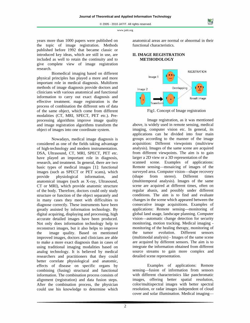
Fig1. Concept of Image registration Image registration, as it was mentioned
above, is widely used in remote sensing, medical imaging, computer vision etc. In general, its applications can be divided into four main groups according to the manner of the image acquisition: Different viewpoints (multiview analysis). Images of the same scene are acquired from different viewpoints. The aim is to gain larger a 2D view or a 3D representation of the scanned scene. Examples of applications: Remote sensing—mosaicing of images of the surveyed area. Computer vision—shape recovery (shape from stereo). Different times (multitemporal analysis). Images of the same scene are acquired at different times, often on regular abasis, and possibly under different conditions. The aim is to find and evaluate changes in the scene which appeared between the consecutive image acquisitions. Examples of applications: Remote sensing—monitoring of global land usage, landscape planning. Computer vision—automatic change detection for security monitoring, motion tracking. Medical imaging - monitoring of the healing therapy, monitoring of the tumor evolution. Different sensors (multimodal analysis) - Images of the same scene are acquired by different sensors. The aim is to integrate the information obtained from different source streams to gain more complex and detailed scene representation.
Examples of applications: Remote sensing—fusion of information from sensors with different characteristics like panchromatic images, offering better spatial resolution, color/multispectral images with better spectral resolution, or radar images independent of cloud cover and solar illumination. Medical imaging—

Journal of Theoretical and Applied Information Technology
© 2005 - 2010 JATIT. All rights reserved.
www.jatit.org
3
combination of sensors recording the anatomical body structure like magnetic resonance image (MRI), ultrasound or CT with sensors monitoring functional and metabolic body activities like positron emission tomography (PET), single photon emission computed tomography (SPECT) or magnetic resonance spectroscopy (MRS). Results can be applied, for instance, in radiotherapy and nuclear medicine. Scene to model registration. Images of a scene and a model of the scene are registered. The model can be a computer representation of the scene, for instance maps or digital elevation models (DEM) in GIS, another scene with similar content (another patient), ‘average’ specimen, etc. The aim is to localize the acquired image in the scene/model and/or to compare them. Examples of applications: Remote sensing—registration of aerial or satellite data into maps or other GIS layers. Computer vision—target template matching with real-time images, automatic quality inspection. Medical imaging— comparison of the patient’s image with digital anatomical atlases, specimen classification. Due to the diversity of images to be registered and due to various types of degradations it is impossible to design a universal method applicable to all registration tasks. Every method should take into account not only the assumed type of geometric deformation between the images but also radiometric deformations and noise corruption, required registration accuracy and application-dependent data characteristics.
The majority of the registration methods
consists of the following four steps. Feature detection. Salient and distinctive objects (closed-boundary regions, edges, contours, line intersections, corners, etc.) are manually or, preferably, automatically detected. For further processing, these features can be represented by their point representatives (centers of gravity, line endings, distinctive points), which are called control points (CPs) in the literature. Feature matching. In this step, the correspondence between the features detected in the sensed image and those detected in the reference image is established. Various feature descriptors and similarity measures along with spatial relationships among the features are used for that purpose. Transform model estimation. The type and parameters of the so-called mapping functions, aligning the sensed image with the reference image, are estimated. The parameters
of the mapping functions are computed by means of the established feature correspondence. Image resampling and transformation. The sensed image is transformed by means of the mapping functions. Image values in non-integer coordinates are computed by the appropriate interpolation technique. The implementation of each registration step has its typical problems. First, we have to decide what kind of features is appropriate for the given task. The features should be distinctive objects, which are frequently spread over the images and which are easily detectable. Usually, the physical interpretability of the features is demanded. The detected feature sets in the reference and sensed images must have enough common elements, even in situations when the images do not cover exactly the same scene or when there are object occlusions or other unexpected changes.
The detection methods should have
good localization accuracy and should not be sensitive to the assumed image degradation. In an ideal case, the algorithm should be able to detect the same features in all projections of the scene regardless of the particular image deformation. Physically corresponding features can be dissimilar due to the different imaging conditions and/or due to the different spectral sensitivity of the sensors. The choice of the feature description and similarity measure has to consider these factors. The feature descriptors should be invariant to the assumed degradations. Simultaneously, they have to be discriminable enough to be able to distinguish among different features as well as sufficiently stable so as not to be influenced by slight unexpected feature variations and noise. The matching algorithm in the space of invariants should be robust and efficient. Single features without corresponding counterparts in the other image should not affect its performance. The type of the mapping functions should be chosen according to the a priori known information about the acquisition process and expected image degradations. If no a priori information is available, the model should be flexible and general enough to handle all possible degradations which might appear. The accuracy of the feature detection method, the reliability of feature correspondence estimation, and the acceptable approximation error need to be considered too. Moreover, the decision about which differences between images have to be removed by registration has to be done. It is desirable not to remove the differences we are

Journal of Theoretical and Applied Information Technology
© 2005 - 2010 JATIT. All rights reserved.
www.jatit.org
4
searching for if the aim is a change detection. This issue is very important and extremely difficult. Finally, the choice of the appropriate type of resampling technique depends on the trade-off between the demanded accuracy of the interpolation and the computational complexity.
III. PET/CT IMAGE PROCESSING STEPS
The PET and CT data are acquired using a GE discovery STE scanner and exported as DICOM images for the post processing. The specifications and the parameters of the acquisition device and the PET and CT data are provided in [5]. The PET and CT images are aligned as described below. A. Image Segmentation
In the first step of the segmentation process, the image data is low pass filtered to smooth out the boundary of the brain in the PET as well as the CT images. To segment the brain in the PET images, adaptive thresholding is applied based on the minimum and the maximum intensity of each image, where different intensity levels correspond to different anatomical structures in the PET brain images. The range of intensities representing the brain is selected after testing the range on several datasets. By applying an adaptive threshold based on the intensity range of the brain, unwanted structures are removed from the image. Figure 2 (a)-(b)show the result of the segmentation process on the PET data. To segment the brain in the CT images, the lower and upper limit of the intensity range is defined for the brain. Starting from the center point of pixels belonging to this intensity range, a region growing technique is used and the entire brain region is filled iteratively from pixel to pixel until the threshold boundary is reached. Once all of the pixels inside the brain are iteratively marked, the rest of the image is set to zero.
(a)
(b) Fig. 2 The result of the segmentation process applied to the PET and CT images, (a) PET image and segmentation (b) PET image and segmentation B. PET/CT Image Registration
Once the preprocessing is complete, edge detection [6] is applied to the filtered images to obtain the boundaries of the PET and CT brain. The registration process is performed in the axial plane first to obtain the shift values along the x and y axes followed by the registration in the sagittal plane for the shift values along the z axis. Using the axial image, the average distance of the left and right boundary between the PET and CT brain is calculated. The magnitude of the average provides the amount of shift required for the CT, and the sign of the average value provides the direction in which the CT needs to be shifted. Then, the average distance for the top and bottom boundary between the PET and CT brain is calculated which gives the amount of shift required for the CT along the y axis. To register the PET and CT images along the z axis, sagittal plane images are used. In this case, the top boundary of the PET brain is aligned with the corresponding boundary of the CT brain. The average distance between the boundaries of the brain from two different modalities is calculated and the CT image is shifted based on the average distance of the two edges. The boundaries of the brain in the axial plane are determined by traversing the image from all sides, and the first non zero pixel found is marked as the boundary of the brain. This process is same for both the PET and CT image. For the sagittal plane, the PET and CT images are traversed only from the top and the first non zero pixel is marked as the top boundary of the brain. In brain studies, the registration along the x, y, and z axes may not be sufficient to generate a CT attenuation correction map that overlaps with the PET brain completely. Although the top boundaries of the PET and CT brain are aligned,

Journal of Theoretical and Applied Information Technology
© 2005 - 2010 JATIT. All rights reserved.
www.jatit.org
5
part of the PET brain is now projected onto the sinus cavity of the CT image. To compensate for this motion, the CT data has to be rotated in the sagittal plane to completely overlap the brain structures of the two modalities. The CT image is rotated in the sagittal plane based on the brain and sinus cavity boundary due to the attenuation error associated with the air parameter. If the PET brain boundary is projected onto the sinus cavity in the CT, then the CT image is rotated counter clockwise until the PET brain is no longer overlapping with the sinus cavity in the CT image. In the other case, if the PET brain boundary is well inside the sinus boundary of the CT, the CT image is rotated clockwise until the edges of the PET and CT brain along the sinus cavity overlap. Fig.3 shows the horizontal slices from an MRI data scan of a human cranium
Fig.3 Horizontal slices from an MRI data scan IV. RESULTS AND DISCUSSION A. Normalized Cross Correlation
We present the results for medical image registration by template matching. The experiments are conducted on gray scale brain image of size 512 x 512. The different size of template is extracted from the original image.. Fig. 4(a) shows the original image and the template of size 80 x 80 are extracted from original image is shown in Fig.4(b) and 4(c). Using normalized cross-correlation the generated image for registration in 4(d) shows the NCC plot . So we can conclude that simple cross correlation is not used for image registration..
For perfect template matching the value of maximum cross correlation coefficient is 0.99. With Noise the maximum cross correlation coefficient is not 0.99 but this value can be considered for match.
(a)
(b) (c)
050
100150
200250
0
100
200
300-0.4
-0.2
0
0.2
0.4
0.6
(d) Fig.4 (a) Original image (b), (c)Templates (d) NCC Plot
The misregistration between PET and CT is the major drawback for the CT based attenuation correction of the PET. In PET/CT studies of the human brain, the misregistration occurs mainly due to the physical motion of the patient, as most patients are unable to keep their heads still during the entire exam. The misregistration between the PET and CT brain

Journal of Theoretical and Applied Information Technology
© 2005 - 2010 JATIT. All rights reserved.
www.jatit.org
6
geometries results in erroneous attenuation correction parameters for the PET reconstruction. The reconstructed PET images obtained using the misaligned CT attenuation map show areas of abnormal metabolic activity in the brain, where the PET brain is projected outside the CT brain. One approach to address the PET/CT misalignment problem is to repeat the CT scan of the brain in case of a misalignment between the PET and the original CT. In this approach, the PET and CT is downloaded to the processing unit and overlaid. If the brains geometries of the PET are CT align properly, then this CT is used for the reconstruction of the PET. However, if the PET and CT have a significant misregistration, then the patient has to go through another CT scan of the brain. In this case, the patient is exposed to an unnecessary radiation dose that can be avoided if software-aligned PET and CT is used. Also, this process can be very time-consuming and inefficient in terms of clinical resources. In contrast, software based PET/CT brain registration has the advantage of being fast with no need for operator intervention, which increases the potential for routine clinical application. Only one CT scan is needed in this case, which is aligned with the PET and then used as the attenuation correction map. This approach also reduces the radiation dose.
The original PET image and its transformed versions are shown in Figure 5. The original image is shown in Fig,5(a). Fig. 5(b) shows the affine transformed image. The image in Fig.5(c) is obtained by applying projective transformation to the original image
(a) (b)
(c) Fig. 5 a) original image b) affine transform c) projective transform
The MR image in various views using rigid registration is shown in Figure 6. Fig. 6(a) shows the coronal view. Fig.6(b) and (c) shows the sagittal and axial views of an MR image
(a) (b)
(c) Fig. 6 a) coronal view b) sagittal view c) axial view V. CONCLUSION
Image registration is an important task
that enables comparative analysis of images acquired at different occasions. We presented a simple and fast registration method that can be applied to 3DMR images of the brain. It is based on the normalized cross correlation and spatial transformation techniques. We have described a algorithm based on Normalized Cross-Correlation (NCC) is the ideal approach to image registration by template matching. It gives perfect template matching in the given reference image. The Maximum Cross-correlation coefficient values from the NCC plot indicate the perfect template matching with noise and without noise condition. This approach gives registered image, if the sensed images do not have any rotation or scaling. We have also presented a simple and fast rigid registration method that can be applied to 3DMR images of the brain. It is based on automatic brain segmentation, automatic mid-sagittal plane location, image alignment. Finally the various transformation techniques are discussed
Journal of Theoretical and Applied Information Technology
© 2005 - 2010 JATIT. All rights reserved.
www.jatit.org
7
REFERENCES [1] M. V. Green, J. Seidel, J. J. Vaquero, E.
Jagoda, I. Lee, and W. C. Eckelman, “High resolution PET, SPECT and projection imaging in small animals,” Comput. Med. Imaging Graph., vol. 25, pp. 79–86, 2001.
[2] C. A. Pelizzari, G. T. Y. Chen, D. R.
Spelbring, R. R. Weichselbaum, and C. T. Chen, “Accurate 3- dimensional registration of CT, PET, and or MR images of the brain,” J. Comput. Assist. Tomog., vol. 13, pp. 20–26, 1989.
[3] R. P. Woods, J. C. Mazziotta, and S. R.
Cherry, “MRI-PET registration with automated algorithm,” J. Comput. Assist. Tomog., vol. 17, pp. 536–546, 1993.
[4] B. A. Ardekani, M. Braun, B. F. Hutton, I.
Kanno, and H. Iida, “A fully-automatic multimodality image registration algorithm,” J. Comput.Assist. Tomog., vol. 19, pp. 615–623, 1995.
[5] W. M. Wells, P. Viola, H. Atsumi, S.
Nakajima, and S. Kikinis, “Multi-modal volume registration by maximization of mutual information,” Med. Image Anal., vol. 1, pp. 35–51, 1996.
[6] F. Maes, A. Collignon, D. Vandermeulen, G.
Marchal, and P. Suetens, “Multimodality image
registration by maximization of mutual information,” IEEE Trans. Med. Imaging, vol. 16, pp. 187–198, 1997.
[7] C. Studholme, D. L. G. Hill, and D. J.
Hawkes, “An overlap invariant entropy measure of 3D medical image alignment,” Pattern Recognit., vol. 32, pp. 71–86, 1999.
[8] N. Hayakawa, K. Uemura, K. Ishiwata, Y. Shimada, N. Ogi, and T. Nagaoka et al., “A PET-MRI registration technique for PET studies of the rat brain,” Nucl. Med. Biol., vol. 27, pp. 121–125, 2000.
[9] C. A. Johnson, J. Seidel, R. E. Carson, W. R.
Gandler, A. Sofer, and M.V. Green et al., “Evaluation of 3D reconstruction algorithms for a small animal PET camera,” IEEE
Trans. Nucl. Sci., vol. 44, pp. 1303–1308,1997.
[10] M. Desco, J. López, C. Benito, A. Santos, P.
Domínguez, and S. Reig et al., “A multimodality
workstation in practice,” in Computer Assisted Radiology and Surgery (CARS’99), H. U. Lemke, M.W. Vannier, K. Inamura, and A. G. Farman, Eds. Amsterdam, The Netherlands: Elsevier Science, 1999, pp. 218–222.
[11] W. H. Press, S. A. Teukolsky, W. T.
Vetterling, and B. P. Flannery, Numerical Recipes in C, 2nd ed. Cambridge, U.K.: Cambridge Univ. Press, 1992, pp. 412–420.
[12] K. S. Arun, T. S. Huang, and S. D. Blostein,
“Least squares fitting of two 3D point sets,” IEEE Trans. Pattern Anal. Image Recogn., vol. 9, pp. 699–700, 1987.
[13] J. West, J. M. Fitzpatrick, M. Y. Wang, B.
M. Dawant, C. R. Maurer, and R. M. Kessler et al., “Comparison and evaluation of retrospective intermodality brain image registration techniques,” J. Comput. Assist.Tomog., vol. 21, pp. 554–566, 1997.
[14] M. Rivera, J. J. Vaquero, A. Santos, J. Ruiz-
Cabello, and F. del Pozo, “MRI visualization of small structures using improved surface coils,” Magn. Res. Imaging, vol. 16, pp. 157–166, 1998.
[15] S.L. Hartmann, M.H. Parks, P.R. Martin,
and B.M.Dawant, “Automatic 3-d segmentation of internal structures of the head in mr images using a combination of similarity and free-form transformations: Part i,methodology and validation normal subjects,” IEEETransactions on Medical Imaging, vol. 18, no. 10, 1999.

Journal of Theoretical and Applied Information Technology
© 2005 - 2010 JATIT. All rights reserved.
www.jatit.org
8
AUTHOR PROFILES:
Prof.B.Balasubramanian is a research scholar . He is working as an Associate professor in the Dept. of Biomedical Engig. at SriRamakrishna Engg. College, Coimbatore-22. . He has a bachelor’s degree in
Instrumentation & control Engineering, a master’s degree in Medical Electronics and currently pursing PhD in Biomedical Engineering field. He has 11 years of industry experience and has 5 years of teaching experience & has guided many many UG and PG projects. His research and teaching interests include Human Anatomy & Physiology, Medical Physics, Biomedical Instrumentation, Bio-signal Processing, Biomedical Image Processing, Artificial Neural Networks, Human Assist Devices and Radiological Equipments .He is a Student member of IEEE Society. He has contributed in publishing several research papers in national Journals and Conferences.
Dr. K. Porkumaran is presently Vice-Principal cum Professor and Head, EEE Department of Dr. N G P Institute of Technology, Kalapatti Road, Coimbatore 641035. He has a bachelor’s
degree in Instrumentation & control Engineering, a master’s degree in Control systems and Ph.D in Control Systems. He has 10 years of teaching experience and has guided many UG and PG projects.His research and teaching interests include Modeling and Simulation, Instrumentation System Design, Linear and Non-Linear Control Systems, Process Dynamics and Control, Neural Networks. He is a Life member of Indian Society for Technical Education (ISTE) and Associate member of Institution of Engineers (AMIE).He has published several research papers in International and national Journals. He has organized many conferences and chaired several technical session. He is the reviewer for international conferences and journals.