Embed Size (px)
Citation preview

Regina Rodman, MD Faculty Advisor: Harold Pine, MD
Grand Rounds Presentation The University of Texas Medical Branch (UTMB Health)
Department of Otolaryngology September 30, 2011
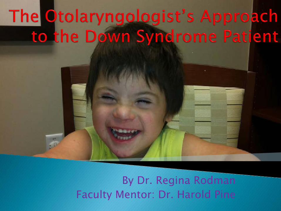
By Dr. Regina Rodman Faculty Mentor: Dr. Harold Pine

Down syndrome is the most common congenital chromosome anomaly, occurring 1/100 births.1
Studies in developed countries document sizable gains in child survival, from 25 years in 19832 to an estimated life expectancy of 50-60 years today.3 ◦ Increased life expectancy largely due to advancements in
ability to repair congenital heart defects. In past 25-30 years there has been a movement
to integrate people with developmental disabilities into the community via group homes rather than institutions.

Attention has been focused on health factors that affect the quality of the patient’s life, and affect their ability to reach full potential.
A survey of parents attending a Down syndrome association conference showed that 50% of Down syndrome children saw an Otolaryngologist regularly.4
Bottom Line: There are more people with Down syndrome in our communities, and almost all of them will require medical care.
Many of these patients have issues with the ears, nose and throat and will need to see an Otolaryngologist.

Down syndrome patients have several morphologic abnormalities that predispose them to problems with the ear, nose and throat.
◦ midface hypoplasia with malformation of the Eustachian tube5 increased number of ear infections and hearing
lossshortened palate6 ◦ relative macroglossia ◦ narrowing of the oropharynx and nasopharynx7 ◦ generalized hypotonia increases the frequency and severity of obstructive sleep
apnea8 ◦ Alterations in the paranasal sinuses ◦ abnormalities of serum immunoglobulins9 ◦ ciliary dyskinesia 10 high incidence of chronic sinusitis

It is likely that the practicing Otolaryngologist will encounter many Down syndrome children, and appropriate treatment can have a significant impact on the quality of life of these patients
Many of the conditions of the Down syndrome child can be treated by the general otolaryngologist, or the general pediatric otolaryngologist.

Absent or hypoplastic nasal bones First trimester: ◦ Present or Not
Second trimester: ◦ nasal bone length ◦ hypoplastic = shorter than 2.5 mm11
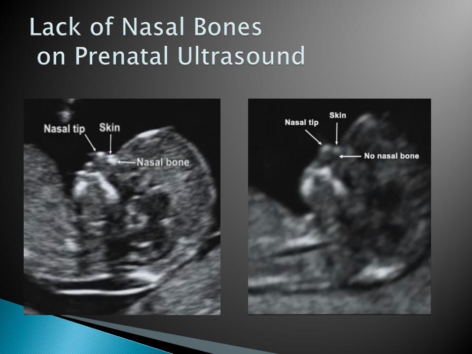

Very important to have sonographers who are formally trained to perform such evaluation. Differences 16.7% reported absent nasal bones in sonographers
without training or quality assurance, to 70% prevalence in studies where sonographers were appropriately trained. 12
Increases ability to detect Down syndrome It has been shown that using sonographic markers of prenasal
thickness, nasal bone length, and nuchal skin fold, increased the detection of Down syndrome in the second trimester by 19-23% compared to serum markers alone.13
Isolated finding of absent nasal bones likely does NOT indicate Down syndrome In one study, 6/6 100% of fetuses with the isolated finding of
absent nasal bones had normal karyotypes, however. 6/8 (75%) of the patients who had absent nasal bones in addition to other abnormal ultrasound findings, did have Down syndrome.14

Stenotic Ear Canals Increased incidence of secretory otitis media Chronic ear disease Secondary hearing loss Ossicular abnormalities Inner ear dysplasia Hearing loss
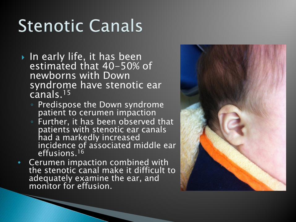
In early life, it has been estimated that 40-50% of newborns with Down syndrome have stenotic ear canals.15 ◦ Predispose the Down syndrome
patient to cerumen impaction ◦ Further, it has been observed that
patients with stenotic ear canals had a markedly increased incidence of associated middle ear effusions.16
• Cerumen impaction combined with the stenotic canal make it difficult to adequately examine the ear, and monitor for effusion.

Recommendation: Down syndrome children should establish care with an Otolaryngologist early in life, as the patient will frequently require microscopic exams and cerumen disimpaction
of the canals under microscopy.17
It is also recommended that those with canal stenosis continue to follow up every 3 months with the Otolaryngologist for evaluation of the middle ear space, to ensure that there is no cerumen impaction, and to monitor for middle ear fluid and infection.17

Experience reported by Cincinnati Children’s Hospital is that the majority of children with stenotic canals grow with age, and by years 2 or 3 this canal is no longer a obstacle to accurate examination. 18
Each patient should be followed regularly by the Otolaryngologist until is it clear that the patient is of appropriate age and size so that s/he is at low risk of serous otitis media and can be easily examined.

There is a high prevalence of serous otitis media in Down syndrome children.19,20
There are several etiologic factors that
explain this increased incidence of ear disease. ◦ Decreased immunity ◦ Midface hypoplasia ◦ Eustacian tube dysfunction General hypotonia Cartilage abnormalities
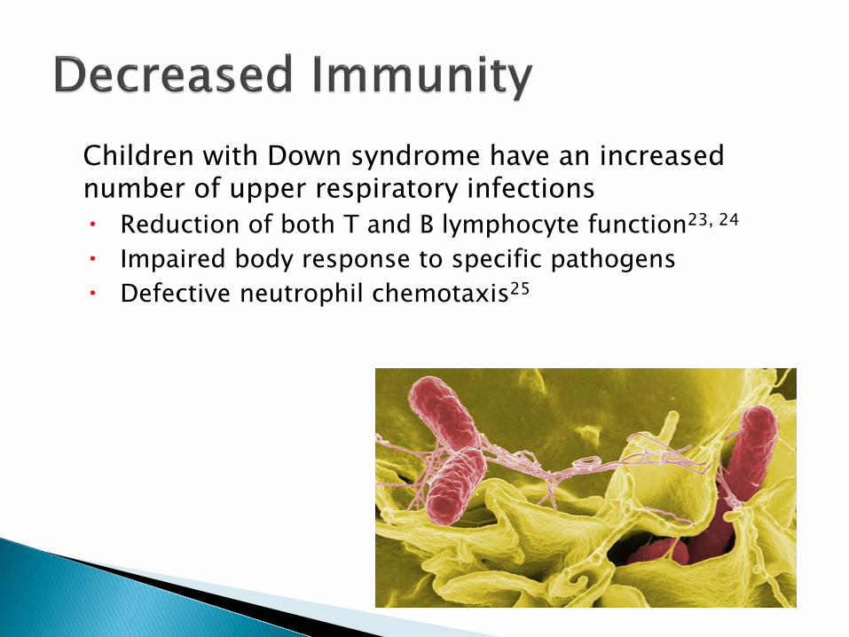
Children with Down syndrome have an increased number of upper respiratory infections Reduction of both T and B lymphocyte function23, 24 Impaired body response to specific pathogens Defective neutrophil chemotaxis25

Midface hypoplasia affects the nasopharynx and the eustachian tube openings.
Bony confines of the nasopharynx are smaller in Down syndrome children.26 ◦ Normal sized soft tissue of the
nasopharynx can only occupy this space at the expense of the airway.
◦ This decrease in post nasal space may cause even small to medium sized adenoids to give rise to Eustachian tube dysfunction.

It has also been demonstrated that the eustachian tube in these patients are extremely small, and collapsed in several portions. 27 ◦ Cartilage cell density in Down syndrome individuals
was decreased at all ages, predisposing the canal to collapse.28 ◦ Generalized hypotonia of these patients can lead to decreased function of the tensor veli palatini muscle of the palate, which is responsible for opening the Eustachian tube29.

Hearing Loss
is more frequent in children with Down
syndrome than in healthy children.

In studies which have conducted audio screening on randomly selected children with Down syndrome, results have shown that 50-90% of Down syndrome children have a hearing impairment.30, 31,32
In one study where 90% of children were found to have at least a mild-moderate hearing loss, only a small percentage of parents (15.2%) reported a positive history of hearing loss.34

Hearing impairment may be masked in patients with intellectual impairment, as speech delays, and lack of response to verbal cues may be attributed to mental retardation.35
Detection of this loss is critical, as it is agreed that the detrimental effects of hearing loss on language development are believed to be greater for those children with learning disabilities compared to children without mental retardation.36

It is recommended that all children with Down syndrome go for routine audiologic screening. AAP recs: ◦ Audiologic testing at birth (ABR/OAE)
◦ then every 6 months up to age 3 years (play
audiometry-debated) ◦ with annual testing after 3 years of age, or when
ear specific pure tone audiometry may be obtained.37,38

The benefits of pressure equalization tubes in Down syndrome patients has been debated over the past decade.
While proven very effective in the general population to decrease the duration of middle ear effusion in the Down syndrome patient, results have been varied.
Many studies published state that they are not effective, however, one can find fault in methodology of each study. ◦ Mean age of 5.439 and 8.240 when placing first set of tubes ◦ Tubes not replaced after extrusion39

Cincinnati Children’s Hospital:41 ◦ Enrolled before the age of 2 years ◦ Followed by an Otolaryngologist every 3-6 months,
depending on the degree of canal stenosis present. ◦ All children were treated for chronic ear infections
and middle ear effusions by placement of PE tubes ◦ Replacement tubes as needed. ◦ At the end of the study two years
later, 93% of the patients had normal hearing.
Recommendation: If surgical management is the chosen path, results must be closely monitored, and the surgeon must be aggressive with re-intervention.

Otolaryngologist must counsel the parents of the patient of the possible increased risks in these patients ◦ cholesteatoma ◦ persistent perforation ◦ atelectatic tympanic membranes ◦ persistent otorrhea
Parents should also know ◦ PE tubes may be placed earlier in the child’s life ◦ expect that the child may need multiple set of tubes throughout
childhood, even into adulthood
Parents should be counseled that reinsertion of tubes is a continuation of treatment, rather than failure of the original attempt.
Parents should be counseled on the importance of follow up with the audiologist and the Otolaryngologist, and the need for aggressive re-intervention.

In addition to middle ear effusions, a component of conductive loss may be caused by abnormalities of the mastoid, or abnormality of the ossicular chain. ◦ Of 107 DS patients only 60% of CHL could be explained by middle
ear effusions or tympanic membrane perforations. 42
Mastoid ◦ Neuroimaging of 59 patients with Down syndrome and found
nonaeration or underaeration of mastoids in the majority (74%) of cases. 43
◦ Lateral cervical spine films study showed 63% of the mastoids examined on with sclerosis and poor aeration. 44
Whether this increase in density is caused by a mastoid
infection that occurred during maximum growth years, or a congenital component is yet to be determined.

Study of post mortem temporal bones43 ossicular abnoormalities attributed to chronic
disease: ◦ erosion of the long process of the incus ◦ erosion manubrium of the malleus ◦ erosion superstructure of the stapes
Interestingly, they also had some findings that
were attributed to congenital deformities: ◦ malformation of the stapes ◦ dehiscence of the facial nerve.
These findings should be considered in children who have a persistent conductive hearing loss despite maximal management of middle ear effusion.

Down syndrome patient predisposes them to cholesteotoma and erosion of the ossicular chain. 45
Ossicular chain reconstruction techniques did significantly improve hearing in this population.
Parents should be counseled that resolution of disease may require several operations. ◦ Largest to date study; 64% of ears were managed with a single surgery,
and 89% of ears were controlled with two surgeries or less.46
Similarly, parents should also be counseled that canal wall
preservation techniques may not be appropriate for this population. ◦ 70%-89% of patients required a canal wall down procedure in a study of 9
patients.45,46

Down syndrome children also have higher rates of mixed hearing loss and sensorineural hearing loss compared to other children, estimated to be 4-9%.47,48
Chronic middle ear disease vs. true neural loss?
The true incidence of sensorineural hearing loss
in Down syndrome children will be determined as future studies evaluate hearing in children who have been aided by early and aggressive care of their middle ear disease.

Uniformly small inner ear structures compared to controls47 hypoplastic cochlea, critically smaller cochlear nerve canal narrowed internal auditory canal hypoplastic lateral semicircular canal with small bony
island hypoplastic vestibules.

BAHA: 18/81 BAHA centers in UK performing surgery on Down syndrome patients. 43 patients, all but one wore the BAHA on a daily basis49 Survey showed 27/28 were very pleased or pleased with
the results50 Glasgow Children’s Benefit Inventory (emotion, physical
health, learning and vitality) showed a significant benefit in all categories in Down syndrome children.
Higher rate of complications 20-50%, all soft tissue problems. ◦ attributed to the fact that patients with learning difficulties have a
tendency to interfere with the area, leading to disturbances of the dressing, sutures and possible graft failure.
◦ A solution to this was proposed, where following BAHA abutment a perforated thermoplastic cage is sewn over the surgical

Cochlear Implants Four patients with Down syndrome had received
cochlear implants in the UK and Ireland.51 ◦ all congenital deafness ◦ all four of the patients had middle ear disease pre
operatively, with two patients requiring PE tubes. ◦ none of the cases had any complications associated with
otitis media. ◦ none had intraoperative difficulties due to anatomy
The outcomes of these four implanted patients have been modest gains in auditory performance, ◦ the eldest child, who has had the implant the longest,
showing the most improvement. As more patients with Down syndrome become
candidates for cochlear implants, patients and families must be counseled about expectations. ◦ abnormalities in the temporal bone of a child with Down
syndrome that may increase the risk of complications. ◦ outcomes may not be as good as children without additional
disabilities, as learning and communication difficulties may prolong the rehabilitation.
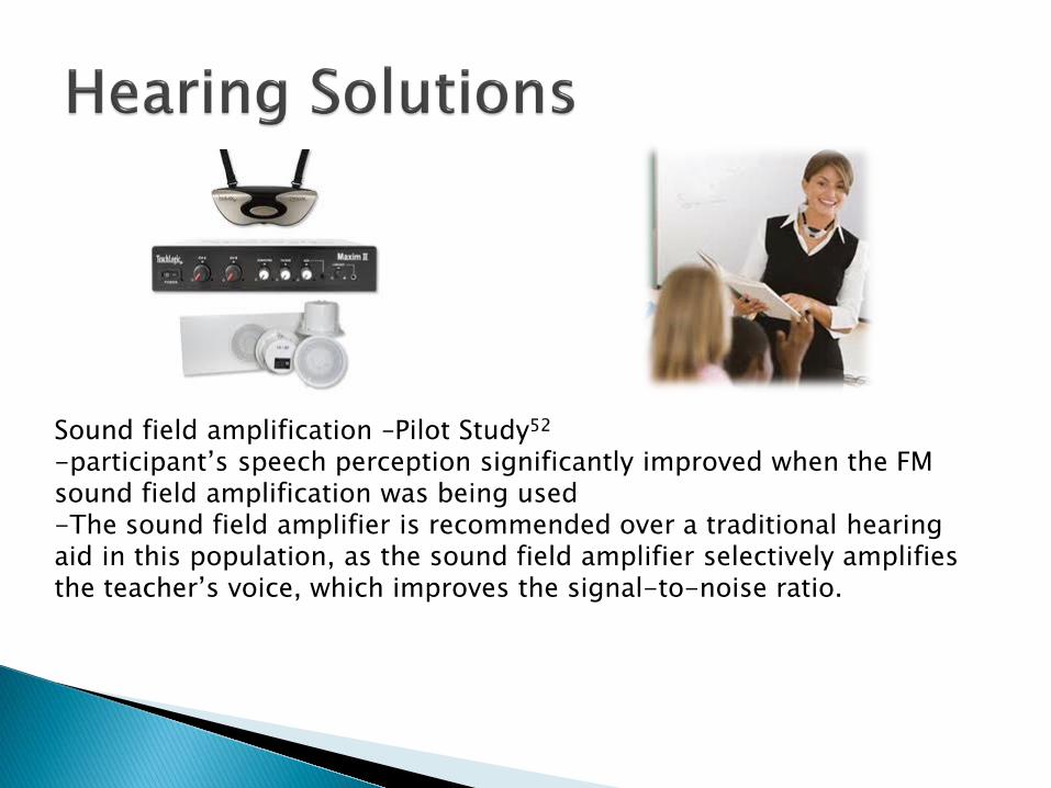
Sound field amplification –Pilot Study52 -participant’s speech perception significantly improved when the FM sound field amplification was being used -The sound field amplifier is recommended over a traditional hearing aid in this population, as the sound field amplifier selectively amplifies the teacher’s voice, which improves the signal-to-noise ratio.

Obstructive sleep apnea syndrome (OSAS) ◦ 0.7% to 2.0% of the general pediatric population53,54 ◦ prevalence in pediatric Down syndrome patients has been estimated at 77-80%55,56
Children with Down syndrome have many predisposing factors of OSAS.55,56,57 ◦ midfacial and mandibular hypoplasia ◦ Relative macroglossia ◦ Glossoptosis (downward displacement or retraction of tongue) ◦ abnormally small upper airway with superficially positioned tonsils and
relative tonsillar and adenoidal encroachment ◦ increased secretions ◦ increased incidence of lower respiratory tract anomalies ◦ obesity ◦ generalized hypotonia resultant collapse of the airway during inspiration.

Relative macroglossia Increased secretions

In Down syndrome patients with relatively narrowed nasal airway, even normal adenoid growth can encroach the airway
OSAS

The effect of obesity on OSAS in DS patients is debated. 2 studies looked at BMI and OSAS
1) The BMI z-score, was higher in the OSA group (2.9) compared to the non OSA group (1.4). However, there were some children with very high BMI z scores* who did not have OSA and some non obsese dhildren with severe sleep apnea.
2) Another study where 91% of patients were not obsese, yet 97% had OSA.
. Based on these results, OSAS is likely a multi-factorial disease with several contributing factors in these patients.
Still, BMI is a modifiable risk factor, and the results of the above studies suggest that weight reduction may show some benefit in the management of OSA in Down syndrome children.
*The BMI z-score, also called BMI standard deviation score, are measures of relative weight adjusted for child age and sex

◦ a worsened trend in word reading speed62 ◦ visual attention ◦ verbal fluency ◦ neurodevelopmental problems 60 daytime somnolence behavioral disturbances school failure developmental delay.
◦ pulmonary hypertension61 ◦ cor pulmonale ◦ congestive heart failure63 Chronic intermittent hypoxemia respiratory acidosis Although we know of no published studies specifically examining these
effects of neurodevelopment and learning on children with Down syndrome, it is logical to expect that this population, who is already predisposed to learning delay and difficulty in school, would be significantly impaired by the effects of sleep apnea. Similarly, children with Down syndrome are predisposed to congenital cardiac anomalies, and are more likely to have pulmonary hypertension than are non syndromic children with the same cardiac anomalies. Again, this may be exacerbated or worsened by OSAS.

He needs a sleep study!

Because of the unreliability of parental reporting, the high prevalence of OSAS in this population, and the negative effects of sleep apnea, it is recommended that all children with Down syndrome between the ages of 3 and 4 years, go for objective testing using full overnight polysomnography for a baseline study.56
◦ 24/35 (69%) parents reported no sleep problems 13/24 (54%) of these did indeed have obstructive sleep apnea on PSG.56
◦ 18/30 (60%) of the children with negative histories also had abnormal polysomnograms. 58

Before entertaining tonsil and adenoid surgery in the Down syndrome population, it is now recommended that a pre-operative polysomnography is obtained.59

Of those 16 patients, all 16 (100%) had abnormal overnight polysomnograms, but the nap study was less sensitive in detecting OSAS, with only 12 (75%) of these patients
having abnormal nap studies.
The degrees of hypoventilation and desaturations were significantly higher in the overnight studies, and thus the nap studies under estimated abnormalities.58
Decreased efficacy in curing sleep apnea in Down syndrome patients ◦ 30-50% still require CPAP, further surgery, or
tracheostomy at a later date.63 When Down syndrome patients and age and
BMI matched controls compared64 ◦ In the Down syndrome group, the AHI showed
improvement after surgery, but was not as significant as the improvement in the control group. ◦ The REM-AHI and lowest SaO2 did not show
significant change in the Down syndrome children

May still be recommended if the parents are given appropriate information about expectations and can give informed consent. ◦ parental expectations is not for complete cure, but for
improvement in symptoms. ◦ may decrease the need for CPAP or oxygen, lower the
setting ◦ It is known that CPAP compliance is low in normal adults65,
and it is even more difficult for children with a developmental disability, who do not understand the disease or the treatment, to be fully compliant.
Therefore, a likelihood of reducing dependence on CPAP, and/or
a 25-50% chance of being weaned completely from a nighttime breathing apparatus, may make the surgery a good option for many Down syndrome patients.

Increased incidence of VPI and hyper nasal speech ◦ 1:2000- 1:3000 rate of complication seen in the general
population ◦ One survey found 2/74 Down syndrome patients developed
transient velopharyngeal insufficiency, and 2/74 other patients hypernasal speech68
◦ were found to have both structural and functional causes of hypernasality69
◦ Structural: high arched, and short soft palate ◦ Functional: hypotonia, slowed motor learning, and oral-motor
developmental delay
Increased post operative complications in Down syndrome children compared to controls67,70
◦ including increased hospital stay ◦ fivefold increase in respiratory complications requiring
intervention ◦ increased duration until adequate PO intake
Recommendation: It is therefore recommended that all Down syndrome patients be admitted to the hospital after adenotonsillectomy for observation.70

Down syndrome patients who underwent T&A plus lateral pharyngoplasty as initial therapy for OSAS. ◦ They found no significant difference between the groups ◦ 48% in the T&A only group to have persistently abnormal
AHI post operatively ◦ 63% in the T&A plus lateral
pharyngoplasty group to have abnormal AHI after surgery.71


Cine MRI was originally used by neurosurgeons to evaluate CSF flow in real time.
Placed supine on the table, sleep is induced by sleep deprivation, spontaneous sleep or sedation, most accurate when snoring
The cine MRI obtains multiple sagittal and axial images in real time, creating a dynamic, three dimensional video of the airway collapse.
This view can appreciate multiple levels of collapse, and it has also been noted that adenoid enlargement and nasopharyngeal obstruction are more prominent on cine MRI.84

One study of Down syndrome patients found that 17.6% of Down syndrome children were reported to have a continual runny nose.39 The narrowing of their nasopharynx adenoid tissue to obstruct the airway
Several studies have shown abnormalities in the immunoglobulin (Ig) levels in Down syndrome23

Treatment similar to the general population (hope you were listening to Dr. Rose) ◦ nasal irrigation ◦ nasal steroids ◦ Antihistamines ◦ decongestants ◦ antibiotics as needed
In patients whose sinusitis and rhinitis are not aided by medical management surgical intervention may be warranted.
Flexible nasopharyngoscopy should be preformed to look for adenoid hypertrophy, which may be obstructing the choanae. This should be done even if the patient has already had an adenoidectomy, as regrowth of adenoid tissue is more common in the Down syndrome patient compared to the general population.84

Orthodontic procedure used to correct a narrow transverse maxillary diameter.
The two maxillary bones are separated at the mid-palatal structure using and intraoral screw mechanism. This leads to a widening of the perimeter of the arch.
Although the major effect is noticed clinically in the area of dentition, the transverse enlargement of the apical bone also affects nasal width.

Usually, these changes result in altered nasal airway flow, with consequently improved nasal ventilation.
In this study 13 patients with Down syndrome used the intraoral maxillary expansion device and 10 patients with Down syndrome did not.
The results showed a significant increase in the total nasal volume, which persisted five months after removal of the device.
The study further reported a significantly improved incidence of 73 ◦ acute otitis media ◦ Adenoiditis and tonsillitis ◦ Snoring ◦ mouth breathing ◦ Restlessness ◦ word articulation ◦ tongue protrusion ◦ facial aesthetics

Laryngomalacia ◦ Down syndrome patients have generalized hypotonia,
leading to flaccidity of the supraglottis, anatomical changes in the epiglottis, arytenoids, and aryepiglottic folds and a high prevalence of GERD.75
Multiple sites of obstruction were seen in 38% of
cases.76 ◦ Tracheomalacia was found in 23/39 (59%),
laryngomalacia in 28% ◦ macroglossia (26% ◦ subglottic stenosis (23%) ◦ congenital tracheal stenosis (5%)

It is important that these cases by managed by a tertiary care center that is familiar with treating the Down syndrome patient and is practiced at comprehensive airway evaluations and a systemic approach to surgery in the Down syndrome child.
Even at centers with high volume of Down syndrome patients, parents should be counseled pre-operatively about the possibility of residual symptoms in children with severe obstruction.

Believed to be more prevalent in the Down syndrome population ◦ 4% of LTRs were DS vs 0.1%-0.15% prevalence78
Congenital narrowing of the subglottis as well acquired deformities.
Same risk factor for SGS: trauma to the subglottis79 ◦ DS child has a higher rate of major surgery (i.e. cardiac
anomalies) (and intubation) ◦ Severe respiratory infections requiring intubation ◦ Usually occurs at a young age, which predisposes them
to subglottic injury and subsequently, subglottic stenosis.

A key factor in preventing subglottic injury, is choosing an appropriate sized endotracheal tube, as age-appropriate endotracheal tubes are too large for the Down syndrome patient. 80,81
◦ A prospective study evaluated 74 children, 42 with Down syndrome, 32 healthy controls, none of whom had wheezing, stridor, or previous intubation before surgery.
◦ The Down syndrome children required an ETT 2-3 sizes smaller than age matched controls.
◦ Further, MRI showed that tracheal diameter was smaller in Down syndrome compared to controls, demonstrating that the overall smaller airway size is not limited to the subglottis, but includes a smaller trachea.
Recommendation: The recommendations based on this prospective
study are that endotracheal tubes at least two sizes smaller should be initially used for intubation in children with Down syndrome.98 To ensure proper sized tube placement, it is critical to confirm the fit of the tube after it is placed by checking for an audible air leak at an inspiratory pressure between 10-30 cm H2O.81

Although AAI is one of the most well known and feared problems associated with Down syndrome, reports of complications associated with AAI are few, and current guidelines and recommendations of airway management and positioning during surgery for the patients are vague.
Incidence of AAI seen on radiography is 14-20% 82, 83 Incidence of symptomatic AAI is much less with only
a few case reports throughout the literature. ◦ 404 patients with Down syndrome that found 14.6% to have
radiographic evidence of AAI, only 1.5% of the patients had symptoms.83

Atlanto axial instability, also called atlantoaxial sublaxation, is the result of increased mobility at the articulation of the first and second cervical vertebrae. A review reports that craniocervical instability, most commonly altantoaxial instability is the result of generalized ligamentous laxity, involving any of the three ligaments of the C1C2 joint.

The radiographic definition of AAI is made by measuring the distance between the anterior surface of the dens to the posterior surface of the tubercle of C1. An anterior atlantodental interval of greater than 4.5 indicates abnormal instability.
Asymptomatic AAI is that which is diagnosed by radiography, but the patient has no neurologic symptoms.
The patient who is symptomatic may experience easy fatiguability, abnormal gait and difficulty walking, neck pain, limited neck mobility, torticollis, clumsiness, lack of coordination, sensory deficits, spasticity, hyperreflexia, clonus, incontinence, and extensor-plantar reflex.


The issue of atlanto axial instability came into wide recognition after the Special Olympics introduced a requirement in 1983 that all individuals with Down syndrome have a lateral neck radiograph before participating, and that those with evidence of instability be banned from certain activities. This was further supported by a statement by the American Academy of Pediatrics in 1984. 84
Radiographs of the neck to be unreliable at identifying
atlantoaxial sublaxation.
A review of case reports of individuals who have experienced catastrophic injury to the spinal cord by the AAP determined that trauma rarely causes the initial symptoms or progression of symptoms, and that nearly all the individuals who have experienced catastrophic injury to the spinal cord have had weeks to years of preceeding, less severe, neurologic abnormalities.83-88

In a 1995 statement, the AAP retired their previous statement, and revised their recommendations to state that evaluation and physical exam by a physician who has cared for the patient longitudinally, is a greater priority than obtaining radiographs when determining a Down syndrome patient’s eligibility for participation in sports.
It is now recommended that the pediatrician perform a careful history and physical examination with attention to myelopathic signs and symptoms at every well-child visit, or when symptoms possibly attributable to spinal cord impingement are reported.38

Because the poor ability of radiographs to detect clinical neurological compromise, it is imperative that every patient have a thorough neurologic exam pre operatively, preferably by a physician who knows the patient well.
While gentle rotation of the head for ear surgery is likely safe, it is still recommended that the patient’s head be supported throughout the procedure, and that extremes of neck positioning be avoided.
When performing tonsillectomy, the patient should remain in a relatively neutral position..

Ear ◦ Routine exam by audiologist and Otolaryngologist ◦ Aggressive intervention and re intervention for OME
Throat ◦ All DS patients should get sleep study around age 3 ◦ T&A still first line for OSA
Airway ◦ Use ETT 2 sizes smaller, check for air leak
Spine ◦ All patients should have pre-op neurologic exam ◦ Support the head at all time

Although these children have disease that is more complex and difficult to treat than the non syndromic pediatric patient, the general Otolaryngologist can have a profound impact on the Down syndrome patient.
Proper management of ear, nose, and throat disorders by the Otolarygologist can support the Down syndrome child’s physical, emotional, and educational development.

1. Down syndrome prevalence at birth: United States, 1983-1990. MMWR. 1994;43:617-622.
2. Roizen NJ, Patterson D, Down’s Syndrome. Lancet. 2003;361:1281-9.
3. Bittles AH, Bower C, Hussain R, Glasson EJ. The four ages of Down Syndrome. Eur J Public Health. 2007;17(2):221-225.
4. Hans PS, Belloso A, Sheehan PZ. Parental Satisfaction with health services provided to children with Down Syndrome in north-west England:an ENT perspective. The Journal of Laryngology and Otology. 2006; 121,382-386.
5. Shibahara Y, Sando I. Congenital anomalies of the Eustachian tube in children with Down Syndrome. Ann Otol Rhinol Laryngol. 1989;98:543-547.
6. Austin J, Preger l, Siris E, taybi H. Short hard palate in newborn: roentgen sign of mongolism. Radiology. 1969;92:775-556.
7. Strome M. Obstructive Sleep Apnea in Down’s Syndrome: a surgical approach. Laryngoscope. 1986;96:1340-1342.
8. Miller J, Capusten B, Lampard R. Changes at the base of the skull and cervical spine in Down Syndrome. J Can Assoc Radiol . 1986;37:85-89.
9. Gershwin ME, Crinella FM, Castles JJ, Trent JKT. Immunologic Characteristics of Down’s Syndrome. J Ment Defic. 1977;21:237
10. Kovesi T, Sinclair B, MacCormick J, Matsinger M, Carpenter B. Primary ciliary dyskinesia associated with Down Syndrome. Chest. 2000;117:1207-1209.
11. Cicero S, Sonek JD, McKenna DS, Croom CS, Johnson L, Nicolaides KH. Nasal bone hypoplasia in trisomy 21 at 15-22 weeks gestation. Ultrasound Obstet Gynecol. 2003; 21(1):15-18. 12. Prefumo F, Sairam S, Bhide A, Thilaganthan B. First trimester nuchal translucency, nasal bones, and trisomy 21 in selected and unselected populations. American Journal Obstetrics and Gynecology. 2006;194:828-833. 13. Miguelez J, Moskovitch M, Cuckle H, Zugaib M, et al. Model-Predicted performance of second-trimester Down syndrome screening with sonographic prenasal thickness. Journal of Ultrasound Medicine. 2010;29:1791-1747. 14. Ting YH, Lao T, Lau TK, Chung MK, Leung TY. Isolated absent of hypoplastic nasal bone in the second trimester fetus: is amniocentesis necessary? The Journal of Maternal-Fetal and Neonatal Medicine. 15. Strome M. Down’s Syndrome-A modern otorhinolaryngological perspective. Laryngoscope 1981. 42:1581-1594. 17. Shott SR. Down Syndrome: Common Otolaryngologic manifestations. Am J Med Genet Part C Semin Med Genet 2006. 142C:131-140. 18. Shott SR. Down Syndrome: Common Otolaryngologic manifestations. Am J Med Genet Part C Semin Med Genet 2006. 142C:131-140. 19. Cunningham C, McArthur K. Hearing Loss and treatment in young Downs syndrome children. Child:Care, Health and Development. 1982;7:357-374. 20. Selikowitz M. Health problems and health checks in school aged children with Down syndrome. Journal of Pediatrics and Child Health 1992;28:383-386.

21. Cunningham C, McArthur K. Hearing Loss and treatment in young Downs syndrome children. Child:Care, Health and Development. 1982;7:357-374.
22. Selikowitz M. Health problems and health checks in school aged children with Down syndrome. Journal of Pediatrics and Child Health 1992;28:383-386.
23. Gershwin ME, Crinella FM, Castels JJ, Trent JK. Immunological characteristics of Down’s syndrome. J of Ment Defic Res. 1977;21:237-248.
24. Chaushu S, Yefenof E, Becker A, Shapira J et al. A link between parotid salivary Ig level and recurrent respiratory infections in young Down’s syndrome patients. Oral Microbiol Immunol 2002;17:172-176.
25. Nespoli L, Burgio GR, Ugazio AG, Maccario R. Immunological feature of Down’s syndrome: a review. J Intellect Disabil Res. 1993;37:543-551.
26. Brown PM, Lewis GT, Parker AJ, Maw A. The skull base and nasopharynx in relation in Down’s syndrome in relation to hearing impairment. Clinical Otolaryngology. 1989;14:241-246.
27. Shibahara Y, Sando I. Congenital anomalies of the Eustachian tube in Down syndrome. Histopathologic case report. Ann Otol Rhinol Laryngol. 1989;Jul;98(7):543-7.
28. Yamaguchi N, Sando I, Hashida Y, Takahashi H, Matsune S, Histologic study of Eustachian tube ccartilage with and without congenital anomalies: A preliminary study. Ann Otol Rhinol Laryngol 99:984-987.
29. Strome M. Down’s Syndrome-A modern otorhinolaryngological perspective. Laryngoscope 1981. 42:1581-1594.
30. Krecicki T, Zalesska-Krecicka M, ubiak K, Gawron W. Brain auditory potentials in children with Down syndrome. Int J Pediatr Otorhinolaryngology. 2005;69(5):615-620.

31. Hess C, Rosanowski F, Eysholdt U, Schuster M. Hearing impairment in children and adolescents with Down syndrome. HNO. 2006;54(3):227-32.
32. Harigai S. Longitudinal studies in hearing-impaired children with Down syndrome. Nippon Jibiinkoka Gakkai Kaiho. 1994;97(12):2208-18.
33. Cunningham C. Hearing loss and treatment in young Down syndrome children. Child care, health and development. 1981;7(6):357-74. 34. McPherson B, Lai S, Leung K, Ng I. Hearing Loss in Chinese school children with Down syndrome. Int J Pediatric Otorhinolaryngology. 2007;71:1905-1915.
35. Balkany TJ, Dows MP, Jafek BW, Krajicek MJ. Hearing loss in Downs syndrome: a treatable handicap more common than generally recognized. Clinical Pediatrics. 1979;18:116-118.
36. Downs MP. The hearing of Downs individuals. Seminars in Speech, Language and Hearing. 1980;1:25-37.
37. Cohen WI. Health care guidelines for individuals with Down syndrome. Down syndrome Q 4:1-16.
38. Bull M, AAP Committee on Genetics. AAP Clinical Report: Health Supervision for children with Down syndrome. Pediatrics 128(2):393-406.
39. Iino Y, Imamura Y, Harigai S, Tanaka Y. Efficacy of tympanostomy tube insertion for otitis media with effusion in children with Down Syndrome. Int J of Pediatric OtoRhinoLaryngology. 1999;49:143-149.
40. Selikowitz M. Short term efficacy of tympanostomy tubes for secretory otitis media in children with Down syndrome. Developmental Medicine and Child Neurology. 1993;35:511-515
Works Cited

41. Shott SR, Joseph A, Heithaus D. Hearing loss in children with Down syndrome. Int J Pediatr Otorhinolaryngology. 2001;61:199-205. 42. Blaser S, Propst E, Martin D, Feigenbaum A, et al. Inner ear dysplasia is common in children with Down syndrome (trisomy 21). The Laryngoscope 2006;116:2113-2119. 43. Glass RB, Yousefzadeh DK, Roizen NJ. Mastoid abnormalities in Down syndrome. Pediatric Radiology. 1989;19;311-312. 44. Balkany T, Mischke R, Downs M, Jafek B. Ossicular abnormalities in Downs syndrome. Otolaryngology Head and Neck Surgery. 1979;87:372-384. 45. O’Malley M, Kaylie D, Himbergen D, Bennett M, Jacksone G. Chronic ear surgery in patients with syndromes and multiple congenital malformations. The Laryngoscope 2007;117:1993-1998. 46. Bacciu A, Pasanisi E, Vincenti V, et al. Surgical treatment of middle ear cholesteotoma in children with Down syndrome. Otol Nerotol 2005;26:1007-1010. 47. Hess C, Rosanowski F, Eysholdt U, Schuster M. Hearing impairment in children and adolescents with Down syndrome. HNO. 2006;54(3):227-32.
48. Cunningham C. Hearing loss and treatment in young Down syndrome children. Child care, health and development. 1981;7(6):357-74 49. Sheehan P, Hans P. IK and Ireland experience of bone anchored hearing adis (BAHA) in individuals with Down syndrome. International Journal of Pediatric Otorhinolaryngology. 2006;70:981-986. 50. McDremott A, Williams J, Kuo M, Peid A, Proops D. The role of bone anchored hearing aides in children with Down syndrome. International Journal of Pediatric Otorhinolaryngology 2008;72:751-757. 51. Hans PS, England R, Prowse S, Young E, Sheehan PZ. UK and Ireland experience of cochlear implants in children with Down syndrome. International Journal of Pediatric Otorhinolaryngology. 2010;74;260-264 52. . Bennetts L, Flynn M. Improving the classroom listening skills of children with Down syndrome by using sound field amplification. Down Syndrome Research and Practice 2002;8(1):19-24. 53. Ali NJ, Pitson DJ, Strandling JR. Snoring, sleep disturbance, and behavior in 4-5 year olds. Arch Dis Child 1993;68:360-366. 54. Gislason T, Benediktsdottir B. Snoring, apneic episodes, and nocturnal hypoxemia among children 6 months to 6 years old. Chest. 1995; 107:963-966.

55.Marcus C, Keens T, Bautista D, Pechmann W, Davidson S. Obstructive Sleep Apnea in Children with Down Syndrome. Pediatrics. 1991;88(1):132-139
56. Shott S, Amin R, Chini B, Heubi C, Hotze S, Akers R. Obstructive sleep apnea: should all children with Down syndrome be tested? Arch Otolaryngol Head Neck Surg 2006;132:432-436.
57. Fink GB, Madaus WK, Walker GF. A quantitative study of the face in Downs syndrome. Am Journal Orthod. 1975;67:540-553.
57. Marcus C, Keens T, Bautista D, Pechmann W, Davidson S. Obstructive Sleep Apnea in Children with Down Syndrome. Pediatrics. 1991;88(1):132-139
58.Roland P, Rosenfield R, Brooks L, Friedman N et al. Polysomnogrpahy for sleep disordered breathing prior to tonsillectomy in children. Otolaryngology- Head and Neck Surgery. 2011;145:(1):S1-S15.
60. Beebe DW, Wells CT, Jeffries J et al. Neuropyschological effects of pediatric obstructive sleep apea. J Int Neuropsychol. 2004;10:962-975.
61. Brouillette RT, Fernbach SK, Hunt CE. Obstructive sleep apnea in infants and children. J Pediatric. 1982;100-31-40.
62. Perkin RM, Anas NG. Pulmonary hypertension in pediatric patients. J Pediatrics. 1984;105:511-522
63. Hals J, hagemo PS, Thaulow E, Sorland SJ. Pulmonary vascular resistance in complete atrioventricular septal defect: a comparison between children with and without Downs syndrome. Acta Pediatrica. 1993;82;595-598.

64. Donnelly L, Shott S, LaRose C, Chini B, Amin R. Causes of persistent obstructive sleep apnea despite previous tonsillectomy and adenoidectomy in children with Down syndrome as depicted on static and dynamic cine MRI. Am Journal Roentgenology 1994;183:175-181. 65. Shete M, Stocks R, Sebelik M, Schoumacher R. Effects of adeno-tonsillectomy on polysomnography patterns in Down syndrome children with obstructive sleep apnea: A comparative study with children without Down syndrome. Int Journal of Pediatric Otorhinolaryngology. 2010; 74:241-244. 66.Wolkove N, Blatzan M, Kamel H, Dabrusin R, Palayew M. Long term compliance with continuous positive pressure airway pressure in parents with obstructive sleep apnea. Can Respir Journal; 2008;15(7):365-369. 67. Kavanagn K, Kahane J, Kordan B. Risks and benefits of adenotonsillectomy for children with Down syndrome. American Journal of Mental Deficiency 1986;91(1)22-29 68. Gelder LV. Open nasal speech following adenoidectomy and tonsillectomy. Journal of Communications Disorders, 1974:4:263-267 69. Gibb AG. Hypernasality (rhinolalia aperta) following tonsil and adenoid removal. Journal of Laryngology and Otology 1958;72:433-451 70.Goldstein N, Armfield D, Kingsley L, Borland L, Alen G, Post C. Postoperative complications after tonsillectomy and adenoidectomy in children with Down syndrome. Arch Otolaryngol Head Neck Surg 1998;124:171-176. 71.Merrell J, Shott S. OSAS in Down syndrome:T&A versus T&A plus lateral pharyngoplasty. International Pediatric Otorhinolaryngology. 2007;71:1197-1203.
71. Shott S, Donnelly L. Cine magnetic resonance imaging: Evaluation of persistent airway obstruction after tonsil and adenoidecomty in children with Down syndrome. The Laryngoscope 2004;114:1724-1729.
72. Donnelly L, Shott S, LaRose C, Chini B, Amin R. Causes of persistent obstructive sleep apnea despite previous tonsillectomy and adenoidectomy in children with Down syndrome as depicted on static and dynamic cine MRI. Am Journal of Roentgenology. 2004;183:175-181.
73. Moura CP, Andrade D, Cunha LM, Tavares MJ et al. Down syndrome: otolaryngological effects of rapid maxiallry expansion. Journal of laryngology and Otology. 2008;122: 1318-1324.
74. Moura CP, Andrade D, Cunha LM, Tavares MJ et al. Rapid maxillary expansion and nasal patency in children with down syndrome. Rhinology. 2005;43:138-142.
75. Mitchell R, Call E, Kelly J. Diagnosis and therapy for airway obstruction in children with Down syndrome. Arch otolarngol Head and Neck Surg 2003;129:642-645.
76. Jacobs I, Gray R, Todd W. Upper airway obstruction in children with Down syndrome. Arch Otolaryngol Head Neck Surgery. 1996;22:945-950.
77. Miller R, Gray S, Cotton R, Myer C, Netterville J. Sunglottic stenosis and Down syndrome. Pediatric Otolaryngology, Am J Otolaryngology. 1990;11:274-277.
78. Miller R, Gray S, Cotton R, Myer C, Netterville J. Sunglottic stenosis and Down syndrome. Pediatric Otolaryngology, Am J Otolaryngology. 1990;11:274-277.
79. Boseley M, Link D, Shott S, Fitton C, Myer C, Cotton R. Laryngotracheoplasty for subglottic stenosis in Down Syndrome children: the Cincinnati experience. International Journal of Pediatric Otorhinolaryngology. 2001;57: 11-15.
80. Kobel M, Creighton RE, Steward DJ. Anaesthetic considerations in Downs syndrome: with 100 patients and a review of the literature. Canada Anaesthesia Society Journal 1982;29:393-398.
81. Shott SR. Down Syndrome: analysis of airway size and guide for appropriate intubation. Laryngoscope 2000;110:585-592.
82. Pueschel S, Scola F. Atlantoaxial instability in individuals with Down syndrome: epidemiologic, radiographic, and clinical studies. Pediatrics. 1987; 80(4):555-560.

83. Miller J, Bapusten B, Lampard R. Changes at the base of skull and cervical spine in Down syndrome. Journal of the Canadian Association of Radiologists. 1986;37:85-89.
84. American Academy of Pediatrics, Committee on Sports Medicine. Atlantoaxial instability in Down Syndrome. Pediatrics. 1984;74:152-154.
85. Selby K, Newton R Gupta S, Hunt L. Clinical predictors and radiological reliability in atlantoaxial sublaxation in Downs syndrome. Archives of Disease in Childhood 1991; 66: 876-878.
86. Riaz S, Drake J, Hedden D. Images in Spine Surgery: Atlantoaxial Instability in Down syndrome. Journal of the Pakistan Medical Association. 2007;57(4):213-214.
87. Hreidarsson S, Magram G, Singer H. Symptomatic atlantoaxial dislocation in Down syndrome. Pediatrics. 1982;69(5):568-571.
88. American Academy of Pediatrics, Committee on Sports Medicine and Fitness. Atlantoaxial instability in Down syndrome: Subject Review. Pediatrics 1995;96:151-154.
WORKS CITED