Embed Size (px)
Citation preview
regie
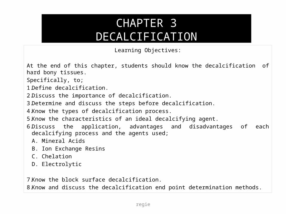
Learning Objectives:
At the end of this chapter, students should know the decalcification of hard bony tissues.Specifically, to;1. Define decalcification.2. Discuss the importance of decalcification.3. Determine and discuss the steps before decalcification.4. Know the types of decalcification process.5. Know the characteristics of an ideal decalcifying agent.6. Discuss the application, advantages and disadvantages of each decalcifying process and the
agents used;A. Mineral AcidsB. Ion Exchange ResinsC. ChelationD. Electrolytic
7. Know the block surface decalcification.8. Know and discuss the decalcification end point determination methods.
CHAPTER 3DECALCIFICATION
regie 2
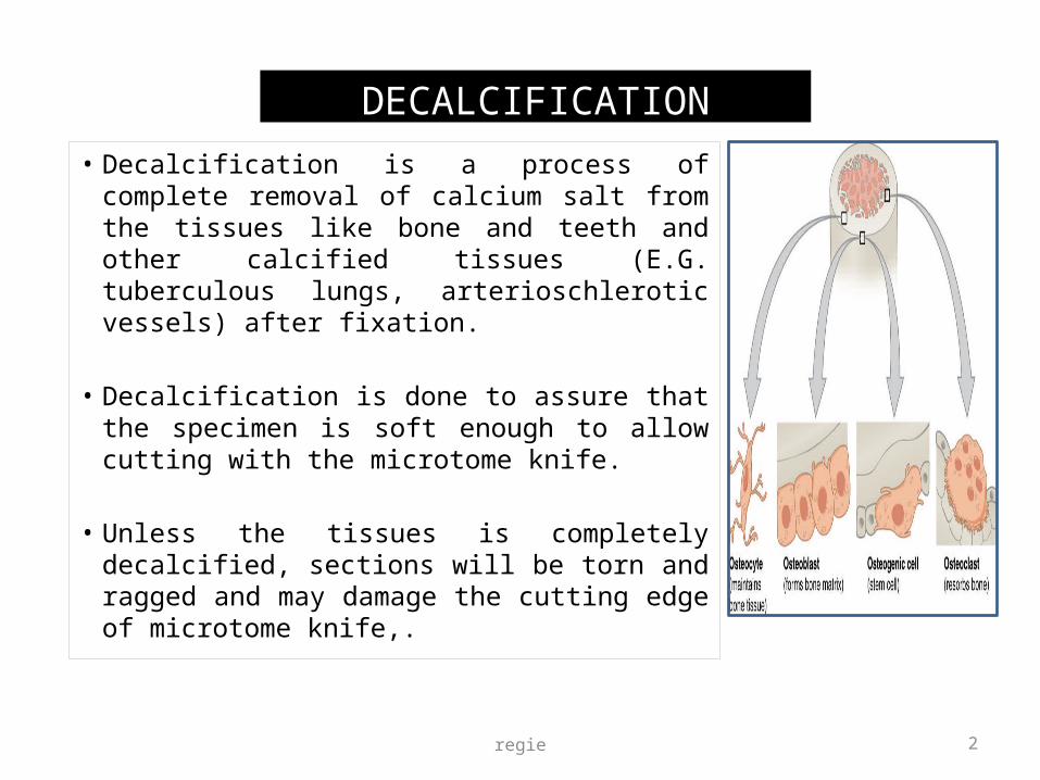
• Decalcification is a process of complete removal of calcium salt from the tissues like bone and teeth and other calcified tissues (E.G. tuberculous lungs, arterioschlerotic vessels) after fixation.
• Decalcification is done to assure that the specimen is soft enough to allow cutting with the microtome knife.
• Unless the tissues is completely decalcified, sections will be torn and ragged and may damage the cutting edge of microtome knife,.
DECALCIFICATION
3
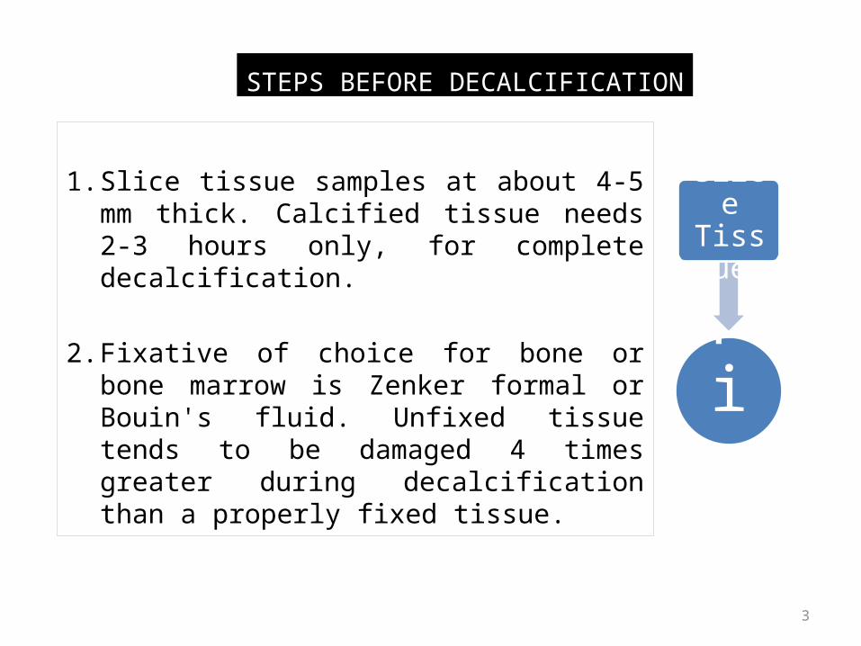
1. Slice tissue samples at about 4-5 mm thick. Calcified tissue needs 2-3 hours only, for complete decalcification.
2. Fixative of choice for bone or bone marrow is Zenker formal or Bouin's fluid. Unfixed tissue tends to be damaged 4 times greater during decalcification than a properly fixed tissue.
STEPS BEFORE DECALCIFICATION
Fix
Slice Tissue
regie 4
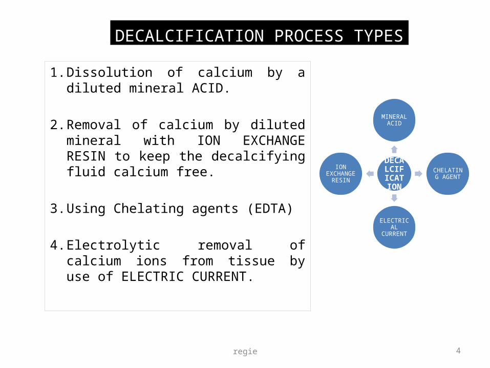
1. Dissolution of calcium by a diluted mineral ACID.
2. Removal of calcium by diluted mineral with ION EXCHANGE RESIN to keep the decalcifying fluid calcium free.
3. Using Chelating agents (EDTA)
4. Electrolytic removal of calcium ions from tissue by use of ELECTRIC CURRENT.
DECALCIFICATION PROCESS TYPES
DECALCIFICATION
MINERAL ACID
CHELATING AGENT
ELECTRICAL CURRENT
ION EXCHANGE
RESIN

regie 5
1. Completely removal of calcium.
2. Absence of damage to tissue cells.
3. Subsequent staining not altered.
4. Shorter time.
GOOD DECALCIFYING AGENT CRITERIA

MINERAL ACIDS:A. STRONG ACID (Nitric Acid , Hydrochloric Acid)B. WEAK ACID (Formic Acid, Acetic Acid, Picric Acid)
• NITRIC ACID: Very rapid, but if used for longer than 24-48 hours cause deterioration of stainability specially of the nucleus. Choice for temporal bones.
Example: 1. PERENYI’S FLUID (nitric acid, absolute alcohol, chromic acid)
Slow for decalcifying hard bone but excellent fluid for small deposits of calcium eg. calcified arteries, coin lesions and calcified glands.
2. Formalin Nitric acidNitric acid causes serious deterioration of nuclear stainability which partially inhibited by formaldehyde.
MINERAL ACIDS
CALCIUMdissolution
Nitric
Formic
AceticPicric
Hydrochloric

7
• HYDROCHLORIC ACID:Slower than nitric acid but still rapid. Fairly good nuclear staining.
• FORMIC ACID:Extensively used as acid decalcifier. Nuclear staining in better, but requires neutralization and thorough washing prior to dehydration.
Example: 1. Gooding and Stelwart's fluid. (Formic acid, Formalin)2. Evans Krajian fluid (Trisodium citrate, formic acid). At pH 2-3.3. Formic Acid Sodium Citrate - Gives better staining result than
nitricacid method, since its less harsh on the cellular properties. Therefore even with over exposure of tissue, it causes little loss of staining qualities.
MINERAL ACIDS

8
The surface of the block to be decalcified is trimmed with scalpel.
The block is then placed in acid solution at 1% hydrochloric acid face downwards.
SURFACE DECALCIFICATION
9
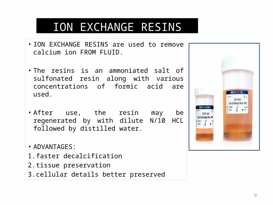
• ION EXCHANGE RESINS are used to remove calcium ion FROM FLUID.
• The resins is an ammoniated salt of sulfonated resin along with various concentrations of formic acid are used.
• After use, the resin may be regenerated by with dilute N/10 HCL followed by distilled water.
• ADVANTAGES:1. faster decalcification 2. tissue preservation 3. cellular details better preserved
ION EXCHANGE RESINS

10
CHELATING AGENTS are organic compounds which binds certain metals. Ethylene-diamene-tetra-acetic acid, disodium salt called VERSENATE has the power of capturing calcium.
This is a slow process but has little or no effect on other tissue elements. Some enzymes are still active after EDTA decalcification.
CHELATING AGENTS
regie 11
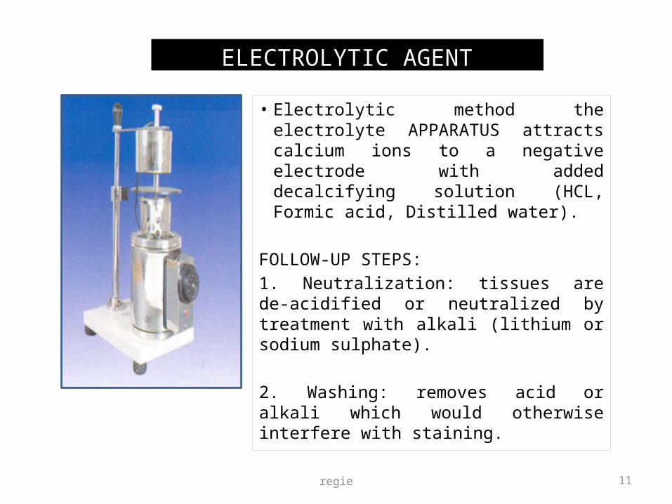
• Electrolytic method the electrolyte APPARATUS attracts calcium ions to a negative electrode with added decalcifying solution (HCL, Formic acid, Distilled water).
FOLLOW-UP STEPS:1. Neutralization: tissues are de-acidified or neutralized by treatment with alkali (lithium or sodium sulphate).
2. Washing: removes acid or alkali which would otherwise interfere with staining.
ELECTROLYTIC AGENT

regie 12
1. FLEXIBILITY Bending, needling or by use of scalpel if it bends easily that means decalcification is complete. This is unreliable, and may cause damage and distortion.
2. X-RAYBest method but very costly. Tissue fixed in mercuric chloride containing fixatives cannot be tested as they will be radio opaque.
3. CHEMICALIt is done to detect calcium FROM the decalcifying fluid. If calcium is tested negative, decalcification in considered complete.
DECALCIFICATION END POINT DETERMINATION METHODS