Embed Size (px)
Citation preview

INTRODUCTION
Malignant soft tissue tumors rarely occur in thehead and neck, and surgeons find it difficult to deter-mine the optimal method for excising these tumors,particularly those located in the face. Malignant softtissue tumors are generally resected along withthick surgical barriers, such as ligaments or the pe-riosteum. However, resection involving such barriersis difficult, due to the serious risks posed when thetumor to be resected is situated adjacent to impor-tant organs in the face. Moreover, accurate diagnosisof soft tissue tumors is often complicated even forwell-trained pathologists due to the histologic char-acteristics of these tumors. Low-grade fibromyxoidsarcoma (LGFMS) is a rare type of sarcoma ; deter-mination of the disease course based on oncologicand histologic features remains controversial (1-3).
Moreover, the efficacy and advantages of adjuvanttherapy for this neoplasm have not been elucidated.We describe here the case of an elderly woman withrecurring facial LGFMS that occurred at a rare lo-cation and an unusual age.
CASE REPORT
An 84-year-old woman noticed a subcutaneous,gradually enlarging tumor on her face. The tumorwas firm and immobile, approximately 3�2 cm insize, and located on the right forehead (Fig. 1).Magnetic resonance imaging (MRI) revealed thatthe mass was connected to the deep temporal fas-cia, appearing as a low-intensity area on T1 imagesand a high-intensity area on T2 images (Fig. 2 a, b).No metastasis was noted on computed tomography.Based on MRI findings, the mass was found to re-semble nodular fasciitis or a tumor such as a heman-gioma or fibroma. We performed an excisional bi-opsy to confirm the diagnosis. Histologic examina-tion of the biopsy specimen revealed fibrous andmyxoid components (Fig. 3 a, b). The fibrous parts
CASE REPORT
Recurring facial low-grade fibromyxoid sarcoma in anelderly patient : A case report
Yoshiro Abe, Ichiro Hashimoto, and Hideki Nakanishi
Department of Plastic, Reconstructive and Aesthetic Surgery, Institute of Health Biosciences, the Uni-
versity of Tokushima Graduate School, Tokushima, Japan
Abstract : Low-grade fibromyxoid sarcoma (LGFMS) is a rare type of sarcoma that is char-acterized by benign-appearing histologic features but a paradoxically aggressive clinicalcourse. These tumors generally occur in young to middle-aged adults, sometimes in chil-dren, but rarely in high-aged adults. LGFMS typically affects the deep soft tissues of thetrunk or lower extremities ; however, it is rarely seen in the face. We here describe acase of LGFMS on the right forehead of an 84-year-old woman. After resection with a 1-cm skin margin, recurrence occurred at 15 months postoperatively. Additional wide ex-cision was subsequently performed with a 2-cm skin margin. Recurrence and metasta-sis have not been observed for 1 year after the second excision. A wide surgical marginshould be considered in cases of LGFMS. J. Med. Invest. 59 : 266-269, August, 2012
Keywords : low-grade fibromyxoid sarcoma, elderly patient, forehead, recurrence, wide excision
Received for publication January 10, 2012 ; accepted March 5,2012.
Address correspondence and reprint requests to Yoshiro Abe,Department of Plastic, Reconstructive and Aesthetic Surgery, In-stitute of Health Biosciences, the University of Tokushima Gradu-ate School, 3 -18-15 Kuramoto, Tokushima-City, Tokushima,770-8503, Japan and Fax : +81-88-633-7297.
The Journal of Medical Investigation Vol. 59 2012
266

comprised spindle-shaped cells in a linear arrange-ment showing whorled and swirling growth patterns,whereas the myxoid parts comprised spindle tostellate-shaped cells with an abundant intercellularmatrix and, in some parts, relatively rich vascularnetworks. The tumor cells contained oval or shortspindle-shaped nuclei without a high degree of atyp-ism or pleomorphism. Immunohistochemical exami-nation revealed diffuse positivity to anti-vimentinantibody, whereas tests for other antibodies werenegative (Table 1). Based on the histologic findings,including the absence of nuclear atypism or pleo-morphism, and the presence of predominant fibrouscomponents, we diagnosed the tumor as LGFMS,not myxofibrosarcoma. Although we recommendedan additional wide excision, the patient refused. Oneyear later, the tumor recurred at the same location.We had previously resected the recurrent tumorwith a 1-cm skin margin, including the temporalbranch of the facial nerve and the temporal muscle,and reconstructed the defect with the remaining
Figure 1 : Photograph showing subcutaneous tumor on the rightforehead. It was approximately 3�2 cm in size.
Figure 2 : (a) MRI revealed the mass (arrows) in the subcuta-neous tissue, appearing as a low-intensity area on T1 images.(b) The mass (arrows) was showed as a high-intensity area onT2 images.
Figure 3 : (a) The tumor composed of fibrous (small arrow) andmyxoid (large arrow) components. (heamatoxylin and eosin stain,�100) (b) The spindle-shaped cells (small arrow) were foundin the fibrous parts, the spindle to stellate -shaped cells (largearrow) with an abundant intercellular matrix in the myxoid parts.(heamatoxylin and eosin stain,�400)
The Journal of Medical Investigation Vol. 59 August 2012 267
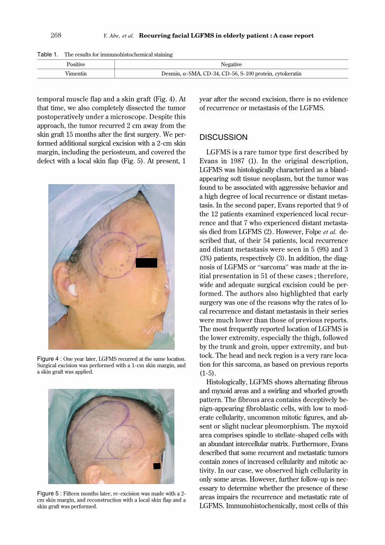
temporal muscle flap and a skin graft (Fig. 4). Atthat time, we also completely dissected the tumorpostoperatively under a microscope. Despite thisapproach, the tumor recurred 2 cm away from theskin graft 15 months after the first surgery. We per-formed additional surgical excision with a 2-cm skinmargin, including the periosteum, and covered thedefect with a local skin flap (Fig. 5). At present, 1
year after the second excision, there is no evidenceof recurrence or metastasis of the LGFMS.
DISCUSSION
LGFMS is a rare tumor type first described byEvans in 1987 (1). In the original description,LGFMS was histologically characterized as a bland-appearing soft tissue neoplasm, but the tumor wasfound to be associated with aggressive behavior anda high degree of local recurrence or distant metas-tasis. In the second paper, Evans reported that 9 ofthe 12 patients examined experienced local recur-rence and that 7 who experienced distant metasta-sis died from LGFMS (2). However, Folpe et al. de-scribed that, of their 54 patients, local recurrenceand distant metastasis were seen in 5 (9%) and 3(3%) patients, respectively (3). In addition, the diag-nosis of LGFMS or “sarcoma” was made at the in-itial presentation in 51 of these cases ; therefore,wide and adequate surgical excision could be per-formed. The authors also highlighted that earlysurgery was one of the reasons why the rates of lo-cal recurrence and distant metastasis in their serieswere much lower than those of previous reports.The most frequently reported location of LGFMS isthe lower extremity, especially the thigh, followedby the trunk and groin, upper extremity, and but-tock. The head and neck region is a very rare loca-tion for this sarcoma, as based on previous reports(1-5).
Histologically, LGFMS shows alternating fibrousand myxoid areas and a swirling and whorled growthpattern. The fibrous area contains deceptively be-nign-appearing fibroblastic cells, with low to mod-erate cellularity, uncommon mitotic figures, and ab-sent or slight nuclear pleomorphism. The myxoidarea comprises spindle to stellate-shaped cells withan abundant intercellular matrix. Furthermore, Evansdescribed that some recurrent and metastatic tumorscontain zones of increased cellularity and mitotic ac-tivity. In our case, we observed high cellularity inonly some areas. However, further follow-up is nec-essary to determine whether the presence of theseareas impairs the recurrence and metastatic rate ofLGFMS. Immunohistochemically, most cells of this
Table 1. The results for immunohistochemical staining
Positive Negative
Vimentin Desmin, α -SMA, CD-34, CD-56, S-100 protein, cytokeratin
Figure 4 : One year later, LGFMS recurred at the same location.Surgical excision was performed with a 1-cm skin margin, anda skin graft was applied.
Figure 5 : Fifteen months later, re-excision was made with a 2-cm skin margin, and reconstruction with a local skin flap and askin graft was performed.
Y. Abe, et al. Recurring facial LGFMS in elderly patient : A case report268

sarcoma are strongly positive for vimentin antibody,but are generally negative for α -smooth muscle ac-tin, desmin, S-100 protein, cytokeratin, CD34, andCD56 antibodies. In addition to these features,
�Zámecník and Michal described the presence ofstrong and diffuse bcl-2 reactivity (5), although wedid not investigate bcl-2 reactivity in the present pa-tient.
The differential diagnosis of LGFMS in terms ofother malignant tumors chiefly includes malignantfibrous histiocytoma and myxofibrosarcoma. Typi-cal malignant fibrous histiocytoma is considerablymore cellular and has greater malignant potentialthan LGFMS, that is, it shows greater nuclear hy-perchromatism, pleomorphism, and mitotic activity.Myxofibrosarcomas are classified into 4 grades,with grade 1 myxofibrosarcoma considered to re-semble LGFMS. However, this tumor can be dif-ferentiated from LGFMS by the greater degree ofnuclear atypia, predominant myxoid component,and absence of metastasis ; further myxofibrosar-coma generally occurs in older adults. Moreover,a t(7 ; 16)(q34 ; p11) translocation resulting in aFUS/CREB3L2 fusion transcript has been reportedin more than 95% of LGFMS cases when combina-tions of classic cytogenetic and molecular cytoge-netic methods, such as reverse transcription polym-erase chain reaction (RT-PCR) and fluorescencein situ hybridization (FISH), are employed. A rareFUS/CREB3L1 variant has also recently been de-scribed in a small subset of LGFMS cases (6). Al-though we did not attempt to analyze these fusiongenes, these methods may be valuable tools in thedifferential diagnosis (7).
Undoubtedly, adequate surgical excision of the tu-mor is necessary, because of the frequent recur-rence of LGFMS. Although a vertical incision thatincludes barriers such as the thick fascia or pe-riosteum is recommended, as based on the criteriafor soft malignant tumors, potential advantages ofa horizontal skin incision remain to be elucidated.In our patient, a 1-cm skin margin was not sufficientand therefore, during the second operation, an ex-tended 2-cm skin margin was employed. At 1 yearafter the second surgery, there is no obvious evi-dence of recurrence ; nonetheless, a long follow-up period is important. Adjuvant radiotherapy orchemotherapy has not been recommended in pre-vious reports ; however, these treatment strategies
may be used in cases of multiple metastases or fre-quent recurrence.
DECLARATION OF INTERESTS
The authors report no conflicts of interest. Theauthors alone are responsible for the content andwriting of the paper.
REFERENCES
1. Evans HL : Low-grade fibromyxoid sarcoma. Areport of two metastasizing neoplasms havinga deceptively benign appearance. Am J ClinPathol 88 : 615-619, 1987
2. Evans HL : Low-grade fibromyxoid sarcoma. Areport of 12 cases. Am J Surg Pathol 17 : 595-600, 1993
3. Folpe AL, Lane KL, Paull G, Weiss SW : Low-grade fibromyxoid sarcoma and hyalinizingspindle cell tumor with giant rosettes. Am JSurg Pathol 24 : 1353-1360, 2000
4. Goodlad JR, Mentzel T, Fletcher CD : Lowgrade fibromyxoid sarcoma : clinicopathologi-cal analysis of eleven new cases in support ofa distinct entity. Histopathology 26 : 229-237,1995
�5. Zámecník M, Michal M : Low-grade fibro-myxoid sarcoma : A report of eight cases withhistologic, immunohistochemical, and ultra-structural study. Ann Diagn Pathol 4 : 207-217,2000
6. Mertens F, Fletcher CDM, Antonescu CR,Coindre JM, Colecchina M, Domanski HA,Downs-Kelly E, Fisher C, Goldblum JR, GuillouL, Reid R, Rosai J, Sciot R, Mandahl N,Panagopoulos I : Clinicopathologic and molecu-lar genetic characterization of low-grade fibro-myxoid sarcoma, and cloning of a novel FUS/CREB3L1 fusion gene. Lab Invest 85 : 408-415,2005
7. Downs-Kelly E, Goldblum JR, Patel RM, WeissSW, Folpe AL, Mertens F, Hartke M, TubbsRR, Skacel M : The utility of fluorescence insitu hybridization (FISH) in the diagnosis ofmyxoid soft tissue neoplasms. Am J Surg Pa-thol 32 : 8-13, 2008
The Journal of Medical Investigation Vol. 59 August 2012 269