Embed Size (px)
Citation preview

RECURRENCE OF TIGHT MITRAL STENOSIS SYNDROME AFTER COMMISSUROTOMY
A REPORT OF SIX CASES WITH REOPERATION
P. SOUL& M.D.,* F. JOLY, M.D.,** J. CARLOTTI, M.D.,** AND
M. SERVELLE, M.D.**
PARIS, FRANCE
S INCE the surgical treatment of tight mitral stenosis has become a successful procedure, possibility of the recurrence of the stenosis has started various
debates. In spite of the fact that some authors6 deny the possibility of a postop- erative resealing of the valve, a few well-documented cases4s6,7,8*g of such an oc- curence have been published during the past few years. In the early stages of surgery of the mitral valve most surgeons recommended beginning the procedure with a digital commissurotomy and completing it with the knife (Brock, Barley, Glover, O’Neill, Dogliotti). Later on it was recognized that a satisfactory opening could be achieved with the employment of finger fracture alone. Usually an open- ing of the mitral area up to two fingerbreadths was thought to give satisfactory results, and postoperative findings have supported such a concept.
In the follow-up of our patients from the clinical and hemodynamic points of view since 19.50, we came to the conclusion that there was no correlation be- tween the surgical opinion of the effectiveness of the procedure and the postop- erative results.2B10p12 On the one hand, complete surgical mitral fractures were followed by only partial hemodynamic restitution, and, on the other hand, com- missurotomy judged to be incomplete anatomically gave very good clinical and hemodynamic results. It became obvious during the long-term follow-up of our operated patients that repeated postoperative catheterizations were of great value in assaying the results of mitral surgery. Especially does this fact gain great importance when the question of the possibility of mitral valve resealing is dis- cussed. Although most of the imperfect commissurotomies manifest themselves only during a long-term follow-up, and often after initial clinical improvement, there are some few cases where the ineffectiveness of the surgical approach may be demonstrated clinically immediately.2~r2
In this paper we intend to (1) confirm in a few cases the unquestionable re-establishment of tight mitral stenosis after a more or less prolonged period from the time of commissurotomy, and (2) discuss the exact causes and the ana- tomic mechanisms which contribute to the reoccurrence of stenosis, along with proof that the valves have resealed.
From the Hdpital Lariboisi&re, Paris. France. Received for publication May 8, 1957. *Professor, F&cult6 de MBdecine de Paris, Medecin des HBpitaux de Paris, and Director of the Center
of Cardiology, HBpital LariboisiBre, Paris. **Assistant. Center of Cardiology, RGpital LariboisiWe, Paris.
695

TABL
E I
-__
I
PRES
SURE
S (M
M.
Hg)
- i
I I
--
-
/
_‘-
02
j PE
RIP
HER
AI
CO
NSU
MPT
ION
S
ATU
RA
TIO
P
CM.S
/M.
(5%
)
CAR
DIA
t O
UTP
U’I
L./M
. D
ATES
R.A
. P.
A.
C.P
. SE
X AG
E
Mile
21
befo
re
C.:
7/
?/52
1s
t c.
: J/
24/5
3 8
mo.
la
ter:
12/1
4/53
33
m
o.
late
r: l/
3/.5
6 2n
d C.
: 2/
10/5
6
I so
/25
I ’ 10
/4
I:;
1 30
/18
(22)
so
/25
(35)
I
(26)
230
96
190
! 93
3.
9 4.
6
4.9
4.9
2.6
--I_
--
- -_
- 20
0 1
96
170
‘~
98
1.50
93
2 Fe
mal
e 22
befo
re
C.:
1st
c.:
7/l
?/53
6/
10/5
3
13
mo.
la
ter:
lO/
7/.5
4 27
m
o.
late
r: 2n
d C.
: 12
/1?/
55
12/
3/55
I 8/
2 (
?5/3
5 (S
O)
(3)
I s/
3.5
(40)
12
/6
(8)
90/5
0 (7
0)
befo
re
C.:
1st
c.:
3/14
/51
4/
9/51
3
mo.
la
ter:
6/22
/51
2nd
C.:
12/2
4/54
180
1 95
180
I 96
3 Fe
mal
e 23
4 Fe
mal
e 32
4.4
4.4
3.2
4.5
4.4
4.6
5.2
I- be
fore
C.
: 1s
t c.
: 2/
l/5
2 l/1
1/52
5 m
o.
late
r: ?/
16/.5
2 17
m
o.
late
r: ?/
24/5
3 44
m
o.
late
r: 2n
d C.
: lO
/ 6/
55
11/2
5/55
220
210
200
~ i:
180
I E
I I
1 43
/20
(36)
i
33/1
5 (2
1)
i I;;
; ;
17/S
(1
2)
, 60
/40
(47)
(
SO/3
5 (4
0)
I 2
I- I
5 Fe
mal
e 28
befo
re
C.
: 1
1st
c.:
12/
l/19/
51
3/51
2n
d C.
: 9/
?/
56
170
I 91
(
48/2
2 (3
0)
; (2
8)
5
6 Fe
mal
e 30
befo
re
C.:
1st
c.:
l/22/
54
1 l/2
6/53
29
mo.
la
ter:
C.:
6/29
/56
2nd
9/14
/56
3 wk
. la
ter:
lO/
5/56
I--~-
- 21
0 91
150
I j 93
I
150
I 96
(12)
130/
?0
(90)
’
Per
oper
at
ive
pres
sure
s:
) see
Ta
blci
il:i(
20/S
I
140/
?0
(100
) !
55/4
0 (S
O)
3
6/3
(4)
~ *.
., 65
/30
(45)
23
j16
(18)
I. 85
zw
5.7
2.6
3.2
1st
C.,
2nd
C.,
and
Irefo
re
C.
= fir
st,
seco
nd,
and
befo
re
com
miss
urot
omy,
re
spec
tivel
y.
R.A.
=
right
au
ricle
; P.
A.
= pu
lmon
ary
arte
ry:
c.1’
. =
cani
\- I
;
lary
pres
sure
. $.
;

E%z “;’ RECURRENCE OF MITRAL STENOSlS AFTER COMMISSUROTOMY 697
CASE MATERIAL
A recurrence of a syndrome of tight mitral stenosis occurring from 9 months up to 3 years after commissurotomy, and involving a second surgical procedure, was observed in 6 cases. These 6 cases were part of a series of 380 commissur- otomies performed during the period from 1950 to 1956. The first group, consist- ing of 180 cases, was operated upon with digital fracture alone. In the second group, comprised of 200 cases (beginning in April, 19.54), 95 per cent of the opera- tions were completed by use of the dilatator. * All 6 cases to be discussed are part of the first group.
CASE l.-This 21-year-old man was admitted to the HGpital Lariboisiere on March 18,
1952, complaining of shortness of breath since 15 years of age. There was no history of rheumatic
or scarlet fever. Exercise tolerance was reduced to 600 meters walking on flat ground. During the
past 6 years he had had several attacks of nocturnal, acute pulmonary edema and bouts of bronchi- tis in winter time. Physical examination revealed normal rhythm, and rumbling mid-diastolic
murmur with accentuation of the first heart sound at the apex. Fluoroscofiy revealed a moderate cardiac enlargement, with a slight bulging of the pulmonary conus, a moderately large left atrium,
and large pulmonary arteries with increased lung hilus shadows. Electrocardiogram showed sinus rhythm, enlarged and high Pi and Pz waves. The QRS axis was -l-80”, and there was slight right
ventricular hypertrophy. Laboratory data included normal sedimentation rate, blood fibrin 2.5 Gm. per liter, and total protein nitrogen 43 mg. per cent (Table II). Hemodynamics, on July 7,
1952, are shown in Table I.
First Operation.-(April 24, 1953.) The mitral leaflets were supple, The mitral area was
just over 1 square centimeter. The stenosis was more marked on the antero-external commissure,
which was somewhat calcified. There was no mitral regurgitation. The postoperative course was very progressive but improvement incomplete. Dyspnea on exertion almost disappeared, but not entirely. There was no more acute pulmonary edema. On fluoroscopy, the bulging of the pulmonary conus was less marked and the pulmonary fields were clearer. The electrocardio-
gram was unchanged. Results of postoperative catheterization on Dec. 14, 1953, are shown in
Table I.
Almost 2% years after commissurotomy, following some days of hard physical work, exer-
tional dyspnea and acute edema reappeared very suddenly and were more severe than before the operation. On examination, the clinical signs, radiologic and electrical findings were the same as
preoperatively. Laboratory data were normal as to sedimentation rate, blood fibrin, electrophore- sis, and C reactive protein, but antistreptolysin units were up to 500 (Table II). A catheteriza-
tlon was performed on Jan. 3, 1956 (Table I).
&operation.-(Feb. 10, 1956.) The mitral area was smaller, less than 1 square centimeter. The valvular tissues were hardened, especially along the borders. The digital opening was very
difficult, but with the instrument a total opening was completed without regurgitation. The postoperative course showed very fast improvement. The lung hilus shadows were cleared up. On the electrocardiogram, P waves returned toward normal, and the QRS axis was +70”. Lab-
oratory data were normal (Table II).
CASE 2.-This 22-year-old woman was admitted to the Hapital on June, 8, 1953, complain- ing of orthopnea, dyspnea on effort, and rare attacks of acute pulmonary edema, over a period of one year. At 17 years of age she had had acute rheumatism, and at 20, mitral stenosis was diagnosed. Physical examination showed normal rhythm, rumbling mid-diastolic murmur,
with accentuation of the first heart sound at the apex and of the second sound at the pulmonary area. F~UO~OSCOPY revealed a moderate cardiac enlargement with prominent pulmonary conus and pulmonary arteries, and increased lung hilus shadows. Electrocardiogram showed sinus rhythm,
*The dilatator was designed by the surgeon, Dr. Marceau Servelle.

enlarged and high I’, and 1’2 wit\ es, and ;I QliS alis uff 140”. ‘l‘herc \vrls marked right I entricular
hypertrophy. Laboratory data gave a normal sedimentation rate, blood fibrin 2.75 Gm. per liter, and antistreptolysin units of 160 (Table II). The hemodynamics, on June 10, 1953, are given in
Table I.
TABLE II
I CASE ~ COMMISSUROTOMIES i SEDIMENTA-
SEX AGE ---
1
ANDDATRS ~ TIONRATE 1 AND 2 HR.
Male
21
B 1 mo. j 5-15 A lmo. i 13-20
2 mo. 6-14
B 1 mo. ~ “-5 A 1 mo. 3-l 1
2
Female
22
1st c. April 1953
2nd C. Feb. 1956
____---
1st c. July 1953
___--
2nd C. Dec. 1955
-___
1st c. April 1951
-
,-
-
_-
-
-
B 1 mo. A 15days
1 mo.
TOTALBLOOD ANTISTREP- FIBRIN ELECTROPHORESIS TOLYSIN
(GM./~,OOO CM?) (UNITS) ---.__-.
2.50 ~ 4.40
- - --__-.-__,~ 2.70 Normal I ,,
I 3.10 Normal 500
2-5 2.75 Normal 20-40 W~itv;l;~lin slightly 160
2-9 I
2nd C. Dec. 1954
1st c. Feb. 1952
2nd C. Nov. 1955
~--
1st c. Jan. 1954
2nd C. Sept. 1956
B 1 mo. A Imo.
4 mo.
2-3 / 2 6-14 )
i - 1.80 Normal 50
2-9 : 3 Normal 125
/ 3
Female
23
__- 4
Female
32
B A 1 mo.
4 mo. 7 mo. 1 yr.
20-40 43-67
2-4
B 6mo. A 15days
2 mo. 4 mo.
--__ 35-70 56-105 36-60
8-16 -__-
66.60 4.60
- -
-I- B 6mo.
3 mo. 2 mo. 1 mo.
~__- 4-13 7-20
3046 12-22
3.60 4.80
-
A lmo. 1438 4.60 2 mo. 4-12 -
B 2mo. 2240 1 mo. 12-29
10 days 6-20 A lmo. 70-90
45 days 12-24 2 mo. 35-60 3 mo. lo-20
3.70 -
4.20 - - -
_- B 3mo.
2 mo. 1 mo.
A 15 days 1 mo.
24 28-59 12-26 39-76
9-10
3.5 6.40
5.40 -
-. With globulin increased
-
loo
Normal Normal Normal
50 200
--
--
-
I-
50
__--- - - - - - - -
____- 50
200 - 125 200
- Normal
- With globulin increased
With globulin normal -
- Normal
- - -
Normal
I- 6
Female
30
Normal - -
With globulin increased -
B = before: A = after: C. = commissurotomy.

E:%:“;’ RECURRENCE OF MITRAL STENOSIS AFTER COMMISSUROTOMY 699
First Operution.-(July 7, 1953.) The mitral leaflets were supple; the mitral area was less
than 1 square centimeter. The digital opening was satisfactory. There was no mitral tegutgi- tation. Postoperative course showed clinical improvement for 1 year. In June 1954, an episode
of infection of the respiratory tract was followed by a reappearance of dyspnea. Catheterization
was performed on Oct. 7, 1954 (Table I).
During the next 18 months there was a deterioration of the situation, with a loss of weight of
10 kilos, and an accentuation of respiratory distress. Examination gave the same findings as
before the operation. A catheterization was performed Dec. 3, 1955 (Table I). Laboratory data
were normal (Table II). Reoperution.-(Dec. 17, 1955.) The mitral valves were hardened but without calcifications.
The mitral area was smaller, less than 1 square centimeter. The digital fracture was difficult, but with the instrument a bilateral commissutotomy was completed up to the mitral ring without
regurgitation. The postoperative course showed very fast clinical improvement with no more
dyspneic troubles. The diastolic tumble disappeared but a faint systolic murmur was heard at
the apex. The electrocardiogram shifted toward the normal. Laboratory data were normal (Table
II). CASE S.-This 23-year-old woman was admitted to the Hapital on Feb. 12, 1951, complaining
of shortness of breath since the age of 7 years. There was otthopnea and severe dyspnea on effort. There was no history of rheumatic or scarlet fever. Physical examination revealed normal
rhythm, and loud diastolic tumble with marked accentuation of the first heart sound at the apex.
Fluoroscopy showed moderate cardiac enlargement with a slight bulging of the pulmonary conus,
large left atrium, and no change of lung hilus shadows. Electrocardiogram indicated sinus rhythm,
enlarged and high PI and PI waves, QRS axis +70”, and incomplete tight bundle branch block.
Hemodynamics, on Match 14, 1951, ate given in Table I. First Operation.-(April 9, 1951.) The mitral valves were hardened, the mitral area was
around 1 square centimeter. The opening of the valve was very difficult, but on the third attempt
a rather good fracture of both commissutes was obtained. Postoperative course was rather stormy,
with several attacks of flutter, a postcommissutotomy syndrome with fever, joint pains, and al- terations of biologic data (Table II). After several months of test and treatment, a marked im-
provement was realized, but the dyspnea on effort was still present. Catheterization data on June
22, 1951, ate given in Table I. During 1953, the patient lost 6 kilos of weight and the symptoms reappeared, with several
attacks of flutter and, later, a persistent auticulat fibrillation accompanied by enlarged liver and
clinical signs of cardiac failure. The radiologic and electrical signs remained unchanged. The laboratory data were modified (Table II).
Reoperution.-(Dec. 24,1954.) The mitral tissues were hardened, especially along the borders. The mitral area was smaller, 0.5 square centimeter. The opening of the valve was difficult, but
with the instrument a complete fracture was obtained. During the postoperative course there
was no improvement. Auticulat fibrillation persisted, with an enlarged heart and several attacks
of rheumatic fever which were difficult to control.
CASE 4.-This 32-year-old woman was admitted to the HBpital on Dec. 24, 1951, complaining of shortness of breath for a period of 10 years. It had become permanent, with attacks of acute pulmonary edema on slightest effort. She had a history of scarlet fever at the age of 12 years
and rheumatic fever at 13. Physical examination indicated normal rhythm, slight aottic insuffi- ciency, and loud diastolic tumble with accentuation of the first heart sound at the apex. Fluoros-
copy showed moderate cardiac enlargement, with a large left atrium, a bulging of the pulmonary conus, and increased lung hilus shadows. Electrocardiogram revealed sinus rhythm, enlarged F’1
and Py waves, and a QRS axis of $60”. There was no tight ventricular hyperttophy. Laboratory data were subnormal (Table II). Hemodynamics, on Jan. 11, 1952, ate given in Table I.
First Operation.-(Feb. 1, 19.52.) The mitral leaflets were supple. The mitral area was 1 square centimeter, with a small regurgitant jet. A satisfactory digital fracture of the commissures
was performed without increased regurgitation. Postoperative course was very rapid with im- pressive improvement, except for persistent dyspnea on effort. The fluotoscopy findings were
better; the electrocardiogram was unchanged. A catheterization (July 16, 1952) showed a partial return toward normal, which persisted (July 24, 19.53, Table I). Normal activity was continued
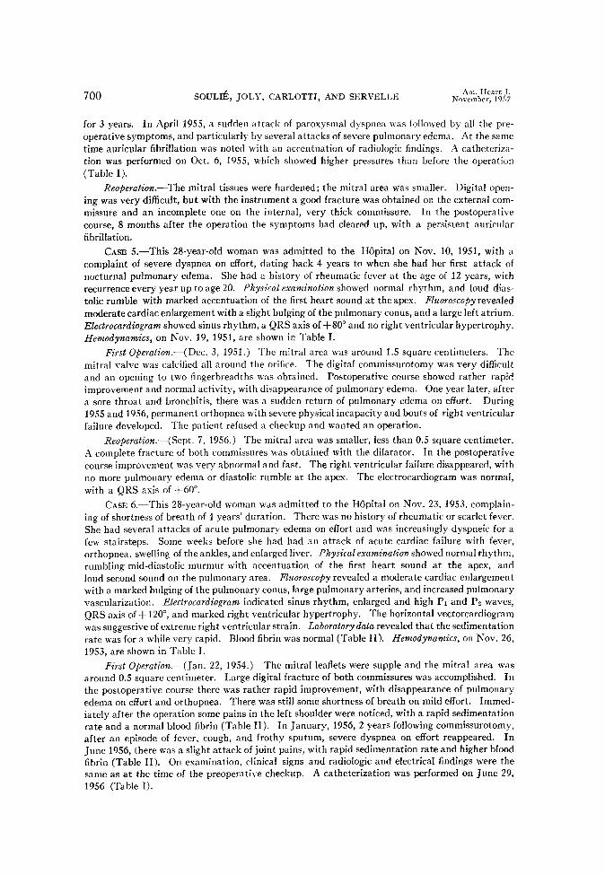
700 SOUL& JOLT, CARLOTTI, AND SERVELLE Am. Heart J,
November, 19.57
for 3 years. In April 1955, a sudden attack of paroxysmal dyspnea was followed by all the pre-
operative symptoms, and particularly by several attacks of severe pulmonary edema. At the same time auricular fibrillation was noted with an accentuation of radiologic findings. A catheteriza- tion was performed on Oct. 6, 1955, which showed higher pressures than before the operation
(Table I).
Reoperution.-The mitral tissues were hardened; the mitral area was smaller. Digital open- ing was very difficult, but with the instrument a good fracture was obtained on the external com-
missure and an incomplete one on the internal, very thick commissure. In the postoperative
course, 8 months after the operation the symptoms had cleared up, with a persistent auricular
fibrillation.
CASE S.-This 28-year-old woman was admitted to the Hapital on Nov. 10, 1951, with a
complaint of severe dyspnea on effort, dating back 4 years to when she had her first attack of
nocturnal pulmonary edema. She had a history of rheumatic fever at the age of 12 years, with
recurrence every year up to age 20. Physical examination showed normal rhythm, and loud dias-
tolic rumble with marked accentuation of the first heart sound at the apex. Flzroroscopy revealed moderate cardiac enlargement with a slight bulging of the pulmonary conus, and a large left atrium,
Electrocardiogram showed sinus rhythm, a QRS axis of -i-SO” and no right ventricular hypertrophy.
Hemodynamics, on Nov. 19, 1951, are shown in Table I.
First Operation.-( Dec. 3, 1951.) The mitral area was around 1.5 square centimeters. The
mitral valve was calcified all around the orifice. The digital commissurotomy was very difficult and an opening to two fingerbreadths was obtained. Postoperative course showed rather rapid improvement and normal activity, with disappearance of pulmonary edema. One year later, after a sore throat and bronchitis, there was a sudden return of pulmonary edema on effort. During 1955 and 1956, permanent orthopnea with severe physical incapacity and bouts of right ventricular
failure developed. The patient refused a checkup and wanted an operation.
IZeoperation.-(Sept. 7, 1956.) The mitral area was smaller, less than 0.5 square centimeter. A complete fracture of both commissures was obtained with the dilatator. In the postoperative course improvement was very abnormal and fast. The right ventricular failure disappeared, with
no more pulmonary edema or diastolic rumble at the apex. The electrocardiogram was normal,
with a QRS axis of +60”.
CASE 6.-This 28-year-old woman was admitted to the HBpital on Nov. 23, 1953, complain-
ing of shortness of breath of 4 years’ duration. There was no history of rheumatic or scarlet fever. She had several attacks of acute pulmonary edema on effort and was increasingly dyspneic for a
few stairsteps. Some weeks before she had had an attack of acute cardiac failure with fever, orthopnea, swelling of the ankles, and enlarged liver. Physical examination showed normal rhythm, rumbling mid-diastolic murmur with accentuation of the first heart sound at the apex, and
loud second sound on the pulmonary area. Fluoroscopy revealed a moderate cardiac enlargement with a marked bulging of the pulmonary conus, large pulmonary arteries, and increased pulmonary
vascularization. Electrocardiogram indicated sinus rhythm, enlarged and high Pr and P? waves,
QRS axis of + 120”, and marked right ventricular hypertrophy. The horizontal vectorcardiogram was suggestive of extreme right ventricular strain. Laboratory data revealed that the sedimentation rate was for a while very rapid. Blood fibrin was normal (Table II). Hemodynamics, on Nov. 26, 1953, are shown in Table I.
First Operation.-(Jan. 22, 1954.) The mitral leaflets were supple and the mitral area was around 0.5 square centimeter. Large digital fracture of both commissures was accomplished. In the postoperative course there was rather rapid improvement, with disappearance of pulmonary
edema on effort and orthopnea. There was still some shortness of breath on mild effort. Immed- iately after the operation some pains in the left shoulder were noticed, with a rapid sedimentation
rate and a normal blood fibrin (Table II). I n J anuary, 19.56, 2 years following commissurotomy,
after an episode of fever, cough, and frothy sputum, severe dyspnea on effort reappeared. In June 1956, there was a slight attack of joint pains, with rapid sedimentation rate and higher blood fibrin (Table II). On examination, clinical signs and radiologic and electrical findings were the same as at the time of the preoperative checkup. A catheterization was performed on June 29,
1956 (Table I).

“N:E: “;’ RECURRENCE OF MITRAL STENOSIS AFTER COMMISSUROTOMY 701
Reoperation.-(Sept. 14, 1956.) The mitral leaflets were still supple. The mitral area was
about 1 square centimeter. The digital opening was 2 square centimeter; with the instrument the
opening was completed up to the valvular ring. The postoperative course was a very abnormally
rapid improvement. The diastolic rumble was no longer present, but there was a faint systolic
murmur at the apex. The vectorcardiogram revealed sudden disappearance of the right ventricular strain. The catheterization, 3 weeks after the second operation (Oct. 5, 1956), showed a consider- able drop of the pressure at rest (Table I). For a short while there was slight alteration of the
laboratory data, but it quickly returned toward normal (Table II).
COMMENTS
To our 6 cases can be added 5 already published by various authors: Don- zelot and associates,4 Santy and co-workers ,g McKusick,8 Keyes and Lam,’ and Gleen and Dineen. The study of these 11 cases will permit consideration of the resealing of the valve, the limits of the syndrome which express it, and the data which make the diagnosis possible (Table I I I).
Frequency.-Actually, the recurrence of mitral stenosis seems rare, judged from a survey of the literature: 1 case out of 178 operations (Bailey); 1 case out of 120 (Keyes and Lam’); 1 case out of 160 (Santyg). For Wood the frequency is around 5 per cent of all cases. Out of our 380 commissurotomies we collected 6 cases. On the other hand, in a postoperative survey of 5 years, Glover and associates6 have found no case of resealing of the valve out of 600 patients operated on.
-However, those facts are not yet definitive; many late postoperative results remain insufficiently studied. It is possible that reseahng of the valves may ap- pear far more frequently. At the same time, a survey over a longer period of time will permit checking the value of the dilatator as to whether its use abolishes the recurrence of the mitral stenosis.
Age.-Almost every instance of resealing was observed in young patients under 30 years of age (8 cases out of 11).
The Physiopathologic Type of Stenosis.-Preoperatively, the circulation pres- sures were less. The more frequent finding was mitral stenosis with pure high capillary pressure (low pressure gradient between mean pulmonary and mean capillary pressure) ; but in 2 cases (Cases 2 and 6) there was a high pulmonary hypertension with a high pressure gradient between mean pulmonary and mean capillary pressure.
The First Operation Findings.-The anatomic condition of the valve varied. Usually, thickened and rigid leaflets were found, but sometimes they were supple (in 4 of our 6 cases). Rarely were calcified deposits noted. The mitral stenosis was always tight, except in 1 case where the mitral area was estimated to be 1.5 square centimeter.5 A slight regurgitant systolic jet was found in 2 cases.4
The First Commissurotomy.-The initial commissurotomy was digital in every instance. The operative reports do not point out any technical difficulties greater than usual. The fracture of at least one commissure was made routinely, but sometimes the internal commissure was incompletely opened, sometimes both commissures. After all, if the fracture seemed satisfactory, as in most of the cases, with excehent and lasting results, the commissurotomy was never completed by a fracture of both commissures up to the mitral valve ring.

702 SOrLIfi, JOl.V, (‘AKLOTTI, AND SERVELLE
Thr Postopemtiue Follow-up Study. 1. Clinical signs: AS a rul?, t-he evolution !Vas made in two successive
periods. During the first period the improvement was such as is usually observed after an efficient commissurotom)-. ‘I’herc was no longer any paroxysmal edema or dyspnea on effort, but in some instances there was still some slight dyspneic bouts on effort. (Often it is a comparative estimation of the functional postopera- tive troubles which permits the patient to appreciate the incomplete improvement due to the first operation.) The duration of the functional improvement was very short in the case of Santy and colleagues; it was negligible in the case of Donzelot and co-workers; but in all other cases the improvement lasted longer: up to 1 year in 4 cases, 2 years in 4 cases, and 3 years in 1 case. In our cases a
TABLEIIT. ELEVENCASESOFRECURRENCEOFMITRALSTENOSIS
CASES
~.~~. ~~~ ~~~~~
I I I I HEMODYNAMICS DUR.4TION OF VERIFI-
~ AGE ~ VALVES COMMISSUROTOMY L.A.ORC.P.(MM.H$ IMPROVEMENT CATION
So& & Colleagues 1 1 20 i Supple ( Opening to 2 I B (26) Reoperation
sq. cm. A 02) ~ 27 mo. /
CT?4 yr.1 --- ~-- --- ---~ 2 22
! ! Supple
~ ____ -. ~_---___ 5 ) 28 Partial cal-
&cation -___ .-- 6 ’ 30 Supple
___ __--- Gleen & Dineen 39 Thickened
_. Donzelot & Colleagues !
-.-, ---- - 29 1 Sclerous
__---- _I- Santy & Colleagues 26 Sclerous
I I- Keyes & Colleagues 1 29 I Calcified
McKusick 42 Sclerous
~--__- ~- ~. -~_~-___-.- - __- Enlarged to 2 1 - 13 mo. 1 Reoperation
fingerbreadths Wi yr.) _- .____- Enlarged to 2 B (22) 1 yr.
fingerbreadths A (15) ---___ -- ~-_.--- Enlarged to 2 ’ B (28) i 3 yr.
fingerbreadths A (IS) .___-___ --___- _ ___- Enlarged to 2 1 F.
fingerbreadths
Opening of both
!
L.A.-B (48-34) commissures A (24.14)
__-I__ Fracture of an- Postoperative
-1 2yr. __--__ 2 yr.
terolateral C.P. (12) commissure
_ ---__--- --__- Good on antero- 15 days
lateral corn- ~ missure
Good on antero- lateral com- missure
Enlarged to 2 fingerbreadths
- G;;te;; antero- B (32)
A (21)
2 mo.
-~- 2 yr.
,~~~ 1 yr.
_:-
Reoperation (3% yr.1
ReoperaBion (4 yr.)
I- Reoperation
(4% yr.1
Reoperation @% Yf.)
Reoperation (3 yr.)
Autopsy (1 mo.)
Reoperation (18 mo.)
Reoperation (2% yr.1
Autopsy (4% yr.1
L.A. = left auricle: C.P. = capillary pressure: B = before commissurotomy; A = after commis- surotomy.
For the ELcst6 cases (personal patients) thehemodynamic dataare givenin the table. Forthe other cases, the mean pulmonary pressure and the mean capillary pressure preoperatively are. respectively: Keyes and Lam: 52 and 35 mm. Hg: McKusick: 63 and 32 mm. Hg; and Gleen and Dineen: 38 and 28 mm. Hg.

k!ZiZ “;” RECURRENCE OF MITRAL STENOSIS AFTER COMMISSUROTOMY 703
peculiar fact was the rather sudden revival of the previous preoperative troubles: after acute nocturnal pulmonary edema in Cases 1 and 4; after removal of the appendix and a common pulmonary infection in Case 3; within a few days after bronchitis in Case 6.
2. Hemodynamic controls: The postoperative hemodynamic study also re- vealed the same two successive periods of evolution. The hemodynamic investi- gation showed immediately either a preoperative drop of pressure in the left auricle (in the case of Keyes and Lam, and our Case 6) or a postoperative clear drop of the capillary and pulmonary artery pressure (in the cases of McKusick and Gleen and Dineen, and 3 of our cases). But there was only a partial improve- ment in 4 cases. After the clinical relapse, the pressures rose again to an even higher level than before the operation (in the case of Keyes and Lam, and 3 of our cases).
The Findings at Reoperation.---Some interesting facts appear when we analyze the operative data:
1. In all the published cases there was a reconstitution of mitral stenosis. In 2 out of 3 autopsied cases a linear healing existed where the fracture had been made previously (the third case will be discussed later).
2. The mitral stenosis was almost regularly found to be tighter than at the first operation; 5 of our cases demonstrated this point quite conclusively.
3. In 4 of our 6 cases, the mitral leaflets were supple at the first operation, and in one of those cases (Case 6) the mitral leaflets were still supple at the second operation, with the mitral area reduced a little. But there were no similar find- ings in the other cases in which there was a profound change of the mitral appa- ratus along with the recurrence of the stenosis. The leaflets were no longer supple, but hardened and thickened, and the contour of the mitral orifice was irregular. Santy himself noticed the “harder” consistency of the stenosed mitral valve, and Gleen and Dineen noted important transformations of the valve itself.5
4. These important changes of the mitral valvular apparatus accompanying the clinical recurrence of the stenosis are of great practical interest. The second operative commissurotomy is more difficult; the digital fracture is harder to accomplish and more limited; usually there is no possibility to open the stenosis as fully as at the first operation. The surgeon must use the dilatating instrument in order to obtain a complete, bilateral fracture.
DISCUSSION OF THE BASIC FACTORS IN THE DEVELOPMENT OF RECURRING STENOSIS
Because of the rarity of such data, the underlying causes for the reconstitu- tion of a hemodynamic and clinical syndrome of tight mitral stenosis following initially successful surgery are not well known.
1. Ila most of the cases, the cause seems to be a refusion of the mitral commis- sures. The post-mortem examinations of a few cases and the findings at the reoperation show it with evidence in 10 cases.
Different mechanisms may preside over the true re-establishment of stenosis: a. The role of an evolutionary rheumatic activity, and its resurgence due
to the operative procedure, must be discussed in the light of McKusick’s case and 3 of our cases. In Case 6, immediately after the first operation and 2% months

after the second one, there was noticed to be transient joint pains with some tcm-
perature, increased sedimentation rate, c ,md increased blood fibrin, with a normal antistreptolysin rate. Such episodes suggest an uncontrolled rheumatic activity, but it lasted for too short a time and had too discontinuous a character to be held with certainty as the cause of the recurrence of the mitral stenosis, In
Case 4, the reappearance of symptoms 3 years after the first operation were coincident with the setup of a persistent auricular fibrillation; the biologic data showed slight disturbance. Case 3 was remarkable because of a rheumatic attack due to the commissurotomy. Th ere was no history of rheumatic fever, but 5 weeks after the operation a sore throat, joint pains, fever, and increased sedimen- tation rate were noted. During the following months 4 similar attacks occurred. The following year, during a pregnancy, a transient auricular fibrillation de- veloped, with attacks, later, of auricular flutter and persistent auricular fibrilla- tion succeeding each other. The heart became enlarged and the biologic data indicated disturbance.
Finally, if the importance of the rheumatic activity was dubious in Cases 4 and 6, it was clear-cut in Case 3. It was likewise in McKusick’s case in which symptoms of rheumatic activity were evident for 3 years.
b. Quite different are Cases 1, 2, 5, where there was no biologic or clinical rheumatic activity before or after the operation. In the case of Santy and that of Keyes and Lam no joint pain or suspicious fever were noticed. A nonspecific mechanism seemed to be patent in those cases. Without any revival of rheumatic activity, the digital fracture provoked in some patients a reaction of sclerous scarring capable of altering the valves, which became thick and again shrank the mitral orifice within a few months, perhaps within a few weeks, making it nar- rower than before (except in one case where the leaflets were supple and the valves less tightly resealed than noted at the first operation). This nonspecific, non- rheumatic mechanism may be dependent upon the special capacity of a particu- lar area to react with a diffuse sclerous scarring. (One knows that the average time for development of mitral stenosis is 5 years, but that it can be far shorter.) On the other hand, it is very well established that continuing hemodynamic disturb- ances are very highly favorable to changing tissues. Might remaining hyper- tension in the left atrium and pulmonary veins segment of the lesser circulation play a part?
c. To those facts it is necessary to add cases in which a local process of thrombosis had been noted; the histologic data of the case of Donzelot and asso- ciates shows that the progressive resealing scar of the valves is due to an organized local clotting.
The mechanism postulated by Margaray7b in the installation of a mitral stenosis deserves to be recalled here; the author believes that on the site of a very small injury made by a rheumatic process frost deposits of fibrin and platelets appear forming a fibrous structure. Such facts induce some authors to propose a systematic anticoagulant therapy during the 2 months following a commissur- otomy.3 This point of view seems especially applicable in cases where an unpaired mitral valve has been fractured incompletely and with difficulty and is able to initiate a continuing inflammatory process.
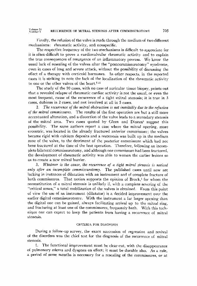
ydLlnmer “;’ RECURRENCE OF MITRAL STENOSIS AFTER COMMISSUROTOMY 705
Finally, the refusion of the valve is made through the medium of two different mechanisms: rheumatic activity, and nonspecific.
The respective frequency of the two mechanisms is difficult to appreciate for it is often difficult to prove a cardiovalvular rheumatic activity and to explain the true consequences of resurgence of an inflammatory process. We know the usual lack of resealing of the valves after the “postcommissurotomy” syndrome, even in cases of long and severe attack, without the possibility of discussing the effect of a therapy with corticoid hormones. In other respects, in the reported cases it is striking to note the lack of the localization of the rheumatic activity to one or the other valves of the heart.3J1
The study of the 10 cases, with no case of auricular tissue biopsy, points out that a revealed relapse of rheumatic cardiac activity is not the usual, or even the most frequent, cause of the recurrence of a tight mitral stenosis; it is true in 3 cases, dubious in 2 cases, and not involved at all in 5 cases.
2. The recurrence of the mitral obstruction is not inevitably due to the refusion of the mitral commissures. The results of the first operation are but a still more accentuated alteration, and a distortion of the valve leads to a secondary stenosis of the mitral area. Two cases quoted by Gleen and Dineen suggest this possibility. The same authors report a case where the mitral opening, more eccentric, was located in the already fractured anterior commissure; the valves became rigid with calcium deposits and a restenosis was built up in the medium zone of the valve, to the detriment of the posterior commissure which had not been fractured at the time of the first operation. Therefore, following an incom- plete bilateral commissurotomy, and although one commissure had been fractured, the development of rheumatic activity was able to worsen the earlier lesions so as to create a new mitral barrier.
3. Whatever is the cause, the recurrence of a tight mitral stenosis is noticed only after an incomplete commissurotomy. The published cases until now are lacking in instances of dilatation with an instrument and of complete fracture of both commissures. That notion supports the opinion of Brock,l for whom the reconstitution of a mitral stenosis is unlikely if, with a complete severing of the “critical zones,” a total mobilization of the valves is obtained. From this point of view the use of an instrument (dilatator) is a decided improvement over the earlier digital commissurotomv. With the instrument a far larger opening than the digital one can be gained, always facilitating arrival up to the mitral ring, and fracturing at least one of the commissures, frequently both. With this tech- nique one can expect to keep the patients from having a recurrence of mitral stenosis.
CRITERIA FOR DIAGNOSIS
During a follow-up survey, the exact succession of regression and revival of the disorders was the chief test for the diagnosis of the recurrence of mitral stenosis.
1. The functional improvement must be clear-cut, with the disappearance of pulmonary edema and dyspnea on effort; it must be durable also. As a rule, a period of some months is necessary for a resealing of the commissures, or at

least for the appearance oi ;I clinical s>,ndrome of the recurring mitral stenosis. The intermediate period usually lasts from 1 to 2 years, sometimes 3 years or more.
2. The control of the pressures of the pulmonary circulation shows, as does the clinical study, the same evolution in two periods: first it brings out either the preoperative fall of pressure in the left atrium or the drop of the capillar\ pressure during the following months; later, with the renewal of symptoms, the pressures rise again, sometimes higher than before the operation.
If the clinical data give a clue, they could not give, however, a secure diagnosis without the hemodynamic findings. This conclusion is supported from another point of view by infrequent but difficult cases where the postoperative hemo- dynamic findings, especially abnormal ones, were due to a postoperative signifi- cant mitral regurgitation.
CONCLUSIONS
The recognized cases of recurrence of mitral stenosis are due to a commis- surotomy efficient from the clinical and hemodynamic results but inefficient from the anatomic standpoint (incomplete opening up to the mitral ring).
SUMMARY
1. Six cases of unquestionable re-establishment of tight mitral stenosis involving a second operation are reported; 5 cases already published by other authors are reviewed.
2. The most important point which marks the recurrence of the stenosis is an evolution of clinical and hemodynamic signs within two periods: (a) a clear- cut regression of the symptoms which last from 1 to 3 years, and (b) a recurrence of the symptoms, which frequently appear abruptly, with an increase of the lesser circulatory pressures, so that a true syndrome of recurrence of tight mitral stenosis can be depicted.
3. The parallel evolution of clinical and hemodynamic signs must be checked upon, because these signs permit the settling of the outline of this problem in making the differential diagnosis with other conditions capable of affecting the future of operated patients.
4. An incomplete digital fracture and unsatisfactory mobilization of the valves seem to be the first condition for recurrent obstruction of the mitral valve. The mitral barrier is rebuilt most frequently from a remanifestation of the val- vular lesions, due either to an acute or low-grade rheumatic activity or more often to a sclerous scarring of nonspecific process.
This study points out the improved value of fracture of the valves by in- strument and the possible help and usefulness of an anti-inflammatory and anti- coagulant treatment.
ADDENDUM
Since the writing of this paper, we have had the opporturlity (1) to collect 3 more cases of
the recurrence of tight mitral stenosis with reoperation similar to the 6 cases presented here, and (2) to look over the current literature and checkups. The report of L. B. Ellis and D. E. Harke+
states that “Evidence will be presented that significant refusion of the valves has occurred rarely
within the period of observation.” C. P. Bailey and H. Goldberg’4 have reported that “We now
y&54 RECURRENCE OF MITRAL STENOSIS AFTER COMMISSUROTOMY 707
can confirm in our own operation series indubitable re-establishment of stenosis in patients who have undergone at least an apparently reasonably adequate commissurotomy; in 6 patients autopsy evidence is available; in 11 others reoperations and hence digital re-examination of the valve by the original operating surgeon has seemed to offer sufficient proof of the development of such re- current stenosis.”
a: :: Z: k. :- 10:
11.
::. 14:
REFERENCES
Brock, R. C.: Brit. Heart J. 14:489, 1952. Carlotti, J., SouliC, P., Joly, F., and Sicot, J. R.:
du retrecissement mitral (56 cas). Etude hemodynamique postoperatoire
Washington, Sept. 1954. Report of the Second World Congress of Cardiology,
Coblentz, B., Hillemand, R., and LenPgre, J.: Arch. mal. coeur 49:535, 1956. Donzelot, E., Dubost, C., Heim de Balsac, R., Metianu, C., and Guillemot, R.: Arch. mal.
coeur 46:300, 1953. Gleen, F., and Dineen, P.: Ann. Surg. 3:40.5, 1956. Glover, R. P., Davila, J. C., O’Neill, T. J. E., and Janton, 0. H.: Circulation 11:14, 19.55. Keyes, J. W., and Lam, G. R.: J.A.M.A. 155:247, 1955. Magaray, F. R.: Brit. Heart J. 1:856, 1951. McKusick, V. A.: A.M.A. Arch. Int. Med. 95:557, 1955. Santy, P., Froment, R., Gallavardin,. L., and Tolot, F.: Arch. mal. coeur 4’7:615, 1954. SouliC, P., Joly, F., Carlotti, J., VOCI, G,, and Servelle, M.: Traitement du retrecissement
mitral par commissurotomie. Indications et resultats post-operatoires (Etude herno- %;;mtque). Report of the First European Congress of Cardiology, London, Sept.
SoulieiFj4Bouvrain, Y., Fortin, P., DiMatteo, J., and Tricot, R.: Arch. mal. coeur 47:49,
Soulie, P., Joly, F., Carlotti, J., and Sicot, J. R.: Arch. mal. coeur 49:729, 1956. Ellis, L. B., and Harken, D. E.: Circulation 14:931, 1956. Bailey, C. P., and Goldberg, H.: Circulation 14:907, 1956.