Embed Size (px)
Citation preview
Reconstructing a solid-solid phase transformationpathway in CdSe nanosheets withassociated soft ligandsZhongwu Wanga,1, Xiao-Dong Wenb, Roald Hoffmannb,1, Jae Sung Sonc, Ruipeng Lia, Chia-Chen Fangd,Detlef-M. Smilgiesa, and Taeghwan Hyeonc
aCornell High Energy Synchrotron Source (CHESS), Wilson Laboratory, Cornell University, Ithaca, NY 14853; bDepartment of Chemistry and ChemicalBiology, Cornell University, Ithaca, NY 14853; cNational Creative Research Initiative Center for Oxide Nanocrystalline Materials and School of Chemicaland Biological Engineering, Seoul National University, Seoul 151-744, Korea; and dDepartment of Materials Science and Engineering, Cornell University,Ithaca, NY 14853
Contributed by Roald Hoffmann, August 10, 2010 (sent for review June 10, 2010)
Integrated single-crystal-like small and wide-angle X-ray diffrac-tion images of a CdSe nanosheet under pressure provide directexperimental evidence for the detailed pathway of transformationof the CdSe from a wurtzite to a rock-salt structure. Two consecu-tive planar atomic slips [(001) h110i in parallel and (102) h1̄01i witha distortion angle of ∼40°] convert the wurtzite-based nanosheetinto a saw-like rock-salt nanolayer. The transformation pressure isthree times that in the bulk CdSe crystal. Theoretical calculationsare in accord with the mechanism and the change in transforma-tion pressure, and point to the critical role of the coordinatedamines. Soft ligands not only increase the stability of the wurtzitestructure, but also improve its elastic strength and fracture tough-ness. A ligand extension of 2.3 nm appears to be the critical dimen-sion for a turning point in stress distribution, leading to theformation ofwurtzite (001)/zinc-blende (111) stacking faults beforerock-salt nucleation.
phase transition ∣ semiconductor ∣ dimensionality
Rational synthesis of materials with defined structure and prop-erties requires a reasonably complete understanding of the
associated kinetic transformations and microscopic mechanisms(1). We need to know these in order to understand how asso-ciated soft-bonded organics may tune structural stability, elasticstrength, and fracture toughness (1–3). The study of solid-solidphase transformations of materials, and their structural stabiliza-tion upon incorporation of soft materials at different scales, canbe very helpful in this regard. For instance, application of pres-sure and temperature converts graphite to a hard diamond phasethat persists at ambient conditions, allowing for a wide range ofknown applications (4). Upon addition of soft materials such assilicon and metals (5, 6), the sintered diamond-dominant nano-composites show improved yield strength and fracture toughness(by several orders of magnitude) without significant diminutionof hardness.
Many fascinating natural substances, such as human bone andother biological materials, also contain exceptionally strongbuilding blocks (2, 3) essential to their function. The subtle inter-play between soft organics and embedded brittle materials isresponsible not only for enhancement of the structural stabilityof embedded brittle materials, but also for improvement of theyield strength and fracture toughness of assembled hierarchicalorganizations (2, 3).
Microscopic mechanisms of transformation are difficult to de-termine. In bulk solids, as metastable higher energy and higherdensity phases nucleate in multiple domains, structural features(such as defects, microstructure, and impurities) may modify sig-nificantly the kinetics and mechanism of a phase transformation.In principle, calculations could provide the energy difference be-tween structural polymorphs at a given pressure or temperature.But putting aside problems in computing activation barriers,
there is often a large discrepancy between theory and experiment.For instance, the nucleation pressure of the bulk zinc-blende torock-salt ZnS phase transformation (with variable microstructureand lattice strain) was observed over a wide pressure range,8 ∼ 16 GPa (7–11). However, the calculated thermodynamictransformation pressure was 14.7 ∼ 16.1 GPa (12–14).
To avoid the effect of structural peculiarities on transformationmechanism, one should ideally carry out measurements on a per-fect defect-free crystal. Small nanocrystals behave as defect-freesmall crystals in which a single domain dominates the nucleationof another structure phase. In a defect-free nanocrystal, if we ex-clude the contribution of the size-dependent surface energy, theexperimental transformation pressure should be equivalent to thecalculated transformation pressure (15). One supportive exampleis that an observed transformation pressure of 15.1 GPa in 15 nmzinc-blende ZnS agrees well with the calculated transformationpressure (15); 15 nm in zinc-blende ZnS proves to be a criticalsize for nanoparticles to behave like a defect-free bulk crystal(16, 17). [We used theories developed in the literature (16, 17)to define the two critical sizes of 15 nm and 4 nm for zinc-blendeZnS. These appear to be the turning point for the particle to bestrain-insensitive and defect-free, respectively (15).]
In this context, the Alivisatos group has performed a rich spec-trum of measurements on spherical CdSe nanoparticles usingsynchrotron X-ray diffraction and optical spectroscopy at well-controlled pressure and temperature (18–21). The observed trans-formation pressure as a function of particle size, and the pressuredependence of the relaxation time provide ways to determine theactivation volume and barrier height (for the wurtzite → rock-saltphase transformation) (9). Several models for the transformationwere then derived, leading to a reasonable description of themicroscopic mechanisms (18, 21). However, one is still not sureexperimentally of the true pathway, because a comparison of ameasured X-ray diffraction (XRD) pattern with several simulatedpatterns (necessary because of different microstructural assump-tions in simulations) is required (21). In parallel investigations,theoretical simulations have suggested several subtle steps thatwere not included in the proposed transformation pathways(22–24). One might not know whether to trust theory or experi-ment here; an experimental key is lacking to open this black boxand single out the true transformation pathway.
Author contributions: Z.W., X.-D.W., and R.H. designed research; Z.W., X.-D.W., R.H., J.S.S.,R.L., C.-C.F., and D.-M.S. performed research; Z.W., J.S.S., R.L., C.-C.F., D.-M.S., and T.H.contributed new reagents/analytic tools; Z.W., X.-D.W., R.H., J.S.S., R.L., C.-C.F., D.-M.S.,and T.H. analyzed data; and Z.W., X.-D.W., and R.H. wrote the paper.
The authors declare no conflict of interest.1To whom correspondence may be addressed. E-mail: [email protected] or [email protected].
This article contains supporting information online at www.pnas.org/lookup/suppl/doi:10.1073/pnas.1011224107/-/DCSupplemental.
www.pnas.org/cgi/doi/10.1073/pnas.1011224107 PNAS ∣ October 5, 2010 ∣ vol. 107 ∣ no. 40 ∣ 17119–17124
CHEM
ISTR
Y
Dow
nloa
ded
by g
uest
on
Nov
embe
r 21
, 202
1

In this study, we show that a technique integrating in situ small-angle X-ray diffraction (SAXRD) and wide-angle X-ray diffrac-tion (WAXRD) can identify the preferred sample orientation andelucidate quite precise details of the phase transformation me-chanism of CdSe nanosheets. [In Fig. 1, we use one SAXRD im-age taken at high pressure in which a full texture is shown forbetter understanding. This is because a large beam stop was usedin the X-ray diffraction image collection at 0 GPa for detectorprotection. Thus half the diffraction ring (Inset SAXRD) wasblocked.]
Results and Discussion1. Experiments and Transformation Pathway.Highly uniform 1.4-nm-thick CdSe nanosheets have been shown to have a defect-freewurtzite structure with a (001) growth direction (25, 26). [In hex-agonal crystal systems, there are two conventions for labeling crys-tal planes, a three-index and a four-index label. In this paper, weconsistently use the three-index notation. CdSeð001Þ ¼ 0001;CdSeð110Þ ¼ ð112̄0Þ.] Two (110) facets dominate the top andbottom surfaces; (001) facets terminate the two side surfaces.The CdSe nanosheets are not pristine; bonded to the (110) sur-faces are organic ligands of octylamine or oleylamine or somemixture of these. There are van der Waals forces between CdSenanosheets, and these lead to lamellar mesostructure (26). [TheCdSe nanosheets bonded at their surface with several types of li-gands that have different chain lengths were synthesized by a low-temperature solution chemical synthesis (25, 26). The length scaleof each sample was characterized by SAXRD.]
The highly oriented samples display a single-crystal-like XRDimage, indicating that the lamellar superstructure CdSe na-nosheets are loaded with expected orientations into a diamondanvil cell (DAC). The in situ synchrotron XRD diffraction imagescollected under pressure reveal a series of pressure- or transfor-mation-tuned texture changes. These allow precise tracking ofthe position and magnitude of atomic distortion and translationduring the wurtzite ↔ rock-salt structural transformation.
Fig. 1 shows the SAXRD and WAXRD images that corre-spond to the shape and lattice of lamellar CdSe nanosheets inour experiment. Two single-crystal-like broad diffraction spotsin the SAXRD image reveal that the basal lamellar layer is par-allel to the force-loading axis of DAC (incident X-ray direction).Correlating the lattice structure of CdSe nanosheets, theWAXRD image indicates that either the (100) or (001) planeis parallel to the diamond anvil surface. The strong spotty diffrac-tion characteristics develop at (002) rather than at (100); this in-dicates that the (100) plane prefers to be oriented parallel to the
diamond anvil. Following such alignment, a lattice orientationwas tuned with the (100) plane perpendicular, and both (110)and (001) planes parallel to the incident X-ray beam.
The SAXRD and WAXRD measurements on the CdSe sam-ples (terminated by a mixture of octylamine and oleylamine, asanalyzed in SI Text and Fig. S1) were then carried out over a pres-sure range of 0–12.7 GPa. The WAXRD images (Fig. 2A) indi-cate that wurtzite CdSe transforms to a rock-salt structure at∼10.7 GPa, a pressure significantly greater than the transforma-tion pressure of 2.5 GPa observed in bulk CdSe (18). It is worthnoting that the wurtzite (002)/zinc-blende (111) stacking fault wasnot detected before the nucleation of the rock-salt phase.
Upon decompression, a mixture of wurtzite and zinc blende isobserved at 3.1 GPa. On further release of pressure, a single wurt-zite phase is fully recovered (Fig. 2B). However, in bulk and othernanocrystal CdSe samples, the CdSe recovered from high pres-sure always has the zinc-blende structure (3), no matter whetherthe starting CdSe sample crystallizes in a wurtzite or zinc-blendestructure. The SEM characterization indicates that pressure andphase transformation do not result in a noticeable fracture ofthe nanosheet; the coexistence of zinc blende and wurtzite isobserved at 3.1 GPa. Most likely one is seeing a series of wurtzite(002)/zinc-blende (111) stacking faults across the original (001)growth direction of the nanosheet.
An azimuthal plot of typical WAXRD images reveals thatCdSe nanosheets produce two strong single-crystal-like diffrac-tion spots located on the W (002) ring (Fig. 3A). In Fig. 3A, thenotation is W ¼ wurtzite, RS ¼ rock salt, and Z ¼ zinc blende.Upon phase transformation at 10.7 GPa, two similar diffractionspots are assigned to the nucleated rock-salt (111) ring and aredeveloped from the two W (002) spots at a shift angle of ∼40°(Fig. 3A). Relative to the RS (111) spots, the two RS (200) spotsemerge at a rotation angle of 55° and 23°, respectively. Releasingthe pressure to 3.1 GPa, the two diffraction spots on the overlap-ping ring of Z (111) and W (002) appear at the same positions asthe starting W (002) spots (Fig. 2A). In Fig. 3B, the WAXRDpatterns of CdSe nanosheets with an intersheet distance of2.3 nm at 8.1 GPa indicate the coexistence of zinc-blende, wurt-zite, and rock-salt CdSe structures.
Using the experimental information on the way pressure tunesthe rotation and translation magnitude of single-crystal-like XRDspots, the transformation pathway from wurtzite to rock salt inthe nanosheets may be unambiguously reconstructed, as shownin Fig. 4 A and B.
There are two sliding steps in the transformation: (i) W CdSeactivates the basal system ð001Þh110i. Pressure forces the Cd andSe atoms to slide relative to each other (move out and in of theplane) in the (001) plane (Fig. 4A). The sequence is similar to theknown deformation mechanism in hcp-type metals (27). In prin-
Fig. 1. (Top) Theoretical correlation of SAXRD and WAXRD to the shapeand lattice orientation of lamellar structure CdSe illuminated by an incidentX-ray beam along different directions. (Bottom, Left and Right) Experimentaldiffraction images indicating the preferred orientations of 1.4-nm-thick CdSenanosheets. The inset in the lower right-hand corner image highlights athigh contrast the spotty feature of the (002) peak.
Fig. 2. Representative high-pressure wide-angle X-ray diffraction patternsof lamellar CdSe nanosheets: (A) compression run, indicating that wurtzitetransforms to rock salt at 10.7 GPa; (B) decompression run, indicating thatthe rock-salt structure of the CdSe nanosheet transforms to a mixture ofzinc-blende and wurtzite structures at 3.1 GPa, and at 0 GPa is completelyconverted back to the wurtzite structure.
17120 ∣ www.pnas.org/cgi/doi/10.1073/pnas.1011224107 Wang et al.
Dow
nloa
ded
by g
uest
on
Nov
embe
r 21
, 202
1

ciple, such a planar shift creates W (001)/Z (111) stacking faults;they are detected only in the “short-ligand”-bonded CdSe na-nosheets, rather than the ones bonded to long ligands.
(ii) In the next step, the (102) plane atoms slide along the h1̄01idirection with an angle of 40°, producing the two observed shiftangles of 55° and 23° (Fig. 3A). The first angle (55°) is consistentwith the ideal intercepting angle of 54.7° between RS (200) andRS (111) (Fig. 4B). The other one (23°) is an approximate averageof the two interception angles between groups of equivalent crys-tallographic planes [e.g., ð2̄00Þ and ð1̄ 1̄ 1̄Þ vs. ð0̄20Þ and ð1̄ 1̄ 1̄Þ]in a rock-salt structure (see SI Text and Fig. S2).
The large lattice strain (revealed by the remnant of the highlytextured W (102) ring in the rock-salt pattern in Fig. 2A) couldexplain the unsymmetrical occurrence of the second diffractionspot. The 3- and 4-fold axes in the rock-salt phase are rotatedby 40° and 5° (or 15°) against the 6-fold axis of the starting wurt-zite structure. The strain-free and highly strained environmentslead to axial rotation by 5° and 15° (4-fold axis in RS against6-fold axis in W), respectively (28, 29), explaining well the ambig-uous observations contrasting bulk single crystal CdS (28) andCdSe (29). [In bulk single crystals, they observed a rotation angleof 5° and 15° from the 6-fold axis of wurtzite in CdS (28) and CdSe(29), respectively, to form the 4-fold axis of rock salt.]
2. Theoretical Simulations of the Phase Transition of CdSe Nanosheets.Using density functional theory (DFT) periodic calculations asimplemented in the Vienna ab initio simulation package (VASP)(see SI Text and Figs. S2–S8), organic ligands are embedded be-tween slabs, bonding to cadmiums at the top and bottom surface
of the CdSe nanosheets. Methylamine is used by us as a modelcoordinating base in the calculations.
Beginning with a seven-layer CdSe slab model covered bymethylamines at 0 GPa, we optimize the slab structure at2.5 GPa, 5 GPa, 7 GPa, 8 GPa, and 15 GPa, respectively, of whichat 2.5 GPa, 7 GPa, and 8 GPa are shown in Fig. 5. The CdSe layersand coordinated methylamine layers are apparent. Following theCdSe geometries, one can see that the wurtzite phase undergoes atransformation to a rock-salt phase at 8 GPa; the phase trans-formation is barrier-free. Because of the lack of an energy barrierto the reverse structural transformation route (rock salt →wurtzite), the pressure-tuned roc-ksalt CdSe phase cannot be re-tained at ambient conditions after compression. This is in agree-ment with our experimental observations.
It is remarkable that the transition (wurtzite → rock salt) in2D nanosheets occurs at ∼3 times higher pressure than thatnecessary for transforming the bulk structures. The calculatedtransition pressure of 2.5 GPa for bulk structure agrees well withexperiment (18). In Fig. 5, the way the atoms shift in the trans-formation is marked by arrows in the 7-GPa geometry (see mag-nification).
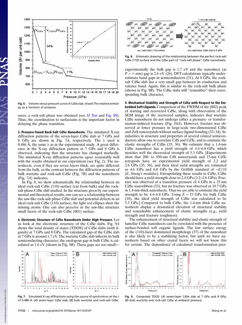
The phase transition may be tracked in another way. Fig. 6shows the calculated volume as a function of pressure (V-P curve)for CdSe nanosheets with adsorbed surfactants. There is an initialsharp decrease in volume between 0 GPa and 2.5 GPa. This vo-lume reduction of 34% is likely due to relatively facile squeezingout of van der Waals space between ligand layers. Such behaviorhas been observed in molecular crystals under pressure (30). An-other volume jump at 7 GPa is observed in the V-P curve, indi-cative of a phase transition. From the detailed atomic coordinatesshown in Fig. 5, it may be seen that this is the wurtzite → rock salttransition in the CdSe nanolayer. In the transition zone, thecorresponding volume reduction is 11%, as shown in Fig. 6. Notethat there is a small enthalpy difference (ΔH ¼ H7 GPa−H8 GPa) of 2.38 eV per unit cell (Cd14Se14), at this transition,as shown in Fig. 6 Inset.
The phase transition is “delayed” by the dimensionality and/orcoordination to surfactant molecules. To check the effect ofsurfactants on the phase transition, the seven-layer CdSe slabwithout methylamines was optimized at 5 GPa and higher pres-
Fig. 3. Azimuthal plot of a typical in situ pressurized WAXRD image of the1.4-nm-thick CdSe nanosheets, and the reconstructed transformation path-way from wurtzite to rocksalt. (A) Azimuthal plot of a typical WAXRD imageof the CdSe nanosheet with an intersheet distance of 2.6 nm. (Note: inset at10.7 GPa has increased contrast for improved definition of spotty texture; thehorizontal strips are noise from diamond anvil surface.) (B) CdSe nanosheetswith an intersheet distance of 2.3 nm at 8.1 GPa, showing the coexistence ofrock salt, wurtzite, and zinc blende at the transition pressure. The (001) peakgives us the intersheet distance.
Fig. 4. (A) Two observed planar atomic slide types: type I—ð001Þh110i andtype II—ð102Þh1̄01i. (B) The resulting rock-salt structure.
Fig. 5. Seven-layer CdSe slab with methylamines optimized at 2.5 GPa,7 GPa, and 8 GPa. Two magnifications of the CdSe slab at 7 GPa and8 GPa (red dotted ellipse zone) are shown, respectively. The atoms shiftsare schematically shown by arrows in the optimized slab structure at7 GPa. Cd atoms are in purple, Se atoms in green, C atoms in black, N atomsin blue, and H atoms in pink.
Wang et al. PNAS ∣ October 5, 2010 ∣ vol. 107 ∣ no. 40 ∣ 17121
CHEM
ISTR
Y
Dow
nloa
ded
by g
uest
on
Nov
embe
r 21
, 202
1
sures; a rock-salt phase was obtained (see SI Text and Fig. S9).Thus, the coordination to surfactants is the important factor indelaying the phase transition.
3. Pressure-Tuned Rock-Salt CdSe Nanosheets. The simulated X-raydiffraction patterns of the seven-layer CdSe slab at 7 GPa and8 GPa are shown in Fig. 7A, respectively. The λ used is0.486 Å, the same λ as in the experimental study. A great differ-ence in the X-ray diffraction pattern at 7 GPa and 8 GPa isobserved, indicating that the structure has changed markedly.The simulated X-ray diffraction patterns agree reasonably wellwith the results obtained in our experiment (see Fig. 2). The na-nosheets, even if they are seven layers thick, are quite differentfrom the bulk, as the contrast between the diffraction patterns ofbulk wurtzite and rock-salt CdSe (Fig. 7B) and the nanosheets(Fig. 7A) indicates.
In Fig. 8, we show schematically the relationship between anideal rock-salt CdSe (110) surface (cut from bulk) and the rock-salt phase CdSe slab studied. In the structure given by our experi-mental and theoretical results, one can see a relationship betweenthe saw-like rock-salt phase CdSe slab and potential defects in anideal rock-salt CdSe (110) surface; the light red ellipses show themissing atoms. One can also perceive in the saw-like structuresmall facets of the rock-salt CdSe (001) surface.
4. Electronic Structure of CdSe Nanosheets Under High Pressure. Letus look at the electronic structure of the CdSe slabs. Fig. 9Ashows the total density of states (TDOS) of CdSe slabs (with li-gands) at 7 GPa and 8 GPa. The calculated gap of the CdSe slabat 7 GPa is around 1.7 eV. The wurtzite CdSe slab inherits its bulksemiconducting character; the analogous gap in bulk CdSe is cal-culated as 1.6 eV (shown in Fig. 9B). These gaps are too small—
experimentally the bulk gap is 1.7 eV and the nanosheet (atP ¼ 1 atm) gap is 2.6 eV (26). DFTcalculations typically under-estimate band gaps in semiconductors (31). At 8 GPa, the rock-salt CdSe slab has a very small gap between its conduction andvalence band. Again, this is similar to the rock-salt bulk phase(shown in Fig. 9B). The CdSe slabs still “remember” their corre-sponding bulk character.
5. Mechanical Stability and Strength of CdSe with Respect to the Em-bedded Soft Lligands.Comparison of the FWHM of the (002) peakof starting and recovered CdSe, along with observation of theSEM image of the recovered samples, indicates that wurtziteCdSe nanosheets do not undergo either a pressure- or transfor-mation-induced fracture (Fig. 10A). However, fracture was ob-served at lower pressure in large-scale two-dimensional CdSeand ZnS nanocrystals without surface-ligand bonding (32–34). Si-milarities in structure and properties of several related semicon-ductors allow one to correlate CdS and CdTe, and to estimate theelastic strengths of CdSe (35, 36). We estimate that a 1.4-nmCdSe nanosheet has a yield strength of 4.4–6.8 GPa, whichmatches well the theoretical strength of 5.3 GPa. [Experimentsshow that 200- to 450-nm CdS nanocrystals and 15-nm CdTetetrapods have an experimental yield strength of 2.2 and2.6 GPa (35, 36), and their ideal yield strengths are estimatedas 4.6 GPa and 6.0 GPa by the Griffith standard of ∼E∕10(E, Young’s modulus). Extrapolating these results to CdSe, CdSeshould have a yield strength close to 2.4 GPa (2.2–2.6 GPa). Frac-ture was observed at a transition pressure of 4 GPa in a 25-nmCdSe nanoribbon (33), but no fracture was observed at 10.7 GPain 1.4-nm-thick nanosheets. Thus we are able to estimate the yieldstrength to be 4.4–6.8 GPa. Using E ¼ 53 GPa for bulk CdSe(18), the ideal yield strength of CdSe was calculated to be5.3 GPa.] Compared to bulk CdSe, the 1.4-nm thick CdSe na-nosheets display a dramatical elevation of structural stabilityand remarkable enhancement of elastic strengths (e.g., yieldstrength and fracture toughness).
The enhancement of structural stability and elastic strength oflamellar CdSe nanosheets can be correlated with the presence ofsurface-bonded soft organic ligands. The low surface energyof the (110)-facet dominated morphology (37) of the nanosheetis also likely to be a stabilizing factor, but until we have na-nosheets based on other crystal facets we will not know thisfor certain. The dependence of calculated transformation pres-
Fig. 6. Volume versus pressure curve of CdSe slab. (Inset) The relative enthal-py as a function of pressure.
Fig. 7. Simulated X-ray diffractions using the source of synchrotron at the λof 0.486 Å: (A) seven-layer CdSe slab; (B) bulk wurtzite and rock-salt CdSe.
Fig. 8. Schematic drawing of the relationship between the perfect rock-saltCdSe (110) surface and the CdSe part of “rock-salt phase” CdSe nanosheets.
Fig. 9. Computed TDOS: (A) seven-layer CdSe slab at 7 GPa and 8 GPa;(B) bulk wurtzite and rock-salt CdSe at ambient pressure.
17122 ∣ www.pnas.org/cgi/doi/10.1073/pnas.1011224107 Wang et al.
Dow
nloa
ded
by g
uest
on
Nov
embe
r 21
, 202
1

sure on sheet thickness (Fig. 10B) shows that reduction ofnanosheet thickness leads to the rock-salt structure nucleatingat ∼14 GPa. [Calculations indicate that the (110) facet has thelowest surface energy (of 0.34 J∕m2) among all crystallographicfacets that may contribute to the total internal energy (33), so weadded this surface energy contribution to calculate the transfor-mation pressure. In practical calculations, an average surface en-ergy of 0.64 J∕m2 was used for the rock-salt phase.] This is slightlygreater than the observed pressure of 10.7 GPa. However, surfaceligands bond to the low energy (110) facets, thus making unlikelya possible role of the surface energy. And this ligand bonding ismainly responsible for the increased stability of the wurtzite struc-ture. A plot of the transformation pressure as a function of ligandlength in Fig. 10C allows us to define a critical length of ∼2.3 nmas responsible for the dramatic alteration of the transformationpressure.
When the thickness of a CdSe nanosheet is less than 2.3 nm, thesheet displays a constant transformation pressure of ∼8.1 GPa.Therefore, it is reasonable to use a short ligand (CH3-NH2) incomputations on CdSe nanosheets, for studying the effect ofthe ligands on the phase transformation. The CH3-NH2 bondedCdSe nanosheets transform from a wurtzite to a rock-salt struc-ture at 8.0 GPa; without ligand bonding, a lower transformationpressure of 5 GPa was found (Fig. S9). This difference suggeststhat in the absence of coordinating stabilizing ligands, the na-nosheets will collapse to the bulk structures, leading to a transitionpressure only slightly greater than the two pressures of 4.0 GPaand 2.5 GPa (observed in 25-nm-thick nanobelt and bulk CdSe),respectively (18, 33). Surface-bonded ligands thus appear to takeon a simple and important role in preventing nanosheets fromdirect interactions.
The elastic properties of lamellar CdSe nanosheets, such asyield strength and fracture toughness, as well as their dependenceon coordinating ligand length, may be both evaluated by variationof internanolayer distance as a function of pressure, using in situpressure SAXRD (Fig. 10D). The distances between nanosheetsinitially display a normal pressure-dependent reduction, but anunusual expansion appears at 6.5 GPa. Once the internanosheet
spacing reaches 3.6 nm at 10.7 GPa, the interlayer distancesremain constant. This unexpected phenomenon can be explainedby the pressure-tuned changes of the chain structures and conse-quent lengths of octylamine and oleylamine. [An octylamine mo-lecule has a chemical formula of C8H19N with a chain length of∼1.2 nm (1.0–1.2 nm), and we could assume that such a shortchain is straight in the absence of external pressure. An oleyla-mine molecule, C18H37N, has a chain length of ∼2.0 nm, but itis mostly buckled with variable angles from case to case, rangingfrom 1.5 to 1.8 nm (Fig. S1).]
The slightly buckled and tilted ligand chains attached to twonearby nanosheets we believe penetrate through the chain-chaingap. When the longer and buckled oleylamine chains arrive at theopposite CdSe nanosheet at 6.5 GPa, the buckled oleylaminechains may open up, and upon compression to 10.7 GPa, theymay straighten (see cartoon in Fig. 11). We are going to studythis process theoretically in the future. Unloading pressure resultsin complete recovery of the internanosheet distance (Fig. 10D),indicative of an elastic deformation during compression from 0 to12.7 GPa. Obviously, additional interplay of surface ligands withbrittle CdSe results in the enhanced elastic properties. The shearstress, as the key tuning factor for material fracture, is absorbedby the sandwiched soft materials. But when soft materials reducethe length scale, the absorption ability weakens and thus en-hances the sheet–sheet interaction.
6. Perspectives and UniqueMaterial Design.Our unique in situ nano-single-crystal-like X-ray diffraction technique, applied here tosurface-bonded CdSe nanosheets, has great potential. The meth-odology is capable of directly monitoring and mapping pressure-driven atomic position changes, allowing for a reasonably straight-forward reconstruction of the transformation pathway.
With the transformation-pathway-associated energy profile inhand, we are able to design previously undescribed groups of me-tastable high-energy structure materials that not only possess highdensity and hardness, but also manifest unique properties. Likethe dense rock-salt CdSe phase, we could either dope other ionsor tune particle size and morphology to build an energy barrierbetween wurtzite and rock salt. This barrier could effectivelyblock the reverse structural change from rock salt to wurtzite,allowing the rock-salt geometry to be retained at ambient condi-tions (38, 39). For instance, cubic diamond persists at ambientconditions due to an energy barrier, but hexagonal diamond facesa small energy barrier and reverts back to graphite (39). Further-more, the low surface energy facets tune the structural stability,and the surface-bonded soft ligands act as a shear stress absorberto enhance elastic strength. With the defined critical length scaleand the recognized interplay between soft materials and brittlecomponents, we may develop superb strong building blocks thatextend elastic strength to its theoretical maximum.
Materials and MethodsPlease see SI Text to this paper for details of the synthesis of the nanosheets,their structural characterization, the setup for the high pressure SAXRD andWAXRD measurements, and the computational modeling of the nanosheetsunder pressure.
Fig. 10. (A) Comparison of the FWHMs of the starting and recovered CdSeand SEM image of the recovered wurtzite from 12.7 GPa, indicative of nofracture through a cycle of pressurization/depressurization; (B) calculatedthermodynamic transition pressure as a function of sheet thickness andresultant saw-like rock-salt layer; (C) observed transformation pressure ina 1.4-nm CdSe nanosheet as a function of intersheet ligand length; (D) pres-sure dependence of intersheet distance.
Fig. 11. Schematic drawing of the pressure-induced changes in mesostruc-tured CdSe nanosheets.
Wang et al. PNAS ∣ October 5, 2010 ∣ vol. 107 ∣ no. 40 ∣ 17123
CHEM
ISTR
Y
Dow
nloa
ded
by g
uest
on
Nov
embe
r 21
, 202
1

ACKNOWLEDGMENTS. We are grateful to a reviewer, Joanna Aizenberg, forcritical comments that led to the improvement of this paper. T.H. thanksthe Korean Ministry of Education, Science and Technology for financialsupport through the National Creative Research Initiative (R16-2002-003-01001-0) and the World Class University (400-2008-0230) Programs of theNational Research Foundation of Korea. This work is based upon research
conducted at CHESS, supported by the National Science Foundation (NSF)/National Institutes of Health under NSF award DMR-0225180. We also thankNSF for its support of our research, Grants CHE-0613306 and CHE-0910623,and TeraGrid resources provided by the National Center for SupercomputingApplications.
1. McMillan PF (2002) New materials from high-pressure experiments. Nat Mater1:19–25.
2. Aizenberg J, et al. (2005) Skeleton of Euplectella sp.: Structural hierarchy from thenanoscale to the macroscale. Science 309:275–278.
3. Peterlik H, Roschger P, Klaushofer K, Fratzl P (2006) From brittle to ductile fracture ofbone. Nat Mater 5:52–55.
4. Bundy FP (1963) Direct conversion of graphite to diamond in static pressure apparatus.J Chem Phys 38:631–635.
5. Zhao YS, et al. (2004) Enhancement of fracture toughness in nanostructured diamond–SiC composites. Appl Phys Lett 84:1356–1358.
6. Riedel R (2000) Handbook of Ceramic Hard Materials (Wiley-VCH, Weinheim,Germany).
7. Desgreniers S, Beaulieu L, Lepage I (2000) Pressure-induced structural changes in ZnS.Phys Rev B 61:8726–8733.
8. Uchino M, et al. (1999) Phase transition and EOS of zinc sulfide (ZnS) under shock andstatic compressions up to 135 GPa. J Phys Chem Solid 60:827–837.
9. Yu SC, Spain IL, Skelton EF (1978) High pressure phase transitions in tetrahedrallycoordinated semiconducting compounds. Solid State Commun 25:49–52.
10. Onodera A, Ohtani A (1980) Fixed points for pressure calibration above 100 kbarsrelated to semiconductor-metal transitions. J Appl Phys 51:2581–2585.
11. Zhou Y, Cambell AJ, Heinz DH (1991) Equations of state and optical properties of thehigh pressure phase of zinc sulfide. J Phys Chem Solids 52:821–825.
12. Jaffe JE, Pandey R, Seel MJ (1993) Ab initio high-pressure structural and electronicproperties of ZnS. Phys Rev B 47:6299–6303.
13. Nazzal A, Qteish A (1996) Ab initio pseudopotential study of the structural phasetransformations of ZnS under high pressure. Phys Rev B 53:8262–8266.
14. Ves S, Schwarz U, Christensen NE, Syassen K (1990) Cubic ZnS under pressure:Optical-absorption edge, phase transition, and calculated equation of state. PhysRev B 42:9113–9118.
15. Wang ZW, GuoQX (2009) Size-dependent structural stability and tuningmechanism: Acase of zinc sulfide. J Phys Chem C 113:4286–4295.
16. Nieh TG, Wadsworth J (1991) Hall–Petch relation in nanocrystalline solids. ScriptaMetall Mater 25:955–958.
17. Gao H, Ji B, Jager IJ, Arzt E, Fratzle P (2003) Materials become insensitive to flaws atnanoscale: Lessons from nature. Proc Natl Acad Sci USA 100:5597–5600.
18. Tolbert SH, Alivisatos AP (1995) The wurtzite to rock salt structural transformation inCdSe nanocrystals under high pressure. J Chem Phys 102:4642–4656.
19. Chen CC, et al. (1997) Size dependence of structural metastability in semiconductornanocrystals. Science 276:398–401.
20. Jacobs K, Zaziski D, Scher EC, Herhold AB, Alivisatos AP (2001) Activation volumes forsolid-solid transformations in nanocrystals. Science 293:1803–1806.
21. Wickham JN, Herhold AB, Alivisatos AP (2000) Shape change as an indicator ofmechanism in the high-pressure structural transformations of CdSe nanocrystals. PhysRev Lett 84:923–926.
22. Bealing C, Martonak R, Molteni C (2009) Pressure-induced structural phase transitionsin CdSe: A metadynamics study. J Chem Phys 130:124712.
23. Grunwald M, Rabani C, Dellago C (2006) Mechanisms of the wurtzite to rocksalt trans-formation in CdSe nanocrystals. Phys Rev Lett 96:255701.
24. Zahn D, Grin Y, Leoni S (2005) Mechanism of the pressure-induced wurtzite to rocksalttransition of CdSe. Phys Rev B 72:064110.
25. Joo J, Son JS, Kwon SG, Yu JH, Hyeon T (2006) Low-temperature solution-phase synth-esis of quantum well structured CdSe nanoribbons. J Am Chem Soc 128:5632–5633.
26. Son JS, et al. (2009) Large-scale soft colloidal template synthesis of 14 nm thick CdSenanosheets. Angew Chem Int Edit 48:6861–6864.
27. Wenk HR, Can Houtte P (2004) Texture and anisotropy. Rep Prog Phys 67:1367–1428.28. Sowa H (2005) The CdSO4, rutile, cooperite and quartz dual nets: interpenetration and
catenation. Solid State Sci 5:73–78.29. Sowa H (2005) The high-pressure behavior of CdSe up to 3 GPa and the orientation
relations between its wurtzite- and NaCl-type modifications. Solid State Sci7:1384–1389.
30. Grochala W, Hoffmann R, Feng J, Ashcroft NW (2007) The chemical imagination atwork in very tight places. Angew Chem Int Edit 46:3620–3642.
31. Hafner J (2008) Ab-initio simulations of materials using VASP: Density-functionaltheory and beyond. J Comput Chem 29:2044–2078.
32. Zaziski D, et al. (2004) Critical size for fracture during solid-solid phase transforma-tions. Nano Lett 4:943–946.
33. Wang ZW, Finkelstein K, Ma C, Wang ZL (2007) Structure stability, fracture, and tuningmechanism of CdSe nanobelts. Appl Phys Lett 90:113115.
34. Wang ZW, et al. (2005) Morphology-tuned wurtzite-type ZnS nanobelts. Nat Mater4:922–927.
35. Fang L, et al. (2007) Mechanical and electrical properties of CdTe tetrapods studied byatomic force microscopy. J Chem Phys 127:184704.
36. Shan ZW, et al. (2008) Ultrahigh stress and strain in hierarchically structured hollownanoparticles. Nat Mater 7:947–952.
37. Manna L, Wang LW, Cingolani R, Alivisatos AP (2005) First-principles modeling of un-passivated and surfactant-passivated bulk facets of wurtzite CdSe: A model system forstudying the anisotropic growth of CdSe nanocrystals. J Phys Chem B 109:6183–6192.
38. Jacobs K, Wickham J, Alivisatos AP (2002) Threshold size for ambient metastability ofrocksalt CdSe nanocrystals. J Phys Chem B 106:3759–3762.
39. Wang ZW, et al. (2008) X-ray induced synthesis of 8H diamond. Adv Mater 20:3303–3307 and references therein.
17124 ∣ www.pnas.org/cgi/doi/10.1073/pnas.1011224107 Wang et al.
Dow
nloa
ded
by g
uest
on
Nov
embe
r 21
, 202
1


![Thermoacoustics of solids: a pathway to solid state engines and … · 2017. 9. 1. · arXiv:1708.09418v1 [physics.app-ph] 30 Aug 2017 Thermoacoustics of solids: a pathway to solid](https://img.dokumen.tips/doc/110x75/5fe19bbf8318fb5a796c1b5a/thermoacoustics-of-solids-a-pathway-to-solid-state-engines-and-2017-9-1-arxiv170809418v1.jpg)