Embed Size (px)
Citation preview

American Academy of O
phthalmology Statement1386 � 2016 by the American Academy of OphthalmologyPublished by Elsevier Inc.
Recommendations on Screening forChloroquine and HydroxychloroquineRetinopathy (2016 Revision)
Michael F. Marmor, MD,1 Ulrich Kellner, MD,2 Timothy Y.Y. Lai, MD, FRCOphth,3 Ronald B. Melles, MD,4
William F. Mieler, MD,5 for the American Academy of Ophthalmology
Background: The American Academy of Ophthalmology recommendations on screening for chloroquine(CQ) and hydroxychloroquine (HCQ) retinopathy are revised in light of new information about the prevalence oftoxicity, risk factors, fundus distribution, and effectiveness of screening tools.
Pattern of Retinopathy: Although the locus of toxic damage is parafoveal in many eyes, Asian patients oftenshow an extramacular pattern of damage.
Dose: We recommend a maximum daily HCQ use of �5.0 mg/kg real weight, which correlates better with riskthan ideal weight. There are no similar demographic data for CQ, but dose comparisons in older literature suggestusing �2.3 mg/kg real weight.
Risk of Toxicity: The risk of toxicity is dependent on daily dose and duration of use. At recommended doses,the risk of toxicity up to 5 years is under 1% and up to 10 years is under 2%, but it rises to almost 20% after 20years. However, even after 20 years, a patient without toxicity has only a 4% risk of converting in the subsequentyear.
Major Risk Factors: High dose and long duration of use are the most significant risks. Other major factorsare concomitant renal disease, or use of tamoxifen.
Screening Schedule: A baseline fundus examination should be performed to rule out preexisting macul-opathy. Begin annual screening after 5 years for patients on acceptable doses and without major risk factors.
Screening Tests: The primary screening tests are automated visual fields plus spectral-domain opticalcoherence tomography (SD OCT). These should look beyond the central macula in Asian patients. The multifocalelectroretinogram (mfERG) can provide objective corroboration for visual fields, and fundus autofluorescence(FAF) can show damage topographically. Modern screening should detect retinopathy before it is visible in thefundus.
Toxicity: Retinopathy is not reversible, and there is no present therapy. Recognition at an early stage (beforeany RPE loss) is important to prevent central visual loss. However, questionable test results should be repeated orvalidated with additional procedures to avoid unnecessary cessation of valuable medication.
Counseling: Patients (and prescribing physicians) should be informed about risk of toxicity, proper doselevels, and the importance of regular annual screening. Ophthalmology 2016;123:1386-1394 ª 2016 by theAmerican Academy of Ophthalmology.
Retinal toxicity from chloroquine (CQ) and its analoguehydroxychloroquine (HCQ) has been recognized for manyyears. Chloroquine toxicity remains a problem in many partsof the world, but is seen less frequently in the United Stateswhere the drug largely has been replaced by HCQ.Hydroxychloroquine is used widely for the treatment ofsystemic lupus erythematosus (SLE), rheumatoid arthritis,and related inflammatory and dermatologic conditions. It isnow being considered for new applications in diabetesmellitus, heart disease, and adjunct cancer therapy. Thus, itis important for ophthalmologists and other physicians tounderstand the prevalence and risk factors for retinopathy.
The American Academy of Ophthalmology recommen-dations for screening that were published in 20111 arerevised in this article to account for new scientific data.The recent publication of a large demographic study hasshown that toxicity is not rare among long-term users ofthe drug, and the risk is highly dependent on the daily doseby weight.2 These data showed that real weight was betterthan ideal weight for calculating dose, and lower risk wasachieved with doses �5 mg/kg real weight. It also hasbeen found that the classic “bull’s-eye” distribution oftoxicity is infrequent in patients of Asian heritage,3,4 whotypically show early damage in a more peripheral pattern.
http://dx.doi.org/10.1016/j.ophtha.2016.01.058ISSN 0161-6420/16

Marmor et al � Screening for CQ and HCQ Retinopathy
Toxicity is of serious ophthalmologic concern because it isnot treatable. Nonetheless, it has been demonstrated thatcentral vision can be preserved if damage is recognizedbefore there are changes in the retinal pigment epithelium(RPE).5 With proper screening, bull’s-eye retinopathy, asclassically described with these drugs, no longer should beseen.
The goal of screening for retinopathy is not to stopvaluable drugs at the first borderline abnormality, but torecognize definitive signs of toxicity at an early enoughstage to prevent a loss of visual acuity. Ophthalmologistsprovide a valuable service not only by screening but also byadvising medical colleagues and patients about risk, safedosing, and appropriate screening procedures. Despite theexistence of published guidelines, screening practices oftenhave been inconsistent or deficient.6,7 The recommendationsin this revision are more concise and practical than the priorversion, to encourage wider compliance.
Hydroxychloroquine and ChloroquineToxicity
The mechanism of CQ and HCQ toxicity is not well un-derstood. High experimental doses have acute effects on themetabolism of retinal cells, but it is not clear how theseshort-term metabolic effects relate to the slow and chronicdamage that characterizes the clinical state of toxicity.Binding to melanin in the RPE may serve to concentrate theagents and contribute to, or prolong, their toxic effects.However, melanin binding also could serve as a mechanismfor removing toxic agents from intracellular sites of damage.Inner and outer retina are damaged by CQ exposure in an-imal studies, but recent work suggests that inner retina is notdamaged significantly as human HCQ toxicity develops.8,9
In clinical practice, the primary damage is to the photore-ceptors, and as the outer nuclear layer degenerates, there issecondarily disruption of the RPE.10 No anatomic featuresof the retina and RPE are known to correlate specificallywith the parafoveal or extramacular patterns of damage asCQ and HCQ toxicity develops. The macular localizationof the disease suggests that light absorption or possiblycone metabolism may play a role, but that is speculation.
The clinical picture of HCQ and CQ toxicity had beencharacterized classically as a bilateral bull’s-eye maculop-athy, an appearance caused by a ring of parafoveal RPEdepigmentation that spares a foveal island. However, this“textbook” pattern should no longer be seen, because rec-ommended screening tests will detect HCQ toxicity longbefore RPE damage is visible by imaging or fundus exam-ination. Although most patients of European descent showinitial photoreceptor damage in the classic parafoveal dis-tribution (Fig 1), most patients of Asian descent will showinitial damage in a more peripheral extramaculardistribution near the arcades (Fig 2).3,4 African-Americansand Hispanics in that study3 showed predominately aparafoveal pattern of damage as in European subjects, butpossibly a greater tendency toward extramacularinvolvement. The numbers of patients of other races weretoo small to draw conclusions.
Visual acuity usually is excellent with either pattern untilsevere stages of damage, and most patients who developHCQ toxicity have no visual symptoms at all. A fewperceptive patients may notice paracentral scotomas whilereading. If drug exposure continues, the area of functionaldisturbance expands, the RPE becomes involved, and themaculopathy can encroach on the foveal center with even-tual loss of visual acuity (Fig 3).2,10 Cystoid macular edemasometimes may develop,11 and advanced cases showwidespread RPE and retinal atrophy with loss of visualacuity, peripheral vision, and night vision.
Hydroxychloroquine and CQ retinopathy can progresseven after the drugs are stopped, although the amount ofprogression and the risk to vision are functions of theseverity of retinopathy at the time it is detected.5,11 It seemsdoubtful that this late progression of damage after stoppingthe drug results from a continued reservoir of the drug,although clearance from the body does take many months.The late progression may represent a gradual decompensa-tion of cells that were injured metabolically during theperiod of drug exposure.
Chloroquine, and less frequently HCQ, can cause whorl-like intraepithelial deposits (verticillata) in the cornea. Thesecorneal changes are not a direct marker for retinal damage,are not associated with visual loss, and in contrast to reti-nopathy are usually reversible.
Statistical Risk of Toxicity
Earlier literature on the prevalence of CQ or HCQ reti-nopathy included few patients on long-term therapy andonly recognized severe toxicity (bull’s-eye changes). Thesereports have been superseded now by a large study of 2361patients who used HCQ for more than 5 years and wereevaluated with 10-2 visual fields or spectral-domain opticalcoherence tomography (SD OCT) so that toxicity could berecognized before there were any visible signs on fundusexamination.2 The overall prevalence of toxicity in thisstudy population was 7.5%, although it varied greatlywith the daily dose and duration of use. Daily dose(more properly, daily use, as measured by actualpharmacy dispensing) was the most critical determinantof risk, and the risk was more closely correlated with realweight than ideal weight. Very thin patients in particularare at increased risk when dose is calculated by idealweight (as previously recommended). Patients in this newstudy2 mostly had been prescribed routine doses of HCQby prior standards, but the average use wasapproximately 5.0 mg/kg of real weight because ofvarying compliance and body habitus. Thus, 5.0 mg ofHCQ/kg real weight corresponds with present medicalprescription practices and should be therapeuticallyeffective for most patients.
Population statistics from the new study showed thatpatients taking HCQ using 4.0 to 5.0 mg/kg real weight hadmarkedly lower cumulative risk of toxicity than those usinghigher levels. KaplaneMeier curves show that patientsstaying with �5.0 mg/kg have less than 1% risk in the first 5years of therapy and less than 2% up to 10 years (Fig 4).
1387

Figure 1. Findings in the left eye of a 48-year-old woman of European descent using hydroxychloroquine (HCQ) at 8 mg/kg for 25 years, showing earlyparafoveal maculopathy from HCQ. Top: 10-2 visual fields over a 4-year period showing changes that were deemed inconsequential until 2009, when shewas finally referred for more comprehensive testing. These could have triggered specialty examination sooner. Middle: The fundus appears normal, but themultifocal electroretinogram (mfERG) shows weak responses in the parafoveal region (especially in the third ring about center: dotted region). Bottom:Spectral-domain optical coherence tomography (SD OCT) showing temporal parafoveal thinning and loss of outer segment structural lines (arrow), andfundus autofluorescence (FAF) showing increased fluorescence paracentrally (arrow). Modified with permission from Marmor MF, Kellner U, Lai TY, et al.Revised recommendations on screening for chloroquine and hydroxychloroquine retinopathy. Ophthalmology 2011;118:415e22.1
Ophthalmology Volume 123, Number 6, June 2016
Beyond this point, the risk increases sharply toapproximately 20% after 20 years. The risk is muchhigher when the daily dose is higher. Although the risk issmaller with low doses, it is not clear that there is anytruly “safe” dosage for long durations of use.
Smoothed response curves (Fig 5) show that the annualincremental risk (for a patient who shows no signs oftoxicity) is less than 1% during the first 10 years oftherapy if use is �5.0 mg/kg and increases to onlyapproximately 4% after 20 years. Thus, this dosagerecommendation is associated with a relatively acceptablerisk of toxicity for patients being screened annually.
Dosage Recommendations
On the basis of the risk data described, we recommend thatall patients using HCQ keep daily dosage <5.0 mg/kg realweight.2 There may be rare instances when higher doses areneeded to manage life-threatening disease or a lower limit isadvisable because of major risk factors (described later).Following this guideline will minimize the risk of retinop-athy and allow long-term use of HCQ for most patients.
1388
Previous recommendations to use ideal body weight for thecalculation of dose were based on the idea that these drugswere not retained in fat; however, the available laboratorystudies show that these drugs store primarily in melanotictissue, liver, and kidney, whereas concentrations are low inmuscle, fat, and a variety of other organs.12,13 Ideal weightformulas result in overdosage in thin individuals, whereasthe recommended formula using real weight accounts forrisk evenly over a broad range of body habitus.2
There are no comparable demographic data for CQ useand toxicity. The mechanisms of action are presumed to besimilar for both drugs, and older clinical literature on anti-malarial toxicity approximately equated 3.0 mg of CQ with6.5 mg of HCQ.14 With this estimation, the equivalent of 5.0mg/kg HCQ would be 2.3 mg/kg CQ. Many reports suggestthat CQ is somewhat more toxic than HCQ, but there are nogood data on pharmacologic equivalence. The higher toxicityof CQ in clinical use may be an artifact of commonprescription practices, which have been biased by theavailable CQ tablet size (250 mg). Almost any patienttaking 1 tablet of CQ will receive more than 2.3 mg/kg.
Because HCQ is available in 200 mg tablets and CQ isavailable in 250 mg tablets, it may seem a challenge to

Figure 2. Findings in the left eye of a 42-year-old Chinese woman showing extramacular retinopathy. She had used 8 mg/kg hydroxychloroquine (HCQ) for8 years and 4 mg/kg for another 2 years. Top: 30-2 fields in grey scale and pattern deviation, showing partial ring scotoma outside the parafoveal region;multifocal electroretinogram (mfERG) showing signal weakness most strikingly in an inferotemporal arc of extramacular responses (traces extend to 20�
eccentricity). Bottom: Autofluorescence image showing increased autofluorescence near the arcades (left arrow) and decreased autofluorescence that signalsearly RPE loss more peripherally (right arrow); Spectral-domain optical coherence tomography (SD OCT) cross-section showing marked loss of outer nuclearlayer and ellipsoid zone corresponding to the increased autofluorescence (left arrow), and beginning retinal pigment epithelium (RPE) disruption at the outeredge of the scan (right arrow). There is no parafoveal damage. Modified with permission from Melles RB, Marmor MF. Pericentral retinopathy and racialdifferences in hydroxychloroquine toxicity. Ophthalmology 2015;122:110e6.3 OS ¼ left eye.
Marmor et al � Screening for CQ and HCQ Retinopathy
prescribe intermediate doses. However, blood levels of thesedrugs only stabilize over many weeks, so that variabledosing will average out over time. Intermediate doses can beobtained easily by splitting tablets or by simply eliminatinga tablet on certain days of the week. In theory, blood levelswould seem an aid to dosing or to the evaluation of poorclearance of these drugs (see “Renal Disease”). However,literature on the measurement of HCQ blood level hasshown it to be an unreliable indicator of medical effective-ness or toxicity.15e17 Hydroxychloroquine is metabolizedby cytochrome P450 enzymes, which can be affected by avariety of drugs, and some of the variability in blood levelsmay relate to these metabolic pathways.18,19
Risk Factors for Toxicity
Major Factors
The most important risk factors are listed in Table 1.Daily Dose and Duration of Use. The most critical risk
factor for the development of HCQ toxicity is excessivedaily dose by weight.2 Dosage >5.0 mg/kg dramaticallyincreases both population risk and annual incremental risk,and extreme doses can be exceedingly dangerous. Tworecent reports on patients receiving HCQ at 800 to 1000mg/day (up to 20 mg/kg) for nonrheumatoid diseasesshowed a 25% to 40% incidence of retinopathy and signsof damage within 1 to 2 years.20,21
Duration of use is linked to dosage as a critical factor.Even patients using a recommended dose have significantrisk after decades of use. Earlier literature had suggested that“cumulative dose” (which combines daily dose and dura-tion) might be a simple indicator of risk,1 but this does nothold up well.2 Risk is most accurately assessed on the basisof duration of use relative to daily dose/weight, as charted inFigures 4 and 5.
Renal Disease. Hydroxychloroquine and CQ are clearedto a large degree by the kidney, so that renal diseaseeffectively increases the circulating level of the drug and therisk of toxicity.2,22 Renal disease is not uncommon in SLEand related diseases, so that careful inquiry is important.Patients with renal disease can have unpredictably highblood drug levels, and both dosage and screening frequencymay need to be adjusted.
Tamoxifen Use. An unexpected finding of the recentlarge study on HCQ use was that concomitant tamoxifen (adrug used for long-term treatment of breast cancer)increased the risk of toxicity approximately 5-fold.2 Thereasons are unclear, although tamoxifen is a retinal toxinin its own right, and there may be adverse metabolicsynergy. Newer estrogen analogs such as anastrozole havenot shown an association with HCQ toxicity to date, butthe number of patients studied has been limited. Patientstaking tamoxifen need careful dosing and screening.
Retinal and Macular Disease. Patients with underlyingretinal disease may be at higher risk for toxicity, althoughthere are no specific data to confirm this. It seems reasonable
1389

Figure 3. Illustration of progressive changes in hydroxychloroquine (HCQ) retinopathy for European patients. Left to right: fundus appearance, spectral-domain optical coherence tomography (SD OCT), 10-2 field pattern deviation, and grey scale. Top to bottom: (A) normal eye; (B) early damage withtemporal SD OCT thinning (arrow) and mild field loss; (C) moderate damage with no fundus changes or retinal pigment epithelium (RPE) loss, but moresevere SD OCT (arrows) and field changes; (D) severe retinopathy with a prominent bull’s-eye macular lesion, RPE damage on SD OCT, and a dense ringscotoma. Reprinted with permission from Melles RB, Marmor MF. The risk of toxic retinopathy in patients on long-term hydroxychloroquine therapy.JAMA Ophthalmol 2014;132:1453e60.2 OCT ¼ optical coherence tomography.
Ophthalmology Volume 123, Number 6, June 2016
not to add a potentially toxic agent to the retina on top of aretinal dystrophy or significant degeneration. The othermajor concern with maculopathy is that it may cause testabnormalities that interfere with the interpretation ofscreening procedures (visual fields, SD OCT, fundus auto-fluorescence [FAF], multifocal electroretinogram [mfERG]).Thus, significant central photoreceptor loss would be acontraindication, whereas isolated drusen (that leave goodvisual fields and intact photoreceptor structure) should notinterfere with screening.
Lesser Factors
Age. Elderly patients might seem to be at higher risk, giventhat aged tissue could be less resistant to the toxic effects ofa medication. However, the recent demographic study foundno significant association between age and risk of toxicity.2
Liver Disease. The liver participates in the metabolismof these agents, but no clear association between liver dis-ease and toxicity has been demonstrated.
1390
Genetic Factors. There have been suggestions that somepatients have a genetic predisposition to HCQ toxicity (e.g.,from abnormalities in the ABCA4 gene),23 but a new reportsuggests that some nonpathogenic ABCA4 polymorphismsactually may be protective.24 Polymorphisms in the cyto-chrome P450 gene might influence blood concentration.18,19
Genetic factors may underlie the difference in disease pre-sentation between European and Asian eyes.
Rationale for Screening
Hydroxychloroquine and CQ retinopathy are not reversible,and cellular damage may progress even after the drugs arestopped. When retinopathy is not recognized until a bull’s-eye appears, the disease can progress for years, often withfoveal thinning and an eventual loss of visual acuity.However, when retinopathy is recognized early, beforeRPE damage, there is only mild and limited progressionafter discontinuing the medication, and the fovea is not

Figure 4. KaplaneMeier curves showing the cumulative risk of retinop-athy over time, with different levels of hydroxychloroquine (HCQ) use.When use is between 4.0 and 5.0 mg/kg, the risk is very low within the first5 to 10 years, but it increases markedly thereafter. Reprinted withpermission from Melles RB, Marmor MF. The risk of toxic retinopathy inpatients on long-term hydroxychloroquine therapy. JAMA Ophthalmol2014;132:1453e60.2
Table 1. Major Risk Factors for Toxic Retinopathy
Daily dosageHCQ >5.0 mg/kg real weightCQ >2.3 mg/kg real weight
Duration of use >5 Yrs, assuming no other risk factorsRenal disease Subnormal glomerular filtration rateConcomitant drugs Tamoxifen useMacular disease May affect screening and susceptibility to HCQ/CQ
CQ ¼ chloroquine; HCQ ¼ hydroxychloroquine.
Marmor et al � Screening for CQ and HCQ Retinopathy
threatened.5 Thus, screening may not “prevent” damage, butif conducted properly it enables the detection of toxicitybefore vision is significantly affected.
It is important to emphasize that HCQ and CQ are usefuldrugs and that they have fewer systemic side effects thanmany of the alternative medications used for immune orinflammatory diseases. Thus, screening can be viewed as ameans of helping patients to continue HCQ or CQ (by notstopping the drugs for uncertain findings) as much as ameans of preventing serious retinal damage (by the earlyrecognition of definitive findings). Although it is importantto be sensitive to signs of damage in a typical pattern, it isalso important to verify such signs with more than 1 test orby repeat testing.
Figure 5. Incremental annual risk of toxicity for a patient at differentlevels of hydroxychloroquine (HCQ) use who is found to be free of reti-nopathy at a given point in time. The annual risk is low within the first 10years of use, but increases with longer durations of therapy. Reprinted withpermission from Melles RB, Marmor MF. The risk of toxic retinopathy inpatients on long-term hydroxychloroquine therapy. JAMA Ophthalmol2014;132:1453e60.2
Screening Frequency
Next, we provide guidelines and recommendations forscreening that we deem a fair balance of risk and cost, butthe exact timing and extent of screening relative to risk andprevalence, and to cost and legal considerations are judg-ments that individual physicians and health plans must ul-timately determine (Table 2).
Baseline Examination. All patients beginning long-termHCQ or CQ therapy should have a baseline ophthalmologicexamination within the first year of starting the drug todocument any complicating ocular conditions and toestablish a record of the fundus appearance and functionalstatus. Most critical is fundus evaluation of the macula torule out any underlying disease that might make use of thesedrugs unwise because of preexisting tissue damage orinterference with the interpretation of screening tests.Although baseline visual fields and SD OCT are alwaysuseful, it is not critical to obtain them at baseline unlessabnormalities are present (e.g., focal macular lesion, glau-coma) that might affect screening tests. The baseline ex-amination also provides an opportunity for advising patientsand prescribing physicians about proper dose levels (and theability to adjust them) and the importance of regularscreening if they continue the medication long-term.
Annual Screening. Given the initial low risk of HCQ orCQ retinopathy, with a proper dose and in the absence ofmajor risk factors, annual screening can be deferred untilthere has been 5 years of exposure. Screening should beginsooner if the risk is high (Table 1). We consider annualexaminations to be sufficient because toxicity developsslowly, and there is time to repeat tests or performadditional tests whenever results are suspicious but notdefinitive.25 More frequent screening may be consideredfor patients with major risk factors. It is important tocheck the dosage relative to weight at every visit and to
Table 2. Screening Frequency
Baseline ScreeningFundus examination within first year of useAdd visual fields and SD OCT if maculopathy is present
Annual ScreeningBegin after 5 yrs of useSooner in the presence of major risk factors
SD OCT ¼ spectral-domain optical coherence tomography.
1391
Ophthalmology Volume 123, Number 6, June 2016
ask about changes in systemic status, such as major weightloss, kidney disease, or tamoxifen use.
Clinical Examination Techniques
Screening techniques that are recommended or that shouldbe avoided are listed in Table 3.
Recommended Screening Tests
We recommend the use of both automated visual fields andSD OCT for routine primary screening, because these arewidely available. Fields potentially are more sensitive, butare subjective and patients differ in the reliability of theirresponses; SD OCT is objective, highly specific, andgenerally sensitive for levels of damage that might bevisually significant. Unless toxic changes are advanced andobvious, at least 1 objective test should confirm subjectivefindings before toxicity is diagnosed.
Subjective, Functional: Automated Threshold VisualFields. Central visual field testing is very sensitive in reliablepatients. We recommend white SITA testing with patterndeviation plots, which distinguish regional loss from back-ground sensitivity better than the grey scale.26 The 10-2 fieldpattern has high resolution within the macula and is excellentfor non-Asian patients (Figs 1 and 5). However, wider testpatterns (24-2 or 30-2) are needed for Asian patients inwhom toxicity often manifests beyond the macula (Fig 2).These larger patterns have only 4 central test spots, andeven a single central spot of reduced sensitivity should betaken seriously. Some practitioners prefer red targets thatare perceptually dimmer, but this comes at a price of more“noise” and a lack of pattern deviation plots.27
Visual fields can vary markedly between visits, and somepatients respond more reliably than others. Proper fieldinterpretation requires a sensitive eye to the characteristicpattern of HCQ and CQ loss in both the parafoveal and
Table 3. Clinical Examination Techniques
Recommended Screening TestsPrimary tests: ideally do both
Automated visual fields (appropriate to race)SD OCT
Other objective tests (as needed or available):mfERGFAF
Newer tests of possible value in futureMicroperimetryAdaptive optics retinal imaging
Not Recommended for ScreeningFundus examinationTime-domain OCTFluorescein angiographyFull-field ERGAmsler gridColor testingEOG
EOG ¼ electro-oculogram; ERG ¼ electroretinogram; FAF ¼ fundusautofluorescence; mfERG ¼ multifocal electroretinogram; SDOCT ¼ spectral-domain optical coherence tomography.
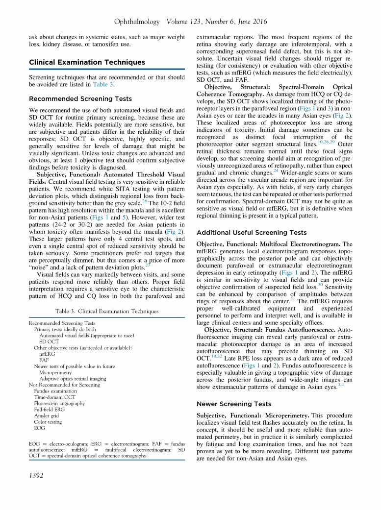
1392
extramacular regions. The most frequent regions of theretina showing early damage are inferotemporal, with acorresponding superonasal field defect, but this is not ab-solute. Uncertain visual field changes should trigger re-testing (for consistency) or evaluation with other objectivetests, such as mfERG (which measures the field electrically),SD OCT, and FAF.
Objective, Structural: Spectral-Domain OpticalCoherence Tomography. As damage from HCQ or CQ de-velops, the SD OCT shows localized thinning of the photo-receptor layers in the parafoveal region (Figs 1 and 3) in non-Asian eyes or near the arcades in many Asian eyes (Fig 2).These localized areas of photoreceptor loss are strongindicators of toxicity. Initial damage sometimes can berecognized as distinct focal interruption of thephotoreceptor outer segment structural lines.10,28,29 Outerretinal thickness remains normal until these focal signsdevelop, so that screening should aim at recognition of pre-viously unrecognized areas of retinopathy, rather than expectgradual and chronic changes.24 Wider-angle scans or scansdirected across the vascular arcade region are important forAsian eyes especially. As with fields, if very early changesseem tenuous, the test can be repeated or other tests performedfor confirmation. Spectral-domain OCT may not be quite assensitive as visual field or mfERG, but it is definitive whenregional thinning is present in a typical pattern.
Additional Useful Screening Tests
Objective, Functional: Multifocal Electroretinogram. ThemfERG generates local electroretinogram responses topo-graphically across the posterior pole and can objectivelydocument parafoveal or extramacular electroretinogramdepression in early retinopathy (Figs 1 and 2). The mfERGis similar in sensitivity to visual fields and can provideobjective confirmation of suspected field loss.30 Sensitivitycan be enhanced by comparison of amplitudes betweenrings of responses about the center.31 The mfERG requiresproper well-calibrated equipment and experiencedpersonnel to perform and interpret well, and is available inlarge clinical centers and some specialty offices.
Objective, Structural: Fundus Autofluorescence. Auto-fluorescence imaging can reveal early parafoveal or extra-macular photoreceptor damage as an area of increasedautofluorescence that may precede thinning on SDOCT.10,32 Late RPE loss appears as a dark area of reducedautofluorescence (Figs 1 and 2). Fundus autofluorescence isespecially valuable in giving a topographic view of damageacross the posterior fundus, and wide-angle images canshow extramacular patterns of damage in Asian eyes.3,4
Newer Screening Tests
Subjective, Functional: Microperimetry. This procedurelocalizes visual field test flashes accurately on the retina. Inconcept, it should be useful and more reliable than auto-mated perimetry, but in practice it is similarly complicatedby fatigue and long examination times, and has not beenproven as yet to be more revealing. Different test patternsare needed for non-Asian and Asian eyes.

Marmor et al � Screening for CQ and HCQ Retinopathy
Objective, Structural: Adaptive Optics Retinal Imaging.Special cameras with enhanced optics to reduce wave-frontdistortion can image the cone array directly and show conedamage with early disease. However, distinguishing damagefrom artifact is still difficult with current instruments, and atthe time of writing, this remains primarily a research tool.
Tests Not Recommended for Screening
Fundus Examination and Photography. Ophthalmoscopyis not a screening tool because photoreceptor damage isdetectable with other techniques well before visible changesin the fundus. A bull’s-eye, by definition, implies RPE lossand an advanced stage of toxicity.
Time-Domain Optical Coherence Tomography. Theresolution is not sufficient to detect early toxic changes.
Fluorescein Angiography. This can recognize RPE de-fects, but these are late changes.
Full-Field Electroretinogram. The full-field electroreti-nogram is a global test of retinal function and will showabnormalities in only very late CQ or HCQ toxicity. It maybe useful to judge the extent of damage beyond the macula.
Amsler Grid. Amsler grid testing is not consistentenough for reliable screening of subtle scotomas.
Color Vision Testing. Color errors may occur but arenot sensitive or specific.
Electro-oculogram. The electro-oculogram has not beenvalidated as a reliable screening test.
Management of Eyes at Risk or withRetinopathy
No diet or medical therapy has proven effective as yet to pre-vent, treat, or reduce risk from HCQ or CQ retinopathy otherthan cessation of the drug. Even stoppage of the drug does notprevent progression of retinopathy, although this is typicallymild if the toxicity is recognized before there is RPE damage.Patients with age-related maculopathy or macular dystrophiessometimes are advised to avoid excessive sun exposure andmaintain intake of lutein and zeaxanthin (which are fovealprotectants). However, the value of such recommendations forpatients at risk fromHCQor CQ exposure, or after retinopathyis recognized and the drug is stopped, is unknown.
Once definitive signs of retinopathy are recognized, thedecision to stop medication should be made in conjunctionwith the patient and the prescribing medical physician toensure that medical risks are managed (e.g., a potential flare-up of SLE). The patient can be advised about the risk offurther visual loss depending on the severity of the reti-nopathy. This risk is minimal if the retinopathy was detectedearly, but significant if there is already a bull’s-eye lesionand some reduction in central foveal thickness, becausedamage can progress for a number of years.5
References
1. Marmor MF, Kellner U, Lai TY, et al. Revised recommen-dations on screening for chloroquine and hydroxychloroquineretinopathy. Ophthalmology 2011;118:415–22.
2. Melles RB, Marmor MF. The risk of toxic retinopathy in pa-tients on long-term hydroxychloroquine therapy. JAMAOphthalmol 2014;132:1453–60.
3. Melles RB, Marmor MF. Pericentral retinopathy and racialdifferences in hydroxychloroquine toxicity. Ophthalmology2015;122:110–6.
4. Lee DH, Melles RB, Joe SG, et al. Pericentral hydroxy-chloroquine retinopathy in Korean patients. Ophthalmology2015;122:1252–6.
5. Marmor MF, Hu J. Effect of disease stage on progression ofhydroxychloroquine retinopathy. JAMA Ophthalmol2014;132:1105–12.
6. Browning DJ. Impact of the revised American Academy ofOphthalmology guidelines regarding hydroxychloroquinescreening on actual practice. Am J Ophthalmol 2013;155:418–28.
7. Nika M, Blachley TS, Edwards P, et al. Regular examinationsfor toxic maculopathy in long-term chloroquine or hydroxy-chloroquine users. JAMA Ophthalmol 2014;132:1199–208.
8. Lee MG, Kim SJ, Ham DI, et al. Macular retinal ganglion cell-inner plexiform layer thickness in patients on hydroxy-chloroquine therapy. Invest Ophthalmol Vis Sci 2014;56:396–402.
9. de Sisternes L, Hu J, Rubin DL, Marmor MF. Localization ofdamage in progressive hydroxychloroquine retinopathy on andoff the drug: inner versus outer retina, parafovea versus pe-ripheral fovea. Invest Ophthalmol Vis Sci 2015;56:3415–26.
10. Marmor MF. Comparison of screening procedures in hydroxy-chloroquine toxicity. Arch Ophthalmol 2012;130:461–9.
11. Kellner S, Weinitz S, Farmand G, Kellner U. Cystoid macularoedema and epiretinal membrane formation during progressionof chloroquine retinopathy after drug cessation. Br J Oph-thalmol 2014;98:200–6.
12. McChesney EW, Shekosky JM, Hernandez PH. Metabolism ofchloroquine-3-14C in the rhesus monkey. Biochem Pharm1967;16:2444–7.
13. McChesney EW. Animal toxicity and pharmacokinetics ofhydroxychloroquine sulfate. Am J Med 1983;75:11–8.
14. Marmor MF, Carr RE, Easterbrook M, et al. Recommenda-tions on screening for chloroquine and hydroxychloroquineretinopathy: a report by the American Academy of Ophthal-mology. Ophthalmology 2002;109:1377–82.
15. Lee JY, Luc S, Greenblatt DJ, et al. Factors associated withblood hydroxychloroquine level in lupus patients: renal func-tion could be important. Lupus 2013;22:541–2.
16. Carmichael SJ, Day RO, Tett SE. A cross-sectional study ofhydroxychloroquine concentrations and effects in people withsystemic lupus erythematosus. Intern Med J 2013;43:547–53.
17. Costedoat-Chalumeau N, Dunogué B, Leroux G, et al.A critical review of the effects of hydroxychloroquine andchloroquine on the eye. Clin Rev Allergy Immunol 2015;49:317–26.
18. Jallouli M, Galicier L, Zahr N, et al. Determinants ofhydroxychloroquine blood concentration variations in sys-temic lupus erythematosus. Arthritis Rheumatol 2015;67:2176–84.
19. Lee Y, Vinayagamoorthy N, Han K, et al. Association ofpolymorphisms of cytochrome P450 2D6 with blood hydrox-ychloroquine levels in patients with systemic lupus erythe-matosus. Arthritis Rheumatol 2016;68:184–90.
20. Leung LS, Neal JW, Wakelee HA, et al. Rapid onset of retinaltoxicity from high-dose hydroxychloroquine given for cancertherapy. Am J Ophthalmol 2015;160:799–805.
21. Navajas EV, Krema H, Hammoudi DS, et al. Retinal toxicityof high-dose hydroxychloroquine in patients with chronicgraft-versus-host disease. Can J Ophthalmol 2015;50:442–50.
1393

Ophthalmology Volume 123, Number 6, June 2016
22. Chiang E, Jampol LM, Fawzi AA. Retinal toxicity found in apatient with systemic lupus erythematosus prior to 5 years oftreatment with hydroxychloroquine. Rheumatology (Oxford)2014;53:2001.
23. Shroyer NF, Lewis RA, Lupski JR. Analysis of the ABCR(ABCA4) gene in 4-aminoquinoline retinopathy: is retinaltoxicity by chloroquine and hydroxychloroquine related toStargardt disease? Am J Ophthalmol 2001;131:761–6.
24. Grassman F, Bergholz R, Mändl J. Common synonymousvariants in ABCA4 are protective for chloroquine inducedmaculopathy (toxic maculopathy). BMC Ophthalmol2015;15:18.
25. de Sisternes L, Hu J, Rubin DL, et al. Analysis of inner andouter retinal thickness in patients using hydroxychloroquineprior to development of retinopathy. JAMA Ophthalmol http://dx.doi.org/10.1001/jamaophthalmol.2016.0155 [Epub aheadof print March 17, 2016].
26. Marmor MF, Melles RB. Disparity between visual fields andoptical coherence tomography in hydroxychloroquine reti-nopathy. Ophthalmology 2014;1257–62.
27. Marmor MF, Chien FY, Johnson MW. The value of red targetsand pattern deviation plots in visual field screening for
1394
hydroxychloroquine retinopathy. Arch Ophthalmol 2013;131:476–80.
28. Kellner S, Weinitz S, Kellner U. Spectral domain opticalcoherence tomography detects early stages of chloroquineretinopathy similar to multifocal electroretinography, fundusautofluorescence and near-infrared autofluorescence. Br JOphthalmol 2009;93:1444–7.
29. Mititelu M, Wong BJ, Brenner M, et al. Progression ofhydroxychloroquine toxic effects after drug therapy cessation:new evidence from multimodal imaging. JAMA Ophthalmol2013;131:1187–97.
30. Lai TY, Ngai JW, Chan WM, Lam DS. Visual field and multi-focal electroretinography and their correlations in patients onhydroxychloroquine therapy. Doc Ophthalmol 2006;112:177–87.
31. Lyons JS, Severns ML. Detection of early hydroxychloroquineretinal toxicity enhanced by ring ratio analysis of multifocalelectroretinography. Am J Ophthalmol 2007;143:801–9.
32. Kellner U, Renner AB, Tillack H. Fundus autofluorescenceand mfERG for early detection of retinal alterations in patientsusing chloroquine/hydroxychloroquine. Invest Ophthalmol VisSci 2006;47:3561–8.
Footnotes and Financial Disclosures
Originally received: January 28, 2016.Final revision: January 28, 2016.Accepted: January 28, 2016.Available online: March 16, 2016. Manuscript no. 2016-197.1 Department of Ophthalmology and Byers Eye Institute, Stanford Uni-versity School of Medicine, Palo Alto, California.2 Zentrum für Seltene Netzhauterkrankungen, AugenZentrum Siegburg,Siegburg, Germany.3 Department of Ophthalmology and Visual Sciences, The Chinese Uni-versity of Hong Kong, Kowloon, Hong Kong.4 Department of Ophthalmology, Kaiser Permanente, Redwood CityMedical Center, Redwood City, California.5 Department of Ophthalmology and Visual Sciences, Illinois Eye and EarInfirmary, University of Illinois, Chicago, Illinois.
Financial Disclosure(s):The author(s) have made the following disclosure(s): T.Y.Y.L.:Consultant � Allergan, Bayer Healthcare, Novartis Pharmaceuticals,Genentech; Grants/grants pending � Bayer Healthcare, Novartis Pharma-ceuticals; Lecture fees � Allergan, Bausch & Lomb, Bayer Healthcare,Novartis Pharmaceuticals; Payment � manuscript preparation fromNovartis Pharmaceuticals.
Abbreviations and Acronyms:CQ ¼ chloroquine; FAF ¼ fundus autofluorescence;HCQ ¼ hydroxychloroquine; mfERG ¼ multifocal electroretinogram;RPE ¼ retinal pigment epithelium; SD OCT ¼ spectral-domain opticalcoherence tomography; SLE ¼ systemic lupus erythematosus.
Correspondence:Flora Lum, MD, Department of Quality of Care and Knowledge BaseDevelopment, American Academy of Ophthalmology, 655 Beach Street,San Francisco, CA 94109-1336. E-mail: [email protected].