Embed Size (px)
Citation preview
9/3/2020
1
Recognizing and Treating Shock in the Prehospital Setting

9/3/2020
2
Special Thanks to Our Sponsor

9/3/2020
3
Your Presenters
Dr. Raymond L. Fowler, MD, FACEP, FAEMS
James M. Atkins MD Distinguished Professor of Emergency Medical Services,
Chief of the Division of EMS Department of Emergency Medicine
University of Texas Southwestern Medical Center
Dallas, TX
Dr. Melanie J. Lippmann, MD FACEP
Associate Professor of Emergency MedicineBrown University
Alpert Medical School Attending Physician
Rhode Island Hospital and The Miriam Hospital Providence, RI

9/3/2020
4
Scenario
SHOCK INDEX?? Pulse ÷ Systolic

9/3/2020
5
What is Shock?
Shock is a progressive state of cellular hypoperfusion in which insufficient oxygen is available to meet tissue demands
It is key to understand that when shock occurs, the body is in distress. The shock response is mounted by the body to attempt to maintain systolic blood pressure and
brain perfusion during times of physiologic distress. This shock response can accompany a broad spectrum of clinical conditions that stress the body, ranging from
heart attacks, to major infections, to allergic reactions.

9/3/2020
6
Causes of Shock
Shock may be caused when oxygen intake, absorption, or delivery fails, or when the cells are unable to take up and use the delivered oxygen to generate sufficient energy to carry out cellular functions.

9/3/2020
7
Causes of Shock
Hypovolemic Shock
Inadequate circulating fluid leads to a diminished cardiac output, which results in an inadequate
delivery of oxygen to the tissues and cells
Distributive Shock
A precipitous increase in vascular capacity as blood vessels dilate and
the capillaries leak fluid, translates into too little peripheral vascular resistance
and a decrease in preload, which in turn reduces cardiac output
9/3/2020
8
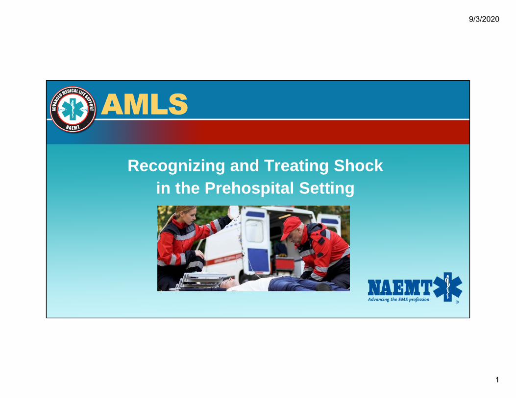
Causes of Shock
Cardiogenic Shock
The heart is unable to circulate sufficient blood to meet the
metabolic needs of the body. Either right or left ventricular failure can lead to cardiogenic shock and
may include dysrhythmias, a cardiac structural disorder, or the
action of certain toxins
Obstructive Shock
Obstruction to the forward flow of blood exists in the great vessels or heart. Significant causes are pericardial tamponade, massive
pulmonary embolism, and tension pneumothorax.
9/3/2020
9
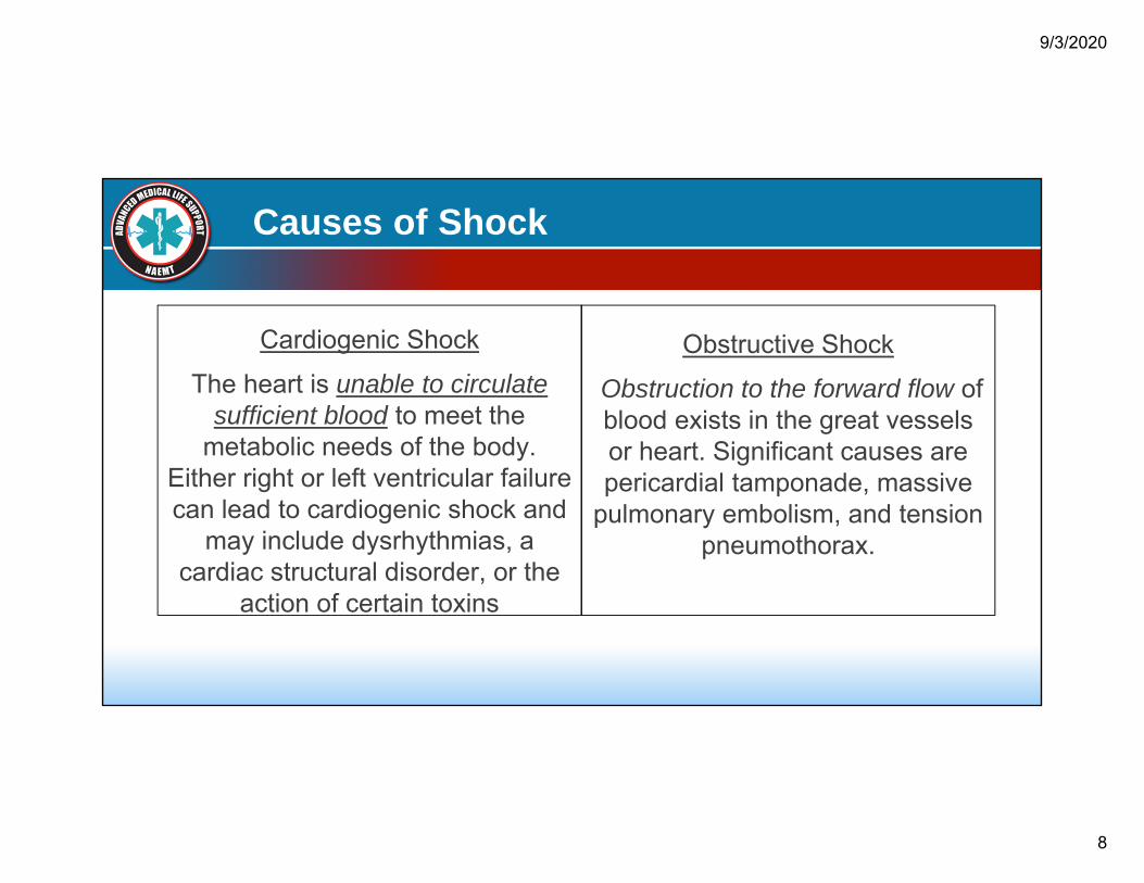
Signs and Symptoms of Shock
Shock occurs in three successive phases—
compensated, decompensated, and
irreversible

9/3/2020
10
AMLS Assessment Pathway
1. Initial Observationsa.Scene Safety Considerationb.Patient Cardinal Presentationc.Primary Survey
i. Consciousness, Airway & Breathing, Circulation/Perfusion
2. First Impressiona. History Takingb. Secondary Survey
i. Vital signs and physical examc. Diagnostic
i. Pulse Oximetry, Capnography, Electrocardiogram, Laboratory Studies, Compensatory Reserve
d. Redefine the Differential Diagnosis
3. Ongoing Managementa. Fluid Resuscitationb. Temperature Regulationc. Vasopressor Administrationd. Administration of Blood Products
i. Transfusion Reactionsii. Hemolytic Reactions
9/3/2020
11
Initial Observations : Scene Safety
Scene Safety Consideration
Scene safety is critical in approaching any patient.
When dealing with a patient who is critically ill, it is easy to miss a safety issue!
Take the time to ensure the scene remains safe for you and all involved.

9/3/2020
12
Initial Observations: Chief Complaint
For the patient suspected of shock you must determine the following:
Do I see any signs of a life threat as I approach my patient?
Are there altered level of consciousness or respiratory distress?
Does the patient’s skin show signs of shock?
(Pale, ashen, diaphoretic, mottled, or hives)
Do the surroundings suggest the possibility of shock? Vomitus? Blood?
9/3/2020
13
Initial Observation: Primary Survey
You should focus your assessment and history collection on the components that will help you identify and treat the life threats and complete these interventions once the patient’s condition stabilizes:
● Level of Consciousness● Airway/Breathing/Circulation/Perfusion● Are the radial, carotid, and femoral pulses
strong, or are they weak, thready, absent?● Is the rate too fast or too slow?● Is the rate regular or irregular?
Always Explain a Tachycardia!!
9/3/2020
14
First Impression: History Taking
Obtaining a thorough history, including an account of the present illness and a comprehensive past medical history,
is essential to determining the “type” of shock.

9/3/2020
15
SAMPLER mnemonic
Signs & Symptoms- What can you see? What is the chief complaint?
Allergies- What have you come in contact with? Meds, Insects, Pollen, Food, Latex?
Medications- Prescriptions, OTC, Herbal, Street
Past Medical History- Has this happened before? Is this a preexisting condition?
Last Oral Intake- When did you eat last? What was it? Nausea or Vomiting?
Events- What were you doing when you started to feel this way?
Risks- Risks to chief complaint (e.g. cardiac disease, COPD, DM)
SAM
PLE
R
9/3/2020
16
OPQRST mnemonic
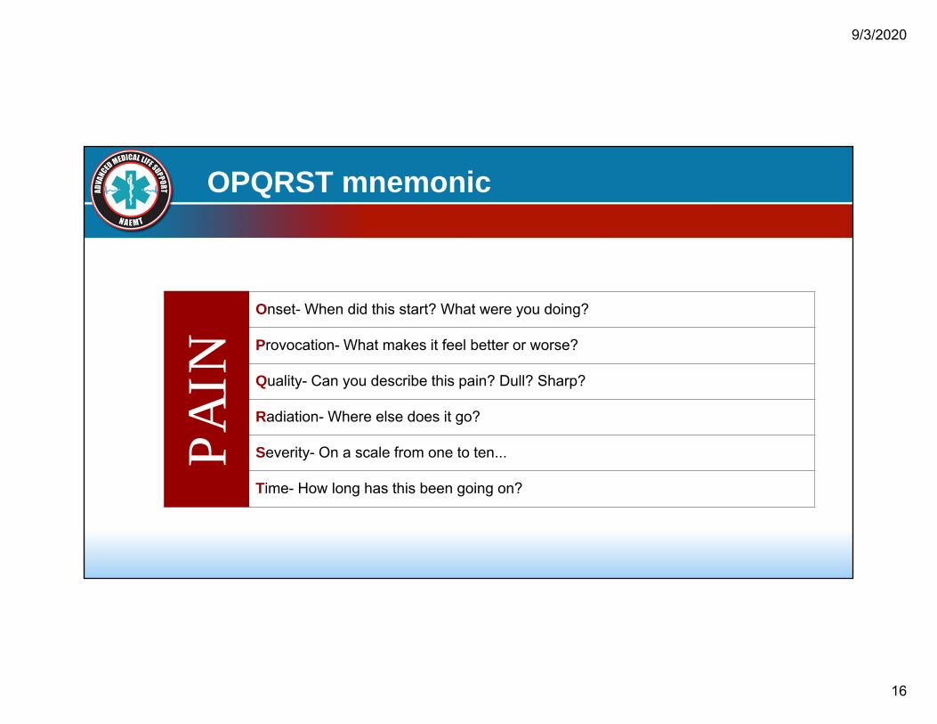
Onset- When did this start? What were you doing?
Provocation- What makes it feel better or worse?
Quality- Can you describe this pain? Dull? Sharp?
Radiation- Where else does it go?
Severity- On a scale from one to ten...
Time- How long has this been going on?
PAIN

9/3/2020
17
Medications Affecting Shock

9/3/2020
18
First Impression: Secondary Survey
Vital Signs
Vital signs (blood pressure, pulse rate, respiratory rate, &
temperature) are essential to determining the patient’s stability and identifying the type of shock. Most common signs are hypotension, tachycardia, tachypnea, and cool skin.
Physical Exam
Physical exam focuses on determining the cause of the shock and selecting proper intervention
9/3/2020
19
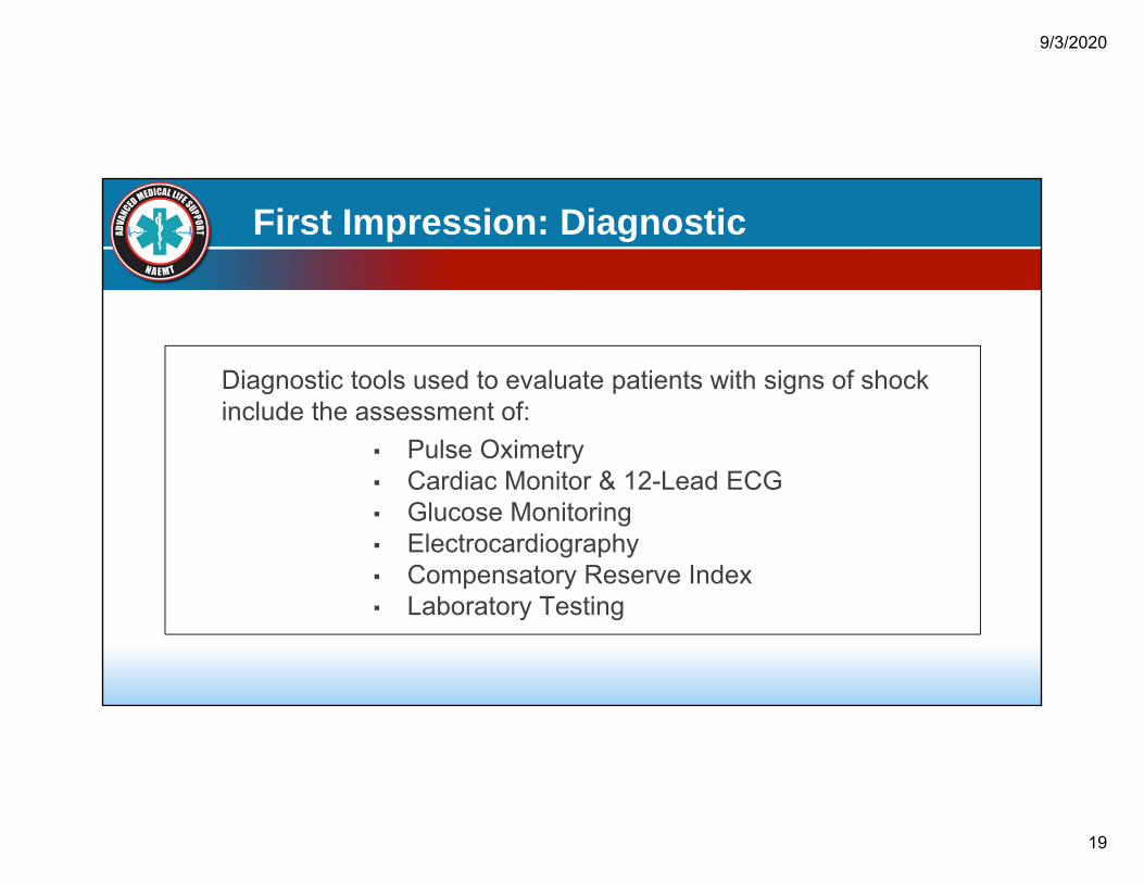
First Impression: Diagnostic
Diagnostic tools used to evaluate patients with signs of shock include the assessment of:
▪ Pulse Oximetry▪ Cardiac Monitor & 12-Lead ECG▪ Glucose Monitoring▪ Electrocardiography▪ Compensatory Reserve Index▪ Laboratory Testing

9/3/2020
20
Monitor &/or 12-Lead ECG for Shock
9/3/2020
21
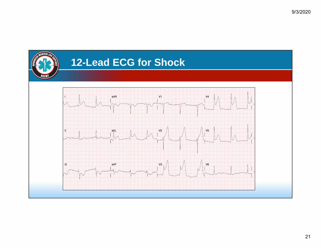
12-Lead ECG for Shock

9/3/2020
22
Glucose Monitoring
As part of the routine evaluation of the patient in shock, obtain a random glucose level for assessment and documentation
Significant hypoglycemia or hyperglycemia should be considered in the differential diagnosis of the cause of your
patient’s clinical shock condition

9/3/2020
23
Compensatory Reserve Index

9/3/2020
24
Compensatory Reserve Index
9/3/2020
25
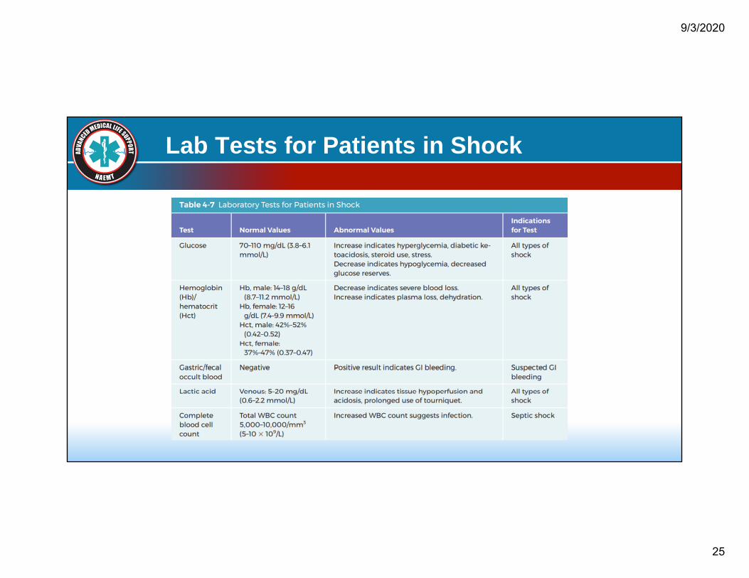
Lab Tests for Patients in Shock

9/3/2020
26
Lab Tests for Patients in Shock

9/3/2020
27
Lab Tests for Patients in Shock
9/3/2020
28
First Impression: Refine the Differential Diagnosis
You may have determined in your primary surveythat the patient was in shock,
but you may not have determined the underlying cause
It is important to move quicklythrough this assessment with a calculated purpose
in order to identify the cause of shock and initiate the necessary treatment

9/3/2020
29
Ongoing Management
The rule of thumb is to assess, intervene, and reassess
During your ongoing management, repeat the primary survey and vital signs, revisit the chief complaint, and
monitor the patient’s response to any treatment you have administered
Always consider the possibility of trauma

9/3/2020
30
Management: Fluid Resuscitation
An initial bolus of 20 to 30 mL/kg (1,000 to 2,000 mL) of isotonic fluid should be given if the patient shows no signs of fluid overload
If the patient is at risk for fluid overload, a more modest bolus of 250 to 500 mL, followed by a reassessment, is appropriate
The purpose of fluid resuscitation should be to enhance perfusion to maintain a MAP at 60 to 70 mm Hg or a systolic pressure at 80 to 90 mmHg
If bleeding is suspected, administration of blood products is indicated
If massive transfusions are required, early administration of fresh-frozen plasma and platelets has been shown to improve survival
9/3/2020
31
Management: Temperature Regulation
The body expends a great deal of energy to maintain a normal temperature. Vasoconstriction shunts blood away from peripheral
tissues, and the body will expend valuable energy trying to stay warm. Keep the patient warm!
The ambulance or resuscitation room should be kept warm, and the patient should be covered with a blanket when practical.
Administration of warmed fluids will assist in maintaining body temperature and should be initiated when feasible.

9/3/2020
32
Management: Vasopressor Administration
Vasopressors, which are medications augmenting blood pressure, are an efficient adjunct treatment in patients
with certain types of shockThe following vasopressors and inotropes may be administered:
Vasopressors • Norepinephrine (preferred agent in most shock scenarios requiring vasopressors)• Epinephrine • Phenylephrine • Dopamine (less preferred)
Inotropes• Dobutamine • Norepinephrine • Epinephrine • Dopamine (less preferred)

9/3/2020
33
Management: Administration of Blood Products
Blood administration may be a viable option
in the prehospital setting
When the patient is actively bleeding, is anemic, is in
shock, and/or has a serious bleeding disorder,
administration of blood products is indicated

9/3/2020
34
Transfusion Reactions
There are generally two complications of blood product administration: infection and immune reactions
Improved methods of screening donors and blood products have decreased problems with the spread of infections
There remains a small risk, especially for cytomegalovirus, which is a common virus that is rarely serious in the general population
9/3/2020
35
Hemolytic Reactions
When the recipient’s antibodies recognize and react to transfused blood as an antigen, the donor RBCs may be
destroyed or hemolyzed. This hemolytic reaction can be quick and aggressive or slower, depending on the immune response
Errors in the blood administration process can create fatal hemolytic reactions. When this occurs, most transfused cells
are destroyed in an overwhelming immune response

9/3/2020
36
Treatment of Hypovolemic Shock
Inadequate circulating fluid leads to a diminished cardiac output, which results in an inadequate delivery of oxygen to
the tissues and cells
9/3/2020
37
Treatment of Distributive shock
Distributive shock stems from a precipitous increase in vascular capacity as blood vessels dilate
and the capillaries leak fluid
Too much vascular space translates into too little peripheral vascular resistance and a decrease in preload, which in turn
reduces cardiac output and results in shock
Causes of Distributive shock include: sepsis, anaphylaxis, neurogenic shock, and toxins
9/3/2020
38
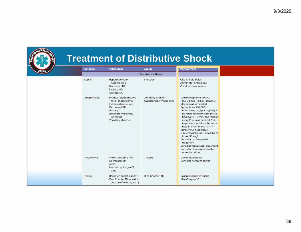
Treatment of Distributive Shock

9/3/2020
39
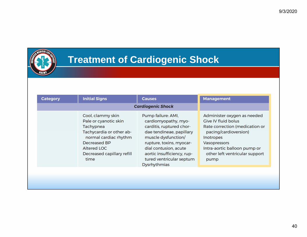
Treatment of Cardiogenic Shock
Occurs when the heart is unable to circulate sufficient blood to meet the metabolic needs of the body
Either right or left ventricular failure can lead to cardiogenic shock and may include dysrhythmias, a cardiac structural
disorder, or the action of certain toxins
Initial treatment of a patient in cardiogenic shock must focus on stabilization of airway, breathing, and circulation
9/3/2020
40
Treatment of Cardiogenic Shock
9/3/2020
41
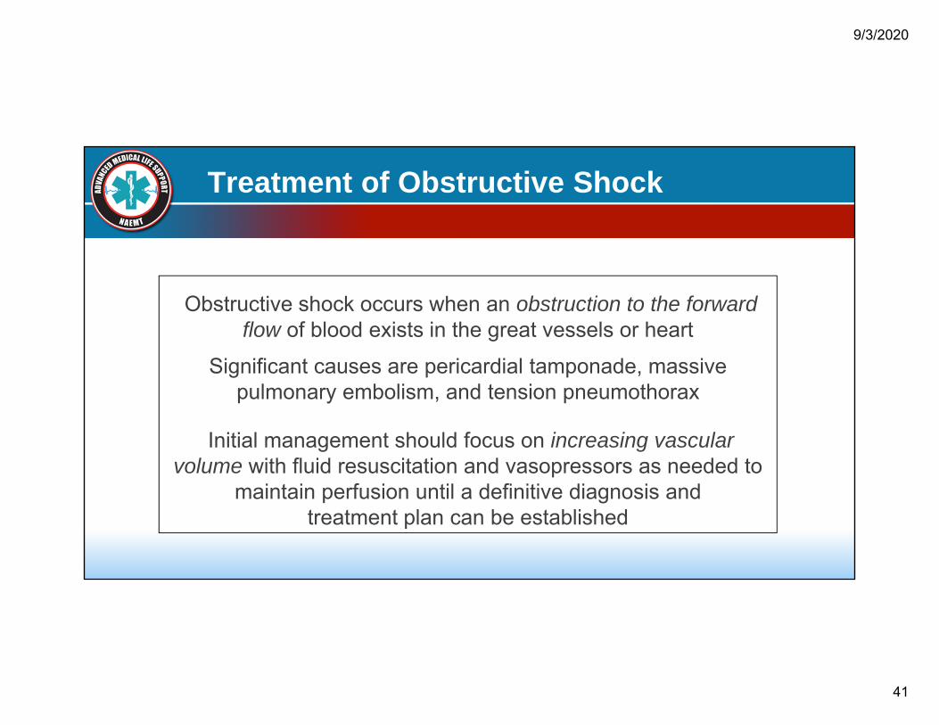
Treatment of Obstructive Shock
Obstructive shock occurs when an obstruction to the forward flow of blood exists in the great vessels or heart
Significant causes are pericardial tamponade, massive pulmonary embolism, and tension pneumothorax
Initial management should focus on increasing vascular volume with fluid resuscitation and vasopressors as needed to
maintain perfusion until a definitive diagnosis and treatment plan can be established

9/3/2020
42
Treatment of Obstructive Shock
9/3/2020
43
Putting it All Together
Early, accurate identification of the patient’s stage and type of shock is essential in managing this condition
Sound clinical reasoning skills, a thorough assessment, and careful but expedient interpretation of diagnostic findings are necessary to provide
effective treatment for the patient in shock
9/3/2020
44
Thank You Presenters & Sponsor
Dr. Raymond L. Fowler, MD, FACEP, FAEMS
James M. Atkins MD Distinguished Professor of Emergency Medical Services,
Chief of the Division of EMS Department of Emergency Medicine
University of Texas Southwestern Medical Center
Dallas, TX
Dr. Melanie J. Lippmann, MD FACEP
Associate Professor of Emergency MedicineBrown University
Alpert Medical School Attending Physician
Rhode Island Hospital and The Miriam Hospital Providence, RI
9/3/2020
45
Contact Us
NATIONAL ASSOCIATION OF EMERGENCY MEDICAL TECHNICIANS1-800-34-NAEMTP: 601-924-7744F: [email protected]