Embed Size (px)
Citation preview

Recognition of Nucleic Acid Bases and Base-pairs byHydrogen Bonding to Amino Acid Side-chains
Alan C. Cheng1,2, William W. Chen1, Cynthia N. Fuhrmann1 andAlan D. Frankel1*
1Department of Biochemistryand BiophysicsUniversity of California513 Parnassus AvenueSan FranciscoCA 94143-0448, USA
2Graduate Group in BiophysicsUniversity of CaliforniaSan Francisco, San FranciscoCA 94143-0448, USA
Sequence-specific protein–nucleic acid recognition is determined, in part,by hydrogen bonding interactions between amino acid side-chains andnucleotide bases. To examine the repertoire of possible interactions, wehave calculated geometrically plausible arrangements in which aminoacids hydrogen bond to unpaired bases, such as those found in RNAbulges and loops, or to the 53 possible RNA base-pairs. We find 32possible interactions that involve two or more hydrogen bonds to the sixunpaired bases (including protonated A and C), 17 of which have beenobserved. We find 186 “spanning” interactions to base-pairs in which theamino acid hydrogen bonds to both bases, in principle allowing particularbase-pairs to be selectively targeted, and nine of these have beenobserved. Four calculated interactions span the Watson–Crick pairs and15 span the G:U wobble pair, including two interesting arrangementswith three hydrogen bonds to the Arg guanidinum group that have notyet been observed. The inherent donor–acceptor arrangements of thebases support many possible interactions to Asn (or Gln) and Ser (or Thror Tyr), few interactions to Asp (or Glu) even though several alreadyhave been observed, and interactions to U (or T) only if the base is in anunpaired context, as also observed in several cases. This study highlightshow complementary arrangements of donors and acceptors can contrib-ute to base-specific recognition of RNA, predicts interactions not yetobserved, and provides tools to analyze proposed contacts or designnovel interactions.
q 2003 Elsevier Science Ltd. All rights reserved
Keywords: RNA structure; non-Watson–Crick base-pairs; DNA–proteininteractions; RNA–protein interactions*Corresponding author
Introduction
The ability of proteins to recognize specific RNAsites is important in many biological systems butthe “rules” governing these interactions are notwell understood. This is in part because the struc-tural database of protein–RNA complexes is stillrelatively limited (despite the recent addition ofthe ribosomal subunit structures) and, perhapsmore importantly, because RNA structures are sodiverse. In addition to Watson–Crick helices,RNAs often contain non-Watson–Crick base-pairs,unpaired bases such as those in bulges and loops,and base-triples or other higher-order tertiary
interactions.1,2 Therefore many of the principlesof protein–DNA recognition inferred from thelarge number of solved structures3,4 may not applyto RNA.
Despite the current gaps in knowledge, it isapparent that one important determinant of speci-ficity in both DNA and RNA complexes is thecomplementary nature of hydrogen bonding inter-actions between polar groups on the protein side-chains and nucleic acid bases. A seminal studyby Seeman et al. conducted before the structure ofeven a single protein–nucleic acid complex hadbeen solved5 systematically examined the possiblehydrogen bonding interactions between aminoacid side-chains and groups along the edges of theWatson–Crick base-pairs. They concluded thatinteractions involving two hydrogen bonds wouldbe required to uniquely distinguish each base-pairfrom the others, and inferred that two hydrogenbonds from a single functional group would
0022-2836/03/$ - see front matter q 2003 Elsevier Science Ltd. All rights reserved
Present address: A. C. Cheng, Pfizer DiscoveryTechnology Center, 620 Memorial Drive, Cambridge, MA02139, USA.
E-mail address of the corresponding author:[email protected]
doi:10.1016/S0022-2836(03)00091-3 J. Mol. Biol. (2003) 327, 781–796
specify a site with higher precision than from twoindependent groups, analogous to the “chelateeffect” in which formation of one bond favorsformation of additional bonds by an increase ineffective concentration.6 Based on their analysis,Seeman et al. predicted two interactions in whichprecisely positioned side-chains in the DNA majorgroove could discriminate amongst all the base-pairs: one in which the guanidinium group of Argdonates two hydrogen bonds to the O6 and N7acceptor groups of guanine and a second in whichthe carboxamide group of Asn (or Gln) hydrogenbonds to the N7 acceptor and N6 donor groupsof adenine. These interactions are indeed themost commonly observed in protein–DNA com-plexes,4,7 – 9 and the importance of such directamino acid–base hydrogen bonds in determiningsequence specificity has been confirmed by manystructure–function studies.
Several detailed studies have analyzed the inter-actions observed in protein–DNA complexes,partly in efforts to determine whether a “recog-nition code” exists for DNA double helices.3,4,8 – 14
It seems clear that while no simple code exists,some common interaction patterns between aminoacids and bases can be found, particularly withina given structural context such as the zinc fingeror helix-turn-helix motif.3,11,15 Hydrogen bondinginteractions to the bases comprise about two-thirds of all base-specific contacts,4 and interactionsinvolving two hydrogen bonds from the side-chain are dominated by the Arg–G and Asn(Gln)–A interactions described above. The only otherfrequent bidentate interaction utilizes the aminogroup of Lys to hydrogen bond to the O6 andN7 acceptors of guanine, although other twohydrogen-bond interactions are found that utilizebifurcated bonds or donors and acceptors fromthe peptide backbone.4 A statistical survey of 28protein–DNA complexes found that side-chainspossessing both donor and acceptor atoms morefrequently use the donor atom for hydrogenbonding.8 In addition to direct amino acid–basehydrogen bonds, it is clear that other types of inter-actions contribute to DNA recognition, includingwater-mediated hydrogen bonds, van der Waalscontacts, and interactions to the sugar–phosphatebackbone, and that the structural context in whichthe interactions are presented is an inherent partof the recognition process.3,4,10,13,14
The analysis of protein–RNA interactions is atan earlier stage and the available structuresrepresent only a small subset of possible RNAtertiary elements, but some characteristics arebeginning to emerge.2,16 – 19 With respect to hydro-gen bonding, perhaps the most obvious differencebetween RNA and DNA complexes is the use ofthe ribose 20OH group in about a quarter of allhydrogen bonds.19 In addition, of all the observedbase-specific interactions, hydrogen bonds appearless dominant than in DNA complexes,17 – 19
probably because a significant number of basesare not stacked within Watson–Crick duplexes
and consequently some bases are sequesteredfrom solvent via van der Waals interactions withthe protein. Nevertheless, the importance of hydro-gen bonding for RNA-binding specificity is asapparent for RNA as it is for DNA. It is inherentlymore difficult to evaluate the possible amino acidhydrogen-bonding interactions to RNA than toDNA5 given the large number of possible RNAbase configurations, and therefore a systematiccomputational approach is required. Here, wereport the calculation of databases of hydrogen-bonding interactions between amino acids andbases or base-pairs that can occur in RNA struc-tures. The databases include interactions betweenunpaired bases, such as those found in bulges orloops, and non-Watson–Crick base-pairs, some ofwhich involve multiple hydrogen bonds that maybe used to uniquely recognize bases in particularstructural contexts. The databases may be usefulnot only for analyzing existing interactions butalso for designing novel interactions in RNA-binding proteins or peptides, in combination withother experimental approaches.
Computational Approach
Database construction
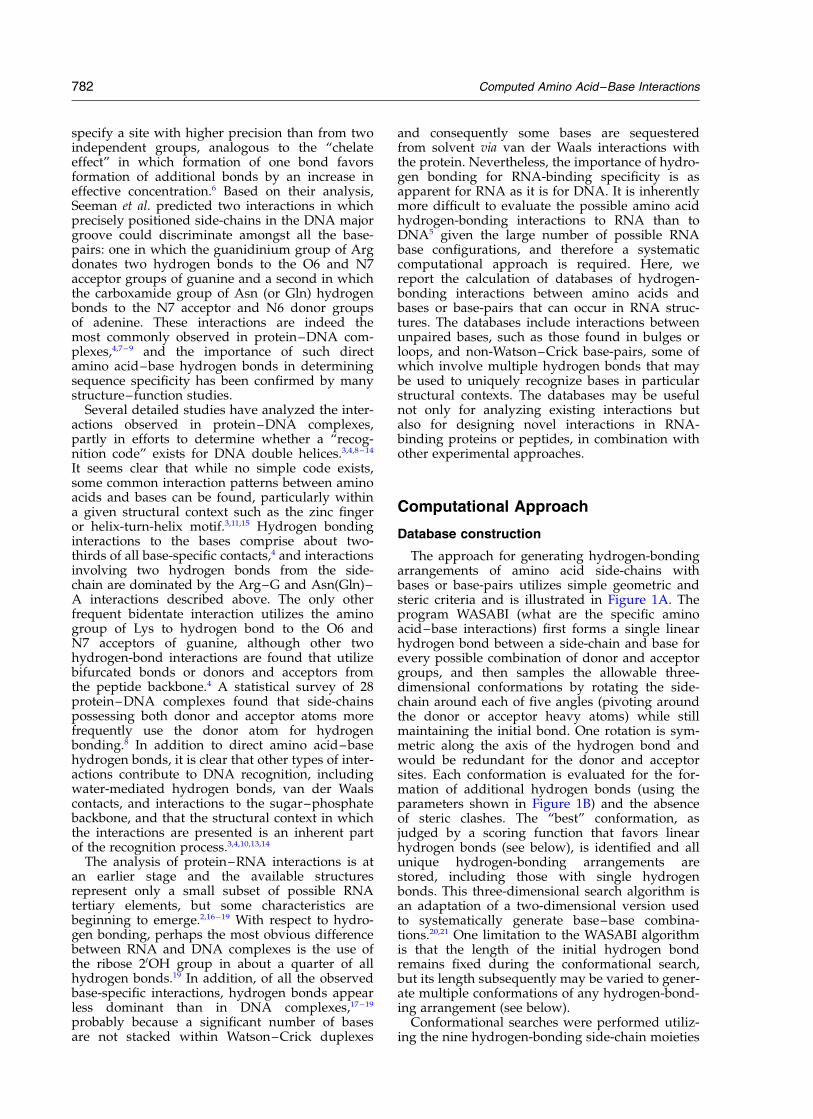
The approach for generating hydrogen-bondingarrangements of amino acid side-chains withbases or base-pairs utilizes simple geometric andsteric criteria and is illustrated in Figure 1A. Theprogram WASABI (what are the specific aminoacid–base interactions) first forms a single linearhydrogen bond between a side-chain and base forevery possible combination of donor and acceptorgroups, and then samples the allowable three-dimensional conformations by rotating the side-chain around each of five angles (pivoting aroundthe donor or acceptor heavy atoms) while stillmaintaining the initial bond. One rotation is sym-metric along the axis of the hydrogen bond andwould be redundant for the donor and acceptorsites. Each conformation is evaluated for the for-mation of additional hydrogen bonds (using theparameters shown in Figure 1B) and the absenceof steric clashes. The “best” conformation, asjudged by a scoring function that favors linearhydrogen bonds (see below), is identified and allunique hydrogen-bonding arrangements arestored, including those with single hydrogenbonds. This three-dimensional search algorithm isan adaptation of a two-dimensional version usedto systematically generate base–base combina-tions.20,21 One limitation to the WASABI algorithmis that the length of the initial hydrogen bondremains fixed during the conformational search,but its length subsequently may be varied to gener-ate multiple conformations of any hydrogen-bond-ing arrangement (see below).
Conformational searches were performed utiliz-ing the nine hydrogen-bonding side-chain moieties
782 Computed Amino Acid–Base Interactions

shown in Figure 1C, including unprotonated andprotonated forms of histidine, and either with thesix RNA bases (A, C, G, U, Aþ, Cþ) or 53 possibleRNA base-pairs generated by Walberer et al.21 Inaddition, we constructed interactions with theadditional DNA base, thymine, and the 17 possiblebase-pairs that utilize thymine.20 These types ofamino acid–DNA interactions may occur inthe context of single-stranded sites or in heliceswith extruded bases.
For our steric parameters, van der Waals radiitaken from AMBER22 were divided by 21/6 toapproximate hard sphere radii;23 these radii werefurther reduced by a factor of 0.8 to include geome-tries slightly outside a reasonable steric range.Polar hydrogen atoms were assigned van derWaals radii of 0.2 A and also were further reducedby the 0.8 steric parameter. Because the polarhydrogen atoms had such small radii, weimplemented a filter that removed conformationsin which two polar hydrogen atoms were closer
than 2.5 A, approximately the distance betweentwo oxygen atoms, thereby eliminating unfavor-able arrangements with two nearby positivecharges. Amino acid side-chains were constructedusing the LEAP package with param96 residuedefinitions.22
The parameters used to define a hydrogen bond(Figure 1B) were chosen based on the analysis ofsmall molecule high-resolution crystalstructures24,25 and on an empirical test of thedonor angle parameter. A maximum distance of3.4 A was used for all hydrogen bonds. We esti-mated acceptor and donor parameters from thecrystal structure analysis, which reported onlyacceptor–hydrogen–donor atom angles, by assum-ing a hydrogen–acceptor length of 2.0 A and adonor–hydrogen bond length of 1.0 and usingdonor angle ¼ sin21(D), where D is as describedpreviously.25 For the nitrogen acceptor angle,values of 0(^22)8 for two-center hydrogen bondsand 0(^45)8 for three-center hydrogen bonds were
Figure 1. A, Schematic of the WASABI search method. Rotations 1–3 illustrate rotations about the donor atom androtations 4 and 5 illustrate rotations about the acceptor atom, conceptually similar to first holding the amino acidfixed and rotating the base (1–3) and then holding the base fixed and rotating the amino acid (4, 5). B, The three par-ameters used to define a hydrogen bond: the acceptor angle, the donor angle, and distance between heavy atoms,with parameters listed for the different atom types. C, The nine hydrogen-bonding moieties of the amino acid side-chains, with arrows indicating donor and acceptor positions.
Computed Amino Acid–Base Interactions 783

three standard deviations from the mean andtherefore included virtually all observed hydrogenbonds. Thus, we used a nitrogen acceptor angle of0(^50)8 to include slightly unreasonable geome-tries and to allow us to evenly sample confor-mations using a 48 step size. We used an oxygenacceptor angle of 0(^90)8 for similar reasons.To determine an appropriate cutoff for the donorangle parameter, we performed a set of WASABIcalculations using angles from 0(^30)8 to 0(^40)8and found that 0(^36)8 generated all known inter-actions (see below) and that at least some of theadditional conformations generated using a0(^38)8 angle appeared reasonable by inspection.A similar empirical approach was used to selectthe 0(^18)8 donor angle parameter used to con-struct the base–base interaction database, whichwas substantially more restrictive due to the planarnature of the conformational search.21
As mentioned above, WASABI generates mul-tiple conformations with the same hydrogenbonding arrangement and thus we devised anempirical scoring function in order to select arepresentative conformation with as planar anarrangement as possible. Scores (S) were calculatedover all hydrogen bonds as follows:
S ¼ S{w1ð1 þ ½A=d�4 2 ½B=d�2Þ þ w2 sin2 ud
þw3 sin2 ua þ w4 sin2 up};
where w1:w2:w3:w4 ¼ 100:30:1:1 for oxygenacceptors and 100:30:10:0 for nitrogen acceptors, ua
is the acceptor angle, ud is the donor angle, up isthe angle between the plane of the side-chain andthe plane of the base or base-pair, d is the distancebetween the heavy atom donor and acceptor, and
A and B are parameterized to mean hydrogenbond distances of 2.95 A for N–O bonds, 2.73 Afor O–O bonds, and 2.90 A for N–N bonds, whichare average distances calculated from the databaseof known protein–nucleic acid complexes andsimilar to those previously reported.24 – 28
Finally, we wished to ensure that each calculatedarrangement could accommodate a nucleotidebackbone and complete amino acid side-chain. Weadded C20-endo or C30-endo ribose sugars (gener-ated using AMBER parameters) to each base in acombinatorial manner, rotating the sugars by 3608around the C10 –N1 bond in 28 increments. Wesimilarly added all amino acid rotamers fromDunbrack & Cohen29 (August 10, 1999 release) in acombinatorial manner and identified any model inwhich no set of sugar and rotamer conformationscould be accommodated sterically. These modelswere analyzed further using DIVERSIGEN asdescribed below. Although we used one hydrogenbond moiety to represent Asn(Gln), Asp(Glu), andSer(Thr) side-chains (Figure 1C), rotamers of allrepresented amino acids also were added for thesefinal steric tests. Interestingly, two interactionsinvolving bifurcated hydrogen bonds with Ser andThr were found to be sterically impossible butcould occur with Tyr. Despite the larger size of theTyr side-chain, the planarity of the aromatic ringmakes the interaction more favorable than withthe Ser or Thr side-chains (see Results).
Diversity generator
Each hydrogen-bonding arrangement is rep-resented in our databases by a single conformation,however many three-dimensional conformationstypically are possible for each arrangement. Weconstructed a diversity generating program, DIVE-RSIGEN, that begins with one conformation andcreates a set of conformations chosen to representthe sterically accessible space for the particularhydrogen-bonding arrangement. For arrangementsthat could not accommodate the nucleotide sugarsand side-chain rotamers (see above), we generatedten representative conformations and tested eachfor its ability to accommodate the sugars and rota-mers. Those few arrangements that could not (seeTable 1) were eliminated from the databases.
To generate conformational diversity, first thelength of each hydrogen bond in a particulararrangement is set to three values, correspondingto short, median, and long distances that cover theexperimentally observed range for each type ofdonor–acceptor pair. Next, the same five anglesvaried in the WASABI search again are incremen-tally varied, beginning with a large step size, andhydrogen bond distances and angles are monitoredand steric tests performed using the parametersdescribed above to retain plausible conformationswith the appropriate hydrogen bonds. Confor-mations generated using each of the startinghydrogen bond lengths are retained, until a totalof ,10,000 are generated (or 1000–4000 in a few
Table 1. Numbers of computed amino acid–base andamino acid–base pair interactions
Single bases Base- pairs
WASABI output 470 7730Remove backbone incompatible 470 7718Remove U/T redundancies 426 5819Remove Asn/Gln, Arg, Hisþ redun
dancies385 5076
Remove Tyr redundancies 344 4612Remove bifurcated interactions 261 3519Remove single H-bond interactions 36 457Remove Aþ, Cþ redundancies 32 423Remove non-spanning base-pair
interactionsN/A 186
The raw output from WASABI was filtered to remove steri-cally restricted conformations, calculational and structuralredundancies, and bifurcated and single hydrogen-bondedinteractions, as described in the text. Bifurcated interactionsrefer only to those in which a donor or acceptor atom on theamino acid is simultaneously involved in two hydrogen bondsto a base and does not include those in which a bifurcatedbond exists between two bases of a base-pair. The narrowdonor angle parameter used to construct the base-pairs limitsthe number of bifurcated bonds.21
784 Computed Amino Acid–Base Interactions
particularly sterically restricted cases). The stepsize used for each of the angles varied is adjustediteratively to achieve the desired ,10,000 confor-mations. These conformations then are clusteredinto the desired number of representatives (10 to,10,000), chosen to cover conformational space ascompletely as possible. It is particularly difficult toachieve a good representation when choosing asmall number of conformations to represent abroad space. To assess whether a chosen set of con-formations reasonably represents the spacesampled, we define similarity of any two confor-mations as the Euclidean distance between thefive parameters of the WASABI search.For conformations a and b, with parameter coordi-nates {a1, a2…a5}, {b1, b2…b5}, Euclidean distance (dE)is defined by dE ¼ ka 2 bk. Thus, we are definingconformational similarity based on hydrogenbonding parameters and not on the r.m.s.d. ofthree-dimensional coordinates. The clustering rou-tine produces conformations within each hydrogenbond class (short, median, long), with their numberproportional to the number of conformationsfound for each in the WASABI search.
Database search of observed interactions
We identified all hydrogen bonding interactionsbetween amino acids and bases or base-pairs inprotein–DNA and protein–RNA complexes in thePDB (July 15, 2002 release; see Table 2). For thissearch, we slightly relaxed the hydrogen bonding
parameters, using a donor angle of 0(^40)8 and amaximum distance of 3.5 A to ensure that noplausible interactions would be missed. Of the 433protein–DNA complexes examined, 378 werecrystal structures, 22 were averaged NMR struc-tures, and 33 were NMR ensembles. Of the 132protein–RNA complexes examined, 103 were crys-tal structures, seven were averaged, NMR struc-tures, and 22 were NMR ensembles. Only crystalstructures with ,3.5 A resolution were used.Hydrogen atoms were added using InsightII(Biosym) or AMBER PROTONATE, and the searchprogram automatically calculated optimized place-ment of hydrogen atoms where rotatable hydroxyland amine groups are involved. Optimized hydro-gen positions were determined by rotating thehydrogen into the plane formed by points D, A,and X (Figure 1B) for each potential hydrogenbond. Polymerases and topoisomerases were con-sidered non-specific binders and were not exam-ined, and for crystal structures with multiplecomplexes in a unit cell, only one representativewas included. For the 30 S and 50 S ribosomalstructures, one representative structure for eachwas chosen (1fjf and 1jj2, respectively), and allothers removed. We made no other attempts toremove other possible sources of redundancy,including similar structures solved by more thanone group, similar structures reported at differentlevels of resolution or refinement, or mutant struc-tures (one interaction was observed only in amutant; see Results and Discussion). For the NMR
Table 2. Nucleic acid-protein complexes from the PDB utilized in this study
DNA complexes (NMR) 185d, 193d, 1a66, 1a6b, 1ahd, 1b69, 1bbx, 1bj6, 1c7u, 1cjg, 1dsc, 1dsd, 1e7j, 1f4s, 1f5e, 1fja, 1g4d,1gcc, 1hry, 1hrz, 1ig4, 1iv6, 1j46, 1j47, 2j4w, 1j5k, 1kqq, 1l1m, 1l1v, 1lcc, 1lcd, 1mse, 1msf, 1nk2,1nk3, 1rcs, 1tf3, 1tn9, 1yui, 1yuj, 2da8, 2ezd, 2eze, 2ezf, 2ezg, 2gat, 2hdc, 2lef, 2stt, 2stw, 3gat,4gat, 5gat, 6gat, 7gat
DNA complexes (crystal) 1a02, 1a0a, 1a1f, 1a1g, 1a1h, 1a1I, 1a1j, 1a1k, 1a1l, 1a3q, 1a6y, 1a73, 1a74, 1aay, 1ais, 1akh, 1am9,1an2, 1an4, 1aoi, 1apl, 1au7, 1awc, 1az0, 1azp, 1azq, 1b01, 1b3t, 1b72, 1b8I, 1b97, 1bc7, 1bc8,1bdh, 1bdi, 1bdt, 1bdv, 1ber, 1bf4, 1bf5, 1bg1, 1bgb, 1bhm, 1bl0, 1bnk, 1bnz, 1bp7, 1bpx, 1bpy,1bpz, 1bsu, 1bua, 1bvo, 1c0w, 1hlo, 1c7y, 1c8c, 1c9b, 1c9r, 1ca5, 1ca6, 1cbv, 1cdw, 1cez, 1cf6,1cgp, 1cit, 1ckq, 1cl8, 1clq, 1cma, 1cqt, 1crx, 1cw0, 1cyq, 1cz0, 1d02, 1d0e, 1d1u, 1d2I, 1d3u,1d5y, 1d66, 1db7, 1db8, 1db9, 1dbc, 1dc1, 1dct, 1ddn, 1dfm, 1dgc, 1dh3, 1diz, 1dnk, 1dp7, 1drg,1dsz, 1du0, 1ea4, 1ecr, 1ej9, 1eqz, 1eri, 1evw, 1ewn, 1ewq, 1eyu, 1f0o, 1f3I, 1f44, 1f4k, 1f4r, 1f5t,1f66, 1f6o, 1fiu, 1fjl, 1fjx, 1fn7, 1flo, 1fok, 1fos, 1fw6, 1g2d, 1g2f, 1g38, 1g9y, 1g9z, 1gdt, 1glu,1gxp, 1h88, 1h89, 1h8a, 1h9d, 1hao, 1hap, 1hbx, 1hcq, 1hcr, 1hdd, 1hlv, 1huo, 1hut, 1huz, 1hw2,1hwt, 1i6j, 1i8m, 1iaw, 1id3, 1if1, 1ig7, 1ig9, 1ign, 1ihf, 1ijs, 1imh, 1ipp, 1ijw, 1j59, 1j5o, 1jb7, 1jey,1jfi, 1jfs, 1jft, 1jgg, 1jh9, 1jj6, 1jj8, 1jko, 1jkp, 1jkq, 1jkr, 1jmc, 1jt0, 1k6o, 1k8g, 1kix, 1ksx, 1ksy,1l1a, 1l2c, 1l2d, 1l3l, 1lat, 1lau, 1le8, 1lli, 1lmb, 1mdy, 1mey, 1mhd, 1mht, 1mj2, 1mjm, 1mjo,1mjp, 1mjq, 1mnm, 1mvm, 1nfk, 1noy, 1oct, 1otc, 1par, 1pdn, 1per, 1pnr, 1pue, 1pvi, 1pyi, 1qai,1qaj, 1qbj, 1qln, 1qp0, 1qp4, 1qp7, 1qp9, 1qpi, 1qps, 1qpz, 1qqa, 1qqb, 1qrh, 1qri, 1qrv, 1qsl,1qum, 1ram, 1rbj, 1rcn, 1rep, 1rtd, 1run, 1ruo, 1rv5, 1rva, 1rvb, 1rvc, 1skn, 1srs, 1ssp, 1svc, 1t7p,1tau, 1tc3, 1tf6, 1tgh, 1tro, 1trr, 1tsr, 1tup, 1uaa, 1ubd, 1vas, 1vkx, 1vol, 1vpw, 1wet, 1xbr, 1yrn,1ytb, 1ytf, 1zaa, 1zay, 1zme, 2bam, 2bpa, 2cgp, 2crx, 2dgc, 2drp, 2gli, 2hap, 2hdd, 2hmi, 2irf,2kfn, 2kfz, 2kzm, 2kzz, 2nll, 2or1, 2pjr, 2pua, 2pub, 2puc, 2pud, 2pue, 2puf, 2pug, 2pvi, 2ram,2rve, 2ssp, 2up1, 3bam, 3cro, 3crx, 3hdd, 3hts, 3mht, 3orc, 3pjr, 3pvi, 4crx, 4dpv, 4mht, 4rve,4skn, 5crx, 5mht, 6cro, 6mht, 6pax, 7mht, 8mht, 9ant, 9mht
RNA complexes (NMR) 1a1t, 1a4t, 1aju, 1akx, 1arj, 1aud, 1biv, 1ck5, 1ck8, 1cn8, 1cn9, 1d6k, 1dz5, 1ekz, 1etf, 1etg, 1exy,1f6u, 1fje, 1g70, 1hji, 1i9f, 1k1g, 1l1c, 1koc, 1mnb, 1qfq, 1ull, 484d
RNA complexes (crystal) 1a34, 1a9n, 1aq3, 1aq4, 1asy, 1asz, 1av6, 1b23, 1b7f, 1bmv, 1c0a, 1c9s, 1cvj, 1cwp, 1cx0, 1d9f,1dfu, 1di2, 1dk1, 1drz, 1dul, 1dzs, 1e6t, 1e7k, 1e7x, 1e8o, 1ec6, 1efw, 1eiy, 1ekc, 1euq, 1euy,1exd, 1f7u, 1f7v, 1f7y, 1f8v, 1feu, 1ffy, 1fjf, 1fjg, 1g1x, 1g2e, 1g59, 1gax, 1gkv, 1gkw, 1gtf, 1gtn,1gtr, 1gts, 1h4q, 1h4s, 1h8j, 1hc8, 1hdw, 1he0, 1he6, 1hp6, 1hq1, 1hys, 1i6h, 1i6u, 1il2, 1j5a, 1jbr,1jbs, 1jbt, 1jid, 1jj2, 1jzx, 1jzy, 1k01, 1k8w, 1knz, 1kog, 1kq2, 1kuo, 1l9a, 1lng, 1lnr, 1mms, 1qa6,1qf6, 1qrs, 1qrt, 1qru, 1qtq, 1qu2, 1qu3, 1ser, 1ttt, 1urn, 1zdh, 1zdi, 1zdj, 1zdk, 2a8v, 2bbv, 2fmt,5msf, 6msf, 7msf
Computed Amino Acid–Base Interactions 785
structures, we scored the presence of an interactionif it was observed in the averaged structure or anymember of an ensemble. Our goal for this studywas to gather all the observed interactions ratherthan to compile precise statistics.
Results
Database construction
To better understand the ways in which RNAsites might be recognized by proteins in a base-specific manner, we calculated extensive databasesof possible hydrogen bonding interactions betweenamino acid side-chains and either the six unpairedbases (A, C, G, U, Aþ, and Cþ) or the 53 possibleRNA base-pairs in planar conformations.21 Althoughour focus is primarily on RNA, we also constructeddatabases that include thymine or the 17 possiblethymine-containing base-pairs that might befound in single-stranded DNA structures. Asimple geometric algorithm (WASABI; Figure 1A)was utilized in which a single hydrogen bond was
first formed between a hydrogen-bonding donoror acceptor of a side-chain moiety (Figure 1C) anda complementary group on a base, followed by asystematic conformational search that identifiedadditional possible hydrogen bonds in stericallyplausible configurations. Each of five hydrogenbond angles (Figure 1B) was varied in 48 stepssuch that no other donor or acceptor on any of theamino acid side-chains would move by more than0.6 A. The hydrogen bonding and steric par-ameters used were slightly beyond what would beconsidered energetically favorable to help generatethorough databases. A single conformation waschosen to represent each unique hydrogen-bond-ing arrangement using a scoring function thatattempted to maintain relatively planar geometrieswhen possible (see Computational Approach). Thedatabases contain all possible amino acid–baseand amino acid–base-pair arrangements with oneor more hydrogen bond (Table 1), but we focusprimarily on interactions containing two or morebonds that have defined orientations and maycontribute to high binding specificity.
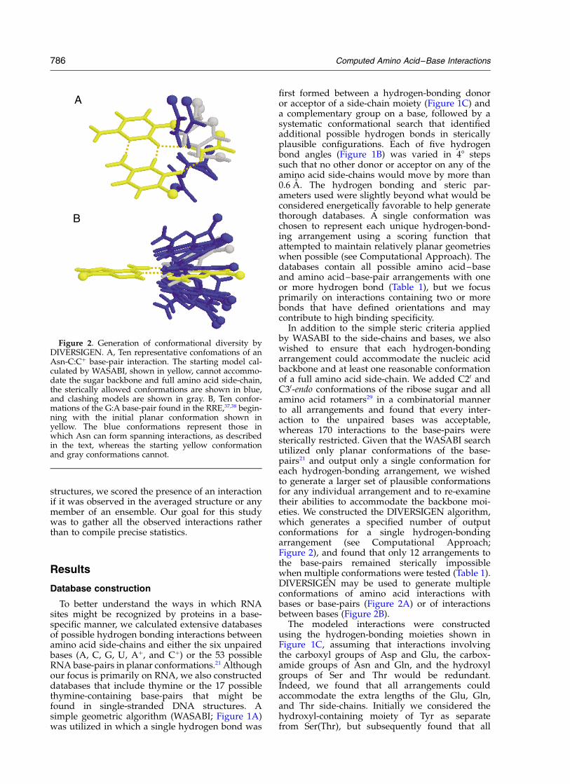
In addition to the simple steric criteria appliedby WASABI to the side-chains and bases, we alsowished to ensure that each hydrogen-bondingarrangement could accommodate the nucleic acidbackbone and at least one reasonable conformationof a full amino acid side-chain. We added C20 andC30-endo conformations of the ribose sugar and allamino acid rotamers29 in a combinatorial mannerto all arrangements and found that every inter-action to the unpaired bases was acceptable,whereas 170 interactions to the base-pairs weresterically restricted. Given that the WASABI searchutilized only planar conformations of the base-pairs21 and output only a single conformation foreach hydrogen-bonding arrangement, we wishedto generate a larger set of plausible conformationsfor any individual arrangement and to re-examinetheir abilities to accommodate the backbone moi-eties. We constructed the DIVERSIGEN algorithm,which generates a specified number of outputconformations for a single hydrogen-bondingarrangement (see Computational Approach;Figure 2), and found that only 12 arrangements tothe base-pairs remained sterically impossiblewhen multiple conformations were tested (Table 1).DIVERSIGEN may be used to generate multipleconformations of amino acid interactions withbases or base-pairs (Figure 2A) or of interactionsbetween bases (Figure 2B).
The modeled interactions were constructedusing the hydrogen-bonding moieties shown inFigure 1C, assuming that interactions involvingthe carboxyl groups of Asp and Glu, the carbox-amide groups of Asn and Gln, and the hydroxylgroups of Ser and Thr would be redundant.Indeed, we found that all arrangements couldaccommodate the extra lengths of the Glu, Gln,and Thr side-chains. Initially we considered thehydroxyl-containing moiety of Tyr as separatefrom Ser(Thr), but subsequently found that all
Figure 2. Generation of conformational diversity byDIVERSIGEN. A, Ten representative confomations of anAsn-C:Cþ base-pair interaction. The starting model cal-culated by WASABI, shown in yellow, cannot accommo-date the sugar backbone and full amino acid side-chain,the sterically allowed conformations are shown in blue,and clashing models are shown in gray. B, Ten confor-mations of the G:A base-pair found in the RRE,37,38 begin-ning with the initial planar conformation shown inyellow. The blue conformations represent those inwhich Asn can form spanning interactions, as describedin the text, whereas the starting yellow conformationand gray conformations cannot.
786 Computed Amino Acid–Base Interactions
Ser(Thr) interactions could be represented by Tyrinteractions despite the extra bulk of the Tyr ring.Interestingly, two Tyr base-pair arrangements can-not occur with Ser, both involving a bifurcatedhydrogen bond to one base that is located close tothe sugar of the second base, illustrating that stericclashes can occur with shorter side-chains thatposition peptide backbone atoms closer to thebases. All interactions with thymine were possiblewith uracil. Thus, the databases were appropriatelyfiltered for all types of redundant interactions,including those with Hisþ and pseudo-
symmetric arrangements with Asn(Gln) and Argmoieties that produce structural redundancies(Table 1).
Amino acid–base interactions
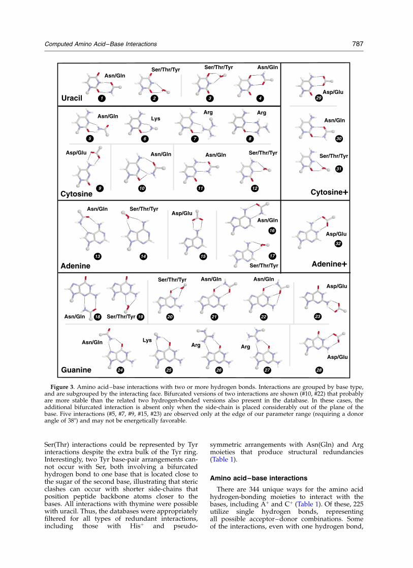
There are 344 unique ways for the amino acidhydrogen-bonding moieties to interact with thebases, including Aþ and Cþ (Table 1). Of these, 225utilize single hydrogen bonds, representingall possible acceptor–donor combinations. Someof the interactions, even with one hydrogen bond,
Figure 3. Amino acid–base interactions with two or more hydrogen bonds. Interactions are grouped by base type,and are subgrouped by the interacting face. Bifurcated versions of two interactions are shown (#10, #22) that probablyare more stable than the related two hydrogen-bonded versions also present in the database. In these cases, theadditional bifurcated interaction is absent only when the side-chain is placed considerably out of the plane of thebase. Five interactions (#5, #7, #9, #15, #23) are observed only at the edge of our parameter range (requiring a donorangle of 388) and may not be energetically favorable.
Computed Amino Acid–Base Interactions 787
require that the amino acid be roughly perpen-dicular to the plane of the base to avoid stericclashes, particularly for His interactions withadenine N3. To simplify analysis of the database,we removed bifurcated hydrogen-bonding inter-actions and those involving the protonatedbases (Aþ, Cþ) that do not form hydrogenbonds to the extra proton (Table 1). Thus, thereare 32 unique interactions involving two ormore hydrogen bonds between amino acid side-chains and the unpaired bases (shown in Figure3). For two of the Asn(Gln) interactions, we pre-sent arrangements that include bifurcated bondsthat probably are more stable than the non-bifur-cated versions in the database. Of the 32 poss-ible interactions, 12 involve Asn(Gln) and eightinvolve Ser(Thr/Tyr). Both types of side-chainsshow potential interactions to all bases exceptAþ, and their dominance likely reflects the highfrequency of adjacent acceptor and donor groupson the bases, as also described for base–baseinteractions.21 Asp(Glu), with two acceptors,shows five interactions, including one with Aþ
not possible with the unprotonated base, andnone with U. Arg, with five hydrogen donors
on its guanidinium group, allows only fourinteractions, all with C and G.
To help evaluate the completeness of our data-base and to determine whether any rules might beinferred from known interactions, we identifiedamino acid–base hydrogen bonds in protein–nucleic acid complexes in the PDB (Table 2), usingslightly relaxed hydrogen bond parameters (seeComputational Approach) to help ensure that noplausible interactions would be missed. Allobserved interactions are found in our database,including 17 of the 32 possible two-hydrogenbonded arrangements (Table 3; Figure 3). Thereare 12 types of interactions in DNA complexes,including five in the major groove and two in theminor groove of Watson–Crick helices, with theArg–G and Asn(Gln)–A interactions (#26 and #13,Figure 3) predicted by Seeman et al.5 dominating,as previously observed.4,7 – 9 Only six types ofDNA interactions are found in which amino acidsform two hydrogen bonds to a Watson–Crick face.A Ser–U interaction is observed in a reversetranscriptase complex (#3, Figure 3),30 a Lys–Cinteraction (#6, Figure 3) is observed in a nucleo-capsid-single-stranded DNA complex,31 an Arg–C
Table 3. Observed amino acid–base interactions
Interaction Face Number (DNA) Number (RNA) Observed interactionsa
Ser/Thr/Tyr-U (#3) WC 1 1 DNA: reverse transcriptase (1d0e) Ser67;RNA: Sxl (1b7f) Tyr164
Asn/Gln-U (#4) WC 0 6 RNA: AspRS (1asy, 1asz) Gln138, (1c0a) Gln46, (1efw)Gln47, (1il2) Gln1046; GlnRS (1euq) Gln517
Lys-C (#6) WC 1 1 DNA: nucleocapsid (1bj6) Lys34;RNA: nucleolin (1fje) Lys94
Arg-C (#8) WC 1 4 DNA: oxoG glycosylase (1fn7) Arg 204RNA: S15/S16/S18 (1ekc,1g1x) Arg74; AspRS (1il2)Arg225; GluRS (1g59) Arg358
Ser/Thr/Lys-C (#12) WC 0 1 RNA: AspRS (1asz) Ser329Asn/Gln-A (#13) Major 83 1 RNA: 50S ribosome (1jj2) Asn44Ser/Thr/Tyr-A (#14) Major 11 7 RNA: U1A (1aud) Tyr12, (1dz5) Ser45, Thr88, Tyr12;
MS2 coat (5msf, 6msf, 7msf) Thr45Asn/Gln-A (#16) WC 3 2 DNA: RNaseB (1rbj) Gln69, Asn71; methyltransferase
(1g38) Asn105RNA: U4 (1e7k) Ser96; Ribozyme (1hp6) Ser91
Ser/Thr/Tyr-A (#17) Major 0 1 RNA: U2B00/A0 (1a9n) Ser91Asn/Gln-G (#18) Minor 7 3 DNA: telomere BP (1otc) Gln135Ser/Thr/Tyr-G (#19) Minor 2 1Lys-G (#25) Major 20 6 RNA: L30 (1ck8, 1cn9) Lys28; 50S (1jj2) Lys35;
ProRS (1h4q, 1h4s) Lys369Arg-G (#26) Major 164 19 DNA: telomere BP (1otc) Arg274;
RNA: AspRS (1c0a, 1il2) Arg222; Rev (1ull) Arg6, (484d)Arg41; Nucleolin (1fje) Arg49
Arg-G (#27) Major 3 4 RNA: Nucleolin (1fje) Arg49Asp/Glu-G (#28) WC 10 15 DNA: telomere BP (1otc) Asp225, Glu45;
(1jb7) Asp223, Asp225, Glu45, (1k8g, 1kix) Asp223,Asp25; UP1 (2up1) Asp42;RNA: TRAP (1c9s, 1gtf, 1gtn) Asp39, Glu36; AspRS (1il2)Glu93; ThrRS (1qf6, 1kog) Glu600; ProRS (1h4q, 1h4s)Asp354, (1h4s) Glu340; 50S (1jj2) Asp105, Glu71
Asp/Glu-Cþ (#29) WC 3 0 DNA: HaeIII (1dct) Glu109; HhaI (1mht, 4mht) Glu119Ser/Thr/Tyr-Cþ (#31) WC 0 2 RNA: U1A (1aud, 1dz5) Tyr12
Numbers refer to the interactions shown in Figure 4. Face refers to the interacting surface of the base (Watson–Crick, major groove,minor groove) in a Watson–Crick helix. The observed cases in DNA and RNA are indicated, with details (protein name, pdbidentifier, residue) provided for bases that are not in a Watson–Crick pair.
a Interactions in the major or minor grooves are listed only if they are found in unpaired or non-Watson–Crick pairing contexts.
788 Computed Amino Acid–Base Interactions

Table 4. Calculated amino acid–base-pair spanning interactions
Base-pairs are listed according to the numbering used by Walberer et al.21 For cases in which the protonated base atom did not forman additional hydrogen bond in the pair, the number of the corresponding unprotonated pair is indicated in parentheses. The Aþ:Cpair (12) is the only case with interactions not observed with the unprotonated partner. The arrangements marked with brackets indi-cate base arrangements in which a U is flipped, presenting essentially an identical donor and acceptor arrangement. The interactionsobserved with these related pairs are identical in all but one case, where steric restrictions differ.
Computed Amino Acid–Base Interactions 789

interaction (#8, Figure 3) is observed in a base exci-sion DNA repair complex,67 Asn (or Gln)–A inter-actions (#16, Figure 3) are observed in RNaseB–DNA and methyltransferase–DNA complexes,32,68
Asp–G interactions (#28, Figure 3) are observed intwo telomere-binding protein complexes,33,34 andAsp (or Glu)–Cþ interactions (#29, Figure 3) areobserved in HhaI and HaeIII methylase complexesin which cytosine bases are extruded from theDNA helix.35,36
Despite the relatively small database of protein–RNA complexes, the diversity of amino acid–baseinteractions already seems apparent. There are 16types of interactions with RNA bases, includingeight in which amino acids form two hydrogenbonds to a Watson–Crick face (Table 3). Of these,Gln–U, Ser–C, and Ser–Cþ interactions (#4, #12,#31; Figure 3) have been observed only in RNAcomplexes, whereas Ser–U, Lys–C, Arg–C, Asn(Gln)–A, and Asp–G interactions (#3, #6, #8, #16and #28) have been observed in both DNA andRNA complexes. In addition to recognition of theWatson–Crick faces of the bases, some interactionsto the major or minor groove faces are found inunpaired or non-Watson–Crick pairing contexts(Table 3), adding further to the diversity of inter-actions seen with RNAs. For recognition of RNAWatson–Crick pairs, the Arg–G interaction is themost common, as for DNA, and the Asn(Gln)–Ainteraction is observed rarely (only one), as notedpreviously.18
Amino acid–base-pair interactions
One potentially attractive strategy to uniquelyrecognize portions of an RNA involves simul-
taneous hydrogen bonding to both partners of anon-Watson–Crick base-pair.21 The Rev-RREinteraction appears to utilize such a strategy torecognize an unusual G:A base-pair.37,38 To system-atically examine the possible amino acid inter-actions with base-pairs, we constructed a databaseusing the 53 possible RNA base-pairs that arebridged by two or more hydrogen bonds (and 17additional pairs that include thymine).21 Afterremoving bifurcated and redundant interactions,as for the unpaired bases, we identified 186“spanning” interactions in which two or morehydrogen bonds bridge across each pair (Table 1).Table 4 lists all interactions by the 53 RNA base-pairs defined by Walberer et al.21 As with theunpaired bases, the most common interactionsutilize Asn(Gln) (77 arrangements), but in contrast,Arg interactions also are common (64 arrange-ments), whereas there are only three possibleAsp(Glu) interactions, two to non-canonical G:Cand C:Aþ purine-pyrimidine pairs, one to a C:Cþ
pyrimidine-pyrimidine pair, and none to anypurine-purine pair. Interestingly, very few Arginteractions are possible with the purine-purinebase-pairs (just three arrangements) but are com-mon to the purine–pyrimidine and pyrimidine–pyrimidine pairs.
Of the 186 possible spanning interactions, ninepotentially form three hydrogen bonds, all usingAsn(Gln) or Arg side-chains (Figure 4). The fourinteractions involving Arg are with G:U wobble orreverse wobble base-pairs (see below), whereasthe five interactions involving Asn(Gln) are withfour unusual base-pairs, two G:G and two G:Cþ
pairs. These six base-pairs are among the mostcommonly used for all spanning interactions
Figure 4. Spanning interactionsutilizing three hydrogen bonds. Thebase-pair numbering is that used byWalberer et al.21 The top set showsinteractions with the G:U wobble,related G:Cþ, and reverse wobblepairs, and the bottom set showsthree interactions of Asn(Gln). Twosymmetric Asn(Gln) interactionsare shown to the symmetric G:Gbase-pair 18, which in principlemight occur simultaneously.
790 Computed Amino Acid–Base Interactions

(base-pairs #8, 10, 18, 20, 31, 32; Table 4), reflectingthe diversity of their donor and acceptor groups.Some of these pairs also are observed to formpotential base-triple interactions in which a third
base, rather than an amino acid, is used to spanthe base-pair.21
Nine examples of spanning interactions havebeen observed (Figure 5), including four toWatson–Crick pairs and two to a G:U wobble pair(discussed below). Six of the nine are in RNA com-plexes and three of these involve a non-canonical,non-wobble base-pair. In the crystal structure of aspliceosomal U2B00–U2A0 protein complex with aU2 snRNA hairpin, Lys20 makes a spanning inter-action to a specificity-determining U:U base-pairlocated in the loop (Figure 5).39 In NMR structuresof an HIV Rev peptide bound to an RRE hairpinor to a related RNA aptamer,37,38 the carboxamideof Asn40 hydrogen bonds to both bases of animportant G:A base-pair (Figure 5). The positionof Asn40 in the two Rev peptide–RNA complexesis well-defined by the NMR data, but the Asn-G:Ahydrogen bonding arrangements appear to differ(Figure 5). It is not yet clear whether the differencein these spanning interactions reflects the slightlydifferent RNA contexts in which the G:A pair ispresented or inaccuracies in the structures. A tightRRE-binding peptide identified from a combina-torial library probably utilizes a Gln side-chain,instead of Asn, in the context of a polyarginineframework to form a spanning interaction to theG:A pair.40
Two of the nine observed spanning interactionswere not found in our database. One of thereported Rev-RRE Asn-G:A interactions (Figure 5)was missing because the G:A pair in the complexis especially non-planar,37 although it was readilyidentified when we first used DIVERSIGEN togenerate ten conformations of the G:A pair (Figure2B) and then used WASABI to generate all possibleamino acid hydrogen bonding interactions. Thiscase illustrates one limitation to approximatingthe base-pairs as nearly planar, as well as anapproach to account for such interactions. The con-struction of subsequent databases may explicitlytake into account the three-dimensional diversityof base-pairings. The second missing interaction,observed in the structure of an EcoRV–DNAcomplex,41 places two polar hydrogen atoms at adistance of 1.87 A in a Thr-A:T spanning inter-action (Figure 5) and was eliminated from the data-base because polar hydrogen atoms closer than2.5 A are considered to have clashing charges. Sub-sequent crystal structures of EcoRV bound to thesame DNA site but with different flankingsequences suggest that Thr186 makes only onehydrogen bond to the O4 of T and that anotherside-chain (Asn185) may hydrogen bond to the N6of the paired A,42 – 45 suggesting that the Thr-A:Tinteraction may not involve two hydrogen bonds.
Spanning interactions to Watson–Crick andwobble base-pairs
Because Watson–Crick base-pairs dominate innucleic acid structures, followed by G:U wobblebase-pairs in RNAs,46,47 we examined their possible
Figure 5. Observed spanning interactions. The topset shows interactions to non-Watson–Crick base-pairsin RNAs, and the bottom set shows interactions toWatson–Crick pairs both in DNA and RNA. PDB identi-fiers are shown in parentheses, and references areprovided in the text. A possible polar hydrogen clash inthe EcoRV complex is indicated in red (see text).
Computed Amino Acid–Base Interactions 791

spanning interactions in more detail. We foundfour possible interactions with the two Watson–Crick base-pairs (Figure 6). Asn(Gln) can spaneither the major or minor groove of a G:C pair andthe major groove of an A:U(T) pair, whereas Argcan span the minor groove of an A:U(T) pair. Twoof these interactions have been observed: an Asn-A:T major groove interaction in a c-Myb–DNAcomplex and Asn–G:C minor groove interactionsin both EndoIV-DNA and Gln tRNA synthetase–tRNA complexes (Figure 5).48 – 50 In the c-Mybcomplex,48 Asn183 hydrogen bonds to both part-ners of an A:T pair in one of 25 members of anNMR ensemble. While seemingly not well popu-lated, the interaction is within the constraints ofthe experimental data and mutation of Asn183to Ala severely reduces binding activity.51 In thecrystal structure of the EndoIV complex,49 anAsn35 interaction to a G:C pair represents the only
direct side-chain–base hydrogen bonds in the com-plex. However, EndoIV is a DNA base excisionrepair endonuclease that recognizes abasic nucleo-tides within a protein pocket, so the role of a base-specific spanning interaction is unclear. In thecrystal structure of a Gln tRNA synthetase mutantbound to its cognate tRNA,50 the mutant Asn235side-chain hydrogen bonds to both bases of theG3:C70 base-pair in the minor groove of theacceptor stem. Asn is able to make an additionalhydrogen bond to the G:C base-pair compared tothe wild-type Asp side-chain, consistent with theobserved decrease in KM corresponding to a gainin binding free energy of ,1.3 kcal/mol.
The wobble G:U base-pair is very commonin RNA structures, and our database contains 16possible spanning arrangements utilizing Arg,Lys, Asn(Gln), and Ser(Thr/Tyr) side-chains(Figure 7). The Arg and Lys interactions can occuronly in the major groove, the Ser(Thr/Tyr) inter-action only in the minor groove, and the Asn(Gln)interactions in both grooves. Seven of the 11Arg–G:U hydrogen bonding arrangements requirea non-planar orientation of the guanidiniumgroup relative to the base pair (Figure 7B). Onespanning interaction between Lys28 and a G:Uwobble pair has been observed in the NMR struc-ture of L30 bound to a hairpin site in its mRNA52
(Figure 5), with the position of Lys28 being well-defined by a large number of NOEs. The Lys28side-chain also appears to make two additionalhydrogen bonds to the surrounding RNA tertiarystructure formed by this terminal G:U pair ofa helix and an adjacent internal loop. Anotherspanning interaction between Lys19 and a G:Uwobble pair has been observed in an SRP19-7SLRNA complex.53
Discussion
The repertoire of interactions
An early study by Seeman et al. defined severalways in which base-pairs might be recognizedin the context of a DNA double helix throughhydrogen-bonding interactions to amino acid side-chains.5 In the case of RNA, bases can be presentedin many more structural contexts, includingunpaired configurations and a variety of non-Watson–Crick arrangements, and thus the numberof possible recognition modes is expected to bequite large. As a first attempt to define the inter-actions possible within complex tertiary structuresand perhaps identify some “rules” of recognition,we performed a systematic search for amino acid–base and amino acid–base-pair interaction patternsbased on geometric and steric criteria. We identi-fied ,5000 plausible interactions, including 32with two hydrogen bonds to a single base and 186with two or three hydrogen bonds that span abase-pair. Only 17 types of interactions to a singlebase and nine that span a base-pair have been
Figure 6. Possible and observed spanning interactionsto the Watson–Crick base-pairs. The Asn–A:T majorgroove interaction has been observed in a c-Myb–DNAcomplex48 and Asn–G:C minor groove interactionshave been observed in EndoIV–DNA49 and Gln tRNAsynthetase–tRNA50 complexes.
Figure 7. Possible spanning interactions to the G:Uwobble pair. A, Interactions in which the side-chains arenearly coplanar with the base-pair and B, interactionsthat require non-planar orientations.
792 Computed Amino Acid–Base Interactions
observed. Among those that recognize unpairedbases are interactions to the Watson–Crick face ofcytosine bases extruded from DNA double helicesin two methylase complexes35,36 and an Asp–Ginteraction in TRAP and threonyl-tRNA synthetaseRNA complexes.54,55 The Asp–G interaction isessential for TRAP binding56 and also is observedin the binding of GTP by G proteins, where bind-ing specificity can be switched to xanthine (XTP)by a compensatory change to the donor and accep-tor arrangement of Asn.57 Our calculated inter-actions include other interesting arrangements inwhich amino acids recognize the Watson–Crickfaces of unpaired bases or span the G:U wobblepair (see below), and some of these are likely tobe observed as the database of RNA–proteincomplexes expands.
The calculated hydrogen bonding arrangementsrepresent a potentially important class of base-specific interactions, although they do not includeinteractions with water molecules, backbonemoieties, hydrophobic groups, or CH· · ·Obonds.17 –19,58 To preliminarily assess whether thecalculated interactions are energetically reasonable,we computed in vacuo interaction energies of the28 arrangements involving the four unpaired,unprotonated bases (Figure 3) using quantumchemical methods (A.C.C. & A.D.F., unpublishedresults). Five interactions near the edge of ourparameter range (388 donor angle) were unstable(see Figure 3 legend), and none of these has beenobserved. Of the remaining 23 arrangements, allbut Lys–G (#25) appear to reside at stable energyminima, with good hydrogen bond geometriesand favorable interaction energies. Thus, ourhydrogen bonding criteria generally result in stablearrangements, although the energetic contributionof any individual interaction clearly will dependon its structural context and, in some cases,may be thermodynamically unfavorable while stillcontributing to binding specificity.59
Importance of spanning interactions
One of the most interesting aspects of the data-bases is the identification of spanning interactionsthat, in principle, can provide unique ways todistinguish among the 53 possible base-pairs.While only nine spanning interactions have beenobserved so far, there is evidence that some areimportant in non-Watson–Crick base-pair recog-nition (Figure 5). In a U2B00/A0 –U2 snRNA com-plex, Lys20 of U2B00 spans a U:U base-pair at thebase of a loop, allowing discrimination betweenU2 and a related U1 hairpin recognized byU1A.39,60 In two Rev peptide–RRE complexes,Asn40 makes a spanning interaction to a G:Abase-pair, and both the Asn and G:A pair are criti-cal for recognition.37,38,61,62 In an L30–rRNA com-plex, Lys28 spans a highly conserved wobble G:Ubase-pair, and mutation of the Lys decreases bind-ing affinity by 20–30-fold.52 Similarly, in anarchaeal SRP19-7SL complex, Lys19 spans a G:Uwobble pair at the base of a tetraloop and is oneof only two base-specific interactions formed inthe complex.53
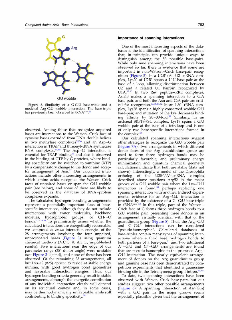
Our calculated spanning interactions suggestother strategies to recognize the G:U wobble pair(Figure 7A). Two arrangements in which differentdonor faces of the Arg guanidinium group areused to form three hydrogen bonds may beparticularly favorable, and preliminary energyminimization and quantum chemical geometrycalculations indicate that both are stable (data notshown). Interestingly, a model of the Drosophilaortholog of the U2B00/A0 –snRNA complexdescribed above positions Arg52 in the majorgroove of a G:U wobble pair where the Lys–U:Uinteraction is found,39 perhaps replacing onespanning interaction with another. Indirect experi-mental evidence for an Arg–G:U interaction isprovided by the existence of a G–G:U base-triplein tRNAAsp.63 In this triple, part of the Watson–Crick face of G forms three hydrogen bonds to aG:U wobble pair, presenting three donors in anarrangement virtually identical with that of theguanidinum group (Figure 8). Thus, the Arg–G:Uand G–G:U interactions can be considered“pseudo-isomorphic”. Calculated databases ofbase-triples contain many types of spanning inter-actions where a third base hydrogen bonds toboth partners of a base-pair,21 and two additionalAþ–G:U and Cþ–G:U arrangements are foundthat are pseudo-isomorphic to the proposed Arg–G:U interaction. The nearly equivalent arrange-ment of donors on the Arg guanidinium groupand guanine base has been demonstrated by com-petition experiments that identified a guanosine-binding site in the Tetrahymena group I intron.64,65
To date, two spanning interactions have beenobserved with Watson–Crick base-pairs but ourstudies suggest two other possible arrangements(Figure 6). A spanning interaction of Asn(Gln)with a G:C pair in the major groove seemsespecially plausible given that the arrangement of
Figure 8. Similarity of a G-G:U base-triple and amodeled Arg-G:U wobble interaction. The base-triplehas previously been observed in tRNAAsp.63
Computed Amino Acid–Base Interactions 793

donors and acceptors on a G:C pair are relativelysymmetric in both the major and minor grooves(Figure 6), and given the precedent of the minorgroove interaction. However, it is unclear howwell such an interaction would discriminatebetween base-pairs because Asn(Gln) can similarlyspan the major groove of an A:U(T) pair (Figure 6).In contrast, the Asn(Gln) minor groove spanninginteraction, observed in the Gln tRNA synthetaseand EndoIV structures, can uniquely distinguishthe donor/acceptor arrangements among all base-pairs using an appropriately positioned side-chain, as can a possible spanning interaction ofArg in the A:T minor groove (Figure 6).
In principle, several side-chains might be used todiscriminate a G:U wobble pair from the Watson–Crick pairs. From inspection of Table 4, Lys, Ser,or Arg are able to form spanning interactions tothe wobble pair but not to the Watson–Crickpairs, whereas Asn can span both types. Thus,if Lys, Ser, or Arg were positioned between thebases of a pair, accurate discrimination mightbe possible. We favor Arg for this purpose, givenits potential to form the three hydrogen-bondedinteraction described above.
Complementarity of donor–acceptor arrangements
In general, the bases and base-pairs display ahigh frequency of adjacent acceptor and donorgroups21 and consequently, arrangements invol-ving the carboxamide group of Asn(Gln) or thehydroxyl group of Ser(Thr/Tyr), which havecomplementary acceptor–donor pairs, are highlyrepresented among the possible doubly hydrogen-bonded interactions (Figure 3, Table 4). Such inter-actions to the single bases are relatively commonlyobserved (Table 3). In contrast, there are few poss-ible interactions to Asp(Glu), reflecting the limitednumber of adjacent donor group arrangements onthe bases. For unpaired bases, six of the 32 possiblearrangements involve Asp(Glu) (Figure 3), but onlythree have favorable hydrogen bond geometries.Of these, two interactions are to the protonatedbases (Aþ and Cþ) and one is to G. Interestingly,Asp-Cþ and Asp-G interactions already have beenobserved (Table 3) despite the involvement of theWatson–Crick face. A previous analysis of DNA–protein complexes revealed that interactions withAsp and Glu are rarely observed, and it wassuggested that this probably reflects unfavorableelectrostatic interactions between the negativelycharged carboxyl group and DNA backbone.13 Itseems that the arrangement of donors on thebases also inherently disfavors hydrogen-bondedAsp(Glu) interactions. The rarity of hydrogenbonding possibilities for Asp(Glu) may present agood strategy for base-specific recognition and,indeed, two out of the three types of interactionswith unpaired bases already have been observeddespite the relatively small size of the RNA struc-
tural database, with the Asp-G interaction appear-ing in five different RNA–protein complexes.
Because donors and acceptors become occupiedin a base-pair, it is instructive to examine thedoubly hydrogen-bonded interactions possibleonly in an unpaired context. Such interactions arecandidates for recognizing bases in bulges orloops. Interestingly, every amino acid interactionto U (or T) can form two hydrogen bonds to abase only in the absence of any type of base-pair-ing (assuming two hydrogen bonds are requiredto form a base-pair). This is a consequence of thefact that U (or T) possesses a total of only threedonor and acceptor groups and thus cannot simul-taneously form two hydrogen bonds to bothanother base and to an amino acid, nor can bifur-cated bonds be made to the middle N3 donorgroup. Thus, U bases in RNA bulges and loops inprinciple could be specified uniquely by twohydrogen bonds and, indeed, seven cases alreadyhave been observed (Table 3).
Utilization of the databases
The databases described may be useful fordeducing specific amino acid–RNA contacts inconjunction with biochemical data, or by analyzingamino acid–base covariations or compensatorymutations, as attempted for an L11 ribosomal pro-tein–rRNA complex.66 In principle, the databasesalso may be used to engineer “isosteric” change-of-specificity variants, or to aid in designing novelsequence-specific binding proteins whose inter-actions are guided largely by hydrogen bondinginteractions. The databases, named NAIL (nucleicacid interaction libraries), have been placed on agraphical web site† along with a set of filtersthat can be used to sort through the databases bycriteria such as: number of hydrogen bonds, typeof amino acid, and type of base or base-pair21.
Acknowledgements
We thank Peter Kollman, David Agard, andWendell Lim, and Bernhard Walberer, SteveLandt, Aenoch Lynn and others members of theFrankel lab for helpful discussions, James Rober-ston for help with quantum chemical calculations,Wei Wang for advice on energetic calculations,David Konerding for computational advice, andValerie Calabro, Chandreyee Das, Steve Landt,Robert Nakamura, and James Robertson for com-ments on the manuscript. We thank the ComputerGraphics Laboratory (UCSF) for use of computingresources. This work was supported by NIH grantsGM56531 and GM47478 (to A.D.F.) and by NIHtraining grants GM08284 and GM08388 (A.C.C.).
† http://www.ucsf.edu/frankel/frankel_homepage.html
794 Computed Amino Acid–Base Interactions

References
1. Hermann, T. & Patel, D. J. (1999). Stitching togetherRNA tertiary architectures. J. Mol. Biol. 294, 829–849.
2. Draper, D. E. (1999). Themes in RNA–protein recog-nition. J. Mol. Biol. 293, 255–270.
3. Pabo, C. O. & Nekludova, L. (2000). Geometricanalysis and comparison of protein–DNA interfaces:why is there no simple code for recognition? J. Mol.Biol. 301, 597–624.
4. Luscombe, N. M., Laskowski, R. A. & Thornton, J. M.(2001). Amino acid–base interactions: a three-dimen-sional analysis of protein–DNA interactions at anatomic level. Nucl. Acids Res. 29, 2860–2874.
5. Seeman, N. C., Rosenberg, J. M. & Rich, A. (1976).Sequence-specific recognition of double helicalnucleic acids by proteins. Proc. Natl Acad. Sci. USA,73, 804–808.
6. Creighton, T. E. (1993). Proteins: Structures andMolecular Properties, W. H. Freeman and Co, NewYork.
7. Pabo, C. O. & Sauer, R. T. (1992). Transcriptionfactors: structural families and principles of DNArecognition. Annu. Rev. Biochem. 61, 1053–1095.
8. Mandel-Gutfreund, Y., Schueler, O. & Margalit, H.(1995). Comprehensive analysis of hydrogen bondsin regulatory protein DNA–complexes: in search ofcommon principles. J. Mol. Biol. 253, 370–382.
9. Lustig, B. & Jernigan, R. L. (1995). Consistencies ofindividual DNA base-amino acid interactions instructures and sequences. Nucl. Acids Res. 23,4707–4711.
10. Suzuki, M. (1994). A framework for the DNA–protein recognition code of the probe helix in tran-scription factors: the chemical and stereochemicalrules. Structure, 2, 317–326.
11. Choo, Y. & Klug, A. (1997). Physical basis of a pro-tein–DNA recognition code. Curr. Opin. Struct. Biol.7, 117–125.
12. Mandel-Gutfreund, Y. & Margalit, H. (1998). Quanti-tative parameters for amino acid–base interaction:implications for prediction of protein–DNA bindingsites. Nucl. Acids Res. 26, 2306–2312.
13. Jones, S., van Heyningen, P., Berman, H. M. &Thornton, J. M. (1999). Protein–DNA interactions:a structural analysis. J. Mol. Biol. 287, 877–896.
14. Kono, H. & Sarai, A. (1999). Structure-based predic-tion of DNA target sites by regulatory proteins.Proteins: Struct. Funct. Genet. 35, 114–131.
15. Suzuki, M. & Yagi, N. (1994). DNA recognition codeof transcription factors in the helix-turn-helix, probehelix, hormone recptor, and zinc finger families.Proc. Natl Acad. Sci. USA, 91, 12357–12361.
16. Steitz, T. A. (1999). The RNA World (Gesteland, R. F.,Cech, T. R. & Atkins, J. F., eds), 2nd edit., pp.427–450, Cold Spring Harbor Laboratory Press,Cold Spring Harbor, NY.
17. Jones, S., Daley, D. T. A., Luscombe, N. M., Berman, H.& Thornton, J. M. (2001). Protein–RNA interactions:a structural analysis. Nucl. Acids Res. 29, 943–954.
18. Allers, J. & Shamoo, Y. (2001). Structure-based analy-sis of protein–RNA interactions using the programENTANGLE. J. Mol. Biol. 311, 75–86.
19. Treger, M. & Westhof, E. (2001). Statistical analysis ofatomic contacts at RNA–protein interfaces. J. Mol.Recog. 14, 199–214.
20. Walberer, B. J. (2000). Construction and analysis of acomplete database of hydrogen-bonded base combi-
nations, PhD thesis, University of California, SanFranscisco.
21. Walberer, B.J., Cheng, A.C., Frankel, A.D. (2003).Structural diversity and isomorphism of hydrogen-bonded base interactions in nucleic acids. J. Mol. Biol.
22. Cornell, W. D., Cieplak, P., Bayly, C. I., Gould, I. R.,Merz, K. M., Ferguson, D. M. et al. (1995). A secondgeneration force field for the simulation of proteins,nucleic acids, and organic molecules. J. Am. Chem.Soc. 117, 5179–5197.
23. Israelachvili, J. N. (1989). Intermolecular and SurfaceForces, Academic Press, New York.
24. Taylor, R., Kennard, O. & Versichel, W. (1983). Geo-metry of the N–H–OvC hydrogen bond. J. Am.Chem Soc. 105, 5761–5766.
25. Taylor, R. & Kennard, O. (1984). Hydrogen-bond geo-metry in organic crystals. Accts. Chem Res. 17,320–326.
26. Saenger, W. (1984). Principles of Nucleic Acid Structure,Springer, New York.
27. Jeffrey, G. A. & Saenger, W. (1991). Hydrogen Bondingin Biological Molecules, Springer, Berlin.
28. Baker, E. N. & Hubbard, R. E. (1984). Hydrogenbonding in globular proteins. Prog. Biophys. Mol.Biol. 44, 97–179.
29. Dunbrack, R. L. & Cohen, F. E. (1997). Bayesianstatistical analysis of protein sidechain rotamerpreferences. Protein Sci. 6, 1661–1681.
30. Najmudin, S., Cote, M. L., Sun, D., Yohannan, S.,Montano, S. P., Gu, J. & Georgiadis, M. M. (2000).Crystal structures of an N-terminal fragment fromMoloney murine leukemia virus reverse transcrip-tase complexed with nucleic acid: functional impli-cations for template-primer binding to the fingersdomain. J. Mol. Biol. 296, 613–632.
31. Morellet, N., Demene, H., Teilleux, V., Huynh-Dinh,T., de Rocquigny, H., Fournie-Zaluski, M. C. &Rocques, B. P. (1998). Structure of the complexbetween the HIV-1 nucleocapsid protein NCp7 andthe single-stranded pentanucleotide d(ACGCC).J. Mol. Biol. 283, 419–434.
32. Ko, T. P., Williams, R. & McPherson, A. (1996).Structure of a ribonuclease B þ d(pA)4 complex.Acta Crystallog. sect. D, 52, 160–164.
33. Horvath, M. P., Schweiker, V. L., Bevilacqua, J. M.,Ruggles, J. A. & Schultz, S. C. (1998). Crystalstructure of the Oyxtricha nova telomere and bind-ing protein complexed with single strand DNA.Cell, 95, 963–974.
34. Ding, J., Hayashi, M. K., Zhang, Y., Manche, L.,Krainer, A. R. & Xu, R. M. (1999). Crystal structureof the two-RRM domain of hnRNP A1 (UP1) com-plexed with single-stranded telomeric DNA. GenesDev. 13, 1102–1115.
35. Klimasauskas, S., Kumar, S., Roberts, R. J. & Cheng,X. (1994). HhaI methyltransferase flips its targetbase out of the DNA helix. Cell, 76, 357–369.
36. Reinisch, K. M., Chen, L., Verdine, G. L. & Lipscomb,W. N. (1995). The crystal structure of HaeIII methyl-transferase covalently complexed to DNA: an extra-helical cytosine and rearranged base pairing. Cell,82, 143–153.
37. Battiste, J. L., Mao, H., Rao, N. S., Tan, R.,Muhandiram, D. R., Kay, L. E. et al. (1996). Alphahelix major groove recognition in an HIV-1 Rev pep-tide–RRE RNA complex. Science, 273, 1547–1551.
38. Ye, X., Gorin, A., Ellington, A. D. & Patel, D. J. (1996).Deep penetration of an alpha-helix into a widened
Computed Amino Acid–Base Interactions 795
RNA major groove in the HIV-1 rev peptide–RNAaptamer complex. Nature Struct. Biol. 3, 1026–1033.
39. Price, S. R., Evans, P. R. & Nagai, K. (1998). Crystalstructure of the spliceosomal U2B00 –U2A0 proteincomplex bound to a fragment of U2 small nuclearRNA. Nature, 394, 645–650.
40. Tan, R. & Frankel, A. D. (1998). A novel glutamine–RNA interaction identified by screening librariesin mammalian cells. Proc. Natl Acad. Sci. USA, 95,4247–4252.
41. Winkler, F., Banner, D., Oefner, C., Tsernoglou, D.,Brown, R., Heathman, S. et al. (1993). The crystalstructure of EcoRV endonuclease and of its com-plexes with cognate and non-cognate DNA frag-ments. EMBO J. 12, 1781–1795.
42. Kostrewa, D. & Winkler, F. K. (1995). Mg2þ binding tothe active site of EcoRV endonuclease: a crystallo-graphic study of complexes with substrate andproduct DNA at 2 A resolution. Biochemistry, 34,683–696.
43. Perona, J. & Martin, A. (1997). Conformationaltransitions and structural deformability of EcoRVendonuclease revealed by crystallographic analysis.J. Mol. Biol. 273, 207–225.
44. Horton, N. C. & Perona, J. J. (1998). Role of protein-induced bending in the specificity of DNA recog-nition: crystal structure of EcoRV endonucleasecomplexed with d(AAAGAT) þ d(ATCTT). J. Mol.Biol. 277, 779–787.
45. Horton, N. C. & Perona, J. J. (1998). Recognition offlanking DNA sequences by EcoRV endonucleaseinvolves alternative patterns of water-mediated con-tacts. J. Biol. Chem. 273, 21721–21729.
46. Masquida, B. & Westhof, E. (2000). On the wobbleG:U and related pairs. RNA, 6, 9–15.
47. Varani, G. & McClain, W. H. (2000). The G:U wobblebase pair. A fundamental building block of RNAstructure crucial to RNA function in diverse systems.EMBO Rep., 1, 18–23.
48. Ogata, K., Morikawa, S., Nakamura, H., Sekikawa,A., Inoue, T., Kanai, H. et al. (1994). Solution struc-ture of a specific DNA complex of the Myb DNA-binding domain with cooperative recognitionhelices. Cell, 79, 639–648.
49. Hosfield, D. J., Mol, C. D., Shen, B. & Tainer, J. A.(1998). Structure of the DNA repair and replicationendonuclease and exonuclease FEN-1: couplingDNA and PCNA binding to FEN-1 activity. Cell, 95,135–146.
50. Arnez, J. G. & Steitz, T. A. (1996). Crystal structuresof three misacylating mutants of Escherichia coliglutaminyl-tRNA synthetase complexed withtRNA(Gln) and ATP. Biochemistry, 35, 14725–14733.
51. Gabrielsen, O. S., Sentenac, A. & Fromageot, P.(1991). Specific DNA binding by c-Myb: evidencefor a double helix-turn-helix-related motif. Science,253, 1140–1143.
52. Mao, H., White, S. A. & Williamson, J. R. (1999).A novel loop-loop recognition motif in the yeast ribo-somal protein L30 autoregulatory RNA complex.Nature Struct. Biol. 6, 1139–1147.
53. Hainzl, T., Huang, S. & Sauer-Eriksson, A. E. (2002).Structure of the SRP19 RNA complex and impli-cations for signal recognition particle assembly.Nature, 417, 767–771.
54. Antson, A. A., Dodson, E. J., Dodson, G., Greaves,R. B., Chen, X. & Gollnick, P. (1999). Structure of thetrp RNA-binding attenuation protein, TRAP, boundto RNA. Nature, 401, 235–242.
55. Sankaranarayanan, R., Dock-Bregeon, A.-C., Romby,P., Caillet, J., Springer, M., Rees, B. et al. (1999). Thestructure of threonyl-tRNA synthetase-tRNA(Thr)complex enlightens its repressor activity and revealsan essential zinc ion in the active site. Cell, 97,371–381.
56. Elliott, M. B., Gottlieb, P. A. & Gollnick, P. (1999).Probing the TRAP–RNA interaction with nucleosideanalogs. RNA, 5, 1277–1289.
57. Powers, T. & Walter, P. (1995). Reciprocal stimulationof GTP hydrolysis by two directly interactingGTPases. Science, 269, 1422–1424.
58. Mandel-Gutfreund, Y., Margalit, H., Jernigan, R. L. &Zhurkin, V. B. (1998). A role for CH· · ·O interactionsin protein–DNA recognition. J. Mol. Biol. 277,1129–1140.
59. Szwajkajzer, D. & Carey, J. (1997). Molecular and bio-logical constraints on ligand-binding affinity andspecificity. Biopolymers, 44, 181–198.
60. Oubridge, C., Ito, N., Evans, P. R., Teo, C. H. &Nagai, K. (1994). Crystal structure at 1.92 A resolu-tion of the RNA-binding domain of the U1A spliceo-somal protein complexed with an RNA hairpin.Nature, 372, 432–438.
61. Iwai, S., Pritchard, C., Mann, D. A., Karn, J. & Gait,M. J. (1992). Recognition of the high affinity bindingsite in rev-response element RNA by the humanimmunodeficiency virus type-1 rev protein. Nucl.Acids Res. 20, 6465–6472.
62. Tan, R., Chen, L., Buettner, J. A., Hudson, D. & Fran-kel, A. D. (1993). RNA recognition by an isolated a
helix. Cell, 73, 1031–1040.63. Westhof, E., Dumas, P. & Moras, D. (1985). Crystallo-
graphic refinement of yeast aspartic acid transferRNA. J. Mol. Biol. 184, 119–145.
64. Yarus, M. (1988). A specific amino acid binding sitecomposed of RNA. Science, 240, 1751–1758.
65. Michel, F., Hanna, M., Green, R., Bartel, D. P. &Szostak, J. W. (1989). The guanosine binding site ofthe Tetrahymena ribozyme. Nature, 342, 391–395.
66. GuhaThakurta, D. & Draper, D. E. (1999). Protein–RNA sequence covariation in a ribosomal protein–rRNA complex. Biochemistry, 38, 3633–3640.
67. Norman, D. P., Bruner, S. D. & Verdine, G. L. (2001).Coupling of substrate recognition and catalysis by ahuman base-excision repair protein. J. Am. Chem.Soc. 123, 359–360.
68. Goedecke, K., Pignot, M., Goody, R. S., Scheidig, A. J.& Weinhold, E. (2001). Structure of the N6-adenineDNA methyltransferase M. TaqI in complex withDNA and a cofactor analog. Nature Struct. Biol. 8,121–125.
Edited by D. E. Draper
(Received 23 October 2002; accepted 23 December 2002)
796 Computed Amino Acid–Base Interactions