Embed Size (px)
Citation preview

NATIONAL GUIDELINES FOR RECOGNITION AND MANAGEMENT OF VIRAL HAEMORRHAGIC FEVERS 2014

National Department of Health Page 2 of 62
Table of Content 1. INTRODUCTION .......................................................................................................................................... 5 2. REFERRAL OF VIRAL HAEMORRHAGIC FEVER PATIENTS .................................................................. 6 3. BACKGROUND TO THE VIRAL HAEMORRHAGIC FEVERS ................................................................... 6 4. DIAGNOSIS ............................................................................................................................................... 14 5. IMMEDIATE ACTION TO BE TAKEN AFTER CLINICAL DIAGNOSIS OF VHF ...................................... 27 6. MANAGEMENT OF VHF PATIENTS ........................................................................................................ 29 7. NOTIFICATION AND CONTROL OF OUTBREAKS OF VHF ................................................................... 42 8. KEY REFERENCES .................................................................................................................................. 58
National Department of Health Page 3 of 62
National Department of Health Page 4 of 62
National Department of Health Page 5 of 62
1. INTRODUCTION Many infections, and even non-infectious diseases, can cause fever and a haemorrhagic state. It is important to distinguish these conditions from viral haemorrhagic fevers (VHFs) caused by the so-called formidable or Class 4 viruses. The VHFs have in common a propensity for person-to-person spread and high mortality rates, which necessitate that special infection control measures (isolation precautions) should be instituted when managing suspected or confirmed cases of the diseases, and work with the viruses is permitted only in biosafety level 4 (BSL4) laboratories. However, not all of the viruses associated with VHFs are uniformly lethal or spread readily between humans: some less pathogenic viruses are placed in Class 4 in countries from which they are absent in order to exercise control over their possible introduction. Many parts of the world have endemic VHFs, and modern travel has made it possible for introduced cases to occur virtually anywhere. The most common VHF in Southern Africa is caused by the tick-borne Crimean-Congo haemorrhagic fever (CCHF or Congo fever) virus, and approximately 5-20 cases of the disease are diagnosed in South Africa each year. Rift Valley fever, a zoonotic disease of sheep and cattle, also occurs in our region, but human infections are generally seen in the context of major outbreaks of disease in livestock which occur at irregular intervals of many years when exceptionally heavy rains favour breeding of the mosquito transmitters of the virus, and human-to-human transmission has not been recorded. The most recent large outbreak in South Africa was in 2010. In addition, the growing tendency for severely ill patients from countries in tropical Africa to seek medical attention in South Africa is leading to increased risk that cases of Lassa, Marburg and Ebola haemorrhagic fevers may be imported inadvertently. Fatal nosocomial infections have occurred in South African hospitals in the past, and to avoid further tragedies health care workers should maintain high standards of infection control and biosafety awareness at all times, and all patient care facilities should institute contingency plans for dealing with VHF patients. The present document, an updated version of guidelines first prepared in 1985, is intended as a guide to the recognition and management of suspected and confirmed cases, and prevention of nosocomial spread, of the indigenous African viral haemorrhagic fevers. The recommendations are not binding except where reference is made to legislation, statutory regulations, or agreed protocol for dealings between separate organizations and institutions, each of which should draft and implement protocols adapted to their own needs.

National Department of Health Page 6 of 62
2. REFERRAL OF VIRAL HAEMORRHAGIC FEVER PATIENTS After it was recognized in the 1980s that Congo fever is indigenous in South Africa, it was arranged that at least one provincial hospital within each province should be designated as a referral centre for the management of VHF patients, but circumstances have changed: ● It can no longer be assumed that VHF patients can automatically be referred to designated
provincial hospitals. ● All private hospitals, and public tertiary and regional hospitals should be adequately resourced
and prepared to handle VHF patients. ● All other public hospitals must have access to public referral hospitals that are adequately
resourced. It is the responsibility of provincial Department of Health and Hospital Services, including the Coordinators of Communicable Disease Control, to formulate and implement provincial policy with regard to referral of VHF patients, including the designation of specific referral hospitals (see section 7). Previous versions of the present document contained a list of designated referral hospitals with contact details of persons with whom to liaise in order to arrange referral of VHF patients. Unfortunately this type of information is subject to abrupt changes, and hence the management of each hospital (specifically infection control officers) should establish for themselves what policy applies in their own province or sub region with respect to referral of VHF patients, and keep up-to-date contact details for the nearest designated VHF referral centre. Do not be caught unprepared.

National Department of Health Page 7 of 62
3. BACKGROUND TO THE VIRAL HAEMORRHAGIC FEVERS Viruses associated with haemorrhagic fevers (Table 3.1), fall into three groups with respect to their reservoir hosts and primary means of transmission, namely, rodent-associated viruses, arthropod-borne viruses, and viruses thought to be associated with bats. 3.1 Rodent-associated viruses The arenaviruses and hantaviruses cause chronic kidney infection in myomorph rodents (rats and mice) with excretion of virus in the urine, and humans become infected from contaminated food or household items, but there may also be occupational or recreational exposure to rodent excreta. 3.1.1 Lassa fever Lassa fever is caused by an arenavirus that is confined to West Africa (Nigeria, Sierra Leone, Guinea and Liberia are particularly affected). Related viruses that occur in rodents elsewhere in Africa were not known to be pathogenic until the recent discovery of Lujo virus in southern Africa. Lassa fever infection is generally associated with a comparatively mild disease with fever and a death rate of 1-2% among cases in the community at large, but some patients develop haemorrhagic disease and deaths rates may approach 20% among hospitalised patients, or exceed 40% in nosocomial outbreaks. Person-to-person spread of infection, which occurs in the home and hospital, appears to require overt contact with infected tissues and body fluids. A physician from Nigeria who was evacuated for treatment in South Africa in 2007 proved to be suffering from fatal Lassa fever, but fortunately there were no secondary infections. Clinical features of Lassa fever The incubation period is usually 7-10 days (range 3-21 days). Over 80% of infections are asymptomatic or mild, but in the rest there is insidious onset of fever, chills, malaise, headache, generalized myalgia and prostration. Within 2-3 days patients develop sore throat, vomiting, abdominal or chest (retrosternal) pains, cough, hypotension and bradycardia. There is characteristic pharyngeal and tonsillar inflammation with vesicular or ulcerative lesions and whitish or yellowish exudate. Conjuctivae are injected, and there is lymphadenopathy, muscle tenderness, pulmonary rales, and sometimes maculopapular rash. From day 5 patients may progress to severe sustained fever and toxaemia with haemorrhages (epistaxis, haematemesis, melaena), puffiness of the face and neck, serous effusions (hydrothorax), disorders of the central nervous system and shock. The acute illness has a duration of 1-3 weeks. Deafness occurs in 25% of patients with some recovery in 1-3 months, and there may be loss of hair and an unsteady gait during convalescence. Clinical pathology of Lassa fever Early leucopenia may be followed by leucocytosis. Proteinuria is common. Abnormalities in platelet counts, prothrombin and clotting time are not marked, but there may be pronounced increases in serum levels of aspartate and alanine transaminases, lactic dehydrogenase and creatine kinase. Viraemia lasts about a week from the time of onset of disease but excretion of virus in urine may extend over 3-10 weeks. Lujo virus: In September-October 2008, there was a nosocomial outbreak of infection with a new arenavirus, Lujo virus, in Johannesburg, involving 5 patients, 4 of whom died, with a clinical course similar to severe Lassa fever. The first patient was transferred from Zambia to South Africa for medical management and the source of her infection remains undetermined, although rodents are suspected. Three cases involved secondary spread of infection from the first patient, and there was one tertiary infection. The secondary and tertiary infections all occurred before isolation precautions were implemented. Several arenaviruses cause hemorrhagic fevers in South America.

National Department of Health Page 8 of 62
Clinical features of Lujo virus Incubation period of 9-13 days; a prodomal illness characterized by fever, headache and myalgia, followed by diarrhoea and pharyngitis and a morbiliform rash on the face and trunk reported in three cases on day 6-8 of illness. Facial swelling occurred in three patients with marked pharyngeal ulceration reported in one patient. There appeared to be an initial clinical improvement after hospital admission in three patients, followed by sudden, rapid deterioration in all patients who died. Bleeding was not a prominent feature. One patient had a petechial rash and another had oozing of blood from venipuncture sites. One patient was treated with intravenous ribavirin and survived. Clinical pathology of Lujo virus At the time of admission all patients had thrombocytopenia (range: 42-104 x 109/L). Liver transaminases (AST and ALT) were raised in all five patients during the course of their illness. Hantaviruses. Several hantaviruses are associated with a group of diseases in Europe and Asia which are known collectively as haemorrhagic fever with renal syndrome (HFRS) (with fatality rates of <1-35%), while another group of hantaviruses is associated with the hantavirus pulmonary syndrome (HPS) (fatality rates ≥50%) in North and South America. Hantaviruses have been poorly studied in Africa, and there is as yet little evidence that they occur here, except possibly for Seoul virus, thought to have been widely disseminated to sea ports with ship-borne rats and occurring in urban settings. Clinical features of HFRS There are 4 clinical forms of the disease, varying in severity (<1-35% fatal) from nephropathia epidemica associated with Puumala virus in Scandinavia, through mild or rat-borne HFRS associated with Seoul virus infection which has been widely disseminated with ship-borne rats, to Far Eastern HFRS associated with Hantaan virus in Asia (also known as Korean haemorrhagic fever), and so-called Balkan HFRS associated with Dobrava virus. The incubation period is 2-3 weeks. Severe disease has five well-marked phases but these overlap and are obscured in mild disease. An initial febrile phase of 3-7 days is marked by high fever, chills, malaise, myalgia, anorexia, dizziness, headache and ocular pain, abdominal and back pain with tenderness in the renal area (peritoneal and retroperitoneal oedema), followed by characteristic flushing of the face neck and chest, with injection of the eyes, palate and pharynx which develops into a fine petechial rash and conjunctival haemorrhage. There is marked proteinuria. A hypotensive phase follows abruptly and lasts hours to 2 days, with tachycardia and classical shock: narrowed blood pressure, cold and clammy skin, dulled senses and confusion; one third of fatal patients enter irreversible shock at this stage. Proteinuria continues and there is mild haematuria, raised haematocrit level, leukemoid reaction and thrombocytopenia. Onset of an oliguric phase of 3-4 days is marked by increasing blood urea and creatinine levels. Blood pressure begins to normalize but hypertension can result from the hypovolaemic state. There may be severe nausea and vomiting, and bleeding tendencies increase: epistaxis, conjunctival haemorrhage, cerebral and gastro-intestinal haemorrhage and extensive purpura. There is hyperkalaemia, hyponatraemia and hypocalcaemia. There may be central nervous symptoms and pulmonary oedema, with 50% of fatalities occurring in this phase. A diuretic phase may last days to weeks, with diuresis of up to 3-6 litres per day, and marks the start of recovery. The convalescent phase lasts 2-3 months with progressive recovery of glomerular filtration rate. Clinical features of HPS Persons who develop HPS are often healthy young adults, but may be of any age and either sex. The incubation period is 2-3 weeks and onset is marked by sudden development of fever, headache, severe myalgia and a cough, which may be productive in some instances. Gastrointestinal manifestations in some patients include abdominal pain, nausea, vomiting and diarrhoea. After 3-6 days of illness there is progressive tachypnoea, tachycardia and hypotension preceding the onset of acute respiratory distress with pulmonary oedema. Patients are generally hospitalized at this stage,
National Department of Health Page 9 of 62
but some die before they can be admitted. On admission patients may have proteinuria, leucocytosis with neutrophilia plus increased myeloid precursors and atypical lymphocytes, haemoconcentration, and thrombocytopenia, and increased prothrombin and partial-thromboplastin times, although there is no rash and very seldom a tendency towards overt or internal bleeding. Within two days of being admitted to hospital most patients develop diffuse bilateral interstitial and alveolar pulmonary infiltration and pleural effusions demonstrable on radiographs, with hypoxaemia, which necessitates intubation, mechanical ventilation and oxygen supplementation. Sometimes there is renal insufficiency and increased serum creatine kinase levels (evidence of skeletal muscle inflammation). Death generally occurs 6-8 days after the onset of illness, often within 48 hours of admission to hospital, but can range from 2 days after the observed onset of illness to more than two weeks. Fatality rates often exceed 40%, and incurable shock and myocardial dysfunction may contribute to the high mortality. Autopsies reveal non-cardiogenic pulmonary oedema and serous pleural effusions, with scant lymphoid infiltration of the lung tissue. Some survivors manifested transient diuresis, but otherwise they make an uneventful recovery without sequelae. 3.2 Arthropod-borne viruses (‘arboviruses’ or ‘insect-transmitted’ viruses) Several haemorrhagic fevers are caused by arboviruses. These are diverse viruses, which have in common the fact that they are transmitted by blood-sucking arthropods (mosquitoes, midges, sand flies and ticks), with various wild and domestic animals serving as reservoir hosts (infected animals which serve as sources of virus for infecting the arthropod vectors). Only a few arboviruses cause haemorrhagic disease. 3.2.1 Crimean-Congo haemorrhagic fever (CCHF or Congo fever) Congo fever is the most frequently observed haemorrhagic fever in South Africa. It is caused by a tick-borne virus, which occurs widely in Africa, Eastern Europe and Asia, within the distribution range of its main vectors, ticks of the genus Hyalomma. These are known as bont-legged ticks in South Africa on account of the distinctive brown and white bands on their legs. The disease is seen most frequently in the Northern Cape, Free State and North West Provinces where the drier climate favours the bont-legged ticks, but cases may occur anywhere in the country: patients infected in the Free State have become ill in KwaZulu-Natal, and abattoir workers have developed the disease within the cities of Cape Town and Johannesburg. The disease has an approximately 30% fatality rate and humans acquire infection from tick bite or from contact of broken skin with fresh infected blood and tissues of livestock (sheep, cattle, ostriches), which themselves undergo benign infection. Meat, which has been bled out and hung to mature according to proper slaughterhouse procedures, is not infectious, and cooking destroys the virus. About 5-20 cases of the disease are diagnosed in South Africa each year, and two South Africans are known to have acquired infection during visits to Namibia and Tanzania. In addition, a patient from the DRC with unrecognised CCHF was treated in South Africa; the diagnosis was only established after his death but fortunately there were no secondary infections. Infection can occur in hospitals where medical staff comes into contact with the blood of patients (needle sticks) or blood-tinged body fluids; there have been three such incidents in South Africa involving 6 nurses, a surgeon and a laboratory technologist, with 3 fatalities. There is no vaccine. Clinical features of Congo fever The incubation period commonly ranges from 1-3 days after tick bite, to 5-6 days after contact with infected blood or other tissues, but may occasionally be longer. People are not always aware of being bitten by ticks (look for ticks or bite marks, including on the scalp and between toes), but infection can also be acquired from merely squashing ticks between the fingers. In contrast to the necrotic eschars that occur at the site of the bites in tick bite fever (rickettsiosis), there may only be slight bruising at bite sites in Congo fever. Unlike many other arbovirus diseases, a high proportion of infections are symptomatic. Onset is usually very sudden, with severe headache, dizziness, neck pain and stiffness, sore eyes, photophobia, fever, rigor and chills, followed rapidly by myalgia with intense backache or leg pains, nausea, sore throat and vomiting. There may be non-localized abdominal pain and diarrhoea at an early stage. Fever is often intermittent and patients may undergo sharp changes of

National Department of Health Page 10 of 62
mood over the first two days, with feelings of confusion and aggression. By day 2-4 patients may exhibit lassitude, depression and somnolence, and have a flushed appearance with injected conjunctivae or chemosis. Tenderness localizes in the right upper quadrant of the abdomen, and hepatomegaly may be discernible. Tachycardia is common and patients may be slightly hypotensive. There may be lymphadenopathy, plus enanthema and petechiae of the throat, tonsils and buccal mucosa. A petechial rash appears on the trunk and limbs by day 3-6 of illness, and this may be followed rapidly by the appearance of large bruises and ecchymoses, especially in the anticubital fossae, upper arms, axillae and groin. Oozing of blood from injection or venipuncture sites, epistaxis, haematemesis, haematuria, melaena, gingival bleeding and bleeding from the vagina or other orifices may commence on day 4-5 of illness, seldom earlier. There may also be internal bleeding, including retroperitoneal and intracranial haemorrhage. Severely ill patients enter a state of hepatorenal and pulmonary failure from about day 5 onwards and progressively becomes drowsy, stuporous and comatose. Jaundice may become apparent during the second week of illness. The mortality rate is approximately 30% and deaths generally occur on day 5-14 of illness. Patients who recover usually begin to improve suddenly on day 9-10 of illness, but asthenia, conjunctivitis, slight confusion and amnesia may continue for a month or longer. Clinical pathology of Congo fever During the first few days of illness there may be leucocytosis or leucopenia, and elevated aspartate and alanine transaminases, gamma-glutamyl transferase, lactic dehydrogenase, alkaline phosphatase and creatine kinase levels, while bilirubin, creatinine and urea levels increase and serum protein levels decline during the second week. Thrombocytopenia, elevation of the prothrombin ratio, activated partial thromboplastin time, thrombin time, elevation of D-dimers and fibrin degradation products, as well as depression of fibrinogen and haemoglobin values are evident very early in the illness, indicating that disseminated intravascular coagulopathy is an early and central event in the pathogenesis of the disease. During the first 5 days of illness any of the following clinical pathology values are highly predictive of fatal outcome: leucocyte counts ≥10x109/L; platelet counts ≤20x109/L; AST ≥200U/L; ALT ≥150U/L; APTT ≥60 seconds; and fibrinogen ≤110mg/dL. Leucopenia does not have the same poor prognostic connotation as leucocytosis at this early stage, and all clinical pathology values may be grossly abnormal after day 5 of illness without necessarily being indicative of a poor prognosis. Viraemia is usually detectable during the first week of illness (range 1-13 days), and viral nucleic acid can be detected in serum by RT-PCR for up to 16 days after onset. Antibody response is rarely demonstrable in fatal illness, and thus detection of antibody is generally a favourable sign. Rift Valley fever (RVF) RVF is a mosquito-borne virus disease of livestock in Africa and Madagascar which affects mainly sheep and cattle, and causes massive outbreaks of abortion and death of young animals at irregular intervals of years when particularly heavy rains favour the breeding of the vectors. Humans acquire infection from contact with infected tissues of farm animals, or less frequently from mosquito bite. Most patients experience benign illness with fever, some with ocular sequelae (usually transient scotomas, but sometimes permanent blindness) and only <1% develop fatal haemorrhagic disease, hepatitis or encephalitis. Nevertheless, outbreaks can be massive and the disease has caused large numbers of human deaths on occasion. The last major outbreak in South Africa occurred in 2010, and particularly affecting farms in Eastern Cape, Free State and Northern Cape Provinces with some spread to the Western Cape, North West and Gauteng Provinces. There were 230 lab confirmed human cases and 26 deaths but is likely that there were a significant number of asymptomatic cases who were not tested. In 1985, one patient infected in Angola and two infected in Zambia were treated in South Africa. In 2000-1, the disease was recognized outside of the African region for the first time in a large outbreak in Saudi Arabia and Yemen. Curiously, there are no records of human-to-human transmission of the virus, although very high levels of virus occur in the blood of patients so that transmission by needle stick is possible. An experimental human vaccine produced in the USA was formerly used on a limited scale in people with occupational exposure to infection in the livestock industry and in laboratories, but it is not currently available.
National Department of Health Page 11 of 62
Clinical features of RVF The incubation period is generally 2-6 days, and the majority of infections are either mild (recognized only in serosurveys or as laboratory infections), or present as moderate to severe febrile illness with sudden onset of severe retro-orbital pain and headache, photophobia, suffused conjunctivae, myalgia, arthralgia, prostration, nausea and tenderness of the liver without hepatomegaly. Fever and prostration often last only 2-3 days, or the disease may run a diphasic course over two weeks. Ocular complications occur in 5-20% of cases 1-3 weeks after onset of illness. Decreased visual acuity or scotomas are associated with retinal haemorrhages, exudate and macular oedema. Vision usually improves over a period of 1-3 months as lesions resolve, but occasionally there can be detached retina and blindness. Less than 0.5% of patients develop encephalitis or haemorrhagic disease with high death rates. Encephalitis occurs as a complication 1-2 weeks after the acute febrile disease, and patients may succumb or undergo sudden or protracted recovery. Haemorrhagic fever with or without neurologic disease, can supervene within a week after the acute febrile stage. There is extensive liver necrosis in these cases, and there may be marked anaemia following massive epistaxis, haematemesis and melaena. Petechiae, ecchymoses and jaundice may be evident. Clinical pathology of RVF There is usually leucopenia, hyperbilirubinaemia, thrombocytopenia, prolongation of clotting parameters and markedly raised serum transaminases. Viraemia commonly lasts 2-3 days but has been recorded for up to 11 days. Chikungunya, yellow fever and dengue viruses These viruses circulate between mosquitoes and non-human primates (monkeys and apes) in forests, but have the unusual ability among arboviruses of utilizing humans as their sole vertebrate hosts in urban outbreaks of disease. Although infections with these three viruses can take a haemorrhagic form, they have not been associated with human-to-human spread, and their main importance is as differential diagnoses for VHF. Chikungunya (CHIK) virus causes outbreaks of illness characterized by fever and joint pain in rural locations where baboons and monkeys occur in Africa, mainly in East Africa, but including South Africa, particularly the Limpopo and Mpumalanga Lowveld, and northern KwaZulu-Natal coast. Pain in a particular joint may last for up to two years after the acute illness. Severe and haemorrhagic forms of the disease have been recorded in a minority of patients in Asia and the Indian Ocean islands where the virus causes large urban epidemics. Chikungunya has been diagnosed in South African tourists returning from abroad, and it is theoretically possible that such a patient could initiate urban outbreaks involving transmission by local mosquitoes, particularly in KwaZulu-Natal. There is no vaccine. Yellow fever (YF) is a well-known mosquito-borne virus, which causes outbreaks of fatal disease with necrotic hepatitis in South America, West Africa, and less frequently East Africa, but it has never been recorded south of Angola. Suitable mosquito vectors occur in eastern South Africa. The fact that a very effective vaccine is available, and is used on international travellers, tends to limit the potential for tourists to spread infection to remote locations, but it is possible that sick patients could be evacuated for treatment in South Africa. Nosocomial infection has never been described, although in endemic areas mosquito transmission could also affect health care workers. Dengue (DEN) is a mosquito-borne virus which causes massive outbreaks of disease with fever, and joint and muscle pains throughout the tropics in South America, the Caribbean, East and West Africa, Indian Ocean islands, India and South East Asia. There are four sub-types of the virus, and a small proportion of patients may develop haemorrhagic disease or a shock syndrome, particularly the very young and the aged, or those who suffer sequential infection with a second sub-type of the virus after an interval when immunity to the initial infection is waning. This latter phenomenon involves so-called immune-enhancement of infection. Suitable mosquito vectors exist in eastern South Africa, and it is theoretically possible for the virus to be introduced into the country and for epidemics to occur here.
National Department of Health Page 12 of 62
The disease has been diagnosed in South Africa on a few occasions in recent years in people who had visited India, the Far East, or Indian Ocean islands. There is no vaccine. 3.3 Viruses believed to be associated with bats There is emerging evidence that the filoviruses (filament-shaped or thread-like viruses), Marburg (MBG) and Ebola (EBO), are associated with bat reservoir hosts. Outbreaks of human disease have sometimes resulted from known contact with infected tissues of non-human primates (chimpanzees and gorillas), but since these animals are equally as susceptible to fatal infection as are humans, it is surmised that they are unlikely to be reservoir hosts. Marburg virus appears to be confined to Africa, whereas the Reston sub-type of Ebola virus, which apparently causes benign infection in humans, was discovered in monkeys imported into the USA from the Philippines. In Africa, Marburg and Ebola viruses appear to be endemic in the tropical region roughly within the area enclosed by Zimbabwe, Angola, Ivory Coast and Kenya: Marburg outbreaks are known to have originated in Uganda, Kenya, DRC, Zimbabwe and Angola, while outbreaks caused by the Sudan, Zaire and Ivory Coast sub-types of Ebola virus have occurred in Sudan, Democratic Republic of Congo, Uganda, Gabon, Congo Republic and Ivory Coast. Two young Australians who are thought to have become infected while hitchhiking in Zimbabwe, developed Marburg disease in South Africa in 1975, and a nurse in Johannesburg acquired infection from them. A doctor, who became infected from contact with Ebola patients in Gabon in 1996, came to South Africa for treatment, and a nurse acquired fatal infection from him. Clinical features of Marburg and Ebola fevers The incubation period is generally 7-10 days (range 2-21 days) and the duration of clinical disease is of similar duration, but convalescence is prolonged. There is sudden onset of fever, severe headache (often frontal initially), sore throat, chest and/or abdominal pain, myalgia, arthritis, malaise, fatigue, nausea and anorexia. Signs exhibited by patients include oral/throat lesions, persistent diarrhoea and vomiting, dehydration, dry cough, conjunctivitis and non-itching maculopapular rash of trunk and limbs with onset on about day 5 of illness and desquamation 4-10 days later. The rash may be difficult to discern in dark-skinned patients, but the desquamation is more apparent and may involve palms and soles. There may be splenomegaly and non-icteric hepatitis with epigastric tenderness. Pregnant women may abort. The more severe and fatal cases progress to a haemorrhagic state by day 5-8 of illness with bleeding from needle puncture or scarified sites, mouth/gingival bleeding, haematemesis, melaena and epistaxis. Central nervous system symptoms include aggressive and altered behaviour, confusion and somnolence. Dehydration is severe in the absence of administration of fluids. Clinical pathology of Marburg and Ebola fevers There may be transient leucopenia followed by marked leucocytosis, reduced platelet counts, raised transaminases, proteinuria and low haemoglobin values. Viraemia has been detected up to day 17 of illness, but persistence of virus has been demonstrated in some organs (liver, and eye with uveitis) for several weeks, and excretion in semen has been recorded for up to 12 weeks after onset of illness

Table 1: Viral haemorrhagic fevers and certain related infections
Family Genus Virus Human disease Vectors Vertebrate hosts Distribution Rodent-associated viruses Arenaviridaee Arenavirus Lassa Lassa fever Rodents W Africa
Lujo Lassa-like Rodents? Southern Africa?
Several other
S. American VHFs Rodents S America
Bunyaviridae Hantavirus Several Asian & European
HFRS Rodents Asia & Europe
Several N & S American
HPS Rodents N & S America
Arthropod-borne viruses Bunyaviridae Phlebovirus Rift Valley fever Rift Valley fever Mosquitoes Ruminants Africa, Madagascar, S
Nairovirus Crimean-Congo HF Crimean-Congo HF Ixodid ticks Ruminants & small mammals
E Europe, Asia, Africa
Alphaviridae Alphavirus Chikungunya Chikungunya Mosquitoes Monkeys Africa, Asia
Flaviviridae Flavivirus Yellow fever Yellow fever Mosquitoes Humans & monkeys S America, W & E Africa
Dengue I, II, III, IV Dengue fever Mosquitoes Humans & monkeys Caribbean, W & E Africa, Omsk HF Omsk HF Ixodid ticks Rodents Siberia, Roumania?
Kyasanur Forest disease Kyasanur Forest disease Ixodid ticks Unknown India, Pakistan
Alkhurma Alkhurma Argasid ticks
Camels, sheep Saudi Arabia (Near East?)
Viruses believed to be associated with bat reservoir hosts Filoviridae Filovirus Marburg Marburg disease Bats Africa
Ebola-Zaire Ebola HF Bats? Africa
Ebola-Sudan Ebola HF Bats? Africa
Ebola-Ivory Coast Ebola HF Bats? Africa
Ebola-Reston Non-pathogenic for man? Bats? Philippines
4. DIAGNOSIS 4.1 Clinical diagnosis of VHF Signs and symptoms of VHF Early signs and symptoms are non-specific, and patients may present with fever, headache, conjuctivitis, pharyngitis, myalgia (especially lower back pain), vomiting, abdominal pain and diarrhoea. Recognition of the syndrome is easier once patients develop a petechial rash or ecchymoses, and other haemorrhagic signs such as epistaxis, haematemesis and melaena. There may be rapid progression to multi-organ failure, altered mental state, jaundice and shock. Important information to bear in mind during clinical diagnosis Not all patients with VHF bleed, and it is more important to recognize a syndrome that may include bleeding, nosocomial transmission, evidence of thrombocytopenia and hepatic dysfunction, notably raised transaminases. Clinicians can seek advice from the medical officer on duty at the National Institute for Communicable Diseases (NICD) (cellular telephone number 082 883 9920). More than 90% of suspected cases of VHF prove to be severe forms of common diseases. Many of the diseases mistaken for VHF are treatable if diagnosed early. There must be systematic elimination of differential diagnoses (see section 4.2). Failure to institute appropriate safety precautions can have severe consequences. However, the unnecessary institution of isolation precautions is expensive and highly disruptive. By the time that VHF is suspected patients have often received prior medical attention during which certain clinical pathology and microbiological tests may have been performed (see section 4.2). Obtaining a history of possible exposure to infection can be crucial to diagnosing VHF. Relatives and cohorts often provide more reliable information than severely ill patients. Detailed and accurate information required during diagnosis ● Age, sex, and place of residence of the patient (VHF infection has not yet been confirmed to
have occurred within South Africa in a child <10 years old). ● Chronic medical conditions and medication, including recent drug and dosage adjustments. ● History of the current illness, including results of prior medical and laboratory investigations. ● Occupation of the patient and possible exposure to infection as in:
- Health care and laboratory workers who tended, or processed specimens from, patients with confirmed or suspected VHF or undiagnosed fever compatible with VHF; and
- Contact with animals or animal tissues by abattoir workers, veterinarians, farm workers, hunters, taxidermists, or persons who work with hides and skins.
● Non-occupational contact with known or suspected cases of VHF, or undiagnosed fever. ● Non-occupational contact with animals or their tissues including blood. ● Residence in or recent travel to tropical or rural environments. ● Handling or being bitten by ticks or insects, especially mosquitoes. ● Recent travel to a country known or likely to be endemic for VHF, particularly involving rural
environments and contact with animals or insects - but remember that some rodent-associated and mosquito-borne VHF viruses can occur in urban environments (see section 3).
● Record exact details of: - The date/s of potential exposure/s to infection. - The date of onset of illness (incubation periods are <1 week for arbovirus infections including
Congo fever, but up to 3 weeks for arenavirus, hantavirus and filovirus infections - see section 3).
- The dates and types of all specimens previously taken and submitted for laboratory examination.

National Department of Health Page 15 of 62
- The results of all clinical pathology and microbiological tests already performed (see section 4.2).
Features that support a diagnosis of VHF • Short duration and rapid progression of the disease: i.e. acute rather than chronic illness. • Lack of evidence in the patient's history or physical examination, which excludes VHF. • Laboratory evidence of leucopenia, thrombocytopenia, coagulation abnormalities, and raised
serum transaminases, but leucocytosis can occur in CCHF, Lassa, Marburg and Ebola haemorrhagic fevers, and relatively normal platelet counts can be seen in Lassa fever.
• The progression of the illness and the timing of bleeding in relation to the onset of symptoms may be important in guiding the diagnosis of VHF versus alternative diagnosis. For example: patients with Congo fever typically bleed three to five days after the onset of illness while patients with meningococcal disease typically bleed within 24 hours after the onset of symptoms.
Features which tend to exclude a diagnosis of VHF Normal platelet counts and normal serum transaminase levels render VHF unlikely. Confirmation of an alternative diagnosis, e.g. a positive blood culture may also render VHF unlikely. However, it is important to remember that bacterial septicaemia can occur as a complication to VHF, and in areas where malaria is endemic patients may test positive for malaria on blood smears while suffering from other infections, including VHF. A scoring system found to be useful in the diagnosis of Congo fever in South Africa is presented in Table 4.1, and a document on answers to frequently asked questions about the disease, compiled for people involved in the livestock industry, is included as Appendix 1. The outcome of the initial assessment may be inconclusive, but the aim should be to decide whether or not to proceed on the assumption that VHF may be involved. The disruptions and expense caused by false alarms should be balanced against the potentially dire consequences of failure to recognize VHF. For submission of specimens for specific laboratory confirmation of VHF see section 4.3.
National Department of Health Page 16 of 62
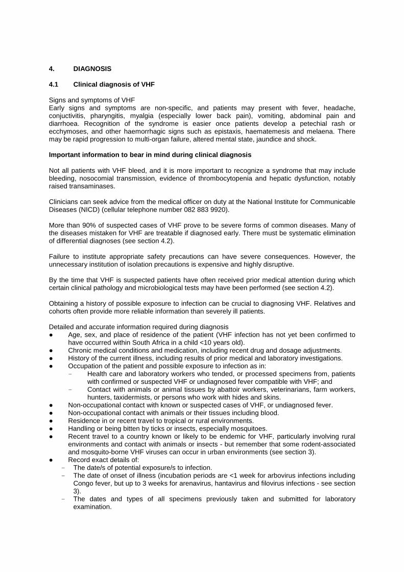
Table 2: Criteria for clinical diagnosis of Crimean-Congo haemorrhagic fever. R Swanepoel, JH Mynhardt and S Harvey 1987
Incubation period following known or potential exposure:
<l week
1 we e k or undetermined
I. HISTORY OF EXPOSURE TO INFECTION: Bitten by tick/s or crushed tick/s with bare hands OR Had direct contact with fresh blood or other tissues of live stock or game animals OR Had direct contact with blood, secretions or excretions of confirmed or suspected CCHF patient (including needle pricks) OR Resided in or visited a rural environment where contact with livestock or ticks was possible, but a specific incident constituting exposure cannot be identified
3 3** 3 2
2** 2*** 2 1
II. SIGNS AND SYMPTOMS: Sudden onset Fever 38℃ on at least one occasion Severe headache Myalgia Nausea and/or vomiting Bleeding tendency: petechial rash, ecchymoses, epistaxis, haematemesis haematuria or melaena
1 1 1 1 1 3
III. CLINICAL PATHOLOGY DURING FIRST 5 DAYS OF ILLNESS: Leukopaenia or leukocytosis WCC<3 x 10⁹/1 or 9 x 10
9/1 Thrombocytopaenia Platelets < 150 x 109/L Platelets < 100 x 10/L OR 50% de cre a s e in e ithe r WCC or platelet counts within 3 days Abnormal PI Abnormal PTT Raised transaminases AST 100 U/L ALT 100 U/L
1 1 2 1 1 1 1 1
*South African tick-borne typhus and ehrlichiosis must be excluded. **Rift Valley fever and anthrax must be excluded. ***Brucellosis, Q fever and anthrax must be excluded. A total score of 12 points or more constitutes an indication for treating a patient as a case of CCHF.

National Department of Health Page 17 of 62
4.2 Differential diagnosis of suspected VHF Procedure to follow when VHF is suspected When VHF is suspected, it is important to obtain and interpret the results of all medical examinations and laboratory tests already performed, but warn laboratory personnel of the suspected diagnosis and ensure that further laboratory tests are only performed with appropriate biosafety precautions (see section 6.2). Another crucial step to take is to ensure that all specimens previously submitted to laboratories are retained for onward transmission to NICD along with newly collected specimens for specific VHF diagnostic tests (see.section 4.3). Diseases commonly confused with VHF Malaria, trypanosomiasis, relapsing fever, plague, yellow fever, other arbovirus infections and leptospirosis, especially after travel to or residence in rural or tropical areas (malaria is most common and can be rapidly fatal if not treated, but it also occurs together with other infections including VHF). Bacterial septicaemias resemble VHF and can be rapidly fatal if not treated; most commonly caused by meningococci, but also by a wide variety of Gram-positive and -negative bacteria, and include typhoid, anthrax, and Capnocytophaga species (dysgonic fermenter 2) infection after dog bite, (septic abortion and tuberculosis with haemoptysis can also resemble VHF). Rickettsioses: tick bite fever (TBF), Q fever, typhus; TBF often occurs in town dwellers who visit rural environments, but can also result from exposure to kennel ticks in urban settings, even where dogs are kept indoors in apartment buildings; TBF can run a fatal course very similar to Congo fever, but has an incubation period of 7-10 days after tick bite as compared to 1-3 days for Congo fever, there is usually a necrotic eschar at the site of the tick bite in TBF and the petechial rash extends to palms and soles; TBF can be treated with broad-spectrum antibiotics. Hepatitis A, B, E, and less often C (westerners travelling in Africa often develop hepatitis A). Fulminant systemic herpes simplex virus infection with hepatitis (with/without vesicular rash); about 60 cases have been seen in RSA with high fatality, mostly in ostensibly healthy young adults; extremely high transaminase levels which may fall terminally after virtually complete destruction of hepatocytes. Less common are severe cytomegalovirus, E-B virus or varicella-zoster virus infections, or haemorrhagic measles. HIV seroconversion sickness, or HIV/AIDS with secondary infections, especially septicaemias. Drug sensitivities and overdoses including anticoagulants (warfarin), other poisons and toxins including haematoxic snake bite envenomation (e.g. boomslang), industrial and agricultural chemical poisoning. Malignant disease, e.g. leukaemia, lymphoma. Idiopathic thrombocytopenic purpura. Heat stroke. Interpretation of clinical pathology results for differentiating VHFs from other diseases Full haematological examination: Findings compatible with VHF include leucopenia, thrombocytopenia, anaemia, altered clotting parameters and increased fibrin degradation products or D-dimers, but disseminated intravascular coagulopathy also occurs in many other conditions, including septicaemia. Granulocytosis suggests bacterial infection, but leucocytosis can occur in CCHF, Lassa, Marburg and Ebola haemorrhagic fevers (see section 3), and in leukaemia.

National Department of Health Page 18 of 62
Examination of a stained blood smear: Malaria, trypanosomiasis, other haemoparasitic diseases and certain bacterial septicaemias (meningococcus, Capnocytophaga, anthrax) can be diagnosed, and differential white cell counts can be performed to provide an indication of leucocytosis/granulocytosis, leucopenia, leukaemia, anaemia, and even thrombocytopenia. Bacteriological blood cultures: It is important that blood cultures should be performed to exclude septicaemia. Samples should be taken before antibiotic therapy is instituted. Septicaemia can be secondary to many conditions including pneumonia, gastroenteritis, perforated ulcers, and abscesses or wound infections. Clinical chemistry tests: Raised serum transaminase levels occur commonly in VHF, and to a lesser extent also raised bilirubin levels, but jaundice and hepatocellular damage have many causes. Extremely high transaminase and bilirubin levels occur in systemic herpes simplex infection with hepatitis. Evidence of severe liver damage is a poor prognostic sign. Proteinuria is common in VHFs, notably in Lassa fever. Specific serodiagnostic tests for non-VHF diseases: Serological tests results should be interpreted with caution, taking into account the sensitivity and specificity of the test and the stage that they are performed during the course of the illness. Notably negative results using the currently available tests for tick bite fever may not exclude the disease. Anti-HA IgM, HbsAg, HBeAg and Anti-HBc are important screening tests for hepatitis A and B. Serodiagnostic tests are available for leptospirosis, salmonellosis, measles, herpesvirus infections and many other diseases which could be confused with VHF. Rapid serum latex agglutination tests can be used to detect bacterial antigen in meningococcal septicaemia. More than one pathology may be present in a patient, and epidemiological information and clinical laboratory findings should guide the diagnostic process. 4.3 Laboratory verification of VHF Specific diagnostic tests for the formidable (Class 4) VHFs are performed only by the Special Pathogens Unit (SPU) at NICD. It is essential that arrangements are made directly with one of the SPU laboratory diagnosticians before specimens are submitted (Laboratory telephone numbers 011 386 6339, 082 903 9131, 082 908 8042 and 082 908 8046; NICD Hotline 082 883 9920), particularly where urgent investigations are warranted after normal work hours (07h30-16h00 Monday to Friday). The staff must be informed of the means of transport of the specimens, tracking or waybill numbers, and expected date and time of delivery. 4.3.1 Source and nature of specimens: Clinical laboratories All specimens that may have been submitted to haematology, microbiology, clinical chemistry and other laboratories before VHF was suspected must be traced and redirected to NICD for virological examination. These specimens are important because VHF viruses are often only present in blood and other tissues in the early stages of the disease, and may be absent later. Live patients Specimens to be taken from live patients specifically for the investigation of suspected VHF should include 5-10ml of clotted blood and 5ml of blood taken with EDTA/sequestrene (lavender top). Throat swabs in viral transport medium may also be useful. Daily samples collected from patients in whom a diagnosis of VHF has already been confirmed provide valuable information, but need not be submitted for urgent tests; the samples can be kept refrigerated and sent to NICD in batches by routine laboratory delivery services with appropriate packaging (see 4.3.2 below).
National Department of Health Page 19 of 62
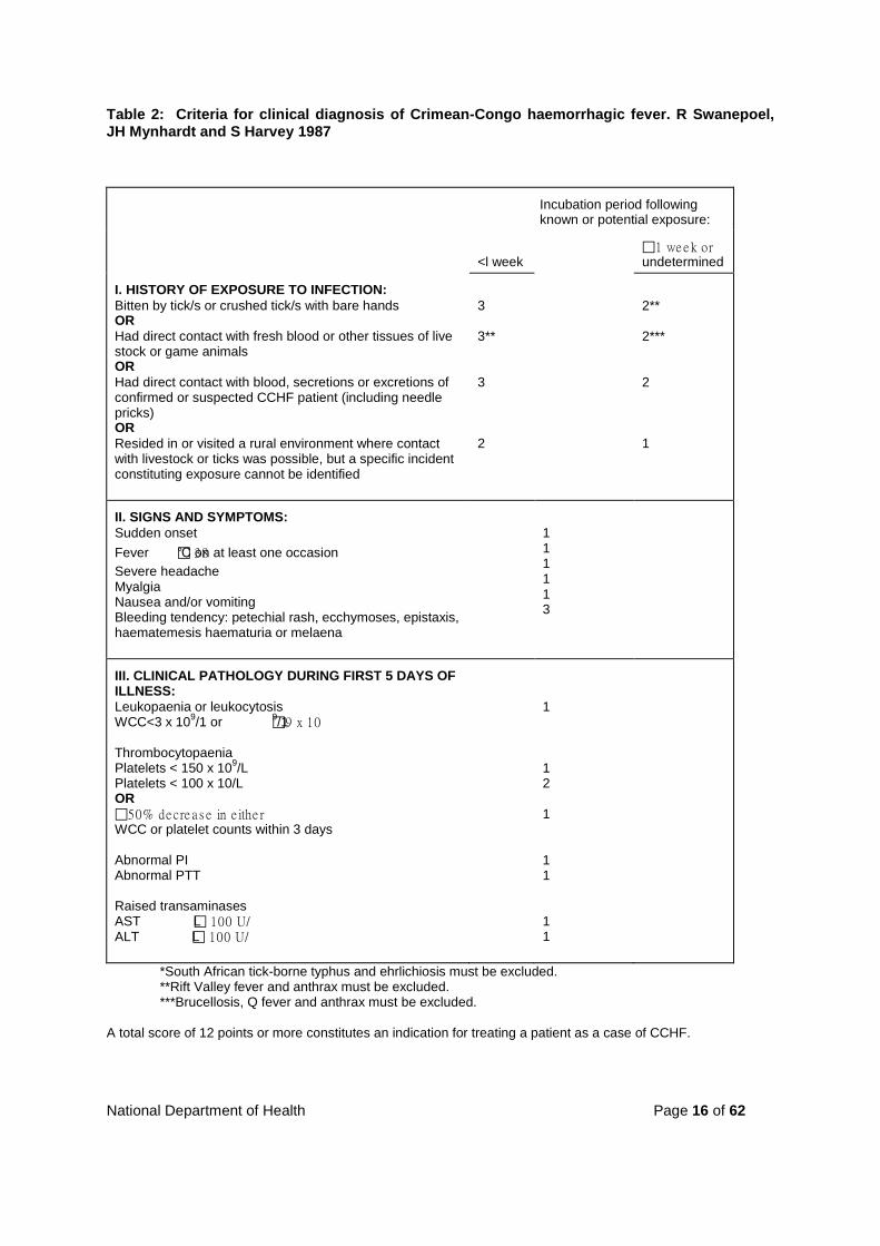
Corpses There is usually reluctance to proceed with a full autopsy until VHF can be excluded, and there is a widespread misconception that post mortem procedures may only be performed with the consent of relatives. However, in terms of the Health Act 61 of 2003 autopsy and removal of organs or tissues 'for determining the cause of death’ may be authorized by the medical practitioner in charge of clinical services in the hospital or authorized institution, or of the mortuary, or by a medical practitioner authorized by the person in charge of such hospital or authorized institution. Minimal specimens taken to eliminate VHF should include blood collected by cardiac puncture and liver samples taken with a biopsy needle; some liver should be placed in fixative for histopathological examination and some placed in a small volume of viral transport medium or physiological saline for virological examination. If possible, some liver tissue should also be placed in 2.5% glutaraldehyde fixative for electromicroscopy. The specimens can be taken in the ward where the death occurred or in a mortuary. Blood tends to ooze from needle puncture sites and these should be taped or sealed (e.g. Opsite®, S & N Pharmaceuticals Pty Ltd). The body should be decontaminated and sealed in double stout plastic body bags as discussed in section 6.5. Labels attached directly to the primary specimen containers (e.g. blood tubes) should be marked clearly with the name of the patient and date of collection of the sample. For removal from the patient facility or mortuary, the specimens should be double-wrapped in zip-lock specimen bags or ordinary clear plastic bags and labeled appropriately, preferably with biohazard stickers to alert staff to the contents, and should be delivered by hand directly to the laboratory responsible for forwarding the specimens to NICD. It may be useful to have a histopathologist examine rapidly fixed (heated formalin) and sectioned liver specimens. Bacterial septicaemia can sometimes be recognized and differentiated from liver disease due to VHF or other causes. Lack of liver lesions suggests that VHF is not involved. 4.3.2 Packaging of specimens for transfer to NICD
National Department of Health Page 20 of 62
UN/WHO approved shipping containers for hazardous specimens are commercially available, e.g. SAF-T-PAK®, or else safe packaging can be improvised as indicated in the text box below (Figures 4.1; 4.2):
The method used for transmitting specimens to NICD depends on the urgency with which diagnostic tests are required, proximity to NICD, and the availability and speed of routine delivery services for transmitting specimens to NICD as operated by the National Health Laboratory Service (NHLS) and private companies (e.g. Ampath, Lancet) For the delivery of specimens for urgent tests from within a few hours distance by road from NICD, it may be necessary to assign a specific vehicle and driver. This applies even to hospitals within close proximity to NICD since routine specimen delivery routes are operated at certain times of day only. Sometimes relatives of patients are willing to deliver specimens when no other rapid means of transport is available. Specimens should be delivered directly to members of SPU staff (contact telephone numbers: 011 386 6339, 082 903 9131, 082 908 8042, 082 908 8046; NICD Hotline 082 883 9920), or after hours left with the security guards at the entrance to NICD by prior arrangement with SPU staff (for map see Figure 4.6).
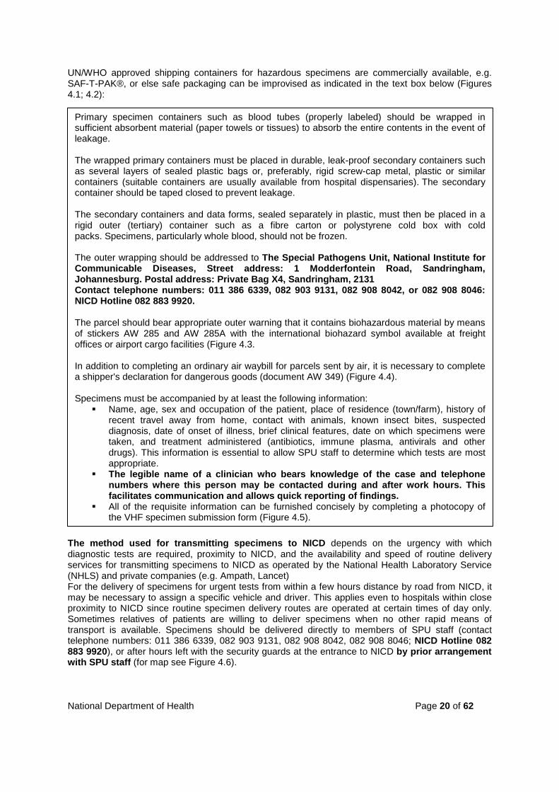
Primary specimen containers such as blood tubes (properly labeled) should be wrapped in sufficient absorbent material (paper towels or tissues) to absorb the entire contents in the event of leakage. The wrapped primary containers must be placed in durable, leak-proof secondary containers such as several layers of sealed plastic bags or, preferably, rigid screw-cap metal, plastic or similar containers (suitable containers are usually available from hospital dispensaries). The secondary container should be taped closed to prevent leakage. The secondary containers and data forms, sealed separately in plastic, must then be placed in a rigid outer (tertiary) container such as a fibre carton or polystyrene cold box with cold packs. Specimens, particularly whole blood, should not be frozen. The outer wrapping should be addressed to The Special Pathogens Unit, National Institute for Communicable Diseases, Street address: 1 Modderfontein Road, Sandringham, Johannesburg. Postal address: Private Bag X4, Sandringham, 2131 Contact telephone numbers: 011 386 6339, 082 903 9131, 082 908 8042, or 082 908 8046: NICD Hotline 082 883 9920. The parcel should bear appropriate outer warning that it contains biohazardous material by means of stickers AW 285 and AW 285A with the international biohazard symbol available at freight offices or airport cargo facilities (Figure 4.3. In addition to completing an ordinary air waybill for parcels sent by air, it is necessary to complete a shipper's declaration for dangerous goods (document AW 349) (Figure 4.4). Specimens must be accompanied by at least the following information:
Name, age, sex and occupation of the patient, place of residence (town/farm), history of recent travel away from home, contact with animals, known insect bites, suspected diagnosis, date of onset of illness, brief clinical features, date on which specimens were taken, and treatment administered (antibiotics, immune plasma, antivirals and other drugs). This information is essential to allow SPU staff to determine which tests are most appropriate.
The legible name of a clinician who bears knowledge of the case and telephone numbers where this person may be contacted during and after work hours. This facilitates communication and allows quick reporting of findings.
All of the requisite information can be furnished concisely by completing a photocopy of the VHF specimen submission form (Figure 4.5).
National Department of Health Page 21 of 62
For delivery of specimens from longer distances it may be possible to utilize routine laboratory delivery services, or a commercial courier service using scheduled road or air transport and door-to-door delivery, depending on the urgency with which tests are required. However, deliveries after normal work hours, and particularly at weekends, can be difficult to arrange. Follow up specimens from patients in whom the diagnosis has already been confirmed or sera from healthy contacts of VHF patients which are sent for routine screening and do not require urgent tests, can be sent to NICD by regular laboratory delivery services with appropriate packaging. 4.3.3 Laboratory tests If emergency tests are warranted and appropriate arrangements have been made ahead of time with SPU staff (telephone numbers 011 386 6339, 082 903 9131, 082 908 8042 and 082 908 8046; NICD Hotline 082 883 9920) tests can be performed after normal work hours, which are 07h30-16h00 on weekdays only. 4.3.4 Interpretation of results In the acute phase of the disease, cases of VHF are diagnosed by identifying virus antigen or nucleic acid in the specimens, or by isolating (culturing) live virus. Virus antigen detection tests are used for certain diseases only and take 3-8 hours to complete. Detection of virus nucleic acid by reverse transcription-polymerase chain reaction (RT-PCR) takes 6-12 hours from the time of receiving the specimen in the laboratory, depending on whether or not there is need for nested (second round) tests. Isolating virus in culture can sometimes be achieved within 2 days but usually takes a week or longer. In the convalescent phase of the disease, cases of VHF are diagnosed by identifying an antibody response. Preliminary IgG antibody tests can be completed within two hours of receipt of specimens and IgM tests within 3 hours, but overnight tests produce more reliable results. All serum samples (acute and convalescent) are routinely tested for antibodies to the full range of African VHF viruses. This is because the clinical histories received are sometimes inaccurate, particularly with respect to the date of onset or duration of illness. It is extremely important to remember that even acute specimens for which virus antigen, RT-PCR and antibody tests are all negative, occasionally yield virus in culture some days later. Failure to appreciate this possibility has led to serious misunderstandings in the past. Sometimes it is necessary to submit a further sample to clarify an ambiguous finding. For example, detection of IgG antibody on its own, without virus or IgM antibody, could indicate past infection not connected to the current illness, but sometimes IgG can appear in circulation slightly before IgM during convalescence. It is almost equally important to eliminate a possible diagnosis of VHF as it is to confirm a diagnosis rapidly: failure to detect virus or viral nucleic acid in serum during the first 7 days of illness, or to demonstrate antibody two weeks after onset, constitutes a fair indication that one of the known African VHFs is not involved. However, viraemia may be of very short duration or absent. Hence, negative findings on samples taken early in the course of disease should be supported by antibody tests on further specimens taken in convalescence. In emergencies results are made known telephonically or by fax as soon as possible, with written confirmation following later (remember to include contact details for the person to whom results should be reported when submitting specimens).
National Department of Health Page 22 of 62
Figure 1: Example of commercially available specimen biosafety packaging.
Figure 2: Example of improvised specimen biosafety packaging.
National Department of Health Page 23 of 62
Figure 3: International biohazard symbol stickers AW 285 and AW 285A

National Department of Health Page 24 of 62
Figure 4: IATA shipper's declaration for dangerous goods (document AW 349).
National Department of Health Page 25 of 62
Figure 5: VHF laboratory investigation request form
.
National Department of Health Page 26 of 62
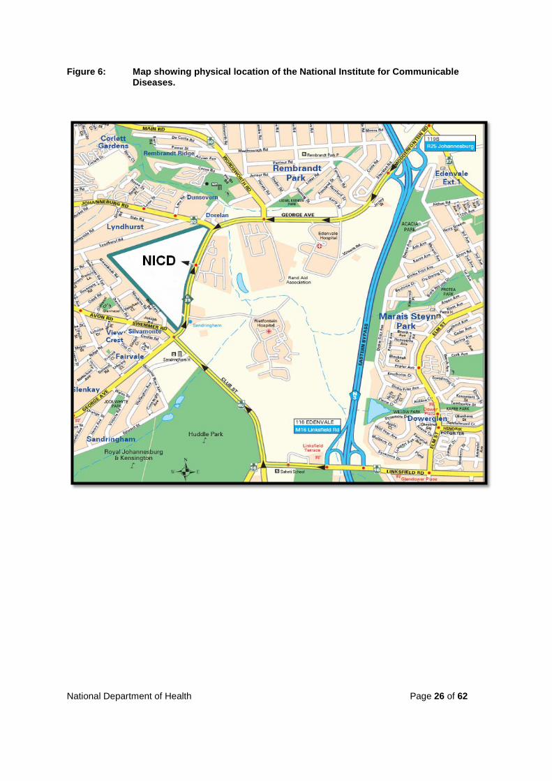
Figure 6: Map showing physical location of the National Institute for Communicable Diseases.
National Department of Health Page 27 of 62
5. IMMEDIATE ACTION TO BE TAKEN AFTER CLINICAL DIAGNOSIS OF VHF As soon as the decision is made to proceed on the basis of a presumptive diagnosis of VHF, measures should be applied to minimize exposure of medical staff, other patients and relatives. Whatever is ultimately decided concerning the management of the case, the immediate course of action should be to: ● Inform the management and infection control officers at the medical facility concerned of the
existence of the suspected case of VHF. ● Isolate the patient and apply infection precautions as best as can be managed under the
circumstances in cooperation with infection control staff (see section 6.3). The precautions must remain in force until the possibility of VHF has been excluded or the patient is no longer under care at the facility concerned.
● Administer such life-saving therapy as may be necessary and possible, e.g. blood/fluid therapy. ● Take steps to verify the diagnosis (see sections 4.3). ● Cooperate with infection control officers in preparing a list of staff members who have had
contact with the patient or fomites, including ambulance, laboratory and cleaning personnel - the contacts must be informed of the risks and precautions to be taken, and placed under observation (see section 7 for definitions of exposure, contact and observation).
● Notify the National Director of Communicable Disease Control (CDC) and the relevant Provincial Coordinator of CDC of the existence of the suspected case of VHF so that they can investigate the circumstances surrounding the incident, place relatives and cohorts and other contacts of the patient/s under observation if indicated, and take necessary actions to control any potential outbreak of VHF in the community at large (see section 7.2 for contact details of the officials).
● Decide whether the patient is to be retained at the primary hospital, or whether to seek transfer to a hospital more suited to managing the case. Decisions to transfer VHF patients cannot be taken unilaterally; see section 7.1 for the criteria and mechanisms for reaching decisions on referral.
● Assess the status of the patient as either low, moderate or high risk with respect to the probability that VHF is involved, the likely outcome of the disease, and the feasibility of safe transfer - sometimes the process of transfer poses too great a threat to the life of the patient or the safety of the personnel involved:
Low risk patients This category has febrile disease with features suggestive of VHF (e.g. thrombocytopenia), but are not necessarily severely ill and lack a history of contact with known VHF patients or animals (other than long-term pets), or animal tissues, or ticks and mosquitoes, and have not left an urban environment for at least 3 weeks prior to onset of illness. There are no haemorrhages, and risk of spread of infection is assessed as low. Moderate risk patients This category has febrile disease with features suggestive of VHF, and are not necessarily severely ill, but have visited or resided in a tropical or rural environment, or have had contact with animals or animal tissues, or ticks and mosquitoes during the 3 weeks preceding onset of illness. They have not had direct contact with known VHF patients or fomites (see section 7.3) but may have an indirect association with such patients, e.g. they have worked, resided in or visited the same places as VHF patients. Although there may be no haemorrhages, it is assessed that infection with a VHF agent may be involved. High-risk patients This category is severely ill with fever and haemorrhagic manifestations (this criterion is sufficient to place patients in the high risk category). In addition, they may have visited or resided in a tropical or rural environment, or have had contact with animals, animal tissues or ticks and mosquitoes during the 3 weeks preceding onset of illness. Alternatively, they may not necessarily be severely ill, but
National Department of Health Page 28 of 62
have had definite exposure to VHF (see section 7.3). This includes a) hospital and laboratory staff who have developed illness within 3 weeks* of last known contact with a confirmed VHF patient or fomites associated with such patients, and b) relatives and close associates of known VHF patients. (*The interval is 2 weeks for arbovirus diseases such as Congo fever, but 3 weeks for Lassa, Marburg and Ebola haemorrhagic fevers.)

National Department of Health Page 29 of 62
6. MANAGEMENT OF VHF PATIENTS 6.1 Medical management of VHF patients The medical management of VHF patients is a subject on which it is difficult to obtain consensus of opinion, and detailed analysis lies beyond the scope of the present document. The following remarks represent an attempt to summarize experience gained mainly in the management of Congo fever patients in South Africa. . 6.1.1 Antiviral therapy Antiviral compounds Ribavirin is a synthetic nucleoside analogue, which has been shown to be of use in treating hantavirus and arenavirus (Lassa fever) infections. There is evidence to suggest that it is of benefit in treating Congo fever patients but the findings are not conclusive, mainly because too few patients have been placed on therapy sufficiently early in the course of the disease for meaningful analysis: since deaths occur from day 5 of illness onwards the disease must be recognized and treated early. In practice, ribavirin therapy has only been attempted in patients with severe disease and a poor prognosis. In order to reach an early decision to institute therapy, it should be noted that during the first 5 days of illness in Congo fever any of the following pathological values are predictive of fatal outcome: leucocyte count ≥10x109/L; platelet count ≤20x109/L; AST ≥200U/L; ALT ≥150U/L; APTT ≥60 seconds; and fibrinogen ≤110mg/dL. After day 5 of illness any value may be grossly abnormal without necessarily being indicative of a poor prognosis. The oral preparation of ribavirin is registered in South Africa for the treatment of viral hepatitis. The drug would therefore be used ‘off- label’ for the treatment of CHF or Lassa fever. The trade name is Copegus, a Roche product, available in 200mg tablets. Ideally all severely ill patients should be treated with the intravenous formulation of ribavirin, but unfortunately it is not currently available in South Africa. It generally has to be sourced and imported when required. Table 6.1 and 6.2 shows the recommended dosage for adults and children.
Table 3: Recommended ribavirin dosage for Lassa- and Crimean-Congo haemorrhagic fevers: Adults including pregnant women.
Administration Loading dose d1 d1-4 d5-10
IV (6) 17 mg/kg (max 1000 mg per dose) 1x *
17 mg/kg (max 1000 mg per dose) q 6h
8 mg/kg (max 500 mg per dose) q 8h
PO (4) 2000 mg 1x 1000 mg q 6h 500 mg q 6h
* The loading dose for intravenous ribavirin has been suggested in other documents as 30 mg/kg
(max 2000 mg per dose) 1x (6, 7). Table 4: Recommended ribavirin dosage for Lassa- and Crimean-Congo haemorrhagic fevers: Children
Administration Loading dose d1 d1-4 d5-10 IV (6) 17 mg/kg 1x 17 mg/kg q 6h 8 mg/kg q 8h
PO (4) 30 mg/kg 1x 15 mg/kg q 6h 7 mg/kg q 6h

National Department of Health Page 30 of 62
Oral ribavirin treatment of CCHF reported by Fisher-Hoch et al. (14): 4000 mg/d d1-4, 2400 mg/d d5-10.
Ribavirin can cause bone marrow depression, raised serum bilirubin values, nausea and malaise, but these effects are generally overshadowed by the signs and symptoms of VHF. Moreover, the drug is teratogenic in animal models, but its use should still be considered in pregnant patients given the potential for lethality in severe infections. Congo fever patients have generally succumbed or recovered before completion of 10 days of treatment, resulting in early termination of the treatment. No other chemotherapeutics are available for the treatment of VHFs, and the use of ribavirin is indicated only for the treatment of hantavirus, arenavirus and Congo fever virus infections. Use of ribavirin is considered to be contraindicated in Rift Valley fever as some patients treated in Saudi Arabia in 2000 succumbed to late-onset viral encephalitis, but the association with ribavirin is not clear. Prophylactic use of ribavirin Oral ribavirin has been used prophylactically in persons deemed to have been exposed to infection with hantaviruses, arenaviruses and Congo fever virus, but the side effects of the drug can cause confusing and distressing illness which is particularly inconvenient when several people are affected. Hence it is advised that prophylaxis should be strictly limited to instances where there are strong indications that there has been exposure to infection, such as needle stick with blood known to be infected. The dosage for prophylaxis is the same as for treatment of infection. Interferon It has been demonstrated that interferons have significant antiviral activitiy against VHF agents in vitro and in animal models, and that there may be high levels of interferon expression in VHF patients. There appears to be no information on the value of interferon therapy in VHFs, but it is cautioned that its use in VHF patients poses difficult clinical challenges.
6.1.2 Immune plasma therapy There is no controlled experimental evidence to indicate that the use of immune plasma is of benefit in VHF, and persons who have recovered from Congo fever generally have low neutralizing antibody activity in their serum which is unlikely to be of therapeutic value. 6.1.3 Supportive treatment Monitoring of vital functions This should include temperature, pulse and respiration rates, chest auscultation and fluid balance (liquid intake/urinary output). The necessity for and frequency of additional monitoring is dictated by the severity of the disease/condition of the patient and whether or not a ventilator and drugs such as diuretics are being used. Laboratory tests to support patient management include full blood counts (with platelet plus haemoglobin values), coagulation, liver function, glucose, creatinine, urea, electrolyte, blood gases and pH determinations on appropriate blood samples. A chest X-ray should be taken on admittance of the patient and repeated if respiratory distress or suspected secondary infection occurs.

National Department of Health Page 31 of 62
Haemoglobin replacement This may be considered when blood haemoglobin levels fall to 8-10g/dL, but some patients tolerate such low levels quite well, and it is more important to treat on the basis of signs and symptoms of anaemia (respiratory distress) than purely on haemoglobin levels. Although fresh blood may be transfused, it is better to use red blood cell concentrate to treat the anaemia of VHF. This helps prevent fluid overload and development of the respiratory distress syndrome. Modern additives to red cell concentrates adequately maintain the levels of phosphates which modulate the oxygen affinity of haemoglobin, so it is not essential to use fresh blood. As a rough guide, one unit of red cell concentrate should raise the haemoglobin level of an average adult by lg/dL. Treatment of disseminated intravascular coagulopathy (DIC) Contrary to our earlier perceptions, DIC appears to be an early and prominent feature of CCHF and other VHFs. There are two views on treatment of DIC: one holds that the administration of coagulation factors merely ‘adds fuel to the fire’, while the other advocates judicious replacement of coagulation factors. The latter opinion appears to be most widely favoured. The use of heparin is considered to be useful in the early hypercoaguable stage of DIC, when there is accelerated partial thromboplastin time (PTT) and decreased prothrombin ratio (PR), but is of no value once the fibrinogen level falls. However, most cases of VHF are not diagnosed sufficiently early for use of heparin to be of value. Moreover, the use of the drug requires constant monitoring of the response and is best avoided by the inexperienced. Thrombocytopaenia is a common feature of VHFs and occurs regularly in CCHF. There is agreement on the need for replacement of platelets, but this should be done only if thrombocytopaenia is accompanied by purpura and active bleeding such as epistaxis, or if platelet counts fall below 20x109/L. A bag of platelet concentrate contains approximately 0.5 - 1.0X10 11 platelets in about 50 ml of plasma. The dosage of platelet concentrate is 1 bag/10kg body mass and transfusion services can be requested to pool the total dose, e.g. 7 bags can be supplied as 1 bag of 350 ml, which can be administered rapidly (10 minutes). Transfusion services ordinarily supply platelets of appropriate ABO group specificity. The treatment may be repeated over a period of days if the patient's platelet level continues to decline or remains critically low. If there is manifest consumption of other coagulation factors (abnormal PTT and PR levels, fibrinogen level <0.8g/L), administer fresh frozen plasma (FFP) or fresh dried plasma (FDP) at the rate of 10ml/kg body mass for the first dose. The treatment may be repeated if the patient continues to bleed or if coagulation factor levels remain markedly abnormal. As a general rule, 2-3 units of FFP or FDP should be administered to augment coagulation factors for every 10 units of red cell concentrate given to the patient. Fibrinogen is not available as a separate product, but apart from its administration in FFP and FDP, it (and other factors) can also be administered in the form of cryoprecipitate. One bag of wet cryoprecipitate contains about 250 mg of fibrinogen and a bottle of dried cryoprecipitate, called anti-haemophilia factor (AHF), which is derived from a pool of 4-6 units of wet cryoprecipitate, contains approximately 1 g of fibrinogen. About 1-2 g fibrinogen (10 bags of wet cryoprecipitate) may be administered as a first dose. Prothrombin complex concentrate (PCC, factor IX complex, Proplex) may be indicated following liver damage. It contains 200 units factor IX in a10 ml volume and a dose of 1 U/kg should increase the blood level of the factor by approximately 1%. Vitamin K should also be administered.

National Department of Health Page 32 of 62
Intravenous fluids Plasma is used to replace coagulation factors, not merely for volume expansion, but it is expensive and haemodynamic goals can be achieved with artificial colloids or even crystalloids. Iso-osmotic albumin solution (4%) may be used for volume expansion. Although 20% albumin has been used to treat hypoproteinaemia following liver damage in CCHF, it is considered better to use an enteral feed that provides sufficient calories and protein according to body mass. If the gut is unavailable for enteral nutrition parenteral feeding may be necessary. Hypoglycaemia was thought to be of critical importance in a number of CCHF patients in South Africa and blood glucose levels should be monitored carefully in severely ill patients. Other therapy There is no information on the effectiveness of steroids to allay the ‘cytokine storm’ underlying the DIC in VHF patients, but there is some support for this approach from animal models. If used, the dose should not exceed 200-300mg hydrocortisone daily. The use of non-steroidal anti-inflammatory drugs is not recommended. Antacids, painkillers, relaxants and tranquillisers are administered as indicated. Antibiotic prophylaxis is generally not indicated, however many patients will have received antibiotics prior to the diagnosis having been made. As with all patients in the ICU regular screening for colonization and infection is necessary. Counselling of patients and relatives is mandatory as this is a highly stressful situation. 6.2 Clinical pathology monitoring of VHF patients Requirements Hospitals which manage suspected or confirmed cases of VHF should have available the services of a laboratory able to conduct the following tests: ● A minimum range of screening tests to eliminate non-VHF diseases:
- Full blood count. - Examination of blood smears for parasites and bacteria. - Blood cultures for septicaemia.
● Haematological and clinical chemistry tests to monitor treatment and progress of patients: - Full blood counts (including platelet and haemoglobin values). - Coagulation studies. - Liver function tests. - Blood glucose tests. - Creatinine, urea, electrolyte determinations. - Blood gases and pH determinations. - Cross matching studies for transfusions.
Ideally the tests should be conducted by a small team of experienced volunteer technologists in a room set aside for the purpose within an existing laboratory, but since the occurrence of VHF is sporadic the expenditure to equip a dedicated unit is not justified. Consequently, the required tests are often conducted within routine laboratory facilities temporarily set aside for the purpose as required. Operational procedures Technologists who conduct clinical pathology tests on specimens from VHF patients should be trained in the donning, removal, and disposal of personal protective equipment (PPE), and entry and exit
National Department of Health Page 33 of 62
procedures from infected areas, as described under isolation precautions for VHF patients (see section 6,3). Only volunteer team members should be present during the testing of specimens from suspected or confirmed VHF patients, and as far as possible manipulation of specimens should be performed in biohazard laminar flow safety cabinets (class IIA). Duty registers should be kept, with staff subjected to the same monitoring as other medical personnel dealing with VHF patients, and incidents constituting potential exposure to infection, including injuries and spillages, should be dealt with as described in sections 6.4 and 7. Decontamination of laboratory equipment including auto analyzers should follow standard operating procedures developed from manufacturer’s instructions. Decontamination of laboratory floors, walls and work surfaces, and disposal of waste materials, should follow procedures described in section 6.4. Specimens for monitoring of VHF patients should be preserved at least until the patient is discharged or a diagnosis is established in a deceased patient, and should then be disposed of in a safe manner (autoclaved or sent for incineration). However, specimens should be offered to the Special Pathogens Unit (SPU) at the National Institute for Communicable Diseases (NICD) rather than destroyed, since much valuable information is gained from the examination of serial samples from VHF patients. Ideally a separate clotted blood sample should be taken daily from confirmed VHF patients for submission to NICD, but these can be submitted together when the patient is discharged. 6.3 Isolation precautions (formerly known as barrier-nursing procedures) Although the VHFs are seldom encountered, the consequences of being unprepared can be extremely serious. All medical institutions should formulate and implement contingency plans for isolating and managing VHF patients, even on a temporary basis. The aims should be to: ● Identify facilities and resources which can be utilized for isolating and managing VHF patients. ● Provide health care workers with training and instructions specific to their duties so that they
are able to act in an informed manner when suspected cases of VHF are encountered. ● Train all staff members to recognize potential cases of VHF, but ensure that critical assessment
of such cases is performed by experienced clinicians and infection control personnel. ● Train suitable volunteers in isolation precautions. Experience has shown that when VHF occurs
in an institution where there has been no prior discussion of VHFs and training in isolation precautions it may be extremely difficult to obtain volunteers. Do not be caught unprepared.
● Ensure that infection control personnel monitor safety practices during isolation precautions and place staff who are in contact with VHF patients or fomites under observation (see section 7.3).
● Establish proper channels of communication so that relevant members of staff at all levels are informed promptly of the existence of a suspected case of VHF, or of the impending arrival at a hospital of such a patient, and of all key developments in the handling of the case.
● Extend the system of communication to outside officials who need to be kept informed, such as Communicable Disease Control officials of the national and provincial Departments of Health (see section 7).
● Make provision for well-informed responses to enquiries from news media (see section 7). Facilities The minimum accommodation required for isolation precautions consists of one room in which the patient may be isolated and an ante-room or adjacent room where staff can don and remove personal protective equipment (PPE). Ideally, isolation units should have separate entrance and exit (‘clean and dirty’) channels, and it is advantageous if the ante-room has a hand-basin and if ablution facilities are located in convenient proximity to the patient's room. The equipments and supplies required in the
National Department of Health Page 34 of 62
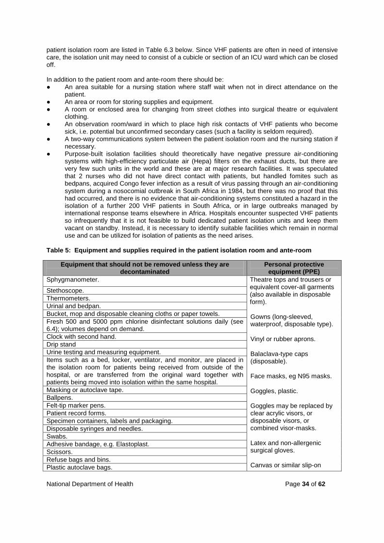
patient isolation room are listed in Table 6.3 below. Since VHF patients are often in need of intensive care, the isolation unit may need to consist of a cubicle or section of an ICU ward which can be closed off. In addition to the patient room and ante-room there should be: ● An area suitable for a nursing station where staff wait when not in direct attendance on the
patient. ● An area or room for storing supplies and equipment. ● A room or enclosed area for changing from street clothes into surgical theatre or equivalent
clothing. ● An observation room/ward in which to place high risk contacts of VHF patients who become
sick, i.e. potential but unconfirmed secondary cases (such a facility is seldom required). ● A two-way communications system between the patient isolation room and the nursing station if
necessary. ● Purpose-built isolation facilities should theoretically have negative pressure air-conditioning
systems with high-efficiency particulate air (Hepa) filters on the exhaust ducts, but there are very few such units in the world and these are at major research facilities. It was speculated that 2 nurses who did not have direct contact with patients, but handled fomites such as bedpans, acquired Congo fever infection as a result of virus passing through an air-conditioning system during a nosocomial outbreak in South Africa in 1984, but there was no proof that this had occurred, and there is no evidence that air-conditioning systems constituted a hazard in the isolation of a further 200 VHF patients in South Africa, or in large outbreaks managed by international response teams elsewhere in Africa. Hospitals encounter suspected VHF patients so infrequently that it is not feasible to build dedicated patient isolation units and keep them vacant on standby. Instead, it is necessary to identify suitable facilities which remain in normal use and can be utilized for isolation of patients as the need arises.
Table 5: Equipment and supplies required in the patient isolation room and ante-room
Equipment that should not be removed unless they are decontaminated
Personal protective equipment (PPE)
Sphygmanometer. Theatre tops and trousers or equivalent cover-all garments (also available in disposable form). Gowns (long-sleeved, waterproof, disposable type). Vinyl or rubber aprons. Balaclava-type caps (disposable). Face masks, eg N95 masks. Goggles, plastic. Goggles may be replaced by clear acrylic visors, or disposable visors, or combined visor-masks. Latex and non-allergenic surgical gloves. Canvas or similar slip-on
Stethoscope. Thermometers. Urinal and bedpan. Bucket, mop and disposable cleaning cloths or paper towels. Fresh 500 and 5000 ppm chlorine disinfectant solutions daily (see 6.4); volumes depend on demand. Clock with second hand. Drip stand Urine testing and measuring equipment. Items such as a bed, locker, ventilator, and monitor, are placed in the isolation room for patients being received from outside of the hospital, or are transferred from the original ward together with patients being moved into isolation within the same hospital. Masking or autoclave tape. Ballpens. Felt-tip marker pens. Patient record forms. Specimen containers, labels and packaging. Disposable syringes and needles. Swabs. Adhesive bandage, e.g. Elastoplast. Scissors. Refuse bags and bins. Plastic autoclave bags.
National Department of Health Page 35 of 62
Plastic cable ties for sealing bags 12 (obtainable from electrical/hardware stores).
shoes. Overshoes (disposable) or stout plastic bags.
Small, clear plastic bags for removal of specimens or other small items from the isolation room. Biohazard labels Disposable eating utensils. Desk, chairs Restricted entry and hazard warning signs. Register to record entry and exit of persons with authorized access to the isolation unit. Much of the PPE is available in disposable plastic or paper form at all hospitals and clinics. Up to 25 changes of protective clothing may be required per day in nursing a patient during the critical phase of VHF illness (not all patients become severely ill or exhibit bleeding tendencies). Some hospitals utilize mended and condemned linen and theatre clothes for nursing VHF patients, but dye the items an obvious colour to help ensure that they are disposed of safely. Ideally, hospitals should keep stocks of the essential items in readiness, but this involves dedication of funds and secure storage space, plus rotation of perishable items. Alternatively, stocks should be secured immediately an emergency arises. Formidable epidemic disease packs (FED packs) containing virtually all of the above PPE items are available commercially, and customized packs can be prepared to order or within the hospital. Safety equipment It is notable that international teams operating under the auspices of the World Health Organization to control outbreaks of Marburg and Ebola haemorrhagic fevers in Africa use only standard PPE items specified above. Almost all VHF patients in South Africa have been nursed without special safety equipment, and all nosocomial infections occurred before the patients were placed under conditions of isolation precautions. In the past, some hospitals in South Africa acquired special safety equipment for protection of staff against nosocomial infection, ranging from containment bed isolators, full-face respirators (gas masks) to battery-operated positive-pressure ventilated respirators (‘pappers’). There are disadvantages associated with each of these items: bed isolators are very expensive and occupy a large floor space; gas masks are tiring to use and tend to become fogged, thus reducing visibility and efficiency; pappers require expensive semi-disposable hoods and interfere with the use of stethoscopes. Nevertheless, the use of pappers may be warranted for particularly hazardous procedures, such as intubation of VHF patients under intensive care. Hoods may be re-used by the same staff member for successive entries into the patient isolation room provided they are disinfected on exit from the isolation room as described below (they should be marked with the name of the user). Power supply points will be required in the ante-room for re-charging batteries, plus a rack or coat hooks for hanging respirators and hoods when not in use. Personnel Ideally, specifically trained, volunteer staff should be used for nursing VHF patients, and personnel who were in contact with the VHF patient/s before isolation precautions were implemented should be utilized first to limit potential exposure of further members of staff. Select persons of calm disposition able to cope with the stress of nursing VHF patients under strict isolation precautions. Nosocomial infections can almost invariably be traced to fundamental lapses in technique, such as needle-sticks, against which most safety equipment cannot protect. Fatigue causes mistakes and hence adequate numbers of staff should be delegated to nursing patients under conditions of strict isolation precautions without seriously depleting the rest of the hospital or unnecessarily exposing too

National Department of Health Page 36 of 62
many individuals to VHF. If the nursing load is too heavy, as when multiple patients are involved, it may be necessary to suspend some or all-routine functions of the hospital. Counselling of staff (plus patients and families) should be offered to alleviate stress. Shifts should be limited to a maximum of 8 hours (6 hours are preferable) to ensure a high degree of efficiency. Intensive nursing of critically ill VHF patients may require 3-5 persons per shift, 1-2 of whom are in the patient's room on a 1-2 hourly rotation. Low profile nursing of moderately ill patients requires less staff and often it is unnecessary to maintain a constant presence in the patient's room. In addition to the staff members who are directly in attendance on the patient, one member of the nursing or administrative staff should remain outside of the isolation area to control communications, logistics and access to the isolation suite. In large hospitals it may be necessary to use security officers to control access to the isolation suite. Domestic and any other staff who have not been specifically instructed in isolation precautions must be excluded from the isolation suite. All medical and auxiliary staff (ambulance and laboratory personnel) who come into contact with a suspected or confirmed VHF patient or fomites, either before or after the institution of isolation precautions, must be placed under observation (see section 7). This should be done formally but the precautionary nature of the measure should be explained carefully. Incidents constituting possible exposure to infection, e.g. needle sticks or other direct contact of skin with patient’s blood or body fluids, must be recorded and promptly brought to the attention of the hospital's infection control team to decide on any action to be taken (see section 7). Baseline blood counts plus serum transaminase tests may be performed for persons who have had contact with a VHF patient or fomites, and serum samples should be kept frozen for later use if suspected infection occurs. However, this should be limited to persons with definite exposure to infection such as a needle-stick with known infected blood. Indiscriminate bleeding of contacts generates undue concern and unreasonable demands from people who have not had genuine exposure to infection. Placing a patient into isolation Explain to the patient and family that isolation precautions are being instituted and make an effort to reassure them. The donning of protective clothing by medical personnel can have a demoralizing effect on lay people. Establish from the clinician in charge whether or not the patient's immediate family will be permitted to visit the patient (under supervision and with proper protective clothing). Inform the family accordingly and arrange for instruction in correct use of protective clothing. Ensure that all staff are informed that the patient is being placed into isolation, institute control over access to the isolation suite and display appropriate warning notices. Henceforth only specifically authorized personnel may have access to the patient and all staff must wear protective clothing when tending the patient. The patient is transferred to the isolation room on his/her bed, and all other items of equipment required from the original ward (e.g. locker, ventilator, monitor, etc.) are moved with the patient. The procedure for receiving VHF patients from outside the hospital is described in section 7. All non-essential items, including the patient's records, are left in the original ward and are decontaminated in the prescribed manner by personnel wearing protective clothing (see section 6.4). New patient records are started and kept outside of the isolation room.
National Department of Health Page 37 of 62
All other patients who were in the original ward with the VHF patient are transferred, preferably to a single other ward, so that the original ward can be decontaminated (see section 6.4). Sometimes it is more convenient to leave the VHF patient in the original ward and convert it into an isolation room. Ensure that the infection control team prepares a register of all persons deemed to have had contact with the VHF patient/s and places contacts under observation (see section 7.3). Ensure that a duty register is kept of all staff shifts and visits to the patient, to ensure traceability of contact with the patient. Dressing for entering the patient isolation room In a change-room or other suitable area close to the entrance to the isolation suite, staff remove all jewellery and replace their street clothes with surgical theatre tops and trousers, or equivalent cover-all garments (washable fabric or disposable), plus canvas or similar slip-on shoes. These clothes are worn for the duration of the work shift and are used to move around in the vicinity of the isolation suite, but an extra layer of protective clothing is donned in the ante-room for entry into the patient isolation room: ● Long-sleeved, waterproof, disposable gown. ● Vinyl or rubber apron if more than light duties are involved, e.g. bleeding patients. ● Two pairs of latex surgical gloves, one worn over the other - the cuffs of the outer pair of gloves
should be pulled over the cuffs of the gown and taped in place with masking tape around the wrist.
● Disposable balaclava-type cap. ● Disposable face-mask, e.g. N95 – cannot be used with facial hair (beards). ● Goggles or acrylic visor, or disposable visor. ● A disposable combination visor-face mask can replace a separate mask and goggles or visor. ● Alternatively, a positive-pressure ventilated respirator (papper) with hood could replace the
balaclava, face-mask and goggles or visor. ● Two pairs of overshoes, one over the other, or heavy duty plastic bags taped to the trouser
legs, or waterproof boots. Needles, other sharp objects, patient's blood, blood-contaminated discharges and equipment soiled with blood constitute the greatest danger and must be handled with extreme care. Gloved hands contaminated with patient's blood or discharges should be dipped into 500 ppm chlorine disinfectant solution (see section 6.4) kept in the isolation room. Gloves must be checked frequently for tears or punctures and if the patient bleeds profusely, both inner and outer gloves must be changed hourly and the hands washed thoroughly in soap and water or surgical scrub disinfectant. Procedure for leaving the patient isolation room The procedure for leaving the isolation room must be followed strictly to prevent contamination of personnel and the environment. Double refuse or autoclave bags (heavy duty), which are used to receive discarded protective apparel, are placed one inside the other in a bin or holder in the ante-room with 20-30 cm of the top of the bags folded back over the rim of the bin or holder to form a clean margin when the bag is sealed. The bin is placed close to the door leading from the patient isolation room. On leaving the patient isolation room, the outer overshoes are removed and placed in the disposal bag. Waterproof overshoes or boots may be dipped into a bucket/tray of 500 ppm chlorine disinfectant (see section 6.4) before being removed. The outer gloves are dipped or washed in 500 ppm chlorine disinfectant (see section 6.4), peeled off and discarded into the disposal bag. The inner gloves are used to remove the other items of protective wear and to place them in the disposal bag as follows: ● Goggles or acrylic visors are removed and placed in 500 ppm chlorine disinfectant (see section
6.4). ● Disposable combination visor-masks are discarded into the disposal bag.

National Department of Health Page 38 of 62
● If a positive-pressure respirator is being worn, an assistant in the ante-room swabs or sprays the outer surface of the hood with 500 ppm chlorine disinfectant (see section 6.4) and with gloved hands helps to remove the respirator; the swabbed hood is hung on a hook or rack to dry; the respirator itself is hung or placed on a suitable surface and connected to a battery charger. Respirator hoods are marked with the names of individual members of the team for re-use, and are discarded for incineration or safe disposal when no longer required.
Facemasks and balaclava caps are removed and placed in the disposal bag. ● Next, aprons and gowns are removed and folded or rolled in the process so that outside
surfaces are on the inside and they are placed in the disposal bag. ● The inner pair of overshoes and finally the inner gloves are removed and placed in the disposal
bag, and the hands are washed thoroughly with soap and water or surgical scrub disinfectant (use of ethyl or isopropyl alcohol is not recommended for disinfection of the hands in nursing VHF patients).
● The inner and outer top rims of the disposal bags are sprayed with 500 ppm chlorine disinfectant (section 6.4) and sealed by a gloved assistant, conveniently with plastic cable-ties obtainable from electrical or hardware stores, or with adhesive tape. The double bags are sealed into a third bag, or several layers of bags if necessary to prevent leakage. It is useful if the outer bag is colour-coded, e.g. red, to indicate that it contains biohazardous material due for incineration. However, red bags are commonly used for waste in hospitals.
● The outer bag is labeled with biohazard stickers and sent for incineration under supervision, or sealed into the container of a commercial biohazardous materials disposal contractor, e.g. Sanumed®.
● Surgical theatre clothes or equivalent cover-all garments and footwear are removed in the outer change room and discarded into laundry or disposal containers as appropriate, and staff don their street clothes. Preferably, staff should be able to take a shower bath before leaving the isolation area.
Procedures for dealing with potentially hazardous incidents All incidents constituting possible exposure to infection, such as needle stick injuries and splashing with patient’s body fluids must be recorded and reported to infection control staff to decide on appropriate action, and an Employer’s Report of an Accident Form (Compensation for Occupational Injuries and Diseases Act, 1993) must be completed and submitted. First aid procedures should be applied as considered necessary, eg bleeding of needle stick or sharp instrument injury sites should be encouraged and wounds bathed in copious 500 ppm chlorine disinfectant (see 6.4). Infection control staff in consultation with senior clinicians and management should decide whether staff members potentially exposed to infection should be placed in quarantine (section 7), subjected to prophylactic treatment (section 6.1), or simply kept under observation (section 7). Patient care facilities should be subjected to routine disinfection, but overt spillages of hazardous materials should be dealt with as they arise (section 6.4). Discharge of patients Provided they are well, Congo fever, Rift Valley fever and other arbovirus disease patients can be removed from strict isolation precautions, or even discharged from hospital, two weeks after onset of illness, but they should remain under supervision and refrain from strenuous activity for a month or more, depending on their progress. Meningism, encephalitis and ocular lesions can occur as late complications of Rift Valley fever. Patients with a diagnosis of any of the other African haemorrhagic fevers should be nursed in isolation for at least 3 weeks after onset of illness. Sexual transmission of Marburg virus in semen has been recorded two months after recovery of the patient and the same could probably occur with Ebola virus. Excretion of Lassa fever virus in urine has been observed to occur over a period of a few

National Department of Health Page 39 of 62
weeks, and hence the discharge of Lassa fever or Lujo virus patients from hospital should be made consequent upon failure to isolate virus or to detect viral nucleic acid by RT-PCR in three consecutive urine samples collected on separate days. The same would apply to haemorrhagic fever with renal syndrome (HFRS) patients, but culture of the viruses is difficult and erratic. Recovery from VHF may be marked by prolonged convalescence and it is advisable that patients should be kept under casual surveillance for about 3 months. They should be warned of the possibilities of their transmitting infection through intimate contact during this time. If convenient, serum samples from recovered patients should be sent for monitoring of antibody levels at intervals after recovery as opportunity arises; useful diagnostic information on the duration of IgM and IgG responses is accumulated in this manner. Patients should be approached about the possibility of donating immune plasma once they are fully recovered, for preparation of control reagents for diagnostic tests, or for possible therapeutic use. Offers to donate plasma can be discussed with the Special Pathogens Unit at NICD (telephone numbers 011 386 6339, 082 903 9131, 082 908 8042 and 082 908 8045). 6.4 Disinfectants and decontamination The use of disinfectants is not a substitute for sterilization by physical means, especially heat as in autoclaving or incineration. However, there are many situations where it is necessary to resort to the use of disinfectants, and the present discussion is limited to requirements for managing VHF patients. The use of brand names does not imply recommendation of a product to the exclusion of similar preparations. It should be remembered that mechanical cleansing is an integral part of proper disinfection: excess organic matter rapidly reduces the efficacy of disinfectants. Choice of general disinfectant Inorganic chlorine, in the form of diluted household bleach, has been used as the disinfectant of choice in controlling outbreaks of VHF in Africa because it is effective, relatively inexpensive and readily available. However, inorganic chlorine corrodes metals and tends to degrade fabrics. Brand-name household bleaches contain 5% sodium hypochlorite and are diluted as follows for use:
A 10% aqueous solution of household bleach (one part bleach plus 9 parts water) yields 0.5% hypochlorite, or approximately 5000 ppm chlorine, and is used for disinfecting overt spillages of contaminated materials, excreta (organic wastes) and surfaces of corpses. A 1:100 aqueous solution of household bleach (one part bleach plus 99 parts water, or 1 part of 10% diluted bleach plus 9 parts of water) yields 0.05% hypochlorite, or approximately 500 ppm chlorine, and is used for disinfecting gloved hands, walls and floors without overt spillages of contaminated materials, clothing, bedding, equipment and instruments, and the outer surfaces of sealed plastic bags containing infected or contaminated materials. One or two crystals of potassium permanganate (Condy’s crystrals) can be added to undiluted household bleach to impart a pink colour to the diluted solutions of disinfectant for easy identification. Fresh stocks of diluted disinfectant should be prepared daily.
‘Organic’ chlorine formulations which contain a detergent, an anti-corrosive agent and chlorine incorporated in or complexed to organic molecules (chloro-cyanurates and chloramines) offer clear advantages over inorganic hypochlorite. A dry granular preparation of this type is sold in South Africa as Biocide D (6g granules per sachet), or as Biocide D Extra (30g granules per sachet) (Johnson Diversey, Germiston):
National Department of Health Page 40 of 62
Disinfectant solutions should be clearly labelled with the concentration of active ingredient and date of preparation. Choice of hand disinfectant Gloved hands are generally rinsed in 500 ppm chlorine, and although this can also be used to rinse bare hands, it is recommended that when surgical gloves are removed after nursing or transportation of VHF patients, or performance of clinical pathology tests, the hands should be thoroughly washed with soap and water, or with a surgical scrub preparation. Ethyl or isopropyl alcohol preparations are not recommended for disinfection of the hands in managing VHF patients, although they can be used as skin disinfectants for injection of patients. Decontamination and disposal of hazardous items A specific individual in each nursing shift, or ambulance crew transporting a VHF patient, should be responsible for supervising decontamination and disposal of biohazardous items. All items leaving the isolation unit (patient's room and anteroom, or ambulance) should be enclosed in double layer autoclave bags (or more layers if necessary to prevent leakage) and sealed with cable ties or adhesive tape. The outer surface should be labelled with biohazard stickers and swabbed with 500 ppm chlorine disinfectant. Disposable items should be sent for incineration under supervision and re-useable items for autoclaving. Crockery and cutlery used for feeding VHF patients should ideally be of the disposable type and incinerated along with food wastes. Bedpans and other containers with patient secretions, excretions and other wastes such as vomitus and blood, should be flooded with copious 5000 ppm chlorine disinfectant, left for at least 30 minutes, and sealed into adequate layers of leak-proof autoclave bags or other secure secondary containers (e.g. stainless steel container). The outer surfaces of the bags or containers should be swabbed with 500 ppm chlorine disinfectant, labelled with biohazard stickers and the items removed for autoclaving and cleaning. Autoclaved wastes can be flushed into municipal sewers. After flushing, bedpans are cleaned with 500 ppm chlorine disinfectant. Thoroughly disinfected wastes (prolonged exposure to copious disinfectant) can also be discarded into sealed disposal pits, or buried. It is convenient to use chemical toilets instead of bed pans for ambulant patients. Vinyl, rubber and other items which are degraded by autoclaving could be discarded and incinerated, or subjected to prolonged immersion in 500 ppm chlorine disinfectant. The hoods of battery-operated positive-pressure ventilated respirators (‘pappers’) are discarded for incineration at the termination of patient treatment, and the respirators are swabbed with 500 ppm chlorine disinfectant, and sealed into labelled bags and sent for gaseous sterilization. Hypodermic and intravenous needles should be used with great care, discarded into rigid-walled disposal containers, flooded with a 5000 ppm chlorine disinfectant (see 6.4), sealed into leak-proof bags, labelled and sent for incineration.
Twenty sachets of Biocide D Extra (600g) dissolved in 10 litres of water yields 0.5% hypochlorite, or approximately 5000 ppm chlorine, and is used for disinfecting overt spillages of contaminated materials, excreta (organic wastes) and surfaces of corpses.
Two sachets of Biocide D Extra (60g) dissolved in 10 litres of water yields 0.05% hypochlorite, or approximately 500 ppm chlorine, and is used for disinfecting gloved hands, walls and floors without overt spillages of contaminated materials, clothing, bedding, equipment and instruments, and the outer surfaces of sealed plastic bags containing infected or contaminated materials.
Fresh solutions should be prepared daily.
National Department of Health Page 41 of 62
Used linen and cloth items of protective wear should be sealed into labelled bags and autoclaved before laundering, but consideration should be given to incinerating grossly contaminated items such as bloodstained mattresses and pillows. Items, which are not visibly soiled, could be soaked in 500 ppm chlorine disinfectant for 30 minutes before laundering. Persons laundering cloth protective apparel or bedding used for VHF patients should don personal protective equipment (PPE) as described for isolation precautions (see section 6.3). Vomitus, blood and other overt spillages on floors and similar impervious surfaces should be flooded with 5000 ppm chlorine disinfectant, covered with paper towels and left for 30 minutes before removal. Floors of VHF patient isolation units should be mopped and drains flushed with 500 ppm chlorine disinfectant daily, or whenever there is spillage of potentially contaminated material. Rinsed mops should be soaked in 500 ppm chlorine disinfectant for 30 minutes. At the termination of patient treatment the walls and all impervious surfaces in isolation units (lockers and tables) should be swabbed in addition to the disinfection of floors and drains. The same procedures should be applied to mortuaries and laboratories handling corpses or samples of suspected or confirmed VHF patients. Patient records, which have been kept in an infected environment, can be bagged and autoclaved, or the information preserved by other means, e.g. copied from records, taped to a window or glass partition, or transmitted via telephone. 6.5 Disposal of corpses Corpses of suspected VHF patients may be processed for immediate disposal if an etiological diagnosis has been confirmed. If a diagnosis has not been established, then in terms of the Health Act 61 of 2003 certain medical practitioners are empowered to authorize the performance of an autopsy to determine the cause of death, as described in section 7 of this document. Usually the autopsy procedures are limited to collecting blood by cardiac puncture and taking liver samples with biopsy needles. After the autopsy specimens have been taken, the corpse may be held under refrigeration in a mortuary if the facilities exist, while laboratory investigations to eliminate VHF proceed. This usually takes a week, and if a diagnosis of VHF is eliminated, it may be deemed safe and/or necessary to proceed with a full autopsy to establish the cause of death. For disposal, corpses are washed with 5000 ppm chlorine disinfectant (see section 6.3 above). Orifices are plugged with gauze and puncture sites are taped or sealed (Opsite®, S & N Pharmaceuticals Pty Ltd). The corpse is enclosed in an impervious body bag and sealed (it is advantageous if the body bag has an air-valve). The attendants change protective clothing, swab the body bag with fresh disinfectant and seal the corpse into a second impervious body bag. After disinfection of the outer body bag, the corpse can be removed for storage in a mortuary or placed in a coffin for disposal. If impervious body bags are not available, adequate layers of stout plastic shrouds may be used. The shrouded corpse should be placed in a coffin packed with absorbent material (sawdust) which is moistened with 5000 ppm chlorine disinfectant (see 6.4). The coffin should be sealed and wiped with 500 ppm chlorine disinfectant (see 6.4). The corpse should be cremated or buried under the supervision of a representative of the provincial Department of Health and Hospital Services, more specifically the office of the Coordinator of Communicable Disease Control.

National Department of Health Page 42 of 62
7. NOTIFICATION AND CONTROL OF OUTBREAKS OF VHF 7.1 Transfer of VHF patients 7.1.1 Arranging transfer of VHF patients Reasons for and against transfer of VHF patients Patients are often transferred through one or more hospitals before VHF is suspected. However, once VHF is suspected or confirmed, the following points must be taken into consideration: Indications for transfer of VHF patients The most important reason for transferring a patient is the need for better medical care. Another valid reason is to achieve greater safety in isolation and nursing of patients. Thus, there are stronger grounds for moving moderate or high risk patients to better facilities, but low risk patients are easier to move safely (see section 5 for discussion of risk categories). The existence of a conveniently-located referral centre which has been specifically designated and equipped to receive VHF patients, is an obvious incentive to transfer patients. Contraindications to the transfer of VHF patients Patients should not be moved when their condition does not allow this to be achieved safely: the process may unduly threaten the life of the patient, or involve too great a risk of spreading infection. It is inadvisable to move patients when there appears to be a continuing outbreak of infection, as in common source outbreaks in abattoirs or on farms, or when there has been definite exposure of contacts (as in nosocomial needle sticks), or when secondary cases have already become manifest. The inference is that further cases may arise and that transfer of patients merely results in creating two or more potential centres of infection where contacts have to be placed under observation. Under certain circumstances, therefore, it is better to second trained staff and the required equipment to the primary hospital, than it is to move patients. Reaching a decision on transfer of VHF patients Decisions are reached with greatest facility where a framework for consultation has been organized as a matter of preparedness. Thus, a pre-arranged panel within the primary hospital should perform the initial clinical evaluation and decide whether there are indications for seeking transfer of patients. The panel should include clinicians, infection control and management representatives. Advice can be sought from the medical officer on duty at the National Institute for Communicable Diseases (NICD) (NICD Hotline 082 883 9920). Once it has been decided to seek transfer of a patient, it should only be necessary to contact one person per telephone at the referral hospital. This person should be authorized to take decisions on accepting transfer of VHF patients, or be able to obtain decisions rapidly. Experience has shown that decisions can be reached with suitable expediency (see requirements for a VHF referral centre on the following pages). Arrangements for transporting the patient should be made at the same time (see 7.1.2 and 7.1.3 below). If the primary hospital does not have information available on provincial policy with regard to referral of VHF patients, or contact details for a referral hospital, information can be sought from the provincial Coordinator of Communicable Disease Control (see section 7.2), who must in any event be informed of transfers of VHF patients.

National Department of Health Page 43 of 62
7.1.2 Non-ambulance transport of low risk VHF patients Before VHF is diagnosed, patients are usually transported to doctors’ rooms or hospital without special precautions. Once VHF is suspected, patients should not be transported without specific precautions to prevent spread of infection. However, there is generally room for judicious improvisation in transporting VHF patients in the early stages of disease. For instance, when febrile illness first occurs in a known VHF contact, there appears to be no valid objection to the patient being taken to hospital in the vehicle of a relative with whom the patient has already had close contact. The safety of those in attendance should nevertheless remain a prime consideration and patients who are severely ill, or who are vomiting or manifesting haemorrhagic signs, should only be transported by an ambulance crew using appropriate personal protective equipment (see 7.1.3 below). 7.1.3 Transport of VHF patients by ambulance There appear to be no strong reasons for the transportation of suspected or confirmed VHF patients by air within South Africa, and this has not occurred during the past decade. Administrative considerations As recommended for hospitals, ambulance and emergency medical services are advised to ensure that staff is trained in the recognition of VHFs, assessment of the condition of patients, and in essential isolation precautions for safe transportation of patients (see 6.3). This applies particularly, but not exclusively, to ambulance crews, which provide a service for designated VHF referral hospitals. Ambulance crews tasked with the transport of VHF patients at referral centres should ideally be composed of volunteer personnel, and be contactable through a designated individual to whom all requests for transport of VHF patients should be channelled. Equipment Ambulance and emergency medical services should keep stocks of personal protective equipment (PPE), most conveniently in the form of formidable epidemic disease (FED) packs, each containing: • Disposable gown 1 • Disposable balaclava type cap 1 • Dust goggles 1 (alternatively a clear acrylic visor or disposable visor) • Disposable plastic aprons 2 • Theatre masks, moulded 2 • Surgical gloves 2 pairs • Overshoes 2 pairs In addition, there should be decontamination (DECON) packs each containing: • Plastic autoclave bags (preferably red) 10 • Sharps disposal container • Biocide D Extra or equivalent disinfectant, 50 sachets • Biohazard labels • Felt tip marker pen • Masking tape 1 roll • Plastic cable ties for sealing bags 12 (obtainable from electrical/hardware stores) • Paper towels 4 rolls • 10 litre plastic bucket (it is a good idea to mark 1 litre graduations on the bucket) Ambulances despatched to transport suspected or confirmed VHF patients should carry 10 FED and 2 DECON packs. Although the ambulance should be stripped of non-essential equipment, it should carry a suction unit, a complete oxygen supply unit and the standard range of equipment for management of patients. Items could be sealed into plastic bags with adhesive tape and opened only if required.

National Department of Health Page 44 of 62
Battery-operated positive-pressure ventilated respirators (‘pappers’) (e.g. Racal ‘Dust Master’, Delta Health & Safety, Kempton Park) with disposable hoods could replace the balaclava, face mask, goggles or visor in high risk situations. Two pappers are required per ambulance crew, and they must be maintained in working order with batteries charged at the base from which the ambulance operates. Pappers can generally be used for up to 8 hours with fully charged batteries. The disposable hoods are relatively expensive. Operational procedures for the transportation of VHF patients by ambulance A minimum ambulance crew of 3 members is required for the transportation of a VHF patient. The clinician requesting transport should advise the ambulance team of the condition of the VHF patient and of the appropriate protective measures to be taken, eg: • Conscious patient, no vomiting, no active visible haemorrhage, in full control of urinary bladder
and bowel function - ambulance crew to use protective clothing as contained in FED packs; and • Patient with disturbed level of consciousness, vomiting, possible haemorrhages or pulmonary
involvement, not in control of urinary bladder or bowel functions - the use of battery-operated positive-pressure ventilated respirators (‘pappers’) with hoods in place of the balaclava cap, masks and goggles or visors is advisable, particularly if there is to be nebulization, suctioning, intubation and manual ventilation of the patient.
The donning and removal of PPE and the use of pappers for safe transport of VHF patients should follow the routines described for isolation precautions for VHF patients in hospitals, with disposal of soiled items into double autoclave bags, sealed with cable ties or adhesive tape and labelled with biohazard stickers (see sections 6.3; 6.4). Re-usable items should be bagged separately from disposable items. Sharp instruments, particularly needles, should be used with great care and disposed of into appropriate sharps disposal containers. On arrival at the location of the patient, the 3 crew members should all don protective clothing, but the driver should avoid contact with the patient and act as a liaison between the other 2 crew members and local hospital staff to ensure safe transfer of the patient into the ambulance. Five of the FED packs should be carried in the driver's compartment and these could be made available to the personnel at the referring hospital if necessary for use in transferring the patient and in decontaminating afterwards (information on hospital decontamination procedures can be found in section 6.4 of this document). Before transferring the patient, the crew should re-assess his/her condition and if necessary consult the clinical team at the referral hospital per telephone if there has been marked deterioration. Patients must be brought by wheeled bed or hospital trolley to the ward entrance and then transferred to the ambulance stretcher, to minimize further contamination of the hospital, and passages should be kept clear during transit of the patient. The receiving hospital should be given an estimated time of arrival by the ambulance crew, and the patient should be taken by shortest route to the appropriate ward through passages which are kept clear during the transit. Decontamination of the ambulance and disposal of hazardous items Crew members decontaminating ambulances should don PPE as contained in FED packs. During or after transport of a VHF patient, vomitus, blood and other spillages should be flooded with disinfectant at a concentration of 5000 ppm available chlorine (20x30g sachets of Biocide D Extra/10L water - see section 6.4), and covered with paper towels for at least 30 minutes before being wiped up. Overt spillages should never be sprayed with disinfectant. Containers with secretions, excretions and other wastes such as vomitus and blood, should be flooded with copious chlorine disinfectant at a concentration of 5000 ppm (20x30g sachets of Biocide D Extra/10L water - see section 6.4) for at least 30 minutes.
National Department of Health Page 45 of 62
All items leaving the ambulance should be enclosed and sealed in adequate layers of autoclave bags to prevent leakage. The outer surfaces of the bags should be wiped with chlorine disinfectant at a concentration of 500 ppm (2x30g sachets of Biocide D Extra/10L water - see section 6.4) and labelled to indicate that the bags contain biohazardous material. Disposable items should be sent for incineration under supervision and re-usable items sent for autoclaving. The ambulance interior should be swabbed, including fittings, with chlorine disinfectant at a concentration of 500 ppm (2x30g sachets of Biocide D Extra/10L water - see section 6.4). It is convenient to dispense 500 ppm chlorine disinfectant from rigid-walled plastic spray bottles for cleaning surfaces which are not visibly contaminated. Crew members who decontaminate ambulances should remove their PPE as described for isolation precautions during nursing of VHF patients in hospitals, with disposal of soiled items into double autoclave bags, sealed with cable ties or adhesive tape and labelled with biohazard stickers (see sections 6,3; 6.4). 7.1.4 Importation of VHF patients and transportation by air South Africa accepted transfer of an American citizen with suspected Ebola fever from Zaire (DRC) in 1976 (laboratory tests proved to be negative), but since then it appears that no country has granted permission for the intentional importation of suspected or known cases of VHF, although technically countries could not exclude their own citizens. However, there have been many unwitting importations of VHF patients worldwide, sometimes resulting in the occurrence of fatal nosocomial infections, also in South Africa. As with other countries, South African regulations pertaining to the importation of suspected or known cases of VHF are intended to give effect to the International Health Regulations of 2005 (IHR 2005), which aim to control national and international spread of contagious diseases. The importation of patients into South Africa by air occurs in two ways: Intentional importation of patients – patients who are referred for medical attention are often assisted by evacuation companies which operate their own air ambulance services, but which may utilize scheduled commercial airline flights for ambulant patients with non-contagious conditions. • It is the responsibility of the aeromedical assistance company to ensure that visas are obtained
for patients if necessary from the Department of Home Affairs. • If the patient’s condition is considered to be non-contagious (eg traumatic, surgical, obstetric or
neoplastic) the pilot of the medical evacuation flight need only submit a general declaration (GENDEC) by facsimile to the Port Health Officer (PHO) at the port of intended entry, most often O.R.Tambo or Lanseria airports.
• If a contagious disease (or suspected VHF) is involved, the aeromedical assistance company (pilot) must obtain prior clearance for importation of the patient through submission of a duly completed request form AC1 by facsimile to the PHO at the intended port of entry. The PHO must obtain expeditious clearance from the provincial Directorate of Health and Hospital Services (specifically the office of the Coordinator of Communicable Disease Control), which may in turn consult, or at least must notify, the national Ministry of Health. In practice, the referring clinicians in the country of origin of the patient, the aero-medical assistance company, as well as health authorities and the referral hospital within South Africa, often seek advice from the medical officer on duty at the NICD (NICD Hotline 082 883 9920) in instances where VHF may be involved.
• The PHO informs the pilot or person who made the request of the decision to permit or decline permission for importation of the patient by facsimile of form PH1, with a reference number. In general, requests for importation of suspected or known cases of VHF will be declined unless there are exceptional circumstances, eg a South African citizen is involved.
Aeromedical assistance companies, and hospitals which accept patients from abroad, are well advised to comply strictly with the legal requirements for their own safety and the safety of others, as

National Department of Health Page 46 of 62
well as to avoid liability to prosecution or litigation. Aeromedical assistance companies have the same obligations as hospitals and ambulance services to ensure that staff is trained in the recognition of VHFs, assessment of the condition of patients, and in essential isolation precautions for safe transportation of patients (see 6.3). Air ambulances should have available the same safety equipment as recommended for road ambulances (e.g. PPE FED packs and DECON packs, plus pappers) (section 7.1.3 above), and apply the same operational principles in transporting patients as described for ambulances. The use of pappers is particularly advisable if there is to be nebulization, suctioning, intubation and manual ventilation of potential VHF patients within the confined space of an air ambulance. Although most instances of intentional importation of potential cases of VHF have involved Gauteng airports commonly utilized for medical evacuation of patients from tropical Africa, the increasing tourist trade and the institution of direct flights to remote destinations implies that vigilance should be maintained at all South African airports. Management of a suspected VHF patient on arrival at the port of entry is discussed below (see procedure below on the arrival of a flight with a suspected or known VHF patient). Unintentional importation of VHF patients - patients who are being medically evacuated to South Africa ostensibly for non-contagious diseases may develop signs and symptoms suggestive of VHF or other formidable infectious disease (eg avian influenza) in transit. It also occurs that patients suffering from suspected VHF (or other notifiable disease) travel to South Africa on scheduled flights on their own initiative, sometimes specifically to seek medical attention here, without declaring their illness to the airline. Hence, aircrew members on commercial and medical evacuation flights should be trained to recognize the following signs and symptoms suggestive of VHF (or other formidable infectious diseases) in passengers: • Fever (≥38.5C) • Severe headache • Abnormal sweating • Rapid breathing • Excessive coughing • Severe vomiting • Diarrhoea • Bleeding - eg nosebleed or vomiting blood The crew should attempt to isolate the patient and to avoid contact between the patient (or secretions and excretions) and other passengers as best as can be managed under the circumstances. The pilot should notify the control tower at the airport of intended arrival of the existence of the patient on board, and the tower should arrange for the flight to be met by a PHO. The crew should complete form AC2 ‘for notification of symptoms of a patient/sick passenger transported per aircraft to South Africa’, and this should be handed to the PHO on arrival. Screening procedures to detect febrile patients are increasingly being instituted within international airports following the occurrence of the SARS and avian influenza pandemics of recent years, and this represents a further method by which potential imported cases of VHF may be detected. Procedure on the arrival of a flight with a suspected or known VHF patient The flight must be met by port health officials, including a medical officer if necessary, to assess the patient and the likelihood that VHF is involved, and the doors kept closed (no disembarkation allowed) until formalities have been completed. A duly completed form AC2 should be handed to the PHO if relevant. Prior arrangements for patients arriving on medical evacuation flights to be transported by ambulance and admitted to referral hospitals, should be permitted to proceed with due warning to the aircraft crew, ambulance crew and the hospital concerned that VHF may be involved, so that appropriate safety procedures can be instituted. If a suspected VHF patient arrives on a scheduled flight without
National Department of Health Page 47 of 62
prior arrangements for admission to a hospital in South Africa, the PHO should arrange for transportation and admission of the patient to a hospital designated for medical management of VHF patients (there should be standing arrangements for PHOs to refer patients to designated hospitals through liaising with authorized contact persons at the hospital – see 7.1.1: reaching a decision on transfer of VHF patients). There should be a designated area within the airport for temporary isolation of patients awaiting transport to a designated hospital. The pilot should permit the public address system to be used to inform the crew and passengers calmly and factually that there is an ill person on board and to explain the precautionary measures which are being taken, before disembarkation is allowed. All passengers and crew members should be given an information sheet plus a Health Alert Notice which is to be handed to a medical clinician should the person develop febrile illness within the ensuing 3 weeks. Contact details for those crew members and passengers on the aircraft deemed to have been exposed to possible infection should be recorded by the PHO and given to the office of the provincial Coordinator of Communicable Disease Control (along with a complete passenger list) so that the persons at risk can be placed under observation if deemed necessary (see section 7). In deciding which persons may have had contact with the patient or secretions and excretions in such a manner as to have been exposed to possible infection (see definitions in section 7) it is advisable to include passengers seated in same row (aisle to aisle) as the patient, plus those seated in the two rows behind and the two rows in front of the patient. People deemed to have been exposed to possible infection should be especially well briefed on the precautionary measures and their responsibilities, if necessary in a room in the airport. The PHO should assess the need for disinfection of affected parts of the aircraft cabin and arrange for this to be conducted as described for ambulances (see under 7.1.3 above). The use of transport isolators for conveyance of passengers by air Transport isolators are not available except through the military services. 7.1.4 Importation of VHF patients into South Africa by land and sea The importation of VHF patients into South Africa by land seems less likely than by air, but did occur in 1975 when two people who had been hitch-hiking in Zimbabwe developed Marburg disease shortly after entering South Africa, with the subsequent occurrence of nosocomial infection in a health care worker in Johannesburg. There appears to be a real but small risk of importing cases of VHF into South Africa by sea, with the rat-borne Seoul hantavirus being the most likely candidate. Moreover, there are well-documented instances where crew members and passengers of ships sailing from tropical destinations were found to be suffering from mosquito-borne infections such as yellow fever and dengue fever. It is believed that a ship sailing down the east coast of Africa ignited a large epidemic of dengue fever in Durban in 1926 (before the causative agent of the disease was known). Consequently, PHOs at sea ports of entry should maintain the same vigilance and apply the same principles of VHF control as prescribed for major airports, including awareness of the possible importation of disease through disembarkation of passengers and crew members at ports, as well as through medical evacuation of sick persons from ships at sea by boat or helicopter. 7.2 Notification of cases of VHF In terms of regulations promulgated under Health Act 61 of 2003, the VHFs are category A notifiable diseases which should be reported to the Department of Health by telephone within 24 hours of being diagnosed, with written notification on form GW17/5 (Figure 7.1) to follow within 5 days. However, it is important for implementation of control measures that additional information should be supplied, including details of clinical presentation, and this can be achieved conveniently by use of a checklist such as shown in Figure 7.2.

National Department of Health Page 48 of 62
Notification should be made by the health care professional tending the patient as soon as possible after it has been decided to proceed on the assumption that VHF may be involved, or after a diagnosis of VHF has been confirmed, depending on which occurs first. Reports should be made to the National Department of Health, Directorate: Communicable Disease Control (CDC), Pretoria, plus the relevant Provincial Coordinator of CDC listed in Table 7.1 below. The National Department of Health will notify World Health Organisation (WHO). Table 6: A list of contacts for notification and reporting of VHF outbreaks
7.3 Public health response to VHF outbreaks 7.3.1 Immediate responsibilities of Provincial CDCs during outbreaks of VHF: ● Ensure that correct laboratory and autopsy investigations are undertaken to establish an
aetiological diagnosis (see section 4.3). ● Investigate the source of the outbreak. ● Trace and place under observation all VHF contacts in the community at large, beginning with
the family and cohorts of VHF patients (see below for definitions of contact and observation). ● Ascertain whether the hospital authorities are treating VHF patients under appropriate
conditions of isolation precautions, and whether the hospital infection control staff have traced and placed all health care workers who have had contact with the patient/s or fomites under observation.
● Participate as necessary in the decision-making process as to whether VHF patients should be treated in the primary hospital (the hospital where the diagnosis of VHF was first suspected) or should be transferred to a referral hospital, and help facilitate approved transfers (see section 7.1).
● Supervise disposal of corpses of VHF patients (see section 6.3.3).
Province Telephone Fax Cell phone Address
National 012 395 8096 012 395 8906 082 578 3107 DoH, P/Bag X828, Pretoria 0001
Northern Cape 053 830 0529 053 830 0655 072 391 3345 DoH, P/Bag X5049,
Kimberley 8301 KwaZulu- Natal 033 846 7461 033 846 7759 083 457 1185 DoH, P/Bag X9051,
Pietermaritzburg 3200
Mpumalanga 013 766 3078 013 766 3473 082 229 8893 DoH, P/Bag X11285, Nelspruit 1200
Gauteng 011 355 3867 011 355 3338 082 335 3134 DoH, P/Bag X085, Marshalltown 2107
North West 018 397 2600 018 397 2693 082 770 3683 DoH, P/Bag X2068, Mmabatho 0273
Limpopo 015 293 6062 015 293 6281 079 491 1909 DoH, P/Bag X9302, Polokwane 0700
Free State 051 408 1734 051 408 1074 083 452 8954 DoH, P.O Box 227, Bloemfontein 9300
Western Cape 021 483 3737 021 483 2682 083 488 0777 DoH, P.O Box 2060,
Cape Town 8000 Eastern Cape 040 608 0857 043 642 1409 083 378 0189 DoH, P/Bag X0038,
Bisho X0038 World Health Organisation 012 305 7725 012 305 7729 P.O Box 13113, Tramshed,
Pretoria, 0126
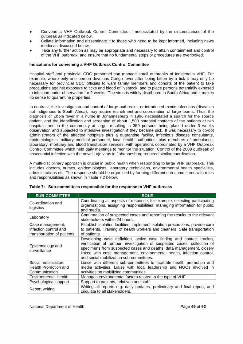
National Department of Health Page 49 of 62
● Convene a VHF Outbreak Control Committee if necessitated by the circumstances of the outbreak as indicated below.
● Collate information and disseminate it to those who need to be kept informed, including news media as discussed below.
● Take any further action as may be appropriate and necessary to attain containment and control of the VHF outbreak, and ensure that no fundamental steps or procedures are overlooked.
Indications for convening a VHF Outbreak Control Committee Hospital staff and provincial CDC personnel can manage small outbreaks of indigenous VHF. For example, where only one person develops Congo fever after being bitten by a tick it may only be necessary for provincial CDC officials to warn family members and cohorts of the patient to take precautions against exposure to ticks and blood of livestock, and to place persons potentially exposed to infection under observation for 2 weeks. The virus is widely distributed in South Africa and it makes no sense to quarantine properties. In contrast, the investigation and control of large outbreaks, or introduced exotic infections (diseases not indigenous to South Africa), may require recruitment and coordination of large teams. Thus, the diagnosis of Ebola fever in a nurse in Johannesburg in 1996 necessitated a search for the source patient, and the identification and screening of about 1,500 potential contacts of the patients at two hospitals and in the community at large, resulting in 350 persons being placed under 3 weeks observation and subjected to intensive investigation if they became sick. It was necessary to co-opt administrators of the affected hospitals plus a quarantine facility, infectious disease consultants, epidemiologists, military medical personnel, local health authorities, plus members of ambulance, laboratory, mortuary and blood transfusion services, with operations coordinated by a VHF Outbreak Control Committee which held daily meetings to monitor the situation. Control of the 2008 outbreak of nosocomial infection with the novel Lujo virus in Johannesburg required similar coordination. A multi-disciplinary approach is crucial in public health when responding to large VHF outbreaks. This includes doctors, nurses, epidemiologists, laboratory technicians, environmental health specialists, administrations etc. The response should be organised by forming different sub-committees with roles and responsibilities as shown in Table 7.2 below. Table 7: Sub-committees responsible for the response to VHF outbreaks
SUB-COMMITTEE ROLE
Co-ordination and logistics
Coordinating all aspects of response, for example: selecting participating organisations, assigning responsibilities, managing information for public and media.
Laboratory Confirmation of suspected cases and reporting the results to the relevant stakeholders within 24 hours.
Case management, infection control and transportation of patients
Establish isolation facilities, implement isolation precautions, provide care to patients. Training of health workers and cleaners. Safe transportation of patients.
Epidemiology and surveillance
Developing case definition, active case finding and contact tracing, verification of rumour, investigation of suspected cases, collection of specimens from suspected cases and deaths, data management, closely linked with case management, environmental health, infection control, and social mobilization sub-committees.
Social mobilisation, Health Promotion and Communication
Liaise with different sub-committees to facilitate health promotion and media activities, Liaise with local leadership and NGOs involved in activities on mobilizing communities.
Environmental Health Manages environmental factors related to the type of VHF. Psychological support Support to patients, relatives and staff.
Report writing Writing all reports e.g. daily updates, preliminary and final report, and circulate to all stakeholders.
National Department of Health Page 50 of 62
7.3.2 Tracing of contacts The purpose of tracing VHF contacts and placing them under observation is to control spread of infection and thus to terminate an outbreak. The office of the relevant provincial Coordinator of CDC is ultimately responsible for tracing and observation of contacts. In practice, infection control officials within hospitals where VHF patients are treated assume responsibility for placing health care workers who have had contact with the patient/s or fomites under observation, and this is done irrespective of whether or not contact took place before or after isolation precautions were instituted. Concurrently, provincial CDC teams operate within the community at large to trace the movements of the VHF patient/s for up to 3 weeks prior to onset of illness in order to establish the source of infection, and to prepare a list of all contacts who are at risk of developing the disease and need to be placed under observation (a period of 3 weeks prior to onset of illness applies for Marburg, Ebola, Lassa and Lujo fevers, but 2 weeks is appropriate for Congo fever and other arbovirus diseases, see section 3). Definitions An outbreak of VHF is the occurrence of one or more cases of VHF. An index patient in an outbreak of VHF is the first patient in whom the disease is recognized, and is not necessarily the primary case, i.e. is not necessarily the first person to have become infected in the outbreak. Recognition of the disease in the index patient results in the discovery of the outbreak. A multiple case outbreak of VHF can arise when there is secondary human-to-human spread of infection from a primary case. Common-source outbreaks occur when more than one primary case of infection arises from exposure to a natural source of infection, e.g. infected animal tissues. A source patient is a patient from whom transmission has occurred to produce secondary infection/s. A contact is a person who has been exposed to an infected person, animal or contaminated environment in such a manner as to have had the opportunity to acquire infection. A case contact is a person who has been exposed to an infected person or his/her secretions, excretions, blood or other tissues in such a way as to be at risk of acquiring infection. A source contact is a person who has been exposed to the same external (non-human) source/s of infection as an infected person. Low risk contacts have had slight or indirect contact with a VHF patient or other source of infection on a single or few occasions. Moderate risk contacts have had close and prolonged contact with a VHF patient or other source of infection. This category includes intimate friends of a VHF patient, relatives and health care workers. High-risk contacts have had what is judged to be definite exposure to VHF infection, e.g. needle-stick with blood from a confirmed case of VHF or similar exposure to animal tissues in a common-source outbreak. Exposure to infection which constitutes contact for purposes of VHF control includes association with an infected person at any time from onset of fever until 3 weeks later in any of the following ways: • Sharing the same residence. • Face-to-face contact (≤1 metre).
National Department of Health Page 51 of 62
• Skin or mucous membrane contact or penetrating injury with the patient's secretions, excretions, blood or other tissues. This includes exposure to animal tissues or insect bites in situations where such exposure is considered to be the source of infection.
In tracing and assessing persons potentially exposed to infection, interviews should be based on questionnaires prepared specifically for the circumstances of the outbreak under investigation (see Figure 7.3 for an example). Persons assessed as having been exposed to infection as defined above, are included on a list of contacts to be placed under observation. 7.3.3 Observation of contacts Observation of contacts of VHF consists of recording temperatures twice daily for 3 weeks (21 days) from the last date of contact with a VHF patient or fomite, and monitoring for signs and symptoms of illness. A 21 day observation period is appropriate for Marburg, Ebola, Lassa and Lujo fevers, but 14 days is adequate for Congo fever, which has a shorter incubation period. Rift Valley fever also has a short incubation period, but the virus seldom causes serious or haemorrhagic disease and person-to-person spread has not been recorded, so active observation is not essential. Persons with ongoing exposure to infection, such as health care workers engaged in nursing of VHF patients, remain under observation while exposure continues to occur, and are kept under observation for the requisite 14 or 21 day period after the last date of potential exposure to infection. Active observation involves contacts being seen twice daily by a medical official charged with this responsibility. Passive observation entails the contact reporting (e.g. by telephone) on their own status to the observation officer. Passive observation is sometimes applied to contacts deemed to be reliable, e.g. health care workers, but this must not be permitted when VHF is involved. It is important to note that the term observation is used in preference to surveillance since the terms active surveillance and passive surveillance are used in a different sense to denote monitoring of a population for the occurrence of a disease either actively through sampling a sub-population or passively through simply testing samples submitted voluntarily to the laboratory. Contacts should be seen at pre-arranged venues and times, which could include their place of employment, e.g. a hospital, or their place of residence, e.g. a farm, and specific arrangements must be made for monitoring of contacts at weekends or other times of absence from duty. All contacts must be seen twice daily at fixed times and any unexplained absences from work or home must be investigated. No medical official should be responsible for monitoring more contacts than can be conveniently managed; in large outbreaks 10 contacts per monitor has been found to be convenient. Temperatures and illnesses reported by contacts should be recorded on a standard list (see an example presented as Figure 7.3), but care should be taken not to ask leading questions: let the contacts describe how they feel. Low to moderate risk contacts of VHF (see definitions in 7.3.2 above), including health care workers, may be kept under active observation in their normal environment and employment, but should not leave the town/district until the observation period has ended. This provision is enforceable in law. High-risk contacts of VHF (see definition in 7.3.2 above) must be kept under active observation or placed under quarantine in a suitable facility for the duration of the quarantine period. It is not universally agreed that there is a need to confine high risk contacts to a quarantine facility, provided they are kept under strict active observation. At most, confinement to a quarantine facility should be applied selectively to those considered to be in imminent danger of developing infection, e.g. medical staff who have had a needle-stick injury with blood known to be infected, or those who have developed non-specific illness, e.g. fever and headache.

National Department of Health Page 52 of 62
Quarantine facilities need not necessarily be in the same complex as VHF isolation units. Old infectious disease hospitals in isolated localities are ideal, and since confinement of essentially healthy people may be involved, it is advantageous to have access to an outdoor area. Any contact who develops fever (temperature of 38°C or over) or signs and symptoms suggestive of VHF, must be placed in isolation and treated as a suspected case. Although monitoring of individual patients ceases on completion of the requisite 14 or 21 day period after the last date of potential exposure to infection, outbreaks are only declared to be over after twice the duration of this period has passed, 28 or 42 days since the last known potential exposure of any person to infection. Hospital infection control and provincial CDC personnel must continue to monitor the situation during this precautionary period. Counselling of contacts and health care workers should be considered to counter stress during outbreaks. 7.4 Communication with the media News media can be disruptive during outbreaks of VHF through disseminating incorrect and alarmist information, and through making undue demands on the time of officials who are heavily engaged in controlling the outbreak. However, with proper planning and liaison, the media can be utilized to dispel misconceptions and to disseminate useful information. This is best achieved by conducting communications with the media on an organized basis, and issuing factual, non-sensational statements through specially appointed spokespersons who confine themselves to their areas of competence. For example: • Spokespersons for the national and provincial Departments of Health can report on control
measures, VHF policy and the status of an outbreak. • Members of NICD staff can provide background information on VHFs, including distribution and
occurrence of the diseases, sources of infection, means of spread and mortality. It is useful to have succinct fact sheets on the diseases available for distribution.
• Senior administrators and clinicians in hospitals treating VHF patients can issue suitably guarded statements on the clinical status of patients, bearing in mind the rights of patients and relatives to preserve their privacy and anonymity.
Information must first be made known to those who have need of it, e.g. it is unacceptable for clinicians or relatives of patients to hear of laboratory findings on the radio, or for officials of the Department of Health to learn of the existence of an outbreak of VHF in the press. Although it is useful to issue approved press statements at set times, it is advisable to have well-informed spokespersons readily available to the media. Refusing to communicate or withholding information does not remove misconceptions. However, officials should not volunteer sensitive information to outsiders or to the media. 7.5 Long-term responsibilities of Provincial CDCs include: ● Drafting contingency plans for managing single or multiple case outbreaks of VHF in the
province. ● Formulating policy as to whether cases of VHF should to be referred to a specifically
designated hospital, or whether they should be managed in the hospital where the diagnosis is first suspected. In practice, decisions will vary with individual circumstances.
● Acting in conjunction with the provincial Department of Health and Hospital Services to identify and secure the use of facilities and resources needed for managing outbreaks and delegating responsibilities, including the designation of specific hospitals as VHF referral centres (see section immediately below).
● Assisting health care facilities to institute planning and training of health care workers in the recognition, transport, isolation and nursing of patients.

National Department of Health Page 53 of 62
Requirements for a VHF referral centre include: ● Tertiary care hospital which has been specifically designated for referral of VHF patients. It is
usually possible to adapt space within a hospital to serve as an isolation unit for nursing of VHF patients with little or no structural alteration (see section 6.3.1).
● Laboratory unit capable of performing essential clinical pathology tests for monitoring the treatment of VHF patients. It is best to use an existing laboratory, preferably but not necessarily within the same complex as the patient isolation unit (see section 6.2).
● Quarantine facility for high-risk contacts of VHF patients, such as health care workers who develop non-specific illness after known exposure to infection. Such persons are moved into the isolation unit if a diagnosis of VHF is confirmed. A quarantine facility is seldom required, and it need not be in the same complex as the isolation unit.
● Mortuary with facility for refrigerated storage of corpses until a diagnosis has been confirmed. This need not be within the same complex as the isolation unit and does not require special facilities provided that the corpse is properly disinfected and shrouded.
● Clinicians, nurses, infection control and laboratory personnel trained for evaluation, management, nursing and laboratory monitoring of VHF patients with due isolation precautions.
● A senior person, logically a clinician (plus an alternate), authorized to take decisions on accepting transfer of VHF patients, with appropriate consultation if necessary.
● An ambulance service with paramedical teams equipped and trained for safe transport of VHF patients.
● Personal protective and safety equipment required for transport and nursing of VHF patients.

National Department of Health Page 54 of 62
Figure 7: Form GW17/5 for notification of scheduled diseases

National Department of Health Page 55 of 62
Figure 8: Suggested form for furnishing extra information on possible, probable or confirmed cases of VHF.
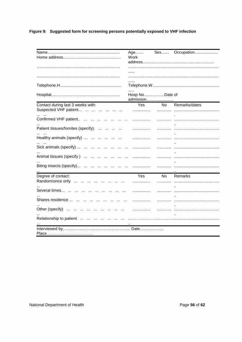
National Department of Health Page 56 of 62
Figure 9: Suggested form for screening persons potentially exposed to VHF infection
Name................................................................ Age....... Sex....... Occupation..................... Home address................................................... Work
address............................................................... .......................................................................... ................................................................................
...... .......................................................................... ................................................................................
...... Telephone.H...................................................... Telephone.W...........................................................
..... Hospital............................................................. Hosp No..................Date of
admission........................ Contact during last 3 weeks with: Yes No Remarks/dates Suspected VHF patient... ... ... ... ... ... ... ... ...
................ ............. .........................................
Confirmed VHF patient.. ... ... ... ... ... ... ... ...
................ ............. ..........................................
Patient tissues/fomites (specify). ... ... ... ... ... ...
................ ............. ..........................................
Healthy animals (specify) ... ... ... ... ... ... ... ...
................ ............. ..........................................
Sick animals (specify) ... ... ... ... ... ... ... ... ...
................ ............. ..........................................
Animal tissues (specify ) ... ... ... ... ... ... ... ...
................ ............. ..........................................
Biting insects (specify)... ... ... ... ... ... ... ... ...
................ ............. ..........................................
Degree of contact: Yes No Remarks Random/once only ... ... ... ... ... ... ... ... ...
................ ............. ..........................................
Several times... ... ... ... ... ... ... ... ... ... ...
................ ............. ..........................................
Shares residence ... ... ... ... ... ... ... ... ... ...
................ ............. ..........................................
Other (specify) ... ... ... ... ... ... ... ... ... ...
................ ............. ..........................................
Relationship to patient ... ... ... ... ... ... ... ...
……………………………...........................................
Interviewed by………………………………………….. Date……………... Place…………………………….
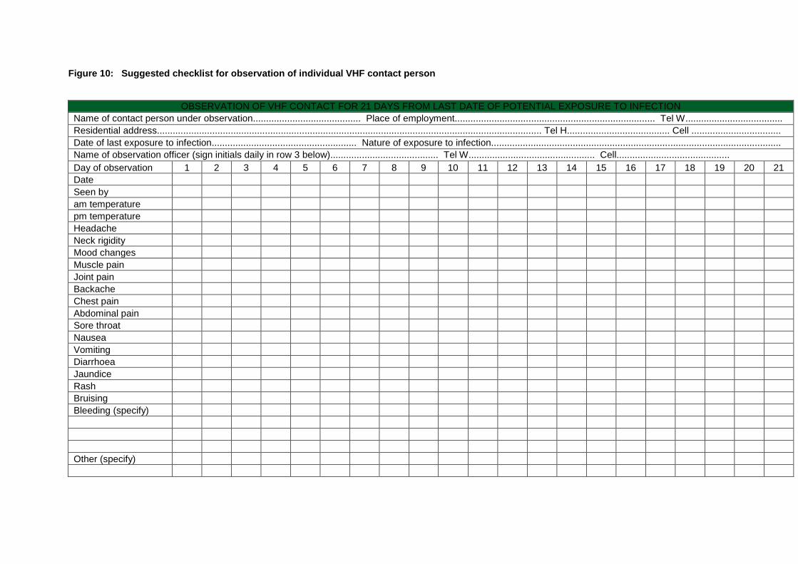
Figure 10: Suggested checklist for observation of individual VHF contact person
OBSERVATION OF VHF CONTACT FOR 21 DAYS FROM LAST DATE OF POTENTIAL EXPOSURE TO INFECTION
Name of contact person under observation......................................... Place of employment............................................................................ Tel W..................................... Residential address.................................................................................................................................................. Tel H....................................... Cell .................................. Date of last exposure to infection....................................................... Nature of exposure to infection.............................................................................................................. Name of observation officer (sign initials daily in row 3 below)......................................... Tel W................................................ Cell........................................... Day of observation 1 2 3 4 5 6 7 8 9 10 11 12 13 14 15 16 17 18 19 20 21 Date Seen by am temperature pm temperature Headache Neck rigidity Mood changes Muscle pain Joint pain Backache Chest pain Abdominal pain Sore throat Nausea Vomiting Diarrhoea Jaundice Rash Bruising Bleeding (specify) Other (specify)

8. KEY REFERENCES Swanepoel, R. (1985) Recognition and management of viral haemorrhagic fevers: a handbook and resource directory. Sandringham: National Institute for Virology, Department of Health, South Africa. Swanepoel, R. (1987) Recognition and management of viral haemorrhagic fevers: a handbook and resource directory, 2nd edition. Sandringham: National Institute for Virology, Department of Health, South Africa. CDC.(1988) Management of patients with suspected viral hemorrhagic fever. MMWR; 37 (no. S-3);1-15. CDC. (1995) Notice to Readers Update: Management of Patients with Suspected Viral Hemorrhagic Fever - United States. MMWR; 44(25);475-479. CDC and WHO. (1998) Infection Control for Viral Haemorrhagic Fevers in the African Health Care Setting. Atlanta: CDC. Siegel JD, Rhinehart E, Jackson M, Chiarello L, and the Healthcare Infection Control Practices Advisory Committee, CDC. (2007) Guideline for Isolation Precautions: Preventing Transmission of Infectious Agents in Healthcare Settings National Guidelines on Epidemic Preparedness and Response. (2009) Department of Health, South Africa.
National Department of Health Page 59 of 62
APPENDIX 1: FREQUENTLY ASKED QUESTIONS ON CRIMEAN-CONGO HAEMORRHAGIC FEVER Q: What is a viral haemorrhagic fever? A: A viral haemorrhagic fever is a disease which is caused by a virus and has a tendency to disturb blood clotting, so that patients may develop uncontrolled bleeding or “haemorrhaging”. Many common diseases can resemble viral haemorrhagic fever, but the term is reserved for a particular group of diseases associated with a high death rate. In Africa, these include Crimean-Congo haemorrhagic fever, Lassa fever, Marburg disease and Ebola fever. Apart from the fact that they cause similar disease, the viruses are not closely related to each other, and are transmitted in a variety of ways. Q: What is Crimean-Congo haemorrhagic fever? A: Crimean-Congo haemorrhagic fever is a tick-borne viral disease of humans which occurs in Africa, Eastern Europe and Asia. Q: Why does it have the name “Crimean-Congo haemorrhagic fever”? A: A disease given the name Crimean haemorrhagic fever was first recognized on the Crimean Peninsula in 1944, although the virus which causes the disease was only identified in 1967. Meanwhile, in 1956 a virus given the name Congo was isolated from a child with fever in the former Belgian Congo (now Democratic Republic of the Congo). In 1969 it was discovered that the two viruses were the same. Consequently, the virus and the disease are called “Crimean-Congo haemorrhagic fever”. The name is often abbreviated to “CCHF”, and in South Africa the disease is commonly called “Congo fever”. Q: Where does the virus come from? A: The virus is transmitted mainly by Hyalomma ticks, which have distinctive brown and white bands on their legs, and are known in South Africa as bont-legged ticks (Afr.: bontpootbosluise). The virus can remain in the ticks for long periods, and even pass through the eggs to infect the next generation of ticks. There are three species of Hyalomma in South Africa, and although they are widely distributed the ticks tend to be most numerous in the drier north-western parts of the country - the Karoo, western Free State, Northern Cape and North-West Province. Immature Hyalomma ticks (larvae and nymphs) feed on ground birds such as guinea fowl, and small mammals up to the size of hares. Adult Hyalommas feed on livestock such as cattle, sheep and goats, as well as wild animals such as antelope, and also ostriches. Animals bitten by infected ticks do not develop the disease, but can circulate the virus in their blood for a few days, up to a week, and thereafter become immune to further infection. Non-infected ticks become infected if they feed on the animals during the short period when virus is in circulation, thus ensuring that the virus is perpetuated. Q: How do humans become infected? A: Humans can become infected from being bitten by infected ticks, or even from squashing ticks if fluid from the ticks gets into cuts and breaks in the skin, or onto mucous membranes. Fortunately immature Hyalommas feed only on small animals and do not bite humans, and adult Hyalommas prefer farm animals and so seldom bite humans. If humans were preferred hosts of Hyalomma ticks, there would be many more cases of the disease. Humans can also become infected if blood from infected livestock or wild animals comes into contact with broken skin (cuts and abrasions) or mucous membranes during the short period that the animals have the virus in circulation. On farms, this usually happens when young animals become infected as a result of being exposed to ticks, and humans are then exposed to blood during procedures such as the castration of calves, slaughtering of lambs, vaccination of animals, the cutting of identity notches in the ears, or the attachment of ear tags. Occasionally animals that have been reared under tick free conditions come into contact with ticks and the virus late in life, and so slaughtering mature animals can also result in human infection. Although the proportion of mature animals that will have virus in circulation may be extremely low, many thousands of animals are slaughtered each day at abattoirs. Hunting and butchering of wild animals can also be a source of human infection.

National Department of Health Page 60 of 62
Similarly, humans can become infected if blood or blood-tinged body fluids and wastes from patients with the disease comes into contact with broken skin or mucous membranes, as occurs when medical care personnel sustain accidental needle sticks. Q: Which people are at risk of becoming infected? A: People who are at particular risk include those involved in the livestock industry, such as farmers and farm labourers, veterinarians, abattoir workers, persons who slaughter animals informally, and hunters. Within abattoirs those who come into contact with fresh blood are at greatest risk. Once carcases have been bled out and hung to mature there is a sudden increase in acidity of the meat and virus cannot be detected in the carcase. Ostriches appear to be the only birds in which there is similar circulation of detectable levels of virus in blood as occurs in mammals. There is no indication that meat processed and matured according to standard abattoir practice constitutes a danger to consumers. Half-fed ticks which detach from the hides of recently slaughtered animals may attach indiscriminately to hosts available in their environment, and thus infect slaughtermen. Apart from people directly involved in the livestock industry, persons at risk of being bitten by ticks include those who live in the countryside and town dwellers who visit the countryside for occupational or recreational purposes, including hunting and hiking. People are not always aware of being bitten by ticks. In patients with Congo fever, ticks have been found attached in concealed sites such as on the scalp and between the toes. Occasionally, no direct evidence can be obtained to indicate that a patient with Congo fever had contact with animal blood or with ticks, and the only evidence to suggest possible exposure to infection is the fact that the patient lived in or visited an environment where such contact was possible. Health care personnel or close associates of a patient can acquire infection from contact of broken skin or mucous membranes with blood or blood-tinged body fluids and wastes of the patient. Although spread of infection to family members has never been recorded in South Africa, it is possible. The only time that infection has been seen in groups of people is when they have been exposed together to a common source of infection, as in slaughtering animals. In contrast, there have been several instances of secondary spread of infection from patients to health care personnel, and this has usually involved needle stick injuries in hospitals. Q: How common is the disease and how often is it fatal? A: In brief, about 1-10 cases of Congo fever are diagnosed each year in South Africa (range 0-20), and 20-25 percent of patients die, but the death rate can be 30-50 per cent if patients do not receive proper medical attention. More exactly, 158 cases of Congo fever have been diagnosed in Southern Africa from the time that the presence of the disease was first recognized in 1981 up until the end of 2000, with one infection having occurred in Zaire, one in Tanzania, ten in Namibia and the remainder in South Africa. Most patients were employed in the livestock industry, and males constituted 129/158 of cases. Marginally the largest group of cases, 67/158 (42,4%), arose from known tick bite or squashing of ticks; a similar number, 66/158 (41,8%), arose from known or potential contact with fresh blood or other tissues of livestock and/or ticks; 7/158 (4,4%) nosocomial infections arose from contact with blood or items contaminated with blood of known Congo fever patients, while in 18/158 (11,4%) cases there was no direct evidence of contact with livestock or ticks, but the patients lived in or visited a rural environment where such contact was possible. Q: What are the signs and symptoms of the disease? A: The disease has a short incubation period followed by a very sudden onset of illness. People usually become sick within 1-3 days of being bitten by a tick, or 5-6 days (occasionally longer) after exposure to the blood of infected livestock or humans. (The short incubation period and sudden onset are among several points of distinction with tick bite fever.)
National Department of Health Page 61 of 62
Patients abruptly develop a severe headache with sore and reddened eyes, fever with cold chills, and intense body pains, particularly involving the muscles of the lower back and thighs. The patients feel extremely unwell and usually take to their beds. Body temperatures do not necessarily remain high and may fluctuate in the course of each day. There may be nausea and vomiting, and sometimes abdominal pain and diarrhoea early in the disease. At this stage, blood tests already show disturbed liver function, and a decrease in blood platelets, which are involved in the clotting of blood. After about 5 days patients may develop a rash of pink blotches on the body, followed by various bleeding tendencies, depending on the severity of the illness. They bruise easily, often have nose bleeds, and may pass blood in the stool and urine. Stools seldom contain fresh blood; they usually have a dark and tarry appearance. Small or large red spots of bleeding into the skin appear, and there may be large confluent areas of bleeding into the skin around injection sites and in skin folds such as in the armpits or groin. Patients may vomit blood and bleed from the gums, and women may develop heavy uterine bleeding. Blood continues to ooze from needle puncture sites. There can also be internal bleeding, including intracerebral bleeding. Patients go into a coma as liver, kidney and lung functions fail, and death occurs 5-14 days after onset of illness, usually from heart failure. Patients who recover show sudden improvement from day 10 of illness onwards. Virus remains detectable in human blood for up to two weeks after the onset of illness, but once the results of blood tests indicate that patients’ body functions have recovered, and they feel well and are no longer bleeding, they can be discharged from hospital. Although there has been no indication that virus continues to be excreted in body fluids, patients should refrain from intimate contact with other people for six weeks after recovery from the disease as a precaution against spread of infection. Convalescent patients should not undertake heavy duties during this period. After recovery patients are immune to further infection. It is not uncommon for recovered patients to remember little or nothing about the events of their illness. Treatment essentially consists of supportive therapy, which comprises intravenous feeding of the patient and replacement of blood and clotting factors. Severely ill patients may be placed on ventilators and other life support systems. The chemotherapeutic drug ribavirin has been used to treat patients on a trial basis, but the intravenous form of the drug which is required for seriously ill patients, is virtually unobtainable since it is only produced intermittently on account of low demand. Q: What action should be taken if a person is suspected of having the disease? A: The disease may be suspected when a person suddenly becomes sick with headache, fever and chills, muscle pains, and possibly nausea, vomiting and diarrhoea, less than a week after being bitten by a tick, squashing ticks, or coming directly into contact with fresh blood or blood-tinged body fluids and organs of livestock, wild animals or human Congo fever patients. A doctor should be consulted immediately the disease is suspected, and if the doctor believes that the suspicion is justified then arrangements should be made to send blood samples (blood taken with the anti-coagulant EDTA, plus clotted blood) expeditiously to the Special Pathogens Unit of the NICD in Johannesburg for confirmation of the diagnosis. It should be remembered that the vast majority of suspected cases prove not to be Congo fever, and if there is doubt the doctor should consult physicians specifically charged with handling viral haemorrhagic fever patients in the province concerned, or members of staff of the Special Pathogens Unit. Certain major hospitals have been designated for the management of haemorrhagic fever patients in each province. Medical personnel should establish for themselves what arrangements exist in their own province. Immediately after a Congo fever or any other haemorrhagic fever is suspected, medical personnel should ensure that they apply strict precautions against infection from blood and other body fluids. On no account should patients suspected to be suffering from any haemorrhagic fever be referred to a hospital without first discussing the case with the relevant clinicians. Specimens should not be sent to the Special Pathogens Unit without first contacting the Unit. The doctor (or other health worker certified as competent to diagnose) who makes the diagnosis has a legal obligation to notify the Local Authority Health Services of the existence of the case on form GW17/5.

National Department of Health Page 62 of 62
Q:What precautions should be applied to persons who have potentially been exposed to infection? A: Local and provincial health officials are responsible for investigating the circumstances surrounding confirmed cases of the disease, and instituting such control measures as may be necessary. Persons in the community at large, including family members, who have been in contact with confirmed Congo fever patients, or who have been exposed to the same potential source of infection, are classified as being at zero, low, moderate or high risk according to defined criteria, and placed under appropriate observation as discussed below. Medical personnel who have been exposed to patients are separately placed under observation of the infection control officials of the institution concerned. Contacts considered to be at high risk would, for instance, include persons who have had accidental injury with a needle contaminated with the blood of a confirmed Congo fever patient. Such persons would be placed under active observation, which consists of reporting twice a day to a designated health official to be monitored for signs and symptoms of the disease and to have their temperature recorded for a period of two weeks after last contact with the patient (calculated to exceed the incubation period of Congo fever by a wide margin of safety - the observation period would be extended to three weeks for most of the other viral haemorrhagic fevers). Low risk contacts of confirmed patients, who have not had closer than one metre face-to-face contact with the patient for instance, may be placed under passive observation, which could consist of reporting to the responsible health official daily by telephone rather than in person. Note that persons under observation are not in quarantine and may continue with their normal duties, including attending to patients. They are only considered to be infectious once they become sick themselves. As soon as they develop signs and symptoms considered to be characteristic of the disease, or a fever of 38,5℃ or greater, they are admitted to hospital as suspected cases. Places such as abattoirs constitute a special case. Since exposure potentially occurs on a continuing basis (although the risk is actually low), there is seldom an indication for placing selected individuals under special observation. Instead, clinics attached to abattoirs should maintain a high degree of awareness of Congo fever and other diseases which can be acquired from livestock, and ensure that there is appropriate investigation of sick members of staff. Family members and co-workers of patients who become infected on farms may be placed under observation depending on their degree of potential exposure to infection, but since the ticks and virus are so widely distributed there is no logic in placing farms under quarantine. Q: What measures can be taken to prevent exposure to infection? A: Persons potentially exposed to tick bite can use certain pyrethroid acaricides to treat clothing such as socks and trousers (acaricides are insecticides used against ticks). Formulations which are generally available from shops that sell equipment for camping and outdoor activities, include aerosol sprays and sachets of concentrated acaricide used to prepare emulsions into which clothing is dipped. Abattoir workers, veterinary staff, farm workers and hunters should use appropriate impervious protective clothing and gloves when engaged in activities which carry a risk of exposure to animal blood. Although it is incumbent upon employers to supply protective clothing and instruction in safety, employees must take responsibility for adhering to safety regulations. Veterinary regulations promulgated for ostrich abattoirs require that birds should be treated with an appropriate acaricide and held in tick-free circumstances for 14 days before slaughter. Similar regulations would be impossible to implement for other livestock. Vast numbers of cattle, sheep and goats are slaughtered each day, and the costs of constructing tick-free holding pens of suitable capacity would be prohibitive, as would the costs and logistics of holding and feeding the animals and supervising the operation. A potential alternative would be the development of a veterinary vaccine that is applied to farm animals as a public health measure, but such research would require special funding. At present there is no human vaccine, and the lack of potential demand for such a vaccine inhibits its development.