Embed Size (px)
Citation preview

http://www.e-hmr.org 223
Jin Young Yang, et al. • Skeletal Muscle Stem Cells for DMD HMR
Hanyang Med Rev 2015;35:222-228222 © 2015 Hanyang University College of Medicine • Institute of Medical Science http://www.e-hmr.org
INTRODUCTION
About 50 years ago, the satellite cell was discovered through electron microscopic inspection of skeletal muscle. Satellite cells are mono-nucleated and located in the edge of the skeletal myofi-bers. These cells were imposed for their association with muscle regeneration. Satellite cells maintain dormant state in the mature muscle and are activated mitotically during muscle injuries, and finally differentiated to myoblasts. It contributes to the myofibrils resulting in muscle recovery from the injury [1]. Collins proved that satellite cells have both differentiation and self-renewal ability [2]. Sequentially, Kuang et al. established that the muscle stem cells achieve self-renewal through asymmetrical divisions. Also, satel-lite cells did not consist of only adult stem cells, rather both adult stem cells and committed progenitor cells [3].
Muscular dystrophies include many diseases, which have skele-tal muscle wasting and limit mobility of patients. Among that, Du-chenne muscular dystrophy (DMD), fragility of respiratory mus-
cles and absence of dystrophin protein in the cardiac muscle re-sults in respiratory or cardiac failure and early death [4]. The un-derlying pathogenesis is already investigated, but the treatment has not been established yet. However, research has accumulated these days and clinical trials are also being performed now.
There are roughly four methods to research DMD treatments, 1) gene therapy replacing the mutated gene, 2) cell therapy which replace affected cells or using bone marrow-derived stem cells and mesoangioblasts, 3) repairing the endogenous gene using exon skipping, skipping premature termination, and 4) drug therapy which compensates for lack of dystrophin, promotes function in dystrophic muscle, improves muscle hypertrophy and minimizes muscle wasting, uses histone deacetylase inhibitors or NO-releas-ing anti-inflammatory drugs [5]. In this article, cell therapies in DMD are focused in a review and current clinical trials using cell therapy are introduced.
Hanyang Med Rev 2015;35:222-228http://dx.doi.org/10.7599/hmr.2015.35.4.222
pISSN 1738-429X eISSN 2234-4446
Muscle stem cells, which are known as satellite cells have heterogeneous components of committed myogenic progenitors, non-committed satellite cells, and mesenchymal stem cells. This distinguishing organization of self-renewal and differentiation capacities encour-ages the remarkable regenerative ability of skeletal muscles. Lately it has been proved that the satellite cell is the derivation of muscle regeneration and with the self-renew function, it roles as a true muscle stem cell. Therefore, stem cell therapy using satellite cells is consid-ered to be ideal therapy for muscular dystrophies, which is deficient in specific muscle pro-tein and causes muscle degeneration. Especially, Duchenne Muscular Dystrophy (DMD), which is caused by mutations at the dystrophin gene, has been targeted by much research. In this article the satellite cell characteristics, regulation of cell function, and stem cell ther-apy for DMD and the present progressive clinical trials will be reviewed.
Key Words: Satellite Cells, Skeletal Muscle; Muscular Dystrophy, Duchenne; Dystrophin; Clinical Trial
Recent Advances in Skeletal Muscle Stem Cells for Duchenne Muscular Dystrophy TreatmentJin Young Yang, Jaemin Jeong
Department of Surgery, Hanyang University College of Medicine, Seoul, Korea
Correspondence to: Jaemin JeongDepartment of Surgery, Hanyang University College of Medicine, 222 Wangsimni-ro, Seongdong-gu, Seoul 04763, KoreaTel: +82-2-2220-0647Fax: +82-2-2220-0224E-mail: [email protected]
Received 4 September 2015 Revised 22 September 2015 Accepted 29 September 2015
This is an Open Access article distributed under the terms of the Creative Commons Attribution Non-Commercial License (http://creativecom-mons.org/licenses/by-nc/3.0) which permits un-restricted non-commercial use, distribution, and reproduction in any medium, provided the origi-nal work is properly cited.
Review
http://www.e-hmr.org 223
Jin Young Yang, et al. • Skeletal Muscle Stem Cells for DMD HMR
Hanyang Med Rev 2015;35:222-228
SATELLITE CELL IDENTIFICATION AND CHARACTERISTICS
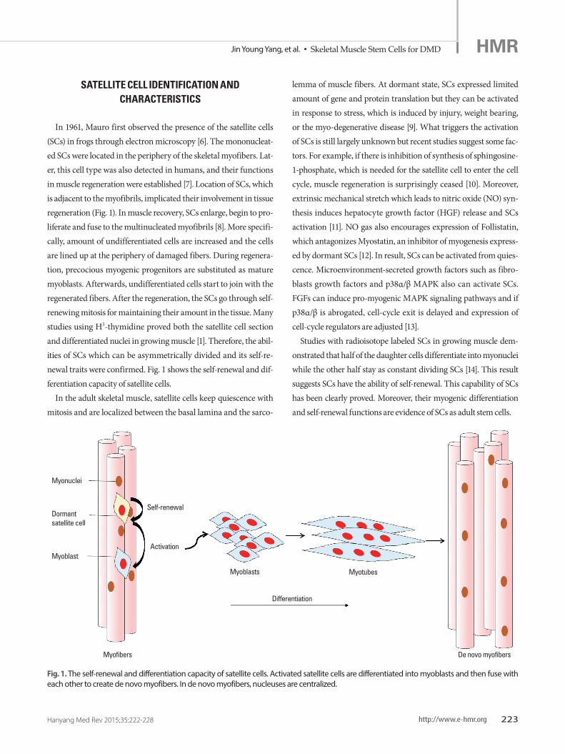
In 1961, Mauro first observed the presence of the satellite cells (SCs) in frogs through electron microscopy [6]. The mononucleat-ed SCs were located in the periphery of the skeletal myofibers. Lat-er, this cell type was also detected in humans, and their functions in muscle regeneration were established [7]. Location of SCs, which is adjacent to the myofibrils, implicated their involvement in tissue regeneration (Fig. 1). In muscle recovery, SCs enlarge, begin to pro-liferate and fuse to the multinucleated myofibrils [8]. More specifi-cally, amount of undifferentiated cells are increased and the cells are lined up at the periphery of damaged fibers. During regenera-tion, precocious myogenic progenitors are substituted as mature myoblasts. Afterwards, undifferentiated cells start to join with the regenerated fibers. After the regeneration, the SCs go through self-renewing mitosis for maintaining their amount in the tissue. Many studies using H3-thymidine proved both the satellite cell section and differentiated nuclei in growing muscle [1]. Therefore, the abil-ities of SCs which can be asymmetrically divided and its self-re-newal traits were confirmed. Fig. 1 shows the self-renewal and dif-ferentiation capacity of satellite cells.
In the adult skeletal muscle, satellite cells keep quiescence with mitosis and are localized between the basal lamina and the sarco-
lemma of muscle fibers. At dormant state, SCs expressed limited amount of gene and protein translation but they can be activated in response to stress, which is induced by injury, weight bearing, or the myo-degenerative disease [9]. What triggers the activation of SCs is still largely unknown but recent studies suggest some fac-tors. For example, if there is inhibition of synthesis of sphingosine-1-phosphate, which is needed for the satellite cell to enter the cell cycle, muscle regeneration is surprisingly ceased [10]. Moreover, extrinsic mechanical stretch which leads to nitric oxide (NO) syn-thesis induces hepatocyte growth factor (HGF) release and SCs activation [11]. NO gas also encourages expression of Follistatin, which antagonizes Myostatin, an inhibitor of myogenesis express-ed by dormant SCs [12]. In result, SCs can be activated from quies-cence. Microenvironment-secreted growth factors such as fibro-blasts growth factors and p38α/β MAPK also can activate SCs. FGFs can induce pro-myogenic MAPK signaling pathways and if p38α/β is abrogated, cell-cycle exit is delayed and expression of cell-cycle regulators are adjusted [13].
Studies with radioisotope labeled SCs in growing muscle dem-onstrated that half of the daughter cells differentiate into myonuclei while the other half stay as constant dividing SCs [14]. This result suggests SCs have the ability of self-renewal. This capability of SCs has been clearly proved. Moreover, their myogenic differentiation and self-renewal functions are evidence of SCs as adult stem cells.
Fig. 1. The self-renewal and differentiation capacity of satellite cells. Activated satellite cells are differentiated into myoblasts and then fuse with each other to create de novo myofibers. In de novo myofibers, nucleuses are centralized.
Myonuclei
Dormant satellite cell
Myoblast
Myoblasts
Myofibers
Differentiation
Myotubes
De novo myofibers
Self-renewal
Activation

http://www.e-hmr.org 225
Jin Young Yang, et al. • Skeletal Muscle Stem Cells for DMD HMR
Hanyang Med Rev 2015;35:222-228224 http://www.e-hmr.org
Jin Young Yang, et al. • Skeletal Muscle Stem Cells for DMDHMR
Hanyang Med Rev 2015;35:222-228
REGULATION OF SATELLITE CELL FUNCTION
1. Niche regulation
Stem cell niche means the microenvironment where stem cells are localized and it controls the fate and function of stem cells. As to satellite cells, the stem cell niche can regulate the asymmetric di-vision and commitment of daughter cells without disturbing the stem cell homeostasis in the niche [15]. Because satellite cells are lo-cated along the myofibers below the basal lamina, the muscle fiber is one of the most important elements of the stem cell niche. All signals such as chemical, mechanical and electrical from the host fiber have been proved to be associated with the regulation of SCs function. The basal lamina also plays as an important role in the niche. It accounts for a major part of the extracellular matrix and is mainly made up of laminin, collagen, and proteoglycans [9]. There-fore, adhesion to the basal lamina is essential for the maintenance of stem cell characters. Another element of the niche is the micro-vasculature that supplies SCs and interstitial cells which interact with SCs [16]. In humans, 68% of satellite cells are localized within 5 μm from capillaries or vascular endothelial cells at both dormant and activated conditions [17]. Meanwhile, asymmetric division of the stem cell depends on cell polarity, which is achieved through cell to cell or cell to ECM interactions within the niche. Each side of SCs expresses different molecules – integrin α7β1 receptors on basal lamina side, M-cadherin on apical side – and this asymmet-ric allocation lets SCs form a structural basis for polarity and leads to the cell fate differences. This asymmetric cell division would also promote the fusion of the differentiating daughter cells [16].
Also, cytokines such as Il-1α, IL-13, TNF-α, and IFN-γ which is secreted by T cells are promoting factors of proliferation of muscle stem cells during muscle regeneration. In the study by Fu et al., these 4 cytokines were injected together into regenerating muscle of Rag1-/- mice and they helped in both decreasing muscle stem cell proliferation and correcting the impaired regenerative respons-es. In addition, muscle stem cells with these cytokines comparably participated more in muscle recovery than freshly isolated muscle stem cells. These results implicate that the muscle stem cells that expanded with the cytokines in vitro could achieve properties of undifferentiated progenitor cells [18].
2. Signaling pathway
In several decades, many signaling pathways such as Wnt, Notch, Bone morphogenetic protein (BMP), TGF-β are proved to be in-
volved in activation of satellite cells. As well as activation, modulat-ing pathways specific to muscle stem cells have also been demon-strated. Maintenance of dormant state, reversible quiescence and self-renewal, asymmetric destination, symmetric proliferation of stem cells are defined [19].
Notch signaling is needed to preserve the dormant state of satel-lite cells, suggesting that niche-derived Notch ligand should bind to a Notch receptor on the dormant satellite cell [20]. In this study, Rbp-j which is the downstream transcripton factor in the Notch pathways was eliminated from adult stem cells in normal muscle. This caused activation and ectopic differentiation of stem cells even if it did not enter the cell cycle and bypassed the transient amplify-ing progenitor stage [20]. Consequently, RBP-J plays as a restrictor of cell cycle entry and a mediator of Notch signaling [21].
Referred from in vitro experiments, it is supposed that multiple signaling pathways are associated with quiescence of the satellite cell. So far, Ang1/Tie2 [22], P38/MAPK [13], myostatin [23], Notch-3 [24], Spry1 [25], are proved to be involved in satellite cell cycles.
Although many signaling pathways that regulate stem cell func-tion of satellite cells are revealed, there might be more unknown pathways [19]. During the cell cycle, many signaling molecules are repeatedly used. Representatively, Numb is used both to decide asymmetric fate and maintain progenitor cells [26]. Therefore, clas-sical Wnt, notch and BMP signals might also be used in another way to regulate stem cell functions [19].
3. Epigenetic regulation of satellite cell function
Regulation of the SC’s function is focused at the epigenetic level, which has cellular and physiological phenotypic trait variations caused by external or environmental factors that alter gene expres-sion without modifications in the DNA sequences. It involves DNA methylation, histone modification, etc. Right after the identifica-tion of SCs, differences in chromatin organization between adult muscle and growing muscle accord with their shift from a dormant to an activated state [27]. The regulators of epigenetics are media-tors of DNA methylation and demethylation, histone acetylases, methylases, miRNAs and so on [19]. These factors would contrib-ute many changes in aged satellite cells that describe age-related declines in function and rejuvenation through exposure to the sys-temic environment [28]. So far, Pax7 functions with the Wdr5-Ash2L-MLL2 histone methyltransferase complex to methylate his-tone H3 lysine 4 at the Myf5 locus [29], and regulation of the re-pressive PRC2, EZH2 to control Pax7 expressions are established.

http://www.e-hmr.org 225
Jin Young Yang, et al. • Skeletal Muscle Stem Cells for DMD HMR
Hanyang Med Rev 2015;35:222-228
BIOMARKERS INVOLVED IN MUSCLE RECOVERY BY SATELLITE CELLS
Many biomarkers have been investigated so far. The most im-portant molecule is Pax7, which regulates self-renewal in satellite cells and maintains myogenic potential. Pax7 is a transcription factor involved in the embryonic development of muscle stem cells and is expressed in both dormant and activated state of satellite cells [30]. It also regulates the expression of Myf5, which is associ-ated with the embryonic myogenesis and stem cell differentiation [31]. Pax3 controls proliferation of the satellite cells along with Pax7 [32]. In addition to those, Barx2 [33], M-cadherin [34], c-Met [35], α7-integrin [36], CD34 [37], CXCR4 [38], syndecan-3, syndecan-4 [39], caveolin-1 [40], calcitonin receptor [41], lamins A and C, and emerin [42] are also expressed biomarkers in stem cells.
Above all, Pax7 is a uniform marker of muscle stem cells. When muscle injury occurs, Pax7+ satellite cells enter into the cell cycle and start to differentiate. Some of those cells return back to the dormant state to supply the cell pool. At that state, the satellite cell lacks MyoD. But during activation and progression, satellite cells begin to expresss MyoD and Myf5 protein, and finally have Myo-genin which is the marker of differentiaton [19].
CURRENT STEM CELL-BASED THERAPY IN DMD PATIENTS
In recent studies, muscle stem cells have been proved to have self-renewable ability at the single-cell level and stem cell functions
[19]. Therefore, satellite cells are being examined to treat muscle diseases. Among many skeletal muscle diseases, some muscular dystrophies which repeat degeneration and regeneration may con-sume pre-existed precursors.
Especially, DMD is a result of frame shift mutations of dystro-phin gene located in the locus Xp21 and about 1/3,500 male birth is affected [4]. If mutations of dystrophin genes occurred, it causes the loss of functional protein on muscle fibers and consequently, the fibers become fragile and necrotize to death. Satellite cells can regenerate the deteriorated fibers but the new muscle fibers still have no dystrophin and will be degenerated once more (Fig. 2). As degeneration-regeneration cycles repeated, satellite cells will be se-nescent and lose their ability of proliferation, differentiation and muscle regeneration [43]. It leads to muscle weakness progressively and eventually the patients could not even use respiratory muscles. Cardiac defects that develop are the most frequent causes of death. Because current treatments of DMD is limited to management of inflammation [44], many studies focus at the regenerative func-tion of satellite cells.
Cell therapy is infusing normal myoblasts and strengthening dystrophin expression on myofibers, so it is considered as one of the possible treatments of DMD. There are two methods in cell therapy, autologous graft and heterologous graft. Autologous graft is using patient’s own muscle progenitor cells, but the mutations should be corrected before re-engraftment. The implanted pro-genitor cells can be involved in the muscle regeneration process and provide the corrected gene to tissue. The advantage of autolo-gous graft is lack of immune response against dystrophin. But the
A B
Fig. 2. Dystrophin (red) is not expressed in mdx mouse. (A) C57BL/10ScSn control mouse. (B) C57BL/10ScSn-Dmd< mdx> .

http://www.e-hmr.org 227
Jin Young Yang, et al. • Skeletal Muscle Stem Cells for DMD HMR
Hanyang Med Rev 2015;35:222-228226 http://www.e-hmr.org
Jin Young Yang, et al. • Skeletal Muscle Stem Cells for DMDHMR
Hanyang Med Rev 2015;35:222-228
proliferation capacities of cells are limited, so these confines should be considered. Whereas heterologous graft using progenitors of normal donors does not require gene correction, thus, continuous immunosuppression is needed. Consequently, neither of these me-thods is perfect for DMD treatment [45].
Besides the graft methods, the insufficient migration and death of the myoblasts are also reasons for the negative results seen in studies [46]. Some countermeasures such as increasing in number of myoblasts injected to the muscle or effect of radiation to defect muscle for boosting the release of myogenic factors are suggested but are still in doubt [47].
Many other cell therapies are currently undergoing. Substitut-ing mutated cells is a process that involves injecting into dystro-phic muscle and leads to the expression of dystrophin by myofi-bers. The limitation of this method is delivering myoblasts sys-temically by local injection [48]. Normal bone marrow-derived stem cells also can be used to regenerate skeletal muscle fibers. With this, bone marrow mesenchymal stem cells have little myo-genic differentiation ability but could be altered to be more myo-genic with high levels of intracellular Notch protein [49]. Donor mesoangioblasts which are intra-arterial transplanted can improve dystrophic muscle. When mesoangioblasts are transplanted to dogs, expression of dystrophin increased up to 70% of the muscle fibers and they had normal contraction force and mobility [50].
PRESENT PROGRESSIVE CLINICAL TRIALS OF CELL THERAPY IN DMD PATIENTS
Recently, many clinical trials in DMD patients have progressed. Of that, some trials with cell therapy are introduced in this review.
1. NCT02241434, stem cell therapy in DMD
Neurogen Brain and Spine Institute have fulfilled clinical trials from January, 2009. This phase 1 trial is aimed to see the effect of autologous bone marrow mononuclear cell therapy in DMD pa-tients and 500 patients are now enrolled. It has single group as-signment, open label test and recipients are 3 to 25 year old males and females from India. Primary completion data is manual mus-cle testing, which will be collected in January, 2016.
2. NCT01834040, study safety and efficacy of BMMNC for the
patient with DMD
Chaitanya hospital, India is now carrying out a phase 1, 2 clini-
cal trial from September, 2014. 30 DMD patients who are 4 to 20 year old males/females whom have consented to bone marrow-de-rived autologous cell therapy are enrolled. This Study is performed as a single arm, single center trial to check the safety and efficacy of BMMNC (100 million per dose) for the patients with DMD. In-tervention method is intralesional/intravenous injection of autolo-gous stem cells and the primary outcome is improvement seen in daily living scales planned to September, 2016. Secondary outcome is improvement of muscular dystrophy seen in specific functional rating scales.
3. NCT02285673, efficacy of umbilical cord mesenchymal stem cells
in DMD
From November, 2013, Acibadem university is carrying out phase 1/2 clinical trials which identify the efficacy of umbilical cord mesenchymal stem cells in DMD patients and whether the wild type gene can be transferred to patients. 10 patients who are 7 to 20 year old males are enrolled who need partial respiratory sup-port during the day (less than or equal to stage 1 NIH, liver, renal and cardiac function). Primary outcome is seen by DMD gene ex-pression that will be collected in February, 2015.
4. NCT02196467, transplantation of myoblasts to DMD patients
In Centre Hospitalier Universitaire de Québec, DMD patients who are males and older than 16 years are enrolled from May, 2014 to examine whether the transplantation of normal myoblasts throu-ghout one muscle (the extensor carpi radialis) of the patients is safe and whether it will improve the strength of that muscle. The ulti-mate aim of this study is to evaluate the safety of a procedure of high-density injections of donor myoblasts throughout a muscle (under immunosuppression by tacrolimus). This study will be im-plemented by single group assessment in a double blind manner and the estimated primary completion date is January, 2018.
5. NCT01834066, study safety and efficacy of bone marrow derived
autologous cells for the treatment of muscular dystrophy
In Chaitanya hospital, 25 patients who have consented to bone marrow derived autologous cell therapy and who are 6 to 25 years old are enrolled to this trial from September, 2014. The interven-tion method is intralesional transfer of autologous stem cell (MNCs) per dose, 6 doses in 3 months. Anticipated primary outcome is sig-nificant improvement in muscle strength by using kinetics muscle testing or by using MMT score. Secondary outcome is seen by im-

http://www.e-hmr.org 227
Jin Young Yang, et al. • Skeletal Muscle Stem Cells for DMD HMR
Hanyang Med Rev 2015;35:222-228
provement of daily living scales and baselines in EMG, which will be collected in November, 2016.
CONCLUSION
Therapeutic research for DMD has been rapidly developed in recent years. Many clinical trials have been started now and results will be available soon. However, there are still some limitations re-maining. First, strategies to repair the dystrophin gene are avail-able only for some mutations and cell and gene therapies have cost problems. Also, local delivery of the therapeutic agent is needed to minimize side effects and maximize the effect of agents, but clini-cal benefit can only be performed using systemic delivery [5]. Sat-ellite cells still have a long way to go, but so far, numerous and bril-liant research are being implemented even now.
ACKNOWLEDGMENTS
This work was supported by No. 2014R1A1A1002599 from Ba-sic Science Research Program through the National Research Foun-dation of Korea funded by MISP.
REFERENCES
1. Snow MH. Myogenic cell formation in regenerating rat skeletal muscle injured by mincing. II. An autoradiographic study. Anat Rec 1977;188: 201-17.
2. Collins CA, Olsen I, Zammit PS, Heslop L, Petrie A, Partridge TA, et al. Stem cell function, self-renewal, and behavioral heterogeneity of cells from the adult muscle satellite cell niche. Cell 2005;122:289-301.
3. Kuang S, Kuroda K, Le Grand F, Rudnicki MA. Asymmetric self-renewal and commitment of satellite stem cells in muscle. Cell 2007;129:999-1010.
4. Emery AE. The muscular dystrophies. Lancet 2002;359:687-95.5. Cossu G, Sampaolesi M. New therapies for duchenne muscular dystro-
phy: challenges, prospects and clinical trials. Trends Mol Med 2007;13: 520-6.
6. Mauro A. Satellite cell of skeletal muscle fibers. J Biophys Biochem Cytol 1961;9:493-5.
7. Kuang S, Rudnicki MA. The emerging biology of satellite cells and their therapeutic potential. Trends Mol Med 2008;14:82-91.
8. Bischoff R. Regeneration of single skeletal muscle fibers in vitro. Anat Rec 1975;182:215-35.
9. Charge SB, Rudnicki MA. Cellular and molecular regulation of muscle regeneration. Physiol Rev 2004;84:209-38.
10. Nagata Y, Partridge TA, Matsuda R, Zammit PS. Entry of muscle satellite cells into the cell cycle requires sphingolipid signaling. J Cell Biol 2006; 174:245-53.
11. Wozniak AC, Anderson JE. Nitric oxide-dependence of satellite stem cell activation and quiescence on normal skeletal muscle fibers. Dev Dyn 2007; 236:240-50.
12. Pisconti A, Brunelli S, Di Padova M, De Palma C, Deponti D, Baesso S, et al. Follistatin induction by nitric oxide through cyclic GMP: a tightly regulated signaling pathway that controls myoblast fusion. J Cell Biol 2006; 172:233-44.
13. Jones NC, Tyner KJ, Nibarger L, Stanley HM, Cornelison DD, Fedorov YV, et al. The p38alpha/beta MAPK functions as a molecular switch to activate the quiescent satellite cell. J Cell Biol 2005;169:105-16.
14. Moss FP, Leblond CP. Satellite cells as the source of nuclei in muscles of growing rats. Anat Rec 1971;170:421-35.
15. Fuchs E, Tumbar T, Guasch G. Socializing with the neighbors: stem cells and their niche. Cell 2004;116:769-78.
16. Kuang S, Gillespie MA, Rudnicki MA. Niche regulation of muscle satel-lite cell self-renewal and differentiation. Cell Stem Cell 2008;2:22-31.
17. Christov C, Chretien F, Abou-Khalil R, Bassez G, Vallet G, Authier FJ, et al. Muscle satellite cells and endothelial cells: close neighbors and privi-leged partners. Mol Biol Cell 2007;18:1397-409.
18. Quarta M, Rando TA. Mimicking the niche: cytokines expand muscle stem cells. Cell Res 2015;25:761-2.
19. Brack AS, Rando TA. Tissue-specific stem cells: lessons from the skeletal muscle satellite cell. Cell Stem Cell 2012;10:504-14.
20. Bjornson CR, Cheung TH, Liu L, Tripathi PV, Steeper KM, Rando TA. Notch signaling is necessary to maintain quiescence in adult muscle stem cells. Stem Cells 2012;30:232-42.
21. Conboy IM, Rando TA. The regulation of Notch signaling controls satel-lite cell activation and cell fate determination in postnatal myogenesis. Dev Cell 2002;3:397-409.
22. Abou-Khalil R, Brack AS. Muscle stem cells and reversible quiescence: the role of sprouty. Cell Cycle 2010;9:2575-80.
23. McCroskery S, Thomas M, Maxwell L, Sharma M, Kambadur R. Myo-statin negatively regulates satellite cell activation and self-renewal. J Cell Biol 2003;162:1135-47.
24. Kitamoto T, Hanaoka K. Notch3 null mutation in mice causes muscle hyperplasia by repetitive muscle regeneration. Stem Cells 2010;28:2205-16.
25. Shea KL, Xiang W, LaPorta VS, Licht JD, Keller C, Basson MA, et al. Spro-uty1 regulates reversible quiescence of a self-renewing adult muscle stem cell pool during regeneration. Cell Stem Cell 2010;6:117-29.
26. Petersen PH, Zou K, Krauss S, Zhong W. Continuing role for mouse numb and numbl in maintaining progenitor cells during cortical neurogenesis. Nat Neurosci 2004;7:803-11.
27. Church JC. Satellite cells and myogenesis; a study in the fruit-bat web. J Anat 1969;105:419-38.
28. Brack AS, Rando TA. Intrinsic changes and extrinsic influences of myo-genic stem cell function during aging. Stem Cell Rev 2007;3:226-37.
29. McKinnell IW, Ishibashi J, Le Grand F, Punch VG, Addicks GC, Green-blatt JF, et al. Pax7 activates myogenic genes by recruitment of a histone methyltransferase complex. Nat Cell Biol 2008;10:77-84.
30. Seale P, Sabourin LA, Girgis-Gabardo A, Mansouri A, Gruss P, Rudnicki MA. Pax7 is required for the specification of myogenic satellite cells. Cell 2000;102:777-86.
31. Beauchamp JR, Heslop L, Yu DS, Tajbakhsh S, Kelly RG, Wernig A, et al. Expression of CD34 and Myf5 defines the majority of quiescent adult skeletal muscle satellite cells. J Cell Biol 2000;151:1221-34.
32. Collins CA, Gnocchi VF, White RB, Boldrin L, Perez-Ruiz A, Relaix F, et al. Integrated functions of Pax3 and Pax7 in the regulation of proliferation, cell size and myogenic differentiation. PLoS One 2009;4:e4475.
33. Meech R, Gonzalez KN, Barro M, Gromova A, Zhuang L, Hulin JA, et al. Barx2 is expressed in satellite cells and is required for normal muscle grow-th and regeneration. Stem Cells 2012;30:253-65.

http://www.e-hmr.org PB
Jin Young Yang, et al. • Skeletal Muscle Stem Cells for DMD HMR
<DOI>228 http://www.e-hmr.org
Jin Young Yang, et al. • Skeletal Muscle Stem Cells for DMDHMR
Hanyang Med Rev 2015;35:222-228
34. Hollnagel A, Grund C, Franke WW, Arnold HH. The cell adhesion mol-ecule M-cadherin is not essential for muscle development and regenera-tion. Mol Cell Biol 2002;22:4760-70.
35. Wozniak AC, Pilipowicz O, Yablonka-Reuveni Z, Greenway S, Craven S, Scott E, et al. C-Met expression and mechanical activation of satellite cells on cultured muscle fibers. J Histochem & Cytochem 2003;51:1437-45.
36. Ozeki N, Lim M, Yao CC, Tolar M, Kramer RH. alpha7 integrin express-ing human fetal myogenic progenitors have stem cell-like properties and are capable of osteogenic differentiation. Exp Cell Res 2006;312:4162-80.
37. Ieronimakis N, Balasundaram G, Rainey S, Srirangam K, Yablonka-Re-uveni Z, Reyes M. Absence of CD34 on murine skeletal muscle satellite cells marks a reversible state of activation during acute injury. PLoS One 2010;5:e10920.
38. Ratajczak MZ, Majka M, Kucia M, Drukala J, Pietrzkowski Z, Peiper S, et al. Expression of functional CXCR4 by muscle satellite cells and secretion of SDF-1 by muscle-derived fibroblasts is associated with the presence of both muscle progenitors in bone marrow and hematopoietic stem/pro-genitor cells in muscles. Stem Cells 2003;21:363-71.
39. Cornelison DD, Filla MS, Stanley HM, Rapraeger AC, Olwin BB. Syn-decan-3 and syndecan-4 specifically mark skeletal muscle satellite cells and are implicated in satellite cell maintenance and muscle regeneration. Dev Biol 2001;239:79-94.
40. Gnocchi VF, White RB, Ono Y, Ellis JA, Zammit PS. Further characteri-sation of the molecular signature of quiescent and activated mouse mus-cle satellite cells. PLoS One 2009;4:e5205.
41. Yamaguchi M, Ogawa R, Watanabe Y, Uezumi A, Miyagoe-Suzuki Y, Tsu-jikawa K, et al. Calcitonin receptor and Odz4 are differently expressed in Pax7-positive cells during skeletal muscle regeneration. J Mol Histol 2012;
43:581-7.42. Frock RL, Kudlow BA, Evans AM, Jameson SA, Hauschka SD, Kennedy
BK. Lamin A/C and emerin are critical for skeletal muscle satellite cell differentiation. Genes&Dev 2006;20:486-500.
43. Decary S, Hamida CB, Mouly V, Barbet JP, Hentati F, Butler-Browne GS. Shorter telomeres in dystrophic muscle consistent with extensive regen-eration in young children. Neuromuscul Disord 2000;10:113-20.
44. Manzur AY, Kinali M, Muntoni F. Update on the management of duch-enne muscular dystrophy. Arch Dis Child 2008;93:986-90.
45. Peault B, Rudnicki M, Torrente Y, Cossu G, Tremblay JP, Partridge T, et al. Stem and progenitor cells in skeletal muscle development, maintenance, and therapy. Mol Ther 2007;15:867-77.
46. Guerette B, Asselin I, Skuk D, Entman M, Tremblay JP. Control of inflam-matory damage by anti-LFA-1: increase success of myoblast transplanta-tion. Cell Transplant 1997;6:101-7.
47. Skuk D, Roy B, Goulet M, Tremblay JP. Successful myoblast transplanta-tion in primates depends on appropriate cell delivery and induction of regeneration in the host muscle. Exp Neurol 1999;155:22-30.
48. Partridge TA, Morgan JE, Coulton GR, Hoffman EP, Kunkel LM. Con-version of mdx myofibres from dystrophin-negative to -positive by injec-tion of normal myoblasts. Nature 1989;337:176-9.
49. Dezawa M, Ishikawa H, Itokazu Y, Yoshihara T, Hoshino M, Takeda S, et al. Bone marrow stromal cells generate muscle cells and repair muscle degeneration. Science 2005;309:314-7.
50. Sampaolesi M, Blot S, D’Antona G, Granger N, Tonlorenzi R, Innocenzi A, et al. Mesoangioblast stem cells ameliorate muscle function in dystrophic dogs. Nature 2006;444:574-9.