Embed Size (px)
Citation preview

i
Reactions of novel self-
assembled iron(II) phosphine
complexes
Andrew Stuart Kirk
A thesis submitted for the degree of Doctor of Philosophy
University of Bath
Department of Chemistry
May 2008
COPYRIGHT
Attention is drawn to the fact that copyright of this thesis rests with its author. A copy of
this thesis has been supplied on condition that anyone who consults it is understood to
recognise that its copyright rests with the author and they must not copy it or use
material from it except as permitted by law or with the consent of the author.
This thesis may be made available for consultation within the University Library and
may be photocopied or lent to other libraries for the purposes of consultation.

ii
Acknowledgements
I would like to thank my supervisors Dr. Andrew Burrows and Dr. Michael Whittlesey for
all their help and ideas over the course of my Ph.D. studies. I would also like to thank
members of the Burrows and Whittlesey research groups past and present, in
particular Dr. Chris Richardson for his help and chemistry advice. My gratitude extends
to Deborah Dodds, Jimmy White, Laura Fisher, and Araminta Ledger, who all
contributed to my work in their undergraduate projects. Thanks also to Dr. Mary
Mahon and Dr. Gabriele Kociok-Köhn for collecting X-ray data and solving the crystal
structures, to Dr. John Lowe for his help with all things NMR, and to Dr. Frank Marken
for his electrochemistry expertise and guidance.
I would like to thank all my friends and colleagues for making my Ph.D. more fun than
work, including Mike Bonné, Kevin Cassar, Hamid Chouja, Gillian Eade, Alberto Fattori,
Cathy Frankis, Rob French, Haniti Hamid, Nathan Hollingsworth, Mathi Kandiah, Stuart
Macdonald, Graeme Nawn, Chris Richardson, Anna Stevenson, Claire Thompson,
Lorena Tomas Laudo, Mariya Zhelyazkova and many, many others. Special thanks go
to Cathy, Graeme and Nathan for giving me somewhere to sleep in Bath for so long,
and of course to Claire for all the fun and good times we’ve had.
Most of all I’d like to thank all my family for their support over the course of my Ph.D.
studies, especially my Mum, Dad, Sister and my Grandparents Ken and Eva. Without
them this achievement would not have been possible.

iii
Contents
Abstract……………………………………………………………………………….vi
Abbreviations………………………………………………………………………..vii
Chapter 1 - Introduction
1.1 Phosphorus-based compounds in chemistry 1
1.2 Transition metal – phosphine interactions in organometallic chemistry 2
1.3 The steric properties of phosphine ligands 3
1.4 The electronic properties of phosphine ligands 7
1.5 Macrocycles and macrocyclic ligands 12
1.5.1 The macrocyclic effect 13
1.5.2 Phosphorus containing macrocycles 14
1.6 Medium-ring heterocyclic diphosphines 21
1.7 Water-soluble phosphine ligands 32
1.7.1 PTA-based phosphine ligands 33
1.7.2 Hydroxyalkylphosphine Ligands 36
1.8 A novel self-assembled iron(II) hydroxymethylphosphine complex 40
1.9 Concluding Remarks 41
1.10 References 43
Chapter 2 - Coordination chemistry and derivatisation reactions of a
novel water-soluble iron(II) macrocyclic phosphine complex
2.1 Introduction: A novel water-soluble iron(II) macrocyclic phosphine complex 50
2.2 Synthesis of [Fe(L1)(H2O)2]SO4 (1) 51
2.3 Coordination chemistry of [Fe(L1)(H2O)2]SO4 (1) with halides 57
2.4 X-ray structures of [Fe(L1)Cl2]·H2O (3·H2O) and
[Fe(L1)Br2]·H2O (5·H2O) 61
2.5 Coordination chemistry of [Fe(L1)(H2O)2]SO4 (1) with pseudo-halides 64
2.6 X-ray structures of [Fe(L1)(NCS)2]·2H2O (6·2H2O) and [Fe(L1)(N3)2]·0.812H2O
(7·0.812H2O) 68
2.7 Reaction of [Fe(L1)(H2O)2]SO4 (1) with sodium carbonate 71
2.8 X-ray structure of [Fe(L1)(κ2-O2CO)]·1.7H2O (8·1.7H2O) 72
2.9 Reactions of [Fe(L1)(H2O)2]SO4 (1) with CO 74
2.10 X-ray structure of [Fe(L1)(CO)2]SO4·H2O (9·H2O) 77

iv
2.11 Reaction of [Fe(L1)Cl2] (3) with CO 79
2.12 Attempts to remove and isolate ligand L1 from the metal 80
2.13 Nuclear Resonance Vibrational Spectroscopic studies of 1 and 8 as models
for the active site Iron-Sulfur Cluster-Free Hydrogenase (Hmd) 82
2.14 Functionalisation of the hydroxymethyl groups on 1 to give the acylated
sulfato complex [Fe(L2)(κ2-O2SO2)] (13) 88
2.15 X-ray structure of [Fe(L2)(κ2-O2SO2)]·1.3(CH3)2CO (13·1.3(CH3)2CO) 90
2.16 Reaction of [Fe(L1)(H2O)2]SO4 (1) with glycine 92
2.17 Synthesis of the dichloride complex [Fe(L2)Cl2] (15) from
[Fe(L2)(κ2-O2SO2)] (13) 94
2.18 X-ray structure of [Fe(L2)Cl2] (15) 95
2.19 Reaction of [Fe(L2)(κ2-O2SO2)] (13) with CO 97
2.20 Reaction of [Fe(L2)Cl2] (15) with CO 99
2.21 Reaction of [Fe(L2)Cl2] (15) with pseudo-halides 100
2.22 X-ray structure of [Fe(L2)(N3)2]·0.5H2O (21·0.5H2O) 102
2.23 Reaction of [Fe(L2)Cl2] (15) with sodium carbonate 105
2.24 Electrochemical Studies 106
2.24.1 Electrochemistry of L1 complexes 107
2.24.2 Electrochemistry of L2 complexes 108
2.25 Conclusion 112
2.26 References 115
Chapter 3 – Self-assembly and coordination chemistry of a new iron(II)
phosphine complex
3.1 Introduction: Further modification of the self-assembly reaction 118
3.2 Synthesis of 1 using THMP and formaldehyde 119
3.3 Synthesis of [Fe(L3)2(κ2-O2SO2)] (23) 119
3.4 X-ray structure of [Fe(L3)2(κ2-O2SO2)]·2H2O (23·2H2O) 122
3.5 Synthesis of cis-[Fe(L3)2Cl2] (24a) 125
3.6 X-ray structure of trans-[Fe(L3)2Cl2] (24b) 127
3.7 Coordination chemistry of cis-[Fe(L3)2Cl2] (24a) with pseudo-halides 129
3.7.1 Reaction of cis-[Fe(L3)2Cl2] (24a) with potassium thiocyanate 130
3.7.2 X-ray structure of trans-[Fe(L3)2(NCS)2] (25b) 132
3.7.3 Reaction of cis-[Fe(L3)2Cl2] (24a) with sodium azide 134
3.8 Coordination chemistry of cis-[Fe(L3)2Cl2] (24a) with carbonate 135
3.9 X-ray structure of [Fe(L3)2(CO3)]·7H2O (27·7H2O) 136
3.10 Reaction of cis-[Fe(L3)2Cl2] (24a) with CO 138

v
3.11 Reaction of cis-[Fe(L3)2Cl2] (24a) with sodium borohydride 141
3.12 X-ray structure of trans-[Fe(L3)2(H)Cl]·1.5C6H6 (29·1.5C6H6) 144
3.13 Attempts to coordinate dinitrogen 146
3.14 Electrochemistry of L3 complexes 147
3.15 Further modification of the self-assembly reaction 154
3.16 Self-assembly reactions in the absence of metals 156
3.17 Conclusions and further work 158
3.18 References 161
Chapter 4 – Experimental section
4.1 General Experimental 162
4.2 Syntheses of chapter 2 compounds 162
4.3 Syntheses of chapter 3 compounds 170
4.4 Electrochemistry 177
4.5 Crystallography 177
4.6 References 178
Appendix - Crystallographic Data 179

vi
Abstract
This thesis describes the synthesis and coordination chemistry of self-assembled
multidentate iron(II) phosphine complexes.
Chapter 1 introduces the background to phosphine ligands, their properties,
interactions with transition metals and applications. The chapter then discusses
macrocyclic and medium ring P,N-containing ligands, as well as some water soluble
phosphines. The chapter also introduces the novel self-assembled macrocyclic
phosphine complex [FeL1(H2O)2]SO4 (1) and its tetradentate cyclic phosphine ligand L1.
Chapter 2 describes the synthesis of [FeL1(H2O)2]SO4 (1) and its coordination
chemistry with a variety of ligands, including halides, pseudo-halides, and CO. 57Fe
labelled versions of complex 1 and the related dicarbonyl complex [FeL1(H2O)2]SO4 (9)
were synthesised as models for the hydrogenase protein Hmd in a Nuclear Resonance
Vibrational Spectroscopy study. Reactions were also undertaken to functionalise the
hydroxymethyl groups in order to alter the properties of the complexes. The reaction of
1 with acetic anhydride afforded complex [Fe(L2)(κ2-O2SO2)] (13), possessing the
acylated ligand L2 and a coordinated sulfate ligand. The coordination chemistry of 13
was explored with a variety of neutral and anionic ligands, including halides, pseudo-
halides, carbonate, and CO. Electrochemical cyclic voltammetric investigations of L1
and L2 complexes were also explored.
Chapter 3 reports the investigations carried out to explore the effect of altering the
reagents of the self-assembly reaction. The self-assembly reaction to synthesise
complex 1 was also attempted with copper(II), nickel(II), copper(II) and zinc(II) salts, as
well as in the absence of a metal template, which all did not lead to the formation of
any isolable species. The syntheses of the novel iron(II) complexes [Fe(L3)2(SO4)] (23)
and cis-[Fe(L3)2Cl2] (24a) containing the new bidentate phosphine ligand L3 are also
reported, as well as the coordination chemistry of 24a with a variety of ligands. The
reaction of 24a with NaBH4 gave the trans hydride-chloride complex
trans-[Fe(L3)2(H)Cl] (29). Electrochemical investigations of the L3 complexes were also
carried out.
Chapter 4 provides the experimental details for the reactions described in chapters 2
and 3.

vii
Abbreviations
δ chemical shift (NMR)
ν frequency (IR and NMR)
{1H} proton decoupled (NMR)
Ǻ angstrom
br broad (NMR)
cod 1,5-cyclooctadiene
CV cyclic voltammetry/cyclic voltammogram
d doublet (NMR)
dppe bis(diphenylphosphino)ethane
EDTA ethylenediaminetetraacetic acid
ES-MS electrospray mass spectrometry
Et ethyl group
EtOH ethanol
Fc+/Fc ferrocenium/ferrocene
HMPB 1,2-bis(bis(hydroxymethyl)phosphino)benzene
HMPE 1,2-bis(bis(hydroxymethyl)phosphino)ethane
HMQC Heteronuclear Multiple Quantum Coherence (NMR)
IR infra-red
J nuclear spin-spin coupling constant (NMR)
L generic ligand
M metal
Me methyl group
m multiplet (NMR)
NMR nuclear magnetic resonance
NRVS nuclear resonance vibrational spectroscopy
Ph phenyl group
PTA 1,3,5-triaza-7-phosphaadamantane
R alkyl or aryl group
s singlet (NMR)
SCE saturated calomel electrode
t triplet (NMR)
THMP tris(hydroxymethyl)phosphine
THPS tetrakis(hydroxymethyl)phosphonium sulfate
TPPMS (3-sulfonatophenyl)diphenylphosphine
TPPTS tris(3-sulfonatopheny)phosphine

Chapter 1 - Introduction
1
Chapter 1 – Introduction
1.1 Phosphorus-based compounds in chemistry
The interest in phosphorus-based compounds of the type PR3 (where R is an alkyl or
aryl group) has existed ever since the first synthesis of trimethylphosphine from methyl
chloride and impure calcium phosphide at 180-300 °C by Thenard in 1847.1 Many later
studied this area of chemistry, including Michaelis who in 1885 discovered the first
synthesis of triphenylphosphine,2 but it was not until Chatt and Mann’s study of the
coordination chemistry of tertiary phosphines to palladium halides3 in the 1930s that
interest in transition metal phosphine chemistry really took off.4 This has ultimately
given birth to a number of new and exciting applications, most notably the use of
phosphines as ligands in transition metal-catalysed reactions. The ability of
phosphines to assist the control and selectivity of these catalytic transformations is in
no small part responsible for much of this interest. Varying the R groups on the
phosphine can dramatically change the steric and electronic properties of the ligand,
and this in turn can alter the ability of the compound to act as a useful ligand in
catalysis.
Phosphines are useful in organometallic and inorganic chemistry largely because the
phosphorus possesses a lone pair that enables the phosphine to create new bonds to
the phosphorus, such as coordination to transition metals via donation of the lone pair
into a vacant metal bonding orbital. The major difference between phosphorus(III)
compounds and their analogous nitrogen-based relatives is that despite both having
pyramidal geometry, amines undergo rapid pyramidal inversion at room temperature.
This inversion is much slower in phosphines, effectively giving them fixed pyramidal
structures.5 This leads to large differences between the properties of phosphines and
amines within coordination chemistry.
In this chapter, Sections 1.2 to 1.4 discuss some fundamental aspects of the
coordination chemistry of phosphine-based ligands to transition metals including steric
and electronic effects. Sections 1.5 and 1.6 introduce the concept of macrocyclic
chemistry including phosphorus-containing macrocycles, and smaller P,N-containing
heterocyclic ligands. Section 1.7 covers the area of water-soluble phosphines, their
coordination chemistry and applications, while Section 1.8 discusses the discovery of a
novel water-soluble iron(II) phosphine complex and its potential applications, which
forms the basis of this thesis.

Chapter 1 - Introduction
2
1.2 Transition metal – phosphine interactions in organometallic chemistry
It has long been known that varying the nature of the R groups on the phosphine (PR3)
can lead to large changes in its ability to coordinate to metals, as well as differences in
chemical and physical properties. Traditionally, the two main ways of measuring these
differences are in terms of the electronic and steric effects of changing the R
substituents. Figure 1.1 shows a representation of the differences between steric and
and electronic effects.
Figure 1.1 – Schematic representation of electronic and steric effects.
Steric effects in terms of tertiary phosphine ligands are loosely defined as the effective
size of the phosphine and its R substituents. Tolman in his seminal papers in the
1970s6, 7 first proposed a parameter called the cone angle θ, which has perhaps been
the most widely used method of determining the relative size of phosphines since its
introduction.
Electronic effects are associated with the nature of the chemical bonding itself. It is
well established that the nature of the bonding of phosphines to transition metals
contains both a σ and a π element, and studies of electronic effects are usually
concerned with how the differences in the bond character influence the coordination
ability of the phosphine and the chemistry of the complexes it forms.
It is important to note, however, that these steric and electronic properties cannot be
regarded as completely separate factors, as they are often intrinsically intertwined and
that altering the sterics of a particular ligand will often have an effect on the electronics
and vice versa. For example, by placing a bulkier R group on a phosphine its steric
properties are altered by definition, but this could have an additional effect on the lone
pair of the phosphorus and thus also alter the electronic properties.
Chapter 1 - Introduction
3
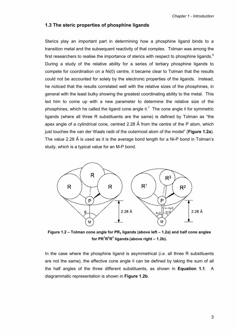
1.3 The steric properties of phosphine ligands
Sterics play an important part in determining how a phosphine ligand binds to a
transition metal and the subsequent reactivity of that complex. Tolman was among the
first researchers to realise the importance of sterics with respect to phosphine ligands.6
During a study of the relative ability for a series of tertiary phosphine ligands to
compete for coordination on a Ni(0) centre, it became clear to Tolman that the results
could not be accounted for solely by the electronic properties of the ligands. Instead,
he noticed that the results correlated well with the relative sizes of the phosphines, in
general with the least bulky showing the greatest coordinating ability to the metal. This
led him to come up with a new parameter to determine the relative size of the
phosphines, which he called the ligand cone angle θ.7 The cone angle θ for symmetric
ligands (where all three R substituents are the same) is defined by Tolman as “the
apex angle of a cylindrical cone, centred 2.28 Ǻ from the centre of the P atom, which
just touches the van der Waals radii of the outermost atom of the model” (Figure 1.2a).
The value 2.28 Ǻ is used as it is the average bond length for a Ni-P bond in Tolman’s
study, which is a typical value for an M-P bond.
Figure 1.2 – Tolman cone angle for PR3 ligands (above left – 1.2a) and half cone angles
for PR1R2R3 ligands (above right – 1.2b).
In the case where the phosphine ligand is asymmetrical (i.e. all three R substituents
are not the same), the effective cone angle θ can be defined by taking the sum of all
the half angles of the three different substituents, as shown in Equation 1.1. A
diagrammatic representation is shown in Figure 1.2b.
Chapter 1 - Introduction
4
2/3
2 3
1
∑=
=i
iθθ
Equation 1.1 – Tolman cone angle equation.
In essence, this asymmetrical cone angle parameter can be described as ‘twice the
mean average of the sum of the three half cone angles’. This equation therefore
assumes that each individual R group contributes to the cone angle in an asymmetrical
ligand PR1R2R3, the exact same amount that it would in the symmetrical ligand PR13.
This may not be completely accurate in reality, but is a good enough approximation for
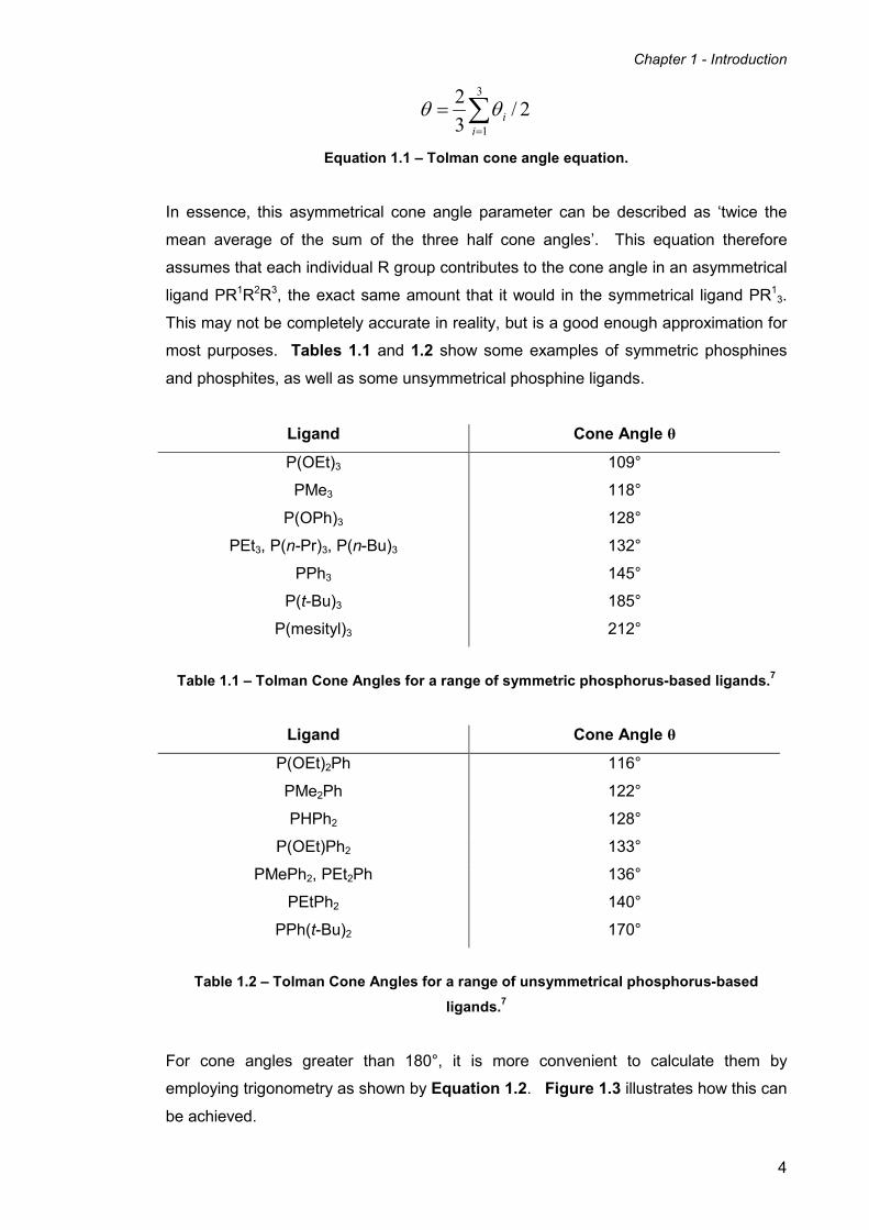
most purposes. Tables 1.1 and 1.2 show some examples of symmetric phosphines
and phosphites, as well as some unsymmetrical phosphine ligands.
Ligand Cone Angle θ
P(OEt)3 109°
PMe3 118°
P(OPh)3 128°
PEt3, P(n-Pr)3, P(n-Bu)3 132°
PPh3 145°
P(t-Bu)3 185°
P(mesityl)3 212°
Table 1.1 – Tolman Cone Angles for a range of symmetric phosphorus-based ligands.7
Ligand Cone Angle θ
P(OEt)2Ph 116°
PMe2Ph 122°
PHPh2 128°
P(OEt)Ph2 133°
PMePh2, PEt2Ph 136°
PEtPh2 140°
PPh(t-Bu)2 170°
Table 1.2 – Tolman Cone Angles for a range of unsymmetrical phosphorus-based
ligands.7
For cone angles greater than 180°, it is more convenient to calculate them by
employing trigonometry as shown by Equation 1.2. Figure 1.3 illustrates how this can
be achieved.

Chapter 1 - Introduction
5
Figure 1.3 – Method of calculating cone angles greater than 180°.7
αθ
α
2180
/
+=
= dhTAN
Equation 1.2 – Trigonometric equation for the calculation of θ greater than 180°.7
It is important to consider that all cone angles in general are derived from the ligand
models and are not determined from the molecules themselves. This has a number of
implications:
a) Phosphine ligands do not necessarily possess ‘cone-shaped’ symmetry as
assumed by the model and method of measurement. The models assume free
rotation of the R groups where possible, which might not (necessarily) be the
case in reality.
b) The model assumes the minimum cone angle possible – it is difficult to
determine how likely this is in reality due to the potential strain caused by
placing the R groups as close together as possible.
c) Bond lengths and angles in the model are fixed, which does not allow for any
flexibility or fluxional motion as in the real molecule.
d) The electronic effects of the ligand are not considered, nor are the steric and
electronic effects of other ligands coordinated to the metal.
Various people have introduced a number of refinements and modifications to address
some of these shortcomings, including determining the cone angles from single-crystal
X-ray crystallographic data,8 ligand profile plots,9 molecular mechanics calculations,10,11
and using other parameters such as the solid angle.12

Chapter 1 - Introduction
6
A more accurate technique introduced by Brown and Lee11 uses molecular mechanics
calculations to determine the steric bulk of ligands. This method involves
computationally measuring the ligand repulsive energies (ER values) in energy-
minimised structures. They found a very good correlation between their ER values and
Tolman’s cone angle, which highlights the importance of Tolman’s work more than forty
years later.
More recently, the steric effects of more complicated phosphorus-based ligands such
as diphosphines and other polydentate phosphine ligands have received considerable
attention.13 This is largely due to the fact they have played an increasingly important
role as ligands for catalytic applications. Measuring the steric size of a bidentate ligand
can be carried out in a similar manner to that of a monodentate ligand, i.e. by
calculating the Tolman cone angle for each half of a diphosphine ligand using
Equation 1.1. The value for the θi/2 for the backbone fragment of the ligand should be
measured by taking the angle between the M-P bond and an imaginary line bisecting
the P-M-P angle (Figure 1.4).14
Figure 1.4 – Tolman half cone angles for diphosphines.
However, it is difficult to define such a parameter that is dependant on the behaviour
and characteristics of the ligand backbone. The angles are usually determined from
the X-ray crystal structure of a metal diphosphine complex, which may or may not differ
significantly from the angles of the complex in solution due to changes in conformation
and flexibility. Therefore all parameters based on crystallographically-determined
angles are essentially properties of the X-ray crystal structure in question, and not
simply the ligand itself.
The P-M-P angle (bite angle) of a bidentate ligand is another important way of
classifying diphosphine ligands, and this angle in particular can vary between different
ligands, but also from metal to metal. This is largely because the bite angle of any

Chapter 1 - Introduction
7
metal-ligand complex is a compromise between the metal’s preferred bite angle and
the one preferred by the ligand.15 The preferred bite angle of a ligand generally
depends on constraints imposed by the ligand backbone, as well as steric factors
determined by the substituents on the phosphorus donor (including the backbone).
Metal preferred bite angles, however, are largely determined by electronic effects. If a
particular ligand/metal combination fails to find a conformation in which both the metal
and ligand are satisfied, this can lead to changes in the geometry of the complex, or
even lead to another coordination mode such as the bridging mode. This is when each
donor atom of a bidentate ligand is coordinated to a different metal centre. Ligands
with unusually large or small bite angles have long been used to synthesise complexes
that would otherwise be unstable with ligands possessing classic bite angles. Figure
1.5 shows some examples of average crystallographically determined bite angles of a
range of diphosphine ligands.
Figure 1.5 – Selected bite angles of diphosphines as determined by X-ray crystallography
(standard deviation in brackets).15
Dierkes and van Leeuwen15 found in a study of a range of bidentate phosphine ligands,
that the bite angles had a pronounced effect on both selectivity and rate of metal
catalysed reactions.
1.4 The electronic properties of phosphine ligands
Before attempting to explain how electronic effects influence the behaviour of
phosphine ligands in terms of their coordination chemistry and reactivity, it is necessary
to discuss the nature of the M-P bond, and its context within coordination chemistry.
The traditionally accepted description of the M-P bond within a transition metal
phosphine complex can be broken down into two main components. The first is the σ-

Chapter 1 - Introduction
8
donation of electron density from the lone pair of the phosphine to a symmetric empty
bonding orbital on the transition metal. The other is the π-acceptor (or π-back bonding)
component, which is when electrons from a filled orbital on the transition metal donate
back into an empty orbital on the phosphorus (See Figure 1.6).
Figure 1.6 - representation of the traditionally accepted σ and π components of the M-P
bond. The phosphorus π-acceptor orbital is a σ* orbital, only part of which is shown.
In the case of an octahedral complex, the metal d orbitals involved in σ bonding with
the six ligands are the eg orbitals. This leaves the t2g orbitals non-bonding and directed
in-between the six M-P bonds. The main part of the M-P bond is the σ bond formed
from a metal d orbital and an sp3 orbital on the phosphorus. This essentially means
that phosphorus ligands act as both Lewis bases (σ-electron donors) and Lewis acids
(π-electron acceptors). This helps phosphine ligands to stabilise metals both in high
and low oxidation states, which is a very useful property in organometallic catalysis.
This ‘synergetic’ σ + π approach was originally successfully applied as a description of
metal carbonyl bonding, and which was then applied to metal-phosphorus bonding as a
result of this success. Despite this, however, the electronic factors in metal-
phosphorus interactions remain contentious.16 All studies generally agree that the M-P
bond contains a σ component, but it is the π-back bonding component which has
received most attention. The traditional Dewar-Chatt-Duncanson model17, 18 describes
the π-back bonding component as the drift of the electrons from filled transition metal d
orbitals to the empty 3d orbitals on the phosphorus. However, some have suggested
that the π-donation of electrons from the metal occurs into either the phosphorus p
orbitals19 or the P-R σ* antibonding orbitals.20 Calculations carried out by Pacchioni
and Bagus21 have also suggested that the d orbitals of the phosphorus are too diffuse
and high in energy to effectively participate solely in the π-back bonding of electron
density, and instead the electron density is being donated into a combination of the P-R
σ* antibonding orbitals and phosphorus d orbitals. Computational models have been
compared with X-ray crystal structural data of various metal-phosphine complexes in
order to determine the accuracy and viability of such calculations. In the study by

Chapter 1 - Introduction
9
Orpen and Connelly,20 twenty four sets of transition metal-phosphine and -phosphite
complexes, whose X-ray crystal structures were known for multiple oxidation states,
were studied for variations in their geometry. They found that the M-P bond lengths
tended to increase upon oxidation of the metal, consistent with there being an
important element of π-back bonding present. Other authors have even suggested that
some phosphine ligands are pure σ-donors22-25 and others even π-donors.22, 23
It is now generally accepted that the nature (or even existence) of this π-back bonding
depends greatly on the nature of the R substituents on the phosphorus atom. When
highly electronegative substituents are present, such as in PF3, the P-X bond is highly
polarised and therefore the P-X σ* orbital is low in energy and mainly centred around
the P atom, favouring back bonding.26, 27 Where various experimental and theoretical
studies have shown that ligands such as PF3, PCl3, P(OR)3 all clearly accept π electron
density from transition metals, the amount of π back bonding towards alkyl- and aryl-
substituted phosphorus ligands is much harder to quantify, with different methods
leading to different conclusions.
Apart from using computational and crystallographic methods to determine the σ-donor
and π-acceptor properties of phosphorus ligands, other experimental techniques, and
therefore other parameters are often employed. One of the most commonly used
spectroscopic techniques is to use infrared spectroscopy to measure the CO stretching
frequency of a range of transition metal carbonyl complexes in which steric factors do
not play a major role. Tolman, in addition to his studies on the role of steric factors of
phosphorus ligands, also carried out investigations into their electronic factors using
this technique.6 This method is based on the strong π-acceptor properties of the
carbonyl ligand, which is able to accept electron density into an antibonding orbital of
the CO bond. The stretching frequency of the carbonyl bond is dependent on the
electron density at the transition metal centre, and this in turn depends on the σ-donor
and π-acceptor properties of the other ligands coordinated to the metal. Strong π-
acceptor and weak σ-donor ligands draw electron density away from the metal,
reducing the amount of π back bonding to the carbonyl and therefore increasing the
stretching frequency of the carbonyl group. Conversely, weak π-acceptor and strong σ-
donor ligands effectively increase the electron density at the metal centre, increasing
the amount of π back bonding to the CO and decreasing the CO stretching frequency.
Tolman’s study was based on complexes in CH2Cl2 solution, where L is any
monodentate phosphorus-based ligand (mainly phosphines and phosphites), either
symmetric or asymmetric. As a result of his study, Tolman introduced the measured

Chapter 1 - Introduction
10
electronic parameter νCO (A1) as a measure of the σ-donor and π-acceptor properties of
a PR3 ligand. νCO (A1) is defined in Equation 1.3.
13
1
1 1.2056)( −=
=
∑+= cmA
i
i
iCOχν
Equation 1.3 – equation relating Tolman electronic parameters ν and χi.
The value 2056.1 cm-1 is the νCO (A1) value for P(t-Bu)3, the ligand with the lowest νCO
(A1) value used in Tolman’s study. χi is the substituent modification parameter, which is
set at zero for P(t-Bu)3. Table 1.3 contains selected values for both νCO (A1) for
selected ligands and χi for their corresponding substituents.
Ligand χi / cm-1 Σ χi / cm
-1 νCO (A1) / cm-1
P(t-Bu)3 0.0 0.0 2056.1
PEt3 1.8 5.4 2061.7
PMe3 2.6 7.8 2064.1
PPh3 4.3 12.9 2068.9
P(OEt)3 6.8 20.4 2076.3
P(OMe)3 7.7 23.1 2079.5
P(OPh)3 9.7 29.1 2085.3
PCl3 14.8 44.4 2097.0
PF3 18.2 54.6 2110.8
Table 1.3 – Examples of Tolman’s χi and νCO (A1) values of PR3 ligands for [Ni(CO)3(PR3)].6
The examples in Table 1.3 clearly show how the electronic parameters are affected by
increasing the electronegativity of the substituents on the phosphorus ligand.
Increased electronegativity of the substituents as the table is descended leads to
increased νCO (A1) as a result of the decrease in σ-donor character and an increase in
π-acceptor character.
Although Tolman’s electronic parameters are a useful way of comparing the electronic
character of a series of ligands, they are a measure of the combined effect of both the
σ-donor and π-acceptor character, and do not separate the two characteristics.
Partially as a result of this and partially independently, others have made attempts to
devise other parameters that can separately characterise the σ and π elements of a
ligand.
Chapter 1 - Introduction
11
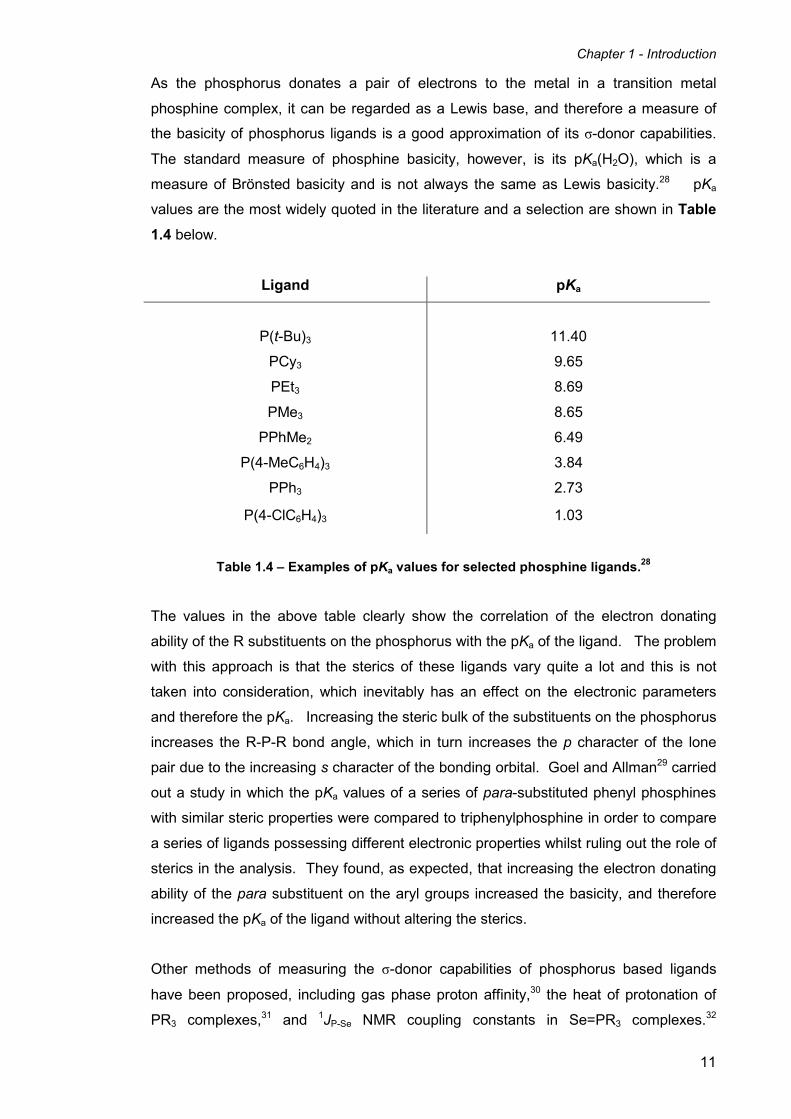
As the phosphorus donates a pair of electrons to the metal in a transition metal
phosphine complex, it can be regarded as a Lewis base, and therefore a measure of
the basicity of phosphorus ligands is a good approximation of its σ-donor capabilities.
The standard measure of phosphine basicity, however, is its pKa(H2O), which is a
measure of Brönsted basicity and is not always the same as Lewis basicity.28 pKa
values are the most widely quoted in the literature and a selection are shown in Table
1.4 below.
Ligand pKa
P(t-Bu)3
11.40
PCy3 9.65
PEt3 8.69
PMe3 8.65
PPhMe2 6.49
P(4-MeC6H4)3 3.84
PPh3 2.73
P(4-ClC6H4)3 1.03
Table 1.4 – Examples of pKa values for selected phosphine ligands.28
The values in the above table clearly show the correlation of the electron donating
ability of the R substituents on the phosphorus with the pKa of the ligand. The problem
with this approach is that the sterics of these ligands vary quite a lot and this is not
taken into consideration, which inevitably has an effect on the electronic parameters
and therefore the pKa. Increasing the steric bulk of the substituents on the phosphorus
increases the R-P-R bond angle, which in turn increases the p character of the lone
pair due to the increasing s character of the bonding orbital. Goel and Allman29 carried
out a study in which the pKa values of a series of para-substituted phenyl phosphines
with similar steric properties were compared to triphenylphosphine in order to compare
a series of ligands possessing different electronic properties whilst ruling out the role of
sterics in the analysis. They found, as expected, that increasing the electron donating
ability of the para substituent on the aryl groups increased the basicity, and therefore
increased the pKa of the ligand without altering the sterics.
Other methods of measuring the σ-donor capabilities of phosphorus based ligands
have been proposed, including gas phase proton affinity,30 the heat of protonation of
PR3 complexes,31 and 1JP-Se NMR coupling constants in Se=PR3 complexes.
32

Chapter 1 - Introduction
12
Puddephatt et al.33 have used the ionisation potentials of a range of phosphorus
ligands as a measure of basicity, i.e. the availability of the lone pair. They found that
the ionisation energies of the phosphorus lone pair in the gas phase followed the
sequence PMe3 > PPhMe2 > PPh2Me > PPh3. This shows a good correlation between
phosphorus lone pair ionisation potential and basicity within a series of closely related
phosphines. However, this technique is not ideal for comparing the relative basicities
of substantially different phosphines such as PCy3 and PF3. Also, as the ionisation
energies are measured in the gas phase, this is not always useful as most chemists
are interested in basicities of phosphine ligands in solution. The basicities of
phosphines in solution differ greatly mainly because they are dominated by solvation
effects, especially in ionising solvents.
1.5 Macrocycles and macrocyclic ligands
A macrocycle is a large cyclic molecule and, in the eyes of a coordination chemist, a
macrocyclic ligand can be defined as “a cyclic molecule consisting of an organic
framework into which heteroatoms capable of coordinating to metals have been
incorporated.”34 More specifically, a macrocyclic ligand should contain at least three or
more potential donor atoms in a ring of at least nine atoms.35 Macrocycles are
commonly found in nature in the form of porphyrins, corrins, and chlorins, but it was not
until 1936 that the first synthesis of a macrocycle (1,4,8,11-tetraazacyclotetradecane)
was reported.36 The popularity of macrocyclic chemistry began to increase in the
1960s, thanks in part to the efforts of Busch37 and Curtis,38 who discovered the
synthesis of nickel(II) diimine Schiff base coordination compounds from the reaction of
nickel(II) diamines and aliphatic ketones. These initial studies were primarily aimed at
mimicking porphyrins and other naturally occurring macrocycles such as corrins,
chlorins, and corphins.
Another area of development for macrocyclic chemistry began in the late 1960s
focused on using macrocycles such as oxygen-based crown ethers (Pedersen),39
mixed oxygen nitrogen bicyclic cryptands (Lehn),40 and cavitands (Cram)41 to model
biological processes such as ion transport. Traditionally macrocycles have been
studied both for their use as models for naturally occurring macrocyclic systems such
as porphyrins, as well as their use in supramolecular chemistry and as receptors for
molecular recognition and complexation. To highlight the importance of macrocyclic
chemistry, the 1987 Nobel Prize in Chemistry was awarded to Cram, Lehn and
Pedersen "for their development and use of molecules with structure-specific
interactions of high selectivity" based on their development of macrocycles in this area.

Chapter 1 - Introduction
13
Today many different kinds of macrocycles have been discovered and studied,
including simple polyaza-macrocycles, heteroatom-based macrocycles such as
polythia-, polyphospha-, and polyarsa-macrocycles, as well as a wide range of mixed-
donor macrocycles.
1.5.1 The macrocyclic effect
It is well known that complexes containing polydentate ligands are more stable than
those complexes containing the equivalent number of similar monodentate ligands.
This is known as the chelate effect.42 It is therefore obvious to ask whether
macrocyclic ligands give rise to more stable complexes than their related linear open-
chain relatives. This is in fact the case and has become known as the macrocyclic
effect. The macrocyclic effect was first reported by Cabbiness and Margerum43 in 1969
by comparing the stability constants of the copper(II) complexes of a tetraamino
macrocyclic ligand and a similar tetraamino acyclic ligand. They determined that the
macrocyclic copper(II) complex was approximately ten thousand times more stable
than its acyclic analogue, despite having a similar sequence of chelate rings. This
large increase in stability cannot be simply attributed to the chelate effect in terms of
entropy, and exists despite the restricted geometry that the macrocycle imposes on the
metal. They also noticed that the rate of coordination for the macrocyclic ligand was
much slower than that of the related open-chain ligand, which is in general the case for
most macrocyclic ligand systems. Macrocyclic complexes are almost invariably more
stable than those of their open-chain counterparts, though the difference decreases
and eventually becomes insignificant when the macrocyclic rings are large and very
flexible.35 Many applications of macrocyclic ligands rely on the macrocyclic effect and
the increased stability of the complexes which they form.
The origins of the macrocyclic effect have been the subject of much discussion. It is
tempting to imagine both the macrocyclic and chelate effects in terms of possessing a
common entropic origin. It is now clear, however, that this is not the case and it is not
possible to describe the macrocyclic effect simply in terms of a single origin.35 Studies
by Paoletti and co-workers44-49 have shown that the macrocyclic effect has both
entropic and enthalpic contributions. Experimentally determined values of ∆H and T∆S
for the formation of nickel(II) high-spin and low-spin complexes of the 1,4,8,11-
tetraazacyclotetradecane macrocycle (cyclam) and its open-chained analogue
increased (i.e. more negative ∆H and more positive T∆S) upon going from the complex
of the open chain ligand to the cyclam complex.

Chapter 1 - Introduction
14
1.5.2 Phosphorus-containing macrocycles
The first phosphorus-containing macrocycles were prepared in 1897 by Stokes,50 when
he discovered cyclophosphazenes (X2P=N)n (n≥5). It is surprising then, that intensive
study of phosphorus macrocycles did not commence until the mid 1970s, despite the
development of crown ethers by Pedersen39 several years earlier in 1967. This is even
after knowing the benefits and complexing ability of macrocyclic ligands. A lack of
progress in this area can potentially be attributed to experimental difficulties, such as
complex multi-step procedures, unstable and air sensitive products, and low yielding
reactions. One of the main driving forces for interest in the field was to create more
efficient macrocycles by introducing tri-coordinated phosphorus atoms into the ring, or
phosphoryl and thiophosphoryl groups possessing high complex forming ability.51
Macrocycles containing tri-coordinated phosphorus groups usually bind very easily to
transition metals, whereas alkali metals can be captured with phosphine oxide or
sulfide groups. Like most phosphine ligands, macrocyclic phosphines are also
extremely useful in stabilising transition metals in their lowest oxidation states. Other
potential uses include molecular recognition, complexation of salts and anions, as well
as homogeneous and phase-transfer catalysis. Thiophosphoryl macrocycles have
even been known to assist the transport of transition metal ions such as Fe3+, Cu2+,
Co2+, and Ni2+ through liquid membranes.52
There are, in general, two separate methods of preparing phosphorus-containing
macrocycles; those utilising classical methods of macrocycle synthesis, and those
taking into account specific properties of the phosphorus groups. It must be noted that
there are many different kinds of phosphorus macrocycle containing different linking
groups such as C-P-C, N-P-N, and O-P-O within the macrocyclic ring, but this review
will concentrate on C-P-C linking groups. The three main strategies for the synthesis of
phosphorus containing macrocycles are:
i) Cyclocondensations
ii) Ring-opening reactions
iii) Template-assisted reactions
Cyclocondensations are reactions where the condensation of two functionalised
species leads to the formation of a macrocycle. For example, a [1+1]
cyclocondensation is the formation of a macrocycle from one molecule of each
reactant, whereas a [2+2] cyclocondensation combines two molecules of each. One
method of forming C-P-C macrocycles by cyclocondensation is to react the lithium salts

Chapter 1 - Introduction
15
of diphosphines with an appropriate dihalogenated species using high dilution
conditions. Kyba et al. used this method from 1977 to 1985 to great effect to prepare a
series of tri-donor nine-membered rings and tetra-donor fourteen-membered rings.
Scheme 1.1 shows the formation of a tri-phosphorus (4) and a tetra-phosphorus
macrocycle (5) from the dilithium salt of the ortho-bis(phenylphosphino)benzene (1)
and the relevant tertiary phosphine dichlorides (2 and 3).53
Scheme 1.1 – Formation of tri- and tetra-phosphorus macrocycles.53
Replacing the phenyl-phosphine based dilithium salt 1 with the dilithium salt of
dithiocatechol allowed the formation of mixed P, S donor macrocyclic ligands.53 Other
P-containing mixed-donor macrocycles have also been prepared with arsenic,54-58
nitrogen,55, 57-61 and oxygen.55, 58, 59, 61
Macrocycles 4 and 5 exist as a number of geometrical isomers. Species 4 exists as
three isomers (two meso and one dl pair), whereas 5 exists as five isomers, three
meso and two dl pairs. Both 4 and 5 have been shown to be capable of coordinating to
transition metals. The tridentate macrocycle 4 binds to transition metals using all three
P-donor atoms in a fac-tridentate manner, whereas 5 is large enough to ligate to the
metal in either a cis- or trans-tetradentate fashion to afford an octahedral complex
(Figure 1.7).

Chapter 1 - Introduction
16
Figure 1.7 – Ligation modes of macrocycles 4 and 5 in octahedral complexes.53
Ring-opening reactions are another useful method of preparing phosphorus-containing
macrocycles. In almost all cases, macrocycles containing C-P-C linkages prepared by
this method lead to nine-membered rings. These are generally synthesised from
phosphorus-containing five-membered rings such as phospholes. The first attempts to
synthesise macrocycles via this method were carried out by Wittig in 1964. He took
neat spirophosphoranes (e.g. 6) and heated them up to their melting point, which
induces the opening of the two five-membered rings, leading to the formation of a C-C
bond to give the phosphorus(III) macrocycle 7.62 The lone pair of the phosphorus can
also undergo addition reactions such as methylation with methyl iodide, to give the
cationic macrocycle 8. The reaction is shown below in Scheme 1.2.
Scheme 1.2 – Ring-opening of a spirophosphorane 6 to form the macrocycle 7 and
subsequent methylation to form 8.62
The ionic macrocycle 8 can also be prepared directly from 6 by heating it with methyl
iodide in a one-pot reaction. Many other substituted variants of 7 and 8 have been
prepared by using spirophosphoranes containing different alkyl and aryl substituents at
a variety of positions on the aromatic rings, as well as the phosphorus itself.
Using a template (such as a transition metal) to assist the formation of a macrocycle
can be a very useful method, and has been part of the field of macrocyclic chemistry
since its inception. In many cases it is possible to successfully replace traditional multi-
step macrocycle syntheses with template-assisted reactions offering less steps and
higher yields. Two different roles exist for the metal in the template process, either

Chapter 1 - Introduction
17
kinetic or thermodynamic in nature.63 In the kinetic role, the ligands are already
coordinated to the metal and arranged in a manner that allows one final condensation
to complete the macrocyclic complex. The thermodynamic role is when the formation
of the macrocycle would not happen at all without the presence of the metal ion. The
cyclisation process can either take place on the phosphorus itself or on a substituent of
the phosphorus atom. The first example of template-assisted macrocycle synthesis
containing a phosphorus atom was prepared by Marty and Schwartzenbach64 in 1970.
Their method involved reacting the tetradentate mercaptophosphine nickel complex 9
with dibromoxylene 10 to form the {P2S2} macrocyclic Ni complex 11 (Scheme 1.3).
Scheme 1.3 – Template-assisted synthesis of the P2S2 macrocyclic ligand complex 11.
This is an example of a kinetic template synthesis with the cyclisation process taking
place at the sulphur atoms, and not at the phosphorus atoms themselves.
An advantage of using a template synthesis is that it forms the metal complex directly,
without the need for further complexation reactions. However, this is fine if the metal
used in the templating process is the desired metal in the final complex, but if the free
macrocycle itself is wanted, then it is necessary to remove the metal via a
decomplexation reaction. In some cases the demetallation has been shown to occur
automatically, but this is rare and in most examples it is necessary to devise a method
for removing the metal. In many instances, it has proven very difficult to separate the
macrocycle from the metal template largely due the macrocyclic effect.
An example where the researchers65 managed to successfully remove the metal from a
phosphorus-containing macrocyclic complex formed by template-assisted assembly is
shown below in Scheme 1.4.

Chapter 1 - Introduction
18
Scheme 1.4 – Template synthesis of a tetradentate P4 macrocyclic Ni complex and
subsequent demetallation.
This process involves the cyclocondensation reaction of the aryl dibromide 10 with a
nickel(II) disecondary phosphine complex 12. In the presence of potassium carbonate
as a base, the reaction gives the fifteen-membered macrocyclic nickel(II) tetradentate
phosphine complex 13. This is also the first example of a template-assisted reaction
where the cyclisation process occurs at the phosphine centre itself. The chloride
anions can then be exchanged with other anions such as BF4- and NCS- via metathesis
reactions. The free macrocycle 14 can be liberated from the metal by treating the
complex with an aqueous solution of sodium cyanide, thus complexing the nickel to
give nickel cyanides and releasing the free ligand.
Another more recent example of the template synthesis and liberation of a tetradentate
macrocyclic phosphine has been demonstrated by Lebbe et al. (Scheme 1.5).66

Chapter 1 - Introduction
19
Scheme 1.5 – Template synthesis and oxidative demetallation of a Ni P4 macrocyclic
complex, followed by reduction to give the free macrocycle.
The tridentate phosphine ligand H(Me)P(CH2)3P(Ph)(CH2)3P(Me)H 15 was reacted with
the divinylaminophosphine (CH2=CH2)2PNEt2 16 in the presence of NiBr2 to form the
nickel(II) tetradentate aminophosphine macrocyclic complex 17. This was then reacted
with an aqueous solution of HBr to replace the NEt2 with an OH group to give 18.
To obtain the free ligand, Lebbe et al. treated 18 with a strong oxidising agent such as
aqueous solutions of H2O2/HCl or Br2. This completely oxidised the four phosphine
donors to phosphine oxide groups to give the free ligand oxide and precipitated the
nickel from solution as Ni(OH)2. Upon subsequent addition of a base such as
potassium hydroxide, the hydroxy group is deprotonated to give the phosphonate oxide
derivative 19, which was converted to the methyl ester 20 by acidification with HCl and
reaction with HC(OMe)3. Excess thionyl chloride was reacted with 20 to form the
phosphinic acid chloride 21. This then reacted cleanly with LiAlH4 in good yield to give
the reduced macrocyclic phosphine ligand 22. NMR spectroscopy showed four
different diastereoisomers, two symmetrical and two unsymmetrical.
Tridentate {P3} ligands have also attracted a lot of recent attention due to their potential
applications in coordination chemistry and homogeneous catalysis. There are three
main methods of preparing these triphosphamacrocycles via a template synthesis; the
1+1+1, 2+1, and 3+0 approaches.67 The 1+1+1 method involves all three chelate rings
Chapter 1 - Introduction
20
of the macrocycle forming successively at the metal centre from three appropriately-
functionalised monodentate phosphines. In the 2+1 closure, only two chelate rings are
formed as one is already present in the pre-bound bidentate phosphine. The 3+0
approach uses a coordinated tridentate phosphine ligand requiring only one ring-
closing step to give the final macrocycle. Though this approach seems to be the
simplest on offer, no literature examples exist to this date for the 3+0 method.67 A good
example of the 1+1+1 method by Norman et al.68 is based on the preparation of a
triphosphacyclododecane using a molybdenum tricarbonyl complex as a template.
However, they failed to release the free macrocycle from the metal due to its high
stability, though Edwards and co-workers69 managed to achieve the stereoselective
liberation of this macrocycle at a later date by the reaction of the molybdenum and
chromium macrocyclic complexes with base.
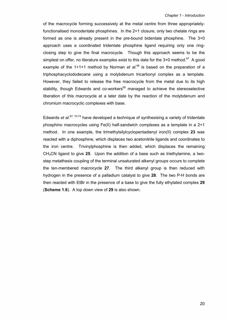
Edwards et al.67, 70-74 have developed a technique of synthesising a variety of tridentate
phosphino macrocycles using Fe(II) half-sandwich complexes as a template in a 2+1
method. In one example, the trimethylsilylcyclopentadienyl iron(II) complex 23 was
reacted with a diphosphine, which displaces two acetonitrile ligands and coordinates to
the iron centre. Trivinylphosphine is then added, which displaces the remaining
CH3CN ligand to give 25. Upon the addition of a base such as triethylamine, a two-
step metathesis coupling of the terminal unsaturated alkenyl groups occurs to complete
the ten-membered macrocycle 27. The third alkenyl group is then reduced with
hydrogen in the presence of a palladium catalyst to give 28. The two P-H bonds are
then reacted with EtBr in the presence of a base to give the fully ethylated complex 29
(Scheme 1.6). A top down view of 29 is also shown.

Chapter 1 - Introduction
21
Scheme 1.6 – Synthesis of a symmetric ten-membered P3 macrocyclic Fe(II) complex.
67
Edwards et al.67, 70-74 also used this same method to produce a range of symmetric and
asymmetric tri-phosphorus 9- to 12- membered rings containing a range of secondary
and tertiary alkyl and aryl phosphine groups. Repeated attempts to liberate these
triphosphorus macrocycles with conventional agents such as CN- were not successful.
The d6 octahedral Fe(II) template appears to be very kinetically inert and resitant to
disruption. One nine-membered ring was successfully liberated as its trioxide by
exhaustive oxidation,72 but none of the free uncoordinated ligands were obtained. The
iron centre was found to undergo reversible one electron oxidation in most of these
complexes, but attempts to scavenge the Fe(III) ions from the oxidised complexes with
various complexing agents (such as CN-, OH-, EDTA4-) were unsuccessful.
1.6 Medium-ring heterocyclic diphosphines
Another interesting class of phosphine ligands is the medium-ring diphosphines.
These are generally defined as compounds in which two phosphorus atoms are
incorporated into a ring of between seven and twelve atoms.75 These medium-sized
rings are usually quite strained and show strong tendancies to undergo transannular
reactions. Currently the state of understanding of these compounds and their

Chapter 1 - Introduction
22
coordination chemistry is quite limited, but it has been proposed that compounds in this
category will show novel properties and reactions.75
Eight-membered rings containing at least two donor atoms are unique in coordination
chemistry, as they occupy a niche between large polydentate macrocycles with the
ability to readily coordinate one or more metal centres and small heterocycles which
posses limited chelating ability.76 They are not themselves technically classed as
macrocycles, as they do not meet the criteria of the minimum number of nine atoms
and three donors. As incorporating donor atoms such as phosphorus into these cyclic
and polycyclic molecules gives rise to rigid steric constraints on the metal, eight
membered rings should have major implications for coordination chemistry and
catalysis.77
Figure 1.8 – Potential coordination modes and conformations of bidentate eight-
membered rings.
Figure 1.8 (above) shows three potential coordination modes and conformations of
polydentate eight-membered ring systems. Another interesting prospect for
tetradentate eight-membered ring systems is the potential to chelate to two different
metals in an exo-ditopic manner.78 In order for the cyclic ligand to obtain this binding
mode the heterocycle must be in the chair/chair conformation (Figure 1.9).
Figure 1.9 – chair/chair conformation of a tetradentate eight-membered heterocycle
coordinating to two metals in an exo-ditopic manner.

Chapter 1 - Introduction
23
An important class of eight-membered bidentate phosphines are the 1,5-diaza-3,7-
diphosphacyclooctanes, first synthesised by Märkl and co-workers in 1980.79 These
were prepared by reacting two equivalents of an alkyl or aryl bis(hydroxymethyl)
phosphine with two equivalents of a primary amine in a modified version of the
Mannich reaction (Scheme 1.7). This Mannich-type synthesis of phosphines is a very
important and convenient method of constructing heterocyclic bis(phosphines) and
other heterocyclic phosphine ligands for transition metal coordination chemistry.80
Scheme 1.7 – General synthesis of 1,5-diaza-3,7-diphosphacyclooctanes.
X-ray crystallographic studies by Wong et al.81 on [CH2P(Ph)CH2N(2-pyridyl)]2 (30) (i.e.
where R = Ph, R’ = o-pyridyl) showed that the free ligand adopts a crown configuration
in the solid state. The lone pairs of the phosphorus were also found to be syn to each
other, which is perfect for chelation to metals. Coordination studies of this ligand with
Mo(CO)4(η2-norbornadiene) gave the octahedral mononuclear complex as expected.
The X-ray structure, however, showed that while the ligand does indeed chelate to the
metal in a bidentate manner, the ligand actually adopts a chair/boat conformation by
inversion of one half of the ligand (31) (Figure 1.10).
Figure 1.10 – Uncoordinated crown conformation of (30) and coordinated chair/boat
conformation of its Mo(CO)4 complex (31).
Chapter 1 - Introduction
24
Attempts by the authors to create heterobimetallic complexes via coordination of a
second metal (such as Zn2+, Co2+, Ni2+) to the ring nitrogen atoms proved unsuccessful.
Further research in this area has been carried out over the past decade using 1,5-
diaza-3,7-diphosphacyclooctanes functionalised with chiral,82 unsaturated,83
ferrocenyl,84 ferrocenylmethyl,85 o-oxyphenyl,86, 87 and amino acid88, 89 substituents at
the heteroatoms. The general method of synthesis involves the Mannich-type
preparation of hydroxymethyl phosphines from primary phosphines and formaldehyde,
followed by the reaction with primary amines. These ligands have been used in
coordination chemistry to prepare a range of asymmetric,82 polymetallic,85 water-
soluble,85-89 and chelate82, 83, 85, 86, 88, 90-94 complexes of transition metals.
Karasik, Hey-Hawkins and co-workers discovered that the type of amine used in the
synthesis of these ligands is crucial in determining the final product.85, 89 Depending on
the amine used in the reaction with a range of aryl phosphines, they obtained either the
linear bis(arylaminomethyl)phosphines 32, the six-membered 1,3-diaza-5-
phosphacyclohexanes 33 and 34, or the 1,5-diaza-3,7-diphosphacyclooctanes 35
(Scheme 1.8).

Chapter 1 - Introduction
25
Scheme 1.8 – Reactions of bis(hydroxymethyl)arylphosphines with primary amines.
Interestingly, using different phosphines in the reaction whilst keeping the amine
constant does not lead to the formation of a different type of product. When 2-
aminobenzoic acid (37) is used as the amine, the linear bis(2-
carboxyphenylaminomethyl)phosphines (32a-g) are generated as the products, even if
the stoichiometry is altered to favour the cyclic 2+2 products. Interestingly however,
when the 4-aminobenzoic acid (38) is instead used as the amine, both the linear bis(4-
carboxyphenylaminomethyl)phosphines (analogous to 32a-g) and the eight-membered
heterocyclic 1,5-diaza-3,7-diphosphacyclooctanes are formed as products (35a-g),
depending on the stoichiometry employed in the reaction. In contrast to the aromatic
amines already mentioned, when the aliphatic amines glycine, D- or L-phenylglycine, L-
alanine (all derivatives of 39), or L-lysine (40) were used in the reaction instead, the
six-membered 1,3-diaza-5-phosphacyclohexanes (33a, b, c, g and 34a, d-g) are
formed. This presumably occurs from the reaction of two equivalents of amine with
Chapter 1 - Introduction
26
one equivalent of bis(hydroxymethyl)phosphine to initially give the linear
bis(aminophosphines), followed by the condensation of a molecule of formaldehyde
(which is present because the reactions to form the bis(hydroxymethyl)phosphines
(36a-g) from the analogous primary phosphines and formaldehyde is done in situ and
the products are not isolated) with the two NH groups to complete the six-membered
ring. It is remarkable that these three different classes of ligand can be formed simply
by altering the substituents on the amine, whereas using different substituted
phosphines does not alter the final product.
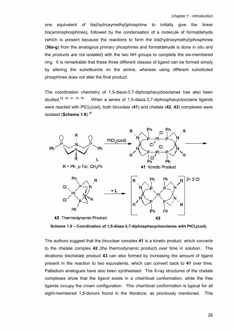
The coordination chemistry of 1,5-diaza-3,7-diphosphacyclooctanes has also been
studied.82, 85, 91, 93, 95 When a series of 1,5-diaza-3,7-diphosphacyclooctane ligands
were reacted with PtCl2(cod), both binuclear (41) and chelate (42, 43) complexes were
isolated (Scheme 1.9).95
Scheme 1.9 – Coordination of 1,5-diaza-3,7-diphosphacyclooctanes with PtCl2(cod).
The authors suggest that the binuclear complex 41 is a kinetic product, which converts
to the chelate complex 42 (the thermodynamic product) over time in solution. The
dicationic bischelate product 43 can also formed by increasing the amount of ligand
present in the reaction to two equivalents, which can convert back to 41 over time.
Palladium analogues have also been synthesised. The X-ray structures of the chelate
complexes show that the ligand exists in a chair/boat conformation, while the free
ligands occupy the crown configuration. This chair/boat conformation is typical for all
eight-membered 1,5-donors found in the literature, as previously mentioned. This

Chapter 1 - Introduction
27
conformation is favoured most likely due to the minimisation of ring strain which occurs
as a result of the ligand chelating to the metal.
The six-membered 1,3-diaza-5-phosphacyclohexane ligands are also good ligands for
transition metals, and behave as monodentate phosphine ligands as would be
expected. cis-Bis(1,3-diaza-5-phosphacyclohexane) platinum and palladium
complexes (44) have been synthesised, as well as monodentate carbonyl complexes of
molybdenum and tungsten (45 and 46) (Figure 1.11).
Figure 1.11 – Complexes of 1,3-diaza-5-phosphacyclohexane ligands.
When primary diamines (48a-c) are used in the ligand synthesis instead of
monoamines, the entirely unexpected formation of macrocyclic
aminomethylphosphines (49a-e) is achieved (Scheme 1.10).96, 97
Scheme 1.10 – Formation of aminomethylphosphino macrocycles (49a-e).
In a similar manner to the reaction using 4-aminobenzoic acid, the 1,5-diaza-3,7-
diphosphacyclooctane motif is formed from two equivalents each of phosphine and
Chapter 1 - Introduction
28
amine. However, in this instance the two remaining primary amino groups on the
alternate ends of the aromatic diamines condense with two further equivalents of
phosphine to form a second 1,5-diaza-3,7-diphosphacyclooctane moiety. The resulting
macrocyclic product is formed in approximately 50% yield, which is very high for a one-
pot synthesis of a highly organised structure from six components,98 especially without
the use of high-dilution techniques. The macrocycle itself contains a large, hollow,
basket-like intramolecular cavity, potentially useful for the construction of organised
coordination compounds containing a catalytic site (such as a transition metal).
Other phosphorus- and nitrogen- containing heterocyclic ligands have also been
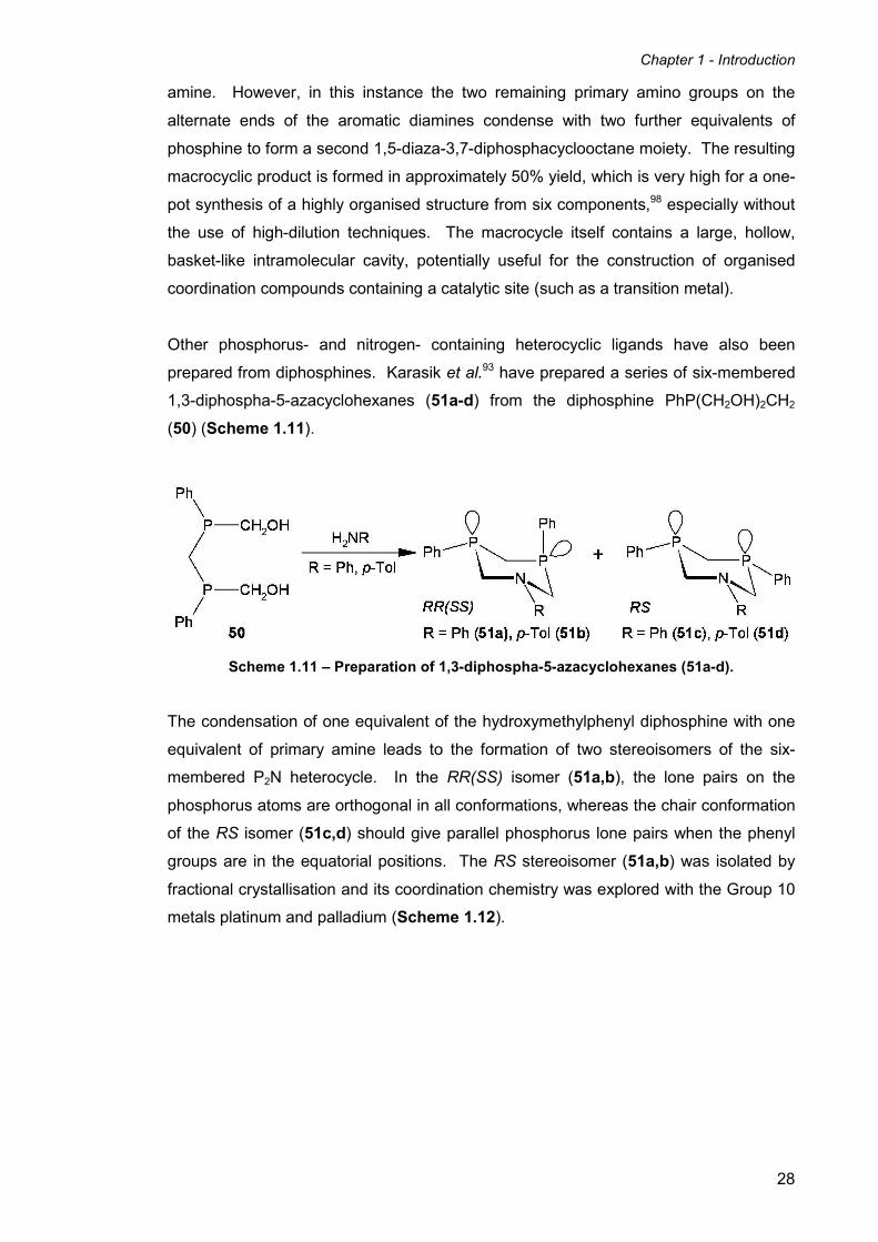
prepared from diphosphines. Karasik et al.93 have prepared a series of six-membered
1,3-diphospha-5-azacyclohexanes (51a-d) from the diphosphine PhP(CH2OH)2CH2
(50) (Scheme 1.11).
Scheme 1.11 – Preparation of 1,3-diphospha-5-azacyclohexanes (51a-d).
The condensation of one equivalent of the hydroxymethylphenyl diphosphine with one
equivalent of primary amine leads to the formation of two stereoisomers of the six-
membered P2N heterocycle. In the RR(SS) isomer (51a,b), the lone pairs on the
phosphorus atoms are orthogonal in all conformations, whereas the chair conformation
of the RS isomer (51c,d) should give parallel phosphorus lone pairs when the phenyl
groups are in the equatorial positions. The RS stereoisomer (51a,b) was isolated by
fractional crystallisation and its coordination chemistry was explored with the Group 10
metals platinum and palladium (Scheme 1.12).

Chapter 1 - Introduction
29
Scheme 1.12 – Coordination chemistry of RS 1,3-diphospha-5-azacyclohexane ligands
(51c,d).
With both platinum and palladium, the resulting complexes are all dinuclear, and no
chelate complexes are observed. This is due to the fact that the six-membered ring is
too rigid to bend to a position where both phosphorus lone pairs could face towards the
same metal centre. This is in contrast to the more flexible 1,5-diaza-3,7-
diphosphacyclooctanes, which are commonly known to chelate to one metal. Their
flexibility allows for the ligand to bend around in order to allow the second phosphorus
lone pair to coordinate to the metal, after the first has already done so.
In order to make an analogue of these heterocyclic {P2N} ligands capable of chelating
to one metal centre, Karasik and co-workers92, 99 targeted a more flexible version based
on the eight-membered 1,5-diaza-3,7-diphosphacyclooctanes. Their strategy was
similar to the synthesis of 1,3-diphospha-5-azacyclohexanes: to react a
hydroxymethylaryl diphosphine with one equivalent of primary amine to form the ring.
In order to make an eight-membered ring, two more carbon atoms are needed, and this
is achieved by using a diphosphine with a propylene spacer group between the two
phosphorus atoms. Scheme 1.13 below shows the reaction of
bis(hydroxymethylarylphosphino)propanes (54a,b) with the primary amines 5-
aminoisophthalic acid and benzylamine.

Chapter 1 - Introduction
30
Scheme 1.13 – Assembly of ligands (55a,b) and (56) from
bis(arylhydroxymethyl)phosphino propanes.
When both the phenyl and the mesityl-substituted diphosphines 54a and 54b
respectively are reacted with 5-aminoisophthalic acid and formaldehyde, the desired 1-
aza-3,7-diphosphacyclooctane ligands (55a,b) are formed. The molecular structures
and NMR spectra of these ligands show that they adopt the familiar crown
conformation both in the solid state and in solution. In contrast to this, when 5-
aminoisophthalic acid is replaced with benzylamine, an entirely unexpected product is
formed. Instead of 2+1 cycloaddition being observed in the case with 5-
aminoisophthalic acid, a 4+2 cycloaddition occurs to give the sixteen-membered P4N2
macrocycle (56) in 51% yield. This reaction is a rare example of a high-yielding self-
assembly of a large phosphine containing macrocycle without employing the use of
metal-templating or high-dilution techniques. The product is isolated as only the RSSR
isomer, despite using a mixture of the (RS)- and (RR/SS)- diastereoisomers of 1,3-
bis(mesitylhydroxymethylphosphino)propane. The coordination chemistry of this
sixteen-membered tetraphosphine macrocycle has yet to be explored, but the 1-aza-
3,7-diphosphacyclooctane ligands exhibit coordination chemistry very similar to that of
the 1,5-diaza-3,7-diphosphacyclooctanes in that they tend to chelate to Group 10
metals such as Pt to give the cis-bidentate complexes upon reaction with [PtCl2(cod)].92
Katti and co-workers88, 100 have also investigated the reactions of a number of
hydroxymethyl phosphines with primary amines, with the intent of linking together

Chapter 1 - Introduction
31
biologically useful molecules such as amino acids, peptides and proteins under mild
conditions. This potentially shows huge scope for applications in the biomedical
sciences, such as the immobilisation of phosphines on peptides to give interesting
peptide- or protein-metal conjugates, and subsequent coordination of a metal centre.
Altering metal-binding sites within peptides and proteins has even been shown to
enhance structural integrity, stabilise conformations that are biologically active, and
overall help to assist new enzymatic activity.101
Scheme 1.14 – Reactions of HMPB (57) and HMPE (58) with primary amines.
The hydroxymethyl phosphines 1,2-bis(bis(hydroxymethyl)phosphino)benzene (57)
(HMPB) and 1,2-bis(bis(hydroxymethyl)phosphino)ethane (58) (HMPE) were reacted
with a range of amino acids and model peptides in ethanol. Two possible structures
were initially proposed for the products of the reactions, one in which each primary
amino moiety reacts with both hydroxymethyl groups on each phosphorus to create two
four-membered rings (59a and 60a). The second possibility is where a CH2OH group
from each adjacent phosphorus atom reacts with the NH2 terminus of the amine to
afford two seven-membered heterocyclic rings (59b and 60b). X-ray structural analysis
confirmed that the second of the two possible structures to be correct (Scheme 1.14).
These results show that the water-soluble phosphines HMPB (57) and HMPE (58),
when reacted with primary amines, kinetically favour monomeric products, and do not
tend to undergo cross-linking to form polymers. This is important in context to using
HMPB (57) and HMPE (58) to modify peptides and proteins to produce P-CH2-N
linkages, without creating cross-linked polymers.
Chapter 1 - Introduction
32
1.7 Water-soluble phosphine ligands
Phosphine ligands able to impart water solubility upon a transition metal complex have
received much attention, most notably in the fields of homogeneous catalysis102 and
biomedicine.103 The abundant nature of water and its non-toxic nature make it an ideal
green solvent for many industrial processes. Water-soluble phosphines have been
prepared by the addition of sulfonate,104 ammonium,105 carboxylate,106 carbohydrate,107
phosphonium and phosphonate,108 and hydroxymethyl109 groups to the parent
phosphine.
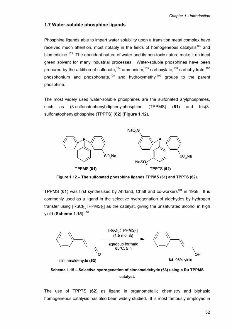
The most widely used water-soluble phosphines are the sulfonated arylphosphines,
such as (3-sulfonatophenyl)diphenylphosphine (TPPMS) (61) and tris(3-
sulfonatopheny)phosphine (TPPTS) (62) (Figure 1.12).
Figure 1.12 – The sulfonated phosphine ligands TPPMS (61) and TPPTS (62).
TPPMS (61) was first synthesised by Ahrland, Chatt and co-workers104 in 1958. It is
commonly used as a ligand in the selective hydrogenation of aldehydes by hydrogen
transfer using [RuCl2(TPPMS)2] as the catalyst, giving the unsaturated alcohol in high
yield (Scheme 1.15).110
Scheme 1.15 – Selective hydrogenation of cinnamaldehyde (63) using a Ru TPPMS
catalyst.
The use of TPPTS (62) as ligand in organometallic chemistry and biphasic
homogeneous catalysis has also been widely studied. It is most famously employed in

Chapter 1 - Introduction
33
the Ruhrchemie/Rhône-Poulenc oxo process, based on a rhodium catalyst (Scheme
1.16). This process has been commercially used for the hydroformylation of alkenes
since 1984.111
Scheme 1.16 – The Ruhrchemie/Rhône-Poulenc oxo process.
One drawback of sulfonated phosphines is that their syntheses from arylphosphines
and oleum are often cumbersome and hard to accurately reproduce, due to the
formation of phosphine oxides during the reaction. Some authors have also found that
the sulfonate groups are non-innocent and can alter other properties of the ligand and
its complexes. For example, Tyler and co-workers discovered that the sulfonate
groups affected the ability of their iron complexes to bind small molecules such as
dinitrogen.112 However, methods of synthesising sulfonated arylphosphines cleanly
with almost no oxide formation have also been proposed.113, 114
1.7.1 PTA-based phosphine ligands
1,3,5-triaza-7-phosphaadamantane (PTA) (66) is a cage-like monodentate phosphine
ligand based on adamantane, first prepared by Daigle et al. in 1974.115 It has received
much attention in the field of coordination chemistry and organometallic catalysis,
largely due to its air stability and solubility in water. Its water solubility is a result of
hydrogen bonding of the nitrogen atoms to water.116 It can be synthesised by reaction
of 1,3,5,7-tetraazaadamantane with tris(hydroxymethyl)phosphine (THMP) (65) (route
(a) in Scheme 1.17).115 Alternatively, it can be made by reacting THMP (65) with a
solution of ammonia and formaldehyde (route (b) in Scheme 1.17).115

Chapter 1 - Introduction
34
Scheme 1.17 – Syntheses of 1,3,5-triaza-7-phosphaadamantane (PTA) (66) from
tris(hydroxymethyl)phosphine (THMP) (65).
PTA (66) has excellent donating ability (similar to PMe3) and has a relatively small cone
angle of 102°.117 Additionally, it is thermally stable up to 260°C.118 In acidic media 66
is protonated to give [PTA(H)]+, which shows greater solubility. For these reasons,
PTA complexes of rhodium and ruthenium have been the subject of much research,
including both monophasic119, 120 and biphasic121-123 hydrogenation of alkenes and
aldehydes. Tang et al.124 have even shown that PTA itself is effective as an
organocatalyst in the Morita-Baylis-Hillman reactions of certain activated alkenes with
electrophiles such as aldehydes and imines.
Additionally, 66 can also be readily modified to give other similar P-CH2-N
heterocycles. Most modifications to date have focused on reactions of the ‘lower rim’
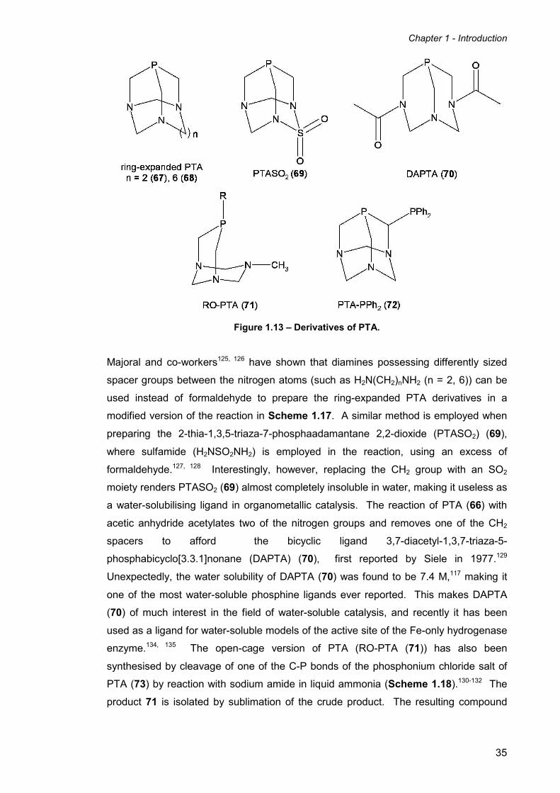
of the ligand, the triazacyclohexane ring. Examples include ring-expanded variants of
PTA (67, 68),125, 126 the sulfone derivative PTASO2 (69),127, 128 the diacetyl DAPTA
(70),117, 129 and a ring-opened version (RO-PTA) (71) (Figure 1.13).130-132 Only one
example of a modification of the ‘upper rim’ of PTA has been reported to date, the
bidentate phosphine PTA-PPh2 (72) (Figure 1.13).133
Chapter 1 - Introduction
35
Figure 1.13 – Derivatives of PTA.
Majoral and co-workers125, 126 have shown that diamines possessing differently sized
spacer groups between the nitrogen atoms (such as H2N(CH2)nNH2 (n = 2, 6)) can be
used instead of formaldehyde to prepare the ring-expanded PTA derivatives in a
modified version of the reaction in Scheme 1.17. A similar method is employed when
preparing the 2-thia-1,3,5-triaza-7-phosphaadamantane 2,2-dioxide (PTASO2) (69),
where sulfamide (H2NSO2NH2) is employed in the reaction, using an excess of
formaldehyde.127, 128 Interestingly, however, replacing the CH2 group with an SO2
moiety renders PTASO2 (69) almost completely insoluble in water, making it useless as
a water-solubilising ligand in organometallic catalysis. The reaction of PTA (66) with
acetic anhydride acetylates two of the nitrogen groups and removes one of the CH2
spacers to afford the bicyclic ligand 3,7-diacetyl-1,3,7-triaza-5-
phosphabicyclo[3.3.1]nonane (DAPTA) (70), first reported by Siele in 1977.129
Unexpectedly, the water solubility of DAPTA (70) was found to be 7.4 M,117 making it
one of the most water-soluble phosphine ligands ever reported. This makes DAPTA
(70) of much interest in the field of water-soluble catalysis, and recently it has been
used as a ligand for water-soluble models of the active site of the Fe-only hydrogenase
enzyme.134, 135 The open-cage version of PTA (RO-PTA (71)) has also been
synthesised by cleavage of one of the C-P bonds of the phosphonium chloride salt of
PTA (73) by reaction with sodium amide in liquid ammonia (Scheme 1.18).130-132 The
product 71 is isolated by sublimation of the crude product. The resulting compound

Chapter 1 - Introduction
36
acts as a P, N bidentate ligand, and complexes of gold and molybdenum have been
isolated and characterised.130-132
Scheme 1.18 – Synthesis of RO-PTA (71).
The only P, P bidentate derivative of PTA is the diphenylphosphine-modified PTA-PPh2
(72).133 It is synthesised by the selective lithiation of one of the CH2 groups of the
‘upper rim’ of PTA, followed by reaction of the lithium salt (PTA-Li) (74) with
chlorodiphenylphosphine (Scheme 1.19). Coordination complexes of PTA-PPh2 (72)
with both molybdenum and tungsten have been reported, and in both cases the ligand
acts as a bidentate phosphine donor, creating a four-membered chelate ring between
the ligand and metal.133
Scheme 1.19 – Synthesis of PTA-PPh2 (72).
1.7.2 Hydroxyalkylphosphine Ligands
Another alternative to using sulfonated aryl phosphines as water-solubilising ligands is
to use hydroxyalkyl phosphines. The most simple hydroxyalkyl phosphine is
tris(hydroxymethyl)phosphine (THMP) (65), which is moderately air stable and
unusually for trialkylphosphines is a crystalline solid at room temperature (mp = 58
°C).136 It can be synthesised by reacting PH3 gas with formaldehyde in the presence of
catalytic amounts of K2[PtCl4] in aqueous solution, which is efficient but requires the
handling of extremely toxic gaseous PH3 in large amounts (Reaction 1, Scheme

Chapter 1 - Introduction
37
1.20).137 An alternative method of synthesising THMP (65) without the use of PH3 is to
react the commercially available [P(CH2OH)4]Cl (THPC) (73) with base (Reaction 2,
Scheme 1.20).138
Scheme 1.20 – Syntheses of tris(hydroxy)methyl phosphine (THMP) (65).
In coordination chemistry THMP (65) acts as a monodentate ligand much like the
similar trialkylphosphines. The cis-dihalo platinum(II) and palladium(II) complexes of
the formula [MX2{P(CH2OH)3}2] can simply and reliably be prepared from the
corresponding [MX2(cod)] salts.136 These same dichloro complexes have also
previously been reported by Chatt et al.109 using the appropriate [MCl4]2- salt (Pt or Pd),
though this route has been reported by Ellis et al.136 to give impure products and poor
yields. Some ruthenium complexes of 65 have also been synthesised which have
been shown to catalyse the hydrogenation of sorbic acid.139 The sorbic acid is reduced
in the presence of [Ru(Cp*)(Cl)(CO){P(CH2OH)3}], with a TOF of 2.6 h-1 giving a 40%
yield of 3-hexenoic acid (80% cis) in 15h. These results are better than the equivalent
TPPTS based catalyst, though they are not exceptional.102 Other THMP complexes of
iridium and rhodium have also been shown to catalyse the hydrogenation of
cinnamaldehyde and the hydroformylation of 1-pentene respectively.140 Higham et al.
have also synthesised ruthenium and rhodium complexes of a range of monodentate
hydroxymethyl phosphines including THMP, bis(hydroxymethyl)phenylphosphine, and
tris(hydroxypropyl)phosphine.141, 142
Using hydroxymethyl groups to impart solubility upon phosphines is an idea that has
also been extended to diphosphines, such as HMPB (57) and HMPE (58), first reported
by Katti and co-workers in 1996.143 They investigated the coordination chemistry of
these ligands with palladium and platinum for potential use in organometallic

Chapter 1 - Introduction
38
catalysis,144 as well as 198Au,145 and Re (75-78) for potential use as water-soluble
complexes for radiopharmaceuticals in nuclear medicine (Scheme 1.21).143, 146
Scheme 1.21 – Synthesis of rhenium HMPB and HMPE complexes.
Hydroxyalkyldiphosphines with longer-chain hydroxyalkyl groups were first produced by
Tyler and co-workers.147 Ligands based on HMPE containing (CH2)xOH groups where
x = 3, 4, 5 where prepared and their coordination chemistry investigated with Ni(II),
Ru(III), and Rh(I). More recently, they have synthesised a range of water-soluble
iron(II) complexes (79a-e and 80a-e) based on these longer-chained hydroxyalkyl
ligands and their related ester variants for potential use as dinitrogen and dihydrogen
binding agents (Scheme 1.22).112, 148, 149
Scheme 1.22 – Synthesis of water-soluble long-chained hydroxyalkyl and methyl ester
bisphosphine iron(II) complexes (79a-e and 80a-e).

Chapter 1 - Introduction
39
Iron(II) chloride and iron(II) sulfate salts were reacted with two equivalents of
diphosphine in either methanol or ethanol to give the cis-Fe(II) bis(diphosphine)
sulfates 79a-e and the trans-Fe(II) bis(diphosphine) dichlorides 80a-e respectively.
The trans-dichlorides 80a-e have been shown to be capable of binding small molecules
such as dinitrogen, carbon monoxide and acetonitrile in the presence of sodium
tetraphenylborate in alcoholic solvents (Scheme 1.23).112 However, the dinitrogen
complexes (81c,e) only forms under 50 psi of dinitrogen gas.
Scheme 1.23 – Coordination chemistry of small molecules with trans-[FeL2Cl2]
complexes 80a-e in alcoholic solvents.
In aqueous solutions however, the trans-dichloride complexes 80a-e first react with the
solvent to form complexes of the general formula trans-[FeL2(H2O)(Cl)]+ (where L is a
water-soluble chelating diphosphine ligand). Subsequent exposure to dinitrogen or
dihydrogen affords the complexes trans-[FeL2(N2)(Cl)]+ and [FeL2(H2)(Cl)]
+ (Reaction 1,
Scheme 1.24).
Scheme 1.24 – Coordination chemistry of N2 and H2 with trans-[FeL2Cl2] complexes in
aqueous solution.
In the case of the dihydrogen complex 85e, the coordinated H2 molecule undergoes
heterolysis and loses a proton to ultimately give the trans-[FeL2(H)(H2)]+ product 87e
Chapter 1 - Introduction
40
(Reaction 2, Scheme 1.24).148, 149 Spectroscopic studies show that following the loss
of the first chloride and replacement with dihydrogen, heterolysis of the H2 occurs to
initially form trans-[FeL2(H)(Cl)] (86e). The second chloride is then replaced by a
second molecule of H2 to give the trans-[FeL2(H)(H2)]+ product (87e). When exposed
to an atmosphere of dinitrogen, the H2 molecule is substituted by N2 to give the trans-
[FeL2(H)(N2)]+ product. Water soluble complexes with coordinated H2 and N2 ligands
are quite rare in the literature, and ultimately show promise as water-soluble gas
scrubbers to separate dinitrogen and dihydrogen from natural gas streams. Solubility
of the complexes in polar solvents is especially important for gas scrubbing
applications due to the need to minimise the solubility of the natural gas in the carrier
solvent to maximise the separation.
1.8 A novel self-assembled iron(II) hydroxymethylphosphine complex
The phosphonium salt tetrakis(hydroxymethyl)phosphonium sulfate [P(CH2OH)4]SO4
(THPS) is used as a biocide in oil wells. It was noticed by oil well engineers that THPS
aids the dissolution of iron sulfide particulates and deposits which are a result of
anaerobic microbial activity and indigenous H2S. It is necessary to remove these
deposits as they can lead to blockage and restrictions with the pipework and upset the
oil/water separation, both of which are undesirable and lead to damage. It was also
discovered that the dissolution of the FeS particulates is accompanied by a red
colouration of the water in the well. Recently, researchers at Albright and Wilson (now
part of Rhodia plc) undertook studies to explain how THPS helps to dissolve iron
sulfide deposits and also to determine the origin of the red colouration observed which
was believed to be due to a water-soluble iron complex.
The reactions of THPS and its related phosphine THMP were performed with a variety
of Fe(II) and Fe(III) salts (e.g. FeS, FeCl2, FeCl3, FeSO4, [Fe(NH4)2(SO4)2]) in order to
simulate the oil well conditions within the laboratory.150 The development of
coordination chemistry of water-soluble phosphines with relatively cheap metals such
as iron is also an attractive prospect due to the financial and environmental benefits
they offer. They found that only the ferrous ammonium sulfate afforded a red
colouration of the aqueous solution when reacted with THPS and THMP. Other
hydroxyalkylphosphonium salts formed no red complexes at all. Crucially however, it
was discoved that the presence of ammonium cations (naturally present in oil well
waters and also as a result of the addition of ammonium bisulfite as an oxygen
scavenger), an iron(II) salt, and THPS or THMP are all of paramount importance to the
formation of the red complex. Following optimisation of the reaction conditions, it was

Chapter 1 - Introduction
41
discovered that two equivalents of THPS (i.e. four equivalents of phosphonium ions)
added to ferrous ammonium sulfate (Mohr’s salt) in aqueous solution at room
temperature, followed by the slow addition of a base (i.e. NaOH) whilst maintaining the
pH between 4.5 and 5 leads to a deep red solution. Deep red crystals suitable for
single crystal X-ray diffraction analysis were isolated when the solution was left at 5°C
for two weeks.
Figure 1.14 – Structure of the Fe(II) phosphine complex 88 and its self assembled ligand
L1.
The molecular structure of 88 shows that the red complex is a dicationic pseudo
octahedral Fe(II) complex with an interesting self-assembled 1,5-diaza-3,7-
diphosphacyclooctane based hydroxymethyl ligand (L1) (Figure 1.14). The ligand acts
as a chelating bidentate ligand with the two ring-phosphorus atoms coordinating to the
metal in a cis equatorial fashion. The two nitrogen atoms of the cyclic ring possess
pendant CH2P(CH2OH)2 groups which each coordinate to the metal via the trans axial
sites. The two remaining equatorial sites are occupied by two water molecules. It is
not known the exact mechanism by which the complex is formed but is believed to
involve a Mannich-like condensation of a [P(CH2OH)4]+ moiety with two ammonium
ions, the whole process being templated by the metal to form the eight-membered ring
and its pendant arms.
1.9 Concluding Remarks
Phosphorus-based ligands represent an extremely important class of ligand in the
fields of organometallic and transition metal coordination chemistry, due to the wide
range of steric and electronic properties that can be altered by changing the
substituents on phosphorus. The study of phosphine ligands in particular has received

Chapter 1 - Introduction
42
much attention due to their ability to stabilise metals in their lowest oxidation states by
accepting π electron density from the metal as well as potential applications as ligands
in transition metal catalysis. 31P{1H} NMR spectroscopy as a characterisation
technique is also useful and convenient for monitoring reactions and identifying
products containing phosphorus nuclei.
The incorporation of multiple phosphine donor atoms into macrocyclic rings has
important implications in coordination chemistry, offering greater stability in terms of
both chelating ability and the additional stability offered by the macrocyclic effect.
Phosphorus donors incorporated into smaller heterocyclic rings show much promise
within the field of coordination chemistry and have been shown to be useful ligands in
coordinating to a wide range of transition metals.
There is increasing interest in performing chemistry in aqueous media for applications
in biomedicine, biphasic catalysis, and natural gas scrubbing. Chemistry in water is
also more financially and environmentally friendly as it does not generate large
amounts of expensive organic solvent waste or consume large amounts of energy
needed to produce them. Water-solublising phosphine ligands such as sulfonated
phosphines, PTA based ligands, and hydroxyalkylphosphines in particular are receiving
much attention in this field.
Chapter 2 of this thesis investigates the coordination chemistry of the water-soluble
Fe(II) cyclic hydroxymethylphosphine complex 88 with a range of neutral and ionic
ligands, and examines the reactivity of the pendant hydroxymethyl groups on the
coordinated ligand L1 to give other functionalised derivative complexes.
Chapter 3 extends the self-assembly process by altering the substrates involved in the
formation of 88, and determines whether related complexes can be formed and
isolated using this method. The formation of an iron(II) complex containing a new
bicyclic bidentate phosphine ligand is described, and its coordination chemistry with a
range of ligands investigated.
Chapter 4 is the experimental section and describes experimental techniques and
procedures used in the synthesis and investigation of the compounds described in
Chapters 2 & 3.

Chapter 1 - Introduction
43
1.10 References
1. P. Thenard, C.R. Hebd. Seances Acad. Sci., Ser. C, 1847, 25, 892.
2. A. Michaelis and H. V. Soden, Annalen, 1885, 229, 295.
3. J. Chatt and F. G. Mann, J. Chem. Soc., 1938, 1949.
4. C. A. McAuliffe, in "Comprehensive Coordination Chemistry", Eds. G.
Wilkinson, R. D. Gillard and J. A. McCleverty, Pergamon Press, 1987, vol. 2, p.
990.
5. J. H. Downing and M. B. Smith, in "Comprehensive Coordination Chemistry II",
eds. J. A. McCleverty and T. J. Meyer, Elsevier Pergamon, 2004, vol. 1, p. 253.
6. C. A. Tolman, J. Am. Chem. Soc., 1970, 92, 2956.
7. C. A. Tolman, Chem. Rev., 1977, 77, 313.
8. E. C. Alyea, S. A. Dias, G. Ferguson and R. J. Restivo, Inorg. Chem., 1977, 16,
2329.
9. G. Ferguson, P. J. Roberts, E. C. Alyea and M. Khan, Inorg. Chem., 1978, 17,
2965.
10. T. L. Brown, Inorg. Chem., 1992, 31, 1286.
11. T. L. Brown and K. J. Lee, Coord. Chem. Rev., 1993, 128, 89.
12. D. White, B. C. Taverner, P. G. L. Leach and N. J. Coville, J. Comput. Chem.,
1993, 14, 1042.
13. P. W. N. M. van Leeuwen, P. J. C. Kamer, J. N. H. Reek and P. Dierkes, Chem.
Rev., 2000, 100, 2741.
14. C. A. Tolman, W. C. Seidel and L. W. Gosser, J. Am. Chem. Soc., 1973, 96, 53.
15. P. Dierkes and P. W. N. M. van Leeuwen, J. Chem. Soc., Dalton Trans., 1999,
1519.
16. M. S. Davies, M. J. Aroney, I. E. Buys, T. W. Hambley and J. L. Calvert, Inorg.
Chem., 1995, 34, 330.
17. M. J. S. Dewar, Bull. Soc. Chim. Fr., 1951, 18, C71.
18. J. Chatt and L. A. Duncanson, J. Chem. Soc., 1953, 1939.
19. S.-X. Xiao, W. C. Trogler, D. E. Ellis and Z. Berkovitch-Yellin, J. Am. Chem.
Soc., 1983, 105, 7033.
20. A. G. Orpen and N. G. Connelly, Organometallics, 1990, 9, 1206.
21. G. Pacchioni and P. S. Bagus, Inorg. Chem., 1992, 31, 4391.
22. M. N. Golovin, M. M. Rahman, J. E. Belmonte and W. P. Giering,
Organometallics, 1985, 4, 1981.
23. M. M. Rahman, H.-Y. Liu, A. Prock and W. P. Giering, Organometallics, 1987,
6, 650.
24. R. J. Angelici and M. D. Malone, Inorg. Chem., 1967, 6, 1731.

Chapter 1 - Introduction
44
25. M. M. Rahman, H.-Y. Liu, K. Eriks, A. Prock and W. P. Giering,
Organometallics, 1989, 8, 1.
26. T. Leyssens, D. Peeters, A. G. Orpen and J. N. Harvey, New J. Chem., 2005,
29, 1424.
27. T. Leyssens, D. Peeters, A. G. Orpen and J. N. Harvey, Organometallics, 2007,
26, 2637.
28. C. A. McAuliffe, in "Comprehensive Coordination Chemsitry", Eds. G.
Wilkinson, R. D. Gillard and J. A. McCleverty, Pergamon Press, 1987, vol. 2, p.
1030.
29. R. G. Goel and T. Allman, Can. J. Chem., 1982, 60, 716.
30. P. B. Dias, M. E. M. d. Piedade and J. A. M. Simoes, Coord. Chem. Rev., 1994,
135-136, 737.
31. R. J. Angelici, Acc. Chem. Res., 1995, 28, 51.
32. J. G. Verkade and J. A. Mosbo, in "Phosphorus-31 NMR Spectroscopy in
Stereochemical Analysis", Eds. J. G. Verkade and L. D. Quin, VCH, 1987, p.
425.
33. R. J. Puddephat, G. M. Bancroft and T. Chan, Inorg. Chim. Acta., 1983, 73, 83.
34. K. Bowman-James, in "Encyclopedia of Inorganic Chemistry", Ed. R. B. King,
Wiley, New York, 2005.
35. E. C. Constable, Coordination Chemistry of Macrocyclic Compounds, Oxford
University Press, 1999.
36. J. V. Alphen, Recl. Trav. Chim. Pays-Bas, 1936, 55, 835.
37. M. C. Thompson and D. H. Busch, J. Am. Chem. Soc., 1962, 84, 1762.
38. N. F. Curtis, J. Chem. Soc., 1960, 4409.
39. C. J. Pedersen, J. Am. Chem. Soc., 1967, 89, 7017.
40. B. Dietrich, J.-M. Lehn and J.-P. Sauvage, Tetrahedron Lett., 1969, 2889.
41. J. Almy, D. C. Garwood and D. J. Cram, J. Am. Chem. Soc., 1973, 95, 2961.
42. A. E. Martell, in "Advances in Chemistry", Oxford University Press, 1967, vol.
62, p. 272.
43. D. K. Cabbiness and D. W. Margerum, J. Am. Chem. Soc., 1969, 91, 6540.
44. L. Fabbrizzi, P. Paoletti and A. B. P. Lever, Inorg. Chem., 1976, 15, 1502.
45. A. Anichini, L. Fabbrizzi, P. Paoletti and R. M. Clay, Inorg. Chim. Acta., 1977,
24, L21.
46. A. Anichini, L. Fabbrizzi, P. Paoletti and R. M. Clay, J. Chem. Soc., Dalton
Trans., 1978, 577.
47. R. M. Clay, M. Micheloni, P. Paoletti and W. V. Steele, J. Am. Chem. Soc.,
1979, 101, 4119.

Chapter 1 - Introduction
45
48. E. Gallori, E. Martini, M. Micheloni and P. Paoletti, Inorg. Chim. Acta., 1980, 40,
X66.
49. M. Micheloni and P. Paoletti, Inorg. Chim. Acta., 1980, 43, 109.
50. H. N. Stokes, Am. Chem. J., 1897, 19, 782.
51. A.-M. Caminade and J. P. Majoral, Chem. Rev., 1994, 94, 1183.
52. R. M. Izatt, K. Pawlak, J. S. Bradshaw and R. L. Bruening, Chem. Rev., 1991,
91, 1721.
53. E. P. Kyba, C. W. Hudson, M. J. McPhaul and A. M. John, J. Am. Chem. Soc.,
1977, 99, 8053.
54. E. P. Kyba and S. S. P. Chou, J. Chem. Soc., Chem. Commun., 1980, 449.
55. E. P. Kyba and S. S. P. Chou, J. Am. Chem. Soc., 1980, 102, 7012.
56. E. P. Kyba and S. S. P. Chou, J. Org. Chem., 1981, 46, 860.
57. E. P. Kyba and S.-Z. Liu, Inorg. Chem., 1985, 24, 1613.
58. E. P. Kyba, C. N. Clubb, S. B. Larson, V. J. Schueler and R. E. Davis, J. Am.
Chem. Soc., 1985, 107, 2141.
59. E. P. Kyba, A. M. John, B. Brown, C. W. Hudson, M. J. McPhaul, A. Harding, K.
Larsen, S. Niedzwiecki and R. E. Davis, J. Am. Chem. Soc., 1980, 102, 189.
60. M. A. Fox, K. A. Campbell and E. P. Kyba, Inorg. Chem., 1981, 20.
61. E. P. Kyba, R. E. Davis, C. W. Hudson, A. M. John, S. B. Brown, M. J.
McPhaul, L.-K. Liu and A. C. Glover, J. Am. Chem. Soc., 1981, 103, 3868.
62. G. Wittig and A. Maercker, Chem. Ber., 1964, 97, 747.
63. M. C. Thompson and D. H. Busch, J. Am. Chem. Soc., 1964, 86, 3651.
64. W. Marty and G. Schwarzenbach, Chimia, 1970, 24, 431.
65. T. A. DelDonno and W. J. Rosen, J. Am. Chem. Soc., 1977, 99, 8051.
66. T. Lebbe, P. Machnitzki, O. Stelzer and W. S. Sheldrick, Tetrahedron, 2000, 56,
157.
67. A. R. Battle, P. G. Edwards, R. Haigh, D. E. Hibbs, D. Li, S. M. Liddiard and P.
D. Newman, Organometallics, 2007, 26, 377.
68. B. N. Diel, R. C. Haltiwanger and A. D. Norman, J. Am. Chem. Soc., 1982, 104,
4700.
69. P. G. Edwards, J. S. Fleming and S. S. Liyanage, Inorg. Chem., 1996, 35,
4563.
70. T. Albers and P. G. Edwards, Chem. Commun., 2007, 858.
71. P. G. Edwards and M. L. Whatton, Dalton Trans., 2006, 442.
72. P. G. Edwards, R. Haigh, D. Li and P. D. Newman, J. Am. Chem. Soc., 2006,
128, 3818.
73. P. G. Edwards, P. D. Newman and D. E. Hibbs, Angew. Chem., Int. Ed., 2000,
39, 2722.

Chapter 1 - Introduction
46
74. P. G. Edwards, P. D. Newman and K. M. A. Malik, Angew. Chem., Int. Ed.,
2000, 39, 2922.
75. R. W. Alder and D. Read, Coord. Chem. Rev., 1998, 176, 113.
76. L. D. Quinn, in "The Heterocyclic Chemistry of Phosphorus", J. Wiley, New
York, 1981.
77. E. H. Wong, Comments Inorg. Chem., 1996, 18, 283.
78. X. Delaigue, J. M. Harrowfield, M. W. Hosseini, A. D. Cian, J. Fischer and N.
Kyritsakas, J. Chem. Soc., Chem. Commun., 1994, 1579.
79. G. Märkl, G. Y. Jin and C. Schoerner, Tetrahedron Lett., 1980, 21, 1409.
80. B. A. Arbuzov and G. N. Nikonov, Adv. Heterocycl. Chem., 1994, 61, 59.
81. J. C. Kane, E. H. Wong, G. P. A. Yap and A. L. Rheingold, Polyhedron, 1999,
18, 1183.
82. A. A. Karasik, R. N. Naumov, O. G. Sinyashin, G. P. Belov, H. V. Novikova, P.
Lönnecke and E. Hey-Hawkins, Dalton Trans., 2003, 2209.
83. F. Z. Baimukhametov, V. F. Zheltukhin, G. N. Nikonov and A. S. Balueva, Zh.
Obshch. Khim, 2002, 72, 1754.
84. A. A. Karasik, Y. S. Spiridonova, D. G. Yakhvarov, O. G. Sinyashin, P.
Lönnecke, R. Sommer and E. Hey-Hawkins, Mendeleev Commun., 2005, 15,
89.
85. A. A. Karasik, R. N. Naumov, R. Sommer, O. G. Sinyashin and E. Hey-
Hawkins, Polyhedron, 2002, 21, 2251.
86. A. A. Karasik, I. O. Georgiev, E. I. Musina, O. G. Sinyashin and J. Heinicke,
Polyhedron, 2001, 20, 3321.
87. J. Heinicke, M. He, A. A. Karasik, I. O. Georgiev, O. G. Sinyashin and P. G.
Jones, Heteroat. Chem., 2005, 16, 379.
88. D. E. Berning, K. V. Katti, C. L. Barnes and W. A. Volkert, J. Am. Chem. Soc.,
1999, 121, 1658.
89. A. A. Karasik, O. G. Sinyashin, J. Heinicke and E. Hey-Hawkins, Phosphorus,
Sulfur, Silicon Relat. Elem., 2002, 177, 1469.
90. A. A. Karasik, A. V. Krashilina, A. T. Gubaidullin, I. A. Litvinov, V. K. Cherkasov,
O. G. Sinyashin and G. A. Abakumov, Russ. Chem. Bull., 2000, 49, 1782.
91. A. A. Karasik, R. N. Naumov, A. S. Balueva, Y. S. Spiridonova, O. N. Golodkov,
H. V. Novikova, G. P. Belov, S. A. Katsyuba, E. E. Vandyukova, P. Lönnecke,
E. Hey-Hawkins and O. G. Sinyashin, Heteroat. Chem., 2006, 17, 499.
92. A. A. Karasik, R. N. Naumov, Y. S. Spiridonova, O. G. Sinyashin, P. Lönnecke
and E. Hey-Hawkins, Z. Anorg. Allg. Chem., 2007, 633, 205.
93. S. V. Bobrov, A. A. Karasik and O. G. Sinyashin, Phosphorus, Sulfur, Silicon
Relat. Elem., 1999, 144-146, 289.

Chapter 1 - Introduction
47
94. E. V. Novikova, A. A. Karasik, E. Hey-Hawkins and G. P. Belov, Russ. J. Coord.
Chem., 2005, 31, 260.
95. A. A. Karasik, S. V. Bobrov, G. N. Nikonov, A. P. Pisarevskii, I. A. Litvinov, A. S.
Dokuchaev, Y. T. Struchkov and K. M. Enikeev, Russ. J. Coord. Chem., 1995,
21, 574.
96. A. S. Balueva, R. M. Kuznetsov, S. N. Ignat'eva, A. A. Karasik, A. T.
Gubaidullin, I. A. Litvinov, O. G. Sinyashin, P. Lönnecke and E. Hey-Hawkins,
Dalton Trans., 2004, 442.
97. D. V. Kulikov, A. A. Karasik, A. S. Balueva, O. N. Kataeva, I. A. Litvinov, E.
Hey-Hawkins and O. G. Sinyashin, Mendeleev Commun., 2007, 17, 195.
98. I. Bauer, O. Rademacher, M. Gruner and W. D. Habicher, Chem. Eur. J., 2000,
6, 3043.
99. R. N. Naumov, A. A. Karasik, O. G. Sinyashin, P. Lönnecke and E. Hey-
Hawkins, Dalton Trans., 2004, 357.
100. K. V. Katti, R. Kannan, K. K. Katti, N. Pillarsetty and C. L. Barnes, Phosphorus,
Sulfur Silicon Relat. Elem., 2002, 177, 1587.
101. B. L. Iverson, S. A. Iverson, V. A. Roberts, E. D. Getzoff, J. A. Tainer, S. J.
Benkovic and R. A. Lerner, Science, 1990, 249, 659.
102. N. Pinault and D. W. Bruce, Coord. Chem. Rev., 2003, 241, 1.
103. K. V. Katti, H. Gali, C. J. Smith and D. E. Berning, Acc. Chem. Res., 1999, 32,
9.
104. S. Ahrland, J. Chatt, N. R. Davies and A. A. Williams, J. Chem. Soc., 1958, 276.
105. R. T. Smith, R. K. Ungar, L. J. Sanderson and M. C. Baird, Organometallics,
1983, 2, 1138.
106. V. Ravindar, H. Schumann, H. Hemling and J. Blum, Inorg. Chim. Acta., 1995,
240, 145.
107. T. N. Mitchell and K. Heesche-Wagner, J. Organomet. Chem., 1992, 436, 43.
108. E. Renaud, R. B. Russell, S. J. Forestier, M. C. Brown and J. Baird, J.
Organomet. Chem., 1991, 419, 403.
109. J. Chatt, G. J. Leigh and R. M. Slade, J. Chem. Soc., Dalton Trans., 1973,
2023.
110. F. Joo and A. Bényei, J. Organomet. Chem., 1989, 363, C19.
111. C. W. Kohlpaintner, R. W. Fischer and B. Cornils, Appl. Catal., A, 2001, 221,
219.
112. W. K. Miller, J. D. Gilbertson, C. Leiva-Paredes, P. R. Bernatis, T. J. R.
Weakley, D. K. Lyon and D. R. Tyler, Inorg. Chem., 2002, 41, 5453.
113. T. Thorpe, S. M. Brown, J. Crosby, S. Fitzjohn, J. P. Muxworthy and J. M. J.
Williams, Tetrahedron Lett., 2000, 41, 4503.

Chapter 1 - Introduction
48
114. H. Gulyás, A. Szöllösy, B. E. Hanson and J. Bakos, Tetrahedron Lett., 2002, 43,
2543.
115. D. J. Daigle, A. B. Pepperman, Jr. and S. L. Vail, J. Heterocycl. Chem., 1974,
17, 407.
116. D. J. Daigle, Inorg. Synth., 1998, 32, 40.
117. D. J. Darensbourg, C. G. Ortiz and J. W. Kamplain, Organometallics, 2004, 23,
1747.
118. A. D. Phillips, L. Gonsalvi, A. Romerosa, F. Vizza and M. Peruzzini, Coord.
Chem. Rev., 2004, 248, 955.
119. D. J. Darensbourg, F. Joó, L. Nádasdi and A. Bényei, J. Organomet. Chem.,
1996, 512, 45.
120. P. Smolenski and F. P. Pruchnik, Appl. Organomet. Chem., 1999, 13, 829.
121. D. J. Darensbourg, F. Joó, M. Kannisto, A. Kathó, J. H. Reibenspies and D. J.
Daigle, Inorg. Chem., 1994, 33, 200.
122. D. J. Darensbourg, F. Joó, M. Kannisto, A. Kathó and J. H. Reibenspies,
Organometallics, 1992, 11, 1990.
123. D. J. Darensbourg, N. W. Stafford, F. Joó and J. H. Reibenspies, J. Organomet.
Chem., 1995, 488, 99.
124. X. Tang, B. Zhang, Z. He, R. Gao and Z. He, Adv. Synth. Catal., 2007, 349,
2007.
125. J. Navech, R. Kraemer and J. P. Majoral, Tetrahedron Lett., 1980, 21, 1449.
126. M. Benhammou, R. Kraemer, H. Germa, J. P. Majoral and J. Navech,
Phosphorus, Sulfur Silicon Relat. Elem., 1982, 14, 105.
127. J. R. Delerno, R. J. Majeste and L. M. Trefonas, J. Heterocycl. Chem., 1976,
13, 757.
128. D. J. Daigle, A. B. Pepperman, Jr. and G. Boudreaux, J. Heterocycl. Chem.,
1974, 11, 1085.
129. V. I. Siele, J. Heterocycl. Chem., 1977, 14, 337.
130. B. Assman, K. Angermaier and H. Schmidbaur, J. Chem. Soc., Chem.
Commun., 1994, 941.
131. B. Assman, K. Angermaier, M. Paul, J. Riede and H. Schmidbaur, Chem. Ber.,
1995, 128, 891.
132. E. Fluck and H. J. Weissgraeber, Chem.-Ztg., 1977, 101, 304.
133. G. W. Wong, J. L. Harkreader, C. A. Mebi and B. J. Frost, Inorg. Chem., 2006,
45, 6748.
134. Z. Wang, J. Liu, C. He, S. Jiang, B. Åkermark and L. Sun, Inorg. Chim. Acta.,
2007, 360, 2411.

Chapter 1 - Introduction
49
135. Y. Na, M. Wang, K. Jin, R. Zhang and L. Sun, J. Organomet. Chem., 2006, 691,
5045.
136. J. W. Ellis, K. N. Harrison, P. A. T. Hoye, A. G. Orpen, P. G. Pringle and M. B.
Smith, Inorg. Chem., 1992, 31, 3026.
137. M. Reuter, L. Orthner, Ger. Pat., 1 035 135, 1958.
138. K. A. Petrov, V. A. Parshina and M. B. Luzanova, Zh. Obshch. Khim, 1962, 32,
553.
139. B. Drießen-Hölscher and J. Heinen, J. Organomet. Chem., 1998, 570, 141.
140. A. Fukuoka, W. Kosugi, F. Morishita, M. Hirano, L. McCaffrey, W. Henderson
and S. Komiya, Chem. Commun., 1999, 489.
141. L. J. Higham, M. K. Whittlesey and P. T. Wood, Dalton Trans., 2004, 4202.
142. L. Higham, A. K. Powell, M. K. Whittlesey, S. Wocadlo and P. T. Wood, Chem.
Commun., 1998, 1107.
143. V. S. Reddy, D. E. Berning, K. V. Katti, C. L. Barnes, W. A. Volkert and A. R.
Ketring, Inorg. Chem., 1996, 35, 1753.
144. V. S. Reddy, K. V. Katti and C. L. Barnes, J. Chem. Soc., Dalton Trans., 1996,
1301.
145. D. E. Berning, K. V. Katti, W. A. Volkert, C. J. Higginbotham and A. R. Ketring,
J. Nucl. Med., 1998, 25, 577.
146. R. Schibli, K. V. Katti, W. A. Volkert and C. L. Barnes, Inorg. Chem., 2001, 40,
2358.
147. G. T. Baxley, W. K. Miller, D. K. Lyon, B. E. Miller, G. F. Nieckarz, T. J. R.
Weakley and D. R. Tyler, Inorg. Chem., 1996, 35, 6688.
148. J. D. Gilbertson, N. K. Szymczak and D. R. Tyler, Inorg. Chem., 2004, 43, 3341.
149. J. D. Gilbertson, N. K. Szymczak, J. L. Crossland, W. K. Miller, D. K. Lyon, B.
M. Foxman, J. Davis and D. R. Tyler, Inorg. Chem., 2007, 46, 1205.
150. J. C. Jeffery, B. Odell, N. Stevens and R. E. Talbot, Chem. Commun., 2000,
101.

Chapter 2 - Coordination chemistry and derivatisation reactions of an iron(II) phosphine complex
50
Chapter 2 – Coordination chemistry and derivatisation
reactions of a novel water-soluble iron(II) phosphine
complex
2.1 Introduction: A novel water-soluble iron(II) macrocyclic phosphine
complex
There is increasing interest in performing chemistry in aqueous media for applications
in biomedicine, biphasic catalysis, and natural gas scrubbing. Chemistry in water is
also more financially and environmentally friendly as it does not generate large
amounts of expensive organic solvent waste, or even consume large amounts of
energy needed to produce the solvents themselves. Water-solubilising phosphine
ligands such as hydroxyalkylphosphines in particular are receiving attention in this field,
mainly due to their catalytic potential and also due to the convenient nature of the 31P{1H} NMR spectroscopic handle that the 31P nucleus provides. The simplest
hydroxyalkylphosphine is tris(hydroxymethyl)phosphine P(CH2OH)3 (THMP), and this
has been used to create water-soluble complexes of metals from groups 7 to 11.1-16
These include complexes of platinum, palladium and nickel,1-5 gold,6, 7 rhenium,8
ruthenium,9, 10, 13, 15, 16 iridium and rhodium,12 and copper.14
The development of coordination chemistry of water-soluble phosphines with relatively
cheap metals such as iron is also an attractive prospect, mainly due to the financial and
environmental benefits that the metals themselves offer. The discovery of the water-
soluble iron(II) hydroxymethylphosphine complex 1, containing the self-assembled
tetradentate cyclic ligand L1 (Figure 2.1), is interesting due to the novel templated
synthesis in which it is formed.
Figure 2.1 – Structure of the Fe(II) phosphine complex 1 and its self-assembled ligand L1.

Chapter 2 - Coordination chemistry and derivatisation reactions of an iron(II) phosphine complex
51
Due to the conformation that ligand L1 imposes on complex 1, two equatorial cis sites
are occupied by solvent water molecules, which should be readily replaced by other
ligands. This cis conformation is ubiquitous in catalysis, as two adjacent sites are often
required for the binding of a substrate (such as an alkene) to the metal before its
subsequent transformation and separation from the metal centre. Therefore 1 shows
promise as a potential catalyst precursor.
Despite the obvious potential of complex 1 as a water-soluble catalyst of Fe(II), and as
a natural gas scrubbing agent by reversibly binding small molecules such as N2, no
extensive studies have been carried out on this system in the literature. Because of
the interest in water-soluble transition metal complexes and the lack of studies carried
out with this system, it was decided to study the coordination chemistry of 1. It was
also decided to investigate the reactivity of the ligand itself and to determine the
possibility of decomplexing the ligand from the metal and isolating it.
2.2 Synthesis of [Fe(L1)(H2O)2]SO4 (1)
Complex 1 was synthesised according to a literature procedure.17 The synthesis can
be carried out using two equivalents of tris(hydroxymethyl)phosphine (THPS) with one
of iron(II) ammonium sulfate as in the literature method (reaction 1, Scheme 2.1), or
using the separate iron(II) and ammonium sulfates (reaction 2, Scheme 2.1). The two
methods work in the same way, and once the reagents have been dissolved there is
essentially no difference between them, as both solutions will contain a mixture of
[P(CH2OH)4]+, [Fe(H2O)6]
2+, NH4+, and SO4
2- ions in the same stoichiometry (4:1:2:4
ratio respectively).
The THPS was added as a 50% aqueous solution. Aqueous sodium hydroxide
solution (1 M) was then added dropwise to the reaction mixture, taking care to keep the
pH of the solution below 5. The slow addition of base converts the THPS into THMP
and formaldehyde, which initiates the reaction. This gave a deep red solution of 1.
The reaction was complete when the pH stabilised at approximately 5, at which point
most of the THPS had been used up.

Chapter 2 - Coordination chemistry and derivatisation reactions of an iron(II) phosphine complex
52
Scheme 2.1 – Formation of [Fe(L1)(H2O)2]SO4 (1).
The deep red solution was then left at 5°C for ca. one week yielding a deep red
microcrystalline solid in approximately 10% yield. When a two-fold excess of THPS
was used, the yield (based on Fe2+ ions) increased to approximately 20% of solid. This
is most likely due to the decreased solubility of the final product in the more viscous
reaction solution.
The 31P{1H} NMR spectrum of the isolated low-spin d6 product 1 in D2O (Figure 2.2)
showed the expected characteristic triplet resonances at δ 20.1 and -1.5 due to the two
pairs of chemically inequivalent phosphines (axial and equatorial).
-10-530 25 20 15 10 5 0 ppm Figure 2.2 – 31P{1H} NMR spectrum of [Fe(L1)(H2O)2]SO4 (1) in D2O at 122 MHz.

Chapter 2 - Coordination chemistry and derivatisation reactions of an iron(II) phosphine complex
53
The 1H NMR spectrum of 1 is more complex, but can be assigned (Figure 2.3). There
are four groups of chemically inequivalent CH2 protons in an 8:8:4:4 ratio; eight CH2
protons on the four axial pendant CH2OH arms (♦), four on the two equatorial pendant
arms (●), four on the two CH2 bridges between the axial phosphorus atoms and the
nitrogens (■), and eight on the four CH2 groups in the eight-membered ring (▲). It may
be expected that the exo- and endo-protons in the eight-membered ring are chemically
inequivalent due to the imposed rigidity of the complexed ligand, as found in other
similar complexed cyclic ligands.18 This is indeed the case and the protons on the CH2
groups within the eight-membered (▲) ring give rise to an AB system at δ 4.45. The
two groups of pendant CH2OH protons give rise to broad singlets at δ 3.52 (●) and 3.00
(♦) due to their ability to rotate freely about the P-C bond. However, the CH2 protons
linking the pendant phosphines to the eight-membered ring (■) must be structurally
rigid due to their inclusion into a six-membered ring formed as a result of the chelation
of the ligand to the metal. Despite this rigidity, these protons give rise to a sharp
singlet at δ 4.23 due to their chemical equivalence.
3.03.13.23.33.43.53.63.73.83.94.04.14.24.34.44.5 ppm Figure 2.3 – 1H NMR Spectrum of [Fe(L1)(H2O)2]SO4 (1) in D2O at 300 MHz.
31P – 1H HMQC spectroscopy indicated that the triplet resonance at δ 20.1 in the 31P{1H} NMR spectrum correlates to the AB resonance at δ 4.45 in the 1H NMR
spectrum assigned as the methylene protons in the eight-membered ring. This
suggests that the resonance at δ 20.1 in the 31P{1H} NMR spectrum is the equatorial
phosphine resonance, due to the close proximity of these nuclei to the methylene
N
P
N
P
PP
OH
OH
OH
OHOH
OH
▲
■
●
♦
▲
▲
▲
▲
■ ■
●
●
♦
♦
♦
♦

Chapter 2 - Coordination chemistry and derivatisation reactions of an iron(II) phosphine complex
54
groups in the eight-membered ring. The 31P – 1H HMQC also showed that the triplet
resonance at δ 20.1 in the 31P{1H} NMR spectrum correlates to the peak at δ 4.23 in the 1H NMR spectrum assigned as the methylene protons linking the pendant phosphines
to the eight-membered ring. This helps to confirm that the resonance at δ -1.5 in the 31P{1H} NMR spectrum is the axial phosphine due to the close proximity to the linking
methylene groups. No 2JP-H coupling of any of the pendant methylene protons to the
phosphines was observed.
-20-15-10-520 15 10 5 0 ppm Figure 2.4 – 31P{1H} NMR spectrum of the self-assembly reaction solution before isolation
of [Fe(L1)(H2O)2]SO4 (1) in D2O at 122 MHz.
The 31P{1H} NMR spectrum of the crude reaction mixture (Figure 2.4) showed a large
number of peaks present suggesting that there are at least four species present in
solution. Jeffery et al.17 observed the formation of a second pair of triplets at -12.0 and
-22.8 ppm with the related reduction in intensity of the resonances associated with 1
when excess base was added. This can be associated with the conversion of 1 to a
new complex, tentatively assigned by Jeffery et al.17 as either the neutral hydroxy
complex 2a, or the oxy-bridged species 2b, which forms due to the base induced
deprotonation of the H2O molecules coordinated to Fe in 1 (Scheme 2.2).

Chapter 2 - Coordination chemistry and derivatisation reactions of an iron(II) phosphine complex
55
Scheme 2.2 – Suggested reaction of 1 with base.
The triplet-like resonances at δ -12.0 and -22.8 (Figure 2.4) were observed as well as
others at δ -5.7, -14.9, and -20.8, though these resonances all appeared to be second-
order and more like doublets of doublets than triplets, in which case they would not
correlate exactly with the structures of 2a or 2b. These other species did not crystallise
from solution and were therefore not possible to characterise them unambiguously.
The addition of two equivalents of a sodium hydroxide solution to an aqueous solution
of 1 gave rise to two triplet resonances at δ 22.8 and 8.6 in the 31P{1H} NMR spectrum.
Other peaks were also visible, which are potentially intermediates and side-products
showing multiple species in solution. When triethylamine was used instead of sodium
hydroxide, a very similar spectrum was observed. Alternatively, when Na4EDTA was
used as the base, the spectrum was much cleaner. 31P{1H} NMR spectroscopy
showed the same triplet resonances at δ 22.8 and 8.6, as well as a broad peak at ca. δ
21 and a related broad triplet at δ 3.8 which were also observed in the reaction with
NaOH.
Kinetic studies were undertaken to determine the relationship of these resonances
(Figure 2.5). The kinetic studies show that when one equivalent of EDTA4- was added
(essentially four equivalents of base due to the four carboxylate groups) the broad peak
and broad triplet at ca. δ 21 and 3.8, respectively, formed very quickly. These peaks
slowly converted over time into the two much sharper triplet resonances at δ 22.8 and
8.6. If a larger excess of Na4EDTA was added the reaction proceeded faster. It is
unlikely that either of these two species are complexes of EDTA, as both are clearly
observed in the similar reaction with NaOH or NEt3 as the base. The kinetic studies
were carried out using EDTA4- instead of NaOH or NEt3 due to the simplicity of the
spectra, with many other unidentifiable peaks observed with these latter two bases,
presumably as a result of side reactions and/or decomposition.

Chapter 2 - Coordination chemistry and derivatisation reactions of an iron(II) phosphine complex
56
456789101112131415161718192021222324 ppm
(a)
(b)
(c)
(d)
(e)
(f)
Figure 2.5 – Kinetic studies of the reaction of [Fe(L1)(H2O)2]SO4 (1) with 1 equiv. Na4EDTA
in D2O at 162 MHz using 31P{1H} NMR spectroscopy after: (a) 5 mins, (b) 1 hr, (c) 2 hrs, (d)
3 hrs, (e) 6 hrs, and (f) 12 hrs.
The structural identity of the two species formed in the study cannot be easily
unambiguously identified. Single crystals could not be grown despite numerous
attempts and a pure sample of neither species could be isolated. One possible
interpretation is that the two triplets at δ 22.8 and 8.6 represent the neutral bis(hydroxy)
complex [Fe(L1)(OH)2] 2a, with the broad resonances potentially belonging to the
intermediate mono(hydroxy), mono(aqua) cation [Fe(L1)(OH)(H2O)]+. However, the
31P{1H} NMR spectrum of the [Fe(L1)(OH)(H2O)]+ cation would be expected to consist
of two doublets of triplets and a doublet of doublets, rather than the two observed
broad triplets. If the hydroxyl ligand was exchanging with the H2O solvent molecules,
the observed broad resonance pattern maybe expected. A similar resonance pattern is
observed in the reaction of 1 with sodium chloride in section 2.3, the broad peaks likely
being a result of the coordinated aqua groups exchanging with chloride anions.
Interestingly, when pyridine was used as the base, not only was the reaction fairly
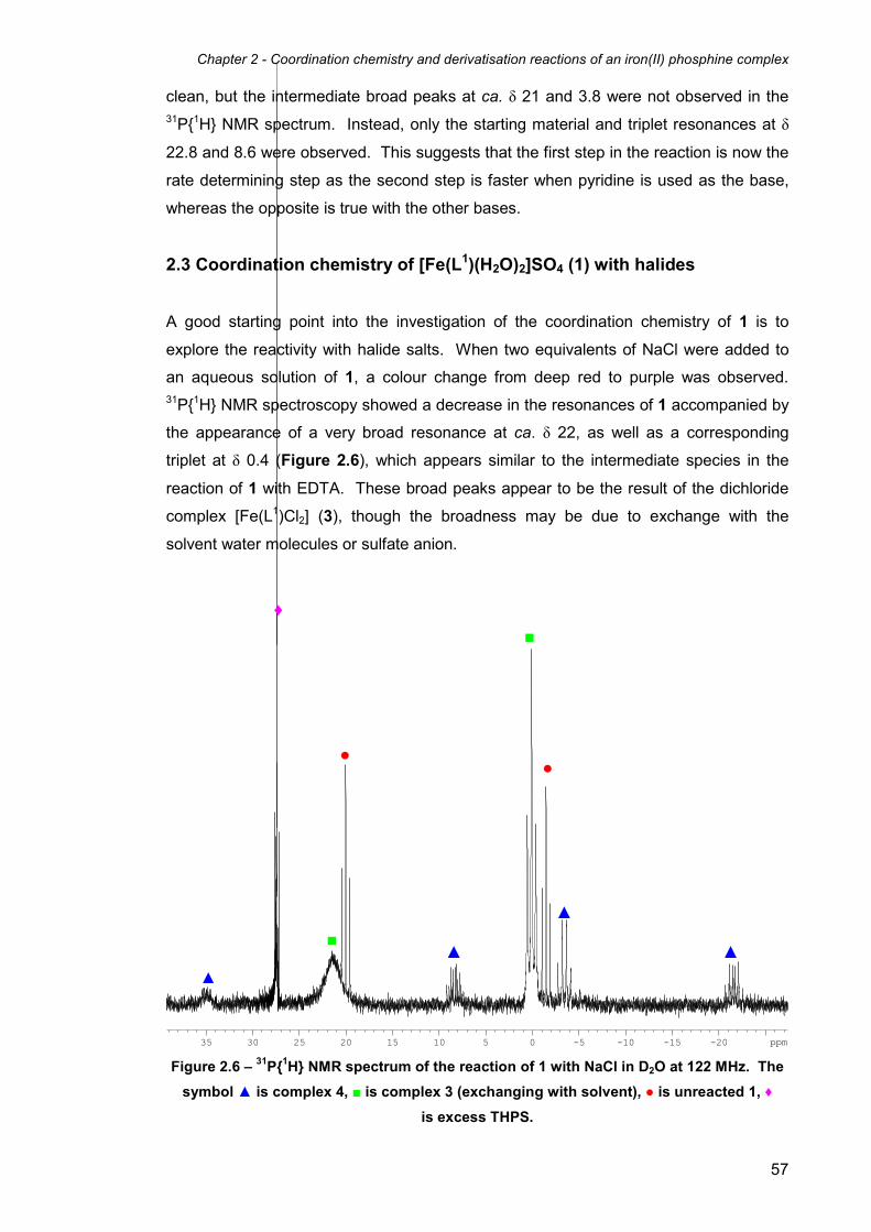
Chapter 2 - Coordination chemistry and derivatisation reactions of an iron(II) phosphine complex
57
clean, but the intermediate broad peaks at ca. δ 21 and 3.8 were not observed in the 31P{1H} NMR spectrum. Instead, only the starting material and triplet resonances at δ
22.8 and 8.6 were observed. This suggests that the first step in the reaction is now the
rate determining step as the second step is faster when pyridine is used as the base,
whereas the opposite is true with the other bases.
2.3 Coordination chemistry of [Fe(L1)(H2O)2]SO4 (1) with halides
A good starting point into the investigation of the coordination chemistry of 1 is to
explore the reactivity with halide salts. When two equivalents of NaCl were added to
an aqueous solution of 1, a colour change from deep red to purple was observed. 31P{1H} NMR spectroscopy showed a decrease in the resonances of 1 accompanied by
the appearance of a very broad resonance at ca. δ 22, as well as a corresponding
triplet at δ 0.4 (Figure 2.6), which appears similar to the intermediate species in the
reaction of 1 with EDTA. These broad peaks appear to be the result of the dichloride
complex [Fe(L1)Cl2] (3), though the broadness may be due to exchange with the
solvent water molecules or sulfate anion.
-20-15-10-535 30 25 20 15 10 5 0 ppm Figure 2.6 – 31P{1H} NMR spectrum of the reaction of 1 with NaCl in D2O at 122 MHz. The
symbol ▲ is complex 4, ■ is complex 3 (exchanging with solvent), ● is unreacted 1, ♦
is excess THPS.
▲
▲
▲
▲ ■
■
● ●
♦

Chapter 2 - Coordination chemistry and derivatisation reactions of an iron(II) phosphine complex
58
The other four resonances observed (apart from unreacted starting materials) were
three doublets of doublets of triplets at δ 35.0, 8.4 and -21.6 and an overlapping
doublet of doublets of doublets at δ -3.3. This coupling pattern combined with the four
different phosphine environments is a result of the presence of excess THPS in the
reaction solution, leading to the formation of the related phosphine THMP (which is in
equilibrium with THPS), one of which coordinates to the iron centre. The two axial
phosphines are therefore magnetically equivalent, with the three equatorial phosphines
all being inequivalent. Figure 2.7 shows three possibilities for the structure of complex
4.
Figure 2.7 – Possible structures of complex 4.
Attempts to crystallise and isolate 3 from aqueous solution resulted in the
reprecipitation of complex 1. This is consistent with 1 existing in equilibrium with 3,
with the equilibrium shifting to the left due to the precipitation of the less soluble 1 in
preference to 3 (Scheme 2.3).
Scheme 2.3 – Equilibrium between [Fe(L1)(H2O)2]SO4 (1) and [Fe(L
1)Cl2] (3) in H2O.
The reaction of 1 with BaCl2 in order to replace the sulfate counter ion with chloride
anions was also attempted. Reacting an aqueous solution of 1 with BaCl2 gave an

Chapter 2 - Coordination chemistry and derivatisation reactions of an iron(II) phosphine complex
59
insoluble white precipitate of BaSO4, which was removed by filtration. The 31P{1H}
NMR spectrum of the reaction solution showed the familiar broad resonance at ca. δ
22, as well as a corresponding triplet at δ 0.4 as in the reaction with NaCl, though this
still could not be isolated and led to the reprecipitation of 1 as before. As the isolation
of the dichloride complex 3 could not be achieved in aqueous solution, reactions with
the pseudo-halide thiocyanate and azide anions were carried out in water (see Section
2.5). These reactions were preformed as a comparison to the chemistry with halides
and in order to see if the corresponding bis(pseudo-halide) complexes could be
isolated from aqueous solution.
When the reaction of 1 with tetramethylammonium chloride as the chloride source was
carried out in methanol in the presence of excess THPS using excess (20 equivalents), 31P{1H} NMR spectroscopy in d4-methanol (Figure 2.8) showed the major complex
formed possessed four phosphine environments, similar to the resonance pattern
observed for complex 4. Though it was not possible to completely unambiguously
characterise this complex (as in the similar reaction in water), the 31P{1H} NMR
resonances of the product are consistent with the proposed structures of 4 (Figure
2.7).
-30-25-20-15-10-540 35 30 25 20 15 10 5 0 ppm Figure 2.8 – 31P{1H} NMR spectrum of the reaction of 1 with Me4NCl in the presence of
excess THPS in d4-methanol at 122 MHz.
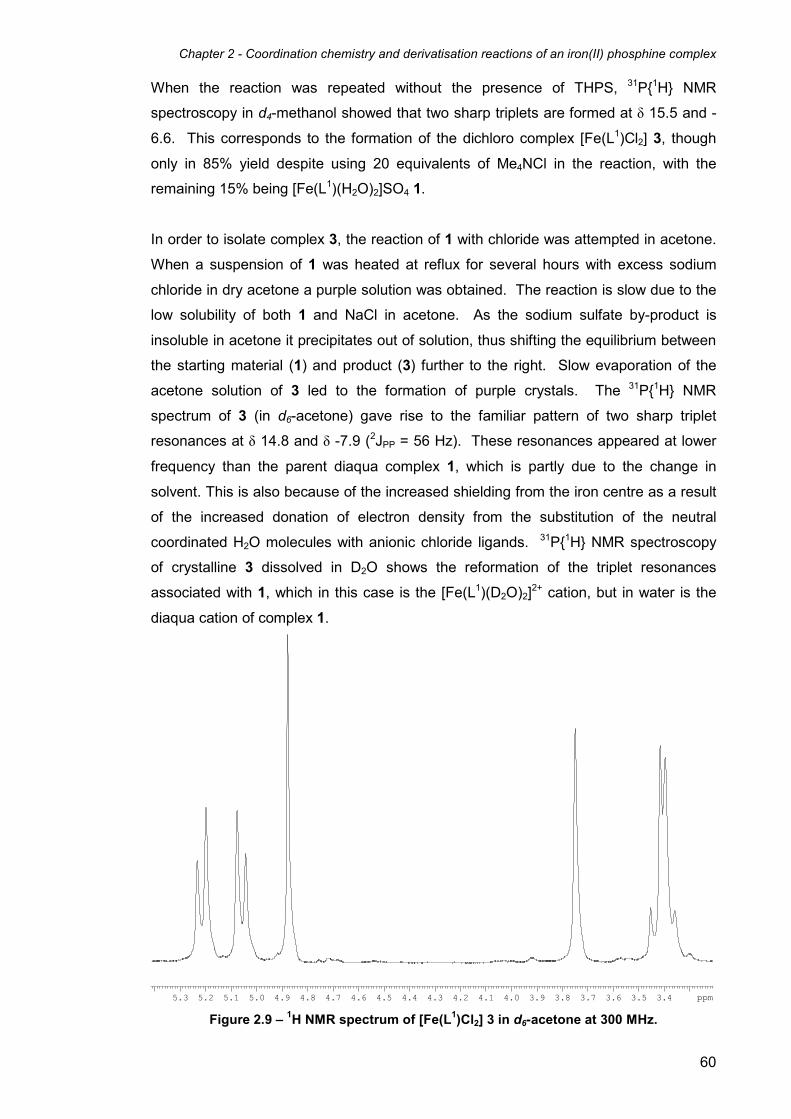
Chapter 2 - Coordination chemistry and derivatisation reactions of an iron(II) phosphine complex
60
When the reaction was repeated without the presence of THPS, 31P{1H} NMR
spectroscopy in d4-methanol showed that two sharp triplets are formed at δ 15.5 and -
6.6. This corresponds to the formation of the dichloro complex [Fe(L1)Cl2] 3, though
only in 85% yield despite using 20 equivalents of Me4NCl in the reaction, with the
remaining 15% being [Fe(L1)(H2O)2]SO4 1.
In order to isolate complex 3, the reaction of 1 with chloride was attempted in acetone.
When a suspension of 1 was heated at reflux for several hours with excess sodium
chloride in dry acetone a purple solution was obtained. The reaction is slow due to the
low solubility of both 1 and NaCl in acetone. As the sodium sulfate by-product is
insoluble in acetone it precipitates out of solution, thus shifting the equilibrium between
the starting material (1) and product (3) further to the right. Slow evaporation of the
acetone solution of 3 led to the formation of purple crystals. The 31P{1H} NMR
spectrum of 3 (in d6-acetone) gave rise to the familiar pattern of two sharp triplet
resonances at δ 14.8 and δ -7.9 (2JPP = 56 Hz). These resonances appeared at lower
frequency than the parent diaqua complex 1, which is partly due to the change in
solvent. This is also because of the increased shielding from the iron centre as a result
of the increased donation of electron density from the substitution of the neutral
coordinated H2O molecules with anionic chloride ligands. 31P{1H} NMR spectroscopy
of crystalline 3 dissolved in D2O shows the reformation of the triplet resonances
associated with 1, which in this case is the [Fe(L1)(D2O)2]2+ cation, but in water is the
diaqua cation of complex 1.
3.43.53.63.73.83.94.04.14.24.34.44.54.64.74.84.95.05.15.25.3 ppm Figure 2.9 – 1H NMR spectrum of [Fe(L1)Cl2] 3 in d6-acetone at 300 MHz.

Chapter 2 - Coordination chemistry and derivatisation reactions of an iron(II) phosphine complex
61
1H NMR spectroscopy of 3 (Figure 2.9) revealed a similar spectrum to that of 1, which
is similar for all complexes of general formula [Fe(L1)X2], although differences in
chemical shift and separation between the AB resonances were observable. The
resonance at δ 3.41 now showed signs of being an AB system, whereas in the 1H NMR
spectrum of 1 the corresponding peak was clearly a singlet at room temperature. This
resonance corresponds to the CH2 protons on the CH2OH groups of the axial
phosphine pendant arms. In complex 1, these groups can freely rotate about the P-C
bond, making both CH2 protons equivalent, leading to a singlet. It is possible that in
complex 3, the large chloride ligands prevent rotation through steric bulk or hydrogen
bonding, which would make the axial PCH2OH protons inequivalent, and lead to an AB
system.
The reaction with KBr to form the dibromo complex [Fe(L1)Br2] 5 was carried out, using
excess KBr in acetone. The 31P{1H} NMR spectrum of 5 dissolved in d6-acetone
showed two triplet resonances at δ 15.6 and -12.2 (2JP-P = 56 Hz). The 1H NMR
spectrum of 5 is similar to the spectrum of 3.
2.4 X-ray structures of [Fe(L1)Cl2]·H2O (3·H2O) and [Fe(L1)Br2]·H2O (5·H2O)
Purple crystals of 3 suitable for single crystal X-ray analysis were isolated by slow
evaporation of an acetone solution in air. The solved crystal structure revealed the
crystals were of the monohydrate, represented in Figure 2.10. Selected bond lengths
and angles are given in Table 2.1. The structure confirmed that the substitution of both
cis-aqua ligands for two chloride groups has taken place, as indicated by the NMR
data. The ligand L1 has remained intact and is coordinated in a tetradentate manner.
This gives a distorted octahedral geometry similar to complex 1, with the oriented Fe-P
bonds in the equatorial plane ranging from 80.190(15)° to 94.863(15)°. This distortion
is in part enforced by the chelating equatorial phosphine donors, which have a
crystallographic bite angle of 80.190(15)°. This angle is lower than the ideal 90° angle
for an octahedral complex, which then causes further distortion within the molecule.
The Fe-P bond lengths (between 2.16 and 2.26 Å) and Fe-Cl bond lengths (ca. 2.40 Å)
in 3 are consistent with related iron(II) phosphine structures, such as
[Fe(P{CH2CH2CH2PMe2}3)Cl2]).19 The Fe-P bond lengths of the axial phosphorus
donors are longer than those of the equatorial phosphine groups. This observation is
expected due to the greater trans influence of phosphines compared to the chloride
anions.20 The C-P-C bond angles for the axial and equatorial phosphine atoms vary
between 98.90(7)° and 102.65(7)°, which are low compared to the ideal angle of 109.5°
for an sp3 hybridised atom. This is because the bonding of the phosphines to the Fe(II)

Chapter 2 - Coordination chemistry and derivatisation reactions of an iron(II) phosphine complex
62
Figure 2.10 –Molecular structure of [Fe(L1)Cl2]·H2O (3·H2O). Thermal ellipsoids are at the
30% probability level. The included water molecule and hydrogen atoms have been
omitted for clarity.
Bond Lengths
Fe(1)-P(1) 2.2553(4) Fe(1)-P(4) 2.2541(4)
Fe(1)-P(2) 2.1836(4) Fe(1)-Cl(1) 2.4006(4)
Fe(1)-P(3) 2.1639(4) Fe(1)-Cl(2) 2.4001(4)
Bond Angles
P(4)-Fe-P(1) 174.612(17) Cl(2)-Fe-Cl(1) 90.374(15)
P(3)-Fe-Cl(1) 174.945(17) P(3)-Fe-P(2) 80.190(15)
P(2)-Fe-Cl(2) 174.760(17)
Table 2.1 – Selected bond lengths (Å) and angles (°) for complex 3·H2O.

Chapter 2 - Coordination chemistry and derivatisation reactions of an iron(II) phosphine complex
63
Figure 2.11 - Molecular structure of [Fe(L1)Br2]·H2O (5·H2O). Thermal ellipsoids are at the
30% probability level. The included water molecule and hydrogen atoms have been
omitted for clarity.
Bond Lengths
Fe(1)-P(1) 2.2679(5) Fe(1)-P(4) 2.2701(5)
Fe(1)-P(2) 2.1646(5) Fe(1)-Br(1) 2.5378(3)
Fe(1)-P(3) 2.1873(5) Fe(1)-Br(2) 2.5405(3)
Bond Angles
P(4)-Fe-P(1) 175.263(18) Br(2)-Fe-Br(1) 90.697(9)
P(3)-Fe-Br(1) 173.612(16) P(3)-Fe-P(2) 80.149(17)
P(2)-Fe-Br(2) 175.810(16)
Table 2.2 – Selected bond lengths (Å) and angles (°) for complex 5·H2O.

Chapter 2 - Coordination chemistry and derivatisation reactions of an iron(II) phosphine complex
64
centre creates strain within the ligand, as a result of the preference of the Fe(II) metal
for the octahedral geometry. Crystals of 5 suitable for single crystal X-ray diffraction
analysis were also isolated by slow evaporation of an acetone solution, and proved to
be of the mono-hydrate. As with 3, the structure is consistent with the NMR data, and
shows that both cis-aqua ligands have been substituted by bromide ligands (Figure
2.11). Selected bond lengths and angles are given in Table 2.2. The molecular
structure of 5 is very similar overall to that of 3, showing a distorted octahedral
geometry, with the cis bond angles in the equatorial plane varying from 80.149(17)° to
95.680(14)°. The crystallographic bite angle of the equatorial phosphine donors of the
L1 ligand is 80.149(17)°, which is the same as the bite angle of the L1 ligand on 3 within
experimental error. The Fe-Br bond lengths are 2.5378(3) and 2.5405(3) Å, which are
significantly longer than the similar Fe-Cl bond lengths in the structure of 3 (2.4006(4)
and 2.4001(4) Å), as would be expected for the larger bromide atoms.
2.5 Coordination chemistry of [Fe(L1)(H2O)2]SO4 (1) with pseudo-halides
In contrast to the reaction with halides, the reaction of 1 with two equivalents of
potassium thiocyanate in aqueous solution gave an orange precipitate of
[Fe(L1)(NCS)2] (6) in high yield (Scheme 2.4).
Scheme 2.4 – Synthesis of isothiocyanato and azido complexes 6 and 7.
The 31P{1H} NMR spectrum of 6 in D2O, in which it is sparingly soluble, showed two
second order resonances at δ 13.7 and δ 9.5. These resonances resemble triplets, but
appear more complex than those for complex 1. Interestingly, the appearance of the
resonances is dependant on the magnetic field strength of the spectrometer. At higher
field strength, less second order character is observed, as was shown by running the 31P{1H} NMR spectrum at 81, 122, and 162 MHz, respectively. This effect is due to the
fact that resonances begin to coalesce when the value of the coupling constant

Chapter 2 - Coordination chemistry and derivatisation reactions of an iron(II) phosphine complex
65
approaches the value of the peak separation (Hz), which is itself dependant on the
spectrometer field strength. Simulations of the 31P{1H} NMR spectra at all three
spectrometer frequencies were carried out and compared to the corresponding
experimental spectra (Figure 2.12). At all three frequencies the experimental and
simulated spectra match each other closely.
Figure 2.12 – Simulated (upper) and experimental (lower) 31P{1H} NMR spectra of
[Fe(L1)(NCS)2] (6) in D2O at different field strengths.
15.0 14.0 13.0 12.0 11.0 10.0 9.0
81 MHz
122 MHz
162 MHz

Chapter 2 - Coordination chemistry and derivatisation reactions of an iron(II) phosphine complex
66
When the 31P{1H} NMR spectrum was run in acetone at 122 MHz there was a larger
separation between the two resonances, which gave rise to a much reduced second
order spectrum (Figure 2.13).
Figure 2.13 – Simulated (upper) and experimental (lower) 31P{1H} NMR spectra of
[Fe(L1)(NCS)2] (6) in d6-acetone at 122 MHz.
When the reaction was carried out with the addition of only one equivalent of
thiocyanate, the 31P{1H} NMR spectrum showed a 1:1 mixture of the starting material 1
and the product 6. This suggests that the first substitution of a water ligand for a
thiocyanate anion to form [Fe(L1)(H2O)(NCS)]+ is the rate determining step, and the
second substitution to form 6 is faster. The IR spectrum of 6 shows two absorption
peaks at 2109 and 2098 cm-1 for ν(NCS), which are due to the symmetric and
asymmetric stretches of the cis equatorially coordinated thiocyanate ligands. The small
frequency separation of the two absorptions has also been observed in the case of
[Fe(pp3)(NCS)2] (pp3 = tris(2-diphenylphosphinoethyl)phosphine).21, 22
The reaction of 1 with sodium azide to give the [Fe(L1)(N3)2] complex (7) required an
excess of N3- anions to drive the reaction to completion (Scheme 2.4). The 31P{1H}
NMR spectrum of 7 showed the expected pair of triplets (δ 16.9 and δ 12.4, 2JP-P = 54
Hz), consistent with two azido ligands occupying the cis sites following the substitution
of both H2O ligands. When two equivalents of sodium azide are used, other products
are observed in the 31P{1H} NMR spectrum (Figure 2.14). A 31P-31P COSY experiment
shows that the triplet-like peak at δ 11.3 couples to two second order resonances at δ
18.3 and 14.4, which appear to be doublets of triplets. Therefore, the resonance at δ
122 MHz

Chapter 2 - Coordination chemistry and derivatisation reactions of an iron(II) phosphine complex
67
11.3 is actually a second-order doublet of doublets, which would be expected for an
ABX2 spin system, such as the [Fe(L1)(N3)(H2O)]
+ complex. Unlike 6, complex 7 did not
precipitate from aqueous solution during the reaction, but can be precipitated by the
addition of acetone. In contrast to the reaction of 1 with chloride in aqueous solution,
precipitation of the product 7 did not lead to reversion back to 1.
6789101112131415161718192021 ppm Figure 2.14 – 31P{1H} NMR spectrum of the reaction of 1 with two equivalents of NaN3 in
D2O at 122 MHz.
In comparison with the thiocyanate chemistry, the 31P{1H} NMR spectrum of the product
of the reaction of one equivalent of azide with 1 showed that the main species (as well
as small amounts of 1 and 7) was now the asymmetric complex represented by the
doublet of doublets at δ 11.3 and two doublets of triplets at δ 18.3 and 14.4, which has
been proposed to be of the asymmetric cation [Fe(L1)(N3)(H2O)]+. This suggests that
the second substitution of a water ligand for an azide anion to give [Fe(L1)(N3)2] (7) is
the rate determining step, in complete contrast of the reaction of complex 1 with
thiocyanate.
The IR spectrum of 7 showed only one azide absorption at 2056 cm-1. As the two
coordinated azide anions are cis to one another, two absorptions would be expected as
a result of the symmetric and asymmetric stretching modes of the ligands. Only one
peak is observed presumably due to both stretching modes being very close in energy.
Chapter 2 - Coordination chemistry and derivatisation reactions of an iron(II) phosphine complex
68
Similar observations have been made by Field et al. for related cis bis(azido) iron(II)
phosphine complexes23.
2.6 X-ray structures of [Fe(L1)(NCS)2]·2H2O (6·2H2O) and
[Fe(L1)(N3)2]·0.812H2O (7·0.812H2O)
Unequivocal characterisation of 6 was provided by X-ray crystallography. Orange
crystals of 6 were grown upon slow evaporation of an acetone solution. The analysis
confirms the substitution of both H2O molecules for thiocyanate anions has occured, as
indicated by the NMR data. The structure is of the dihydrate and shows that the
thiocyanato groups on 6 are bound via the harder nitrogen atoms of the NCS- ligand,
rather than the softer sulfur donors (Figure 2.15). This is expected due to the better
match between the relatively hard iron(II) centre and the nitrogen donors. The Fe-N-C
bond angles in 6 are almost linear (173.5(3) and 175.5(3)°), reflecting the sp
hybridisation of the orbitals on the N atom of the thiocyanate ligand. These values are
within the range reported for iron-thiocyanate complexes, though greater than the
mean values reported in the Cambridge Structural Database for compounds of this
type (164.4(9.6)°). Other main bond lengths and angles are largely similar to those in
the structures of 3 and 5 (Table 2.3).
Red crystals of 7 were grown by adding diethyl ether to a water/acetone solution. As
expected from the NMR data, the structure confirms the substitution of both aqua
groups for azide ligands has occurred (Figure 2.16). The structure of 7·0.812H2O has
two molecules of the iron complex in the asymmetric unit, the main difference between
them being the Fe-N-N bond angles. For the molecule containing Fe(1), the angles are
144.32(8) and 145.30(18)°, whereas in the molecule containing Fe(2) the angles are
more diverse at 138.9(2) and 152.4(2)° (Tables 2.4a and 2.4b). These angles are all
above the mean average values for iron azide complexes found in the Cambridge
Structural Database (125.8(9.5)°),24, 25 and are all higher than the 120° angle that would
be expected for the sp2 hybridised terminal nitrogen atom on the azide group. The two
molecules of complex 7 are interconnected into a three-dimensional structure through a
combination of O-H···O and O-H···N hydrogen bonds, with the latter involving the azido
groups as acceptors.

Chapter 2 - Coordination chemistry and derivatisation reactions of an iron(II) phosphine complex
69
Figure 2.15 - Molecular structure of [Fe(L1)(NCS)2]·2H2O (6·2H2O). Thermal ellipsoids are
at the 30% probability level. The included water molecules and hydrogen atoms have
been omitted for clarity.
Bond Lengths
Fe(1)-P(1) 2.2320(10) Fe(1)-N(2) 1.952(3)
Fe(1)-P(2) 2.1840(10) N(1)-C(1) 1.160(4)
Fe(1)-P(3) 2.1907(11) S(1)-C(1) 1.660(4)
Fe(1)-P(4) 2.2469(10) N(2)-C(2) 1.162(5)
Fe(1)-N(1) 1.960(3) S(2)-C(2) 1.656(4)
Bond Angles
P(1)-Fe(1)-P(4) 174.32(4) Fe(1)-N(1)-C(1) 175.5(3)
N(1)-Fe(1)-P(2) 175.02(10) Fe(1)-N(2)-C(2) 173.5(3)
N(2)-Fe(1)-P(3) 175.67(9) N(1)-C(1)-S(1) 178.1(3)
N(2)-Fe(1)-N(1) 90.30(13) N(2)-C(2)-S(2) 178.3(4)
P(3)-Fe(1)-P(2) 80.99(10)
Table 2.3 – Selected bond lengths (Å) and angles (°) for complex 6·2H2O.

Chapter 2 - Coordination chemistry and derivatisation reactions of an iron(II) phosphine complex
70
Figure 2.16 - Molecular structure of [Fe(L1)(N3)2]·0.812H2O (7·0.812H2O) showing molecule
1 of 2 independent molecules in the asymmetric unit. Thermal ellipsoids are at the 30%
probability level. The included water molecules and hydrogen atoms have been omitted
for clarity.
Bond Lengths (molecule 1)
Fe(1)-P(1) 2.2313(7) Fe(1)-N(6) 2.028(2)
Fe(1)-P(2) 2.1846(7) N(3)-N(4) 1.169(3)
Fe(1)-P(3) 2.1753(7) N(4)-N(5) 1.182(3)
Fe(1)-P(4) 2.2453(7) N(6)-N(7) 1.173(3)
Fe(1)-N(3) 2.029(2) N(7)-N(8) 1.185(3)
Bond Angles (molecule 1)
P(1)-Fe(1)-P(4) 174.71(3) N(4)-N(3)-Fe(1) 144.32(18)
N(3)-Fe(1)-P(2) 177.61(6) N(7)-N(6)-Fe(1) 145.30(18)
N(6)-Fe(1)-P(3) 177.68(6) N(3)-N(4)-N(5) 176.1(3)
N(6)-Fe(1)-N(3) 85.48(8) N(6)-N(7)-N(8) 174.4(3)
P(3)-Fe(1)-P(2) 80.92(2)
Table 2.4a – Selected bond lengths (Å) and angles (°) for complex 7·0.812H2O (molecule
1).

Chapter 2 - Coordination chemistry and derivatisation reactions of an iron(II) phosphine complex
71
Bond Lengths (molecule 2)
Fe(2)-P(5) 2.2611(7) Fe(2)-N(14) 2.032(2)
Fe(2)-P(6) 2.1783(7) N(11)-N(12) 1.180(3)
Fe(2)-P(7) 2.1847(7) N(12)-N(13) 1.180(3)
Fe(2)-P(8) 2.2324(7) N(14)-N(15) 1.161(3)
Fe(2)-N(11) 2.052(2) N(15)-N(16) 1.185(3)
Bond Angles (molecule 2)
P(5)-Fe(2)-P(8) 175.48(3) N(12)-N(11)-Fe(2) 138.9(2)
N(11)-Fe(2)-P(6) 178.00(7) N(15)-N(14)-Fe(2) 152.4(2)
N(14)-Fe(2)-P(7) 174.44(8) N(13)-N(12)-N(11) 176.2(3)
N(11)-Fe(2)-N(14) 84.12(10) N(16)-N(15)-N(14) 176.3(3)
P(6)-Fe(2)-P(7) 80.77(3)
Table 2.4b – Selected bond lengths (Å) and angles (°) for complex 7·0.812H2O (molecule
2).
2.7 Reaction of [Fe(L1)(H2O)2]SO4 (1) with sodium carbonate
The reaction of an aqueous solution of 1 with sodium carbonate afforded a red solution
of the neutral complex [Fe(L1)(κ2-O2CO)] (8), which is formed as a result of the
replacement of the coordinated water molecules with a bidentate carbonate ligand
(Scheme 2.5). The isolation of 8 was achieved by the addition of acetone to an
aqueous solution.
Scheme 2.5 – Reaction of 1 with sodium carbonate to give the κ2-carbonato complex 8.
The 31P{1H} NMR spectrum of 8 in D2O exhibited the anticipated pair of triplets at δ 23.5
and δ 9.4 (2JP-P = 54 Hz). The IR spectrum of 8 showed a carbonate absorption band
at 1681 cm-1.

Chapter 2 - Coordination chemistry and derivatisation reactions of an iron(II) phosphine complex
72
The reaction of a red aqueous solution of 8 with hydrochloric acid gave a purple
solution, accompanied by the evolution of carbon dioxide. The 31P{1H} NMR spectrum
showed the formation of a broad peak and a broad triplet at ca. δ 22 and 0.4
respectively, as observed in the reaction of 1 with sodium chloride in aqueous solution.
This evolution of the gas suggests that the carbonate ligand had reacted with the acid
to give a molecule each of CO2 and H2O, as well as two chloride ligands. However, the
familiar broad 31P{1H} NMR resonance pattern suggests that the exchange between the
H2O solvent molecules is occurring, and attempts to isolate the dichloride resulted in
the formation of the 31P{1H} NMR resonances associated with 1.
2.8 X-ray structure of [Fe(L1)(κ2-O2CO)]·1.7H2O (8·1.7H2O)
Crystals of 8·1.7H2O suitable for X-ray analysis were grown from the slow diffusion of
acetone into an aqueous solution of the iron complex. The diffraction revealed the
structure to be of the hydrate 8·1.7H2O, and the structure contains two independent
molecules of 8 in the asymmetric unit. Both H2O molecules have been substituted with
the bidentate carbonate ligand (Figure 2.17), as suggested by NMR spectroscopy.
The octahedral structure of 8 is much more distorted than the structures of 3, 5, 6 and
7 due to the bidentate carbonate ligand, which constrains the structure giving a O(1)-
Fe(1)-O(2) angle of 63.8(2)° (63.9(2)° for O(10)-Fe(2)-O(11) in molecule 2), which is far
less than the standard 90° angle expected for a normal octahedral structure. The
equatorial crystallographic bite angle is 81.38(10)° (80.49(9) for molecule 2), which is
similar to the structures of 3, 5, 6 and 7. Other bond lengths and angles are similar to
the previously crystallographically-characterised complexes in this chapter (Tables
2.5a and 2.5b). The structure is organised into a three-dimensional network, with the
hydroxymethyl and uncoordinated carbonate oxygen atoms acting as hydrogen bond
acceptors. The included water molecules in the structure are also involved in the
hydrogen bonding.

Chapter 2 - Coordination chemistry and derivatisation reactions of an iron(II) phosphine complex
73
Figure 2.17 - Molecular structure of [Fe(L1)(κ2-O2CO)]·1.7H2O (8·1.7H2O) showing molecule
1 of two independent molecules in the asymmetric unit. Thermal ellipsoids are at the
30% probability level. The included water molecules and hydrogen atoms have been
omitted for clarity.
Bond Lengths (molecule 1)
Fe(1)-P(1) 2.223(3) Fe(1)-O(2) 2.073(5)
Fe(1)-P(2) 2.232(3) O(1)-C(1) 1.307(9)
Fe(1)-P(3) 2.175(3) O(2)-C(1) 1.286(10)
Fe(1)-P(4) 2.162(2) O(3)-C(1) 1.271(9)
Fe(1)-O(1) 2.044(5)
Bond Angles (molecule 1)
P(1)-Fe(1)-P(2) 174.30(9) P(3)-Fe(1)-P(4) 81.38(10)
P(4)-Fe(1)-O(1) 171.50(19) O(1)-Fe(1)-O(2) 63.8(2)
P(3)-Fe(1)-O(2) 170.85(18) O(3)-C(1)-O(2) 123.9(8)
P(4)-Fe(1)-O(2) 107.76(17) O(3)-C(1)-O(1) 121.8(8)
P(3)-Fe(1)-O(1) 107.05(18) O(2)-C(1)-O(1) 114.2(7)
Table 2.5a – Selected bond lengths (Å) and angles (°) for complex 8·1.7H2O (molecule 1)
(standard deviation in brackets).

Chapter 2 - Coordination chemistry and derivatisation reactions of an iron(II) phosphine complex
74
Bond Lengths (molecule 2)
Fe(2)-P(5) 2.216(2) Fe(2)-O(11) 2.057(5)
Fe(2)-P(6) 2.243(3) O(10)-C(14) 1.301(10)
Fe(2)-P(7) 2.164(2) O(11)-C(14) 1.288(11)
Fe(2)-P(8) 2.186(3) O(12)-C(14) 1.269(9)
Fe(2)-O(10) 2.032(5)
Bond Angles (molecule 2)
P(5)-Fe(2)-P(6) 173.96(9) P(6)-Fe(2)-P(7) 80.49(9)
P(7)-Fe(2)-O(10) 170.6(2) O(10)-Fe(2)-O(11) 63.9(2)
P(8)-Fe(2)-O(11) 172.79(18) O(10)-C(14)-O(12) 121.3(9)
P(7)-Fe(2)-O(11) 106.72(18) O(11)-C(14)-O(12) 125.3(9)
P(8)-Fe(2)-O(10) 108.9(2) O(10)-C(14)-O(11) 113.4(7)
Table 2.5b – Selected bond lengths (Å) and angles (°) for complex 8·1.7H2O (molecule 2).
2.9 Reactions of [Fe(L1)(H2O)2]SO4 (1) with CO
Stirring an aqueous solution of 1 saturated with CO for 24 hours resulted in a yellow
solution. 31P{1H} NMR spectroscopy of the solution showed two new triplets to lower
frequency than for 1, at δ -14.9 and δ -39.4 (2JP-P = 35 Hz), consistant with the
formation of the iron(II) dicarbonyl complex [Fe(L1)(CO)2]SO4 9. The lower value for
the 2JP-P coupling constant going from 1 to 9 is expected due to the high trans influence
of the π-acceptor carbonyl ligands, which weakens the bonds between the equatorial
phosphine groups and the iron centre. The identity of 9 is supported by the IR
spectrum, which displays two carbonyl stretches at 2059 and 2022 cm-1.

Chapter 2 - Coordination chemistry and derivatisation reactions of an iron(II) phosphine complex
75
Scheme 2.6 – Reaction of 1 with CO to give the dicarbonyl complex 9, via the
intermediate mono-carbonyl complexes 10 and 11.
When 1 is exposed to an atmosphere of CO for a shorter period of time, the
intermediate mono-carbonyl complexes 10 and 11 (Scheme 2.6) can be observed by 31P{1H} NMR spectroscopy, in addition to resonances for 1 and 9. Complexes 10 and
11 both display three resonances, consisting of two sets of doublets of triplets and one
doublet of doublets (Figure 2.18). Upon further exposure to CO, the intermediates 10
and 11 are fully converted to the dicarbonyl 9.

Chapter 2 - Coordination chemistry and derivatisation reactions of an iron(II) phosphine complex
76
-40-35-30-25-20-15-10-515 10 5 0 ppm Figure 2.18 – 31P{1H} NMR spectrum of the reaction of [Fe(L1)(H2O)2]SO4 (1) with CO in
D2O at 122 MHz. Symbols ▲ and ■ signify complexes 10 and 11, ● signifies complex 9.
The relationship between these resonances was confirmed by 31P-31P COSY NMR
spectroscopy. The A2MX resonance patterns are consistent with both intermediates 10
and 11 having chemically inequivalent equatorial phosphine groups as a result of two
different ligands occupying the cis sites on the iron (i.e. they have the general formula
[Fe(L1)XY]n+). As 1 possesses C2 symmetry and a mirror plane, it is not immediately
obvious why there should be two separate mono(carbonyl) intermediates, as
substitution of either of the two H2O ligands should result in the aqua carbonyl complex
[Fe(L1)(CO)(H2O)]SO4 (10). This is an obvious intermediate in the stepwise
replacement of both aqua ligands on 1 to form 9. The second intermediate has been
identified as the mono(carbonyl) mono(sulfato) complex [Fe(L1)(CO)(κ1-OSO3)] (11) on
the basis of mass-spectral evidence. The positive ion electrospray mass spectrum of
the mixture of 10 and 11 showed peaks at m/z = 625.0 and 603.0, which can be
assigned as [M+Na]+ and [M+H]+, where M is [Fe(L1)(CO)(κ1-OSO3)] (11). When the
reaction of 1 with CO is carried out in the presence of excess sulfate, poor conversion
to 9 is observed and instead the proportion of the intermediates 10 and 11 is
maximised. This adds additional evidence to the existence of the proposed
[Fe(L1)(CO)(κ1-OSO3)] moiety (11). This is further backed up by the observation of the
analogous sulfato carbonyl complex trans-[Fe(L)(CO)(κ1-OSO3)], where L =
bis(hydroxypropylphosphino)ethane (DHPrPE (81b) in Chapter 1, section 1.7.2) as a
result of the reaction of cis-[Fe(L)2(SO4)] with CO.26 Although 10 and 11 can be
identified as [Fe(L1)(CO)(H2O)]SO4 and [Fe(L1)(CO)(κ1-OSO3)] respectively, it is not
possible to unambiguously assign 10 and 11 to one set of resonances each, as they
▲
▲
▲ ■
■
■
●
●

Chapter 2 - Coordination chemistry and derivatisation reactions of an iron(II) phosphine complex
77
only differ in 31P chemical shift and 2JP-P coupling constant (i.e. 10 or 11: δ 10.5 (dt, 2JP-P
= 74, 2JP-P = 39 Hz), -0.93 (dd, 2JP-P = 39,
2JP-P = 56 Hz), -29.2 (dt, 2JP-P = 74,
2JP-P = 56
Hz); 10 or 11: δ 11.8 (dt, 2JP-P = 68, 2JP-P = 40 Hz), -4.4 (dd,
2JP-P = 40, 2JP-P = 50 Hz), -
35.5 (dt, 2JP-P = 68, 2JP-P = 50 Hz).
2.10 X-ray structure of [Fe(L1)(CO)2]SO4·H2O (9·H2O)
Yellow crystals suitable for X-ray analysis were grown by evaporation of an aqueous
solution. The structural analysis showed the complex to be the hydrate 9·H2O. As
expected, both aqua groups have been substituted with neutral carbonyl ligands to give
the dicationic dicarbonyl complex (Figure 2.19). The Fe-P bonds trans to the CO
ligands are longer than in the other complexes, due to the high trans influence of CO.
As a result the crystallographic bite angle of the two equatorial phosphorus atoms
(79.42(3)°) is smaller than that in the related dichloride complex 3 (80.190(15)°). In
other crystallographically characterised complexes of L1 the Fe-P bond lengths to the
axial phosphorus atoms are significantly longer than those to the equatorial
phosphines. The difference is much smaller in 9, also due to the high trans influence of
CO (Table 2.6).

Chapter 2 - Coordination chemistry and derivatisation reactions of an iron(II) phosphine complex
78
Figure 2.19 - Molecular structure of [Fe(L1)(CO)2]SO4·H2O (9·H2O). Thermal ellipsoids are
shown at the 30% probability level. The included water molecule and hydrogen atoms
have been omitted for clarity.
Bond Lengths
Fe(1)-P(1) 2.2771(7) Fe(1)-C(1) 1.804(2)
Fe(1)-P(2) 2.2660(7) Fe(1)-C(2) 1.824(3)
Fe(1)-P(3) 2.2628(7) C(1)-O(1) 1.129(3)
Fe(1)-P(4) 2.2653(6) C(2)-O(2) 1.120(3)
Bond Angles
P(3)-Fe(1)-P(4) 174.81(3) C(1)-Fe(1)-C(2) 95.74(11)
P(2)-Fe(1)-C(1) 171.32(8) Fe(1)-C(1)-O(1) 178.6(2)
P(1)-Fe(1)-C(2) 172.34(8) Fe(1)-C(2)-O(2) 178.8(2)
P(1)-Fe(1)-P(2) 79.42(3)
Table 2.6 – Selected bond lengths (Å) and angles (°) for complex 9·H2O.

Chapter 2 - Coordination chemistry and derivatisation reactions of an iron(II) phosphine complex
79
2.11 Reaction of [Fe(L1)Cl2] (3) with CO
Reaction of an acetone solution of [Fe(L1)Cl2] (3) with CO in the presence of 1
equivalent of sodium tetraphenylborate gave an orange precipitate containing the
mono(cationic) carbonyl complex [Fe(L1)(CO)Cl]+. The 31P{1H} NMR spectrum in D2O
shows the usual pattern for a hetero-substituted L1 complex, i.e. two doublets of triplets
(δ 4.6 and -30.1), and a doublet of doublets (δ -6.2) (Figure 2.20). Despite the
presence of the tetraphenylborate, the complex crystallised as the chloride salt
[Fe(L1)(CO)Cl]Cl 12, as confirmed by microanalysis. The IR spectrum (KBr disc)
showed the CO stretching frequency for 12 at 1987 cm-1.
-30-25-20-15-10-55 0 ppm Figure 2.20 - 31P{1H} NMR spectrum of [Fe(L1)(CO)Cl]Cl (12) in D2O at 162 MHz.
In contrast with the reaction of 1 with CO, the reaction of 3 with CO only affords one
mono(carbonyl) complex due to the absence of the sulfate anion. The dicarbonyl
complex [Fe(L1)(CO)2]Cl2 was not formed because of the precipitation of 12 after the
first substitution of chloride for CO, which is due to the insolubility of the ionic complex
in acetone.
After 24 h, the 31P{1H} NMR spectrum of the D2O solution of 12 showed the formation
of some of the [Fe(L1)(CO)(D2O)]2+ cation due to the substitution of the chloride ligand
with D2O, as well as the formation of some [Fe(L1)(D2O)2]SO4 (1) as a result of the
substitution of both coordinated CO and Cl- ligands.

Chapter 2 - Coordination chemistry and derivatisation reactions of an iron(II) phosphine complex
80
2.12 Attempts to remove and isolate ligand L1 from the metal
The tetradentate macrocyclic phosphine ligand L1 is a very interesting ligand because
of its ability to impart water-solubility and to form highly stable iron(II) complexes.
Ligand L1 has never been isolated as a free ligand and would be very difficult to
synthesise without the metal template. It would be desirable to isolate the ligand so
that its coordination chemistry can be explored with other metals, though it is not
known whether or not the free ligand would be stable in solution. One method of
removing the ligand from the iron(II) centre is by introducing a ligand with a higher
binding constant than L1 that will preferentially coordinate to the iron(II) centre.
Ethylenediaminetetraacetate (EDTA4-) is a hexadentate ligand that is expected to form
a more stable complex with the iron than L1.
Two equivalents of Na4EDTA were added to an aqueous solution of 1 to give a deep
red solution. 31P{1H} NMR spectroscopy (D2O) showed peaks at δ 21.3 (broad) and 4.3
(t). The appearance of two small triplets at δ 24.1 and 10.0 was also observed. After
24 h, the two triplets at δ 24.1 and 10.0 had grown to be the major product,
accompanied by the reduction in intensity of the peaks at δ 21.3 and 4.3. This is
described in the kinetic NMR experiment in Section 2.2. This is very similar to the 31P{1H} NMR spectrum of the reaction with hydroxide, suggesting that EDTA4- in this
case is deprotonating the water ligands to give the neutral di-hydroxy species 2a.
When a large excess of Na4EDTA was added, the solution turned brown and, similar to
the reactions with base, led to an insoluble brown precipitate believed to be iron(III)
hydroxide. Orange/brown crystals suitable for X-ray diffraction were isolated from
vapour diffusion of iso-propanol into an aqueous solution of complex 1 with two
equivalents of Na4EDTA after about six weeks.
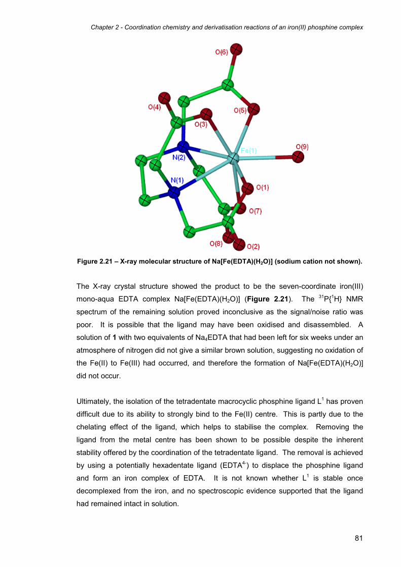
Chapter 2 - Coordination chemistry and derivatisation reactions of an iron(II) phosphine complex
81
Figure 2.21 – X-ray molecular structure of Na[Fe(EDTA)(H2O)] (sodium cation not shown).
The X-ray crystal structure showed the product to be the seven-coordinate iron(III)
mono-aqua EDTA complex Na[Fe(EDTA)(H2O)] (Figure 2.21). The 31P{1H} NMR
spectrum of the remaining solution proved inconclusive as the signal/noise ratio was
poor. It is possible that the ligand may have been oxidised and disassembled. A
solution of 1 with two equivalents of Na4EDTA that had been left for six weeks under an
atmosphere of nitrogen did not give a similar brown solution, suggesting no oxidation of
the Fe(II) to Fe(III) had occurred, and therefore the formation of Na[Fe(EDTA)(H2O)]
did not occur.
Ultimately, the isolation of the tetradentate macrocyclic phosphine ligand L1 has proven
difficult due to its ability to strongly bind to the Fe(II) centre. This is partly due to the
chelating effect of the ligand, which helps to stabilise the complex. Removing the
ligand from the metal centre has been shown to be possible despite the inherent
stability offered by the coordination of the tetradentate ligand. The removal is achieved
by using a potentially hexadentate ligand (EDTA4-) to displace the phosphine ligand
and form an iron complex of EDTA. It is not known whether L1 is stable once
decomplexed from the iron, and no spectroscopic evidence supported that the ligand
had remained intact in solution.

Chapter 2 - Coordination chemistry and derivatisation reactions of an iron(II) phosphine complex
82
2.13 Nuclear Resonance Vibrational Spectroscopic studies of 1 and 8 as
models for the active site Iron-Sulfur Cluster-Free Hydrogenase (Hmd)
The importance of vibrational spectroscopy as a method for determining the molecular
structures and functionalities of molecules is well established. In biochemistry,
however, more advanced techniques are needed due to the complexity of biomolecules
such as enzymes. In particular, site-selective techniques are needed to probe active
sites of biomolecules to gain information about their structure. Established techniques
such as resonance Raman,27 difference infrared,28, 29 and femtosecond coherence
spectroscopy30 offer a certain degree of site selectivity, but the information which they
provide is limited. This is because the selection rules of infrared and Raman
spectroscopies do not allow the observation of many important vibrational modes. For
example, in-plane vibrational Fe modes for haem derivatives are effectively forbidden
in IR and Raman spectroscopies, but could be expected to provide important
information about bond strengths. Some other haem vibrations have (just simply) not
been identified despite being allowed by selection rules.
Nuclear Resonance Vibrational Spectroscopy (NRVS) is a recent synchrotron-based
spectroscopic technique combining nuclear excitation and vibrational motion.31 NRVS
(also known as Nuclear Resonant Inelastic X-ray Scattering – NRIXS) is selective for
the vibrations of Mössbauer active nuclei such as 57Fe, 83Kr, 119Sn and 157Eu. The
isotope of most interest is 57Fe due to its importance at the centre of haems and other
important biomolecules, and this has provided most of the results to date. Despite this
dependence on Mössbauer active nuclei, NRVS gives the complete set of vibrational
modes of the probe nucleus, including those forbidden by selection rules in IR and
Raman spectroscopies. The technique is also highly selective due to its dependence
on Mössbauer active nuclei, meaning that only vibrational modes involving the active
nucleus are observed, and there is no interference from the vibrations involving only
non-active nuclei, for example in ligands. Additional information in NRVS spectra can
be obtained from the peak intensities, which are related to both the magnitude and
direction of the vibrations, giving the technique an extra dimension. In the case of 57Fe,
for example, all iron-ligand modes are observed, including the in-plane Fe vibrations in
haems and the iron-imidazole stretch in porphyrins, both of which are not observed by
Raman spectroscopy.31 Despite existing for a relatively short period of time, NRVS is
showing potential to be an important technique for the characterisation of biomolecules,
especially those containing iron-based active sites.

Chapter 2 - Coordination chemistry and derivatisation reactions of an iron(II) phosphine complex
83
In collaboration with Prof. Stephen Cramer at the University of California, Davis and the
Lawrence Berkeley National Laboratory in the USA, NRVS studies were carried out to
investigate the possibility of using complex 1 as a model for the active site of the iron-
containing enzyme H2-forming methylene-tetrahydromethanopterin dehydrogenase
(Hmd). The X-ray molecular structure of Hmd has not yet been solved and therefore
current knowledge of its active site structure is obtained from the interpretation of IR,32
Mössbauer,33 and X-ray absorption spectra.34 These techniques have revealed
important information about the iron in Hmd, and it is now known that the iron is low-
spin, and is believed to be complexed by two CO, one S, and one or two O/N based
ligands (Figure 2.22).35 It is still not known, however, whether the low-spin iron in Hmd
is Fe(II) or Fe(0). As it is believed that the iron is five coordinate, this may suggest it is
Fe(0), as low-spin Fe(II) is usually six-coordinate. The proposed vacant site is believed
to be the H2 activation site, and is modelled trans to the CO ligand, as it is believed that
the function of the CO is to increase the acidity of the H2 ligand trans to it.36 This is still
highly ambiguous, however, as X-ray absorption spectroscopy cannot differentiate
between penta- and hexa-coordinated geometries.
Figure 2.22 – Model of the proposed structure of the active site of H2-forming methylene-
tetrahydromethanopterin dehydrogenase (Hmd).
In order to find out more about the Fe-based active site of Hmd, highly selective
techniques such as NRVS are required. In order to interpret results, simple models
consisting of discrete iron complexes with similar ligands and geometries are needed
for comparison of their NRVS spectra. As one possibility is that the active site of Hmd
contains an low-spin Fe(II) with two carbonyl and two N or O based ligands (such as
H2O), complexes 1 and 9 are ideal candidates for such models.
As NRVS needs a Mössbauer active nucleus, 57Fe analogues of 1 and 9 were
synthesised. This was achieved by first making 57FeSO4 from the reaction of 57Fe
metal powder with concentrated sulfuric acid. The 57FeSO4 was then used in the
reaction with THPS and ammonium sulfate to give 57Fe labelled 1, [57Fe(L1)(H2O)2]SO4.

Chapter 2 - Coordination chemistry and derivatisation reactions of an iron(II) phosphine complex
84
The 57Fe analogue of 9, [57Fe(L1)(CO)2]SO4, was prepared by the exposure of an
aqueous solution of [57Fe(L1)(H2O)2]SO4 to carbon monoxide, as in the synthesis of 9
from 1. The 31P{1H} NMR spectra of 57Fe labelled 1 and 9 showed the extra splitting
associated with the coupling of the spin ½ 57Fe nucleus with the 31P nuclei to give two
sets of doublets of triplets as expected.
-520 15 10 5 0 ppm
18.519.0 ppm -2.5 -3.0 ppm
Figure 2.23 – 31P{1H} NMR spectrum of [57Fe(L1)(H2O)2]SO4 in D2O at 202 MHz.
Figure 2.23 shows the 31P{1H} NMR spectrum of [57Fe(L1)(H2O)2]SO4 in D2O. The
extra splitting due to the coupling with the 57Fe nucleus can clearly be seen, giving a 1JFe-P coupling of 37 Hz to both the equatorial and axial phosphine environments. The 31P{1H} NMR spectrum of the dicarbonyl complex [57Fe(L1)(CO)2]SO4 (Figure 2.24) is
similar in appearance to that of the diaqua complex, though in this case the 1JFe-P
coupling differs slightly for the axial and equatorial phosphines. The 1JFe-P coupling
constant for the coupling of the equatorial phosphines to the iron is 26 Hz, while the
axial phosphine coupling constant is 28 Hz. In both cases, residual triplets belonging
to the non-57Fe labelled complexes can also be observed due to the 57Fe metal powder
used in the synthesis only being approximately 95% 57Fe. Studies of 1JFe-P coupling
constants of iron phosphine complexes are relatively rare in the literature, and the
values reported here are relatively low compared to others reported for iron-phosphine
complexes. For example, a study on the 1JFe-P coupling constants of a range of Fe(II)
porphyrin complexes of the formula [57Fe(TPP)(PMe3)(L)] (TPP = tetraphenylporphyrin)

Chapter 2 - Coordination chemistry and derivatisation reactions of an iron(II) phosphine complex
85
ranged between 35 and 59 Hz depending on the ligand trans to the phosphine.37, 38
The 1JFe-P coupling increases from L = CO (36 Hz) < PMe3 (45 Hz) < N-MeIm (59 Hz, N-
MeIm = N-methylimidazole), suggesting that increasing the π-acceptor capability of the
ligand trans to the phosphorus nucleus decreases the 1JFe-P coupling constant. This
correlates to the 57Fe analogues of 1 and 9, with the dicarbonyl complex having the
lower 1JFe-P value. The additional phosphine donors in 1 and 9 both cis and trans to the
other donors have better π-acceptor capabilities than the N donor porphyrins, hence
the lower overall 1JFe-P values reported for the 57Fe analogues of 1 and 9.
-14 -16 -18 -20 -22 -24 -26 -28 -30 -32 -34 -36 -38 -40 ppm
-15.5 ppm -40.0 ppm
Figure 2.24 – 31P{1H} NMR spectrum of [57Fe(L1)(CO)2]SO4 in D2O at 162 MHz.
NRVS spectra of solid samples of 57Fe analogues of 1 and 9 were recorded in
collaboration with Prof. Cramer and coworkers. Figure 2.25 shows the NRVS
spectrum of [57Fe(L1)(H2O)2]SO4 (red) compared to other iron-phosphine complexes
(blue and black). As NRVS is a relatively new technique, the exact position of iron-
aqua vibrational modes is not known. In the NRVS spectra of [57Fe(edt)(CO)2(dppv)]
(blue) and [57Fe(edt)(CO)2(PMe3)2] (black) (where edt = -S(CH2)2S
-, dppv = cis-
Ph2HC=CHPPh2) (measured in collaboration with Prof. Rauchfuss and coworkers), the
peaks between 450 and 650 cm-1 are mainly contributions from the Fe-CO stretching
and bending modes, which are clearly not visible in the spectrum of
[57Fe(L1)(H2O)2]SO4. Preliminary DFT simulations of the NRVS spectra of Hmd carried
out by Prof. Cramer and coworkers indicated that the proposed Fe-OH2 peaks should
occur around 380 cm-1.
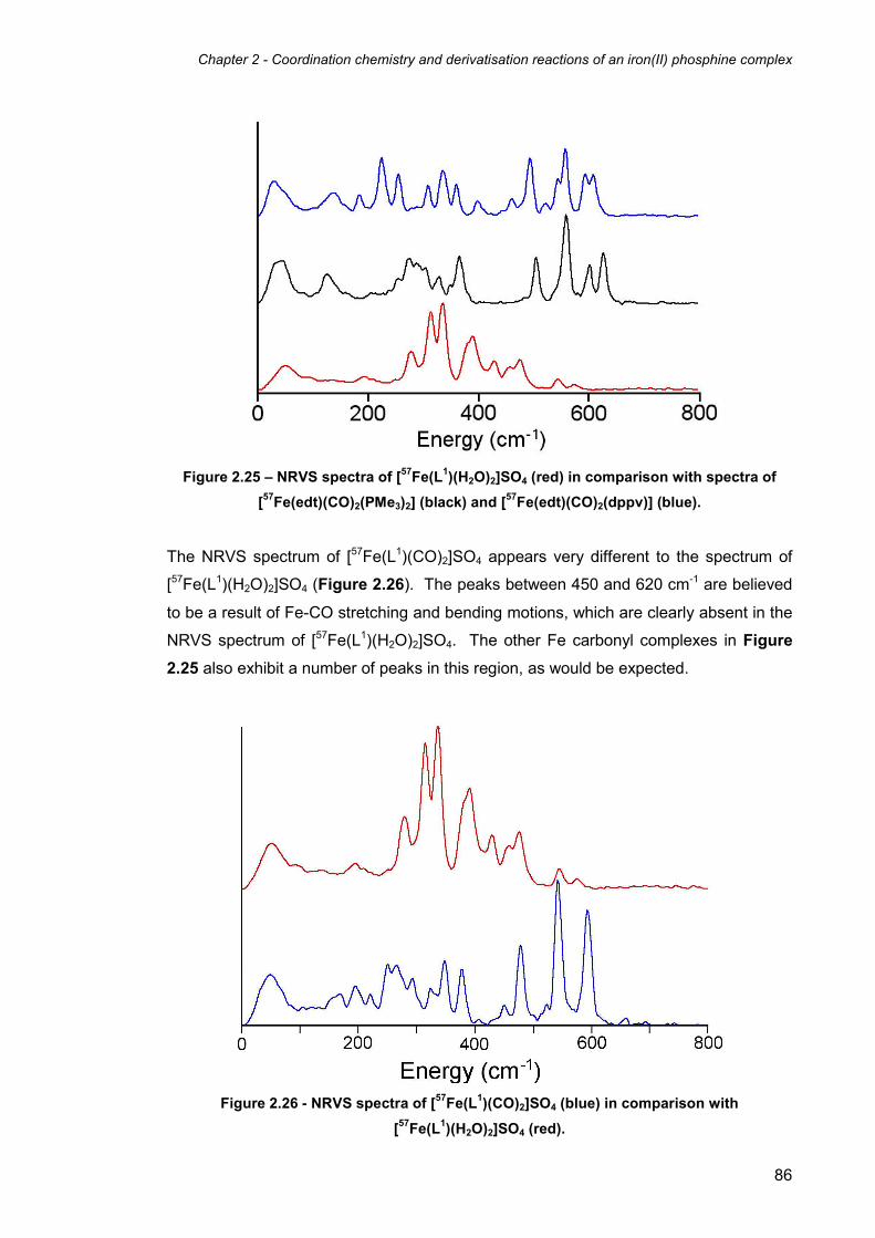
Chapter 2 - Coordination chemistry and derivatisation reactions of an iron(II) phosphine complex
86
Figure 2.25 – NRVS spectra of [57Fe(L1)(H2O)2]SO4 (red) in comparison with spectra of
[57Fe(edt)(CO)2(PMe3)2] (black) and [57Fe(edt)(CO)2(dppv)] (blue).
The NRVS spectrum of [57Fe(L1)(CO)2]SO4 appears very different to the spectrum of
[57Fe(L1)(H2O)2]SO4 (Figure 2.26). The peaks between 450 and 620 cm-1 are believed
to be a result of Fe-CO stretching and bending motions, which are clearly absent in the
NRVS spectrum of [57Fe(L1)(H2O)2]SO4. The other Fe carbonyl complexes in Figure
2.25 also exhibit a number of peaks in this region, as would be expected.
Figure 2.26 - NRVS spectra of [57Fe(L1)(CO)2]SO4 (blue) in comparison with
[57Fe(L1)(H2O)2]SO4 (red).

Chapter 2 - Coordination chemistry and derivatisation reactions of an iron(II) phosphine complex
87
Further simulations carried out by Prof. Cramer and coworkers suggested that the
largest peaks at 312 and 336 cm-1 in the NRVS spectrum of 57Fe labelled 1 are the
symmetric and asymmetric Fe-OH2 stretching vibrations. To test this theory, the 18OH2
labelled version of the complex was synthesised, to determine whether the peaks at
312 and 336 cm-1 were shifted upon exchange of the 16OH2 for the 18OH2 ligands. In
order to prepare the 18OH2 labelled product, [57Fe(L1)(H2O)2]SO4 was recrystallised
from >95% 18OH2 to give [57Fe(L1)(18OH2)2]SO4. The exchange of the
16OH2 for the 18OH2 ligands could not easily be confirmed by traditional characterisation methods.
NMR spectroscopy would not be able to distinguish the difference as both 16O and 18O
have nuclear spins of zero. Mass spectrometry should theoretically determine if the
substitution had taken place as long as the 18OH2 sample was run in 18OH2 to prevent
exchange post-synthesis. However, this does not work as the electrospray mass
spectrum of [Fe(L1)(H2O)2]SO4 1 shows the main peak at m/z = 574.99, corresponding
to the molecular ion [[Fe(L1)(SO4)] + H]+, where the sulfate anion has coordinated to the
metal by displacing the coordinated aqua ligands. This prevents the technique from
being useful in determining whether the substitution had taken place.
Figure 2.27 – NRVS spectra of [57Fe(L1)(H2O)2]SO4 (red) in comparison with the
18OH2
labelled version [57Fe(L1)(18OH2)2]SO4 (blue).
The NRVS spectrum of [57Fe(L1)(18OH2)2]SO4 compared to the non-18OH2 labelled
version (Figure 2.27) unexpectedly shows that all the peaks between 300 and 600 cm-1
have been shifted as a result of the substitution. The reason for the shift of all the
peaks in this region is hard to determine. It could be that the OH groups on the ligand
have been exchanged with 18OH groups as a result of the recrystallisation. Ultimately it

Chapter 2 - Coordination chemistry and derivatisation reactions of an iron(II) phosphine complex
88
is difficult to determine with absolute certainty that the features at 312 and 336 cm-1 are
those related to the Fe-OH2 vibrational modes.
2.14 Functionalisation of the hydroxymethyl groups on 1 to give the
acylated sulfato complex [Fe(L2)(κ2-O2SO2)] (13)
In order to explore the reactivity of the hydroxymethyl groups on 1, reactions were
carried out in order to convert them into different functionalities. Functionalised
derivatives of complex 1 could also potentially exhibit different solubilities from 1, which
could allow the comparison of the different complexes in a range of solvents.
Complex 1 reacted with acetic anhydride upon heating to 100°C to give [Fe(L2)(κ2-
O2SO2)] (13), in which all six hydroxymethyl groups have been acylated (Scheme 2.7).
Scheme 2.7 – Reaction of 1 with acetic anhydride affords the acylated sulfato complex
[Fe(L2)(κ2-O2SO2)] (13).
Complex 13 was isolated by reducing the volume of acetic anhydride solvent under
reduced pressure followed by precipitation of the crude product with Et2O. Washing
with further diethyl ether and recrystallising from CH2Cl2/Et2O yielded the purified
product 13. As expected, complex 13 is more soluble in organic solvents (such as
acetone, chloroform, and dichloromethane) than its parent complex 1 (which is
insoluble in these solvents).
The 31P{1H} NMR spectrum of 13 in CDCl3 showed the expected pair of triplet
resonances at δ 15.3 and 0.9 (2JP-P = 50.5 Hz). The 1H NMR spectrum showed the
appearance of two singlets at δ 2.2 and 2.1 with a 2:1 integral ratio corresponding to
the four methyl groups on the axial phosphine pendant arms and two methyl groups on
the equatorial phosphines, as can be seen in the diagram of L2 (Figure 2.28). Similar
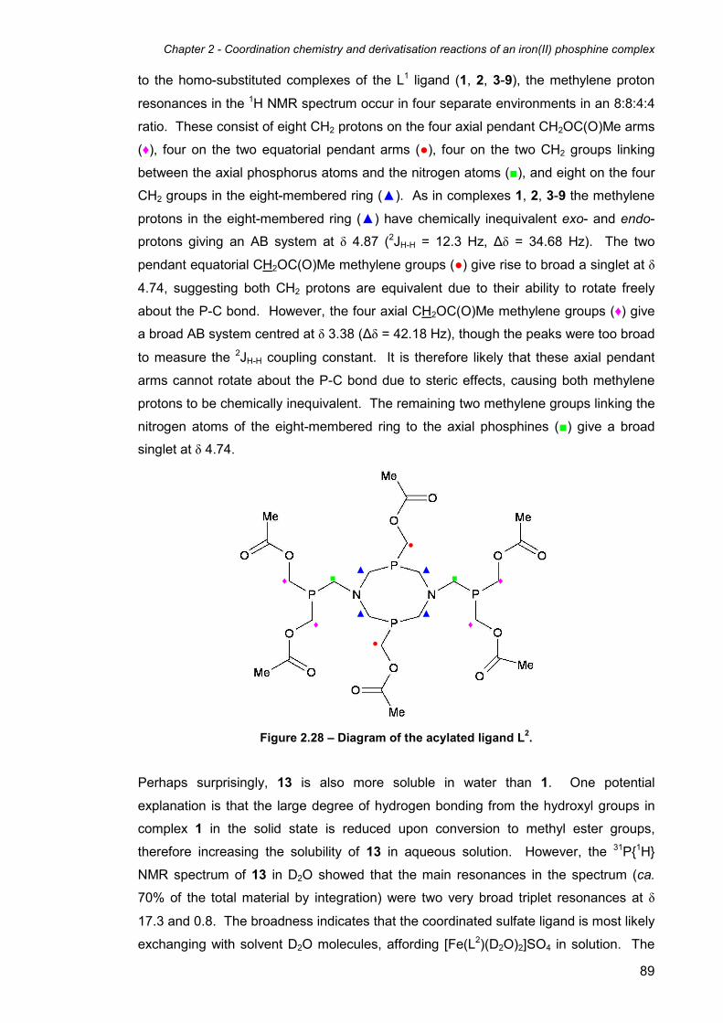
Chapter 2 - Coordination chemistry and derivatisation reactions of an iron(II) phosphine complex
89
to the homo-substituted complexes of the L1 ligand (1, 2, 3-9), the methylene proton
resonances in the 1H NMR spectrum occur in four separate environments in an 8:8:4:4
ratio. These consist of eight CH2 protons on the four axial pendant CH2OC(O)Me arms
(♦), four on the two equatorial pendant arms (●), four on the two CH2 groups linking
between the axial phosphorus atoms and the nitrogen atoms (■), and eight on the four
CH2 groups in the eight-membered ring (▲). As in complexes 1, 2, 3-9 the methylene
protons in the eight-membered ring (▲) have chemically inequivalent exo- and endo-
protons giving an AB system at δ 4.87 (2JH-H = 12.3 Hz, ∆δ = 34.68 Hz). The two
pendant equatorial CH2OC(O)Me methylene groups (●) give rise to broad a singlet at δ
4.74, suggesting both CH2 protons are equivalent due to their ability to rotate freely
about the P-C bond. However, the four axial CH2OC(O)Me methylene groups (♦) give
a broad AB system centred at δ 3.38 (∆δ = 42.18 Hz), though the peaks were too broad
to measure the 2JH-H coupling constant. It is therefore likely that these axial pendant
arms cannot rotate about the P-C bond due to steric effects, causing both methylene
protons to be chemically inequivalent. The remaining two methylene groups linking the
nitrogen atoms of the eight-membered ring to the axial phosphines (■) give a broad
singlet at δ 4.74.
Figure 2.28 – Diagram of the acylated ligand L2.
Perhaps surprisingly, 13 is also more soluble in water than 1. One potential
explanation is that the large degree of hydrogen bonding from the hydroxyl groups in
complex 1 in the solid state is reduced upon conversion to methyl ester groups,
therefore increasing the solubility of 13 in aqueous solution. However, the 31P{1H}
NMR spectrum of 13 in D2O showed that the main resonances in the spectrum (ca.
70% of the total material by integration) were two very broad triplet resonances at δ
17.3 and 0.8. The broadness indicates that the coordinated sulfate ligand is most likely
exchanging with solvent D2O molecules, affording [Fe(L2)(D2O)2]SO4 in solution. The
▲
▲
▲
▲
♦ ♦
♦ ♦
●
●
■ ■

Chapter 2 - Coordination chemistry and derivatisation reactions of an iron(II) phosphine complex
90
remaining peaks in the 31P{1H} NMR spectrum consisted of a number of triplets and
multiplets which could not be easily indentified, suggesting that other reactions are
occurring upon the solution of 13 in water. Attempts to isolate the di-aqua complex
[Fe(L2)(H2O)2]SO4 from aqueous solution proved unsuccessful.
2.15 X-ray structure of [Fe(L2)(κ2-O2SO2)]·1.3(CH3)2CO (13·1.3(CH3)2CO)
Red crystals of [Fe(L2)(κ2-O2SO2)]·1.3(CH3)2CO (13·1.3(CH3)2CO) were grown upon
layering an acetone solution with hexane. The X-ray structure (Figure 2.29) shows
that all six hydroxyl groups have been functionalised to give methyl ester
functionalities. The ligand structure itself otherwise remains intact and coordinates to
the iron in a similar manner to the L1 ligand in complexes 1 to 12. Additionally, the
sufate anion has coordinated to the iron centre, displacing the two water molecules to
give an overall neutral complex. The presence of the acyl groups in place of the
hydroxyl groups has little effect on the coordination sphere of the complex, with the
bond angles and bond lengths remaining largely similar (Table 2.7). The absence of
the hydroxymethyl groups in 13 means that there are no strong hydrogen bond donors,
and as a result, the structure contains no strong intermolecular interactions. Instead,
the supramolecular structure is dominated by C-H···O hydrogen bonds where the ester
carbonyl moieties act as the hydrogen bond acceptors.

Chapter 2 - Coordination chemistry and derivatisation reactions of an iron(II) phosphine complex
91
Figure 2.29 - Molecular structure of [Fe(L2)(κ2-O2SO2)]·1.3(CH3)2CO (13·1.3(CH3)2CO).
Thermal ellipsoids are at the 30% probability level. The included acetone molecule and
hydrogen atoms have been omitted for clarity.
Bond Lengths
Fe(1)-P(1) 2.2256(16) Fe(1)-O(2) 2.046(4)
Fe(1)-P(2) 2.2362(16) S(1)-O(1) 1.513(5)
Fe(1)-P(3) 2.1803(17) S(1)-O(2) 1.523(5)
Fe(1)-P(4) 2.1776(17) S(1)-O(3) 1.463(5)
Fe(1)-O(1) 2.047(4) S(1)-O(4) 1.451(5)
Bond Angles
P(1)-Fe(1)-P(2) 174.54(6) O(3)-S(1)-O(1) 110.2(3)
O(1)-Fe(1)-P(3) 174.07(15) O(4)-S(1)-O(1) 111.1(3)
O(2)-Fe(1)-P(4) 175.33(14) O(3)-S(1)-O(2) 111.4(3)
O(2)-Fe(1)-O(1) 69.74(15) O(4)-S(1)-O(2) 111.5(3)
P(3)-Fe(1)-P(4) 80.32(5) O(4)-S(1)-O(3) 111.3(3)
O(1)-S(1)-O(2) 100.87(19)
Table 2.7 – Selected bond lengths (Å) and angles (°) for complex 13·1.3(CH3)2CO.
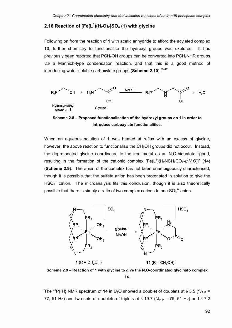
Chapter 2 - Coordination chemistry and derivatisation reactions of an iron(II) phosphine complex
92
2.16 Reaction of [Fe(L1)(H2O)2]SO4 (1) with glycine
Following on from the reaction of 1 with acetic anhydride to afford the acylated complex
13, further chemistry to functionalise the hydroxyl groups was explored. It has
previously been reported that PCH2OH groups can be converted into PCH2NHR groups
via a Mannich-type condensation reaction, and that this is a good method of
introducing water-soluble carboxylate groups (Scheme 2.10).39-42
Scheme 2.8 – Proposed functionalisation of the hydroxyl groups on 1 in order to
introduce carboxylate functionalities.
When an aqueous solution of 1 was heated at reflux with an excess of glycine,
however, the above reaction to functionalise the CH2OH groups did not occur. Instead,
the deprotonated glycine coordinated to the iron metal as an N,O-bidentate ligand,
resulting in the formation of the cationic complex [Fe(L1)(H2NCH2CO2-κ2N,O)]+ (14)
(Scheme 2.9). The anion of the complex has not been unambiguously characterised,
though it is possible that the sulfate anion has been protonated in solution to give the
HSO4+ cation. The microanalysis fits this conclusion, though it is also theoretically
possible that there is simply a ratio of two complex cations to one SO42- anion.
Scheme 2.9 – Reaction of 1 with glycine to give the N,O-coordinated glycinato complex
14.
The 31P{1H} NMR spectrum of 14 in D2O showed a doublet of doublets at δ 3.5 (2JP-P =
77, 51 Hz) and two sets of doublets of triplets at δ 19.7 (2JP-P = 76, 51 Hz) and δ 7.2

Chapter 2 - Coordination chemistry and derivatisation reactions of an iron(II) phosphine complex
93
(2JP-P = 76, 77 Hz). These resonances are of the A2MX pattern similar to those
observed for all of the asymmetrically substituted variants of 1, such as 10, 11, and 12.
This is consistent with the proposed cationic structure of 14. It is likely that the
hydroxyl groups cannot be functionalised by this method because the phosphines are
coordinated to the metal. The reason why uncomplexed hydroxymethylphosphines can
be functionalised is due to the availability of the phosphine lone pair and the presence
of catalytic amounts of formaldehyde (Scheme 2.10). These are both believed to be
fundamental requirements for the Mannich-type reaction of hydroxymethylphosphines
with primary amines.
Scheme 2.10 – Mannich-type condensation reaction of a hydroxylmethylphosphine with a
primary amine and formaldehyde.
The above mechanism shows the role of formaldehyde and the lone pair of the
phosphorus in the reaction of a hydroxymethylphosphine with a primary amine. The
formaldehyde is needed for the initial nucleophilic attack of the amine, to eventually
form the carbenium cation via loss of H2O. The lone pair of the phosphine attacks the
positively charged carbon atom of the carbenium ion to give the substituted
phosphonium cation. The addition of base to the phosphonium cation leads to the
substituted phosphine and the elimination of a molecule of formaldehyde, hence the
formaldehyde acts as a catalyst. This is in contrast to the functionalisation of 1 with
acetic anhydride, which is a direct reaction of the hydroxyl groups with the anhydride to
form the ester.
Chapter 2 - Coordination chemistry and derivatisation reactions of an iron(II) phosphine complex
94
2.17 Synthesis of the dichloride complex [Fe(L2)Cl2] (15) from
[Fe(L2)(κ2-O2SO2)] (13)
In order to investigate the coordination chemistry of 13 and compare it with that of the
parent complex 1, reactions of 13 were carried out with a variety of neutral and anionic
ligands. Complex 13 differs from 1 not only in that it has a less polar acylated ligand
backbone, but also because it possesses a coordinated chelating sulfate ligand giving
a neutral complex. The chemistry of 13 can therefore be investigated in organic
solvents. The coordinated sulfate anion on 13 can be substituted by reaction with
[PPN]Cl (PPN = bis(triphenylphosphoranylidene)ammonium) in dichloromethane to
give a purple solution of the dichloride species [Fe(L2)Cl2] (15) (Scheme 2.11).
Scheme 2.11 – Reaction of 13 with [PPN]Cl to give the dichloro complex [Fe(L2)Cl2] (15).
The 31P{1H} NMR spectrum of 15 showed the expected pair of triplets, shifted to δ 16.5
and δ -1.6 (2JP-P = 59 Hz), suggesting the formation of the di-substituted product. In
contrast to the reaction of 1 with chloride, there is no equilibrium between 13 and 15,
and 15 can be isolated by the addition of diethyl ether to the dichloromethane solution.
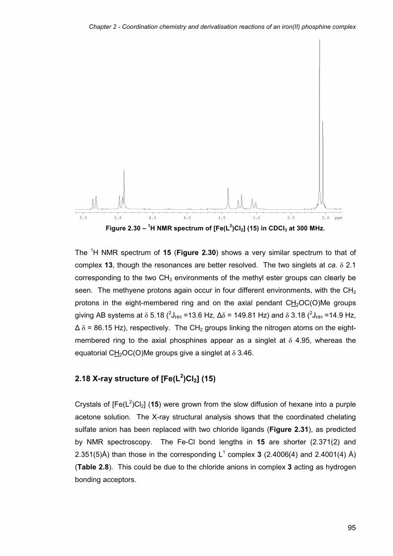
Chapter 2 - Coordination chemistry and derivatisation reactions of an iron(II) phosphine complex
95
2.02.53.03.54.04.55.05.5 ppm
Figure 2.30 – 1H NMR spectrum of [Fe(L2)Cl2] (15) in CDCl3 at 300 MHz.
The 1H NMR spectrum of 15 (Figure 2.30) shows a very similar spectrum to that of
complex 13, though the resonances are better resolved. The two singlets at ca. δ 2.1
corresponding to the two CH3 environments of the methyl ester groups can clearly be
seen. The methyene protons again occur in four different environments, with the CH2
protons in the eight-membered ring and on the axial pendant CH2OC(O)Me groups
giving AB systems at δ 5.18 (2JHH =13.6 Hz, ∆δ = 149.81 Hz) and δ 3.18 (2JHH =14.9 Hz,
∆ δ = 86.15 Hz), respectively. The CH2 groups linking the nitrogen atoms on the eight-
membered ring to the axial phosphines appear as a singlet at δ 4.95, whereas the
equatorial CH2OC(O)Me groups give a singlet at δ 3.46.
2.18 X-ray structure of [Fe(L2)Cl2] (15)
Crystals of [Fe(L2)Cl2] (15) were grown from the slow diffusion of hexane into a purple
acetone solution. The X-ray structural analysis shows that the coordinated chelating
sulfate anion has been replaced with two chloride ligands (Figure 2.31), as predicted
by NMR spectroscopy. The Fe-Cl bond lengths in 15 are shorter (2.371(2) and
2.351(5)Å) than those in the corresponding L1 complex 3 (2.4006(4) and 2.4001(4) Å)
(Table 2.8). This could be due to the chloride anions in complex 3 acting as hydrogen
bonding acceptors.

Chapter 2 - Coordination chemistry and derivatisation reactions of an iron(II) phosphine complex
96
Figure 2.31 - Molecular structure of [Fe(L2)Cl2] (15) showing molecule 1 of two
independent half-molecules in the asymmetric unit. Thermal ellipsoids are at the 30%
probability level. Hydrogen atoms have been omitted for clarity.
Bond Lengths
Fe(1)-P(1) 2.164(2) Fe(2)-P(3) 2.219(2)
Fe(1)-P(2) 2.2305(18) Fe(2)-P(5) 2.161(2)
Fe(1)-Cl(1) 2.371(2) Fe(2)-Cl(2) 2.351(5)
Bond Angles
P(2)-Fe(1)-P(2)’ 173.97(14) P(3)-Fe(2)-P(3)’ 174.25(16)
P(1)-Fe(1)-Cl(1)’ 173.89(10) P(5)-Fe(2)-Cl(2)’ 174.75(10)
Cl(1)-Fe(1)-Cl(1)’ 92.65(12) Cl(2)-Fe(2)-Cl(2)’ 91.17(13)
P(1)-Fe(1)-P(1)’ 80.44(13) P(5)-Fe(2)-P(5)’ 80.67(13)
Table 2.8 – Selected bond lengths (Å) and angles (°) for complex 15 (molecules 1 and 2).

Chapter 2 - Coordination chemistry and derivatisation reactions of an iron(II) phosphine complex
97
2.19 Reaction of [Fe(L2)(κ2-O2SO2)] (13) with CO
Despite the broad resonances observed in the 31P{1H} NMR spectrum of [Fe(L2)(κ2-
O2SO2)] 13 in D2O, the reaction of 13 with CO in aqueous solution was carried out.
The red aqueous solution took on a yellow colouration (similar to the reaction of 1 with
CO) after stirring under an atmosphere of CO for two hours. Surprisingly, the 31P{1H}
NMR spectrum in D2O (Figure 2.32) showed a relatively clean spectrum with three
sharp resonances coupling in an AMX2 spin system; two doublets of triplets at δ 1.4
(2JP-P = 37.7 Hz, 2JP-P = 71.3 Hz) and -31.6 (
2JP-P = 53.0 Hz, 2JP-P = 71.3 Hz) and a
doublet of doublets at δ -3.5 (2JP-P = 53.0 Hz, 2JP-P = 37.7 Hz). These resonances are
reminiscent of the 31P{1H} NMR spectra of the mono-carbonyl complexes
[Fe(L1)(H2O)(CO)]SO4 (10) and [Fe(L1)(CO)(κ1-OSO3)] (11) formed in the reaction of
complex 1 with CO in aqueous solution. It is therefore likely that the complex formed in
the reaction is another mono-carbonyl complex, either [Fe(L2)(H2O)(CO)]SO4 (16) or
[Fe(L2)(CO)(κ1-OSO3)] (17). In agreement with this, the IR spectrum of the complex
showed only one carbonyl stretching peak at 2001 cm-1.
-35-30-25-20-15-10-55 0 ppm Figure 2.32 – 31P{1H} NMR spectrum of the reaction of [Fe(L2)(κ2-O2SO2)] (13) with CO in
D2O at 162 MHz.
When the solution was stirred under an atmosphere of CO for a further four days,
another two doublets of triplets at δ 3.4 (2JP-P = 39.4 Hz, 2JP-P = 72.8 Hz) and -29.6 (
2JP-
P = 53.5 Hz, 2JP-P = 72.8 Hz) and a doublet of doublets at δ -2.3 (
2JP-P = 39.4 Hz, 2JP-P =

Chapter 2 - Coordination chemistry and derivatisation reactions of an iron(II) phosphine complex
98
53.5 Hz) appeared in the 31P{1H} NMR spectrum, with the second set of resonances
accounting for ca. 25% of the total material. The appearance of the second set of
resonances suggests the conversion of one of the mono-carbonyl complexes (16 or 17)
to the other, though it is difficult to determine which way round this reaction occurs. It
is more likely, however, that the carbonyl sulfate complex 17 is initially formed, with the
abundant solvent water molecules eventually replacing the coordinated sulfate ligand
to give the carbonyl aqua complex 16. Despite the prolonged exposure of the reaction
to CO, no triplets relating to the di-carbonyl species [Fe(L2)(CO2)]SO4 were observed.
Scheme 2.12 – Reaction of [Fe(L2)(κ2-O2SO2)] (13) with CO in H2O.
The reaction was then repeated in acetonitrile in order to investigate the effect of
solvent in the reaction. Firstly, the 31P{1H} NMR spectrum of 13 was recorded in
CD3CN in order to better understand the structural identity of 13 in acetonitrile solution.
The 31P{1H} NMR spectrum indicated that the main species present (ca. 85% of the
material by integration) was the mono-acetonitrile complex [Fe(L2)(CD3CN)(κ1-OSO3)],
represented by the familiar AMX2 coupling pattern (two doublets of triplets at δ 8.0 and
5.2 and a doublet of doublets at δ -0.7). The remaining 15% of the material consisted
of two triplet resonances representing the starting material [Fe(L2)(κ2-O2SO2)] 13.
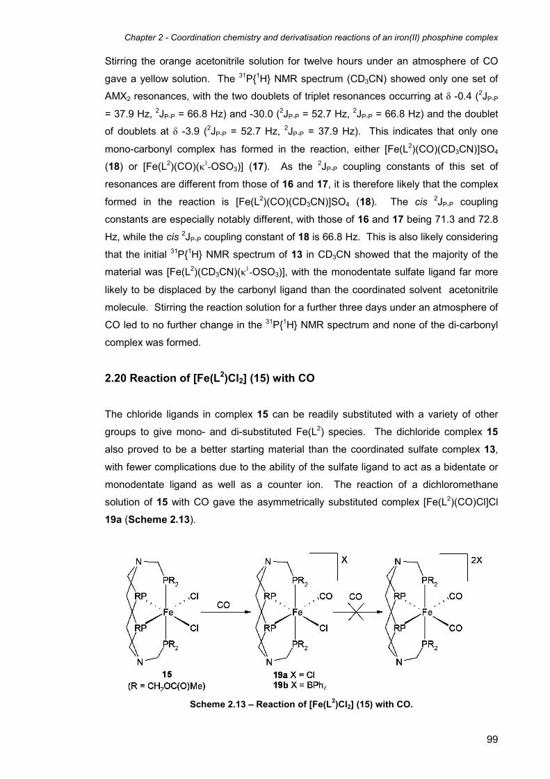
Chapter 2 - Coordination chemistry and derivatisation reactions of an iron(II) phosphine complex
99
Stirring the orange acetonitrile solution for twelve hours under an atmosphere of CO
gave a yellow solution. The 31P{1H} NMR spectrum (CD3CN) showed only one set of
AMX2 resonances, with the two doublets of triplet resonances occurring at δ -0.4 (2JP-P
= 37.9 Hz, 2JP-P = 66.8 Hz) and -30.0 (2JP-P = 52.7 Hz,
2JP-P = 66.8 Hz) and the doublet
of doublets at δ -3.9 (2JP-P = 52.7 Hz, 2JP-P = 37.9 Hz). This indicates that only one
mono-carbonyl complex has formed in the reaction, either [Fe(L2)(CO)(CD3CN)]SO4
(18) or [Fe(L2)(CO)(κ1-OSO3)] (17). As the 2JP-P coupling constants of this set of
resonances are different from those of 16 and 17, it is therefore likely that the complex
formed in the reaction is [Fe(L2)(CO)(CD3CN)]SO4 (18). The cis 2JP-P coupling
constants are especially notably different, with those of 16 and 17 being 71.3 and 72.8
Hz, while the cis 2JP-P coupling constant of 18 is 66.8 Hz. This is also likely considering
that the initial 31P{1H} NMR spectrum of 13 in CD3CN showed that the majority of the
material was [Fe(L2)(CD3CN)(κ1-OSO3)], with the monodentate sulfate ligand far more
likely to be displaced by the carbonyl ligand than the coordinated solvent acetonitrile
molecule. Stirring the reaction solution for a further three days under an atmosphere of
CO led to no further change in the 31P{1H} NMR spectrum and none of the di-carbonyl
complex was formed.
2.20 Reaction of [Fe(L2)Cl2] (15) with CO
The chloride ligands in complex 15 can be readily substituted with a variety of other
groups to give mono- and di-substituted Fe(L2) species. The dichloride complex 15
also proved to be a better starting material than the coordinated sulfate complex 13,
with fewer complications due to the ability of the sulfate ligand to act as a bidentate or
monodentate ligand as well as a counter ion. The reaction of a dichloromethane
solution of 15 with CO gave the asymmetrically substituted complex [Fe(L2)(CO)Cl]Cl
19a (Scheme 2.13).
Scheme 2.13 – Reaction of [Fe(L2)Cl2] (15) with CO.

Chapter 2 - Coordination chemistry and derivatisation reactions of an iron(II) phosphine complex
100
The 31P{1H} NMR spectrum of 19a (Figure 2.33) shows the expected A2MX coupling
pattern with a doublet of triplets at δ -6.4 (2JP-P = 66, 40 Hz), a doublet of doublets at δ -
11.8 (2JP-P = 55, 40 Hz), and a doublet of triplets at δ -35.7 (2JP-P = 66, 55 Hz).
Exposure to further CO did not result in the formation of the dicarbonyl [Fe(L2)(CO)2]2+
cation. Attempts to isolate 19a as a solid resulted in the reformation and crystallisation
of 15 via the loss of the coordinated CO molecule. However, when the reaction was
carried out in the presence of sodium tetraphenylborate in acetone, sodium chloride
was precipitated and removed as a white solid, leaving the tetraphenylborate salt
[Fe(L2)(CO)Cl]BPh4 19b in solution.
-5 -10 -15 -20 -25 -30 -35 ppm Figure 2.33 - 31P{1H} NMR spectrum of [Fe(L2)(CO)Cl]BPh4 (19b) in CDCl3 at 162 MHz.
The 31P{1H} NMR spectrum of 19b is identical to that of 19a, confirming the presence of
the same [Fe(L2)(CO)Cl]+ cation. The addition of diethyl ether to a dichloromethane
solution led to the precipitation of 19b as a yellow solid.
2.21 Reaction of [Fe(L2)Cl2] (15) with pseudo-halides
The dichloride complex 15 reacted with KNCS or NaN3 to form the disubstituted
thiocyanato or azido complexes 20 or 21, respectively (Scheme 2.14).

Chapter 2 - Coordination chemistry and derivatisation reactions of an iron(II) phosphine complex
101
Scheme 2.14 – Reaction of 15 with thiocyanate and azide.
The reaction of 15 with two equivalents of potassium thiocyanate was carried out in a
water/acetone mixture, affording an orange solution of [Fe(L2)(NCS)2] (20). Slow
evaporation of the acetone of the water/acetone mixture resulted in the formation of
orange crystals of 20. In contrast to the other L2 complexes, 20 is sparsely soluble in
most common solvents, but dissolves readily in DMSO. Surprisingly, the 31P{1H} NMR
spectrum of 20 in d6-DMSO showed a singlet resonance (at δ 4.1), instead of the
expected pair of triplets associated with the A2MX coupling pattern (Figure 2.34).
3.63.73.83.94.04.14.24.34.44.5 ppm Figure 2.34 – 31P{1H} NMR spectrum of complex 20 in d6-DMSO at 122 MHz.
However, the 1H and 13C{1H} NMR spectra of 20 are consistent with all other
complexes containing the L2 ligand, suggesting it is intact and coordinated to the metal
in the same manner. The appearance of 20 as a singlet in the 31P{1H} NMR spectrum
is believed to be due to the coincidental occurrence of both phosphine resonances at
the same chemical shift. The simulated 31P{1H} NMR spectrum of 20 with both

Chapter 2 - Coordination chemistry and derivatisation reactions of an iron(II) phosphine complex
102
phosphine environments simulated at the same chemical shift also shows a singlet.
The IR spectrum (KBr disc) revealed a single NCS stretching band at 2108 cm-1. This
is in contrast to [Fe(L1)(NCS)2] (6), the IR spectrum of which shows both symmetric and
asymmetric stretches (at 2098 and 2109 cm-1). This suggests that the symmetric and
asymmetric NCS stretches in 20 are very similar in energy.
Complex 15 also reacted with sodium azide in a water/acetone mixture in a similar
manner to potassium thiocyanate to give a red solution of the bis(azido) complex
[Fe(L2)(N3)2] 21. The 31P{1H} NMR spectrum of 21 showed two second-order
resonances at δ 10.0 and δ 8.6 (Figure 2.35). This is again due to the two phosphorus
resonances occurring at similar chemical shifts and is field dependant. The simulated 31P{1H} NMR spectrum 21 matches the experimental data, affording a 2JP-P value of 57
Hz.
Figure 2.35 – Simulated (upper) and experimental (lower) 31P{1H} NMR spectra of
[Fe(L2)(N3)2] (21) in d6-acetone at 162 MHz.
2.22 X-ray structure of [Fe(L2)(N3)2]·0.5H2O (21·0.5H2O)
Orange crystals of [Fe(L2)(N3)2]·0.5H2O (21·0.5H2O) suitable for X-ray analysis were
isolated by slow evaporation of a water/acetone solution. The structure shows the two
chloride ligands have been substituted with azide anions, without any alteration to the
L2 ligand (Figure 2.36). The asymmetric unit contains one half of an iron complex
molecule. The Fe-N bond lengths in 21 are similar to those found in the related L1
complex [Fe(L1)(N3)2] (7), suggesting that the electronic properties of both 21 and 7 are
10.6 10.2 9.8 9.4 9.0 8.6 8.2

Chapter 2 - Coordination chemistry and derivatisation reactions of an iron(II) phosphine complex
103
comparable (Table 2.9). The Fe-P bond lengths in the structure of 21 are fairly similar
to that of complex 7. However, the difference between axial and equatorial bond
lengths is less in the structure of 21 (2.2279(5) Å (axial) and 2.1951(6) Å (equatorial))
compared to the structure of 7 (2.2313(7), 2.2453(7) Å (both axial) and 2.1846(7),
2.1753(7) Å (both equatorial) for molecule 1; 2.2611(7), 2.2324(7) Å (both axial) and
2.1783(7), 2.1847(7) Å (both equatorial) for molecule 2). The closer similarities of the
axial and equatorial Fe-P bond lengths in the structure of 21 compared to that of 7 may
be related to the closer similarities in the chemical shift difference of the axial and
equatorial environments in the 31P{1H} NMR spectrum. Therefore, it is possible that the
difference in 31P chemical shift of the axial and equatorial triplet resonances in solution
may be directly proportional to the difference between the axial and equatorial Fe-P
bond lengths in the solid state X-ray structure.
The crystallographic Fe-N-N bond angle of complex 21 (144.07(16)°) is similar to the
Fe-N-N bond angles of molecule 1 of the structure of complex 7 (144.32(18) and
145.30(18)°). However, the Fe-N-N bond angles of molecule 2 of 7 are more varied,
with one angle smaller (13.9(2)°) and one larger (152.4(2)°). As with the structure of
complex 7, the Fe-N-N bond angle in the structure of 21 is in between the bond angles
that would expected for either sp or sp2 hybridisation of the terminal nitrogen atoms of
the azide ligands (120 and 180° respectively). As the Fe-N-N angle is closer to 120°,
the hybridisation of the azide terminal nitrogen is most likely closer to sp2 than sp, as
would be expected for an azide ligand.

Chapter 2 - Coordination chemistry and derivatisation reactions of an iron(II) phosphine complex
104
Figure 2.36 - Molecular structure of [Fe(L2)(N3)2]·0.5H2O (21·0.5H2O). Thermal ellipsoids
are at the 30% probability level. Included water molecule and hydrogen atoms have been
omitted for clarity.
Bond Lengths
Fe(1)-P(1) 2.2279(5) N(2)-N(3) 1.190(3)
Fe(1)-P(2) 2.1951(6) N(3)-N(4) 1.163(3)
Fe(1)-N(2) 2.092(19)
Bond Angles
P(1)-Fe(1)-P(1)’ 175.12(4) N(3)-N(2)-Fe(1) 144.07(16)
N(2)-Fe(1)-P(2) 178.08(7) N(4)-N(3)-N(2) 173.8(2)
N(2)-Fe(1)-N(2)’ 82.24(11) P(2)-Fe(1)-P(2)’ 79.43(3)
Table 2.9 – Selected bond lengths (Å) and angles (°) for complex 21.

Chapter 2 - Coordination chemistry and derivatisation reactions of an iron(II) phosphine complex
105
2.23 Reaction of [Fe(L2)Cl2] (15) with sodium carbonate
The reaction of [Fe(L2)Cl2] (15) with excess potassium carbonate in an acetone/water
solution did not proceed at room temperature. Reacting the solution for two hours at
reflux gave a red brown solution, which had seemingly decomposed. The 31P{1H} NMR
spectrum (d6-acetone) of the solution showed a wide range of broad resonances and
singlets, confirming the decomposition.
Scheme 2.15 – Reaction of [Fe(L2)Cl2] (15) with potassium carbonate.
When the reaction was repeated in DMSO, the solution took on a red colouration after
stirring for ten hours at room temperature. The 31P{1H} NMR spectrum (d6-DMSO)
showed the main product gives two triplet resonances at δ 15.9 and 7.5 (2JP-P = 55 Hz)
comprising ca. 60% of the material by integration. This is most likely the coordinated
carbonate complex [Fe(L2)(κ2-O2CO)] (22) (Scheme 2.15). The spectrum included a
number of singlet resonances and other broad peaks, suggesting some decomposition
of the complex. This decomposition is most likely base-induced, caused by the high
pH as a result of the addition of basic carbonate anions. Attempts to purify and isolate
[Fe(L2)(κ2-O2CO)] (22) proved unsuccessful.
Chapter 2 - Coordination chemistry and derivatisation reactions of an iron(II) phosphine complex
106
2.24 Electrochemical Studies
The use of electrochemical measurements in inorganic chemistry to help determine the
ability of metal complexes to undergo electron-transfer processes has now become as
routine a method of characterisation as NMR, vibrational spectroscopies and elemental
analysis.43 Electrochemistry can provide important information on a transition metal
complex, including:
• whether the complex is redox active or not.
• the electrode potentials at which the redox processes take place.
• determining whether the redox processes lead to stable species.
The first target therefore in inorganic electrochemistry is to study the effects of electron
addition and removal processes on the transition metal complex being studied. As
metal-ligand bonds are primarily covalent in character, it is possible that the addition or
removal of electrons within the molecule can lead to the cleavage of bonds or the
formation of new ones. This essentially means that the complex being studied does
not necessarily remain the same during the electron-transfer process, potentially
leading to structural changes or even the formation of entirely new species.43 The
structural changes are controlled by the frontier orbitals of the complex. For example,
in an oxidation process when an electron is removed from the highest occupied
molecular orbital (HOMO), structural changes that can occur are dependant on the
degree of bonding character of the HOMO, i.e. the more bonding character the HOMO
possesses, the greater the structural changes that can occur. This also applies to the
reduction process, the structural changes depending on the degree of anti-bonding
character of the lowest unoccupied molecular orbital (LUMO) to which the electron is
added. If the orbitals involved in the electron-transfer process are non-bonding then
the changes to the complex are expected to be minimal.
Cyclic Voltammetry (CV) is the most common electrochemical technique employed to
study electron-transfer processes and can yield both kinetic and thermodynamic
information. CV works by applying a potential across electrodes immersed in a
solution containing the species being studied, and varying that potential with time. A
plot of current vs. potential is then recorded to give the relevant current-potential
curves. These curves reveal both the potential at which such redox processes occur,
as well as the size of the currents generated, these being related to the concentration
of the species being studied. Finally, the shape of the curve allows the determination
of whether the process is straightforward, or whether it involves potential complications

Chapter 2 - Coordination chemistry and derivatisation reactions of an iron(II) phosphine complex
107
such as adsorption processes. Cyclic Voltammetric studies of the Fe(II) complexes of
L1 and L2 were undertaken to find out more about their redox properties and to
determine whether the related oxidised Fe(III) analogues would be stable and
potentially isolable.
2.24.1 Electrochemistry of L1 complexes
A 1 mM aqueous solution of [Fe(L1)(H2O)2]SO4 1 was prepared, using 0.1 M potassium
chloride as the electrolyte, which gives the dichloride complex 3 in solution. Cyclic
voltammograms were recorded using a 1 mm platinum electrode using an initial scan
rate of 100 mVs-1. The initial scan showed an irreversible oxidation peak at ca. 0.3 -
0.4 V vs. SCE (Figure 2.36). In the second scan and other subsequent scans this
peak was not observed. Only upon polishing of the electrode was this peak observed
again, followed by its disappearance in subsequent scans. This signifies that
irreversible structural changes are occurring to the iron complex during or quickly after
the oxidation process, instead of the simple reversible Nernstian redox process
associated with the oxidation of the Fe(II) metal centre to Fe(III) and its subsequent
reduction back to Fe(II). The fact that the electrode needs polishing in order to
repeatedly observe this peak could be due to the decomposition products of the
oxidation reaction coating the electrode and essentially blocking further electrochemical
processes occurring at the electrode surface. Replacing the aqua groups with other
ligands such as azido, cyano and carbonato groups did not serve to stabilise any
oxidation products, though slight shifts in potential were observed. The same
experiments were attempted using 0.1 M sodium sulfate solution as the electrolyte,
though [Fe(L1)(H2O)2](SO4) 1 is not soluble enough by itself to make a 1 mM solution at
room temperature. Observed cyclic voltammograms for a saturated solution of 1 using
the Na2SO4 electrolyte gave a much smaller oxidation peak as expected, but otherwise
gave similar voltammograms.

Chapter 2 - Coordination chemistry and derivatisation reactions of an iron(II) phosphine complex
108
Figure 2.36 – First scan of the CV of a 1 mM aqueous solution of [Fe(L1)(H2O)2]SO4 (1),
using a 1 mm Pt electrode at 100 mVs-1. 0.1 M KCl was used as the electrolyte.
Further experiments were carried out to determine the approximate timescale for the
decomposition process. Theoretically, by increasing the scan rate of the
voltammogram, the length of time available for the oxidised form to decompose will
also be decreased. Therefore at high scan rates it can be determined whether the
decomposition is effectively prevented due to insufficient time, giving a more reversible
voltammogram. By repeating the above experiment at 1 Vs-1 (ten times the initial scan
rate), the voltammogram shows signs of becoming more reversible. This suggests that
if the oxidised Fe(III) complex is denied enough time to decompose then a reversible
voltammogram may be possible. However, a completely reversible voltammogram was
not observed even at such high scan rates.
2.24.2 Electrochemistry of L2 complexes
The electrochemistry of the iron complexes containing the L2 ligand was explored in
order to compare and contrast to the largely irreversible voltammograms observed in
the cyclic voltammograms of the related L1 complexes. The L2 ligand is more lipophilic
than L1 due to the presence of the less polar methyl ester groups, making it readily
soluble in organic solvents such as 1,2-dichloroethane and acetonitrile. With the
exception of [Fe(L2)(κ2-O2SO2)] (13), the L2 complexes are largely insoluble in water, so
the similar study of the L2 complexes could not be carried out in aqueous solution.
Complex 13 was not studied in H2O as the 31P{1H} NMR spectrum in D2O showed that
multiple complexes were formed, as well as the main product showing broad peaks
suggesting that the coordinated sulfate ligand was exchanging with solvent H2O
molecules.

Chapter 2 - Coordination chemistry and derivatisation reactions of an iron(II) phosphine complex
109
Cyclic voltammetry of the dichloro complex [Fe(L2)Cl2] was initially carried out in
1,2-dichloroethane. A 1 mM 1,2-dichloroethane solution of [Fe(L2)Cl2] (15) was made
up containing 0.1M tetrabutylammonium hexafluorophosphate as an electrolyte, on a 1
mm platinum electrode using an initial scan rate of 50 mVs-1 ((i) in figure 2.37). Cyclic
voltammograms were repeated at 100, 200, and 500 mVs-1 ((ii), (iii), and (iv) in figure
2.37 respectively).
Figure 2.37 – Cyclic voltammograms of [Fe(L2)Cl2] (15) in 1,2-dichloroethane, using a 1
mm Pt electrode at 50, 100, 200, and 500 mVs-1 ((i), (ii), (iii), and (iv) respectively). 0.1 M
Bu4NPF6 was used as the electrolyte.
Interestingly, the above voltammogram shows a fully reversible Nernstian Fe(II)/Fe(III)
redox process at ca. 0.1 V vs. Fc+/Fc with no apparent decomposition of the complex.
This is in direct contrast to the cyclic voltammogram of [Fe(L1)(H2O)2](SO4) 1 and
related complexes, though it is not immediately clear whether this is due to the
difference in functionality of the esterified ligand L2, or the difference of solvent
between aqueous solution and dichloroethane. Figure 2.37 also shows the
dependence of the voltammograms on the scan rate, with the faster scan rates giving
proportionally larger peak areas, and therefore peak currents. The cyclic
voltammograms appear fully reversible at all four scan rates, suggesting that the
reversibility of the Fe(II)/Fe(III) redox system in [Fe(L2)Cl2] (15) in dichloroethane is not
dependant on scan rate, unlike with the L1 complexes. The same experiment was

Chapter 2 - Coordination chemistry and derivatisation reactions of an iron(II) phosphine complex
110
repeated in acetonitrile to determine the effect of using a more polar, more coordinating
solvent (Figure 2.38, vide infra).
Figure 2.38 – Cyclic voltammograms of [Fe(L2)Cl2] (15) in acetonitrile, using a 3 mm
diamond electrode at 100 mVs-1 after 1, 10, and 60 minutes ((a), (b), and (c), respectively).
0.1 M Bu4NPF6 was used as the electrolyte.
When the experiment was conducted after one minute, two redox processes were
observed ((a) in Figure 2.38). The oxidation/reduction peaks at 0.1 V vs. Fc+/Fc seem
fully reversible and correlate with the Fe(II)/Fe(III) redox process of the dichloro
complex [Fe(L2)Cl2] (15) as seen in the voltammograms carried out in 1,2-

Chapter 2 - Coordination chemistry and derivatisation reactions of an iron(II) phosphine complex
111
dichloroethane. However, a second redox process is apparent at ca. 0.7 V vs. Fc+/Fc,
the identity of which is less immediately obvious.
After a total of ten minutes, the colour of the acetonitrile solution had changed from
purple to a more purple/orange colour. CV of the solution at this point showed that the
second redox process had grown in intensity with respect to the peak at ca. 0.1 V vs.
Fc+/Fc ((b) in Figure 2.38). It is therefore reasonable to suggest that the second redox
process results from the cationic mono-chloro mono-acetonitrile complex
[Fe(L2)(MeCN)Cl]+, which forms as a result of the replacement of one of the chloride
anions coordinated to the iron centre by an acetonitrile ligand. Additionally, the redox
process at ca. 0.7 V vs. Fc+/Fc does not appear completely reversible, with the
oxidation peak seeming slightly larger than the complementary reduction peak,
suggesting that the complex is structurally altered upon reduction from Fe(III) back to
Fe(II).
Recording the cyclic voltammogram after 1 hr showed that most of the [Fe(L2)Cl2] (15)
complex had reacted to form [Fe(L2)(MeCN)Cl]+, at which point the solution had taken
on an orange colouration. The irreversibility of the [Fe(L2)(MeCN)Cl]+ Fe(II)/Fe(III)
redox process was even more apparent from this voltammogram. The asymmetry of
this second peak could also be due to the oxidation of traces of water present in the
acetonitrile solvent catalysed by either [Fe(L2)Cl2] (15) or [Fe(L2)(MeCN)Cl]+, as the
oxidation of water occurs at a similar potential.
31P{1H} NMR studies of [Fe(L2)Cl2] (15) were carried out in CD3CN in order to confirm
the formation of [Fe(L2)(CD3CN)Cl]+. The study showed conversion over time of the
two triplet resonances associated with 15 to two second order doublets of triplets at δ
4.1 and 3.1 and a doublet of doublets at δ -6.0 respectively, the expected coupling
pattern for an asymmetric complex such as [Fe(L2)(CD3CN)Cl]+. Despite leaving the
solution for 48 hrs, no formation of the di-acetonitrile complex was observed.
Overall the electrochemical studies have provided an interesting insight into the
stability and reactivity of the iron(II) complexes studied. The contrast in
electrochemical behaviour between the L1 and L2 complexes is particularly interesting,
with the L1 complexes exhibiting largely irreversible behaviour solution at most
standard scan rates in aqueous solution, whilst the equivalent L2 complexes showing
reversible Nernstian Fe(II)/(III) redox processes in non-coordinating organic solvents
such as dichloroethane. In a more coordinating organic solvent such as MeCN, the L2
complexes also exhibit reversible Fe(II)/(III) redox behaviour, though the substitution of
Chapter 2 - Coordination chemistry and derivatisation reactions of an iron(II) phosphine complex
112
a chloride ligand for an acetonitrile molecule also occurs, which can be monitored with
CV over time.
No cyclic voltammetry studies of similar iron hydroxymethylphosphine complexes
appear in the literature to date, so it is difficult to compare the results with related
complexes. It is also therefore difficult to draw conclusions as to why the L1 complexes
give irreversible voltammograms, more specifically why the electrode blocking occurs
after the oxidation of the L1 complexes. As this does not happen with the related L2
complexes, it is reasonable to suggest that the derivatisation of the CH2OH groups into
methyl ester groups prevents the decomposition of the complex upon oxidation. This
decomposition could be because the hydroxymethylphosphine groups have an affinity
for the electrode surface, thus coating it and causing the electrode blocking, or it could
simply be a matter of stability. However, these two sets of results cannot be directly
compared to one another as the conditions (i.e. solvent, electrolyte) in which they are
studied are not identical.
2.25 Conclusion
A comparison of the coordination chemistry of the complexes of both the L1 and L2
ligands shows many similarities, with some notable differences. Firstly, due to the
differences in polarity of the ligands, the majority of the coordination chemistry of the L1
complexes was carried out in aqueous solution, whereas most of the coordination
chemistry of the L2 complexes took place in organic solvents such as dichloromethane
and acetone.
Substituting the aqua groups on complex 1 with chloride and bromide ligands to form
the dihalide complexes required the use of an organic solvent (acetone) to prevent the
ligand exchange with solvent H2O molecules that occurs in aqueous solution. The
acylated equivalent of complex 1, [Fe(L2)(κ2-O2SO2)] (13), also reacted with a chloride
salt to form the dichloride complexes in dichloromethane, though via the substitution of
the coordinated chelating sulfate ligand instead of two H2O ligands. It can therefore be
concluded that both L1 and L2 complexes behave fairly similarly when reacting with
halides in organic solvent, despite the differences in the ligands.
In contrast to the reactions of complex 1 with halides, the reactions of 1 with pseudo-
halides in aqueous solution did not give reversible equilibria with the solvent molecules
as indicated by sharp triplet resonances in the 31P{1H} NMR spectra. This is partially
due to the insolubility of the products relative to the starting materials (with the notable

Chapter 2 - Coordination chemistry and derivatisation reactions of an iron(II) phosphine complex
113
exception of [Fe(L1)(NCS)2] (6)), which is the reverse of the reaction of 1 with halides.
The reactions of complexes 1 and 15 with potassium thiocyanate in aqueous solution
(1) and an acetone/water solution (15) proceeded similarly and both led to the
precipitation of di-thiocyanate complexes [Fe(L1)(NCS)2] (6) and [Fe(L2)(NCS)2] (20)
respectively. Similarly, complexes 1 and 15 also reacted with sodium azide in H2O (1)
and acetone/water (15) to both form the diazide complexes [Fe(L1)(N3)2] (7) and
[Fe(L2)(N3)2] (21) respectively, though 21 precipitated from solution whereas 7 was very
soluble. The formation of complex 7 also required an excess of sodium azide to drive
the reaction to completion, partially due to the fact that mono-azide intermediate
complexes were formed on the way to 7. The observance of the mono-azide
intermediates in this reaction suggests that it is more favourable for an azide ligand to
coordinate to another molecule of complex 1, rather than to coordinate as a second
azide ligand. This suggests the substitution of the second aqua ligand on 1 is the rate
determining step. As mono-thiocyanato complexes are never observed as
intermediate complexes in the reactions of 1 and 15 with potassium thiocyanate, the
rate determing step in these reactions is the first substitution of an aqua or chloride
ligand for a thiocyanate anion. This also applies to the reaction of 15 with sodium
azide.
The reaction of complex 1 with sodium carbonate in aqueous solution led to the
formation of the chelating carbonate complex [Fe(L1)(κ2-O2CO)] (8). No equilibrium
with the solvent was observed, despite the solubility of 8 in aqueous solution. This is
partially due to the carbonate ligand, which formed a highly thermodynamically stable
complex due to the chelate effect. In contrast to this, the similar reaction of [Fe(L2)Cl2]
(15) with potassium carbonate in acetone/water led to complete decomposition of the
starting material. However, when the reaction was repeated in DMSO, the appearance
of two new triplet resonances suggested the formation of the carbonate complex
[Fe(L2)(κ2-O2CO)] (22) had occurred. Complex 22 could not be isolated or purified,
however, due to the decomposition of ca. 40% of the material.
The reaction of complex 1 with CO offers interesting contrasts when comparing to the
similar reaction of the L2 complex 13. Complex 1 reacted with CO in aqueous solution
to afford the di-carbonyl complex [Fe(L1)(CO)2]SO4 (9). When the reaction was
stopped before completion, two mono-carbonyl intermediates were observed which
could be identified as [Fe(L1)(CO)(H2O)]SO4 (10) and [Fe(L1)(CO)(κ1-OSO3)] (11). In
contrast, the reaction of 13 with CO in aqueous solution gave only one mono-carbonyl
complex, most likely the carbonyl sulfate complex [Fe(L1)(CO)(κ1-OSO3)] (17). Further
exposure of 17 to CO did not lead the di-carbonyl complex. It is not clear why the di-

Chapter 2 - Coordination chemistry and derivatisation reactions of an iron(II) phosphine complex
114
carbonyl complex formed in aqueous solution in the case of the L1 complex, whereas
with the similar L2 complex the second substitution did not occur. When
[Fe(L1)(CO)(κ1-OSO3)] (17) was left in aqueous solution, the slow conversion to
[Fe(L1)(CO)(H2O)]SO4 (17) occurred via substitution of the coordinated sulfate ligand
for an aqua ligand. In comparison, 13 dissolved in acetonitrile (mainly
[Fe(L1)(MeCN)(κ1-OSO3)] in solution by 31P{1H} NMR spectroscopy) reacted to rapidly
give [Fe(L1)(CO)(MeCN)]SO4 (18) via the substitution of the coordinated sulfate ligand
with a highly-coordinated acetonitrile solvent ligand.
While the coordination chemistry of both [Fe(L1)Cl2] (3) and [Fe(L2)Cl2] (15) with CO in
acetone gave similar mono-carbonyl mono-chloride complexes [Fe(L1)(CO)Cl]Cl (12)
and [Fe(L2)(CO)Cl]Cl (19a) respectively, the main difference in these reactions lies in
the relative solubilities of the starting materials and products. Complex 3 was sparsely
soluble in acetone, and formed the even less soluble product 12. This therefore
precipitated, automatically preventing reversion back to 3. In contrast, complex 19a
formed in the reaction of 15 with CO is soluble and stays in solution. This allows the
back reaction to 15 to occur when the atmosphere of CO is removed. Only when the
coordinating chloride anion was removed was the Fe(L2)(CO)Cl]+ cation stable in
solution and could be isolated as the tetraphenylborate salt 19b.
In contrast to the reaction of 1 with acetic anhydride, attempts to functionalise the
CH2OH groups on 1 with the amino acid glycine to afford carbamate moieties led to the
formation of the N,O-coordinated glycine complex 14 via substitution of the aqua
groups. However, the hydroxymethyl groups of 14 remained unfunctionalised. This is
presumably because the functionalisation of a hydroxymethyl phosphine with an amine
(i.e. the Mannich reaction) requires the availability of the phosphorus lone pair, which is
unavailable when complexed to the iron centre.
The Nuclear Resonance Vibrational Spectroscopy (NRVS) of 57Fe versions of
complexes [Fe(L1)(H2O)2]SO4 1 and [Fe(L1)(CO)2]SO4 9 showed interesting results.
Peaks relating to the Fe-H2O and Fe-CO modes were tentatively assigned after
comparison with other aqua and carbonyl complexes. The H2O groups on
[57Fe(L1)(H2O)2]SO4 were then exchanged with 18OH2 in an attempt to confirm the
identity of the Fe-aqua vibrational modes by looking for a shift in frequency of the
relevant peaks.
Electrochemical studies of both the L1 and L2 series of complexes provided insight into
their redox behaviour. Complexes of the L1 ligand proved to give largely irreversible
Chapter 2 - Coordination chemistry and derivatisation reactions of an iron(II) phosphine complex
115
cyclic voltammograms in aqueous solution, likely as a result of electrode blocking due
to decomposition of the complexes upon oxidation of the Fe(II) to Fe(III). In contrast,
the L2 complexes showed completely reversible cyclic voltammograms in 1,2-
dichloroethane, showing that the equivalent Fe(III) complexes are stable in solution.
Cyclic voltammetry of the L2 complexes in MeCN showed that a chloride ligand is
replaced with an acetonitrile ligand over time. As a result of these electrochemical
studies, the isolation of the 17 electron Fe(III) L2 complexes would appear to be
feasible. This could be achieved by either chemical or bulk electrochemical methods.
Investigations could then be carried out to determine whether these complexes show
any catalytic activity or the ability to reversibly bind small molecules such as dinitrogen.
2.26 References
1. J. Chatt, G. J. Leigh and R. M. Slade, J. Chem. Soc., Dalton Trans., 1973,
2023.
2. K. N. Harrison, P. A. T. Hoye, A. G. Orpen, P. G. Pringle and M. B. Smith, J.
Chem. Soc., Chem. Commun., 1989, 1096.
3. J. W. Ellis, K. N. Harrison, P. A. T. Hoye, A. G. Orpen, P. G. Pringle and M. B.
Smith, Inorg. Chem., 1992, 31, 3026.
4. E. Costa, M. Murray, P. G. Pringle and M. B. Smith, Inorg. Chim. Acta., 1993,
213, 25.
5. P. A. T. Hoye, P. G. Pringle, M. B. Smith and K. Worboys, J. Chem. Soc.,
Dalton Trans., 1993, 269.
6. S. Komiya, H. Awata, S. Ishimatsu and A. Fukuoka, Inorg. Chim. Acta., 1994,
217, 201.
7. D. E. Berning, K. V. Katti, C. L. Barnes and W. A. Volkert, Chem. Ber., 1997,
130, 907.
8. D. E. Berning, K. V. Katti, L. J. Barbour and W. A. Volkert, Inorg. Chem., 1998,
37, 344.
9. L. Higham, A. K. Powell, M. K. Whittlesey, S. Wocadlo and P. T. Wood, Chem.
Commun., 1998, 1107.
10. B. Drießen-Hölscher and J. Heinen, J. Organomet. Chem., 1998, 570, 141.
11. A. Salvani, P. Frediani, M. Bianchi, F. Piacenti, L. Pistolesi and L. Rosi, J.
Organomet. Chem., 1999, 582, 218.
12. A. Fukuoka, W. Kosugi, F. Morishita, M. Hirano, L. McCaffrey, W. Henderson
and S. Komiya, Chem. Commun., 1999, 489.
13. Y. Kayaki, T. Suzuki and T. Ikariya, Chem. Lett., 2001, 1016.

Chapter 2 - Coordination chemistry and derivatisation reactions of an iron(II) phosphine complex
116
14. D. S. Bharathi, M. A. Sridhar, J. S. Prasad and A. G. Samuelson, Inorg. Chem.
Commun., 2001, 4, 490.
15. L. J. Higham, M. K. Whittlesey and P. T. Wood, Dalton Trans., 2004, 4202.
16. V. Cadierno, P. Crochet, S. E. García-Garrido and J. Gimeno, Dalton Trans.,
2004, 3635.
17. J. C. Jeffery, B. Odell, N. Stevens and R. E. Talbot, Chem. Commun., 2000,
101.
18. A. A. Karasik, R. N. Naumov, R. Sommer, O. G. Sinyashin and E. Hey-
Hawkins, Polyhedron, 2002, 21, 2251.
19. M. Antberg and L. Dahlenburg, Inorg. Chim. Acta., 1985, 104, 51.
20. F. A. Cotton, G. Wilkinson, C. A. Murillo and M. Bochmann, "Advanced
Inorganic Chemistry", Wiley-Interscience, New York, 1999.
21. M. Bacci, S. Midollini, P. Stoppioni and L. Sacconi, Inorg. Chem., 1973, 12,
1801.
22. A. B. P. Lever, B. S. Ramaswamy, S. H. Simonsen and L. K. Thompson, Can.
J. Chem., 1970, 48, 3076.
23. L. D. Field, B. A. Messerle and R. J. Smernik, Inorg. Chem., 1997, 36, 5984.
24. F. H. Allen and O. Kennard, Automat. News., 1993, 8, 31.
25. D. A. Fletcher, R. F. McMeeking and D. Parkin, J. Chem. Comput. Sci., 1996,
36, 746.
26. W. K. Miller, J. D. Gilbertson, C. Leiva-Paredes, P. R. Bernatis, T. J. R.
Weakley, D. K. Lyon and D. R. Tyler, Inorg. Chem., 2002, 41, 5453.
27. T. G. Spiro, Ed., "Biological Applications of Raman Spectroscopy", Wiley-
Interscience, New York, 1988.
28. A. Dong and W. S. Caughey, Meth. Enzymol., 1994, 232, 139.
29. A. Barth and C. Zscherp, Q. Rev. Biophys., 2002, 35, 369.
30. L. Zhu, J. T. Sage and P. M. Champion, Science, 1994, 266, 629.
31. W. R. Scheidt, S. M. Durbin and J. T. Sage, J. Inorg. Biochem., 2005, 99, 60.
32. E. J. Lyon, S. Shima, R. Boecher, R. K. Thauer, F. W. Grevels, E. Bill, W.
Roseboom and S. P. J. Albracht, J. Am. Chem. Soc., 2004, 126, 14239.
33. S. Shima, E. J. Lyon, R. K. Thauer, B. Mienert and E. Bill, J. Am. Chem. Soc.,
2005, 127, 10430.
34. M. Korbas, W. Meyer-Klaucke, S. Vogt, E. Bill, E. J. Lyon, R. K. Thauer and S.
Shima, J. Biol. Chem., 2006, 281, 30804.
35. S. Shima and R. K. Thauer, Chem. Rec., 2007, 7, 37.
36. J. Huhmann-Vincent, B. L. Scott and G. J. Kubas, Inorg. Chim. Acta., 1999,
294, 240.

Chapter 2 - Coordination chemistry and derivatisation reactions of an iron(II) phosphine complex
117
37. L. M. Mink, J. R. Polam, K. A. Christensen, M. A. Bruck and F. A. Walker, J.
Am. Chem. Soc., 1995, 117, 9329.
38. L. M. Mink, K. A. Christensen and F. A. Walker, J. Am. Chem. Soc., 1992, 114,
6930.
39. A. W. Frank and D. J. Daigle, Textile Res. J., 1982, 52, 751.
40. W. Henderson, G. M. Olsen and L. S. Bonnington, J. Chem. Soc., Chem.
Commun., 1994, 1863.
41. D. E. Berning, K. V. Katti, C. L. Barnes and W. A. Volkert, J. Am. Chem. Soc.,
1999, 121, 1658.
42. A. A. Karasik, R. N. Naumov, O. G. Sinyashin, G. P. Belov, H. V. Novikova, P.
Lönnecke and E. Hey-Hawkins, Dalton Trans., 2003, 2209.
43. P. Zanello, "Inorganic Electrochemistry", The Royal Society of Chemistry,
Cambridge, 2003.
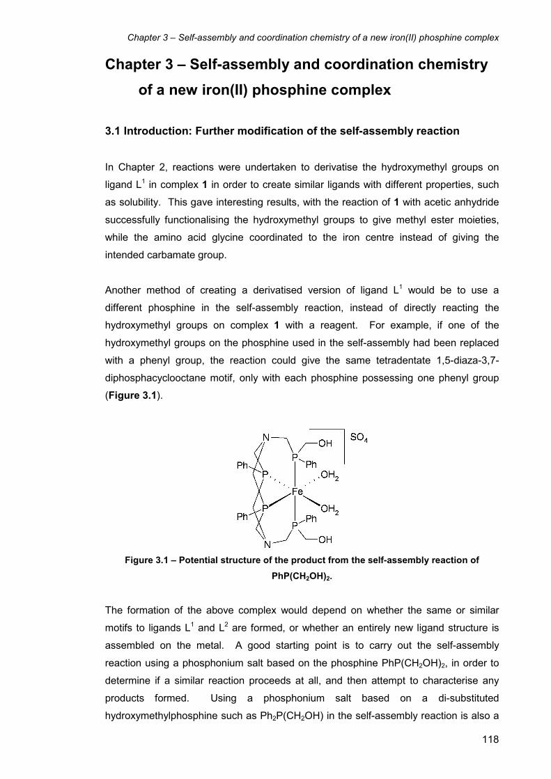
Chapter 3 – Self-assembly and coordination chemistry of a new iron(II) phosphine complex
118
Chapter 3 – Self-assembly and coordination chemistry
of a new iron(II) phosphine complex
3.1 Introduction: Further modification of the self-assembly reaction
In Chapter 2, reactions were undertaken to derivatise the hydroxymethyl groups on
ligand L1 in complex 1 in order to create similar ligands with different properties, such
as solubility. This gave interesting results, with the reaction of 1 with acetic anhydride
successfully functionalising the hydroxymethyl groups to give methyl ester moieties,
while the amino acid glycine coordinated to the iron centre instead of giving the
intended carbamate group.
Another method of creating a derivatised version of ligand L1 would be to use a
different phosphine in the self-assembly reaction, instead of directly reacting the
hydroxymethyl groups on complex 1 with a reagent. For example, if one of the
hydroxymethyl groups on the phosphine used in the self-assembly had been replaced
with a phenyl group, the reaction could give the same tetradentate 1,5-diaza-3,7-
diphosphacyclooctane motif, only with each phosphine possessing one phenyl group
(Figure 3.1).
Figure 3.1 – Potential structure of the product from the self-assembly reaction of
PhP(CH2OH)2.
The formation of the above complex would depend on whether the same or similar
motifs to ligands L1 and L2 are formed, or whether an entirely new ligand structure is
assembled on the metal. A good starting point is to carry out the self-assembly
reaction using a phosphonium salt based on the phosphine PhP(CH2OH)2, in order to
determine if a similar reaction proceeds at all, and then attempt to characterise any
products formed. Using a phosphonium salt based on a di-substituted
hydroxymethylphosphine such as Ph2P(CH2OH) in the self-assembly reaction is also a

Chapter 3 – Self-assembly and coordination chemistry of a new iron(II) phosphine complex
119
possibility, although this would have to give a different ligand motif to L1 and L2. This is
because the neutral phosphine Ph2P(CH2OH) only possesses one CH2OH group and
so it can only undergo one condensation reaction to form a single P-CH2-N linkage.
Therefore, the most complex ligand that could possibly form with Ph2P(CH2OH) is the
tridentate phosphine ligand N(CH2PPh2)3.
3.2 Synthesis of 1 using THMP and formaldehyde
Reactions were carried out to investigate the possibility of using a phosphine directly in
the self-assembly reaction itself, rather than using the related phosphonium salt. This
was attempted to avoid the additional reaction step needed to convert a phosphine into
a phosphonium salt, only to then convert it back again by the addition of base.
Therefore, the self-assembly reaction to form complex 1 was carried out adding
tris(hydroxymethyl)phosphine (THMP) dropwise in the presence of formaldehyde,
rather than using the phosphonium salt THPS. THMP was synthesised following a
literature procedure,1 involving the reaction of dry tetrakis(hydroxymethyl)phosphonium
chloride (THPC) with triethylamine and subsequent removal of the triethylammonium
chloride, formaldehyde, and excess triethylamine via distillation.
When the self-assembly reaction to synthesise complex 1 (Scheme 2.1 in chapter 2)
was carried out using THMP in the presence of formaldehyde instead of THPS
(keeping the conditions and stoichiometry constant), a red solution was formed
identical in appearance to when THPS was used. A red crystalline solid was formed
after ca. 5 days at 5° C, which showed the characteristic triplet resonances at δ 20.1
and -1.5 in the 31P{1H} NMR spectrum in D2O. This showed that complex 1 had
successfully been formed using THMP and formaldehyde instead of THPS.
3.3 Synthesis of [Fe(L3)2(κ2-O2SO2)] (23)
The self-assembly reaction was then attempted using PhP(CH2OH) instead of THPS.
An aqueous solution of the phosphine was slowly added to an aqueous solution of
iron(III) sulfate and ammonium sulfate. No reaction was apparent after the addition of
a few drops of phosphine, so formaldehyde solution was added to determine whether
its presence was necessary in order to ‘catalyse’ the reaction, as it is a key component
of the mechanism of the Mannich reaction (see Chapter 2, section 2.16). The pale
green solution immediately turned a purple colour, quickly giving rise to a purple
precipitate. Subsequent addition of the remaining aqueous PhP(CH2OH)2 solution
resulted in the precipitation of further material. The pH of the reaction solution was
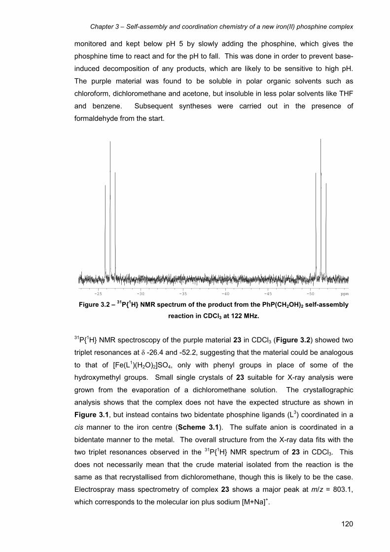
Chapter 3 – Self-assembly and coordination chemistry of a new iron(II) phosphine complex
120
monitored and kept below pH 5 by slowly adding the phosphine, which gives the
phosphine time to react and for the pH to fall. This was done in order to prevent base-
induced decomposition of any products, which are likely to be sensitive to high pH.
The purple material was found to be soluble in polar organic solvents such as
chloroform, dichloromethane and acetone, but insoluble in less polar solvents like THF
and benzene. Subsequent syntheses were carried out in the presence of
formaldehyde from the start.
-25 -30 -35 -40 -45 -50 ppm Figure 3.2 – 31P{1H} NMR spectrum of the product from the PhP(CH2OH)2 self-assembly
reaction in CDCl3 at 122 MHz.
31P{1H} NMR spectroscopy of the purple material 23 in CDCl3 (Figure 3.2) showed two
triplet resonances at δ -26.4 and -52.2, suggesting that the material could be analogous
to that of [Fe(L1)(H2O)2]SO4, only with phenyl groups in place of some of the
hydroxymethyl groups. Small single crystals of 23 suitable for X-ray analysis were
grown from the evaporation of a dichloromethane solution. The crystallographic
analysis shows that the complex does not have the expected structure as shown in
Figure 3.1, but instead contains two bidentate phosphine ligands (L3) coordinated in a
cis manner to the iron centre (Scheme 3.1). The sulfate anion is coordinated in a
bidentate manner to the metal. The overall structure from the X-ray data fits with the
two triplet resonances observed in the 31P{1H} NMR spectrum of 23 in CDCl3. This
does not necessarily mean that the crude material isolated from the reaction is the
same as that recrystallised from dichloromethane, though this is likely to be the case.
Electrospray mass spectrometry of complex 23 shows a major peak at m/z = 803.1,
which corresponds to the molecular ion plus sodium [M+Na]+.

Chapter 3 – Self-assembly and coordination chemistry of a new iron(II) phosphine complex
121
Scheme 3.1 – Self-assembly synthesis of [Fe(L3)2(κ2-O2SO2)] (23) (left) and its self-
assembled ligand L3.
Following the X-ray structural analysis, the ratio of phosphine to iron sulfate to
ammonium sulfate to formaldehyde was adjusted to 4:1:2:2 respectively in order to
match the stoichiometry of the product and give an optimal yield (Scheme 3.1).
Though the formaldehyde is essentially catalytic (with respect to the Mannich reaction
as shown on page 93), the methylene group between the nitrogen atoms on each
ligand is a result of the condensation of a molecule of formaldehyde, and is therefore
also a reagent.
2.83.03.23.43.63.84.04.24.44.64.85.05.25.4 ppm Figure 3.4 – 1H NMR spectrum of [Fe(L3)2(κ
2-O2SO2)] (23) at 500 MHz in CDCl3. The
different chemical environments of the ligand methylene groups shown arise from its
coordination to the metal and cis octahedral geometry of the complex.
N
P
N
P PhPh
▲ ▲ ■ ■
●
● ♦
♦
◙ ◙
◙
▲
■ ●
♦

Chapter 3 – Self-assembly and coordination chemistry of a new iron(II) phosphine complex
122
The 1H NMR spectrum of complex 23 (Figure 3.4) showed five different AB signals
arising from the five different methylene proton environments, four from the P-CH2-N
groups (▲, ■, ●, ♦) and one from the N-CH2-N moiety (◙). This means that all ten
methylene protons on the L3 ligand backbone are magnetically inequivalent in the 1H
NMR spectrum. The N-CH2-N methylene protons give rise to the AB system centred at
ca. δ 4.1 (◙). The four sets of P-CH2-N methylene resonances (▲, ■, ●, ♦) cannot be
individually assigned to a particular P-CH2-N group, which are only magnetically
inequivalent due to the cis geometry of the complex.
3.4 X-ray structure of [Fe(L3)2(κ2-O2SO2)]·2H2O (23·2H2O)
Purple crystals of 23·2H2O were grown by slow evaporation of a dichloromethane
solution. The asymmetric unit of the structure contains one half molecule of the
complex and a molecule of H2O, with the other half being generated by symmetry. The
structure shows the octahedral iron(II) complex with two of the bidentate L3 phosphine
ligands arranged in a cis configuration, with the remaining two coordination sites
occupied by a coordinated bidentate sulfate ligand, giving an overall neutral molecule
(Figure 3.5). The L3 ligand contains two fused six-membered PN2C3 rings, with both
rings sharing the two nitrogen atoms and the N-CH2-N methylene group. A phenyl
group is the third substituent on both of the phosphine donors.
The two fused six-membered PN2C3 rings that make up the backbone of each L3 ligand
both possess chair conformations. This enables the lone pairs of the phosphines to
point at (almost) 90° to each other, which allows the donation of electron density to the
empty orbitals on the metal. The ligand can also be thought of as an eight-membered
P2N2C4 ring (i.e. a 1,5-diaza-3,7-diphosphacyclooctane) with a methylene bridge
between the two nitrogen atoms. This bridging CH2 moiety is formed from the
condensation of a formaldehyde molecule with the remaining N-H group on each side
of the eight-membered ring, losing one molecule of water for each ligand. This means
that the formaldehyde plays two roles in the self-assembly of the L3 ligand; acting as a
catalyst in the Mannich reaction involved in the self-assembly process, as well as a
reagent that is an essential part of the final ligand structure.
The crystallographic bite angle of L3 in this structure is 83.41(6)°. This angle is
significantly larger than the average equatorial crystallographic bite angle (i.e. of the
phosphines in the eight membered P2N2C4 ring) of the L1 and L2 complexes in chapter
2 (80.51(9.5) and 80.06(7)°, respectively). However, this cannot be directly compared

Chapter 3 – Self-assembly and coordination chemistry of a new iron(II) phosphine complex
123
as the bite angle of the L3 ligand is not in the equatorial plane. The bite angle of L3 is
fairly similar to average values of other similar diphosphine ligands in the literature,
such as dppe (82.55(3.65)°) and dpp-benzene (81.95(3.25)°), as shown in Chapter 1,
Figure 1.5.2 The crystallographic bite angle of the coordinated sulfate molecule is
68.4(2)°, which is marginally smaller than the crystallographic bite angle of the
coordinated sulfate molecule in [Fe(L2)(κ2-O2SO2)] (complex 13 in chapter 2), which is
69.74(15)°. Due to these constrained bite angles the octahedral geometry of the
complex is distorted, giving a wide range of cis equatorial bond angles between 69.74
and 99.55°, which are far from the ideal 90° angles in a perfect octahedron.
The analogous L2 sulfate complex 13 has similar Fe-P bond lengths to 23, with
2.1840(18) Å (equatorial) and 2.2829(16) Å (axial) for 23 compared to 2.1803(17) and
2.1776(17) Å (equatorial) and 2.2256(16) and 2.2362(16) Å (axial) for 13. As with the
complexes of L1 and L2 in chapter 2, the axial Fe-P bonds of 23 are noticeably longer
than their equatorial equivalents due to the greater trans influence of the trans
phosphine donor compared to the oxygen donor of the sulfate ligand trans to the
equatorial phosphines. This effect is even more pronounced in 23, with ca. 0.1 Å
difference between the axial and equatorial Fe-P bond lengths. The Fe-O bond lengths
of 13 and 23 are also similar, with 2.061(4) Å for 23 compared to 2.047(4) and 2.046(4)
Å for complex 13.
The phenyl groups on the ligand do not face one another and there is no intramolecular
π-π stacking in each ligand, neither does there appear to be any intermolecular π-π
interactions between individual discrete molecules. The two phenyl groups on each
ligand are oriented almost perpendicular to one another (i.e. by rotation about the P-C
bond attaching the aromatic ring to the phosphine), and each ligand is positioned on
the metal in a way which minimises the inter-ligand steric interactions between the
aromatic rings.

Chapter 3 – Self-assembly and coordination chemistry of a new iron(II) phosphine complex
124
Figure 3.5 – Molecular structure of cis-[Fe(L3)2(κ2-O2SO2)]·2H2O (23·2H2O). The
asymmetric unit contains ½ a molecule of complex 1 with one full water molecule.
Thermal ellipsoids are at the 30% probability level. Included water molecules and
hydrogen atoms have been omitted for clarity.
Bond Lengths
Fe(1)-P(1) 2.2829(16) S(1)-O(1) 1.502(4)
Fe(1)-P(2) 2.1840(18) S(1)-O(2) 1.452(4)
Fe(1)-O(1) 2.061(4)
Bond Angles
P(1)-Fe(1)-P(2) 83.41(6) P(1)-Fe(1)-P(1)’ 177.00(11)
O(1)-Fe(1)-P(2) 96.20(11) O(1)-S(1)-O(1)’ 101.0(3)
O(1)-Fe(1)-O(1)’ 68.4(2) O(1)-S(1)-O(2) 109.8(2)
P(2)-Fe(1)-P(2)’ 99.55(10) O(2)-S(1)-O(2)’ 112.5(4)
Table 3.1 – Selected bond lengths (Å) and angles (°) for complex 23·2H2O (standard
deviation in brackets).

Chapter 3 – Self-assembly and coordination chemistry of a new iron(II) phosphine complex
125
3.5 Synthesis of cis-[Fe(L3)2Cl2] (24a)
Initial attempts to displace the coordinated sulfate ligand in 23 were unsuccessful. In
direct contrast to the iron(II) complexes of L2, when PPNCl was added to a
dichloromethane solution of 23 no reaction occurred. [Fe(L3)2(κ2-O2SO2)] (23) does not
appear to be the best precursor for the investigation of further coordination chemistry.
In order to synthesise a bis-L3 dichloride complex, iron(III) chloride and ammonium
chloride (rather than the equivalent sulfate salts) were reacted with PhP(CH2OH)2 and
formaldehyde as in Scheme 3.1 in section 3.2. Using the same stoichiometry, the
reaction gave a similar purple precipitate as in the formation of complex 23.
-26 -28 -30 -32 -34 -36 -38 -40 -42 -44 -46 -48 -50 ppm Figure 3.6 – 31P{1H} NMR spectrum of the dichloride complex cis-[Fe(L3)2Cl2] (24a) in
CDCl3 at 122 MHz.
The 31P{1H} NMR spectrum of the purple solid in CDCl3 (Figure 3.6) showed two triplet
resonances (at ca. δ -32 and -46) as would be expected, suggesting the dichloride
complex cis-[Fe(L3)2Cl2] (24a) had been successfully formed. However, the
resonances were broad suggesting the complex is fluxional or unstable. This
fluxionality could be the result of the ability of the complex to isomerise between the cis
and trans isomers of the complex, as it is not seen in the case of the chelating sulfate
complex 23, which cannot switch between the cis and trans isomers. Complex 24a
partially dissolves in aqueous solution to give a purple solution. When the 31P{1H} NMR
spectrum was recorded in D2O two sharper resonances were observed at δ -17.8 and -
38.2 (Figure 3.7), although these were now doublets of doublets rather than triplets.
As complex 24a is partially soluble in aqueous solution at neutral pH but does not
possess any highly soluble functional groups, it is likely that the chloride ligands are
being displaced with solvent H2O molecules to give the dicationic diaqua complex

Chapter 3 – Self-assembly and coordination chemistry of a new iron(II) phosphine complex
126
cis-[Fe(L3)2(H2O)2]Cl2, which should be far more polar and therefore soluble in water.
Complex 24a is increasingly soluble with decreasing pH, possibly due to protonation of
the nitrogen groups. If an excess of sodium chloride is added to the aqueous solution
of cis-[Fe(L3)2(H2O)2]Cl2, the dichloride complex cis-[Fe(L3)2Cl2] (24a) precipitates from
solution. In comparison, the similar bidentate sulfate complex 23 is completely
insoluble in aqueous solution. This is presumably due to the fact that the chelating
bidentate sulfate ligand cannot be as easily displaced as the monodentate chloride
ligands on 24a to give a cationic water-soluble complex.
-16 -18 -20 -22 -24 -26 -28 -30 -32 -34 -36 -38 ppm Figure 3.7 – 31P{1H} NMR spectrum of cis-[Fe(L3)2(H2O)2]Cl2 in D2O at 122 MHz.
A closer inspection of the resonances reveals that they have a small splitting of ca. 7
Hz, making the peaks appear like doublets of doublets. This suggests that both
equatorial phosphines are magnetically inequivalent and that each axial phosphine
couples to each equatorial phosphine with a slightly different coupling constant.
Attempts to grow crystals of the purple material from most organic solvents or from
aqueous solution proved unsuccessful. However, it was observed that despite the
complex being largely insoluble in diethyl ether, washings of the purple solid gave a
very dilute pale yellow ether solution. Slow evaporation of the ether solution gave
yellow crystals suitable for X-ray crystallography. The resulting structure showed that
complex was the trans isomer of the dichloride complex trans-[Fe(L3)2Cl2] (24b), with
two identical L3 bidentate phosphine ligands as in complex 23. Due to the very low
solubility of 24b in diethyl ether, a good 31P{1H} NMR spectrum proved very difficult to
obtain. When dissolved in other more polar organic solvents such as dichloromethane,
Chapter 3 – Self-assembly and coordination chemistry of a new iron(II) phosphine complex
127
chloroform or acetone, the yellow crystals of 24b dissolved to give a purple solution. 31P{1H} NMR spectroscopic analysis of this solution showed the familiar broad triplet
resonances at ca. δ -32 and -46, suggesting that the fluxional cis dichloride complex
24a had been reformed. Dissolving the trans complex 24b in D2O also gave a purple
solution, the 31P{1H} NMR spectrum of which displayed two doublet of doublet
resonances at δ -17.8 and -38.2. This suggests that the diaqua complex
cis-[Fe(L3)2(H2O)2]Cl2 had formed, identical to when the cis isomer 24a was dissolved
in aqueous solution.
3.6 X-ray structure of trans-[Fe(L3)2Cl2] (24b)
The X-ray analysis of the yellow crystals of 24b grown from the evaporation of an Et2O
solution confirmed the trans geometry of the complex (Figure 3.8). The complex in the
structure is unsolvated, and the asymmetric unit contains a total of 1.5 molecules of
24b, comprising of one full molecule and one half molecule residing on a mirror plane.
The three independent crystallographic bite angles for the L3 ligand in the structure are
82.01(2) and 81.67(2)° for the full molecule, and 80.840(17)° for the half molecule.
These are all notably lower than in cis-[Fe(L3)2(κ2-O2SO2)] (1) (83.41(6)°), and are in
general closer in value to the crystallographic bite angles for the L1 and L2 complexes
in chapter 2. Similar to complex 1 as well as many complexes in chapter 2, the
geometry of 24b is quite distorted from a regular octahedron, with the cis equatorial P-
Fe-P bond angles ranging between 80.840(17) and 99.160(17)° for the half molecule
(molecule 1) and between 81.67(2) and 99.98(2)° for the full molecule (molecule 2).
This is most notable in the full molecule in the structure, with the two L3 ligands being
staggered in relation to one another, both being tilted in opposite directions away from
the equatorial plane. The phenyl rings on each L3 ligand in this molecule are also tilted
away from the axial plane in opposite directions from each other. In contrast, the L3
ligands on the half molecule almost completely eclipse one another in the equatorial
plane, with the phenyl groups lying perpendicular to the equatorial plane, giving a more
regular octahedral geometry. In both molecules, the phenyl groups face one another
and are between 3.30 and 3.64 Å in the symmetrical half molecule, though the less
symmetrical full molecule has phenyl groups which do not directly face each other so
the distance is harder to judge, though the distance between rings is approximately 3.4
Å in this case. All these distances are typical values for face to face π-π stacking
interactions between two aromatic rings.3

Chapter 3 – Self-assembly and coordination chemistry of a new iron(II) phosphine complex
128
Figure 3.8 – Molecular structure of trans-[Fe(L3)2Cl2] (24b). The asymmetric unit contains
1.5 molecules of complex 20b, consisting of one full and one half molecule (molecule 2
(full molecule) is shown). Thermal ellipsoids are at the 30% probability level. Hydrogen
atoms have been omitted for clarity.
Bond Lengths (molecule 1)
Fe(1)-Cl(1) 2.3598(5) Fe(1)-P(2) 2.2954(5)
Fe(1)-P(1) 2.2674(5)
Bond Angles (molecule 1)
Cl(1)-Fe(1)-Cl(1)’ 180.00(2) P(2)-Fe(1)-P(2)’ 180.000(17)
P(1)-Fe(1)-P(2) 80.840(17) P(1)-Fe(1)-P(2)’ 99.160(17)
P(1)-Fe(1)-P(1)’ 180.000(17) P(2)-Fe(1)-P(1)’ 99.160(17)
Table 3.2 – Selected bond lengths (Å) and angles (°) for complex 24b (standard deviation
in brackets).

Chapter 3 – Self-assembly and coordination chemistry of a new iron(II) phosphine complex
129
Bond Lengths (molecule 2)
Fe(2)-Cl(2) 2.3675(5) Fe(2)-P(4) 2.2476(5)
Fe(2)-Cl(3) 2.3610(5) Fe(2)-P(5) 2.2615(5)
Fe(2)-P(3) 2.2469(5) Fe(2)-P(6) 2.2506(5)
Bond Angles (molecule 2)
Cl(2)-Fe(2)-Cl(3) 177.47(2) P(5)-Fe(2)-P(6) 81.67(2)
P(3)-Fe(2)-P(5) 168.17(2) P(3)-Fe(2)-P(6) 99.98(2)
P(4)-Fe(2)-P(6) 169.12(2) P(4)-Fe(2)-P(5) 98.60(2)
P(3)-Fe(2)-P(4) 82.01(2)
Table 3.3 – Selected bond lengths (Å) and angles (°) for complex 24b (standard deviation
in brackets).
The six different equatorial Fe-P bond lengths in the structure of 24b 2.2674(5),
2.2954(5), 2.2469(5), 2.2476(5), 2.2615(5) and 2.2506(5) Å (arising from the 1.5
independent molecules in the asymmetric unit) are closer in value to the axial rather
than the equatorial Fe-P bond lengths in the structure of 23. This is because all six Fe-
P bonds are trans to another phosphine as a result of the complex possessing trans
geometry, and is again a result of the phosphine having a greater trans influence than
the oxygen of the sulfate ligand on 23. However, the Fe-P bond lengths of the similar
complex trans-[Fe(dmpe)2Cl2] (dmpe = 1,2-bis(dimethylphosphino)ethane) are 2.241(1)
and 2.230(1) Å,4 which are shorter than those reported for 24b. The shorter Fe-P bond
lengths in trans-[Fe(dmpe)2Cl2] can be explained partially by the lower steric
requirement for the smaller dimethylphosphine groups compared to the larger aromatic
groups on L3 in 24a, and partially because of the increased σ-donor ability of the dmpe
ligand compared to L3. The Fe-Cl bonds lengths of 24b (2.3596(5), 2.3675(5), and
2.3610(5) Å), however, are similar in value to the Fe-Cl bond lengths of [Fe(L2)Cl2] (15)
(2.371(2) and 2.351(5) Å). This is despite the fact that the chloride ligands in 24b are
trans to each other whereas they are trans to a phosphine donor in 15. The Fe-Cl
bond length in trans-[Fe(dmpe)2Cl2] is 2.352(1) Å,4 which is largely similar to those of
24a.
3.7 Coordination chemistry of cis-[Fe(L3)2Cl2] (24a) with pseudo-halides
In order to compare and contrast the coordination chemistry of the L3 complexes with
that of the related L1 and L2 complexes, [Fe(L3)2Cl2] was reacted with a range of neutral
and anionic ligands.
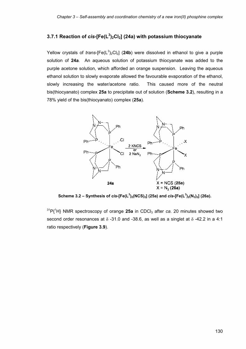
Chapter 3 – Self-assembly and coordination chemistry of a new iron(II) phosphine complex
130
3.7.1 Reaction of cis-[Fe(L3)2Cl2] (24a) with potassium thiocyanate
Yellow crystals of trans-[Fe(L3)2Cl2] (24b) were dissolved in ethanol to give a purple
solution of 24a. An aqueous solution of potassium thiocyanate was added to the
purple acetone solution, which afforded an orange suspension. Leaving the aqueous
ethanol solution to slowly evaporate allowed the favourable evaporation of the ethanol,
slowly increasing the water/acetone ratio. This caused more of the neutral
bis(thiocyanato) complex 25a to precipitate out of solution (Scheme 3.2), resulting in a
78% yield of the bis(thiocyanato) complex (25a).
Scheme 3.2 – Synthesis of cis-[Fe(L3)2(NCS)2] (25a) and cis-[Fe(L
3)2(N3)2] (26a).
31P{1H} NMR spectroscopy of orange 25a in CDCl3 after ca. 20 minutes showed two
second order resonances at δ -31.0 and -38.6, as well as a singlet at δ -42.2 in a 4:1
ratio respectively (Figure 3.9).

Chapter 3 – Self-assembly and coordination chemistry of a new iron(II) phosphine complex
131
-26 -28 -30 -32 -34 -36 -38 -40 -42 -44 -46 -48 ppm
-26 -28 -30 -32 -34 -36 -38 -40 -42 -44 -46 -48 ppm Figure 3.9 – 31P{1H} NMR spectroscopy of cis- and trans-[Fe(L3)2(NCS)2] (25a) and (25b) in
CDCl3 at 122 MHz after ca. 20 minutes (top) and ca. 48 hours (bottom).
The resonance pattern corresponds to the cis-[Fe(L3)2(NCS)2] complex (25a), whereas
the singlet represents the related trans isomer trans-[Fe(L3)2(NCS)2] (25b). Over time
the ratio of cis to trans isomers in CDCl3 solution decreased, as shown by the 31P{1H}
NMR spectrum recorded after 48 hr. (Figure 3.9), showing that the cis:trans ratio had
changed from 4:1 to 2:3. Orange crystals suitable for X-ray crystallography also grew
over time, and which proved to be of the trans isomer 25b.
1H NMR spectroscopic analysis of the mixture of cis and trans isomers showed a
complex spectrum. As in the 1H NMR spectrum of cis-[Fe(L3)2(κ2-O2SO2)] (23), cis-
[Fe(L3)2(NCS)2] (25a) gave rise to five separate AB resonances arising from the four P-
CH2-N methylene groups and the one N-CH2-N group. The trans isomer 25b,
displayed one AB system and a singlet as a result of the increased symmetry of the
trans isomer, in which all four P-CH2-N methylene groups are magnetically equivalent.
The two protons on each P-CH2-N methylene group are, however, magnetically
inequivalent, as one is essentially axial and the other is equatorial in the PN2 six-
membered rings of the L3 ligand. The N-CH2-N methylene group gave rise to a singlet
as both protons on this methylene group are magnetically equivalent.

Chapter 3 – Self-assembly and coordination chemistry of a new iron(II) phosphine complex
132
3.7.2 X-ray structure of trans-[Fe(L3)2(NCS)2] (25b)
The X-ray analysis of the orange crystals isolated from the CDCl3 solution of 25b
showed the structure to be of the trans isomer of the bis(isothiocyanato) complex trans-
[Fe(L3)2(NCS)2] (25b) (Figure 3.10), with the structure being unsolvated. The
asymmetric unit of the structure contains one half molecule of 25b. The coordinated
thiocyanate ligands bind to the iron centre via the nitrogen atoms, which is consistent
with the L1 isothiocyanato complex [Fe(L1)(NCS)2] (6 in Chapter 2).
The Fe-N bond length in 25b is 1.9387(14) Å, which is noticeably shorter than the Fe-N
bond lengths in complex 6 (1.960(3) and 1.952(3) Å). This is because the thiocyanate
ligands in 6 are trans to phosphine donors, which have a greater trans influence than
the nitrogen of the other thiocyanate ligand in complex 25b. The greater trans
influence of the phosphine groups compared to nitrogen causes the Fe-N bond in 6 to
become longer and therefore weaker. As a result, it would therefore be expected that
the Fe-P bonds in the equatorial plane in complex 25b to be longer than those of 6 in
Chapter 2 as they are now positioned trans to other phosphine donors from another L3
ligand. This is indeed the case, with the equatorial Fe-P bond lengths in 6 being
2.1840(10) and 2.1907(11) Å, compared to 2.2453(4) and 2.2592(4) Å for complex
25b. This phenomenon is also observed for similar cis and trans iron(II) thiocyanato
tetraphosphine complexes in the literature. The Fe-N bond lengths of trans-[Fe(rac-
prP4)(NCS)2] (rac-prP4 = 1,1,4,8,11,11-hexaphenyl-1,4,8,11-tetraphosphaundecane)
are 1.932(2) and 1.933(2) Å compared to 1.973(2) and 1.964(2) Å for cis-α-[Fe(rac-
prP4)(NCS)2].5
The conformation of 25b is similar to that of the full molecule in the X-ray structure of
trans-[Fe(L3)2Cl2] 24b, i.e. the two L3 ligands on the metal are staggered with respect to
each other and are both tilted in opposite directions away from the equatorial plane.
The phenyl groups are also twisted from the axial plane and face the equivalent phenyl
group on the opposite ligand, suggesting that π-π interactions of ca. 3.3 Å are also
occurring in this structure.
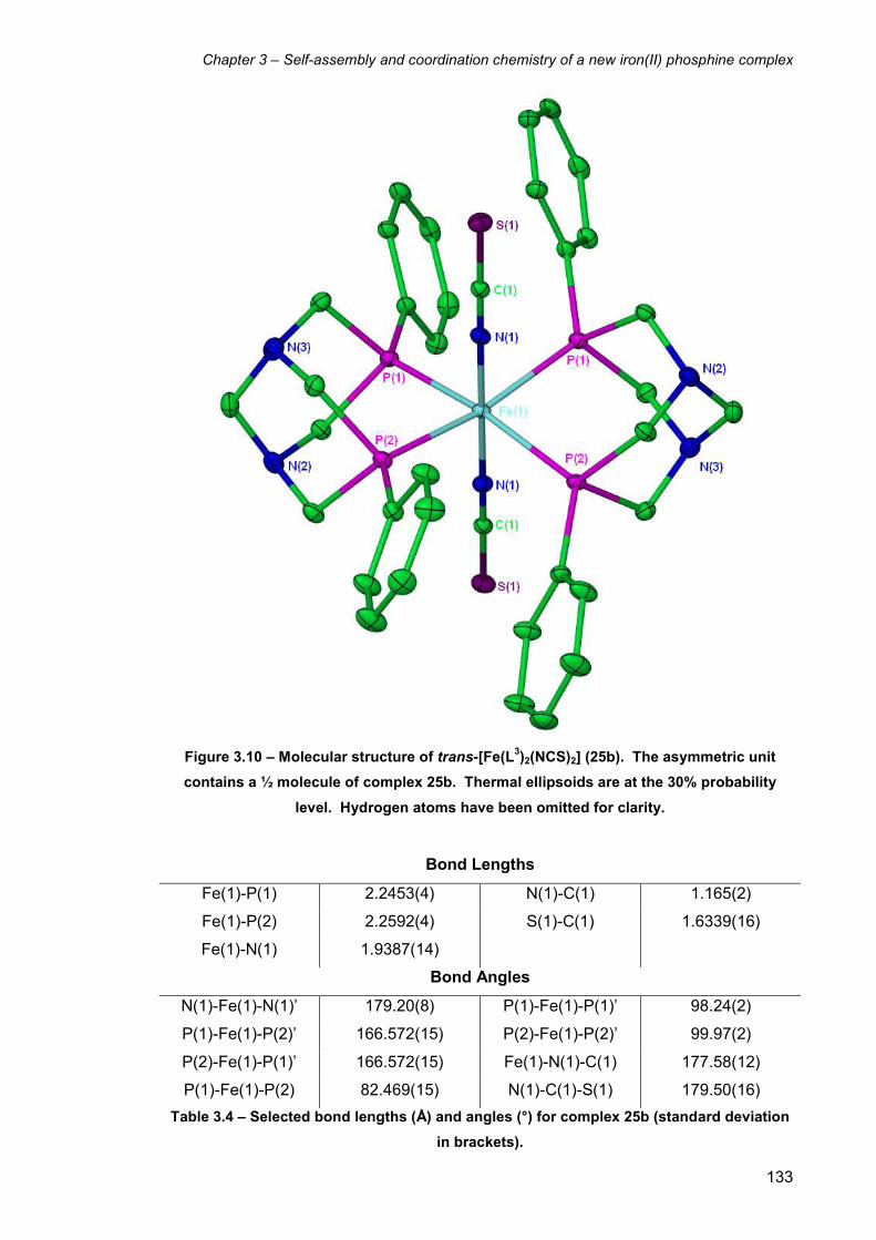
Chapter 3 – Self-assembly and coordination chemistry of a new iron(II) phosphine complex
133
Figure 3.10 – Molecular structure of trans-[Fe(L3)2(NCS)2] (25b). The asymmetric unit
contains a ½ molecule of complex 25b. Thermal ellipsoids are at the 30% probability
level. Hydrogen atoms have been omitted for clarity.
Bond Lengths
Fe(1)-P(1) 2.2453(4) N(1)-C(1) 1.165(2)
Fe(1)-P(2) 2.2592(4) S(1)-C(1) 1.6339(16)
Fe(1)-N(1) 1.9387(14)
Bond Angles
N(1)-Fe(1)-N(1)’ 179.20(8) P(1)-Fe(1)-P(1)’ 98.24(2)
P(1)-Fe(1)-P(2)’ 166.572(15) P(2)-Fe(1)-P(2)’ 99.97(2)
P(2)-Fe(1)-P(1)’ 166.572(15) Fe(1)-N(1)-C(1) 177.58(12)
P(1)-Fe(1)-P(2) 82.469(15) N(1)-C(1)-S(1) 179.50(16)
Table 3.4 – Selected bond lengths (Å) and angles (°) for complex 25b (standard deviation
in brackets).

Chapter 3 – Self-assembly and coordination chemistry of a new iron(II) phosphine complex
134
3.7.3 Reaction of cis-[Fe(L3)2Cl2] (24a) with sodium azide
The reaction of 24a with sodium azide was carried out in a similar manner to the
reaction of 24a with potassium thiocyanate. An aqueous solution of sodium azide was
added to an ethanol solution of cis-[Fe(L3)2Cl2] (24a), resulting in a purple/red
suspension presumably of cis-[Fe(L3)2(N3)2] (26a). As before, the ethanol was allowed
to evaporate, resulting in the formation of a purple/red precipitate.
-24 -26 -28 -30 -32 -34 -36 -38 -40 -42 -44 -46 ppm Figure 3.11 – 31P{1H} NMR spectrum of cis- and trans-[Fe(L3)2(N3)2] (26a, 26b) in CDCl3 at
122 MHz after ca. 20 minutes. The spectrum was identical after 48 hours.
31P{1H} NMR spectroscopy of the purple/red solid in CDCl3 after ca. 20 minutes showed
two second order resonances at δ -27.4 and -36.9, as well as a singlet at δ -43.2 in an
11:1 ratio (Figure 3.11). As with the thiocyanate reaction, the reaction was monitored
over time, although the 31P{1H} NMR spectrum was unchanged after 48 hours, showing
that the isomerisation process is not as favourable for the bis(azido) complexes (26a
and 26b). As a result, the crystallisation of neither the cis or trans isomers could be
achieved. The formation of both cis and trans isomers of 26 is in contrast to the
synthesis of the similar bis(azido) complex trans-[Fe(dmpe)2(N3)2] (dmpe =
bis(dimethylphosphino)ethane), which showed no evidence for the formation of the cis
isomer and only the trans isomer was observed in the 31P{1H} NMR spectrum.6
The 1H NMR spectrum of the mixture of 26a and 26b showed a similar series of
resonances to that of the bis(thiocyanato) complexes 25a and 25b. Therefore, one AB
system and a singlet were observed for the trans isomer 25b, and five AB resonances
for the cis isomer 25a. IR spectroscopy of the mixture showed a peak at 2043 cm-1,
which is likely to be of the cis isomer 25a due to the 11:1 cis:trans ratio. The cis isomer

Chapter 3 – Self-assembly and coordination chemistry of a new iron(II) phosphine complex
135
would be expected to show both symmetric and antisymmetric stretches, though one
peak was also observed for [Fe(L1)(N3)2] (7) in Chapter 2 (section 2.5) and by Field et
al. in related cis bis(azido) iron(II) phosphine complexes.7
3.8 Coordination chemistry of cis-[Fe(L3)2Cl2] (24a) with carbonate
The reaction of an ethanol solution of cis-[Fe(L3)2Cl2] (24a) with an aqueous solution of
sodium carbonate gave a pink suspension of [Fe(L3)2(κ2-O2CO)] (27) (Scheme 3.3).
Scheme 3.3 – Reaction of cis-[Fe(L3)2Cl2] (24a) with sodium carbonate to give the
carbonato complex [Fe(L3)2(κ2-O2CO)] (27).
The 31P{1H} NMR spectrum of the pink precipitate in CDCl3 showed two triplet
resonances at δ -20.2 and -41.4 with 2JP-P = 72.0 Hz (Figure 3.12).
-15 -20 -25 -30 -35 -40 -45 ppm Figure 3.12 – 31P{1H} NMR spectrum of cis-[Fe(L3)2(κ
2-O2CO)] (27) in CDCl3 at 122 MHz.

Chapter 3 – Self-assembly and coordination chemistry of a new iron(II) phosphine complex
136
The 1H NMR spectrum of 27 showed the expected pattern for a bis L3 complex
possessing cis geometry, i.e. four AB resonances for the P-CH2-N methylene protons
and one AB system for the N-CH2-N methylene protons (Figure 3.13).
2.83.03.23.43.63.84.04.24.44.64.85.05.2 ppm
Figure 3.13 – Selected 1H NMR spectrum of cis-[Fe(L3)2(κ2-O2CO)] (27) in CDCl3 at 500
MHz. The different chemical environments of the ligand methylene groups shown arise
from its coordination to the metal and cis octahedral geometry of the complex.
The N-CH2-N protons give rise to the second order AB system centred at δ 3.9 (◙), with
the four remaining P-CH2-N methylene proton groups giving four sets of AB systems
between δ 5.2 and 2.6 (some of which overlap) (▲, ■, ●, ♦). The connectivity of these
resonances was confirmed by 1H COSY NMR spectroscopy. As with 23, the four P-
CH2-N methylene groups (▲, ■, ●, ♦) cannot be individually assigned to a particular
P-CH2-N group, which are only magnetically inequivalent due to the cis geometry of the
complex.
3.9 X-ray structure of [Fe(L3)2(CO3)]·7H2O (27·7H2O)
Purple crystals of 27 were grown from the slow evaporation of the aqueous reaction
filtrate over ca. eight weeks, and are of the heptahydrate (27·7H2O). The structure
confirms that the two chloride ligands have been replaced with a bidentate carbonate
molecule, giving a cis octahedral geometry as expected (Figure 3.14). The chelating
carbonate ligand has a small bite angle (65.01(9)°), which results in a structure that is
far from a regular octahedral geometry. The cis equatorial bond angles range between
65.01(9) and 100.98(3)°. The crystallographic bite angles of the L3 ligand in the
N
P
N
P PhPh
▲
▲ ■
■ ●
● ♦
♦
◙ ◙
▲
■ ●
♦
◙

Chapter 3 – Self-assembly and coordination chemistry of a new iron(II) phosphine complex
137
Figure 3.14 – Molecular structure of [Fe(L3)2(κ2-O2CO)]·7H2O (27·7H2O). Thermal
ellipsoids are at the 30% probability level. Hydrogen atoms and included water
molecules have been omitted for clarity.
Bond Lengths
Fe(1)-P(1) 2.2046(9) Fe(1)-O(2) 2.016(2)
Fe(1)-P(2) 2.2631(10) O(1)-C(20) 1.317(4)
Fe(1)-P(3) 2.2573(10) O(2)-C(20) 1.315(4)
Fe(1)-P(4) 2.1910(9) O(3)-C(20) 1.246(4)
Fe(1)-O(1) 2.029(2)
Bond Angles
P(1)-Fe(1)-P(2) 82.61(3) P(1)-Fe(1)-O(1) 99.29(7)
P(3)-Fe(1)-P(4) 82.89(3) P(4)-Fe(1)-O(2) 95.08(7)
O(1)-Fe(1)-O(2) 65.01(9) O(1)-C(20)-O(2) 111.4(3)
P(1)-Fe(1)-P(4) 100.98(3)
Table 3.5 – Selected bond lengths (Å) and angles (°) for complex 27·7H2O (standard
deviation in brackets).

Chapter 3 – Self-assembly and coordination chemistry of a new iron(II) phosphine complex
138
structure are 82.61(3) and 82.89(3)°, which are similar to those in complexes 23, 24b,
and 25b, though somewhat less than that in the coordinated sulfate complex 23. In
comparison with the X-ray structure of [Fe(L1)(κ2-O2CO)] (complex 8 in Chapter 2), the
Fe-O iron-carbonate bonds are similar (2.029(2) and 2.016(2) Å for 27 compared to
2.044(5) and 2.073(5) Å for complex 8). The O-Fe-O bond angle in complex 27
(65.01(9) Å) is larger than that of 8 (63.8(2) Å).
3.10 Reaction of cis-[Fe(L3)2Cl2] (24a) with CO
The reaction of an aqueous solution of cis-[Fe(L3)2Cl2] (24a) with CO resulted in a
colour change from a purple to a yellow solution over ca. 1 hr, suggesting that one or
both of the coordinated chlorides may have been displaced by a carbonyl ligand to give
a complex such as 28a (Scheme 3.4) or a dicarbonyl such as [Fe(L3)2(CO)2]Cl2.
Scheme 3.4 – Possible product from the reaction of cis-[Fe(L3)2Cl2] (24a) with CO in
aqueous solution.
The 31P{1H} NMR spectrum of the yellow solution in D2O showed a singlet resonance at
δ -51.1 (Figure 3.15), consistent with formation of a trans L3 complex. However, this
does not determine whether the trans complex is mono- or di-substituted, meaning that
the identity of the complex could be one of three likely structures, (i) [Fe(L3)2(CO)2]2+,
(ii) [Fe(L3)2(CO)(H2O)]2+, or (iii) [Fe(L3)2(CO)(Cl)]
+.

Chapter 3 – Self-assembly and coordination chemistry of a new iron(II) phosphine complex
139
-42 -43 -44 -45 -46 -47 -48 -49 -50 -51 -52 -53 -54 -55 -56 -57 -58 -59 -60 ppm Figure 3.15 - 31P{1H} NMR spectrum following reaction of 24a with CO in D2O at 122 MHz.
1H NMR spectroscopy showed two sets of AB doublets at δ 5.1, 4.6 and 4.0, 3.7, as
well as a singlet at δ 4.1 (Figure 3.16). This indicates that the complex is mono-
substituted i.e. product (ii) or (iii), since the 1H NMR spectrum of (i) would give one pair
of doublets and one singlet in a 2:2:1 ratio because the four P-CH2-N proton groups on
each L3 ligand would be equivalent.
3.73.83.94.04.14.24.34.44.54.64.74.84.95.05.15.2 ppm Figure 3.16 – 1H NMR spectrum following reaction of 24a with CO in D2O at 500 MHz.
When the reaction was repeated with 13CO the same yellow solution was produced, but
now the 31P{1H} NMR spectrum showed a doublet at δ -51.1 with 2JP-C = 25.8 Hz. The
doublet indicates that only one 13CO ligand is bound to the iron metal. The 13C{1H}
NMR spectrum is consistent with this, showing a quintet 13CO resonance at δ 214.7.

Chapter 3 – Self-assembly and coordination chemistry of a new iron(II) phosphine complex
140
IR spectroscopy showed a single 12CO stretching frequency at 1939 cm-1, which would
be expected for a mono-carbonyl complex. The IR spectrum of the corresponding 13CO complex showed the equivalent 13CO stretching frequency at 1895 cm-1. This
value is very close to the calculated 13CO value for the complex, which is 1896 cm-1.
Mass spectrometry of the complex showed the major peak to be at m/z = 747.1, which
equates to the chloride carbonyl complex cation [Fe(L3)2(CO)Cl]+. As a result of the
NMR, IR, and mass spectral evidence the complex has been identified as the trans-
[Fe(L3)2(CO)(Cl)] Cl (28a)
When the trans-[Fe(L3)2Cl2] complex (24b) was reacted with CO in diethyl ether
solution (Scheme 3.5), a pale orange precipitate was formed suggesting the formation
of a different product to that found in aqueous solution.
Scheme 3.5 – Reaction of trans-[Fe(L3)2Cl2] (24b) with CO in diethyl ether solution.
The 31P{1H} NMR spectrum of the orange solid showed four main resonances at δ -
39.4, -55.3, -59.4, and -69.0, which are all doublets of doublets of doublets (Figure
3.17). The resonance at δ -69.0 appears simplified due to two of the 2JP-P values being
very similar. This coupling pattern means that all four phosphine groups on the
complex are inequivalent, indicating that the product is cis-[Fe(L3)2(CO)Cl]Cl (28b).

Chapter 3 – Self-assembly and coordination chemistry of a new iron(II) phosphine complex
141
-40 -45 -50 -55 -60 -65 -70 ppm Figure 3.17 - 31P{1H} NMR spectrum of cis-[Fe(L3)2(CO)Cl]Cl (28b) in CDCl3 at 122 MHz.
When 28b was redissolved in D2O a yellow solution is immediately observed, the 31P{1H} NMR spectrum of which showed a singlet resonance at δ -51.1. This is the
same resonance as observed for complex 28a, meaning that cis-[Fe(L3)2(CO)Cl]Cl
(28b) undergoes an isomerisation in water to give trans-[Fe(L3)2(CO)Cl]Cl (28a).
Another example of the synthesis of both cis and trans carbonyl chloride complexes
under different conditions is provided by Bellerby et al.8 When trans-[Fe(dmpe)2Cl2]
was reacted with CO in an acetone solution, the corresponding cis carbonyl chloride
cis-[Fe(dmpe)2(CO)Cl]+ was formed, which was isolated as the tetraphenylborate salt
upon addition of NaBPh4. In contrast, when trans-[Fe(dmpe)2Cl2] was reacted with CO
in an acetone solution at reflux, the majority of the material isomerised to give
trans-[Fe(dmpe)2(CO)Cl]+ as the major product, though some cis-[Fe(dmpe)2(CO)Cl]
+
was also formed. When depe was used as the ligand, trans-[Fe(depe)2Cl2] reacted
with CO in methanol to give trans-[Fe(depe)2(CO)Cl]BPh4 in the presence of NaBPh4,
and no cis isomer was formed.
3.11 Reaction of cis-[Fe(L3)2Cl2] (24a) with sodium borohydride
Two equivalents of sodium borohydride were added to a suspension of cis-[Fe(L3)2Cl2]
(24a) in ethanol under a nitrogen atmosphere (Scheme 3.6). The purple solution
quickly changed colour to orange, followed by formation of an orange precipitate over
ca. 1 hr. The reaction mixture was stirred at room temperature for a further 1 hr to
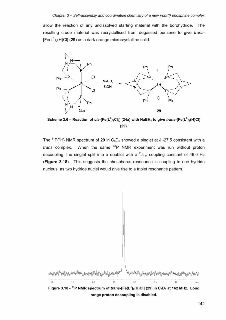
Chapter 3 – Self-assembly and coordination chemistry of a new iron(II) phosphine complex
142
allow the reaction of any undissolved starting material with the borohydride. The
resulting crude material was recrystallised from degassed benzene to give trans-
[Fe(L3)2(H)Cl] (29) as a dark orange microcrystalline solid.
Scheme 3.6 – Reaction of cis-[Fe(L3)2Cl2] (24a) with NaBH4 to give trans-[Fe(L
3)2(H)Cl]
(29).
The 31P{1H} NMR spectrum of 29 in C6D6 showed a singlet at δ -27.5 consistent with a
trans complex. When the same 31P NMR experiment was run without proton
decoupling, the singlet split into a doublet with a 2JP-H coupling constant of 49.0 Hz
(Figure 3.18). This suggests the phosphorus resonance is coupling to one hydride
nucleus, as two hydride nuclei would give rise to a triplet resonance pattern.
-10 -15 -20 -25 -30 -35 -40 ppm Figure 3.18 - 31P NMR spectrum of trans-[Fe(L3)2(H)Cl] (29) in C6D6 at 162 MHz. Long
range proton decoupling is disabled.

Chapter 3 – Self-assembly and coordination chemistry of a new iron(II) phosphine complex
143
The 1H NMR spectrum of the solid showed a quintet at δ -28.45 due to the coupling of
the hydride with the four equivalent phosphorus nuclei, confirming the presence of the
hydride in the complex (Figure 3.19). The 2JP-H coupling constant matches that of the 31P{1H} NMR experiment.
These observations are similar to those of Tyler and coworkers for the similar trans-
[Fe(DMeOPrPE)2(H)Cl] complex (DMeOPrPE = 1,2-bis(bis-
(methoxypropyl)phosphino)ethane), the (uncoupled) 31P NMR spectrum of which
showed a doublet at δ 83.5 with a 2JP-H coupling constant of 45 Hz, indicative of cis P-H
coupling. The 1H NMR spectrum showed a quintet at δ -31.8.9 Holah and coworkers
also found similar observations in the complex trans-[Fe(dppm)2(H)Cl] (dppm =
bis(diphenylphosphino)methane), the 31P{1H} NMR spectrum of which showed a singlet
at δ 24.2. The 1H NMR spectrum showed a quintet at δ -21.2 with 2JP-H = 44 Hz.10
-27.4 -27.6 -27.8 -28.0 -28.2 -28.4 -28.6 -28.8 -29.0 -29.2 -29.4 ppm Figure 3.19 – Hydride region of the 1H NMR spectrum of trans-[Fe(L3)2(H)Cl] (29) in C6D6 at
400 MHz.
Mass spectrometry of the product was carried out in order to fully characterise it and
determine which ligand occupies the site trans from the hydride. The ES-MS of 29 was
first carried out in acetonitrile and gave the major peak at m/z = 685.16, which fits the
molecular weight of the five coordinate complex [Fe(L3)2(H)]+. This unfortunately does
not give light to the identity of the other ligand, which has been removed during the
ionisation process. The mass spectrum was re-recorded in dichloromethane under an
inert atmosphere and now afforded the major peak at m/z = 720.13, corresponding to
trans-[Fe(L3)2(H)Cl]+.

Chapter 3 – Self-assembly and coordination chemistry of a new iron(II) phosphine complex
144
The reaction of 24a with six equivalents of NaBH4 in EtOH in an attempt to synthesise
the dihydride complex was also attempted. The 31P{1H} NMR spectrum of the reaction
solution showed three broad resonances at ca. δ -4, -6 and -29 in a 1:3:1 ratio,
respectively. Attempts to isolate any complexes from the reaction solution proved
unsuccessful.
3.12 X-ray structure of trans-[Fe(L3)2(H)Cl]·1.5C6H6 (29·1.5C6H6)
The orange microcrystalline solid of 29 formed from benzene solution upon leaving it at
room temperature for a few days. The crystals did not decompose in air even after
exposure for 24 hours, though the ethanolic solution of unpurified 29 turned brown and
decomposed upon exposure to air after a few minutes. The X-ray analysis showed the
expected octahedral iron complex with two bidentate L3 ligands arranged in a trans
configuration. One of the axial sites is occupied by a chloride ligand, while the opposite
trans axial site does not have significant electron density. This lack of electron density
is expected for a hydride ligand, which possesses no electrons other than the electron
pair used in bonding to the iron centre. In collaboration with the NMR and mass
spectral data, it can therefore be concluded that the complex is indeed the hydride
chloride complex trans-[Fe(L3)2(H)Cl] (29) (Figure 3.20).
The asymmetric unit of the structure contains two independent half molecules of
complex 29. The complex crystallises with one half molecule of benzene with full
occupancy, as well as one whole molecule of benzene exhibiting 1:1 disorder. The
compound is therefore 29·1.5C6H6. The hydride ligands in the structure cannot be
located by their electron density, therefore the hydride nuclei H(1) and H(2) have been
modelled equidistant from Fe(1) and Fe(2) respectively, at approximately 1.46 Å.
The two Fe-Cl bond lengths in the structure are 2.461(2) and 2.454(2) Å for the Fe(1)-
Cl(1) and Fe(1)-Cl(2) bonds, respectively. Only two other X-ray crystal structures of
similar complexes of the formula trans-[Fe(L)2(H)Cl] (where L = any bidentate
phosphine ligand) have been reported in the literature to date, of trans-[Fe(dppe)2(H)Cl]
(dppe = bis(diphenylphosphino)ethane)11 and trans-[Fe(depe)2(H)Cl] (depe =
bis(diethylphosphino)ethane).12 Both structures have similar Fe-Cl bond lengths to 29,
which are 2.404(2) Å for trans-[Fe(dppe)2(H)Cl] and 2.4044(12) Å for trans-
[Fe(depe)2(H)Cl].
The three crystallographically independent Fe-Cl bond lengths for the trans dichloride
complex trans-[Fe(L3)2Cl2] (24b) are 2.3598(5) Å for the half molecule, as well as

Chapter 3 – Self-assembly and coordination chemistry of a new iron(II) phosphine complex
145
Figure 3.20 – Molecular structure of trans-[Fe(L3)2(H)Cl]·1.5C6H6 (29·1.5C6H6). The
asymmetric unit contains two independent ½ molecules of complex 29, showing
molecule 1 of 2. Thermal ellipsoids are at the 30% probability level. Hydrogen atoms
(except H(1)) and benzene solvent molecules have been omitted for clarity.
Bond Lengths (molecule 1)
Fe(1)-P(1) 2.2050(14) Fe(1)-Cl(1) 2.461(2)
Fe(1)-P(2) 2.1595(13)
Bond Angles (molecule 1)
P(1)-Fe(1)-P(1)’ 172.16(9) P(1)-Fe(1)-P(2)’ 98.20(5)
P(2)-Fe(1)-P(2)’ 153.72(9) P(2)-Fe(1)-P(1)’ 98.20(5)
P(1)-Fe(1)-P(2) 83.59(5)
Table 3.6 – Selected bond lengths (Å) and angles (°) for complex 29·1.5C6H6 (standard
deviation in brackets).
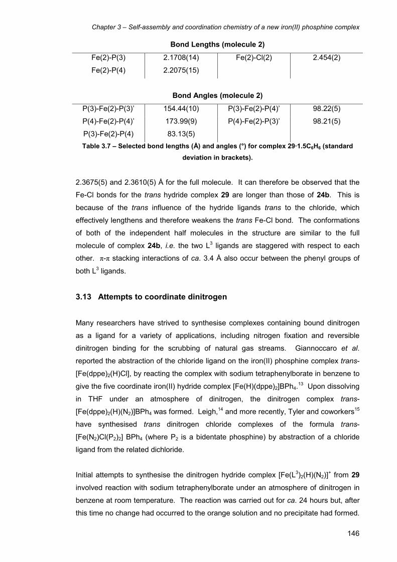
Chapter 3 – Self-assembly and coordination chemistry of a new iron(II) phosphine complex
146
Bond Lengths (molecule 2)
Fe(2)-P(3) 2.1708(14) Fe(2)-Cl(2) 2.454(2)
Fe(2)-P(4) 2.2075(15)
Bond Angles (molecule 2)
P(3)-Fe(2)-P(3)’ 154.44(10) P(3)-Fe(2)-P(4)’ 98.22(5)
P(4)-Fe(2)-P(4)’ 173.99(9) P(4)-Fe(2)-P(3)’ 98.21(5)
P(3)-Fe(2)-P(4) 83.13(5)
Table 3.7 – Selected bond lengths (Å) and angles (°) for complex 29·1.5C6H6 (standard
deviation in brackets).
2.3675(5) and 2.3610(5) Å for the full molecule. It can therefore be observed that the
Fe-Cl bonds for the trans hydride complex 29 are longer than those of 24b. This is
because of the trans influence of the hydride ligands trans to the chloride, which
effectively lengthens and therefore weakens the trans Fe-Cl bond. The conformations
of both of the independent half molecules in the structure are similar to the full
molecule of complex 24b, i.e. the two L3 ligands are staggered with respect to each
other. π-π stacking interactions of ca. 3.4 Å also occur between the phenyl groups of
both L3 ligands.
3.13 Attempts to coordinate dinitrogen
Many researchers have strived to synthesise complexes containing bound dinitrogen
as a ligand for a variety of applications, including nitrogen fixation and reversible
dinitrogen binding for the scrubbing of natural gas streams. Giannoccaro et al.
reported the abstraction of the chloride ligand on the iron(II) phosphine complex trans-
[Fe(dppe)2(H)Cl], by reacting the complex with sodium tetraphenylborate in benzene to
give the five coordinate iron(II) hydride complex [Fe(H)(dppe)2]BPh4.13 Upon dissolving
in THF under an atmosphere of dinitrogen, the dinitrogen complex trans-
[Fe(dppe)2(H)(N2)]BPh4 was formed. Leigh,14 and more recently, Tyler and coworkers15
have synthesised trans dinitrogen chloride complexes of the formula trans-
[Fe(N2)Cl(P2)2] BPh4 (where P2 is a bidentate phosphine) by abstraction of a chloride
ligand from the related dichloride.
Initial attempts to synthesise the dinitrogen hydride complex [Fe(L3)2(H)(N2)]+ from 29
involved reaction with sodium tetraphenylborate under an atmosphere of dinitrogen in
benzene at room temperature. The reaction was carried out for ca. 24 hours but, after
this time no change had occurred to the orange solution and no precipitate had formed.

Chapter 3 – Self-assembly and coordination chemistry of a new iron(II) phosphine complex
147
The 31P{1H} NMR spectrum of the solution showed no change to the singlet resonance
at δ -27.5. The reaction of cis-[Fe(L3)2Cl2] (24a) with sodium tetraphenylborate under
dinitrogen was also attempted, although again no reaction with N2 occurred as
indicated by 31P{1H} NMR spectroscopy. Further work in this area needs to be carried
out in order to successfully coordinate dinitrogen.
3.14 Electrochemistry of L3 complexes
Cyclic voltammetry studies were carried out on a range of L3 complexes in order to
determine more about their redox properties, as well as to establish whether the related
Fe(III) complexes were stable and potentially isolable. The comparison of
electrochemical data of the L3 complexes to those of the L1 and L2 ligands from
Chapter 2 should also yield important information.
A 1 mM 1,2-dichloroethane solution of [Fe(L3)2(κ2-O2SO2)] (23) was prepared, using
0.1 M tetrabutylammonium hexafluorophosphate as the electrolyte. Cyclic
voltammograms were recorded using a 1 mm platinum electrode and an initial scan
rate of 20 mVs-1. The voltammogram showed an irreversible peak at ca. 0 V vs Fc+/Fc,
which suggests that the complex is disassembling upon oxidation. The
voltammograms were then repeated at 50, 100, and 200 mVs-1 to determine the effect
of varying the scan rate on the voltammogram (Figure 3.21). As the scan rate of the
voltammogram is increased, the Fe(II)/Fe(III) redox peak becomes more reversible.
The voltammogram at 200 mVs-1 shows a redox system, which is far more reversible
than at 20 mVs-1, though it is not a fully reversible Nernstian redox process. This
phenomenon shows some similarities to the cyclic voltammetry of the complexes of L1
in Chapter 2, i.e. shows a more reversible redox process at higher scan rates due to
the decrease in the time available for the oxidised species to decompose. The main
difference between the voltammograms of [Fe(L3)2(κ2-O2SO2)] (23) and those of the L
1
complexes is that for 23 there is no need to polish the electrode in between scans in
order to observe the redox process. This means that the decomposed complex which
results from oxidation does not stick to the electrode and cause electrode blocking.
The very small redox process at ca. -0.4 V vs Fc+/Fc could not be identified, though is
believed to be a result of either a small impurity or a new species as a result of some
decomposition of complex 23. The complex was insoluble in acetonitrile, thus
preventing the cyclic voltammetry being investigated in this solvent.
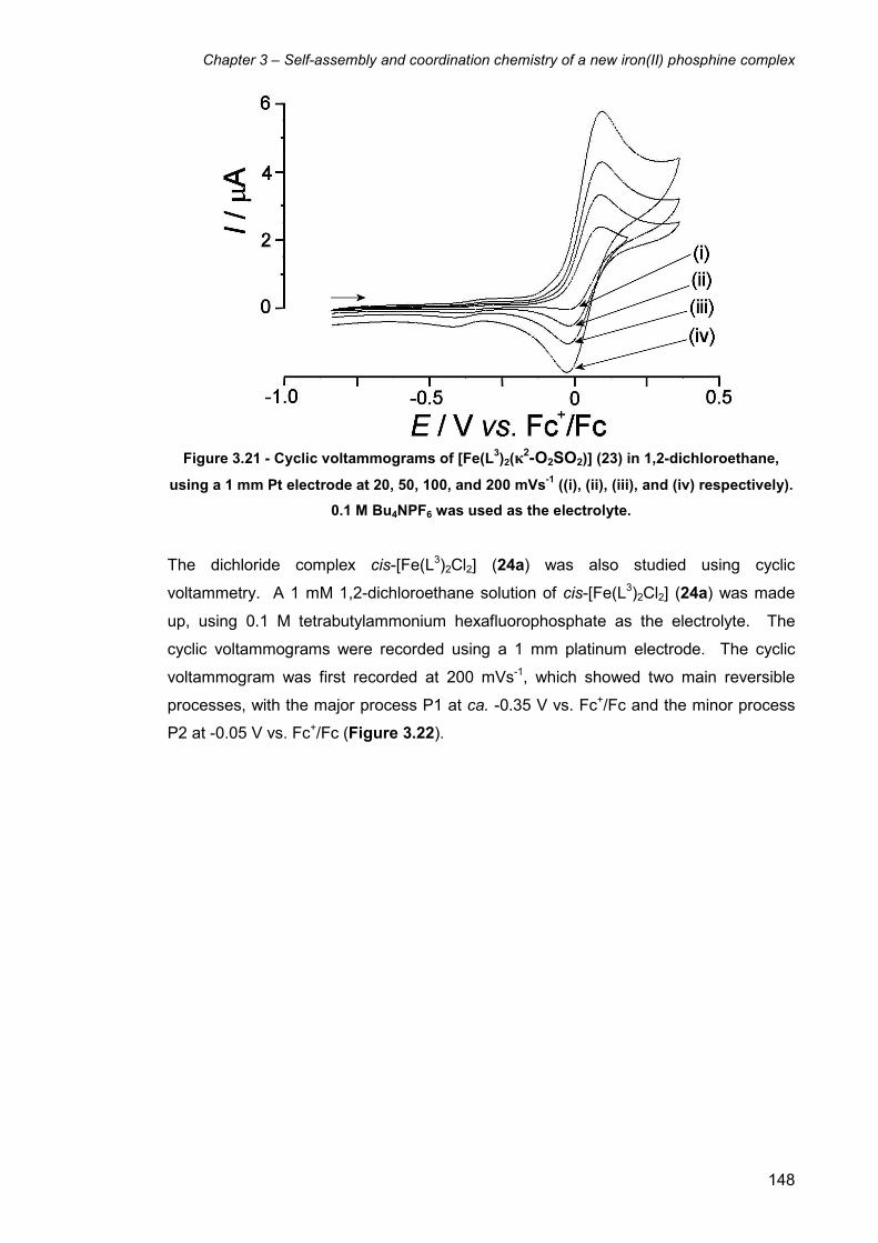
Chapter 3 – Self-assembly and coordination chemistry of a new iron(II) phosphine complex
148
Figure 3.21 - Cyclic voltammograms of [Fe(L3)2(κ2-O2SO2)] (23) in 1,2-dichloroethane,
using a 1 mm Pt electrode at 20, 50, 100, and 200 mVs-1 ((i), (ii), (iii), and (iv) respectively).
0.1 M Bu4NPF6 was used as the electrolyte.
The dichloride complex cis-[Fe(L3)2Cl2] (24a) was also studied using cyclic
voltammetry. A 1 mM 1,2-dichloroethane solution of cis-[Fe(L3)2Cl2] (24a) was made
up, using 0.1 M tetrabutylammonium hexafluorophosphate as the electrolyte. The
cyclic voltammograms were recorded using a 1 mm platinum electrode. The cyclic
voltammogram was first recorded at 200 mVs-1, which showed two main reversible
processes, with the major process P1 at ca. -0.35 V vs. Fc+/Fc and the minor process
P2 at -0.05 V vs. Fc+/Fc (Figure 3.22).

Chapter 3 – Self-assembly and coordination chemistry of a new iron(II) phosphine complex
149
Figure 3.22 - Cyclic voltammogram of cis-[Fe(L3)2Cl2] (24a) in 1,2-dichloroethane, using a
1 mm Pt electrode at 200 mVs-1. 0.1 M Bu4NPF6 was used as the electrolyte.
When the voltammogram of 24a was run at lower scan rates, reversible redox
processes were still observed, meaning that the reversibility of the processes are not
dependant on scan rate. Unlike the sulfate complex [Fe(L3)2(κ2-O2SO2)] (23), the
dichloride complex [Fe(L3)2Cl2] can potentially switch between the cis isomer 24a and
the trans isomer 24b. As the purple cis isomer 24a was used in the experiment and
the 1,2-dichloroethane solution was purple, it seems highly likely that the main
Fe(II)/Fe(III) redox process P1 at ca. -0.35 V vs. Fc+/Fc is of the cis isomer 24a. As the
Fe(II)/Fe(III) redox process for [Fe(L3)2(κ2-O2SO2)] (23) occurs at a more negative
potential (ca. -0.35 V) than for cis-[Fe(L3)2Cl2] (24a), it can be concluded that the
dichloride complex 24a is therefore easier to oxidise than 23.
The assignment of the complex represented by the main redox process P1 at ca. -0.35
V vs. Fc+/Fc as cis-[Fe(L3)2Cl2] (24a) can be backed up by the 31P{1H} NMR spectrum
of 24a in section 3.4, which showed that 24a gives two broad triplet resonances in
DCM/CDCl3 at δ -31.6 and -41.3 (Figure 3.6) arising from the cis isomer. It therefore
seems logical that the smaller redox process at ca. 0.05 V vs. Fc+/Fc could be due to
the trans isomer 24b. Cyclic voltammetry of the trans complex 24b in
1,2-dichloroethane would be ideal for comparison. However, the complex reverted
back to the cis isomer 24a in 1,2-dichloroethane, as indicated by the deep purple
colouration of the solution when the yellow crystals of (24b) were dissolved. Therefore,
it is the solvent that dictates whether the complex is the cis or trans isomer, and

Chapter 3 – Self-assembly and coordination chemistry of a new iron(II) phosphine complex
150
clomplex 24 forms the cis isomer 24a in almost all common organic solvents apart from
diethyl ether.
Cyclic voltammetry studies of cis-[Fe(L3)2Cl2] (24a) were also carried out in acetonitrile
using the same conditions. A 1 mM acetonitrile solution of cis-[Fe(L3)2Cl2] (24a) was
made up using 0.1 M tetrabutylammonium hexafluorophosphate as the electrolyte.
The cyclic voltammograms were recorded using a 1 mm platinum electrode with the
initial scan rate at 100 mVs-1. The cyclic voltammogram now showed only one
reversible redox process at ca. -0.35 V vs. Fc+/Fc relating to the cis isomer 24a
(Figure 3.23).
Figure 3.23 - Cyclic voltammogram of cis-[Fe(L3)2Cl2] (24a) in acetonitrile, using a 1 mm
Pt electrode at 100 mVs-1. 0.1 M Bu4NPF6 was used as the electrolyte.
No redox system at ca. -0.1 V vs. Fc+/Fc was observed in the voltammogram, i.e. there
appeared to be no appearance of the trans isomer 24b. Therefore, this indicates that
the complex stays as the cis isomer 24a when in acetonitrile solution. As acetonitrile is
more polar than dichloroethane, it would be expected that the increased polarity of the
acetonitrile solvent would lead to an increased amount of the more polar cis isomer
24a. In contrast with the cyclic voltammetry of [Fe(L2)Cl2] (15) in acetonitrile, no ligand
substitution was observed. 31P{1H} NMR spectroscopy of 24a in CD3CN showed the
familiar broad triplets associated with 24a, also suggesting no substitution had
occurred.

Chapter 3 – Self-assembly and coordination chemistry of a new iron(II) phosphine complex
151
Cyclic voltammetry studies of the trans hydride chloride complex trans-[Fe(L3)2(H)Cl]
(29) were also carried in order to investigate the stability of the related Fe(III) complex.
A 1 mM 1,2-dichloroethane solution of trans-[Fe(L3)2(H)Cl] (29) was made up using 0.1
M tetrabutylammonium hexafluorophosphate as the electrolyte. The cyclic
voltammograms were recorded using a 1 mm platinum electrode with the initial scan at
10 mVs-1. The voltammogram showed a number of redox processes, with the main
process occurring at ca. -0.85 V vs. Fc+/Fc, which is the Fe(II)/Fe(III) redox process for
the trans hydride chloride complex 29. The experiment was repeated at 20, 50, 100,
and 200 mVs-1 (Figure 3.24), which showed that the peak was seemingly reversible at
all scan rates measured. Therefore the oxidised Fe(III) version of complex 29 is stable
even at low scan rates, making it a rare example of a stable 17 electron metal(III)
hydride complex formed from the oxidation of the corresponding neutral 18-electron
species.16 Another example is the 17 electron radical cation iron hydride
[Fe(Cp*)(dppe)H]PF6 (dppe = 1,2-bis-(diphenylphosphino)ethane).17
Figure 3.24 - Cyclic voltammograms of [Fe(L3)2(H)Cl] (29) in 1,2-dichloroethane, using a 1
mm Pt electrode at 10, 20, 50, 100, and 200 mVs-1 ((i), (ii), (iii), (iv), and (v) respectively).
0.1 M Bu4NPF6 was used as the electrolyte.
After leaving the 1,2-dichloroethane solution for 30 mins still under argon, the cyclic
voltammogram was again recorded. The colour of the solution changed over time from
a bright orange colour to a darker orange/purple solution. The voltammogram (at 100
mVs-1) showed that the other redox processes had grown with respect to the peak at
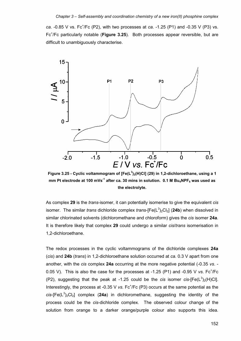
Chapter 3 – Self-assembly and coordination chemistry of a new iron(II) phosphine complex
152
ca. -0.85 V vs. Fc+/Fc (P2), with two processes at ca. -1.25 (P1) and -0.35 V (P3) vs.
Fc+/Fc particularly notable (Figure 3.25). Both processes appear reversible, but are
difficult to unambiguously characterise.
Figure 3.25 - Cyclic voltammogram of [Fe(L3)2(H)Cl] (29) in 1,2-dichloroethane, using a 1
mm Pt electrode at 100 mVs-1 after ca. 30 mins in solution. 0.1 M Bu4NPF6 was used as
the electrolyte.
As complex 29 is the trans-isomer, it can potentially isomerise to give the equivalent cis
isomer. The similar trans dichloride complex trans-[Fe(L3)2Cl2] (24b) when dissolved in
similar chlorinated solvents (dichloromethane and chloroform) gives the cis isomer 24a.
It is therefore likely that complex 29 could undergo a similar cis/trans isomerisation in
1,2-dichloroethane.
The redox processes in the cyclic voltammograms of the dichloride complexes 24a
(cis) and 24b (trans) in 1,2-dichloroethane solution occurred at ca. 0.3 V apart from one
another, with the cis complex 24a occurring at the more negative potential (-0.35 vs. -
0.05 V). This is also the case for the processes at -1.25 (P1) and -0.95 V vs. Fc+/Fc
(P2), suggesting that the peak at -1.25 could be the cis isomer cis-[Fe(L3)2(H)Cl].
Interestingly, the process at -0.35 V vs. Fc+/Fc (P3) occurs at the same potential as the
cis-[Fe(L3)2Cl2] complex (24a) in dichloromethane, suggesting the identity of the
process could be the cis-dichloride complex. The observed colour change of the
solution from orange to a darker orange/purple colour also supports this idea.

Chapter 3 – Self-assembly and coordination chemistry of a new iron(II) phosphine complex
153
Additionally, the more negative potential of the hydride complex 29 compared to the
dichloride complex 24a shows that complex 29 is far easier to oxidise than 24a, which
would be expected for a hydride complex. In general, all the Fe(II) L3 complexes
oxidise at lower potentials than for the Fe(II) L2 complexes ([Fe(L2)Cl2] (15) oxidises at
ca. 0.1 and 0.2 V vs Fc+/Fc in dichloroethane and acetonitrile respectively), meaning
that the L3 complexes are easier to oxidise. One potential explanation is the chelate
effect, with complexes of a tetradentate ligand (such as L2) expected to be more
energetically stable than two similar bidentate ligands (such as L3).
One possible explanation is that complex 29 undergoes isomerisation to give the cis
isomer, which then undergoes a metathesis reaction with a molecule of HCl generated
by the chlorinated solvent to give the cis-dichloride complex 24a and a molecule of
dihydrogen. Another possibility is that the metathesis occurs directly with the solvent
itself. A similar reaction occurs when the molybdenum hydride complex cis-
[HMo(CO)2(L)(C5H5)] (L = P(C6H5)3, P(OC6H5)3, or P(OCH3)3) reacts with carbon
tetrachloride to give the chloride complex cis-[ClMo(CO)2(L)(C5H5)].18 As the rate of
formation of the two processes at ca. -1.25 (P1) and -0.35 V vs. Fc+/Fc (P3) seems to
be equivalent, it could be concluded that the rate of isomerisation from trans-
[Fe(L3)2(H)Cl] (29) to cis is approximately equivalent to the rate of substitution of the
hydride ligand for the chloride. This of course assumes that the reaction does proceed
in this order and the cis isomer is not formed independently from the cis-[Fe(L3)2Cl2]
complex (24a).
When the cyclic voltammograms of the L1, L2, and L3 complexes are compared, the
most noticeable difference is the fact that the L1 complexes do not give reversible
voltammograms. This is most likely a result of the instability of the L1 Fe(III) analogues
that are produced in the oxidation process. This instability could be a result of the
affinity of the hydroxymethyl groups for the working electrode, as the similar L2 and L3
complexes (which do not possess hydroxymethyl groups) generally give reversible
CVs. The L3 complexes generally exhibited redox activity at lower potential (between
ca. 0.5 and 1.0 V) than their L1 and L2 counterparts, signifying that the Fe(II) L3
complexes are easier to oxidise and therefore their Fe(III) analogues would be more
difficult to reduce. Due to their stability as 17-electron Fe(III) complexes, further work
to isolate the Fe(III) variants of L2 and L3 either by chemical or bulk electrochemical
methods should provide interesting results.

Chapter 3 – Self-assembly and coordination chemistry of a new iron(II) phosphine complex
154
3.15 Further modification of the self-assembly reaction
Other possibilities exist for the modification of the self-assembly reaction to form new
products, such as using a different metal salt, using a substitutued amine/ammonium
salt instead of ammonium sulphate, using a substituted aldehyde instead of
formaldehyde, and even using a different counter ion other than sulfate. One
possibility is to attempt the self-assembly reaction using a different metal salt, in order
to determine whether the same or similar ligand motif would be formed, or even an
entirely new ligand structure.
The self-assembly reaction to form complex 1 has two important factors that have
allowed 1 to be isolated and characterised. The first is that complex 1 crystallises from
solution, which allows its isolation and separation from any other complexes that are
formed. The second factor is that 1 is diamagnetic as a result of its d6 low-spin
octahedral structure, which allows it to be easily characterised by NMR spectroscopy
without the broad NMR spectra associated with paramagnetic complexes. If a complex
formed in an analogous self-assembly reaction does not preferentially crystallise from
solution or is paramagnetic (or both), then its isolation and identification would be
harder. Therefore it would be useful to attempt similar reactions with diamagnetic
metal salts such as zinc(II) sulfate (d10).
The self-assembly reaction was attempted with Zn(II)SO4 as the templating metal salt
along with the same stoichiometries of THPS and ammonium sulfate as was used to
form 1. This was attempted in order to determine whether the reaction would give a
zinc(II) analogue of complex 1, or even any product at all. The addition of sodium
hydroxide to the colourless solution over time did not lead to any change in colour of
the solution, which would be expected for a d10 complex assuming no oxidation had
occurred (unlikely for zinc(II)). Following completion of the reaction the solution was
left at 5° C for ca. one week, but no material crystallised from the solution. The 31P{1H}
NMR spectrum of the reaction mixture both before and after the week showed a large
number of singlet resonances suggesting the presence of a variety of phosphine-
containing species. There was no indication that any complexes similar to complex 1
had formed, and as no complexes had crystallised from solution, the isolation,
separation and identification of the different species would be very difficult.
Other metals were attempted in the self-assembly reaction, including copper(II),
nickel(II), and cobalt(II) sulfates. In all cases a colour change occurred, but no material

Chapter 3 – Self-assembly and coordination chemistry of a new iron(II) phosphine complex
155
crystallised from solution despite leaving the solutions at 5° C for ca. one week. The 31P{1H} NMR spectra of the solutions all showed broad resonances due the metals all
being paramagnetic, and therefore no useful structural information could be obtained
from them. Due to these reactions yielding no useful results it was decided to not
investigate this area further.
To determine whether the self-assembly reaction would occur with the addition of a
substituted amine instead of an ammonium salt, the reaction was attempted with
glycine. As with replacing the phosphine/phosphonium salt in the reaction with a
substituted variant, changing the amine/ammonium salt could lead to a number of
possible outcomes. An analogous ligand to L1 could form maintaining the eight-
membered ring system, an entirely different new ligand could form, or even no reaction
at all. One possible structure is that if two glycine molecules condense with two
phosphine moieties, the familiar eight-membered ring could form with the pendant
gycine molecules coordinating to the axial coordination sites (Figure 3.26). Assuming
the carboxylic acid groups would deprotonated upon the addition of base and water
molecules take up the remaining two equatorial coordination sites, the complex would
be of neutral charge.
Figure 3.26 – Proposed structure of the potential product from the self-assembly reaction
using glycine instead of ammonium sulfate.
When the reaction was attempted, addition of sodium hydroxide to the aqueous
solution of FeSO4, THPS, and glycine gave a red colouration to the solution,
suggesting the formation of some iron(II) complex or complexes had occurred. The
reaction was stopped at ca. pH 6 in order to prevent any decomposition that occurs at
higher pH with the other complexes. 31P{1H} NMR spectroscopy of the solution showed
a large amount of unreacted THPS present, as well as number of broad peaks between
δ 50 and δ -200. The red solution was left at 5° C for ca. one week, though no solid
crystallised from solution. Further NaOH was added to the solution in an attempt to
Chapter 3 – Self-assembly and coordination chemistry of a new iron(II) phosphine complex
156
produce more of the product/products, though this did not lead to the crystallisation of
any material from solution. Without isolating any complex from solution, it is very
difficult to determine the nature of any products formed in solution. Also, glycine is
zwitterionic, which could also have a large effect on the outcome of the reaction.
3.16 Self-assembly reactions in the absence of metals
The self-assembly reactions to synthesise both complexes 1 and 23 were attempted
without the presence of a metal template to determine the outcome of the reaction.
The reaction between THPS, ammonium sufate, and sodium hydroxide was carried out
without the presence of the iron(II) sulfate as a metal template. Following the addition
of the sodium hydroxide, no obvious visual change of the colourless solution occurred.
The 31P{1H} NMR spectrum of the solution in D2O showed a large number of singlet
peaks between δ 50 and -150, suggesting that many species were present in solution.
The solution was left at 5° C for ca. one week to see if any of these compounds would
crystallise. No solid material precipitated from solution despite extended time at 5° C
and the addition of further NaOH.
The self-assembly reaction using bis(hydroxymethyl)phenylphosphine to form complex
23 was also carried out in the absence of the iron(II) sulfate. Following addition of the
bis(hydroxymethyl)phenylphosphine to an aqueous solution of ammonium sulfate and
formaldehyde, a white precipitate formed. The 31P{1H} NMR spectrum of the white
solid in CDCl3 showed a number of singlet resonances, indicating either that many
species had precipitated from solution, or maybe one compound had initially formed
but reacted with the CDCl3 solvent to give multiple species that could not be easily
identified.
In order to attempt to synthesise the L3 ligand without the use of the iron(II) template,
the reaction between PhP(CH2OH)2, formaldehyde and ammonia was attempted using
ethanol as the solvent instead of water. As ammonium sulfate is not soluble in ethanol,
ammonia was used as an alternative. It was hoped that using an organic solvent such
as ethanol for the reaction may give only one product, as the solvent H2O molecules
maybe interfering in the reaction in the previous case in aqueous solution. The
bis(hydroxymethyl)phenylphosphine was added dropwise to an ethanolic solution of
ammonia and formaldehyde, which did not result in the formation of a precipitate as in
the similar reaction in aqueous solution. The reaction was then brought to reflux, which
resulted in the precipitation of a white solid after ca. five minutes. The reflux was
continued for a further two hours until no further solid precipitated. The 31P{1H} NMR
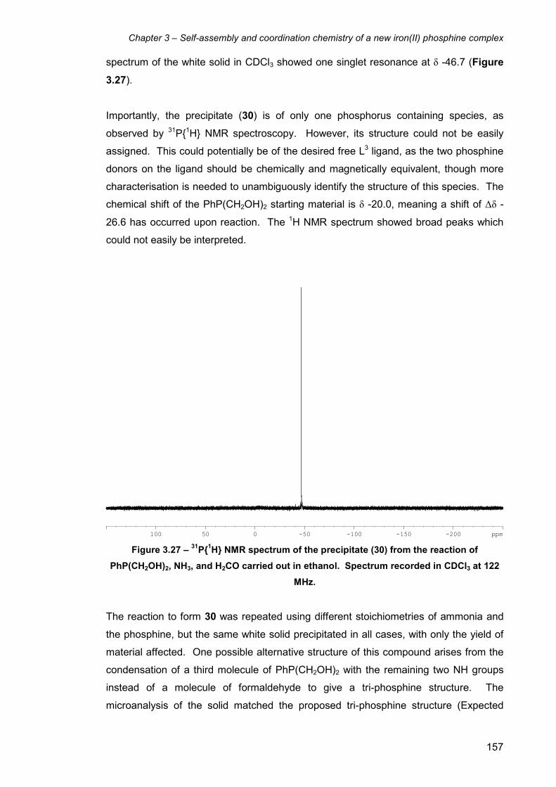
Chapter 3 – Self-assembly and coordination chemistry of a new iron(II) phosphine complex
157
spectrum of the white solid in CDCl3 showed one singlet resonance at δ -46.7 (Figure
3.27).
Importantly, the precipitate (30) is of only one phosphorus containing species, as
observed by 31P{1H} NMR spectroscopy. However, its structure could not be easily
assigned. This could potentially be of the desired free L3 ligand, as the two phosphine
donors on the ligand should be chemically and magnetically equivalent, though more
characterisation is needed to unambiguously identify the structure of this species. The
chemical shift of the PhP(CH2OH)2 starting material is δ -20.0, meaning a shift of ∆δ -
26.6 has occurred upon reaction. The 1H NMR spectrum showed broad peaks which
could not easily be interpreted.
-200-150-100-50100 50 0 ppm Figure 3.27 – 31P{1H} NMR spectrum of the precipitate (30) from the reaction of
PhP(CH2OH)2, NH3, and H2CO carried out in ethanol. Spectrum recorded in CDCl3 at 122
MHz.
The reaction to form 30 was repeated using different stoichiometries of ammonia and
the phosphine, but the same white solid precipitated in all cases, with only the yield of
material affected. One possible alternative structure of this compound arises from the
condensation of a third molecule of PhP(CH2OH)2 with the remaining two NH groups
instead of a molecule of formaldehyde to give a tri-phosphine structure. The
microanalysis of the solid matched the proposed tri-phosphine structure (Expected
Chapter 3 – Self-assembly and coordination chemistry of a new iron(II) phosphine complex
158
(found) for C24H27N2P3·H20: C 63.4(64.0), H 6.43(6.20), N 6.16(6.04) %), though this is
not enough evidence to fully support this conclusion.
Electrospray mass spectrometry of 30 was carried out in order to fully identify its
structure. The spectrum shows a large number of peaks, including a prominent peak at
m/z = 272.1. 30 was then reacted with sulfur in order to help determine its structure.
The 31P{1H} NMR spectrum of the material after stirring with excess sulfur after four
days in CDCl3 showed the singlet resonance had shifted to δ 33.8. The electrospray
mass spectrum of the phosphine sulphide was recorded, which showed only two main
peaks at m/z = 304.2 and 585.5. If the mass of sodium (23) is subtracted from these
values we get 281.2 and 462.5. As one value is double the other, it is likely that these
peaks are [M+Na]+ (304.2) and [2M+Na]+ (585.5), meaning the molecular ion [M] of the
phosphine sulfide is 281.2. When the mass of sulfur (ca. 32) is subtracted from the
peak at m/z = 304.2, we get a peak at approximately m/z = 272.1, which is what we
see in the mass spectrum of 30 before the addition of sulfur (i.e. [M+Na]+ of the
phosphine). Therefore the molecular ion [M] of the phosphine itself without the sodium
ion is m/z = 272.1 – 23.0 = 249.1.
30 was also reacted with one equivalent of sulfur in an attempt to determine the
number of phosphine donors of the compound. The 31P{1H} NMR spectrum after the
reaction showed two broad phosphine resonances in a 1:1 ratio at ca. δ 35 and -43,
suggesting that the ligand is a bidentate phosphine, though this would rule out the
possibility of the tri-phosphine structure. Despite the efforts to fully characterise the
ligand made in the reaction, full structural characterisation remained elusive. Further
work needs to be done in order to identify the structure of 30.
3.17 Conclusions and further work
The investigation of the self-assembly reaction by altering the starting materials (such
as the phosphine/phosphonium salt or amine/ammonium salt) gave some interesting
results. Using bis(hydroxymethyl)phenylphosphine in the presence of formaldehyde
instead of using THPS gave the complex [Fe(L3)2(κ2-O2SO2)] 23, containing the new
bidentate phosphine ligand L3. Ligand L3 consists of two fused PN2 six-membered
rings with a phenyl group as the third substituent on each phosphine. A sulfate ligand
coordinates to the Fe centre in a bidentate manner and occupies the remaining
equatorial sites. The equivalent dichloride complex cis-[Fe(L3)2Cl2] (24a) can be
synthesised by using iron(II) chloride and ammonium chloride instead of the sulfate
salts.
Chapter 3 – Self-assembly and coordination chemistry of a new iron(II) phosphine complex
159
Further reactions involving changes to the other starting materials in the self-assembly
reaction proved less successful. Attempts to replace the ammonium salts with other
amines or ammonium salts did not lead to the isolation of any analogous phosphine
complexes. Replacement of the sulfate counter ions for chlorides, or even using other
metals in the self-assembly reaction to form 1 did not lead to the isolation of any
complexes. However, replacing the sulfate salts with chloride salts in the reaction to
form 23 gave the related cis-dichloride complex 24a.
A comparison of the coordination chemistry of the L3 complexes shows some
similarities to the coordination chemistry of the L1 and L2 complexes as all three series
of complexes have four phosphine donors coordinated to a low-spin iron(II) centre.
However, there are also differences due to the nature of the bidentate L3 ligand versus
the tetradentate L1 and L2 ligands, such as the potential for cis/trans isomerisation.
The solubility of the L3 complexes are in general similar to those of the L2 complexes,
i.e. largely soluble in fairly polar organic solvents such as acetone, dichloromethane,
and chloroform. The trans-dichloride L3 complex 24b could be prepared by extraction
with diethyl ether, despite the low solubility of the complex in that solvent. However,
the cis complex 24a reformed upon dissolving in all tested organic solvents except
Et2O. Both 24a and 24b dissolve in H2O to give the same species, most likely the
diaqua complex cis-[Fe(L3)2(H2O)2]Cl2. The sulfate complex 23, however, was
completely insoluble in water. This is also in direct contrast with the L2 complexes, with
the sulfate complex 13 being more soluble in H2O than the dichloride complex 15.
The chloride ligands on complexes 24a and 24b can be readily substituted with other
ionic and neutral ligands. However, the sulfate ligand on the similar sulfate complex 23
is not readily substituted, which is most likely due to the extra stability that the chelate
effect affords. As a result, complex 24a proved to be a useful precursor for the
synthesis of other L3 complexes. Again, there are contrasts between the L2 and L3
systems, as the coordinated sulfate ligand on the L2 complex 13 can be readily
substituted, whereas the sulfate ligand on 23 cannot. The reason for the greater
reactivity of complex 13 compared to 23 is not entirely clear.
The reactions of 24a with thiocyanate and azide salts in an water/acetone mixture led
to the precipitation of the bis-thiocyanato and bis-azido complexes respectively. In
both cases the 31P{1H} NMR spectra showed a mixture of both cis and trans isomers,
though in the case of the bis-thiocyanato complexes, the cis/trans ratio decreases over
time as a result of the crystallisation of the trans isomer 25b. This is in contrast to the
Chapter 3 – Self-assembly and coordination chemistry of a new iron(II) phosphine complex
160
L1 and L2 bis-thiocyanato and bis-azido complexes, which cannot undergo
isomerisation due to the tetradentate phosphine ligand.
The reactions of the L3 dichloride complexes 24a and 24b with CO are similar to those
of the L1 and L2 dichloride complexes 3 and 15, in that they all form mono-carbonyl
mono-chloride complexes. Interestingly, the trans dichloride 24b gives the cis mono-
carbonyl mono-chloride complex 28b upon reaction with CO in diethyl ether, whereas
the cis dichloride 24a gives the trans mono-carbonyl mono-chloride complex 28a upon
reaction with CO in aqueous solution (via the cis di-aquo complex). Again, the L1 and
L2 dichloride complexes 3 and 15 cannot undergo isomerisation, and only the cis
complexes [Fe(L1)(CO)Cl]Cl (12) and [Fe(L2)(CO)Cl]Cl (19) are formed. Reactions of
24a with carbonate produced the chelating carbonate complex 27, in contrast to the
analogous L2 reaction, in which the complex was unstable to the basic carbonate anion
and decomposed.
The trans hydride chloride complex trans-[Fe(L3)2(H)Cl] (29) was synthesised by the
reaction of 24a with sodium borohydride. Further investigations into the reactivity of 29
need to be carried out in order to find out more about its coordination chemistry, such
as the reactivity towards dinitrogen.
Attempts to synthesise the free bidentate L3 ligand proved unsuccessful. Further
investigation therefore needs to be carried out in this area, as well as efforts to remove
the ligand intact from the metal. If the L3 ligand could be synthesised or isolated, its
coordination chemistry could be investigated with a wide range of metals.
The electrochemistry of the L3 compounds measured gave some interesting results.
Like the L2 complexes, cyclic voltammetry of the L3 complexes in general gave
reversible voltammograms, unlike the L1 complexes. Cyclic voltammetry of the cis-
dichloride complex 24a in dichloroethane showed two reversible redox processes
which presumably arise from the cis and trans isomers, with the cis isomer being far
more abundant. When the CV is repeated in acetonitrile, the redox process due to the
trans isomer is not observed, presumably due to the more polar nature of acetonitrile
compared to dichloroethane. No substitution of a chloride ligand of 24a with an
acetonitrile ligand in acetonitrile was observed electrochemically or by 31P{1H} NMR
spectroscopy, though this was not studied extensively. Similar to the L1 diaqua
complex with the sulfate counter ion (1), the voltammograms of the L3 sulfate complex
23 were irreversible at lower scan rates and more reversible at higher scan rates,
though the irreversibility of the L1 complexes was far greater. CVs of the hydride
Chapter 3 – Self-assembly and coordination chemistry of a new iron(II) phosphine complex
161
complex trans-[Fe(L3)2(H)Cl] (29) gave reversible voltammograms at all scan rates
measured, suggesting the related Fe(III) complex is relatively stable, though this
undergoes further isomerisation and substitution reactions with the dichloroethane
solvent. The Fe(III) version of complex 29 is therefore a rare example of a 17 electron
iron hydride complex. Further attempts to isolate this complex and explore its reactivity
could lead to some interesting results.
3.18 References
1. L. Higham, PhD Thesis, University of East Anglia, 2000.
2. P. Dierkes and P. W. N. M. v. Leeuwen, J. Chem. Soc., Dalton Trans., 1999,
1519.
3. H. W. Roesky and M. Andruh, Coord. Chem. Rev., 2003, 236, 91.
4. M. Di Vaira, S. Midollini and L. Sacconi, Inorg. Chem., 1981, 20, 3430.
5. C. M. Habeck, C. Hoberg, G. Peters, C. Näther and F. Tuczek,
Organometallics, 2004, 23, 3252.
6. L. D. Field, A. V. George, S. R. Pike, I. E. Buys and T. W. Hambley,
Polyhedron, 1995, 14, 3133.
7. L. D. Field, B. A. Messerle and R. J. Smernik, Inorg. Chem., 1997, 36, 5984.
8. J. M. Bellerby, M. J. Mays and P. L. Sears, J. Chem. Soc., Dalton Trans., 1976,
1232.
9. J. D. Gilbertson, N. K. Szymczak, J. L. Crossland, W. K. Miller, D. K. Lyon, B.
M. Foxman, J. Davis and D. R. Tyler, Inorg. Chem., 2007, 46, 1205.
10. Y. Gao, D. G. Holah, A. N. Hughes, G. J. Spivak, M. D. Havighurst, V. R.
Magnuson and V. Polyakov, Polyhedron, 1997, 16, 2797.
11. J.-G. Lee, G.-S. Jung and S.-W. Lee, Bull. Korean. Chem. Soc., 1998, 19, 267.
12. B. Wiesler, F. Tuczek, C. Näther and W. Bensch, Acta Crystallogr., Sect. C:
Cryst. Struct. Commun. , 1998, C54, 44.
13. P. Giannoccaro and A. Sacco, Inorg. Synth., 1977, 17, 69.
14. A. Hills, D. L. Hughes, M. Jimenez-Tenorio and G. J. Leigh, J. Organomet.
Chem., 1990, 391, C41.
15. W. K. Miller, J. D. Gilbertson, C. Leiva-Paredes, P. R. Bernatis, T. J. R.
Weakley, D. K. Lyon and D. R. Tyler, Inorg. Chem., 2002, 41, 5453.
16. P. Hamon, J.-R. Hamon and C. Lapinte, J. Chem. Soc., Chem. Commun.,
1992, 1602.
17. P. Hamon, L. Toupet, J.-R. Hamon and C. Lapinte, Organometallics, 1992, 11,
1429.
18. A. Bainbridge, P. J. Craig and M. Green, J. Chem. Soc. A., 1968, 2715.

Chapter 4 – Experimental section
162
Chapter 4 – Experimental section
4.1 General Experimental
Reactions were typically carried out in air, though manipulations on a Schlenk line
under dinitrogen were used as indicated. All chemical reagents were obtained from
Aldrich and used without further purification unless otherwise stated.
[Fe(L1)(H2O)2]SO4 (1) was prepared according to the literature procedure.1 NMR
spectra were recorded at 298 K on Bruker Avance 300, 400, and 500 MHz NMR
spectrometers, and referenced to residual protio solvent signals for 1H NMR spectra, δ
2.05 (acetone-d6), 4.80 (D2O), 7.24 (CDCl3), 2.50 (DMSO-d6), 7.15 (C6D6) and to
solvent resonances for 13C{1H} NMR: δ 29.9 (acetone-d6), 49.5 (D2O + 1% MeOH), 77.2
(CDCl3), 39.5 (DMSO-d6).2 31P{1H} NMR chemical shifts were referenced externally to
85% H3PO4 (δ 0.0). The low frequency 31P{1H} NMR spectrum (81 MHz) of complex 6
was carried out by Dr. Alan Boyd at Heriot-Watt University. The 31P{1H} NMR
simulations for complexes 6, 20, and 21 were carried out by Dr. John Lowe.
Microanalyses were carried out by Mr Alan Carver (Chemistry Department, University
of Bath). IR spectra were recorded as KBr discs on a Nicolet Protégé 460 FTIR
spectrometer. Electrospray mass spectrometry was carried out by the EPSRC
National Service at the University of Swansea for complex 7, and at the University of
Bath Mass Spectrometry Service on a Bruker MicrOTOF or MicrOTOF-Q spectrometer
for all other complexes reported.
Phenylphosphine (98%) was purchased from Strem Chemicals, Inc, USA. THPS (50%
aqueous solution) and THPC (80% aqueous solution) were kindly provided by Rhodia
Ltd. 57Fe metal powder and 18OH2 were kindly provided by Prof. Stephen Cramer and
coworkers at the University of California, Davis and Lawrence Berkeley National
Laboratories, California, USA. NRVS spectra were also carried out by Prof. Stephen
Cramer and coworkers.
4.2 Syntheses of chapter 2 compounds
Synthesis of [Fe(L1)Cl2] (3)
[Fe(L1)(H2O)2]SO4 (1) (50 mg, 0.0733 mmol) and NaCl (86 mg, 1.47 mmol, 20 Eq) were
heated to reflux in acetone (50 mL) for four hours, generating a purple suspension.
After cooling, the solution was filtered to remove the excess NaCl and Na2SO4 side-

Chapter 4 – Experimental section
163
products. Purple needle-like crystals were grown by leaving the acetone solution to
evaporate at room temperature. Yield = 20 mg (49%).
1H NMR (400 MHz, acetone-d6): δ 5.14 (AB, 8H, ∆ν = 62 Hz,
2JH-H = 13.7 Hz), 4.88 (s,
4H), 3.75 (s, 4H), 3.41 (AB, 8H, ∆ν = 23 Hz, 2JH-H =15.2 Hz).
31P{1H} NMR (122 MHz, acetone-d6): δ 14.8 (t,
2JP-P = 56 Hz), -7.9 (t, 2JP-P = 56 Hz).
Anal. Calc. For C12H30N2O6FeP4Cl2·0.5H2O: C, 25.8; H, 5.60; N, 5.02%. Found: C,
25.9; H, 5.72; N, 5.26%.
Synthesis of [Fe(L1)Br2] (5)
Same as the synthesis of 3 only using KBr instead of NaCl.
1H NMR (300 MHz, acetone-d6): δ 4.54 (AB, 8H, ∆ν = 27 Hz,
2JH-H =13.2 Hz), 4.32 (s,
4H), 3.62 (s, 4H), 3.25 (AB, 8H, ∆ν = 23 Hz, 2JH-H = 15.5 Hz).
31P{1H} NMR (122 MHz, acetone-d6): δ 15.6 (t,
2JP-P = 56 Hz), -12.2 (t, 2JP-P = 56 Hz)
Anal. Calc. For C12H30N2O6FeP4Br2: C, 22.6; H, 4.74; N, 4.39%. Found: C, 22.5; H,
4.89; N, 3.98%.
Synthesis of [Fe(L1)(NCS)2] (6)
[Fe(L1)(H2O)2]SO4 (1) (30 mg, 0.044 mmol) and potassium thiocyanate (18 mg, 0.185
mmol) were heated to reflux in acetone (15 mL) for ca. 18 hours, slowly dissolving the
[Fe(L1)(H2O)2]SO4 to give an yellow/orange solution with an orange precipitate. The
solvent was removed under reduced pressure. The orange solid was washed with
Et2O (10 mL) and collected by filtration, then washed with cold H2O (2 mL) and further
Et2O (5 mL). Orange crystals of 6 were isolated upon leaving an acetone solution of
the product to evaporate slowly. Yield = 23 mg (81%).
1H NMR (400 MHz, acetone-d6): δ 4.45 (AB, 8H, ∆ν = 21 Hz,
2JH-H =13.6 Hz), 4.33 (s,
4H), 3.83 (s, 4H), 3.37 (m, 8H, ∆ν = 86 Hz, 2JH-H = 14.9 Hz).
13C{1H} NMR (100 MHz, acetone-d6): δ 140.7 (s, NCS), 60.1 (m), 58.9 (m), 50.0 (m),
44.9 (m).
Chapter 4 – Experimental section
164
31P{1H} NMR (122 MHz, acetone-d6): δ 12.6 (second order,
2JP-P = 51 Hz), 4.6 (second
order, 2JP-P = 51 Hz).
Anal. Calc. For C14H30N4O6FeP4S2·3H2O: C, 25.9; H, 5.60; N, 8.64%. Found: C, 26.1;
H, 5.23; N, 8.71%.
IR (cm-1): 2109 (νNCS), 2098 (νNCS).
Synthesis of [Fe(L1)(N3)2] (7)
Sodium azide (60 mg, 0.92 mmol) was added to a solution of [Fe(L1)(H2O)2]SO4 (1)
(100 mg, 0.15 mmol) in H2O (10 mL) under an atmosphere of N2 and the solution
stirred for 1h at ambient temperature. A red solid of [Fe(L1)(N3)2] (7) was isolated upon
the addition of acetone (5 mL). Repeated attempts to record an elemental analysis
gave consistently high %N values due to the difficulty in removing excess sodium azide
leftover from the reaction.
1H NMR (400 MHz, D2O): δ 4.54 (s, 8H), 4.36 (s, 4H), 3.72 (s, 4H), 3.19 (AB, 8H, ∆ν =
16 Hz, 2JH-H =16.0 Hz).
13C{1H} NMR (76 MHz, D2O): δ 58.9 (s), 58.0 (m), 50.7 (m), 46.4 (m).
31P{1H} NMR (122 MHz, D2O): δ 16.9 (t,
2JP-P = 54 Hz), 12.4 (t, 2JP-P = 54 Hz).
IR (cm-1): 2056 (νN3).
Electrospray MS: m/z 580.0 [M+Na]+.
Synthesis of [Fe(L1)(κ2-O2CO)] (8)
Sodium carbonate decahydrate (47 mg, 0.164 mmol) was added to a solution of
[Fe(L1)(H2O)2]SO4 (1) (100 mg, 0.147 mmol) in H2O (20 mL), giving a red solution of
[Fe(L1)(κ2-O2CO)] (8) almost immediately. A red microcrystalline solid of 8 was isolated
from solution by the addition of acetone. Yield = 80 mg (94%).
1H NMR (400 MHz, D2O): δ 4.41 (s, 4H), 4.40 (AB, 8H, ∆ν = 21 Hz,
2JH-H =13.7 Hz),
3.70 (s, 4H), 3.14 (AB, 8H, ∆ν = 24 Hz, 2JH-H =16.0 Hz).

Chapter 4 – Experimental section
165
13C{1H} NMR (100 MHz, D2O): δ 164.6 (s, carbonate), 59.7 (m), 56.3 (m), 50.9 (m),
46.3 (m).
31P{1H} NMR (122 MHz, D2O): δ 23.5 (t,
2JP-P = 54 Hz), 9.4 (t, 2JP-P = 54 Hz).
Anal. Calc. For C13H30N2O9FeP4·2.5H2O: C, 26.8; H, 6.05; N, 4.80%. Found: C, 26.4;
H, 5.65; N, 4.58%.
IR (cm-1): 1681 (νCO), 1620 (νCO).
Synthesis of [Fe(L1)(CO)2](SO4) (9)
A solution of [Fe(L1)(H2O)2]SO4 (1) (30 mg, 0.044 mmol) in H2O (30 mL) was purged
with CO for 30 mins and stirred for a further 3 hours under an atmosphere of CO,
resulting in a yellow solution of 9. The solution was reduced to dryness, giving a yellow
microcrystalline solid, which was washed with acetone. Yellow crystals suitable for
X-ray crystallography were grown from an aqueous solution of 9 under an atmosphere
of CO at 5°C. Yield = 27 mg (93%).
1H NMR (400 MHz, D2O): δ 4.73 (s, 4H,), 4.53 (s, 8H), 3.71 (s, 4H), 3.67 (AB, 8H, ∆ν =
71 Hz, 2JH-H =15.0 Hz).
13C{1H} NMR (101 MHz, D2O): δ 204.2 (m, CO), 59.9 (m), 58.7 (m), 49.3 (m), 46.5 (m).
31P{1H} NMR (122 MHz, D2O): δ -14.9 (t,
2JP-P = 35 Hz), -39.4 (t, 2JP-P = 35 Hz).
Anal. Calc. For C14H30N2O12FeP4S·2H2O: C, 25.2; H, 5.14; N, 4.20%. Found: C, 25.4;
H, 5.08; N, 4.11%.
IR (cm-1): 2059 (νCO), 2022 (νCO).
Synthesis of [Fe(L1)(CO)Cl](Cl) (12)
[Fe(L1)Cl2] (3) (7 mg, 0.013 mmol) was dissolved in acetone (20 mL) with heating. The
purple solution was purged with carbon monoxide for 10 mins and stirred for 15
minutes, affording an orange precipitate. The solid 12 was collected by filtration and
washed with acetone (10 mL). Yield = 7 mg (97%).

Chapter 4 – Experimental section
166
1H NMR (400 MHz, D2O): δ 4.74 (s, 2H), 4.56 (AB, 4H, ∆ν = 20 Hz, 2JH-H = 13.1 Hz),
4.42 (AB, 4H, ∆ν = 28 Hz, 2JH-H = 13.5 Hz), 4.31 (s, 2H), 3.74 (AB, 4H, ∆ν = 27 Hz, 2JH-H
= 15.3 Hz), 3.61 (AB, 4H, ∆ν = 120 Hz, 2JH-H =16.3 Hz), 3.26 (AB, 4H, ∆ν = 105 Hz, 2JH-
H =15.2 Hz).
31P{1H} NMR (122 MHz, D2O): δ 5.3 (dt,
2JP-P = 74 Hz, 2JP-P = 41 Hz), -5.5 (dd,
2JP-P =
54 Hz, 2JP-P = 41 Hz), -29.4 (dt, 2JP-P = 74 Hz,
2JP-P = 54 Hz).
Anal. Calc. For C13H30N2O7FeP4Cl2: C, 27.1; H, 5.24; N, 4.85%. Found: C, 26.9; H,
5.25; N, 4.52%.
IR (cm-1): 1987 (νCO).
Synthesis of 57FeSO4.7H2O
57Fe metal powder (320 mg, 5.61 mmol) was stirred in conc. H2SO4 solution (8 mL of
4.5 M) for 2 hrs under an atmosphere of dinitrogen. The solution took on a pale green
colouration with a pale green precipitate with stirring, and ca. half of the solvent was
removed after the evolution of hydrogen gas had finished. The suspension was left at
5°C for ca. 24 hrs, after which time more of the pale green solid had formed. Solid was
filtered, washed with cold H2O (2 mL) and dried in a desiccator. Yield = 888 mg (57%).
Synthesis of [57Fe(L1)(H2O)2]SO4
The synthesis was carried out exactly the same as for 1, only 57FeSO4.7H2O was used.
Yield = 350 mg (16%).
Synthesis of [57Fe(L1)(CO)2]SO4
The synthesis was carried out exactly the same as for 9, only [57FeL1(H2O)2]SO4 was
used instead of 1. Yield = 70 mg (67 %).
Synthesis of [Fe(L2)(κ2-O2SO2)] (13)
[Fe(L1)(H2O)2]SO4 (1) (100 mg, 0.147 mmol) was heated in acetic anhydride (30 mL) at
120°C under an atmosphere of N2 for 2 hrs, slowly dissolving the complex over time to
give a red solution. The solution was reduced in volume to ca. 1-2 mL under vacuum,

Chapter 4 – Experimental section
167
and a red solid was precipitated on addition of diethyl ether (30 mL) and the solution
decanted. The crude red solid of 13 was washed thoroughly with diethyl ether (20 mL)
and recrystallised from CH2Cl2/Et2O. Yield = 83 mg (69%).
1H NMR (400 MHz, CDCl3): δ 4.87 (AB, 8H, ∆ν = 35 Hz,
2JH-H = 12.3 Hz), 4.74 (s, 4H),
3.89 (s, 4H), 3.57 (br, 4H), 3.22 (br, 4H), 2.18 (s, 6H, equatorial OCH3), 2.17 (s, 12H
axial OCH3).
13C{1H} NMR (100 MHz, CDCl3): δ 170.5 (s, equatorial ester C=O), 170.4 (s, axial ester
C=O), 58.6 (m), 57.0 (m), 51.1 (m), 46.8 (m), 20.6 (s, axial methyl), 20.5, (s,
equatorial methyl).
31P{1H} NMR (122 MHz, CDCl3): δ 15.3 (t,
2JP-P = 51 Hz), 0.9 (t, 2JP-P = 51 Hz).
Anal. Calc. For C24H42N2O16FeP4S: C, 34.9; H, 5.13; N, 3.39%. Found: C, 34.7; H, 5.17;
N, 3.16%.
IR (cm-1): 1744 (νC=O), 1219 (νC-O).
Synthesis of [Fe(L1)(H2NCH2CO2-κ2N,O)]HSO4 (14)
[Fe(L1)(H2O)2]SO4 (1) (100 mg, 0.147 mmol) was stirred in H2O (20 mL) for 10 mins,
dissolving some of the solid. A solution of glycine (13.2 mg, 0.176 mmol) and NaOH
(7.4 mg, 0.185 mmol) in H2O (3 mL) was added dropwise over an hour under an
atmosphere of N2, keeping the pH below 7. The addition of the glycine/NaOH solution
dissolved all the remaining 1, resulting in an orange solution. An orange solid of 14
was isolated by the addition of acetone (20 mL) to the solution. The orange solid was
dried under vacuum at 50° C to remove any water. The solid was then washed with
acetone (10 mL), filtered and dried in a dessicator. Yield = 89 mg (89%).
1H NMR (400 MHz, D2O): δ 4.47 (s, 2H), 4.46 (br m, 4H), 4.23 (AB, 4H, ∆ν = 53 Hz,
2JH-
H =13.9 Hz), 4.22 (s, 2H), 3.53 (AB, 4H, ∆ν = 206 Hz, 2JH-H =15.6 Hz), 3.32 (AB, 4H, ∆ν
= 31 Hz, 2JH-H =14.8 Hz), 3.0 (AB, 4H, ∆ν = 60 Hz, 2JH-H =15.6 Hz).
31P{1H} NMR (122 MHz, D2O): δ 19.7 (d of t,
2JP-P = 76 Hz, 2JP-P = 51 Hz), 7.2 (d of t,
2JP-P = 76Hz, 2JP-P = 56 Hz), 3.5 (d of d,
2JP-P = 51 Hz, 2JP-P = 56 Hz).

Chapter 4 – Experimental section
168
Anal. Calc. For C14H35N3O12FeP4S·2H2O: C, 24.5; H, 5.74; N, 6.13%. Found: C, 24.7;
H, 5.40; N, 6.27%.
IR (cm-1): 1629 (νCO).
Synthesis of [Fe(L2)Cl2] (15)
Bis(triphenylphosphoranylidene)ammonium chloride (PPNCl) (308 mg, 0.537 mmol)
was added to a red solution of [Fe(L2)(κ2-O2SO2)] (13) (200 mg, 0.242 mmol) in CH2Cl2
(20 mL), resulting in a purple solution of [Fe(L2)Cl2] (15). A purple solid of 15 was
isolated by the addition of diethyl ether, which was isolated by filtration and washed
with MeOH (20 mL) and Et2O (10 mL). Yield = 103 mg (50%).
1H NMR (400 MHz, CDCl3): δ 5.18 (AB, 8H, ∆ν = 150Hz,
2JH-H =13.6 Hz), 4.95 (s, 4H),
3.46 (s, 4H), 3.18 (AB, 8H, ∆ν = 86 Hz, 2JH-H =14.9 Hz), 2.13 (s, 12H, axial OCH3), 2.08
(s, 6H, equatorial OCH3).
13C{1H} NMR (100 MHz, CDCl3): δ 170.7 (s, axial ester C=O), 170.3 (s, equatorial ester
C=O), 61.2 (m), 59.2 (m), 52.7 (m), 47.4 (m), 20.8 (s, axial methyl), 20.4 (s, equatorial
methyl).
31P{1H} NMR (122 MHz, CDCl3): δ 16.5 (t, 2JP-P = 59 Hz), -1.6 (t,
2JP-P = 59 Hz).
Anal. Calc. For C24H42N2O12FeP4Cl2·0.5CH2Cl2: C, 34.9; H, 5.14; N, 3.32%. Found: C,
34.9; H, 5.15; N, 3.10%.
IR (cm-1): 1743 (νC=O), 1215 (νC-O).
Synthesis of [Fe(L2)(CO)Cl]BPh4 (19)
Sodium tetraphenylborate (22 mg, 0.064 mmol) was added to a purple solution of
[Fe(L2)Cl2] (15) (50 mg, 0.059 mmol) in acetone (10 mL). The solution was stirred
under an atmosphere of carbon monoxide at room temperature overnight, resulting in a
yellow solution with a white suspension of sodium chloride. The NaCl was removed by
filtration, the yellow solution of 19 was reduced to dryness and washed with diethyl
ether. Yield = 49 mg (74%).

Chapter 4 – Experimental section
169
1H NMR (400 MHz, acetone-d6): δ 7.34 (m, 8H, Ar), 6.93 (m, 8H, Ar), 6.78 (m, 4H, Ar),
5.19 (s, 2H), 5.02 (AB, 4H, ∆ν = 17 Hz, 2JH-H =13.5 Hz), 4.97 (m, 4H, ∆ν = 58 Hz, 2JH-H
=14.1 Hz), 4.74 (s, 2H), 4.06 (AB, 4H, ∆ν = 22 Hz, 2JH-H =16.0 Hz), 3.92 (AB, 4H, ∆ν =
146 Hz, 2JH-H =15.4 Hz), 3.62 (AB, 4H, ∆ν = 106 Hz, 2JH-H =15.4 Hz), 2.22 (s, 3H,
equatorial OCH3), 2.19 (s, 6H, axial OCH3), 2.15 (s, 3H, equatorial OCH3), 2.13 (s, 6H,
axial OCH3).
13C{1H} NMR (100 MHz, acetone-d6): δ 170.9 (m, axial and equatorial ester C=O),
165.0 (q, Ar, 1JC-B = 49.5 Hz), 137.1 (s, Ar), 126.1 (q, Ar, 3JC-B = 2.4 Hz), 122.3 (s, Ar),
60.8 (m), 58.8 (m), 51.7 (m), 51.1 (m), 46.8 (m), 20.7 (s, axial OCH3), 20.7 (s, axial
OCH3), 20.6 (s, equatorial OCH3), 20.5 (s, equatorial OCH3).
31P{1H} NMR (122 MHz, acetone-d6): δ -6.4 (dt,
2JP-P = 66, 40 Hz), -11.8 (dd, 2JP-P = 40,
55 Hz), -35.7 (dt, 1P, 2JP-P = 54, 66 Hz).
Anal. Calc. For C49H62N2O13FeP4ClB: C, 52.9; H, 5.61; N, 2.52%. Found: C, 52.6; H,
5.59; N, 2.49%.
IR (cm-1): 1993 (νCO), 1751 (νC=O), 1204 (νC-O).
Synthesis of [Fe(L2)(NCS)2] (20)
A solution of potassium thiocyanate (8 mg, 0.0823 mmol) in H2O (1 mL) was added to a
purple solution of [Fe(L2)Cl2] (15) (30 mg, 0.0356 mmol) in acetone (2 mL), resulting in
an orange solution of [Fe(L2)(NCS)2] (20). An orange microcrystalline solid of 20 was
isolated by slow evaporation of the acetone from the acetone/water solution. Yield = 26
mg (85%).
1H NMR (400 MHz, DMSO-d6): δ 4.75 (AB, 8H, ∆ν = 44 Hz,
2JH-H =13.5 Hz), 4.55 (s,
4H), 3.68 (s, 4H), 3.32 (AB, 8H, ∆ν = 49 Hz, 2JH-H =15.2 Hz), 2.14 (s, 6H, equatorial
OCH3), 2.13 (s, 12H axial OCH3).
13C{1H} NMR (100 MHz, DMSO-d6): δ 170.4 (s, equatorial ester C=O), 170.3 (s, axial
ester C=O), 59.0 (m), 50.0 (m, 4C), 45.7 (m), 20.6 (s, axial OCH3), 20.5, (s, equatorial
OCH3).
31P{1H} NMR (122 MHz, DMSO-d6): δ 4.1 (s).
Chapter 4 – Experimental section
170
Anal. Calc. For C26H42N4O12FeP4S2·H2O: C, 36.12; H, 5.13; N, 6.48%. Found: C, 35.8;
H, 4.98; N, 5.74%
IR (cm-1): 2108 (νNCS), 1748 (νC=O), 1213 (νC-O).
Synthesis of [Fe(L2)(N3)2] (21)
A solution of sodium azide (5 mg, 0.0769 mmol) in H2O (1 mL) was added to a purple
solution of [Fe(L2)Cl2] (15) (30 mg, 0.0356 mmol) in acetone (2 mL), resulting in an red
solution of [Fe(L2)(N3)2] (21). Red crystals of 21 were isolated by slow evaporation of
the acetone from the acetone/water solution. Yield = 27 mg (91%).
1H NMR (400 MHz, acetone-d6): δ 5.00 (AB, 8H, ∆ν = 44 Hz,
2JH-H =13.5 Hz), 4.82 (s,
4H), 3.82 (s, 4H), 3.41 (s, 8H), 2.17 (s, 12H, axial OCH3), 2.10 (s, 6H, equatorial
OCH3).
13C{1H} NMR (100 MHz, acetone-d6): δ 171.2 (s, axial ester C=O), 171.0 (s, equatorial
ester C=O), 60.2 (m), 59.8 (m), 52.7 (m), 47.9 (m), 20.9 (s, axial OCH3), 20.6 (s,
equatorial OCH3).
31P{1H} NMR (162 MHz, acetone-d6): δ 10.0 (second order), 8.6 (second order).
Anal. Calc. For C24H42N8O12FeP4·H2O: C, 34.63; H, 5.33; N, 13.46%. Found: C, 34.7;
H, 5.10; N, 13.3%.
IR (cm-1): 2047 (νN3), 1735 (νC=O), 1219 (νC-O).
4.3 Syntheses of chapter 3 compounds
Synthesis of P(CH2OH)3 (THMP)3
[P(CH2OH)4]Cl (50.0g of 80% aqueous solution, 262 mmol) solution was evaporated to
dryness on a rotary evaporator, and dried further with an azeotropic toluene distillation
for 2 hrs. Dry triethylamine (100 mL) was added to the white solid of [P(CH2OH)4]Cl,
and the reaction was stirred at 60°C for two hours. The resulting white precipitate of
[NEt4]Cl was removed by filtration. The reaction solution was then vacuum distilled at
atmospheric pressure, which removed the excess triethylamine and sublimed the
formaldehyde out in the condenser. The resulting colourless oil of P(CH2OH)3 was left

Chapter 4 – Experimental section
171
to cool and crystallised into a white solid upon leaving at -20°C for ca. 7 days. Yield =
29.0 g (89%).
Synthesis of PhP(CH2OH)2
Phenylphosphine (10.0 g, 90.8 mmol) was added dropwise to a solution of
formaldehyde (15.1 g of 37% solution, 186 mmol) in degassed ethanol (80 mL) under
an atmosphere of dinitrogen. The reaction mixture was stirred for 5 hrs, after which the
solvent was removed in vacuo, giving a colourless oil. The oil crystallised into a white
solid of PhP(CH2OH)2 upon leaving at -20°C for ca. 48 hrs. Yield = 13.5 g (87%).
Synthesis of [Fe(L3)2(κ2-O2SO2)] (23)
Iron(II) sulphate heptahydrate (101 mg, 0.363 mmol), ammonium sulphate (97 mg,
0.734 mmol), and formaldehyde solution (66 mg of 37% solution, 0.813 mmol) were
dissolved in H2O (20 mL) under an atmosphere of N2 to give a pale green solution.
PhP(CH2OH)2 was separately dissolved in degassed H2O (10 mL), which was then
slowly added dropwise over ca. 30 mins to the iron-containing solution whist stirring,
taking care to keep the pH below ca. 5.5. The addition of the aqueous phosphine
solution gave rise to a purple colouration of the solution, which increased in intensity
with further addition of the phosphine solution. As the solution became darker, a purple
solid began to precipitate, which continued with the addition until there was a large
amount of the purple precipitate. The reaction was left to stir for one hour further, the
purple product then collected by filtration and washed with 2 mL cold H2O. The purple
solid 23 was then dried in a dessicator under reduced pressure overnight. Yield = 93
mg (33%).
1H NMR (500 MHz, CDCl3): δ 7.4-7.1 (m, 20H, Ar), 5.27 (d, 2H, P-CH2-N AB1, ∆ν = 821
Hz, 2JH-H = 13.9 Hz), 4.92 (d, 2H, P-CH2-N AB2, ∆ν = 736 Hz, 2JH-H = 12.9 Hz), 4.17 (d,
2H, N-CH2-N AB, ∆ν = 76 Hz, 2JH-H = 13.9 Hz), 4.01 (d, 2H, N-CH2-N AB, ∆ν = 76 Hz,
2JH-H = 13.9 Hz), 4.03 (d, 2H, P-CH2-N AB3, ∆ν = 228 Hz, 2JH-H = 13.9 Hz), 3.63 (d, 2H,
P-CH2-N AB1, ∆ν = 821 Hz, 2JH-H = 13.9 Hz), 3.53 (d, 2H, P-CH2-N AB4, ∆ν = 411 Hz,
2JH-H = 13.9 Hz), 3.53 (d, 2H, P-CH2-N AB3, ∆ν = 228 Hz, 2JH-H = 13.9 Hz), 3.45 (d, 2H
P-CH2-N AB2, ∆ν = 736 Hz, 2JH-H = 12.9 Hz), 2.71 (d, 2H, P-CH2-N AB4, ∆ν = 411 Hz,
2JH-H = 13.9 Hz).
31P {1H} NMR (122 MHz, CDCl3): δ -26.4 (t,
2JP-P = 72 Hz), -51.2 (t, 2JP-P = 72 Hz).

Chapter 4 – Experimental section
172
Anal. Calc. for C34H40N4O4FeP4S1·H2O: C, 51.1; H, 5.30; N, 6.99%. Found: C, 50.7; H,
5.25; N, 6.28%
Electrospray MS: m/z = 803.1 [M+Na]+.
Synthesis of cis-[Fe(L3)2Cl2] (24a) and trans-[Fe(L3)2Cl2] (24b)
The reaction was carried out as for the synthesis of 23, except using iron(II) chloride
tetrahydrate (335 mg, 1.69 mmol) and ammonium chloride (362 mg, 6.77 mmol) in the
same stoichiometry, instead of the equivalent chloride salts. The purple product was
then collected by filtration and washed with 2 mL cold H2O. The purple solid of cis-
[Fe(L3)2Cl2] (24a) was dried in a desiccator under reduced pressure overnight. Yield =
1.01g (79%).
31P {1H} NMR (122 MHz, CDCl3): -32.4 (br t), -46.5 (br t).
31P {1H} NMR (122 MHz, D2O - cis-[Fe(L
3)2(H2O)2]Cl2): δ -17.9 (t, 2JP-P = 74 Hz), -38.2 (t,
2JP-P = 74 Hz).
cis-[Fe(L3)2Cl2] was placed in a cellulose extraction thimble inserted into a Soxhlet
extractor fitted with a condenser and round bottomed flask. Diethyl ether was placed in
the RBF and the extraction was left for 24 hours. This gave a yellow solution of trans-
[Fe(L3)2Cl2], which gave yellow crystals upon evaporation of the ether solution at room
temperature. No NMR spectra of trans-[Fe(L3)2Cl2] (24b) were recorded as the
complex isomerises to the cis isomer 24a when dissolved in most organic solvents, and
is not soluble enough in Et2O to be able to get a concentrated enough sample for NMR
analysis.
Anal. Calc. for C34H40N4FeP4Cl2: C, 54.0; H, 5.34; N, 7.42%. Found: C, 53.4; H, 5.33;
N, 7.23%
Electrospray MS: m/z = 684.2 [M-2Cl]+.
Synthesis of cis-[Fe(L3)2(NCS)2] (25a) and trans-[Fe(L3)2(NCS)2] (25b)
cis-[Fe(L3)2Cl2] (24a) (50 mg, 0.66 mmol) was dissolved in ethanol (10 mL) giving a
purple suspension. Excess potassium thiocyanate (129 mg, 1.33 mmol) was added to
the reaction mixture, reacting any undissolved 24a and giving an orange precipitate of

Chapter 4 – Experimental section
173
25a. The orange solid was collected and washed with cold H2O (5 mL). Yield = 43 mg
(78%). Single crystals of 25b suitable for X-ray diffraction were grown from a CDCl3
solution over ca. 48 hrs. 31P{1H} and 1H NMR spectroscopy showed a mixture of 26a
and 26b in an 4:1 ratio after 20 mins in solution, which changed to 2:3 ratio after 48
hrs.
1H NMR (25a) (300 MHz, CDCl3): δ 7.8-6.6 (m, 20H, Ar), 4.6 (d, 2H, P-CH2-N AB1, ∆ν
= 391 Hz, 2JH-H = 13.9 Hz), 4.3 (d, 2H, P-CH2-N AB2, ∆ν = 387 Hz, 2JH-H = 13.4 Hz), 4.0
(d, 2H, P-CH2-N AB3, ∆ν = 161 Hz 2JH-H = 13.8 Hz), 3.9 (d, 2H, N-CH2-N AB, ∆ν = 24
Hz, 2JH-H = 13.9 Hz), 3.8 (d, 2H, N-CH2-N AB, ∆ν = 24 Hz, 2JH-H = 13.9 Hz), 3.5 (d, 2H,
P-CH2-N AB4, ∆ν = 309 Hz, 2JH-H = 13.8 Hz), 3.5 (d, 2H, P-CH2-N AB3, ∆ν = 161 Hz,
2JH-H = 13.8 Hz), 3.3 (d, 2H, P-CH2-N AB1, ∆ν = 391 Hz, 2JH-H = 13.9 Hz), 3.0 (d, 2H P-
CH2-N AB2, ∆ν = 387 Hz, 2JH-H = 13.4 Hz), 2.4 (d, 2H, P-CH2-N AB4, ∆ν = 309 Hz,
2JH-H
= 13.8 Hz).
31P{1H} NMR (25a) (122 MHz, CDCl3): δ -31.0 (t, 2P,
2JP-P = 77 Hz), -38.6 (t, 2P, 2JP-P =
77 Hz).
1H NMR (25b) (300 MHz, CDCl3): δ 7.8-6.6 (m, 20H, Ar), 4.3 (d, 8H, P-CH2-N AB, ∆ν =
221 Hz, 2JH-H = 13.4 Hz), 4.0 (s, 4H, N-CH2-N), 3.5 (d, 8H, P-CH2-N AB, ∆ν = 221 Hz, 2JH-H = 13.4 Hz).
31P{1H} NMR (25b) (122 MHz, CDCl3): δ -42.2 (s, 4P).
Anal. Calc. for C36H40N6FeP4S2·4H2O: C, 49.6; H, 5.54; N, 9.63%. Found: C, 49.2; H,
4.74; N, 9.41%
IR (cm-1): 2098 (νNCS).
Electrospray MS: m/z = 742.1 [M-NCS]+.
Synthesis of cis-[Fe(L3)2(N3)2] (26a) and trans-[Fe(L3)2(N3)2] (26b)
cis-[Fe(L3)2Cl2] (24a) (50 mg, 0.066 mmol) was dissolved in ethanol (10 mL) giving a
purple suspension. Excess sodium azide (86 mg, 1.32 mmol) was added to the
reaction mixture, reacting any undissolved 24a and giving a red suspension of 26a.
The red solid was collected and washed with cold H2O (5 mL). As with complex 7,
repeated attempts to record an elemental analysis gave consistently high %N values

Chapter 4 – Experimental section
174
due to the difficulty in removing excess sodium azide leftover from the reaction. 31P{1H}
and 1H NMR spectroscopy showed a mixture of 26a and 26b in an 11:1 ratio, which did
not change over time.
1H NMR (26a) (300 MHz, CDCl3): δ 7.5-7.1 (m, 20H, Ar), 4.7 (d, 2H, P-CH2-N AB1, ∆ν
= 30 Hz, 2JH-H = 13.6 Hz), 4.6 (d, 2H, P-CH2-N AB2, ∆ν = 473 Hz, 2JH-H = 13.6 Hz), 3.9
(d, 2H, P-CH2-N AB3, ∆ν = 155 Hz, 2JH-H = 13.6 Hz), 3.9 (d, 2H, N-CH2-N AB, ∆ν = 25
Hz, 2JH-H = 13.6 Hz,), 3.9 (d, 2H, N-CH2-N AB, ∆ν = 25 Hz, 2JH-H = 13.6 Hz,), 3.5 (d, 2H,
P-CH2-N AB4, ∆ν = 293 Hz, 2JH-H = 13.6 Hz), 3.4 (d, 2H, P-CH2-N AB3, ∆ν = 155 Hz,
2JH-H = 13.6 Hz), 3.4 (d, 2H, P-CH2-N AB1, ∆ν = 30 Hz, 2JH-H = 13.6 Hz), 3.0 (d, 2H, P-
CH2-N AB2, ∆ν = 473 Hz, 2JH-H = 13.6 Hz), 2.5 (d, 2H, P-CH2-N AB4, ∆ν = 293 Hz,
2JH-H
= 13.6 Hz).
31P{1H} NMR (26a) (122 MHz, CDCl3): δ -27.4 (t,
2JP-P = 75 Hz), -36.9 (t, 2JP-P = 75 Hz).
1H NMR (26b) (300 MHz, CDCl3): δ 7.5-7.1 (m, 20H, Ar), 4.3 (d, 8H, P-CH2-N AB, ∆ν =
186 Hz, 2JH-H = 14.1 Hz), 4.2 (s, 4H, N-CH2-N), 3.6 (d, 8H, P-CH2-N AB, ∆ν = 186 Hz, 2JH-H = 14.1 Hz).
31P{1H} NMR (26b) (122 MHz, CDCl3): δ -43.2 (s).
IR (cm-1): 2043 (νN3 - mainly trans isomer).
Electrospray MS: m/z = 684.2 [M-2N3]+.
Synthesis of [Fe(L3)2(κ2-O2CO)] (27)
cis-[Fe(L3)2Cl2] (24a) (107 mg, 0.142 mmol) was dissolved in ethanol (20 mL) giving a
purple suspension. Excess sodium carbonate (170 mg, 1.60 mmol) was added,
reacting any undissovled 24a and giving a pink suspension. The resulting pink solid
was recrystallised from chloroform. Yield = 61 mg (59%) Single crystals suitable for X-
ray diffraction grew from the aqueous reaction filtrate over ca. 8 weeks.
1H NMR (500 MHz, CDCl3): δ 7.6-7.1 (m, 20H, Ar), 5.16 (d, 2H, P-CH2-N AB1, ∆ν = 918
Hz, 2JH-H = 12.9 Hz,), 4.76 (d, 2H, P-CH2-N AB2, ∆ν = 680 Hz, 2JH-H = 12.9 Hz,), 4.03
(d, 2H, P-CH2-N AB3, ∆ν = 277 Hz, 2JH-H = 12.9 Hz,), 4.00 (d, 2H, N-CH2-N AB, ∆ν = 33
Hz, 2JH-H = 14.5 Hz), 3.94 (d, 2H, N-CH2-N AB, ∆ν = 33 Hz, 2JH-H = 13.9 Hz), 3.48 (d,
2H, P-CH2-N AB4, ∆ν = 401 Hz, 2JH-H = 13.9 Hz), 3.48 (d, 2H, P-CH2-N AB3, ∆ν = 277

Chapter 4 – Experimental section
175
Hz, 2JH-H = 12.9 Hz), 3.40 (d, 2H, P-CH2-N AB2, ∆ν = 680 Hz, 2JH-H = 12.9 Hz), 3.32 (d,
2H P-CH2-N AB1, ∆ν = 918 Hz, 2JH-H = 12.9 Hz), 2.68 (d, 2H, P-CH2-N AB4, ∆ν = 401
Hz, 2JH-H = 13.9 Hz).
13C{1H} NMR (125 MHz, CDCl3): δ 163.7 (s, O2CO), 131-128 (m, Ar), 72.8 (s, N-CH2-
N), 53.3 (m, P-CH2-N), 51.2 (m, P-CH2-N), 50.9 (s, P-CH2-N), 48.0 (s, P-CH2-N).
31P {1H} NMR (122 MHz, CDCl3): δ -20.2 (t,
2JP-P = 72 Hz), -41.4 (t, 2JP-P = 72 Hz).
Anal. Calc. for C35H40N4O3FeP4: C, 56.5; H, 5.42; N, 7.53%. Found: C, 56.1; H, 5.29;
N, 7.46.
IR (cm-1): 1556 (νC=O).
Electrospray MS: m/z = 701.2 [M-CO2]+.
Synthesis of trans-[Fe(L3)2(CO)Cl]Cl (28a)
cis-[Fe(L3)2Cl2] (24a) (100 mg, 0.133 mmol) was dissolved in H2O (20 mL) and purged
with CO for ca. 20 mins. The reaction was stirred for ca. two hrs, resulting in a yellow
solution. A yellow solid was precipitated from solution by the addition of acetone. The
yellow solid of 28a was washed with additional acetone (5 mL) and dried in a
desiccator. Yield = 87 mg (83%).
1H NMR (400 MHz, D2O): δ 7.1-6.9 (m, 20H, Ar), 4.9 (d, 4H, P-CH2-N AB1, ∆ν = 423
Hz, 2JH-H = 13.5 Hz), 4.4 (d, 4H, P-CH2-N AB2, ∆ν = 156 Hz, 2JH-H = 14.5 Hz), 3.9 (s,
4H, N-CH2-N), 3.8 (d, 4H, P-CH2-N AB2, ∆ν = 156 Hz, 2JH-H = 14.5 Hz), 3.5 (d, 4H, P-
CH2-N AB1, ∆ν = 423 Hz, 2JH-H = 13.5 Hz).
13C{1H} NMR (trans-[Fe(L3)2(
13CO)Cl]Cl, 126 MHz, D2O): δ 214.7 (quintet, 13CO), 132-
129 (m, Ar), 72.2 (s, N-CH2-N), 52.3 (s, P-CH2-N), 49.1 (s, P-CH2-N).
31P{1H} NMR (122 MHz, D2O): δ -51.0 (s).
Anal. Calc. for C35H40N4O1FeP4Cl2·6H2O: C, 46.6; H, 5.81; N, 6.21%. Found: C, 46.3;
H, 5.47; N, 6.06%.
IR (cm-1): 1939 (ν12CO), 1895 (ν13CO).

Chapter 4 – Experimental section
176
Electrospray MS: m/z = 747.1 [M]+.
Synthesis of cis-[Fe(L3)2(CO)Cl]Cl (28b)
trans-[Fe(L3)2Cl2] (24a) (10 mg, 0.013 mmol) was dissolved in diethyl ether (20 mL)
giving a pale yellow solution. CO was purged through the reaction mixture for ca. 20
mins and the reaction mixture was stirred for a further 4 hrs, after which time a pale
pink precipitate began to form. The pink solid was filtered off after a further 7 days of
stirring, washed with Et2O (5 mL) and dried in air. Yield = 5 mg (48%).
31P{1H} NMR (122 MHz, CDCl3): δ -39.4 (ddd,
2JP-P = 91, 38, 45 Hz), -55.3 (ddd, 2JP-P =
91, 61, 48 Hz), -59.4 (ddd, 2JP-P = 38, 61, 120 Hz), -69.0 (ddd, 2JP-P = 45, 48, 120 Hz).
Anal. Calc. for C35H40N4O1FeP4Cl2: C, 53.6; H, 5.15; N, 7.15%. Found: C, 53.0; H,
5.54; N, 4.91%.
IR (cm-1): 1974 (νCO).
Electrospray MS: m/z = 747.1 [M]+.
Synthesis of trans-[Fe(L3)2(H)Cl] (29)
cis-[Fe(L3)2Cl2] (24a) (200 mg, 0.265 mmol) and sodium borohydride (20 mg, 0.529
mmol) were placed in a Schlenk tube under dinitrogen, to which was added 10 mL of
degassed ethanol, dissolving the starting materials to give an orange solution. Upon
stirring for ca. 1hr, all the starting materials had reacted resulting in an orange
suspension. The solution was filtered by cannula under dinitrogen and the solid
washed with 5 mL of degassed ethanol and dried under vacuum. Yield = 128 mg
(67%).
1H NMR (400 MHz, C6D6): δ 7.0-6.8 (m, 20H, Ar), 5.5 (d, 4H, P-CH2-N AB1, ∆ν = 806
Hz, 2JH-H = 12.9 Hz), 3.9 (s, 4H, N-CH2-N), 3.9 (d, 4H, P-CH2-N AB2, ∆ν = 107 Hz, 2JH-H
= 13.3 Hz), 3.6 (d, 4H, P-CH2-N AB2, ∆ν = 107 Hz, 2JH-H = 13.3 Hz), 3.5 (d, 4H, P-CH2-
N AB1, ∆ν = 806 Hz, 2JH-H = 12.9 Hz), -28.6 (quintet, hydride, 2JP-C = 49 Hz).
31P NMR (162 MHz, C6D6): δ -27.5 (d,
2JP-H = 49 Hz).

Chapter 4 – Experimental section
177
Anal. Calc. for C34H41N4FeP4Cl1: C, 56.6; H, 5.73; N, 7.77%. Found: C, 55.4; H, 5.36;
N, 6.08%.
Electrospray MS: m/z = 720.1 [M]+.
4.4 Electrochemistry
All reagents used for electrochemical analyses were of analytical or electrochemical
grade purity. The supporting electrolyte used for the analyses of the L1 complexes in
aqueous solution was potassium chloride, whereas tetrabutylammonium
hexafluorophosphate was used as the supporting electrolyte for measurements of the
L2 and L3 complexes in acetonitrile and dichloroethane solutions. Solvents used
(acetonitrile, dichloroethane, H2O) were purged with argon to deoxygenate the solution
prior to use. All experiments were conducted at 22 ± 2°C.
Voltammetric experiments were performed using an Ecochemie Autolab potentiostat.
For the purpose of these experiments a three electrode cell setup was used,
comprising of working, counter and reference electrodes. The working electrode used
was a 3mm diameter platinum electrode unless otherwise stated. The counter
electrode consisted of a spiral of platinum wire. All potentials were referenced to
internal cobaltocenium hexafluorophosphate, which was referenced to ferrocene (E1/2 =
0.00 V) but were measured using a platinum wire pseudo reference (a platinum wire
which was immersed and equilibrated in the electrolyte containing solvent used for the
experiment).
4.5 Crystallography
Data for complexes 3, 5, 6, 7, 8, 13, 21, 24b, and 27 were collected on a Nonius Kappa
CCD diffractometer. Data for complexes 15 and 25b were collected at Daresbury SRS
Station 9.8 and 23 recorded at Daresbury SRS Station 16.2 SMX by Dr. Mary F.
Mahon and Dr. John E. Warren. Data for complex 29 were recorded at Daresbury SRS
Station 16.2 SMX Dr. Ross W. Harrington. Data for complex 9 was collected by Dr.
Michael Ruf on a Bruker SMART 1000 CCD diffractometer at Bruker in Germany. All
structures were solved by Dr. Mary F. Mahon or Dr. Gabriele Kociok-Köhn. The
solution of all structures was carried out using SHELX-97 while refinements were
completed using SHELXL-97.

Chapter 4 – Experimental section
178
4.6 References
1. J. C. Jeffery, B. Odell, N. Stevens and R. E. Talbot, Chem. Commun., 2000,
101.
2. H. E. Gottlieb, V. Kotlyar and A. Nudelman, J. Org. Chem., 1997, 62, 7515.
3. L. Higham, PhD Thesis, University of East Anglia, 2000.

179
Appendix – Crystallographic Data
Table 1. Crystal data and structure refinement for [Fe(L1)Cl2] (3).
Compound [Fe(L1)Cl2] (3)
Identification code k06adb1
Empirical formula C12 H32 Cl2 Fe N2 O7 P4
Formula weight 567.03
Temperature 150(2) K
Wavelength 0.71073 Å
Crystal system monoclinic
Space group P 21/n
Unit cell dimensions a = 8.00200(10) Å, α = 90°
b = 17.3350(2) Å, β = 101.0040(10)°
c = 15.6440(2) Å, γ = 90°
Volume 2130.15(5) Å3
Z 4
Calculated density 1.768 Mg/m3
Absorption coefficient 1.298 mm-1
F(000) 1176
Crystal size 0.60 x 0.15 x 0.03 mm
Theta range for data collection 3.34 to 30.06°
Limiting indices -11<=h<=11, -24<=k<=24, -22<=l<=22
Reflections collected / unique 40074 / 6235 [R(int) = 0.0475]
Completeness to theta = 30.06 99.7 %
Max. and min. transmission 0.9621 and 0.5098
Refinement method Full-matrix least-squares on F2
Data / restraints / parameters 6235 / 0 / 285
Goodness-of-fit on F2 1.131
Final R indices [I>2σ(I)] R1 = 0.0310, wR2 = 0.0614
R indices (all data) R1 = 0.0364, wR2 = 0.0631
Largest diff. peak and hole 0.499 and -0.600 eÅ -3

180
Table 2. Crystal data and structure refinement for [Fe(L1)Br2] (5).
Compound [Fe(L1)Br2] (5)
Identification code k06adb7
Empirical formula C12 H32 Br2 Fe N2 O7 P4
Formula weight 655.95
Temperature 150(2) K
Wavelength 0.71073 Å
Crystal system monoclinic
Space group P 21/n
Unit cell dimensions a = 8.11200(10) Å, α = 90°
b = 17.2920(2) Å, β = 101.75°
c = 15.8760(2) Å, γ = 90°
Volume 2180.26(5) Å3
Z 4
Calculated density 1.998 Mg/m3
Absorption coefficient 4.688 mm-1
F(000) 1320
Crystal size 0.40 x 0.10 x 0.08 mm
Theta range for data collection 3.52 to 30.02°
Limiting indices -11<=h<=11, -24<=k<=24, -22<=l<=22
Reflections collected / unique 46023 / 6361 [R(int) = 0.0446]
Completeness to theta = 30.02 99.8 %
Max. and min. transmission 0.7055 and 0.2557
Refinement method Full-matrix least-squares on F2
Data / restraints / parameters 6361 / 0 / 280
Goodness-of-fit on F2 1.122
Final R indices [I>2σ(I)] R1 = 0.0249, wR2 = 0.0501
R indices (all data) R1 = 0.0291, wR2 = 0.0513
Largest diff. peak and hole 0.556 and -0.590 eÅ -3

181
Table 3. Crystal data and structure refinement for [Fe(L1)(NCS)2] (6).
Compound [Fe(L1)(NCS)2] (6)
Identification code k02dd01
Empirical formula C14 H34 Fe N4 O8 P4 S2
Formula weight 630.30
Temperature 150(2) K
Wavelength 0.71073 Å
Crystal system monoclinic
Space group P 21/a
Unit cell dimensions a = 15.7040(5) Å, α = 90°
b = 9.7750(3) Å, β = 110.309(2)°
c = 17.6430(5) Å, γ = 90°
Volume 2539.95(13) Å3
Z 4
Calculated density 1.648 Mg/m3
Absorption coefficient 1.057 mm-1
F(000) 1312
Crystal size 0.50 x 0.40 x 0.30 mm
Theta range for data collection 3.65 to 27.48°
Limiting indices -20<=h<=20, -12<=k<=12, -22<=l<=22
Reflections collected / unique 33960 / 5799 [R(int) = 0.1231]
Completeness to theta = 27.48 99.5 %
Max. and min. transmission 0.81 and 0.76
Refinement method Full-matrix least-squares on F2
Data / restraints / parameters 5799 / 2 / 318
Goodness-of-fit on F2 1.012
Final R indices [I>2σ(I)] R1 = 0.0465, wR2 = 0.0889
R indices (all data) R1 = 0.0938, wR2 = 0.1054
Largest diff. peak and hole 1.134 and -0.577 eÅ -3

182
Table 4. Crystal data and structure refinement for [Fe(L1)(N3)2] (7).
Compound [Fe(L1)(N3)2] (7)
Identification code k05ak2
Empirical formula C12 H31.75 Fe N8 O6.88 P4
Formula weight 577.93
Temperature 150(2) K
Wavelength 0.71073 Å
Crystal system monoclinic
Space group C 2/c
Unit cell dimensions a = 33.1620(4) Å, α = 90°
b = 9.60700(10) Å, β = 93.6860(10)°
c = 27.8860(4) Å, γ = 90°
Volume 8865.75(19) Å3
Z 16
Calculated density 1.732 Mg/m3
Absorption coefficient 1.023 mm-1
F(000) 4812
Crystal size 0.30 x 0.25 x 0.03 mm
Theta range for data collection 2.94 to 27.47°
Limiting indices -42<=h<=42, -12<=k<=12, -36<=l<=36
Reflections collected / unique 54628 / 9979 [R(int) = 0.0443]
Completeness to theta = 27.47 98.1 %
Max. and min. transmission 0.9700 and 0.7489
Refinement method Full-matrix least-squares on F2
Data / restraints / parameters 9979 / 9 / 656
Goodness-of-fit on F2 1.076
Final R indices [I>2σ(I)] R1 = 0.0384, wR2 = 0.0809
R indices (all data) R1 = 0.0497, wR2 = 0.0849
Largest diff. peak and hole 0.756 and -0.650 eÅ -3

183
Table 5. Crystal data and structure refinement for [Fe(L1)(κ2-O2CO)] (8).
Compound [Fe(L1)(κ2-O2CO)] (8)
Identification code k03dd02
Empirical formula C13 H33.40 Fe N2 O10.70 P4
Formula weight 568.75
Temperature 150(2) K
Wavelength 0.71073 Å
Crystal system monoclinic
Space group P 21/c
Unit cell dimensions a = 8.6900(1) Å, α = 90°
b = 16.3080(2) Å, β = 91.948(1)°
c = 32.2980(5) Å, γ = 90°
Volume 4574.51(10) Å3
Z 8
Calculated density 1.652 Mg/m3
Absorption coefficient 0.995 mm-1
F(000) 2376
Crystal size 0.25 x 0.20 x 0.20 mm
Theta range for data collection 3.68 to 27.50°
Limiting indices -8<=h<=10, -17<=k<=21, -40<=l<=39
Reflections collected / unique 15715 / 7791 [R(int) = 0.1020]
Completeness to theta = 27.50 74.2 %
Max. and min. transmission 0.84 and 0.77
Refinement method Full-matrix least-squares on F2
Data / restraints / parameters 7791 / 0 / 586
Goodness-of-fit on F2 1.022
Final R indices [I>2σ(I)] R1 = 0.0793, wR2 = 0.1532
R indices (all data) R1 = 0.1985, wR2 = 0.1985
Largest diff. peak and hole 1.048 and -0.727 eÅ -3

184
Table 6. Crystal data and structure refinement for [Fe(L1)(CO)2]SO4 (9).
Compound [Fe(L1)(CO)2]SO4 (9)
Identification code fepco
Empirical formula C4.67 H10.67 S10.33 Fe0.33 N0.67 O4.33 P1.33
Formula weight 216.07
Temperature 293(2) K
Wavelength 0.71073 Å
Crystal system orthorhombic
Space group Pbca
Unit cell dimensions a = 16.3440(9) Å, α = 90°
b = 14.2436(7) Å, β = 90°
c = 20.7566(10) Å, γ = 90°
Volume 4832.1(4) Å3
Z 8
Calculated density 1.791 Mg/m3
Absorption coefficient 1.068 mm-1
F(000) 2696
Crystal size 0.22 x 0.18 x 0.08 mm
Theta range for data collection 1.96 to 28.28°
Limiting indices -20<=h<=21, -10<=k<=18, -27<=l<=27
Reflections collected / unique 33269 / 5982 [R(int) = 0.0731]
Completeness to theta = 28.28 100.0%
Max. and min. transmission 0.91 and 0.79
Refinement method Full-matrix least-squares on F2
Data / restraints / parameters 5982 / 0 / 414
Goodness-of-fit on F2 0.978
Final R indices [I>2σ(I)] R1 = 0.0368, wR2 = 0.0815
R indices (all data) R1 = 0.0569, wR2 = 0.0882
Largest diff. peak and hole 0.657 and -0.686 eÅ -3

185
Table 7. Crystal data and structure refinement for [Fe(L2)(κ2-O2SO2)] (13).
Compound [Fe(L2)(κ2-O2SO2)] (13)
Identification code ko5ak1
Empirical formula C27.90 H49.80 Fe N2 O17.30 P4 S
Formula weight 901.89
Temperature 150(2) K
Wavelength 0.71073 Å
Crystal system monoclinic
Space group P n
Unit cell dimensions a = 9.4460(2) Å, α = 90°
b = 16.4950(3) Å, β = 90.274(1)°
c = 13.4620(3) Å, γ = 90°
Volume 2097.51(7) Å3
Z 2
Calculated density 1.428 Mg/m3
Absorption coefficient 0.631 mm-1
F(000) 943
Crystal size 0.15 x 0.15 x 0.07 mm
Theta range for data collection 3.61 to 27.48°
Limiting indices -12<=h<=12, -21<=k<=21, -17<=l<=17
Reflections collected / unique 39066 / 9522 [R(int) = 0.0563]
Completeness to theta = 27.48 99.7%
Max. and min. transmission 0.94 and 0.86
Refinement method Full-matrix least-squares on F2
Data / restraints / parameters 9522 / 46 / 517
Goodness-of-fit on F2 1.096
Final R indices [I>2σ(I)] R1 = 0.0599, wR2 = 0.1251
R indices (all data) R1 = 0.0686, wR2 = 0.1281
Largest diff. peak and hole 0.610 and -0.566 eÅ -3
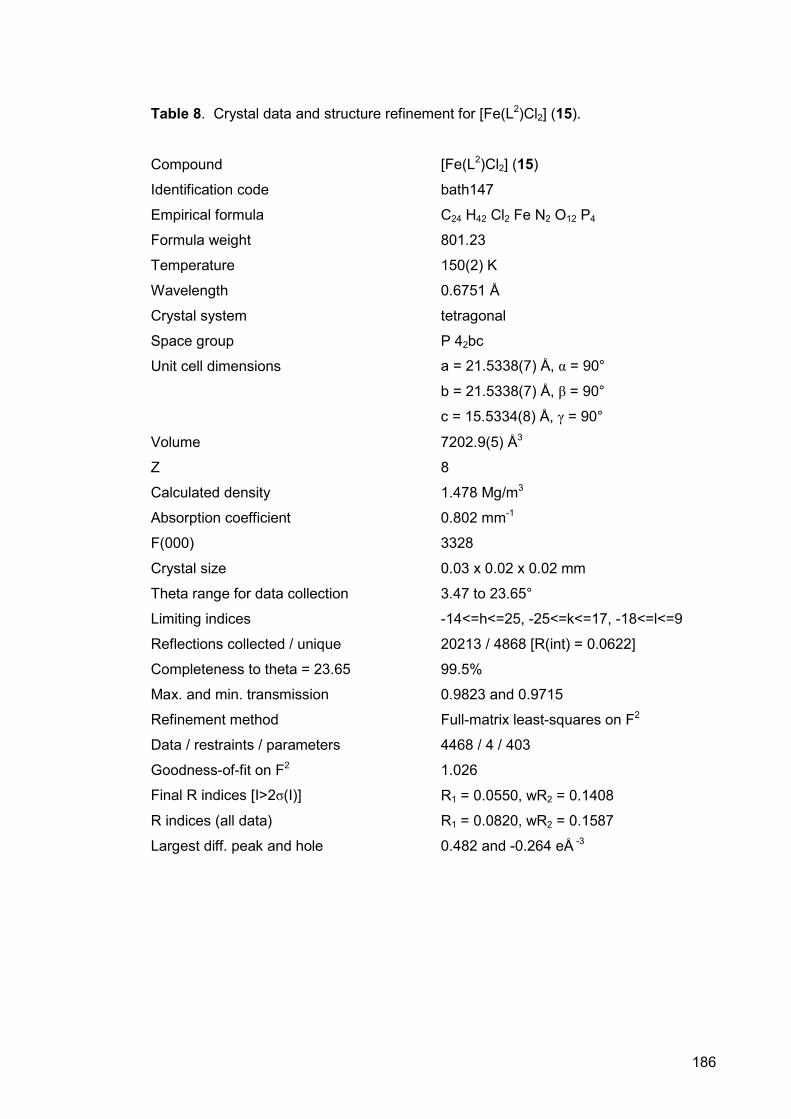
186
Table 8. Crystal data and structure refinement for [Fe(L2)Cl2] (15).
Compound [Fe(L2)Cl2] (15)
Identification code bath147
Empirical formula C24 H42 Cl2 Fe N2 O12 P4
Formula weight 801.23
Temperature 150(2) K
Wavelength 0.6751 Å
Crystal system tetragonal
Space group P 42bc
Unit cell dimensions a = 21.5338(7) Å, α = 90°
b = 21.5338(7) Å, β = 90°
c = 15.5334(8) Å, γ = 90°
Volume 7202.9(5) Å3
Z 8
Calculated density 1.478 Mg/m3
Absorption coefficient 0.802 mm-1
F(000) 3328
Crystal size 0.03 x 0.02 x 0.02 mm
Theta range for data collection 3.47 to 23.65°
Limiting indices -14<=h<=25, -25<=k<=17, -18<=l<=9
Reflections collected / unique 20213 / 4868 [R(int) = 0.0622]
Completeness to theta = 23.65 99.5%
Max. and min. transmission 0.9823 and 0.9715
Refinement method Full-matrix least-squares on F2
Data / restraints / parameters 4468 / 4 / 403
Goodness-of-fit on F2 1.026
Final R indices [I>2σ(I)] R1 = 0.0550, wR2 = 0.1408
R indices (all data) R1 = 0.0820, wR2 = 0.1587
Largest diff. peak and hole 0.482 and -0.264 eÅ -3
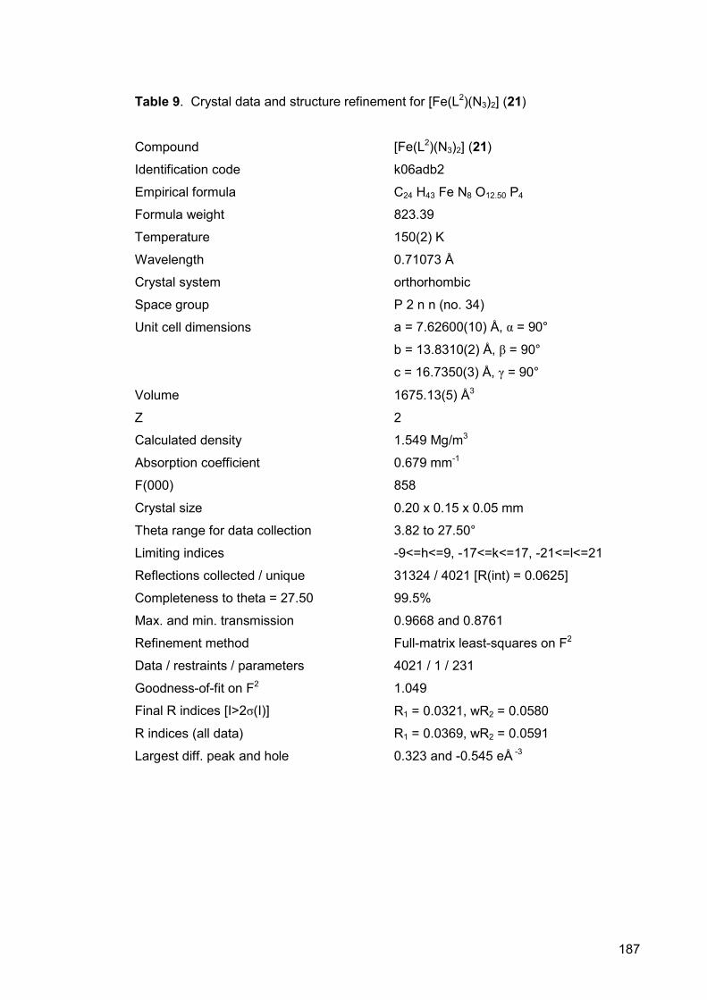
187
Table 9. Crystal data and structure refinement for [Fe(L2)(N3)2] (21)
Compound [Fe(L2)(N3)2] (21)
Identification code k06adb2
Empirical formula C24 H43 Fe N8 O12.50 P4
Formula weight 823.39
Temperature 150(2) K
Wavelength 0.71073 Å
Crystal system orthorhombic
Space group P 2 n n (no. 34)
Unit cell dimensions a = 7.62600(10) Å, α = 90°
b = 13.8310(2) Å, β = 90°
c = 16.7350(3) Å, γ = 90°
Volume 1675.13(5) Å3
Z 2
Calculated density 1.549 Mg/m3
Absorption coefficient 0.679 mm-1
F(000) 858
Crystal size 0.20 x 0.15 x 0.05 mm
Theta range for data collection 3.82 to 27.50°
Limiting indices -9<=h<=9, -17<=k<=17, -21<=l<=21
Reflections collected / unique 31324 / 4021 [R(int) = 0.0625]
Completeness to theta = 27.50 99.5%
Max. and min. transmission 0.9668 and 0.8761
Refinement method Full-matrix least-squares on F2
Data / restraints / parameters 4021 / 1 / 231
Goodness-of-fit on F2 1.049
Final R indices [I>2σ(I)] R1 = 0.0321, wR2 = 0.0580
R indices (all data) R1 = 0.0369, wR2 = 0.0591
Largest diff. peak and hole 0.323 and -0.545 eÅ -3

188
Table 10. Crystal data and structure refinement for [Fe(L3)2(κ2-O2SO2)] (23)
Compound [Fe(L3)2(κ2-O2SO2)] (23)
Identification code bath229
Empirical formula C34 H44 Fe N4 O6 P4 S
Formula weight 816.52
Temperature 150(2) K
Wavelength 0.69040 Å
Crystal system monoclinic
Space group C 2/c
Unit cell dimensions a = 15.422(3) Å, α = 90°
b = 9.5514(18) Å, β = 100.469(2)°
c = 24.333(5) Å, γ = 90°
Volume 3524.5(11) Å3
Z 4
Calculated density 1.539 Mg/m3
Absorption coefficient 0.721 mm-1
F(000) 1704
Crystal size 0.03 x 0.03 x 0.01 mm
Theta range for data collection 3.65 to 22.55°
Limiting indices -16<=h<=17, -10<=k<=9, -21<=l<=26
Reflections collected / unique 7752 / 2513 [R(int) = 0.0981]
Completeness to theta = 22.55 99.6%
Max. and min. transmission 0.991 and 0.975
Refinement method Full-matrix least-squares on F2
Data / restraints / parameters 2513 / 2 / 235
Goodness-of-fit on F2 0.976
Final R indices [I>2σ(I)] R1 = 0.0522, wR2 = 0.1117
R indices (all data) R1 = 0.1064, wR2 = 0.1314
Largest diff. peak and hole 0.362 and -0.370 eÅ -3

189
Table 11. Crystal data and structure refinement for trans-[Fe(L3)2Cl2] (24b)
Compound trans-[Fe(L3)2Cl2] (24b)
Identification code k06adb9
Empirical formula C68 H80 Cl4 Fe2 N8 P8
Formula weight 1510.66
Temperature 150(2) K
Wavelength 0.71073 Å
Crystal system monoclinic
Space group P 21/c
Unit cell dimensions a = 21.9030(2) Å, α = 90°
b = 10.38000(10) Å, β = 97.99°
c = 22.1870(2) Å, γ = 90°
Volume 4995.32(8) Å3
Z 3
Calculated density 1.507 Mg/m3
Absorption coefficient 0.838 mm-1
F(000) 2352
Crystal size 0.45 x 0.30 x 0.25 mm
Theta range for data collection 3.15 to 27.49°
Limiting indices -27<=h<=28, -13<=k<=13, -28<=l<=28
Reflections collected / unique 63321 / 11443 [R(int) = 0.0500]
Completeness to theta = 27.49 99.7%
Max. and min. transmission 0.8178 and 0.7041
Refinement method Full-matrix least-squares on F2
Data / restraints / parameters 11443 / 0 / 610
Goodness-of-fit on F2 1.046
Final R indices [I>2σ(I)] R1 = 0.0334, wR2 = 0.0731
R indices (all data) R1 = 0.0519, wR2 = 0.0806
Largest diff. peak and hole 0.575 and -0.525 eÅ -3
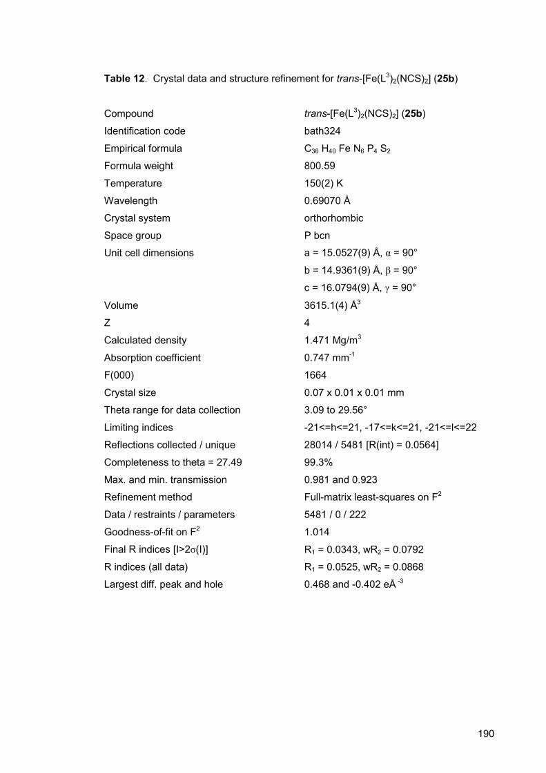
190
Table 12. Crystal data and structure refinement for trans-[Fe(L3)2(NCS)2] (25b)
Compound trans-[Fe(L3)2(NCS)2] (25b)
Identification code bath324
Empirical formula C36 H40 Fe N6 P4 S2
Formula weight 800.59
Temperature 150(2) K
Wavelength 0.69070 Å
Crystal system orthorhombic
Space group P bcn
Unit cell dimensions a = 15.0527(9) Å, α = 90°
b = 14.9361(9) Å, β = 90°
c = 16.0794(9) Å, γ = 90°
Volume 3615.1(4) Å3
Z 4
Calculated density 1.471 Mg/m3
Absorption coefficient 0.747 mm-1
F(000) 1664
Crystal size 0.07 x 0.01 x 0.01 mm
Theta range for data collection 3.09 to 29.56°
Limiting indices -21<=h<=21, -17<=k<=21, -21<=l<=22
Reflections collected / unique 28014 / 5481 [R(int) = 0.0564]
Completeness to theta = 27.49 99.3%
Max. and min. transmission 0.981 and 0.923
Refinement method Full-matrix least-squares on F2
Data / restraints / parameters 5481 / 0 / 222
Goodness-of-fit on F2 1.014
Final R indices [I>2σ(I)] R1 = 0.0343, wR2 = 0.0792
R indices (all data) R1 = 0.0525, wR2 = 0.0868
Largest diff. peak and hole 0.468 and -0.402 eÅ -3
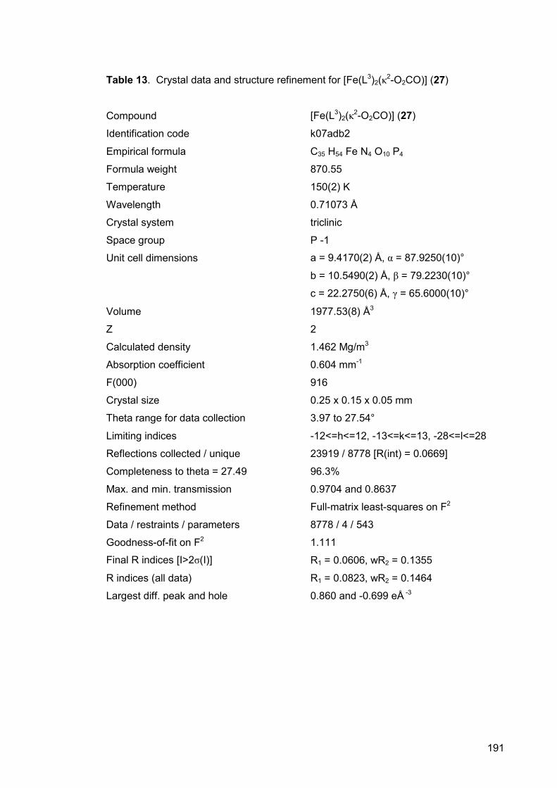
191
Table 13. Crystal data and structure refinement for [Fe(L3)2(κ2-O2CO)] (27)
Compound [Fe(L3)2(κ2-O2CO)] (27)
Identification code k07adb2
Empirical formula C35 H54 Fe N4 O10 P4
Formula weight 870.55
Temperature 150(2) K
Wavelength 0.71073 Å
Crystal system triclinic
Space group P -1
Unit cell dimensions a = 9.4170(2) Å, α = 87.9250(10)°
b = 10.5490(2) Å, β = 79.2230(10)°
c = 22.2750(6) Å, γ = 65.6000(10)°
Volume 1977.53(8) Å3
Z 2
Calculated density 1.462 Mg/m3
Absorption coefficient 0.604 mm-1
F(000) 916
Crystal size 0.25 x 0.15 x 0.05 mm
Theta range for data collection 3.97 to 27.54°
Limiting indices -12<=h<=12, -13<=k<=13, -28<=l<=28
Reflections collected / unique 23919 / 8778 [R(int) = 0.0669]
Completeness to theta = 27.49 96.3%
Max. and min. transmission 0.9704 and 0.8637
Refinement method Full-matrix least-squares on F2
Data / restraints / parameters 8778 / 4 / 543
Goodness-of-fit on F2 1.111
Final R indices [I>2σ(I)] R1 = 0.0606, wR2 = 0.1355
R indices (all data) R1 = 0.0823, wR2 = 0.1464
Largest diff. peak and hole 0.860 and -0.699 eÅ -3

192
Table 14. Crystal data and structure refinement for trans-[Fe(L3)2(H)Cl] (29)
Compound trans-[Fe(L3)2(H)Cl] (29)
Identification code ssg1322
Empirical formula C43 H50 Cl Fe N4 P4
Formula weight 838.05
Temperature 150(2) K
Wavelength 0.68840 Å
Crystal system monoclinic
Space group C 2/c
Unit cell dimensions a = 26.873(3) Å, α = 90°
b = 14.5374(17) Å, β = 111.244(1)°
c = 22.100(3) Å, γ = 90°
Volume 8047.0(17) Å3
Z 8
Calculated density 1.383 Mg/m3
Absorption coefficient 0.637 mm-1
F(000) 3512
Crystal size 0.02 x 0.01 x 0.01 mm
Theta range for data collection 3.44 to 23.91°
Limiting indices -31<=h<=31, -17<=k<=17, -26<=l<=26
Reflections collected / unique 27709 / 6833 [R(int) = 0.1122]
Completeness to theta = 27.49 99.6%
Max. and min. transmission 0.995 and 0.978
Refinement method Full-matrix least-squares on F2
Data / restraints / parameters 6833 / 1 / 516
Goodness-of-fit on F2 0.958
Final R indices [I>2σ(I)] R1 = 0.0562, wR2 = 0.1229
R indices (all data) R1 = 0.1217, wR2 = 0.1530
Largest diff. peak and hole 0.690 and -0.312 eÅ -3