Embed Size (px)
Citation preview

1
UNIVERSITÀ DI PISA
Dipartimento di Farmacia
Corso di Laurea Specialistica in Chimica e Tecnologia
Farmaceutiche
Tesi di Laurea:
Rational design, synthesis and evaluation of
heterocyclic derivatives as inhibitors of serine
palmitoyltransferase
Relatore: Correlatore:
Dott.ssa Sara Del Carlo Prof.ssa Clementina Manera
Dott. Giuseppe Saccomanni
Candidato:
Reinhold Baci (N° MATRICOLA 408866)
Settore Scientifico Disciplinare: CHIM-08

2
ANNO ACCADEMICO 2012 – 2013
SECRETAZIONE DELL’ ELABORATO SCRITTO DELLA TESI DI LAUREA
“Il contenuto di questa tesi di laurea è strettamente riservato, essendo presenti argomenti tutelati dalla legge come
segreti. Pertanto tutti coloro che ne prendono conoscenza sono soggetti all’obbligo, sanzionato anche penalmente
dagli articoli 325 e 623 del codice penale, di non divulgare e di non utilizzare le informazioni acquisite.”

3
INDEX

4
1. INTRODUCTION 1
1.1 Sphingolipids 2 1.2 Ceramide 3 2. Serine palmitoyltransefarse (SPT) 4 2.1 Bacterial SPT 5 2.2 Eukaryotic SPT 6 2.3 Substrate 7 3. Reaction mechanism 8
4. Inhibitors 11 4.1. Penicillamine 12 4.2. Cycloserine 14 4.3. Myriocin 19
5. Retinitis pigmentosa 26
6. Alzheimer 28
7. Hereditary Sensory Neuropathy Type 1 31
8. Melanoma 34 INTRODUCTION TO EXPERIMENTAL PART 37
EQUIPMENT USED 51 EXPERIMENTAL PART 53 REFERENCES 80

5
INTRODUCTION

6
1.1 Introduction
More than 100 hundred years ago, Johann L. W. Thudicum described a not well
identified aliphatic alkaloid in the brain called sphingosine1. The structure of this
substance was characterized only 50 years later by Carter at al.2
In the last years, structural and distribution studies have highlighted the critical roles of
sphingolipids in membranes micro domains formation, skin barrier function and
modulate on of cellular events like apoptosis, proliferation and differentiation3-5
.
These discoveries along with bioactive signal function observation, increase research
attraction and attention on sphingolipids metabolism6-11
.
1.2. Sphingolipids
Sphingolipids (SPL) are one of eight categories of the LIPID MAPS classification
system and they are described as lipids containing a sphingoid base (1,3-dihydroxy-2-
amino alkane) linked by an amido group to a fatty acid and with polar headgroup, like
phosphate and carbohydrate12
. SPL distribution appears ubiquitous in eukaryotic. In
particular, SPL alkane group is usually an eighteen carbons chain; C-2 and C-3, in the
polar headgroup, are chiral and the configuration is 2S, 3R, D-Eritro. Indeed, in
mammal cells, sphingosine appear to be the major sphingoid base, followed by
dihydrosphingosine, while phytosphingosine, the third major type of sphingoid bases,
has been found mainly in plants, although it was revealed in large amount in mammal
tissues like stomach and kidney. Sphingolipids containing cholinephosphoceramide,
known as sphingomielyn, represent 5-10% of mammalian membrane’s phospholipids.
SPL are produced also by some prokaryotes, plants, bacteria and fungi through different
biosynthetic pathways 1,12
. In fungal and plants inositol-phosphoceramide is the most
abundant constituent of the membrane phospholipids; these observations suggest a
relevant structural dissimilarity in the polar headgroup in different species. Conversely
the polar headgroup of glycerophospholipids are structurally very similar between
mammal cells and fungi12
(Fig.1).

7
Fig. 1: Biosynthetic pathway of sphingolipids in mammalian and fungal cells.
1.2. Ceramide
Several studies performed by Braun13
, and Stoffel 14
, independently, have demonstrated
in 1968 that the second step of sphingolipid biosynthesis is the NADPH-dependent
reduction of a C-3 carbonyl group leading to dihydrosphingosine15
. In mammalian cells,
dihydrosphingosine undergoes to an N-acylation reaction forming dihydroceramide.
Afterwards, an alkyl chain oxidation leads to ceramide which successively
functionalized at the polar headgroup to give sphingomielyn or different
glycosphingolipids. Conversely, in fungi, dihydrosphingosine undergoes to oxidation
and then to N-acylation forming phytoceramide which is further converted in inositol
phosphophytoceramide and more structurally complex derivates. In fungi, the acyl-
chain is mainly represented by C-26 hydroxy fatty acid, whereas mammal ceramide
deerivatives are saturated C-16/C-24 fatty acid chain16,17
. Cells produce sphingolipids
through two different metabolic ways: catabolic and anabolic pathways. The catabolic
pathway involves hydrolyses of complex molecules, as sphingomielyn and
glucosphingolipid, to obtain ceramides. The anabolic pathway (also called de novo
biosynthesis) provides an enzymatic cascade which leads to the formation of ceramides
starting from simple and abundant substrates like L-serine and palmitoyl-CoA1.

8
2. Serine palmitoyltransefarse (SPT)
Sphingolipids biosynthesis is different between species (i.e. S. cerevisiae produces
phosphoinositol headgroup sphingolipids only, while P. pastoris both glucosylceramide
and phosphoinositol sphingolipids headgroup), whereas the first enzymatic step of de
novo biosynthesis is conserved across all species producing sphingolipids2 (Fig.2). The
common step of the de novo synthesis is an enzymatic condensation carried out by
serine palmitoyltransferase (SPT), a pyridoxal-5’-phosphate (PLP) dependent enzyme.
SPT is an enzyme belonging to alfa-oxoamine synthase (AOS) family which is a PLP
dependent family.
Fig. 2: POAS family. Reactions catalyzed by POAS family members.
SPT catalyzes a condensation between L-serine and typically C16 acyl-CoA thioester
(palmitoyl-CoA), to give a Claisen-like C18 condensation product: 3-
ketodihydrosphingosine (KDS). Successively KDS is rapidly reduced to
dihydrosphingosine in presence of NADPH1,2
. SPT plays a key role in the biosynthesis
of sphingolipids indeed regulation of SPT catalyzed step prevents accumulation of
sphingolipids metabolites (i.e. sphingoid base) while inhibition of later biosynthetic
steps lead to metabolite accumulation which are death effectors in various experimental
models and pathological conditions. Therefore, SPT condensation reactions represent
the rate-limiting step of sphingolipids biosynthesis1.

9
2.1. Bacterial SPT
The first three dimensional structure of SPT was obtained by the isolation of the
homodimeric (and water soluble) SPT from the Gram-negative bacterium S.
paucimobilis (spSPT)18
. The crystal structure reveals that the holo-SPT monomer (or
internal aldimine) consists of three domains: the N-terminal, the central catalytic
domain and the C-terminal domain. The N-terminal domain is consist of 80 residues (an
α-helix followed by 3 β-sheets). This domain is linked with the central domain also
called catalytic domain. In the central domain is recognized the Lys265
residue
responsible of PLP cofactor binding, while 7 β-sheet is composed by about 200
residues. The C-terminal domain is strictly linked with N-terminal domain by binding
interactions19
. Three dimensional crystal of the holo-enzyme revealed a homodimeric
structure containing a conserved Lys265
-binding-PLP residue. However, for the correct
positioning of the PLP cofactor, (crucial for the catalytic activity), π-stacking
interactions between the pyridine ring and His159
are required. PLP interacts also with
other residues such as Asn138
, Asp231
, His234
, Thr262
, Gly134
and Tyr135
. Furthermore, an
Arg379
is needed for the positioning of the L-serine in the catalytic site because of
crucial interaction with the carboxy moiety of the substrate1.
Other SPT homologues, like Sphingobacterium multivorum SPT (SmSPT) and
Sphingobacterium wittichii (SwSPT), exhibit difference at few levels. SmSPT have a
70% sequence identity and in active site the external aldimine (formed by the PLP and
serine) is linked by two water molecules to two aminoacid residues, Ser81
and Met271
,
and (not to a Lys)20
. Three-dimensional structure of SwSPT reveals a larger active site
than in SpSPT, probably because of the use of a larger acylated-ACP thioester
substrate21,22
.

10
2.2. Eukaryotic SPT
Eukaryotic SPT is a heterodimer consisting in two subunits, LCB1 and LCB2, both
linked to endoplasmatic reticulum (ER) and they are encoded by two different genes,
lcb1 (SPTLC1) and lcb2 (SPTLC2), composed by 15 exons of 85 kbp size localized on
chromosome 9, arm q21-q22, and 12 exons of 110 kbp size localized on chromosome
14, arm q24.3-q3 respectively. These genes encode for proteins of 53 and 63 kDa, with
20 % sequence identity that is probably critical for dimerization23,24
. LCB proteins have
95% identity between mammals and 40% between mammals and yeast. The sequencing
of SPTLC1 and SPTLC2 genes in Saccharamyces cerevisiae revealed that the active
site of eukaryotic SPT is localized in the LCB2 subunit; this observation is suggested by
detection of conserved residues of AOS’s family (one lysine, two hystidine and an
aspartate) which are essential to lock the PLP cofactor in the active site1,2
. LCB1
subunit appears crucial for SPT’s catalytic activity. The lack of this subunit expression
or a missense mutation in SPTLC1 gene causes alteration of SPT’s catalytic activity25
.
Furthermore, CHO cells line defective in SPTLC1 transcription express lower levels of
LCB2. To obtain an overexpression of LCB2 is required an overexpression of LCB1
and LCB2. Moreover, it appears to be unstable when it is not associated with the LCB1.
On contrary an high level of LCB1 do not require an overexpression of LCB2. Both
monomers have only one highly hydrophobic transmembrane domain (TMD). The
catalytic site has a cytosolic orientation indeed indirect immunocytochemical analysis
indicated that C-termini and N-termini of LCB1 have cytosol and lumen orientation
respectively26
. LCB1 and LCB2 are ER membrane integrated proteins type I. Another
isoform of LCB2 called LCB3 is expressed only in certain tissues.
In yeast this enzyme is constituted by the so called “SPOTS COMPLEX” that’s encodes
for SPT–ORM1/2-Tsc3-Sac1 (phosphatase)27
. Therefore, LCB1 and LCB2 subunits are
associated with a third subunit, Tsc3, which is required for the maximal SPT activity.
This heterodimeric structure is associated with oromucosoids proteins (ORM1/2) which
are able to negatively regulate SPT.
Recently for human SPT two novel small subunits named ssSPTa and ssSPTb were
discovered. These subunits can enhance activity >10 fold when bound to LCB1-LCB2
heterodimer. Moreover orosomucoid-like (ORMDL) proteins appear to be able to
interact with the LCB1-LCB2 heterodimer28
.

11
2.3. Substrate
Palmitoyl-CoA is the more kindred acyl-CoA thioester substrate of SPT in mammalian
cells; on the other hand pentadecanoyl-CoA and heptadecanoyl-CoA are also good
substrates less abundant. L- and D- serine are SPT amino acid substrates. SPT is able to
form the Schiff’s bases with both enantiomers but the alfa-deprotonation step (before
acylation) proceeds only with L-serine substrate. Thus L-serine is the common SPT’s
substrate in the de novo synthesis. The hydroxyl, carboxyl and amino groups of L-serine
appear to be necessary for interaction with SPT; neither L-alanine, L-serinamide, D,L-
serinol and nor L-serine methyl ester not in the formation of [ 3H]KDS from L-[
3H]serine, indicating that the hydroxyl, amino, and carboxyl groups of L-serine are
responsible for the recognition of the amino acid substrate by the SPT enzyme29
.

12
3. Reaction mechanism
SPT catalyzes a Claisen like condensation of L-serine and palmitoyl-CoA which leads
to 3-KDS as final product. SPT reaction mechanism proceeds across six step: 1)
formation of the Schiff base between L-serine and PLP, 2) L-serine α-hydrogen
removing; 3) nucleophilic attack to palmitoyl-CoA (formation a transient acylated
adduct), 4) decarboxylation, 5) protonation of α-carbanion and 6) KDS release (Fig.3).
Assays conducted with purified enzyme from chinese hamster ovary cell, suggested that
one molecule of mammalian SPT is capable of catalyzing maximally of 80 cycles of
these steps per minute30.
Fig. 3: Catalytic mechanism of SPT.
The PLP cofactor catalyses all the reaction mechanism. Interaction of PLP aldehyde
group and Lys265
lead to a Schiff base (internal aldimine) in the active site.
Spectrophotometric analysis of holo SPT revealed two UV absorption peaks which
represent two tautomers of the Schiff base PLP-Lys265
: the enolamine (338 nm) and
ketoenamine (426 nm). Moreover, Crystalline structure of S. Multivorum and S.
Paucimobilis SPT highlighted that PLP has Van der Walls interaction with His159
and
Ala233
.

13
Fig. 4: Spectrophotometric analysis of holo SPT
Addiction of L-serine to the holo-enzyme causes drastically changes in the UV spectra
due to the formation of a new Schiff base between PLP and L-serine (external
aldimine). Two intermediate adducts are identified: first adduct has an absorption
similar to holo-SPT while the other one has a maximum peak at 426 nm (which
represent the ketoenaminic tautomer of the new Schiff base) (Fig.4). The interaction of
PLP with L-serine causes a rotation of pyridine ring or a torsion of the Schiff base C4-
C4’ bound.
Once the external aldimine is formed, L-serine is conformationally stabilized by
interaction of the carboxyl group with His159
Nε2 (hydrogen bond) and by interaction of
the hydroxylic group with the PLP phosphate group and a molecule of water. These
interactions fixed the PLP-L-serine complex allowing a perpendicular orientation (80°)
of Cα-COO bond to imine-pyridine plane.
The next step of the reaction mechanism is represented by the formation of a new C-C
bond via a Claisen like condensation. Two different intermediate may be involved in the
nucleophilic substitution: the decarboxylated intermediate or that one generated after
removal of the -H. In order to understand the intermediate involved in the nucleophilic
substitution reaction some experiments have been developed in presence of a palmitoyl-
CoA analogue S-(2-oxoheptadecyl)-CoA. This compound has a methylene group
inserted between the sulphur atom and the carbonyl groups which doesn’t allow the
nucleophilic substitution. Data obtained performing the enzymatic reaction in presence

14
of S-(2-oxoheptadecyl)-CoA and deuterated solvent showed the α-hydrogen switch with
the deuterated solvent increase of about 100 times (high peak) at 426 nm. Therefore, α-
deprotonation of external aldimine is improved in presence of palmitoyl-CoA substrate
and a significative amount of quinoid adduct have been formed. This suggests that the
quinoid adduct attack palmitoyl-CoA thioester to form the C-C bond. A faster α-
deprotonation of external aldimine in presence of palmitoyl-CoA reduce the risk of
intermediates accumulation which can form pyridoxamine-5’-phosphate (PMP) after
transamination reaction (PMP can’t be converted to PLP thereby the enzyme is
inactivated). According to Dunathan hypothesis, the -deprotonation process takes
place when the bond involved in the nucleophilic substitution is perpendicular to the
imine-pyridine place. The reaction takes place thanks to the complete overlap between
the -orbital (occupied) of the designed bond and the free -orbital of the conjugate
imine-pyridine system. The increase of -deprotonation process in presence of
palmitoyl-CoA is related to a strictly stereochemical control of the reaction. In the
external aldimine conformation the C-H bond is 40° rotated with respect to the imine-
pyridine plane avoiding the -deprotonation process. This conformation is generated by
interaction of the carboxyl group of L-serine with His159
as depicted in the SPT-PLP-
serine crystal structure. Computational study showed that palmitoyl-CoA induced
conformational change is related to the displacement of the hydrogen interaction of L-
serine with His159
: palmitoyl-CoA interacts with His159
(hydrogen bond) whereas L-
serine performs a new interaction with the guanidine group of Arg390
rotating the C-N
bond of 50°. In this new conformation the C-H bond of L-serine is perpendicular to
the imine-pyridine plane promoting the -deprotonation.
The nucleophilic substitution of quinoid intermediate to the acyl-CoA thioester
carboxylic group leads to a β-keto-acid derivates. The interaction between palmitoyl-
CoA and His159
promotes the C-C bond formation by acidic catalysis played by His159
:
this residue exchange an hydrogen with the carboxylic group of palmitoyl-CoA
inducing the right positioning. The product forms an hydrogen bond with His159
(the
oxygen of the carbonyl group interact with the hydrogen of His159
). Then β-keto-acid
derivates undergoes to decarboxylation and release of the final adduct (KDS).
Generally PLP catalyzed reactions occur through decarboxylation process catalyzed by
the electron withdrawing immine-pyridine system. Instead SPT decarboxylation
reaction is promoted by the correct position of the carboxyl group by His159
.1

15
4. Inhibitors
Natural inhibitors of SPT have been discovered. Sphingofungine, lipoxamicina
(neoenactin M1), and myriocin (ISP-1/thermozymocidin) (Fig.5) are potent and highly
selective inhibitors of both fungine and mammals SPT in cell-free models with
nanomolar IC50. These compounds are structurally similar to the postulated transitory
adduct formed by the L-serine and palmitoyl-CoA during the condensation suggesting
the crucial role of a transitory adduct of these compound with PLP in the strong
inhibition activity.
Indeed, the inhibitory activity of sphingofungine B is highly dependent on
stereochemistry31,32
.
The crucial role of sphingofungine B stereochemistry has been demonstrated: the C14
hydroxyl group of sphingofungine B yields potent inhibitory activity but it’s not crucial
for the activity. On the other hand, the configuration of the stereogenic centers in the α,
β, γ and δ positions from carbonilic group are essential for the inhibitory activity32,33
.
Viridiofungins were first isolated by Harris and co-workers in 1993 from the fungus,
Trichoderma viride. This family of alkyl citrates exhibited broad spectrum of anti-
fungal properties with minimum fungicidal concentrations in the range of 1–20 μg/mL
against a number of species. Furthermore, viridiofungins inhibited rat and yeast
squalene synthesis. This antifungal activity is unrelated to the inhibition of ergosterol
biosynthesis. Instead, viridiofungins showed very potent (nanomolar range) inhibitory
activity against serine palmitoyltransferase34
.
Fig. 5: Structure of natural SPT inhibitors.

16
4.1. Penicillamine
Penicillamine (Pen) is an α-amino acid and a characteristic degradation product of
penicillins, used in Wilson's disease as copper chelator to form mixed disulfides with
cystein. The enantiomer used as pharmaceutical drug is D-Pen. It has been well
established that both enantiomers and racemic mixture exerts anti-PLP activity by
inhibiting several PLP-dependent enzymes such as alanine aminotransferase, aspartate
aminotransferase, glutamate decarboxylase, hystidine decarboxylase and serine
hydroxymethyl transferase. L-Pen has the stronger inhibitory activity thanks to the
structure similarity with the enzyme’s naturally substrate (L-amino acids) (Fig.6).
Fig. 6: L-amino acids inhibitors of SPT.
A recent research developed by Campopiano group, have clarified Pen inhibitory action
and mechanism on SPT from Sphingomonas Paucimobilis (spSPT). Incubation of D-
Pen and L-Pen with spSPT results in different percentage of enzyme inhibition:
incubation of 5 mM L-Pen with spSPT reduces enzyme activity to 3% while incubation
with D-Pen decreases enzyme activity to 34%. Pen inhibition is reversible and the
mechanism of SPT inactivation occurs by disabling the PLP cofactor. Indeed,
incubation of enzyme inactivated by D-Pen and L-Pen with a buffer containing 50 µM
of PLP, restore the SPT activity to 80% and 57 % respectively. Inability to restore 100%
of the activity using a dialyzing buffer is due, probably, to possible additional
competitive reactions that irreversibly modify the proteins. The Pen thiol-group plays a
crucial role in the inhibition mechanism. The thiol-group is more nucleophilic than the
hydroxyl group of the L-Ser and it’s able to interact with PLP aldehyde leading to
PLP:thiazolidine (PLP:TA) adduct (Fig.7).

17
Fig. 7: Addition of L-Pen to SPT leads to formation of a PLP:TA adduct via an external aldimine intermediate (P
represents group phosphate).
The inhibition reaction could be monitored observing the disappearance of free PLP
peaks at 390 nm and appearance of a new peak at 333 nm (thiazolidine adduct). Using
the same spectrophotometric approach, a time dependent disappearance of holo SPT
ketoenamine (420 nm) and appearance of a thiazolidine adduct have been observed after
addiction of L-Pen to holo-SPT. ESI-MS analysis of these samples confirmed the
formation of a PLP:TA adduct. Moreover L-Pen and L-cystein activities have been
compared highlighting a faster inhibitory activity of L-Pen that could be related to the
presence of gem-dimethyl group enhancing cycle formation through a Thorpe-Ingold
effect35
(Fig.35).
Fig. 8: (A) UV-visible spectrum of 20 mM holo-SPT (solid line) shows typical peaks at 335 nm and 420 nm due to
enolimine and ketoenamine forms of the PLP cofactor respectively. Addition of 10mM L-Cys to holo- SPT (broken
lines) led to formation of a thiazolidine adduct (333 nm peak) over a 30 minute period, with concomitant loss of the
420 nm peak (B) improved thiazolidine formation in SPT over 30 minutes by addition of 10 mM L-Pen.

18
4.2. Cycloserine
β-chloro-L-alanine and L-cycloserine has been used as SPT inhibitors. These
compounds are potent inhibitors of several PLP-dependent enzymes, therefore their use
as specific inhibitors is limited36,37
.
Cycloserine (Fig.9a) is cyclic α-aminoacid well known to inhibit many PLP-dependent
enzymes (transaminase, racemase and decarboxylase)38-40
. It exists as two enantiomers:
D-cycloserine (DCS) and L-cycloserine (LCS).
Fig. 9a: “Conformations” and “ structures” of D- and L- α-amino acids inhibitors of SPT.
The mechanism of antibacterial activity is based on the inhibition of alanine racemase, a
PLP-dependent enzyme that synthesize D-alanine for the formation of D-alanyl-D-
alanine dipeptide (an essential component of the peptidoglycane layer of the M.
tuberculosis)41
. Furthermore, DCS is an agonist of N-methyl-D-aspartic acid (NMDA)
receptors that is implicated in several CNS pathologies and it’s used in neurological
models of these pathologies42
; LCS has been prepared synthetically and used as a
modulator of lipids metabolism in biological research, indeed it is a potent inhibitor of
SPT activity. Furthermore, it has been demonstrate that LCS inhibits mouse brain SPT
activity in vivo after intraperitoneal injection43
. Some authors propose a cycloserine
inhibition mechanism based on aromatization of the PLP:cycloserine external aldimine
(“aromatization mechanism”) by α-deprotonation that give rise to a 3-
hydroxyisoxazole–PMP adduct: the cycloserine ring remains intact and covalenty linked
to the PLP.

19
A first study developed by Ikushiro44
revealed a change in SPT absorption 10 minutes
later the addiction of LCS: the peak corresponding to the ketoenamine tautomer of the
internal aldimine (426 nm) decreased whereas a new peak at 380 nm increased. On the
basis of this data a mechanism of action was hypothesized: in a first attend LCS reacts
with PLP forming and external aldimine that undergoes to decyclization of the
isoxazolidone ring (forming a transient oxime intermediate with absorption at 380 nm)
leading to pyridoxamine 5’-phosphate (PMP) and β-aminooxypyruvate (Fig.9b).
Fig. 9b: Proposed mechanism of LCS inactivation of SPT by Ikushiro et al.
In 2010 the research group of Campopiano45
have highlighted the enantiospecific
inhibition of LCS and DCS and they have identified a novel decarboxylative ring-
opening mechanism for inactivation of SPT. In particular the data show that in order to
obtain the same inhibition of SPT activity (to the same extent and over the same time)
15-fold higher concentration of DCS is needed in comparison with LCS showing a clear
enantiospecific difference of the enzyme active site (Fig.10).
Fig 10: Secondary plot of 1/kapp versus 1/[inhibitor] for LCS and DCS.

20
SPT activity (monitored by continuous DTNB assay) is reduced to 1% and 22 % after 2
hours incubation with 5 mM of DCS and LCS respectively. Furthermore incubation of
treated samples in a buffer containing 25 M of PLP caused the recovery of SPT
activity (83 % and 79 % of DCS and LCS inactivated samples respectively). Incubation
of the inhibited enzyme in buffer (without PLP) doesn’t cause any further change of
activity (the enzyme is still inactivated). These data confirm the disabling of PLP
activity and the absence of further covalent modifications occur. The incubation of
enzyme with DCS and LCS showed different UV-spectra. After 30 minutes from the
addiction of 5 mM LCS new peaks appear at 330 and 380 nm and no more changes
were observed when the incubation time was about 8 hours. Incubation of DCS with
SPT causes the loss of the 425 nm peak and the appearance of a new peak at 380 nm.
This peak shifted to 365 nm over time and at the same time a broad shoulder at 330 nm
appeared (Fig.11A). These results suggest the formation of different species and the
displace of the new PLP-Lys265
bond. In contrast, the UV-VISis spectra of free PLP in
the presence of both enantiomers show the same peak at 360 nm (Fig.11B).
Fig. 11: (A) SPT and 5 mM of LCS at time 0 (solid line), 30 seconds (long dash), 1 minute (dotted line) and 30
minutes (dash dot). (B) SPT and 5 mM of DCS at time 0 (solid line), 30 seconds (long dash), 2 hours (dotted line) and
10 hours (dash dot).
21
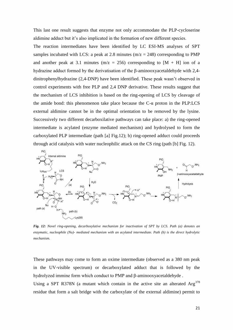
This last one result suggests that enzyme not only accommodate the PLP-cycloserine
aldimine adduct but it’s also implicated in the formation of new different species.
The reaction intermediates have been identified by LC ESI-MS analyses of SPT
samples incubated with LCS: a peak at 2.8 minutes (m/z = 248) corresponding to PMP
and another peak at 3.1 minutes (m/z = 256) corresponding to [M + H] ion of a
hydrazine adduct formed by the derivatisation of the β-aminooxyacetaldehyde with 2,4-
dinitrophenylhydrazine (2,4-DNP) have been identified. These peak wasn’t observed in
control experiments with free PLP and 2,4 DNP derivative. These results suggest that
the mechanism of LCS inhibition is based on the ring-opening of LCS by cleavage of
the amide bond: this phenomenon take place because the C-α proton in the PLP:LCS
external aldimine cannot be in the optimal orientation to be removed by the lysine.
Successively two different decarboxilative pathways can take place: a) the ring-opened
intermediate is acylated (enzyme mediated mechanism) and hydrolysed to form the
carboxylated PLP intermediate (path [a] Fig.12); b) ring-opened adduct could proceeds
through acid catalysis with water nucleophilic attack on the CS ring (path [b] Fig. 12).
Fig. 12: Novel ring-opening, decarboxylative mechanism for inactivation of SPT by LCS. Path (a) denotes an
enzymatic, nucleophile (Nu)- mediated mechanism with an acylated intermediate. Path (b) is the direct hydrolytic
mechanism.
These pathways may come to form an oxime intermediate (observed as a 380 nm peak
in the UV-visible spectrum) or decarboxylated adduct that is followed by the
hydrolyzed immine form which conduct to PMP and β-aminooxyacetaldehyde .
Using a SPT R378N (a mutant which contain in the active site an alterated Arg378
residue that form a salt bridge with the carboxylate of the external aldimine) permit to

22
isolated intermediate and 40 fold reduced rate when compared with the wild-type
enzyme, it was observed transient formation of a quinonoid species (formed by
decarboxylation) at 510 nm during DCS inactivation, while inactivation with LCS
(much faster inactivator in comparison with DCS) show a similar spectrum to the wild-
type enzyme at 380 nm45
.
In addition these researches has been conducted with modeling to isolated the PMP
adduct (Fig.13).
Fig.13: Left: Fo-Fc electron density for the PMP molecule. This was calculated from molecular replacement model
which was refined and had omitted the co-factor. The map is contoured in green at 3s (0.2 Å3). Also shown are the
side chains of Lys265, His159, Asp231 and His234. Monomer A is shown in ribbon in cyan and monomer B in
magenta. The additional Fo Fc electron density ‘‘blob’’ is shown in blue, contoured at 2.7s (0.2 Å 3). A molecule of
the β-aminooxyacetalaldehyde identified by mass spectrometry is placed in the density. Carbons are colored yellow,
nitrogen blue, oxygen red and phosphorous orange. Right: Overlay of SPT:L-ser (2bwj) and the LCS inactivated
form. The loop containing R378 adopts the ‘‘swung-out’’ conformation in the LCS form, in contrast to the ‘‘swung
in’’ conformation in SPT:L-ser. The color scheme for the LCS inhibited form is as (left). For the SPT:L-ser structure,
monomer A is colored light blue. Carbons are colored green, and other atoms are colored the same as in (left). The
main chain of R378 adopts a very different conformation from the SPT:L-ser structure because the salt contact with
L-ser is missing. A well-ordered molecule (red sphere) in the LCS structure is found in the same location as the L-ser
carboxylate. The side chain of L-ser points towards the unfitted blob at the active site.

23
4.3. Myriocin
Natural products and their derivatives are known to be excellent tools for biochemicals
and pharmacologicals studies. Myriocin [(2S, 3R, 4R, 6E)-2-amino-3,4-dihydroxy-2-
(hydroxymethyl)-14oxo-6-eicosenoic acid] (Fig.14a) also known as thermozymocidin
and ISP-1, has potent immunosuppressant properties in addition to antibacterial and
antifungal activity.
Fig. 14a: myriocin structure
Firstly isolated by thermophilic moulds Myriococcum albomyces and Mycelia sterilia,
myriocin remains the most valuable and widely used chemical probes in sphingolipids
research as demonstrated by several studies: the identification of the two SPT subunits
(SPTLCB1 and SPTLCB2) by Screiber et al.27
and of multi-protein membrane-bound
SPT complex (SPOTs complex) by Breslow at al.28
has been performed using myriocin.
Kawasaki and collagues determined an IC50 value of 15 nM using cytotoxic T-cell line
(CTLL-2)46
. Moreover, a recent study has demonstrated that myriocin reduces ceramide
levels in rd10 mouse model of retinitis pigmentosa (RP) and therefore can rescue
photorecepetors death47
. Despite its use, the molecular basis of myriocin inhibition of
SPT is largely unknown. Campopiano and coworkers have recently shed light in
molecular mechanism of SPT inhibition by myriocin using a soluble and recombinant
form of the enzyme from Sphingomonas paucimobilis (spSPT). Uv-vis analyses after
addition of five-fold molar excess of myriocin to holo-SPT revealed that the two
characteristic peak at 333 nm and 420 nm (corresponding to the ketoenamine and
enolimine forms of the external aldimine) led to an increase absorbance with maximum
at 430 nm and the disappereance of the peak at 333 nm. This data reflects a
transamination reaction between PLP and myriocin that lead to the formation of a stable
an external aldimine mimicking the β-keto acid intermediate, Fig.14b and confirmed by
LC ESI-MS analyses Fig.14.b-B.

24
Fig. 14b: SPT inhibition occurs via formation of a PLP-myriocin aldimine. (A) UV-Vis spectrum of 40 μM SPT
before (solid line) and after 200 μM myriocin addition (dotted line). (B) The proposed structure of the inhibitory
complex - a PLP-myriocin aldimine, 11. (C) Detection of the PLP-myriocin aldimine by LC-MS. Top, Extracted Ion
Chromatogram at m/z 631. Bottom, high resolution mass spectrum of the PLPmyriocin aldimine, obtained by
summing the spectra between t = 8-12 minutes. ([M+H]+, C29H48N2O11P; predicted m/z 631.29902; observed error
4.0 ppm). * denotes a contaminant.

25
Interstingly PLP-myriocin aldimine (11) is relatively stable at 25°C. However, after 16
hours of incubation a decrease of 430 nm peak and increase of 331 nm and 400 nm
peaks is revelead (Fig.15).
Fig.15: UV-vis analysis of the degradation of the PLP-myriocin external aldimine in wild-type SPT. The PLP-
myriocin external aldimine (solid line) is stable for 90 minutes, before a decrease at 430 nm is observed, which is
accompanied by a concomitant increase at 331 and 400 nm over 16 hours (dotted and dashed lines).
SPT:PLP-myriocin complex has a noncovalent, reversible nature and very slow off rate
(koff) when incubated with myriocin for 10 minutes: sample dialysis in 25 µM PLP
containg buffer for 24 hours restored enzyme activity of 60% (accompanied with UV-
Vis spectrum back to the internal aldimine). On the other hand a covalent and
irreversible nature of myriocin-enzyme interaction has been observed when samples
were incubated for 16 hours (Fig.16). Moreover, experiments demonstrate that myriocin
is a competitive inhibitor for both L-serine and palmitoyl-CoA and a Ki of 967±98 nM.
Fig.16: Relative enzymatic activity after removal of inhibiting species by dialysis. SPT was inhibited with 200 μM
myriocin and incubated for 10 minutes (white bars) or 16 hours (grey bars) at 25 °C before removal of myriocin by
extensive dialysis. The enzymatic activity was then determined at 0 hours, 3 hours, and 24 hours after dialysis.

26
To better understand the role of the Lys 265 in the dual reaction mechanism of
myriocin, an incubation with five-fold molar excess of myriocin and catalytically-
inactive SPT (K265A SPT) which present active site without Lys residue was
performed. After 16 hours the UV-Vis spectra remained unchanged in contrast with
wild-type SPT suggesting that the initial SPT:PLP-myriocin inhibitor complex breaks
down to form a second species that also inhibits wild-type SPT (Fig.17).
Fig. 17: UV-vis analysis of SPT K265A (40 μM) showed two absorbance maxima at 326 and 402 nm (solid line).
Upon addition of 200 μM myriocin, an immediate shift to a single peak at 425 nm occurred (dotted line), indicating
the formation of a PLP-myriocin aldimine complex. Over 16 hours this spectrum remains unchanged (dashed line),
indicating that the PLP-myriocin aldimine complex is not degraded by this mutant enzyme.
A retro-aldol like mechanism of SPT:PLP:myriocin complex which selectively and
covalently modifies the Lys265
(crucial for the enzyme-catalysed reaction) leading to
irreversible inactivation of the enzyme was hypothesized (Fig.18).
Mass spectrometry analyses showed a covalent adduct (14 in Fig.18) of SPT displaying
a mass of 47.509 Da susceptible to NaBH4 of the covalent SPT-octadecenal imine
adduct and ketone group. Peptide mass fingerprinting identified Lys265
as the site of
modification. Trypsin digest and mass spectrometry analysis identify three peptide
species which displayed monoisotopic masses consistent with Lys265
modified by Δ
mass +282.24 (Fig.19).

27
Fig. 18: Myriocin reacts with PLP in the active site to form the inhibitory PLP-myriocin aldimine 11, this species is
stable for greater than an hour at physiological temperature with inhibition being reversible upon addition of excess
PLP.
Fig. 19: PLP-myriocin aldimine 11 (Fig.18) decomposes over 16 hours, at physiological temperature, to produce a
long chain aldehyde 12 (Fig.18) that react with the active site lysine to form an imine, thus rendering the enzyme
inactive. This covalent modification can be classed as suicide inhibition.

28
Due to the timescale of crystallization, the PLP-myricin aldimine degrades into the
wilde-type enzyme and catalytically inactive K265A has been used to capture external
aldimine which results stable and decompose only after seven days with a
decarboxylation reaction. This slowness relative to wild-type complex is due to the not
optimal orientation (“Dunathan conformation”) suggesting the crucial role of the Lys265
in the decarboxylation of the SPT:PLP-myriocin complex (Fig.20).
Fig. 20: Decarboxylation mechanism to account for PLP-decarboxymyriocin external aldimine observed in the
crystal structure of SPT K265A.
The crystal structure obtained highlights that the conserved residues His159
, Asp231
and
His234
are all in the same relative positions within the active site. Moreover, the CH2OH
head group of myriocin interacts with the 5′-phosphate of PLP. The 3,4-cis-diol of
decarboxymyriocin makes hydrogen bonds to the protein, notably the 3-hydroxy group
of myriocin with the important catalytic residue His159
; this interaction would be
expected to be preserved in the wild type SPT:myriocin complex. The hydrogen bond
network that surrounds and includes the 4-hydroxy group of decarboxymyriocin may be
changed by the presence of Lys265
but at least some of the same network seems certain
to persist and this too involves the same residues that interact with the carboxylate of
the PLP-L-serine external aldimine. These interactions rationalize the competitive
inhibition with L-serine. Accompanying these interactions are movements of the side
chains of Tyr73
, Arg378
and Arg390
as well as a displacement of a key conserved stretch
of amino acids (RPPATP) that constitute a mobile loop that undergoes conformational
changes during the catalytic cycle. The 6,7 trans double bond geometry of myriocin is
clearly defined and we can see electron density for the carbon chain up to C9 which sits
in the hydrophobic cleft adjacent to PLP. The carbon tail of myriocin binds in a similar
orientation to the decanoyl-tail of the PLP-product external aldimine observed bound in
the crystal structure of the related AOS enzyme CqsA from Vibrio cholera consistent
with hypothesis that myriocin mimics the condensation intermediate (Fig. 21).

29
Fig. 21: The structure of SPT K265A PLP-decarboxymyriocin aldimine inhibitory complex. (A) The biological SPT
dimer of the decarboxylated myriocin complex. The protein is shown as a cartoon with one subunit colored pale
green and the other pale blue. The PLP external aldimine of the decarboxylated myriocin (15) is shown in space fill
with carbons colored yellow, nitrogen blue, phosphorous orange. The N terminii are marked as dark blue spheres
and C-terminii as red spheres. (B) Left, The Fo-Fc map (blue chicken wire contoured at 1.8σ, carve radius 1.5A)
calculated from a model which had never contained either PLP or myriocin. Atoms are colored as figure 6A. Right,
the final Fo-Fc map contoured at 0.85σ with a carve radius 1.8 Å. (C) Detailed representation of the active site
interactions, carbon atoms are colored yellow and shown in sticks for the PLP-decarboxymyriocin aldimine 15.
Carbon atoms in protein side chains are shown as white sticks, others atoms are colored as figure 6A. The
hydrocarbon chain of the myriocin inserts into a hydrophobic pocket. (D) Superposition of the PLP-
decarboxymyriocin external aldimine (colored as before) with the PLP-L-serine external aldimine 2W8J (cartoon
pale orange, protein side chain carbons dark grey, aldimine carbons colored green, other atoms are colored as
before). The key catalytic Lys265 residue is mutated to Ala in the decarboxylated myriocin complex.
This structure rationalizes the retro-aldol degradation of the PLP-myriocin external
aldimine 11 into corresponding to the C18 aldehyde 12. This mechanism requires a
base to abstract the proton from the 3-hydroxy group of myriocin but Lys265
would be in
wrong face to perform this role. However, the absolutely conserved His159
is positioned
2.6 A away from the 3-hydroxy of myriocin and probably initiates the cleavage of the
C2-C3 bond with the electrons sinking into the PLP ring. In the other hand deprotonated
Lys265
is positioned to attack the newly formed C18 aldehyde species 12 to form a
covalent aldimine adduct 14 and modifies the key catalytic lysine residue irreversibly
and block access to the active site preventing regeneration by PLP on a biologically-
relevant timescale46
.

30
5. Retinitis Pigmentosa
Retinitis pigmentosa (RP) is a general term related to a wide range of rod and cone
dystrophies characterized by progressive night blindness, visual field constriction and
loss of acuity leading to an altered electroretinogram (ERG)48
. Currently, there is no
therapy able to stop the disease’s evolution based more frequently on mutations of
genes involved in rod photoreceptors function and metabolism. Rods and cone
photoreceptors cells death occurs through both apoptotic and non-apoptotic
mechanisms49
.
In many neurodegenerative and inflammatory diseases, an increase of the intracellular
levels of ceramide, a well-characterized death effector, has been revealed. A direct
genetic link between retinal degeneration and sphingolipid mediated apoptosis has been
highlighted by the discovery of a mutation in CERKL (a gene expressing ceramide
kinase-like protein) which causes loss of function and autosomal recessive RP50
.
Ceramide levels are increased by de-novo biosynthesis or by activated intracellular
sphingomyielinase which hydrolyze complex sphingolipids leading to ceramide. The
Drosophilla model of RP (knock out of the gene encoding for one subunit of SPT) is
characterized by a decrease of ceramide intracellular levels and protective effects in
retinal function and morphology. Also the injection of ceramidase, a ceramide
hydrolyzing enzyme, results in a decrease of ceramide intracellular levels and protective
effects51
. In the murine 661W photoreceptor cell line, oxidative stress can increase
ceramide levels leading to cell death via mitochondrial apoptotic pathway activation and
caspase cascade52
. Therefore, inhibition of ceramide biosynthesis, accordingly with
biochemical analyses, may represent a therapeutic approach for the treatment of this
disease in humans.
A useful model for human RP is represented by rd10 mice line53
which presents a
missense mutation of the beta subunit of the rod-specific phosphodiesterase gene, and
mimics a form of human autosomal RP. Rd10 mice retinal ceramide levels increased at
the third week of life, which correspond to the maximum photoreceptor death period as
well as the human RP. These high levels are constant for the following period, whereas
in wild-type mice ceramide levels decrease during the same period reaching a plateau
after full retinal maturity. Rod death starts about at 12 days of life (P12) and reach the
maximum peaks at 24 days of life (P24). Indeed ERG (electroretinogram) generated by

31
rods can be recorded up to 25 days of life (P25) until 45 days of life (P45) due to death
of the retinal cones. Rod degeneration presents the characteristic features of apoptosis54
.
In rd10 mice model of RP, single intraocular injections of 0.5 nmol (1 μL of 3.77 mM
solution in DMSO) of myriocin, significantly decrease ceramide levels (17.5%) than in
control, and rescued photoreceptors from apoptotic death whereas the ceramide levels in
wild type are not significantly reduced (determined with the diacylglycerol kinase test).
Eye drops consisting in a suspension of solid lipid nanoparticles (SLNs) loaded with
myriocin, carried the drug across ocular tissues permitting trans-ocular drug
administration: the non-invasive and long term treatment of these drops ameliorate the
reduction of function loss and of ceramide levels (40.6%). Histological analyses (Fig.
22) demonstrate that myriocin treated rd10 mice presents normal retinal morphologies
and excellent maintenance of ganglion cell morphology and structure: prolonged
treatment (over 20 days) with solid lipid nanoparticles increase photoreceptor survival,
preserving photoreceptor morphology (rhodopsin and cone immunoreactivity in well-
organized outer segments of rods and cones and presence of well-organized dendrites in
rod bipolar cells) and extends the ability of the retina to respond to light (demonstrated
by the ERG)55
. These data highlight that SPT inhibitors could be an useful tool for the
treatment of RP.
Fig. 22: Effects of myriocin-SLNs on retinal morphology. (A and B) Vertical retinal sections from rd10 mice treated
with control SLNs (A) andmyriocin-SLNs (B) for 10 d (from P14 to P24). The outer nuclear layer (ONL) of the
myriocin-treated retina is thicker because it contains more photoreceptor rows than the control retina. These
micrographs are from the same animals whose ERG data are shown in Fig. 4B. INL, inner nuclear layer; OPL, outer
plexiform layer. (C) Quantification of photoreceptor rows at P24 and P30 in rd10 mice treated with control SLNs or
myriocin-SLNs. Data are mean and SE. *P = 0.002, **P = 0.003, t test.

32
6. Alzheimer
Alzheimer's disease (AD) is the most common form of dementia. It was firstly
described in 1906 by the german neuropathologist Alois Alzheimer. AD is characterized
by short-term memory impairment, language disturbance (aphasia), confusion,
irritability and loss of both judgment and reasoning ability, including deficiency of
acetylcholine. The anatomical consequence of the cholinergic deficit is the atrophy and
degeneration of subcortical cholinergic neurons (especially in the basal forebrain). This
brain region provide cholinergic innervation to the cerebral cortex involving multiple
neurotransmitter systems, including serotonin, glutamate, and neuropeptides. Not only
cholinergic neurons are involved in AD, cortical and hippocampal targets are also
interested. In the early stages (clinically silent stages) neurons degeneration and plaques
formation in the hippocampal cortex take place and in late stages limbic and neocortical
sites (conclamated AD) are involved.
Microscopic analyses of the affected parts of the brain have demonstrated the presence
of a large amount of intracellular neurofibrillary tangles (NFT) consisting in
microtubule-associated protein tau in the hyperphosphorilated and insoluble form.
Moreover there are extracellular amyloid in the form of senile plaques (SP) consisting
of a core of amyloid β-peptides (Aβ). For that reason AD represent one of the 20
clinically defined amyloidosis disease. The severity of impairment is roughly
proportional to tangles and plaques abundance. The observed deposited fibrils were in a
misfolded form β-sheet called β-amyloid. These components appear elevated in the
hippocampus and in the associative regions of the cortex. Indeed, neural injury is most
severe in the hippocampus and neocortex. Astroglia and hippocampal neurons exposed
to β-amyloid show characteristic changes of apoptosis. The loss of neurons is not
uniform but it varies dramatically in relation to functional regions. Mutations in the
genes encoding for the amyloid precursor protein (APP), a type 1 cell surface
glycoprotein which has neurotrophic and neuroprotective activity, and proteins known
as the presenilins (PS1 and PS2), which may be involved with secretases (α, β and γ)
enzyme in APP processing, lead to the formation of the Aβ. The presenilins and their
mutated forms (in the PS1 gene on chromosome 14 and in the PS2 gene on chromosome
1) participate in APP processing leading to production, among others, of 4-kDa β-
amyloid peptide of varied amino acid lengths, predominantly 40-42 (called Aβ40 and
Aβ42 respectively). The production of Aβ from APP appears to result from alterated

33
proteolytic cleavage of APP by the β-site APP-cleaving enzyme 1 (BACE 1)56-60
.
Apolipoprotein E (apo E) has been identified as the first of what are likely to be many
genetic risk factors for AD, involved in transport of cholesterol and lipids in blood. The
mechanism by which the apo E 4 protein increases the risk of AD is unknown, but a
secondary function of the protein in β-amyloid aggregation or processing of APP has
been suggested. Lipid metabolism and high fat diet are risk factors for AD. Indeed,
adequate intracellular ceramide is required for dendritic differentiation and survival of
Purkinje cells61,62
while exogenous ceramides induce neurons and astroglia death in
culture63-66
. Elevated ceramide levels are show to be a risk factor for AD67-71
.
Significant increase of the death-effector ceramide has been observed in AD patients
neurons exposed to Aβ compared with control suggesting a molecular interaction
between these two species. Aβ accumulation may cause oxidative stress as
demonstrated by increased levels of the lipid peroxidation product 4-hydroxynonenal
(4-HNE) in neurons exposed to Aβ72-74
and alteration of membrane lipid metabolism
increas ceramide and sphingomielyn levels; this phenomen lead to synaptic function
alteration, degeneration and neurons death (Fig.23). Moreover, alteration of the
ceramide and cholesterol metabolism increases the γ-secretase’s cleavage of APP and
enhances the production of Aβ4275
.
Fig.23: Pathways of metabolism of sphingomyelin, ceramide and cholesterol, their modulation by oxidative stress,
and their possible roles in neuronal death in AD. The production of sphingomyelin from serine and palmitoyl CoA is
catalyzed by the enzyme serine palmitoyltransferase (SPT). Ceramides are synthesized as precursors to
sphingomyelin and are also generated by the hydrolysis of sphingomyelin by sphingomyelinases (SMase). Exposure
of cells to reactive oxygen species and Aβ induces ceramide production. CAPK, ceramide-activated protein kinase;
CAPP, ceramide-activated protein phosphatase

34
.
In pathological conditions, ceramide facilitate the mislocation of BACE 1 and γ-
secretase from outside to the lipid rafts, where secretase cleavage of APP leading to the
Aβ. Membrane ceramide stabilize BACE 1 affecting the activity of the γ-secretase76-80
.
Moreover, SPT appear increase and positively correlates with Aβ in human autopsy
brain cortices, directly regulates their concentration in the serum and brain. Utilizing
wild-type hybrid mice (C57/Bl6 x C3H), during a high-fat-diet of 5 months (starting at
4 months age) it was show an increase of SPT levels whereas in the same model a high-
fat-diet of 3 months has not showed the same increase suggesting that the duration of
high-fat diet consumption could have effect on metabolic processes81
.
Also in these studies L-cycloserine (LCS) has demonstrated in vitro and in vivo
reduction of cerebroside levels which largely consist in ceramide with polar head and
single glucidic residue82
. Furthermore, cognitive enhancement has been observed in AD
patients in a duble-blind controlled trial with treatment of cycloserine (100 mg/day for
14 days)83. In an early-onset transgenic mice model, TgCRND8, encoding a double
mutant form of APP 695 (KM670/671NL1V717F) under the control of the PrP gene
promoter84
, LCS has demonstrated to reduce Aβ42 ceramide levels in comparison with
mice fed a high fat diet and also in comparison with mice fed a control chow diet. LCS
administration cause a decrease of cortical SPT protein levels with a significant positive
correlation with ceramide and Aβ42 levels in all the study groups. Furthermore, SPT
appear to surround the SP in TgCRND8 mice and humans supporting the involvement
of the enzyme in the Aβ formation. Moreover, LCS cause a decrease of the
hyperphosphorilated tau protein reducing levels GSK3b (a kinase that mediates
phosphorylation of tau). The administration of large doses (100 mg/kg) of LCS reduce
immediately brain SPT levels with weight loss while low dose (25 mg/kg) show side
effects without weight loss. Chronic LCS (by an intraperitoneal surgically implanted
osmotic pump) administration doesn’t change brain histology, morphology,
myelination, or memory in healthy mice with the LDH serum levels unchanged85
.

35
7. Hereditary Sensory Neuropathy Type 1 (HSAN 1)
HSAN 1 is a neuropathy characterized by loss of pain, temperature sensation in hands
and feet accompanied with skin ulcers, infections and high pain86
. In addition,
degeneration of motor neuron occurs with consequent atrophy and weakness of distal
muscles of hands and legs87,88
. Today it has been identified missense mutation at the
SPTLC1 gene encoding the first subunits of SPT89-91
. Analysing 24 HSAN1 families
patients, four missense mutations have been reported corresponding to C133W, C133Y,
V144D, and G387A but recently have been reported two others mutations associated
with HSAN 1. The most frequent mutation observed is an C133W, while G387A is a
relatively not common mutation and doesn’t look like disease-causing mutation92
.
Heterozygous SPTLC1 and SPTLC2 knock-out mice doesn’t develop the neuropathy.
This data changed the common think that HSAN 1 is due to a loss of SPT function and
haploinsufficiency should be reflected in reduced total sphingolipid levels. Indeed, the
total sphingolipids levels are not increased in SPTLC133W transgenic mice that
develop an age-dependent peripheral neuropathy with motor and sensory impairments93
.
Cells can also generate ceramide by the degradation of sphingomyelin from external
sources and therefore they are principally able to compensate reduced de novo ceramide
synthesis94
. To analyze total sphingolipids levels and the correlation with de novo
synthesis of ceramide, it has been utilized the SPT inhibitor myriocin and the ceramide
synthase (CerS) inhibitor fumosin B1 (FB1) which lead to accumulation of
sphingolipids (permitting to observe the type of sphingolipids accumulated). In the
HEK133W and HEK133Y expressing the SPTLC 1 mutant line cell, result in 50%
reduction of the sphingolipids levels utilizing FB-1 in comparison to wild-type cell not
expressing the SPTLC1 mutant. Furthermore, presence of unusually peak in cell lines
expressing the mutant SPTLC1 but not in the wild-type line cells have been revealed.
This peak disappears of cell lines treatment with myriocin suggesting direct correlation
between SPT and the peak revealed in mutant cell lines. When extracted and analyzed
the peak revealed two different metabolites with mass to charge ratio (m/z) of 462.3 and
448.3, these correspond to 16 and 30 Da smaller than sphiganine (SA); m/z=478.3) with
loss mass of oxygen (16 Da) or hydroxymethyl group (30 Da), respectively. Several
analyses reveal that these metabolites have a sphingoid backbone with lack of the
hydroxyl group at the C1 therefore called deoxy-sphingoid bases (DSBs). The two
identified metabolites originate from the conjugation of palmitoyl-CoA with alanine and

36
glycine instead of serine. This reaction would result in the formation of the two atypical
sphingolipids with the lack of the hydroxyl and hydroxymethyl groups at C1 (Fig.24).
Fig. 24: (A) Products of the SPT reaction using serine, alanine, or glycine as substrates. The conjugation of
palmitoyl-CoA with alanine and glycine leads to the formation of the two DSBs: m18:0 and m17:0. (B) Chemical
structure of the DSBs. The numbers of hydroxyls are designated by m (for mono-) and d (for di-) followed by the
number of carbons. The second number indicates the double bonds. For example, d18:0 stands for sphinganine, and
d18:1 stands for sphingosine. All shown metabolites were also found in the N-acetylated form.
Indeed, when HEK133W and HEK133Y cells treated with alanine (10 mM) and glycine
(10 mM), and the de novo synthesis is blocked with FB1, the levels of 1-deoxy-
sphinganine and 1-deoxymethyl-sphinganine were increased, respectively, 4-fold and
10-fold suggesting that the HSAN1 mutations induce a shift in the substrate affinity of
SPT from serine toward alanine and glycine (Fig.25).
Fig. 25: (C) Accumulation of m18:0 and m17:0 in HEKC133W cells after supplementing the culture medium with
alanine or glycine. HEKC133W cells were cultured using either standard medium (-) or medium that was
supplemented with 10mM alanine (-ala) or 10mM glycine (-gly). De novo synthesis was blocked with FB1 for 24 h,
and the accumulated lipids were analyzed by LC-MS. (D) Accumulation of DSB in HEK cells expressing mutant
forms of SPT. HEKempty, HEKL1, HEKC133W, and HEKC133Y cells were treated with FB1 for 24 h, and the
extracted lipids were quantified by LC-MS. Error bars in C and D indicate S.E. p values ≤ 0.01 were labeled with **.

37
DSBs are successively metabolized but not in the classical pathway. The lack of the
hydroxyl and hydroxymethyl groups inhibit the formation of higher substituted
sphingolipids, such as phospho- and glycosphingolipids, but these substrate can’t be
degraded by the classical pathway because of the inability to format a phosphoester
bond at C1. Furthermore, DSBs are substrate for ceramide synthase, N-acylated and also
desaturated by ceramide desaturase (DES), which results in the formation of deoxy-
ceramide and deoxy-methyl-ceramide (demonstrated by the elevated levels of deoxy-
sfingosine and deoxy-methyl-sphingosine in lipid extraction). Indeed, in the human
HSAN 1 plasma higher levels of unsaturated DSBs (than saturated type) has been
detected. Furthermore, the highest DSBs levels are correlates with the most severe
HSAN1 phenotype while moderately elevated DSBs levels are correlated with moderate
HSAN 1 phenotype. Quantitative analyses revealed a dose-dependent effect of DSBs on
neuritis growth and also in a significant dose-dependent reduction of neuritis length.
Indeed, when DRG (dorsal root ganglia) cultured neurons are added of SA (1 µM), 1-
deoxy-sphinganine (1 µM) and 1-deoxy-methylsphinganine (1 µM) it results in a
reduction of cells that presents 1 or more neuritis (30% reduction) compared with the
control take place suggesting that DSBs disturb neuritis formation. Furthermore, when
DSBs are added to SA cultured cell, it results in significant regression of the already
formed neuritis. Immune-fluorescence analyses suggest that DSBs change the stability
and dynamics of neurofilament formation. Indeed, actin and neurofilament are co-
locatizated over the whole length of the neuritis in neurons which are cultured in the
presence of SA while the neuritis of neurons that are cultured in the presence of 1-
deoxy-sphinganine has a clearly disturbed cytoskeletal structure. The neurofilament
straining is significantly shortened and only partly co-localized with the actin, whereas
the actin is detected over the whole length of the neuritis suggesting that the
pathological mechanism in HSAN1 is the accumulation of these neurotoxic metabolites
rather than the reduced de novo sphingolipid synthesis95

38
8. Melanoma
Melanoma is the most common and the most aggressive skin cancer originating from
neural crest–derived melanocytes, pigment cells present normally in the epidermis and
sometimes in the dermis. Melanoma can metastasize to any organ, the brain being a
particularly common site. Metastatic melanoma is generally incurable, with survival in
patients with visceral metastases generally less than 1 year. This is mainly due to
multidrug and radiotherapy resistance by deregulation of apoptotic pathways. Ceramide
and sphingosine have been implicated to be lipid signaling molecules in regulation of
apoptosis, while sphingosine- 1-phosphate has been reported to be a trigger for signal
transduction pathways of cell proliferation90
. Ceramide has a central role in both
apoptotic and mitogenic pathways (especially phosphorylated ceramide/sphingosine)
and is often generated in response to chemotherapeutics and radiotherapeutics via
hydrolysis of sphingomyelin by activated sphingomyelinase (SMase)91,92
. Moreover, an
increased cellular capacity for ceramide glycosylation has been identified as a multidrug
resistance mechanism. Indeed, multidrug resistant cell types have been reported to
display an altered sphingolipid composition, frequently represented by increased level
of glucosylceramide93
. Natural products have been proposed as possible treatment for
multi-drug resistant melanoma. Myriocin, ISP-1 or thermozymocidin, in addition of the
inhibitor activity of the first step of de novo sphingolipid biosynthesis pathway, which
reduces the intracellular pool of sphingolipid intermediates, also decreases extracellular
sphingomyelin, sphingosine-1-phosphate (which has been reported to induce prostate
cancer cell migration94,95
and glycosphingolipids levels96,97
. Myriocin has been shown to
inhibit proliferation of a certain IL-2- dependent mouse cytotoxic T-cell line98
.
Treatment of B16F10 cells (originating from a murine malignant melanoma) with
myriocin cause cell numbers decrease and inhibition of cell proliferation by
approximately 70% compared to that of controls. The inhibition resulted in both
concentration- and time-dependent relation. Furthermore, cell migration when
determined by a wound-healing assay result in an inhibition by 69% to 73% compared
to those of concurrent controls. Incorporation of [3H] thymidine in B16F10 cells result
decreased when cultured with 10 µM myriocin suggest a decreased DNA synthesis of
about 50% compared to control. DNA synthesis appears correlated with cell population
growth in a time-dependent manner. When analyzed, accumulation of cells in G2 /M
occurred after addition of myriocin. The percentage of B16F10 cells, treated with

39
myriocin, in the G2/M phase appear greatly increased in comparison with control
suggesting that myriocin inhibit cell proliferation by G2/M arrest during cell cycle
progression. Myriocin-induced inhibition of cell proliferation may occur through both
inhibition of cdc25C and activation of p53 and p21waf1
/ cip1
signalling pathways leading
to cell cycle arrest at G2/M phase. Notably, no cell death for apoptosis occurs.
The mechanism myriocin induce cell cycle arrest by, is through regulation of the
activity of the protein controlling cell cycle. Cdc2 and cyclin B1 are essential for the
regulation of the crossing from G2 to M phases. These proteins are usually inactivated
through phosphorylation by Weel’s protein-kinase, whereas de-phosphorilation causes
activation and cell cycle promotion. The tumor suppressor protein p53 arrest the cell
cycle in G2/M phase after DNA damage, hypoxia and expression of mutant oncogenes
(Fig.26)99
.
Fig. 26: Proposed signaling pathways involved in cell cycle arrest induced by myriocin in malignant melanoma cells.
In B16F10 cells treated with myriocin, the levels of p53 result increased that bring to
activation or decrease transcription of several genes involved in its upstream control.
Indeed, one of the p53 transcription target cyclin-dependent kinase inhibitor (CIK) p21waf1 /

40
cip1 found overexpressed in B16F10 cells treated with myriocin, binding to and inhibiting
activity of the cyclin B1/cdc2 with consequently cell cycle arrest in G2 phase100
.
Chk2 (checkpoint kinase 2) and PLK1 (serine/threonine-protein kinase) can modulate
Cdc25 by stimulating the Cdc2/ciclin B1 complex, directly inhibited by Weel’s protein
kinase101
. The levels of these proteins in B16F10 cells appear to be not altered in
comparison with control suggest that the myriocin-induced cell cycle arrest occurr through
the p53- p21 signalling pathway102
. In addition, when treated with caffeine, well known to
stimulate several phosphatase leading to de-phosphorilation of p53, the B16F10 cells
treated with myriocin revealed a decreased of p53, p-p53 and p21waf1
/ cip1
. However,
caffeine did not reverse the effect of myriocin on cell proliferation.
The decreased of ceramide levels and the reduced level of cdc2/ciclin B1 in B16F10
cells was reversed by adding C8-ceramide, suggesting the essential role of endogenous
sphingolipids in the tumor growth. Myriocin decrease ERK phosphorylation, well-
known to stimulate tumourigenesis, and modulate PKC activity103
. Furthermore,
myriocin decrease the level of gangliosides GD3, highly expressed in malignant
melanoma cells and enhances their properties in mice and this could play role in the
anti-proliferative activity of myriocin in B16F10 cells104
.

41
INTRODUCTION TO
EXPERIMENTAL PART
42
Sphingolipids (SLs) are components of membrane lipid layer ubiquitously distributed in
mammalian, bacteria and fungi cells. SLs metabolites modulate different cellular events,
such as proliferation, differentiation and apoptosis. Therefore, SLs metabolism
modulators may represent powerful tools in biochemical and pharmacological studies.
The first step of sphingolipid biosynthesis is represented by the condensation of L-
serine and palmitoyl-CoA, catalysed by serine palmitoyltransferase (SPT). Several
natural compounds have been found to be powerful SPT inhibitors. However, these
substances have structure similarities to the sphingolipid metabolite sphingosine, which
modulate several biochemical events in cellular metabolism. Several natural products
have been identified as SPT inhibitors even if these compounds are able to interfere in
other biochemical pathways. L-cycloserine and β-chloro-L–alanine have been studied as
SPT inhibitors in intact cells. Unfortunately these compounds inhibit several PLP-
dependent enzymes avoiding a clinical use. Natural compounds as myriocin and
viridofungines have been studied as powerful SPT inhibitors. Myriocin inhibits
sphingolipids biosynthesis through inhibition of SPT rate and furthermore, Myriocin is
a potent immunosuppressant (more than 100 fold than cycloserine according to several
studies) which may preclude a long term using in chronic pathologies. However,
myriocin has been revealed an important tool which allowes investigation of SPT as
new therapeutic target for treatment of atherosclerosis and retinitis pigmentosa (RP).
Viridofungines are natural SPT inhibitors less active than myriocin but these derivatives
are able to inhibit several enzymes susceptible to tri- and di-carbossilic acids like
squalene synthase.34 Looking at non-natural SPT inhibitors the only data reported are
represented by a patent111
developed by Bolton and colleagues. Neurodegenerative
disease treatment represent a huge focus of pharmaceutical research. In the last few
years SPT role as innovative target in neurodegenerative disease treatment has been
highlighted.
Our research group, have developed different compounds as new SPT inhibitors.
Referring to literature compounds of formula 1 (described in the patent111
) have been
evaluated as starting point to advance new SPT inhibitor. Several structural analogues
(not present in the patent) have been synthesized. Biological data showed interesting
indications of substituent’s type and position on the central core. One of the most
interesting derivatives obtained is represented by compound 2 which have inhibitory
activity in the order of micromolar range. The aim of the present thesis work, was to

43
evaluated the influence of central core (2-oxo-benzimidazole) in the inhibitor-enzyme
interaction. During thesis work have been developed compound of general formula 3, 4,
5 and 6 (Fig.27). Such modifications have been mainly directed toward the oxo-
benzimidazole core in order to verify electronic and conformational influences of the
bicycle nucleus in interaction with enzyme.

44
N
OO
NR1
R2
NN
R1
R2
NN
NR1
R2
N
R2
NR1
R
NN
HO
N
O
Cl
N
NH
N
N
NN
HO
N
O
Cl
Fig. 27
1
2
3 4
5 6

45
Results and discussion
The derivatives of general formula 3 (Scheme 1) were synthesized starting from 4-
chloro-3-nitro-benzonitrile which is subjected to a SNAr with 4-amino-1-Boc piperidine,
leading to 7. In order to reduce reaction time, the synthetic procedures was developed
using a microwave system obtaining very good yields (90 %) which reduced with H2
and Pd/C to obtain derivative 8 which subsequently was cyclizated in presence of
trimethyl ortoformate at 148 °C for 1h to obtain the desired compound 9. The crude
mixture was purified by flash chromatography leading to 60% yield. To improve
reaction yield , derivative 8 was cyclizated using a microwave system in presence of
trimethyl ortoformate and formic acid at 180°C for 40 minutes. After flash-
chromatography purification the desired compound 9 was obtained in good yields (75
%). Then the carbammic group of compound 9 has been hydrolyzed in acidic
environment using a trifluoroacetic acid and dichloromethane mixture (65% yields) or
using a solution of hydrochloric acid in hydro-alcoholic solution (80% yields) leading to
compound 10. The desired compound was obtained after neutralization of the crude
mixture as precipitate. Moreover a synthetic procedure involving direct cyclization of 8
with formic acid to obtain compound 10 has performed. Unfortunately purification of
the crude mixture was difficult in relation to compound solubility.
Compound 10 was reacted with p-chlorophenylacetyl chloride or 2-bromo-p-
chloroacetophenone in order to obtain compounds 11a and 11b respectively. The
derivative 11a was obtained in good yields (65%) after purification by flash-
chromatography. Conversely, the crude product of 11b was very complex and the
desired compound was obtained in very low yields (18%). Unfortunately subsequent
modification of the procedure has not improved reaction yields. As described in
literature, the cyano-derivatives 11a and 11b were converted to compounds 12a and
12b after reaction with azido(trimethyl)silane and tetra- N-butylamino fluoride (TBAF)
at 130°C for 48h112
. The final compound 12a was filtered off after treatment of the
crude mixture with a 2M aqueous solution of hydrochloric acid (40% yields) without
needs of further purification process. Compound 12b was subjected to different
purification procedures (flash-chromatography, crystallization etc.) without success.
Nowadays, procedures to obtain the compound 11b are in progress.

46
SCHEME 1
O
N NH2
NH
N
O
O
NN
N
N
O
N N
N
NH
N N
N
N
R
NN
N
NH
N
N
N
R
NO2N
Cl
NO2
O
NH
N
O
N
a: R= p-chlorophenylacetyl b: R= p-chlorophenylacyl
Reagents and conditions: (i) DIPEA, DMF, 4-amino-1-Boc-piperidine, microwave, 130°C, 15 min, 5
bar; (ii) THF, H2, EtOH, Pd/C, r.t., 4h; (iii) toluene, trimethyl ortoformiate, formic acid, microwave,
180°C, 40 minutes, 9 bar; (iv) hydrochloric acid, ethyl acetate; methanol, r.t. 1h; (v) a: DMF, THF,
triethylamine, p-chlororphenylacetyl chloride, r.t. 2h; b: DMF, THF, triethylamine, 2-bromo-p-
chloroacetophenone r.t. 2h; (vi) azido(trimethyl)silane, TBAF, N2, 120°C, 48h.
7 8
9 10
11 a,b 12 a,b
iii
iv
v
vi
i ii
4-chloro-3-nitro-benzonitrile

47
Different synthetic strategies were evaluated to synthesized compounds of general
formula 4, 5 and 6 and it was identified as building block molecule the indoline
derivatives 14. This molecule is an useful tool to synthesize indole derivatives
substituted in position 1 with a piperidine ring whose insertion is diffucult to obtain
using other synthetic approach (such as reaction between the indole core and a 4-
methylsulfonyloxy-pipertidine derivative)113
. As well as indole system, the insertion of
piperidine group on the isatine core is difficult to perform, whereas direct oxidation of
substituted indole lead to isatine derivatives in good yield114
. On the basis of these
observations, a synthetic strategy characterized by the use of 14 as building block has
been pursued (Fig. 28).
NN O
OR2
NN
R2
R1
N
R2
NR1
N
OO
NR1
R2
Fig. 28
A commercial available indoline was reacted (Scheme 2) with bromine in
dichloromethane at -30°C leading to the 5-bromo-indoline compound 13 (accordingly
with literature)115
.
14
4
5
6
48
The crude mixture was purified by flash-chromatography (70% yield ). Afterwards, a
direct reductive amination reaction between the derivative 13 and 1-Boc-4-piperidone
has been performed. The immine produced was reduced in situ by addiction of sodium
triacetoxyborohydride. This reductive agent was selected in order to avoid reduction of
the ketone and formation of toxic by product (produced by reducing agent such as
sodium cyanoborohydride)116
. The desired compound 14 was obtained in good yields
(80%) as a white powder after purification by flash-chromatography. As previously
described, the piperidine protective group (Boc) of 14 was hydrolyzed in acidic
environment and the pure product was obtained after alcalinization as precipitate which
was collected, identified and characterized as 15. Finally, 15 reacted with p-
chlorophenylacetyl chloride or 2-bromo-p-chloroacetophenone in presence of
triethylamine to obtain compound 16a and 16b respectively. Purification of crude
mixtures showed the same problem noticed for 12a and 12b. The derivative 16a was
purified by flash chromatography whereas the derivative 16b was a complex mixture
hardly purified. Procedures to obtain the desired compound 16b are currently in
progress. [The synthetic procedures have revealed that the b (p-chloroacetophenyl)
substituent makes the compounds difficult to purify and characterize: for this reason
synthetic procedure characterized by use of the substituent a (p-chlorophenylacetyl)
only has been pursued].
Subsequently, accordingly with literature117
the bromo-derivative 14 was converted to
the cyano-derivative 17 avoiding the use of toxic reagents such as sodium cyanide: it
was heated in presence of potassium hexacyanoferrate(II) trihydrate,
tetrakis(triphenylphosphine)palladium(0), and 1,8-diazabicyclo[5.4.0]undec-7-ene
(DBU) in a mixture of tert-butanol and water under nitrogen atmosphere to obtain the
compound 17 which was purified by flash-chromatography (75% yields) (Scheme 2).
The compound 18 was obtained applying the same reaction conditions and purification
procedures used to obtain 15. Compound 18 was subsequently reacted with p-
chlorophenylacetyl chloride in presence of triethylamine. The crude product was
purified by flash-chromatography to yield the desired compound 19a (60%) whose
inhibitory activity was evaluated. Reaction of derivative 19a with azido(trimethy)silane
and TBAF in nitrogen atmosphere led compound 20a (Scheme 2).

49
SCHEME 2
NH
NH
Br
NN
O
O
Br
NNH
Br
NN
R
Br
NN
O
O
N
NNH
N
NN
N
R
N
N
NH
N
N
NR
a: R= p-chlorophenylacetyl b: R= p-chlorophenylacyl
Reagents and conditions: (i) dichloromethane, bromine, -30°C, 2h; (ii) dichloromethane, 1-Boc-4-
piperidone, acetic acid (glacial), sodium triacetoxyboronhydride, 0°C, 15 minutes; r.t. 3h; (iii)
hydrochloric acid (32%), ethyl acetate, methanol, 4h; (iv) a: DMF, THF, p-chlorophenylacetyl chloride,
triethylamine, r.t., 3h, b: DMF, THF, triethylamine , 2-Bromo-p-chloroacetophenone, r.t., 3h. (v)
hydrochloric acid (32%), ethyl acetate, methanol, 4h; (vi) hydrochloric acid (32%), ethyl acetate,
methanol, 4h; (vii) DMF, THF, p-chlorophenylacetyl chloride, triethylamine, r.t., 3h; (viii)
azido(trimethyl)silane, TBAF, 120°C, 48h.
13
14
15
16 a,b
i
ii
iii
iv
20a
v
v
i
17
18
19a
v
i
i
viii
Indoline

50
In order to obtain indole derivatives of formula 5 (Scheme 3), compound 14 was
oxidized using 2,3-dichloro-5,6-dicyano-1,4-benzoquinone (DDQ) at 0°C for 3 h
leading to the compound 21 as described in letterature118
. The pure product was
obtained after purification by flash-chromatography in very good yield (90%). As well
as performed for indoline derivatives, indole derivatives substituted with different
functional group in position 5 were synthesised. As described previously, 21 was
hydrolyzed to 22 and reacted with p-chlorophenylacetyl chloride; the crude mixture was
purified by flash chromatography leading to compound 23a (60% yields). Moreover,
compound 21 was subjected to the same synthetic iter applied to obtain compounds 17
(scheme 2) to obtain the cyano-indole derivatives 24 which was deprotected leading to
compound 25 (Scheme 3). Subsequently, applying the same reaction conditions as
described previously in scheme 2, starting from compound 25, compounds 26a and 27a
were obtained consequently. These compounds were purified by flash-chromatography
(26a) or precipitation (27a).

51
SCHEME 3
NN
O
O
Br
NN
O
O
Br
N
N
N
O
ON
Br
NH
NN
R
Br
NNH
N
NN
R
N
N
N
NH
N
N
NR
R= p-Chlorophenylacetyl
Reagents and conditions : (i) THF, DDQ, 0°C, 3h; (ii) ethyl acetate, methanol; hydrochloric acid (32%),
r.t., 2h; (iii) DMF, THF, triethylamine, p-chlorophenylacetyl chloride, r.t., 12h; (iv) potassium
hexacyanoferrate(II) trihydrate, TPP-Pd; tert-butanol/ water, DBU, r.t. 10 min, 85°C, 24h; (v) ethyl
acetate, methanol; hydrochloric acid 32 %, 30°C, 3h (vi) DMF, THF, triethylamine, p-chlorophenylacetyl
chloride, r.t., 12h; (vii) azido(trimethyl)silane, TBAF, 120°C, 48h.
27a
14
21
22
23a
24
25
26a
i
ii
iii
vii
iv
v
vi

52
To synthesize the isatine derivatives of general formula 6, the 5-bromo-indole
derivative 21 was oxidized with chromium oxide (VI) at room temperature for 2h
(Scheme 4). After neutralization and purification by flash chromatography of the
reaction mixture the desired compound 28 was obtained as orange powder (28% yields).
The bromo-isatine derivative 28 was heated in presence of potassium
hexacyanoferrate(II) trihydrate, tetrakis(triphenylphosphine)palladium(0), and 1,8-
diazabicyclo[5.4.0]undec-7-ene (DBU) in a mixture of tert-butanol and water under
nitrogen atmosphere to obtain the compound 29. Unfortunately, a complex reaction
mixture was obtained not allowing isolation of the desired compound. Purification
procedures to obtain the isatine derivative 29, as precursor for final compounds of
general formula 6a are still under development.

53
SCHEME 4
NN
O
O
Br
O O
O
O
Br
N
N
OO
N
O
O
N
OO
N R
R2
R= p-Chlorophenylacetyl
Reagents and conditions : (i) acetone, acetic acid, water, chromium(VI) oxide, r.t., 2h; (ii) potassium
hexacyanoferrate(II) trihydrate, TPP-Pd; tert-butanol/ water, DBU, r.t. 10 minutes; 85°C, 24h.
21 28
6
i
ii
29

54
The inhibitory activity on SPT of the final products was evaluated using a radioactive
assay at the research lab of Prof. Ghidoni (Lab Biochem & Mol Biol, San Paolo
Medical School, University of Milan). The enzyme source used was a HEK293 cell
lysate. Each compound was tested in final concentration of 50 M (Table 1). The
reference compound 2 showed higher inhibitory activity (74% at 50M) than all the
developed compounds.
TABLE 1
Table 1
Comp. R2 R
1 Formula %
Inhibition
11a CN p-chlorophenylacetyl 3 0%
12a Tetrazole p-chlorophenylacetyl 3 33%
11b CN p-chloroacetophenyl 3 57%
16a Br p-chlorophenylacetyl 4 0%
19a CN p-chlorophenylacetyl 4 1%
20a Tetrazole p-chlorophenylacetyl 4 31%
23a Br p-chlorophenylacetyl 5 14,29%*
26a CN p-chlorophenylacetyl 5 25,92%*
27a Tetrazole p-chlorophenylacetyl 5 29,04%*
*Preliminary data.

55
EQUIPMENT USED

56
• Melting points were determined on a Kofler hot stage apparatus and are
uncorrected.
• 1H NMR and
13C NMR spectra were recorded with a Bruker AC-200
spectrometer in δ unit from TMS as an internal standard.
• Analytical TLC was carried out on Merck 0.2 mm precoated silica-gel glass
plates (60 F-254) and location of spots was detected by illumination with a UV lamp.
• Microwave used was a Biotage Initiator EXP EU, 355301, 11443-11X, 400W,
2450 MHz.

57
EXPERIMENTAL PART

58
tert-Butyl 4-(2-nitro-4-cyanophenylamino)-piperidine-1-carboxylate (7)
In a microwave vial containing 3.5 mL of N,N-diisopropylaethylamine, (DIPEA,
d=0.742) and 4 mL of dimethylformamide (DMF), 1.826 g (10 mmol) of 4-chloro-3-
nitrobenzonitrile were dissolved. Afterwards, 2.4 g of 4-amino-1-Boc-piperidine were
added and the mixture was placed in microwave device. Reaction conditions: 15
minutes; 130°C; 5 bar. After cooling, the reaction mixture was added of 50 mL of ethyl
acetate and 10 mL of tetrahydrofuran (THF). The organic layer was washed with 0.5 M
solution of hydrochloric acid (2x40 mL) and brine in a separatory funnel. The organic
layer was dried over magnesium sulfate, filtrated and evaporated in vacuo leading to the
desired compound as a yellow powder.
The titled compound was obtained as a pure product crystallizing achieved using
toluene.
Yield: 90%
M.W.: 346.38; C22H22O4N4
Mp: 143-145°C
1H NMR (DMSO, 200 MHz): δ 8.52 (s, 1H, Ar), 8.25-8.21 (m. 1H, Ar), 7.86-7.82 (m,
1H, Ar), 7.35-7.30 (m, 1H), 3.95-3.88 (m, 2H), 3.1-2.8 (m, 2H), 1.96-1.85 (m, 2H),
1.55-1.48 (m, 9H, Boc)

59
tert-Butyl 4-(2-amino-4-cyanophenylamino)-1-piperidinecarboxylate (8)
An amount of 1.21 g (3.49 mmol) of tert-butyl 4-(2-nitro-4-cyanophenylamino)-1-
piperidinecarboxylate (7) was solubilized in 120 mL of a solution of THF (5%) and
absolute ethanol (95%). The solution obtained was added of Pd/C, placed under
hydrogen and stirred at room temperature for 4h. Afterwards, the solution was filtrated
on a silica pad and washed with ethyl acetate (100 mL). The organic phase was
eliminated in vacuum leading to a white powder.
The title compound was obtained crystallized using isopropanol.
Yield: 90%
M.W.: 316.4; C24H2402N4
Mp: 160°-162° C
1H NMR (CDCl3, 200 MHz): 7.17-7.13 (d, 1H, Ar), 6.96 (s, 1H, Ar), 6.63-6.58 (m, 1H,
Ar), 4.106-3.92 (m, 4H), 3.47-3.29 (m, 2H), 3.02-2.96 (m, 2H), 2.08- 2.02 (m, 2H), 1.6-
1.26 (m, 9H, Boc).
60
tert-Butyl 4-(5-cyano-1H-benzimidazol-1-yl)piperidine-1-carboxylate (9)
Method A
An amount of 0.632 g (2 mmol) of tert-butyl 4-(2-amino-4-cyanophenylamino)-1-
piperidinecarboxylate (8) was solubilized in 5 mL of trimethyl ortoformiate (45 mmol,
d=0.97 g/mL). The solution was heated at 148°C (reflux) for 1h. Afterwards, the
solution was cooled and the solvent evaporated in vacuum. The crude mixture was
purified by flash chromatography (eluent ethyl acetate). The combined fractions were
evaporated leading to the pure product as a white powder.
Yield A: 60 %
Method B
A solution of 0.88 g (2.78 mmol) of tert-butyl 4-[(2-amino-4-
cyanophenyl)amino]piperidine-1-carboxylate (8) in toluene (5 mL) was added of 3.7
mL (27.8 mmol, d=0.97 g/mL) of trimethyl ortoformiate and 10 μL (0.265 mmol,
d=1.22 g/mL) of formic acid. The solution was placed in microwave equipment (180°C;
40 minutes and 9 bar). After cooling, the reaction was added of water and extracted with
toluene. The organic layer was dried over magnesium sulfate and evaporated in vacuum.
The crude mixture was purified by flash chromatography (ethyl acetate and n–hexane;
5:1). The combined fractions were evaporated leading to the pure product as a white
powder.
Yield B: 75 %
M.W.: 326.39; C18H22O2N4
Mp: 115-116°C
1H-NMR (200 MHz CDCl3): δ 8.15 (s, 1H, N=CH-N); δ 8.11 (s, 1H, Ar); δ 7.55 (dd,
2H, Ar), δ 4.39 (m, 4H), δ 3.0-2.95 (t, 2H), δ 2.95 (t, 2H), δ 1.50 (s, 9H, Boc).

61
1-(piperidin-4-yl)-1H-benzimidazole-5-carbonitrile (10)
Method A:
An amount of 0.51 g (1.56 mmol) of tert-butyl 4-(5-cyano-1H-benzimidazol-1-
yl)piperidine-1-carboxylate (9) was solubilized in 25 mL of ethyl acetate and methanol
mixture (1:1). The solution was added, of 6 mL of a 32% solution of hydrochloric acid.
After 1h the solution was concentrated in vacuum and the residue was added of a
saturated solution of sodium bicarbonate until pH=8. The obtained solution was
extracted with chloroform and the organic layer was dried over magnesium sulfate,
filtrated and solvent evaporated in vacuum. The title compound was collected as a white
powder.
Yield A: 65 %
Method B:
0.326 g (1 mmol) of tert-butyl 4-(5-cyano-1H-benzimidazol-1-yl)-piperidine-1-
carboxylate (9) was solubilized in 10 mL of dichloromethane. After that, 0.8 mL of
trifluoroacetic acid was added and the reaction stirred at room temperature. After 2h the
solution was concentrated in vacuum, and the residue added a saturated solution of
sodium bicarbonate until pH=8. The obtained solution was extracted with chloroform
and the organic layer was dried over magnesium sulfate, filtrated and solvent eliminated
in vacuum. The title compound was collected as a white powder after dried over in
vacuum overnight.
Yield B: 80 %
M.W.: 226. 28; C13H14N4
M.p.: 170°-172°C
1H-NMR: DMSO δ 8.732 (s, 2H, NHH, br), 8.55 (s, 1H, N=CH-N) 7.95(d, 1H, Ar), δ
7.912 (d, 1H, Ar), 4.80 (m, 1H, N-CHCC), 3.52 (m, 2H), 3.18 (m, 2H), δ 2.49 (m, 2H),
2.23 (m, 2H);
13C-NMR: DMSO δ 169.3 (C=O), 143.7 (C), 142.7(CH), 135.8 (C), 133.3 (C), 133.2
(C), 130.2 (CH2), 129.3 (CH2), 126.5 (CH), 126.1 (CH), 119.7 ( C ), 111.1 (CN), 106.3
(C ), 54.3 (CH2), 45.5 (CH ), 41.5 ( CH ), 40.6 (CH2), 32.7 (CH), 31.9 (CH).

62
1-(1-(2-(p-chlorophenyl)acetyl)piperidin-4-yl)-1H-benzimidazole-5-carbonitrile
(11a)
An amount of 0.23 g (1.0 mmol) of 1-(piperidin-4-yl)-1H-benzimidazole-5-carbonitrile
(10) was solubilized in 2 mL of DMF and 4 mL of THF. The mixture was stirred at
room temperature and added of 280 L (2 mmol, d=0.726 g/mL) of triethylamine and
176 L (1.2 mmol, d=1.292 g/mL) of 4’-chlorophenilacetyl chloride. After 2h the
mixture was concentrated in vacuum and cold water was added to the residue. The crude
product was extracted with chloroform which was dried over magnesium sulfate and
concentrated in vacuum. The residue (302 mg) was purified using a BUCHI Sepacore
chromatography (10:0.05 ethyl acetate/acetonitrile). The combined fractions were
evaporated leading to the pure product as a white powder.
Yield: 65 %
M.W.: 377.87; C21H19N3Cl
M.P.: 177-179 °C
1H-NMR (200 MHz, CDCl3): δ 8.15 (s, 1H, N=CH-N), δ 8.03 (s, 1H, Ar), δ 7.58-7.54
(d, 1H, Ar), δ 7.43-7.21 (m, 7H, Ar), 4.98 (d, 1H, N-CHCC), 4.43-4.37 (m, 1H), 4.14-
4.07 (m, 1H), 3.78 (s, 2H), 3.51-3.44 (m, 1H), 3.31-3.18 (m, 1H), 2.85-2.72 (m, 1H),
2.19-1.71 (m, 9H), 1.25-1.18 (m, 2H).

63
1-{1-[(p-Chlorobenzoyl)methyl]piperidin-4-yl}-1H-benzimidazole-5-carbonitrile
(11b)
0.230 g (1.0 mmol) of 1-(piperidin-4-yl)-1H-benzimidazole-5-carbonitrile (10) were
dissolved in 2 mL of DMF. The obtained solution was added of 280 L (2 mmol,
d=0.726 g/mL) of triethylamine and 2 mL of a solution of 2-bromo-p-
chloroacetophenone (0.25 mg) in THF and stirred at room temperature. After 2h the
reaction mixture was concentrated in vacuum. The residue was added of cold water and
extracted with dichloromethane. The solvent was removed in vacuum and the crude
mixture was dissolved in methanol, adsorbed on silica gel and purified in flash
chromatography (ethyl acetate/n-hexane 1:7). The combined fractions were evaporated
leading to the pure product as a white powder.
Yield: 18 %
M.W.: 378.85; C21H19ON4Cl
M.P.: 157°-159° (crystalized in isopropanol and obtained)
1H NMR (200 MHz CDCl3): δ 8.16 (s. 1H, Ar), 7.99-7.90 (m, 1H, Ar), 7.55-7.26 (m,
5H, Ar), 4.67-4.66 (m, 1H), 3.91 (s, 2H, CH2), 3.25-3.19 (m, 2H), 2.52-2.05 (m, 3H).

64
2-(p-chlorophenyl)-1-{4-[5-(1H-tetrazol-5-yl)-1H-benzimidazol-1-yl]piperidin-1-
yl}ethanone (12a)
In a screw cap vial, 0.151 g (0.4 mmol) 1-(1-(2-(p-chlorophenyl)acetyl)piperidin-4-yl)-
1H-benzimidazole-5-carbonitrile (11a) were added and stirred under nitrogen
atmosphere. Then 110 L (0.4 mmol, d=0.868 g/mL) of azido(trimethyl)silane and 200
L (0.2 mmol) of tetra-n-butylammonium floride (TBAF) in THF. Then, the mixture
was heated at 120°C for 48h. The crude mixture (brown solid) was cooled at room
temperature and washed with a 2M aqueous solution of hydrochloric acid (3 X 5 mL)
and ice leading to a yellow precipitate which was filtered and washed with water. The
titled compound was crystallized using acetonitrile leading to a yellow powder.
Yield: 40 %
M.W.: 421.89; C21H20ON7Cl
Mp: 225°-227° C
1H-NMR: DMSO δ 9.20 (s, 1H, N=CH), 8.50 (s, 1H, Ar), 8.16-8.15 (m, 2H, Ar), 7.40-
7.30 (m, 4H, Ar), 4.90-4.50 (m, 1H, N-CH), 4.20-4.10 (m, 1H), 3.90-3.80 (s, 2H, CH2),
3.40-3.10 (m, 2H) 2.79-2.73 (m, 2H), 2.13-1.94 (m, 4H).

65
1-(p-chlorophenyl)-2-{4-[5-(1H-tetrazol-5-yl)-1H-benzimidazol-1-yl]piperidin-1-
yl}ethanone (12b)
In a screw cap vial, 0.10 g (0.26 mmol) of 1-{1-[2-(p-chlorophenyl)-2-
oxoethyl]piperidin-4-yl}-1H-benzimidazole-5-carbonitrile (11b) were added of 70 L
of azido(trimethyl)silane (0.52 mmol, d=0.868 g/mL) and 130 L (0.13 mmol) of
tetrabutylamino floride (TBAF) in THF and stirred under nitrogen atmosphere. The
mixture was heated at 120°C for 48h. Then the crude mixture (brown solid) was cooled
at room temperature and washed with a 2M aqueous solution of hydrochloric acid (3 X
5 mL) leading to a yellow powder. The solid product was filtered and washed with
water.
The title compound was hardly to purify.

66
5-Bromoindoline (13)
1.9 mL (16.8 mmol, d=1.063 g/ml) of indoline were solubilized in 120 mL of
dichloromethane and cooled at -30 °C. 40 mL (16.8 mmol, d=3.119 g/ml) of a bromine
solution in dichloromethane were added dropwise in 30 min. The reaction was stirred
for further 2 h at -30°C. Then a solution of 0.1 N sodium thiosulfate (2x40 mL) was
added in order to quench the reaction. The organic phase was dried over magnesium
sulfate, filtered and evaporated in vacuum. The crude mixture was purified by flash
chromatography (5:1; n-hexane/ethyl acetate). The combined fractions were evaporated
in vacuum leading to the pure product as a yellow powder.
Yield: 70%
M.W.: 198.06; C8H8NBr
M.P.: 39°-40°C
1H NMR (200 MHz DMSO): δ 7.12 (s, 1H, Ar), 7.03 (d, 1H, Ar), 6.99 (d, 1H, Ar),
6.44-6.39 (s, br, 1H, NH), 3.64(t, 2H, CH2), 3.14(t, 2H, CH2);
1H NMR (200 MHz CDCl3): δ 7.21 (s, 1H, Ar), 7.09 (dd, 1H, Ar), 6.50(d, 1H, Ar), 3.57
(t, 3H, CH2, NH), 3.02 (t, 2H, CH2);
13C NMR (200 MHz DMSO): δ 151.20 (C), 131.12 (C), 128.66 (CH), 126.22 (CH),
109.05 (CH), 106.50 (C), 46.03 (CH2), 28.49(CH2).

67
tert-Butyl 4-(5-bromo-indolin-1-yl)piperidine-1-carboxylate (14)
An amount of 1.72 g (8.9 mmol) of 5-bromoindoline (13) were dissolved in 20 mL of
dichloromethane and cooled at 0°C under stirring. Then 1.95 g (9.79 mmol, 1.1 eq) of
1-Boc-4-piperidone, 1.3 mL of glacial acetic acid and 2.83 g (13.35 mmol, 1.5 eq) of
sodium triacetoxy borohydride were added. After 15 min the reaction was kept at room
temperature and stirred for further 3h. Then the reaction was stopped by addiction of 10
mL of saturated solution of sodium bicarbonate. After the separation from the organic
phase, the aqueous layer was extracted with dichloromethane. The organic phases were
collected, dried over magnesium sulfate and the solvent was evaporated in vacuum. The
crude mixture was purified by flash chromatography (5:1, n-hexane/ethyl acetate). The
combined fractions were evaporated leading to the pure product as a white powder.
Yield: 80%
M.W.: 381; C18H25O2N2
M.P.: 91°-93 °C
1H NMR (200 MHz CDCl3): δ 7.14 (s, 1H, Ar), 7.11 (d, 1H, Ar), 6.28 (d, 1H, Ar),
4.27-4.22 (m, 1H, CH), 3.36 (tr, 2H, CH), 2.97-2.76 (m, 4H, CH), 1.79-1.74 (m, 3H,
CH), 1.64-1.48 (s, 12H, CH3);
1H NMR (200 MHz DMSO): δ 7.11-7.07(m, 2H, Ar), 6.44-6.40 (d, 1H, Ar), 4.06-3.99
(m, 1H, CH), 3.60-3.33 (m, 1H, CH), 2.89-2.81 (m, 4H, CH), 1.65-1.60(m, 3H, CH),
1.47-1.22 (m, 13H, CH);

68
5-Bromo-1-(piperidin-4-yl)-indoline (15)
A mixture of 1.143 g (3 mmol) of tert-butyl 4-(5-bromo-indolin-1-yl)piperidine-1-
carboxylate (14) were solubilized in 8 mL of ethyl acetate/methanol (1:1) and 2 mL of
hydrochloric acid 32 % and stirred at room temperature. After 4 h a saturated solution
of sodium bicarbonate (until pH=8) was added obtaining a precipitate. The solid was
filtered in vacuum and dryed overnight. The titled compound was collected as white
powder.
Yield: 90%
M.W.: 280.188; C13H17N2Br
M.P.: 180-182°C
1H NMR (200 MHz, DMSO): δ 8.97 (br, NH), 7.14(s, 1H, Ar), 7.10 (dd, 1H, Ar), 6.49
(dd, 1H, Ar), 3.70 (m, 1H), 3.35-3.28(m, 6H), 2.93-2.85 (m, 4H), 1.86-1.79 (m, 2H).
13C NMR (200 MHz DMSO): δ 149.06 (C), 132.04 (C), 128.77 (CH), 126.40 (CH),
107.83 (CH), 106.90 (C), 49.02 (CH2), 45.83 (CH), 42.26 (CH2), 26.85 (CH2), 23.49
(CH2).

69
1-[4-(5-bromo-indolin-1-yl)piperidin-1-yl]-2-(p-chlorophenyl)ethanone(16a)
0.28 g (1.0 mmol) of 5-bromo-1-(piperidin-4-yl)-indoline (15) were dissolved in 2 mL
of DMF and 4 mL of THF and stirred at room temperature. 276 µL (2 mmol, d=0.726
g/mL) of triethylamine and 176 µL (1.2 mmol) of p-chlorophenylacetyl chloride were
added. After 3 h the solution was concentrated in vacuum and cold water was added.
The crude mixture was extracted with dichloromethane, dried over magnesium sulfate,
filtered and concentrated. The brown semi-solid obtained was purified by flash
chromatography (,ethyl acetate/n-hexane; 7:2). The combined fractions were evaporated
leading to the pure product as a yellow light powder.
Yield: 55 %
M.W.: 433.756; C20H21ON2BrCl
M.P.: 143°-145°C
1H NMR (200 MHz CDCl3): δ 7.29-7.14 (m, 7H, Ar), 4.83-4.76 (m, 1H), 3.98-3.92 (m,
1H), 3.73 (s, 2H, CH2), 3.50-2.92 (m, 5H), 2.67.2.61 (m, 1H), 1.86-1.54 (m, 4H).

70
2-[4-(5-bromo-indolin-1-yl)piperidin-1-yl]-1-(p-chlorophenyl)ethanone (16b)
An amount of 0.28 g (1.0 mmol) of 5-bromo-1-(piperidin-4-yl)-indoline (15) was
solubilized in acetonitrile (10 mL). The solution was added of 280 µL (2 mmol,
d=0.727 g/mL) of triethylamine and 2 mL of a solution of 2-bromo-p-
chloroacetophenone (0.467 mg) in THF and stirred at 75 °C (reflux). The mixture was
put to react overnight. Then the reaction mixture was added of cold water and extracted
with dichloromethane. The solvent was removed in vacuum. The crude product was
hard to purify.

71
tert-Butyl 4-(5-cyano-indolin-1-yl)piperidine-1-carboxylate (17)
A vial was charged with 1.144 g (3 mmol) of tert-butyl 4-(5-bromo-indolin-1-
yl)piperidine-1-carboxylate (8), 0.507 g (1.2 mmol) of potassium ferrocyanide
trihydrate and 86 mg (0.075 mmol) of tetrakis(triphenylphosphyne)palladium(0). The
vial was capped, evacuated and backfilled with nitrogen. Subsequently, under nitrogen,
9 mL of a solution of tert-butanol/ water (1:1) and 110 µL (0.75 mmol, d=1.018 g/mL)
of 1,5-Diazabiciclo[5.4.0]undec-5-ene (DBU) were added. The vial was stirred 10 min
at room temperature and then at 85°C (in pre-heated oil bath) for 24 h. The reaction
mixture was filtered and the cake was rinsed with 20 mL of methanol and 20 mL of
dichloromethane. The organic solution was dried over magnesium sulfate, filtered and
evaporated in vacuum. The crude product obtained was solubilized in methanol,
adsorbed on silica gel and purified by flash chromatography (ethyl acetate). The
combined fractions were evaporated leading to the pure product as a white powder.
Yield: 85%
M.W.: 327.43; C18H25O2N3
M.P.: 130-132 °C
1H NMR (200 MHz, DMSO): δ 7.38 (d, 1H, Ar), 7.27 (s, 1H, Ar), 6.54 (d, 1H, Ar),
4.06-3.99 (m, 2H), 3.69-3.50 (m, 1H), 3.46-3.33 (m, 2H), 2.95-2.80 (m, 4H), 1.661-1.38
(m, 12H,).
13C NMR (200 MHz, DMSO): δ 153.42 (C=O), 153.07 (C), 132.53 (CH), 129.89 (CH),
126.403 (CH), 120.14 (C), 105.09 (CH2), 95.61 (CH), 78.09 (CH2), 50.93 (CH2), 45.43
(CH2), 27.49 (CH3), 27.06 (CH2), 25.09 (CH2).

72
4-(5-cyano-indolin-1-yl)piperidine (18)
An amount of 0.9 g (2.75 mmol) of tert-butyl 4-(5-cyano-indolin-1-yl)piperidine-1-
carboxylate (17) was dissolved in 11 mL of ethyl acetate-methanol mixture (1:1) and
added of 3 mL of hydrochloric acid 32 % for 2 h. Once the reaction was completed
(TLC monitoring)ba saturated solution of sodium bicarbonate was added in order to
reach pH=8. After vacuum filtration and drying over overnight, the title compound was
collected as a white powder.
Yield: 80%
M.W.:227.3; C14H17N3
M.P.:143-145°C
1H NMR (200 MHz, DMSO): δ 8.94 (br, NH), 7.39-7.35 (d, 1H, Ar), 7.27 (s, 1H, Ar),
6.53-6.48 (d, 1H, Ar), 3.54-3.45 (m, 3H), 3.02-2.88 (m, 5H), 1.57-1.45 (m, 5H).

73
1-{1-[2-(p-chlorophenyl) acetyl] piperidin-4-yl}-indolin-5-carbonitrile (19a)
0.227 g of 4-(5-cyano-indolin-1-yl)piperidine (18) (1.0 mmol) are dissolved in 1 mL of
DMF and 4 mL of THF,.280 µL of triethylamine (2 mmol, d=0.726 g/mL) and 176 µL
(1.2 mmol, d =1.292 g/mL) of p-chlorophenylacetyl chloride were. The reaction was
stirred at room temperature overnight. The mixture was concentrated in vacuum and
added of cold water. The crude mixture was washed with water (3x 10 mL) and
extracted with dichloromethane. The organic layer was dried over magnesium sulfate
and filtered. The solvent was removed under vacuum. The crude product was purified
by flash chromatography (ethyl acetate). The combined fractions were evaporated
leading to the pure product as a yellow powder.
Yield: 40%
M.W.: 379.86; C22H22ON3Cl
M.P.: 163-165 °C
1H NMR (200 MHz CDCl3): δ 7.34-7.18 (m, 6H, Ar), 6.32-6.28 (d, 1H, Ar), 4.86-4.79
(m, 1H), 4.01-3.94 (m, 1H), 3.73 (s, 2H), 3.65-3.37 (m, 3H), 3.14-2.89 (m, 3H), 2.69-
2.56 (m, 1H), 1.79-1.58 (m, 3H).
13C NMR (200 MHz CDCl3): δ 169.10 (C=O), 153.86(C), 133.6 (CH), 133.03 (C),
130.68 (CH), 130.24 (CH2), 129.09 (CH2), 127.82 (CH), 121.01 (C-CN), 105.68 (CH2),
98.65 (C), 52.89 (CH2), 46.85 (CH2), 45.81 (CH), 41.79 (CH), 28.49 (CH), 28.13 (CH),
27.5 (CH).

74
2-(p-chlorophenyl)-1-{4-[5-(1H-tetrazol-5-yl)-1H-indolin -1-yl]piperidin-1-
yl}ethanone (20a)
A screw cap vial was filled with nitrogen and charged with 0.15 g (0.39 mmol) of 1-{1-
[2-(p-chlorophenyl) acetyl] piperidin-4-yl}-indolin-5-carbonitrile (19), 104 µL of
azido(trimethyl)silane (0.4 mmol, d=0.868 g/mL) and 200 µL (0.195 mmol) of 1M
TBAF solution in THF. The vial was capped and heated at 120°C for 48 h. Afterwards
the crude mixture (brown semisolid) was cooled at room temperature and washed with a
2M solution of hydrochloric acid (3 X 5 mL) to give a yellow powder. The solid
product was filtrated in vacuum and washed with water and purified by flash
chromatography (ethyl acetate/acetonitrile; 10:1). The combined fractions were
evaporated leading to the pure product as a yellow powder.
Yield: 20%
M.W.: 424.91; C22H23ON6Cl
M.P.: 208-210 °C
1H NMR (200 MHz, DMSO): δ 8.26 (s, 1H, Ar), 7.80-7.76 (m, 1H, Ar), 7.62-7.60 (d,
1H, Ar), 7.41-7.26 (m, 4H, Ar), 6.65-6.63 (d, 1H), 4.74-4.57 (m, 1H), 4.16-4.00 (m, 1
H), 3.80-3.75 (m, 2H, OC-CH2-Ar), 3.32-3.18 (m, 2H), 2.49-1.82 (m, 6H), 1.22-1.13
(m, 3H).
13C NMR (200 MHz, DMSO): δ 211.14 (C), 167.86 (C=O), 134.36 (C), 131.30 (C),
131.16 (C), 131.05 (C), 130.32 (CH2), 127.50 (CH2), 126.24 (C), 119.25 (CH), 114.50
(C), 110.20 (CH), 101.62 (CH), 78.50 (C), 66.95 (CH), 52.88 (C), 52.10 (CH).

75
tert-Butyl 4-(5-bromo-1H-indol-1-yl)piperidine-1-carboxylate (21)
tert-Butyl-4-(5-bromo-indolin-1-yl)piperidine-1-carboxylate (14) (0.381 g,1.0 mmol)
was dissolved in 4 mL of THF. Afterwards, 3mL of a solution of 2,3-dichloro-5,6-
dicyano-1,4-benzoquinone (DDQ) (0.27 g, 1.2 mmol) in THF was added dropwise and
the solution was stirred at 0°C for 3 h. The reaction mixture was diluted with ethyl
acetate (40mL) and was washed with sodium bicarbonate saturated solution (2x20mL)
and brine (20mL). The organic layer was dried over magnesium sulfate and the solvent
was evaporated in vacuum. The crude product was purified by flash chromatography
(petroleum ether/ethyl acetate, 5:1). The combined fractions were evaporated leading to
the pure product as a white powder.
Yield: 90%
M.W.: 379.31; C18H23O2N2Br
M.P.: 91-93 °C
1H NMR (200 MHz, DMSO): δ 7.72-7.71 (d, 1H, Ar), 7.58-7.54 (m, 2H, Ar, CH=C ),
7.25-7.20 (dd, 1H, Ar), 6.44-6.43 (d, 1H, C=CH), 4.58-4.57 (m, 1H, N-CH), 4.14-4.07
(m, 2H, CH2), 3.33-2.95 (m, 2H, CH2), 1.89-1.76 (m, 4H), 1.42 (s, 9H, Boc).
13C NMR (200 MHz, DMSO): δ 153.11 (C=O), 133.34 (C), 129.12 (C), 126.00 (CH),
122.65 (CH), 121.94 (CH), 111.12 (CH), 110.99 (C), 100.16 (CH), 78.24 (CH), 51.86
(CH2), 31.34 (CH2), 27.55 (CH3).

76
4-(5-bromo-1H-indol-1-yl)piperidine (22)
An amount of 0.38 g (1.0 mmol) of tert-Butyl 4-(5-bromo-1H-indol-1-yl)piperidine-1-
carboxylate (21) was solubilized in 5 mL of ethyl acetate and methanol mixture (1:3)
and added of 0.5 mL of hydrochloric acid (32 %) and stirred at 30°C. After 3 h, the
solution was concentrated in vacuum and a saturated solution of sodium bicarbonate
was added until pH=8 leading to formation of a precipitate. After vacuum filtration and
drying over vacuum overnight, the title compound was collected as a white powder.
Yield: 80%
M.W.: 279.05: C13H15N2Br
M.P.:139-141 °C
1H NMR (200 MHz, DMSO): δ 7.71 (s, 1H, Ar), 7.68-7.44 (m, 2H, Ar, CH=C), 7.24-
7.202(m, 1H, Ar), 6.45-6.45 (s, 1H, C=CH), 4.55-4.52 (m, 1H, N-CH), 3.198 (m, 2H),
2.87-2.85 (m, 2H), 1.96-1.76 (m, 6H)

77
1-[4-(5-bromo-1H-indol-1-yl)piperidin-1-yl]-2-(p-chlorophenyl)ethanone (23a)
4-(5-bromo-1H-indol-1-yl)piperidine (22) (0.09 g, 0.3 mmol) was dissolved in 5 ml of a
DMF and THF mixture(3:2). The solution obtained, was added of 84 µL (0.6 mmol,
d=0.726 g/mL) of triethylamine and 53 µL (0.39 mmol, d=1.292 g/mL) of p-
chlorophenylacetyl chloride. The mixture was stirred at room temperature overnight.
The solution was concentrated in vacuum, and cold water was added. The crude product
was extracted in chloroform dried over magnesium sulfate and the solvent was removed
in vacuum. The crude product was purified by flash chromatography (,ethyl
acetate/petroleum ether; 5:2). The combined fractions were evaporated leading to the
pure product as a light yellow powder.
Yield: 60%
M.W.: 432.58; C21H20ON2ClBr
M.P.:145-147°C
1H NMR (200 MHz, CDCl3): δ 7.74 (s, 1H, Ar), 7.35-7.06 (m, 6, Ar, CH=C), 6.46-6.45
(d, 1H, C=CH), 4.92-4.86 (m, 1H), 4.42-4.31 (m, 1H, CH-N), 4.07-4.00 (m, 1H), 3.76
(s, 2H), 3.26-3.13 (m, 2H), 2.82-2.69 (m, 2h), 2.16-1.89 (m, 2H,).

78
tert-Butyl 4-(5-cyano-1H-indol-1-yl)piperidine-1-carboxylate (24)
Tert-butyl 4-(5-bromo-1H-indol-1-yl)piperidine-1-carboxylate (21) (0.57 g, 1.5 mmol),
potassium ferrocyanide trihydrate (0.253 g, 0.6 mmol) and
tetrakis(triphenylphosphyne)palladium(0) (0.05 g, 0.375 mmol) were charged in a vial
which was capped, evacuated and filled with nitrogen. Then, 4.5 mL of a solution of
water/tert-butanol (1:1) and 56 µL (0.375 mmol, d=1.018 g/mL) of DBU were added.
The solution obtained was stirred at room temperature for 10 min and subsequently it
was heated at 85°C in a pre-heated bath oil for 24 h. The reaction was filtered and the
cake was rinsed with 20 mL of methanol and 20 mL of dichloromethane. The solution
obtained was dried over magnesium sulfate, filtrated and evaporated in vacuum. The
crude mixture was adsorbed on silica gel and purified by flash chromatography (ethyl
acetate/petroleum ether 1:4). The combined fractions were evaporated leading to the
pure product as a white powder
Yield: 80%
M.W.: 326.33; C18H23O2N3
M.P.: 121-123 °C
1H NMR (200 MHz, CDCl3): δ 7.98 (s, 1H, Ar), 7.43-7.42 (d, 2H), 7.31-7.29 (d, 1H),
6.62-6.60 (d, 1H), 4.44-4.43 (m, 1H), 2.98-2.86 (m, 3H), 2.10-1.99 (m, 6H), 1.41 (s,
9H).
13C NMR (200 MHz, DMSO-d6): 153.07 (C=O), 136.16 (CH), 127.25 (CH2), 127.02
(CH2), 125.38 (CH2), 122.98 (CH2), 119.98 (C), 110.52 (CH), 101.69 (CH2), 100.58
(CH), 78.24 (CH2), 51.86 (CH2), 31.26 (CH3).

79
4-(5-cyano-1H-indol-1-yl)piperidine (25)
Tert-butyl 4-(5-cyano-1H-indol-1-yl)piperidine-1-carboxylate (24) (0.33 g, 1 mmol)
was dissolved in 5 mL of a methanol/ethyl acetate solution (3:1) and then 1 mL of
hydrochloric acid 32% was added. The solution was stirred at 30 °C for 3 h. Then the
reaction was quenched with a saturated solution of sodium bicarbonate (15 mL) until
pH=8 leading to a precipitate. After filtration and drying over overnight, the titled
compound was collected as a white powder
Yield: 90 %
M.W.:225.03; C14H14N3
M.P.: 148-150°C
1H NMR (200 MHz, DMSO-d6): δ 8.09 (s, 1H, Ar), 7.82-7.47 (m, 4H), 6.64 (s, 1H),
4.71 (m 2H), 3.32-3.26 (m, 3H), 3.10-2.96 (m, 3H).
80
1-{1-[(p-chlorophenyl)acetyl]piperidin-4-yl}-1H-indole-5-carbonitrile (26a)
0.225 g (1.0 mmol) of 4-(5-cyano-1H-indol-1-yl)piperidine (25) were dissolved in 1 mL
of DMF and 7 mL of THF. The solution obtained was cooled at 0 °C and added of 600
µL (4.3 mmol, d=0.726 g/mL) of triethylamine and 179 µL (1.2 mmol, d=1.292 g/mL)
of p-chlorophenylacetyl chloride. The reaction was stirred at 0 °C for 1 h and kept to
room temperature overnight. Then cold water was added and the crude product was
extracted with chloroform which was dried over sulfate magnesium and filtered. The
crude product was purified by flash chromatography (ethyl acetate). The combined
fractions were evaporated leading to the pure product as a white powder.
Yiled: 65%
M.W.: 377.56; C22H20ON3Cl
M.P.:174-172°C
1H NMR (200 MHz, CDCl3): δ 8.07-8.06 (d, 1H, Ar), 7.78-7.73 (d, 1H), 7.67-7.66 (d,
1H, Ar), 7.49-7.23 (m, 7H), 6.61-6.60 (d, 1H, C=CH), 4.76-4.75 (m, 2H), 3.31 (s, 2H),
3.31-3.22 (m, 3H), 1.96-1.89 (m, 2H).

81
2-(p-chlorophenyl)-1-{4-[5-(1H-tetrazol-5-yl)-1H-indole -1-yl]piperidin-1-
yl}ethanone (27a)
A screw cap vial was charged with 0.094 g (0.25 mmol) of 1-{1-[(p-
chlorophenyl)acetyl]piperidin-4-yl}-1H-indole-5-carbonitrile (26a), 70 µL (0.5 mmol,
d=0.868 g/ml) of azido(trimethyl)silane and 125 µL (0.125 mmol) of a 1M solution of
TBAF in THF, under nitrogen. The mixture was stirred at 120 °C for 48 h. Then the
crude mixture (brown semisolid) was cooled at room temperature and washed with a
2M aqueous solution of hydrochloric acid (3 X 5 mL) leading a green precipitate. The
solid was filtrated and washed with water and purified by flash chromatography (ethyl
acetate). The combined fractions were evaporated leading to the pure product as a white
powder.
Yield: 16 %
M.W.: 424.9; C22H21ON6Cl
M.P.: 214-216°C
1H NMR (200 MHz, DMSO-d6): δ 8.24(s, 1H, Ar), 7.78 (m, 2H,), 7.35(s, 1H, Ar), 7.29-
7.25 (m, 4H), 6.619 (s, 1H, CH=C), 4.61 (m, 1H), 3.77-3.30 (m, 2H), 2.47 (m, 2H),
1.92-1.48 (m, 6H).
13C NMR (200 MHz, DMSO-d6): δ 173.85 (C=O), 141.89 (C), 140.46 (CH2), 133.54
(CH2), 132.24 (C), 125.25 (CH), 120.76 (C), 116.21 (CH), 107.63 (CH), 73.14 (CH2),
73.13 (CH2), 58.09 (CH), 50.22 (CH), 37.82 (CH).

82
tert-Butyl 4-(5-bromo-2,3-dioxo-2,3-dihydro-1H-indol-1-yl)piperidine-1-
carboxylate (28)
An amount of 0.5 g (1.31 mmol) of tert-butyl 4-(5-bromo-1H-indol-1-yl)piperidine-1-
carboxylate (21) was solubilized in 6 mL of acetone, 13 mL of acetic acid and 4.5 mL
of water. The solution was slowly added of (over 45 min) 1.7 g (13 mmol) of
chromium(VI) oxide. The reaction mixture was stirred at room temperature for 2 h, and
added of ice and extracted with dichloromethane. The organic phases were washed with
water and saturated solution of sodium bicarbonate and dried over magnesium sulfate.
The solvent was removed in vacuum and the residue was purified by flash
chromatography (n-hexane/ethyl acetate; 3:1). The combined fractions were evaporated
leading to the pure product as an orange powder.
Yield: 28 %
M.W.: 409.32; C18H19O2N2Br
M.P.:165-167°C
1H NMR (200 Mhz, CDCl3): δ 7.41-7.34 (m, 2H, Ar), 7.04-6.99 (d, 1H, Ar), 4.19 (m,
1H), 3.62-3.30 (m, 2H), 3.21-3.05 (m, 4H), 2.67-2.55 (m, 2H), 1.23 (s, 9H).

83
tert-Butyl 4-(5-cyano-2,3-dioxo-2,3-dihydro-1H-indol-1-yl)piperidine-1-carboxylate
(29)
tert-Butyl 4-(5-bromo-2,3-dioxo-2,3-dihydro-1H-indol-1-yl)piperidine-1-carboxylate
(28) (0.17 g, 0.42 mmol), potassium ferrocyanide trihydrate (0.71 g, 0.17 mmol) and
tetrakis(triphenylphosphyne)palladium(0) (0.02 g, 0.01 mmol) were charged in a vial,
capped, evacuated and filled with nitrogen. 2 mL of a solution of water/tert-butanol
(1:1) and 16 µL (0.1 mmol, d=1.018 g/mL) of DBU were added and stirred at room
temperature for 10 min. Then the mixture was heated at 85°C in a pre-heated bath oil
and stirred for 24 h. The reaction mixture was filtered and the cake was rinsed with 20
mL of methanol and 20 mL of dichloromethane. The solution obtained was dried over
magnesium sulfate, filtrated and solvents removed in vacuum. The crude product was
brown oil hardly to purify.

84
REFERENCES

85
1. Hanada K.; Serine palmitoyltransferase, a key enzyme of sphingolipid metabolism;
Biochim Biophys Acta.; 2003 Jun 10; 1632(1-3); 16-30.
2. Yard BA, Carter LG, Johnson KA et al.; J Mol Biol.; 2007 Jul 27; 370(5); 870-86.
3. Hannun YA, Luberto; C. Ceramide in the eukaryotic stress response; Trends Cell
Biol.; 2000 Feb; 10(2); 73-80.
4. Mathias S, Peña LA, Kolesnick; RN Signal transduction of stress via ceramide;
Biochem J.; 1998 Nov 1; 335 (Pt 3); 465-80.
5. Spiegel S, Merrill AH Jr; Sphingolipid metabolism and cell growth regulation;
FASEB J.; 1996 Oct; 10(12); 1388-97.
6. London E, Brown DA; Insolubility of lipids in triton X-100: physical origin and
relationship to sphingolipid/cholesterol membrane domains (rafts); Biochim Biophys
Acta.; 2000 Nov 23; 1508(1-2); 182-95.
7. Hanada K, Nishijima M, Akamatsu Y; Both sphingolipids and cholesterol
participate in the detergent insolubility of alkaline phosphatase, a
glycosylphosphatidylinositol-anchored protein, in mammalian membranes; J Biol
Chem.; 1995 Mar 17; 270(11); 6254-60.
8. Fukasawa M, Nishijima M, Itabe H, et al; Reduction of sphingomyelin level without
accumulation of ceramide in Chinese hamster ovary cells affects detergent-resistant
membrane domains and enhances cellular cholesterol efflux to methyl-beta –
cyclodextrin; J Biol Chem. ; 2000 Nov 3; 275(44); 34028-34.
9. Helms JB, Zurzolo C.; Lipids as targeting signals: lipid rafts and intracellular
trafficking; Traffic.; 2004 Apr; 5(4):247-54.
10. Norlén L.; Skin barrier structure and function: the single gel phase model; J Invest
Dermatol.; 2001 Oct; 117(4); 830-6.
11. Alfred H. Merrill, Jr.; Sphingolipid and Glycosphingolipid Metabolic Pathways in
the Era of Sphingolipidomics; Chem Rev.; 2011 October 12; 111(10); 6387–6422.
12. Jonathan Lowther, James H. Naismith, Teresa M. Dunn et al.; Structural,
mechanistic and regulatory studies of serine palmitoyltransferase; Biochemical
Society Transactions; (2012) 40; (547–554).
13. Braun PE, Snell EE.; Keto intermediates in synthesis of sphingosine and
dihydrosphingosine by cell-free extracts of Hansenula ciferri; J Biol Chem.; 1968 Jul
25; 243(14); 3775–3783;
14. Stoffel W, LeKim D, Sticht G; Biosynthesis of dihydrosphingosine in vitro; Physiol
Chem.; 1968 May; 349(5); 664-70.
86
15. J .N. Kanfe; Sphingolipid metabolism: J.N. Kanfer, S. Hakomori (Eds.),
Sphingolipid Biochemistry, Plenum, New York, NY, USA; 1983; pp. 167–247.
16. Dickson RC.; Sphingolipid functions in Saccharomyces cerevisiae: comparison to
mammals; Annu. Rev. Biochem.; 1998; 67; 27-48.
17. Zabini, Mead JF.; The biosynthesis of sphingosine I. The utilization of carboxyl-
labeled acetate; J Biol Chem. ; 1953 Nov; 205(1); 271-7.
18. Ikushiro H, Hayashi H, Kagamiyama H.; A water-soluble homodimeric serine
palmitoyltransferase from Sphingomonas paucimobilis EY2395T strain.
Purification, characterization, cloning, and overproduction; J Biol Chem.; 2001 May
25; 276(21); 18249-56.
19. Yard BA, Carter LG, Johnson KA et al.; The structure of serine
palmitoyltransferase: gateway to sphingolipid biosynthesis; J. Mol. Biol.; 2007 Jul
27; 370(5); 870-86.
20. Ikushiro H, Islam MM, Okamoto A.; Structural insights into the enzymatic
mechanism of serine palmitoyltransferase from Sphingobacterium multivorum; J
Biochem.; 2009 Oct; 146(4); 549-62.
21. Geiger O, González-Silva N, López-Lara et al.; Amino acid-containing membrane
lipids in bacteria; Prog. Lipid Res.; 2010 Jan; 49(1); 46-60.
22. Lowther J, Naismith JH, Dunn TM et al.; Structural, mechanistic and regulatory
studies of serine palmitoyltransferase; Biochem Soc Trans.; 2012 Jun 1; 40(3); 547-
54.
23. Weiss B, Stoffel W.; Human and murine serine-palmitoyl-CoA transferase--cloning,
expression and characterization of the key enzyme in sphingolipid synthesis; Eur. J.
Biochem.; 1997 Oct 1; 249(1); 239-47.
24. Hanada K, Hara T, Nishijima M, et al.; A mammalian homolog of the yeast LCB1
encodes a component of serine palmitoyltransferase, the enzyme catalyzing the first
step in sphingolipid synthesis; J. Biol. Chem.; 1997 Dec 19; 272(51); 32108-14.
25. Hanada K, Hara T, Fukasawa M et al.; Mammalian cell mutants resistant to a
sphingomyelin-directed cytolysin. Genetic and biochemical evidence for complex
formation of the LCB1 protein with the LCB2 protein for serine
palmitoyltransferase; J. Biol. Chem.; 1998 Dec 11; 273(50); 33787-94.
26. Mandon EC, Ehses I, Rother J, et al.; Subcellular localization and membrane
topology of serine palmitoyltransferase, 3-dehydrosphinganine reductase, and

87
sphinganine N-acyltransferase in mouse liver; J Biol Chem.; 1992 Jun 5; 267(16);
11144-8.
27. Han G, Gupta SD, Gable K, Niranjanakumari S et al.; Identification of small
subunits of mammalian serine palmitoyltransferase that confer distinct acyl-CoA
substrate specificities; Proc. Natl. Acad. Sci. U S A.; 2009 May 19; 106(20); 8186-
91.
28. Breslow DK, Collins SR, Bodenmiller B et al.; Orm family proteins mediate
sphingolipid homeostasis; Nature; 2010 Feb 25; 463(7284); 1048-53.
29. Hanada K, Hara T, Nishijima M.; Purification of the serine palmitoyltransferase
complex responsible for sphingoid base synthesis by using affinity peptide
chromatography techniques; J Biol Chem.; 2000 Mar 24; 275(12); 8409-15.
30. K. Hanada, T. Hara, M. Nishijima; Purification of the serine palmitoyltransferase
complex responsible for sphingoid base synthesis by using affinity peptide
chromatography techniques; J. Biol. Chem.; 2000; 275; 8409– 8415.
31. Zweerink MM, Edison AM, Wells GB, et al; Characterization of a novel, potent,
and specific inhibitor of serine palmitoyltransferase; J Biol Chem.; 1992 Dec 15;
267(35); 25032-8;
32. Shuh Kobayashi, Takayuki Furuka, Takaomi Hayashi et al. ; Catalytic Asymmetric
Syntheses of Antifungal Sphingofungins and Their Biological Activity as Potent
Inhibitors of Serine Palmitoyltransferase (SPT); J.Am.Chem.Soc; 1998; 120; 908-
919.
33. Miyake Y, Kozutsumi Y, Nakamura S et al.; Serine palmitoyltransferase is the
primary target of a sphingosine-like immunosuppressant, ISP-1/myriocin; Biochem.
Biophys. Res. Commun.; 1995 Jun 15;211(2); 396-403;
34. Arun K. Ghosh and Jorden Kass.; A Stereoselective Synthesis of (−)-Viridiofungin
A Utilizing a TiCl4-Promoted Asymmetric Multicomponent Reaction; J.
Antibiotics. 1997; 50; 339–343;
35. Jonathan Lowther, Ashley E. Beattie, Pat R. R. Langridge-Smith et al.; L-
Penicillamine is a mechanism-based inhibitor of serine palmitoyltransferase by
forming a pyridoxal-50-phosphate-thiazolidine adduct; Med. Chem. Commun.;
2012; 3; 1003.
36. K S Sundaram and M Lev; Comparative inhibition of bacterial and microsomal 3-
ketodihydrosphingosine synthetases by L-cycloserine and other inhibitors;
Antimicrob Agents Chemother.; 1984 August; 26(2): 211–213.

88
37. Kimberly A.Medlock and Alfred H.Merrill,Jr; Inhibition of serine
palmitoyltransferase and long-chain base biosynthesis in intact Chinese hamster
ovary cells by β-chloroalanine; Biochemistery; 27 (1988); 7079-7084.
38. Fenn TD, Stamper GF, Morollo AA et al.; Different modes of action of inhibitors of
bacterial D-amino acid transaminase. A target enzyme for the design of new
antibacterial agents; J Biol Chem;. 1981 May 10; 256(9); 4263-8.
39. Ringe D.; A side reaction of alanine racemase: transamination of cycloserine;
Biochemistry.; 2003 May 20;42(19); 5775-83.
40. Malashkevich VN, Strop P, Keller JW et al.; Crystal structures of dialkylglycine
decarboxylase inhibitor complexes; J Mol Biol.; 1999 Nov 19; 294(1); 193-200.
41. Neuhaus FC, Lynch Jl.; The enzymatic synthesis of d-alanyl-d-alanine. 3. On the
inhibition of d-alanyl-d-alanine synthetase by the antibiotic d-cycloserine;
Biochemistry.; 1964 Apr; 3; 471-80.
42. A Sheinin, S Shavit, M Benveniste; Subunit specificity and mechanism of action of
NMDA partial agonist d-cycloserine; Neuropharmacology; 2001; 41; 151-158.
43. Sundaram KS, Lev M.; Comparative inhibition of bacterial and microsomal 3-
ketodihydrosphingosine synthetases by L-cycloserine and other inhibitors;
Antimicrob Agents Chemother.; 1984 Aug; 26(2); 211-3.
44. Ikushiro H, Hayashi H, Kagamiyama H; Reactions of serine palmitoyltransferase
with serine and molecular mechanisms of the actions of serine derivatives as
inhibitors; Biochemistry; 2004, 43(4):1082-1092.
45. Lowther J, Yard BA, Johnson KA, Carter LG et al.; Inhibition of the PLP-dependent
enzyme serine palmitoyltransferase by cycloserine: evidence for a novel
decarboxylative mechanism of inactivation; Mol Biosyst.; 2010 Sep; 6(9); 1682-93.
46. Wadsworth JM, Clarke DJ, McMahon SA, Lowther JP et al.; The Chemical Basis of
Serine Palmitoyltransferase Inhibition by Myriocin; J Am Chem Soc.; 2013 Sep 11.
47. Strettoi E, Gargini C, Novelli E, et al.; Inhibition of ceramide biosynthesis preserves
photoreceptor structure and function in a mouse model of retinitis pigmentosa; Proc
Natl Acad Sci U S A. 2010 Oct 26;107(43):18706-11.
48. Hartong DT, Berson EL, Dryja TP; Retinitis pigmentosa; Lancet. 2006 Nov 18;
368(9549); 1795-809.
49. Corrochano S, Barhoum R, Boya P. et al.; Attenuation of vision loss and delay in
apoptosis of photoreceptors induced by proinsulin in a mouse model of retinitis
pigmentosa; Invest Ophthalmol Vis Sci.; 2008 Sep; 49(9); 4188-94.

89
50. Tuson M, Marfany G, Gonzàlez-Duarte R.; Mutation of CERKL, a novel human
ceramide kinase gene, causes autosomal recessive retinitis pigmentosa (RP26); Am J
Hum Genet.; 2004 Jan; 74(1); 128-38.
51. Acharya U, Patel S, Koundakjian E, et al.; Modulating sphingolipid biosynthetic
pathway rescues photoreceptor degeneration; Science.; 2003 Mar 14;299(5613);
1740-3.
52. Sanvicens N, Cotter TG.; Ceramide is the key mediator of oxidative stress-induced
apoptosis in retinal photoreceptor cells; J Neurochem.; 2006 Sep; 98(5); 1432-44.
53. Chang B, Hawes NL, Pardue MT et al.; Two mouse retinal degenerations caused by
missense mutations in the beta-subunit of rod cGMP phosphodiesterase gene; Vision
Res.; 2007 Mar; 47(5); 624-33
54. Barhoum R, Martínez-Navarrete G, Corrochano S, Germain F et al.; Functional and
structural modifications during retinal degeneration in the rd10 mouse;
Neuroscience.; 2008 Aug 26;155(3); 698-713.
55. Vassar, R., Bennett, B.D., Babu-Kahn, S., et al. ?-Secretase cleavage of Alzheimer's
amyloid precursor protein by the transmembrane aspartic protease BACE.
Science; 1999; 286:735-741.
56. Arnold, S.E., Hyman, B.T., Flory, J., Damasio, A.R et al.; The topographical and
neuroanatomical distribution of neurofibrillary tangles and neuritic plaques in the
cerebral cortex of patients with Alzheimer's disease. Cereb. Cortex, 1991, 1:103-116
57. Braak, H., and Braak, E.; Pathology of Alzheimer's disease; Neurodegenerative
Diseases.; Saunders, Philadelphia; 1994; pp. 585-614
58. Burger’s medicinal chemistery, 6th edition
59. Goodman&Gilman, farmacological bases of therapy, 11th edition
60. Furuya S, Mitoma J, Makino A, et al.; Ceramide and its interconvertible metabolite
sphingosine function as indispensable lipid factors involved in survival and
dendritic differentiation of cerebellar Purkinje cells; J Neurochem.; 1998 Jul;71(1);
366-77.
61. Irie F, Hirabayashi Y.; Application of exogenous ceramide to cultured rat spinal
motoneurons promotes survival or death by regulation of apoptosis depending on its
concentrations; J Neurosci Res.; 1998 Nov 15; 54(4); 475-85.
62. Brugg B, Michel PP, Agid Y, et al.; Ceramide induces apoptosis in cultured
mesencephalic neurons; J Neurochem.; 1996 Feb;66(2):733-9.

90
63. Mangoura D, Dawson G.; Programmed cell death in cortical chick embryo
astrocytes is associated with activation of protein kinase PK60 and ceramide
formation; J Neurochem.; 1998 Jan; 70(1; 130-8.
64. Sastry PS, Rao KS ; Apoptosis and the nervous system; J Neurochem. 2000 Jan;
74(1); 1-20.
65. Satoi H, Tomimoto H, Ohtani R, et al.; Astroglial expression of ceramide in
Alzheimer's disease brains: a role during neuronal apoptosis; Neuroscience.; 2005;
130(3); 657-66.
66. Gulbins E, Kolesnick R.; Raft ceramide in molecular medicine; Oncogene; 2003 Oct
13; 22(45); 7070-7.
67. Kalvodova L, Kahya N, Schwille P et al.; Lipids as modulators of proteolytic
activity of BACE: involvement of cholesterol, glycosphingolipids, and anionic
phospholipids in vitro; J Biol Chem.; 2005 Nov 4;280(44); 36815-23.
68. Sun X, Beglopoulos V, Mattson MP, et al.; Hippocampal spatial memory
impairments caused by the familial Alzheimer's disease-linked presenilin 1 M146V
mutation; Neurodegener Dis. ; 2005;2(1):6-15.
69. Puglielli L, Ellis BC, Saunders AJ; Ceramide stabilizes beta-site amyloid precursor
protein-cleaving enzyme 1 and promotes amyloid beta-peptide biogenesis; J Biol
Chem.; 2003 May 30; 278(22); 19777-83.
70. Julien C, Tremblay C, Phivilay A, et al.; High-fat diet aggravates amyloid-beta and
tau pathologies in the 3xTg-AD mouse model; Neurobiol Aging.; 2010 Sep; 31(9);
1516-31.
71. Lovell MA, Ehmann WD, Mattson MP et al.; Elevated 4-hydroxynonenal in
ventricular fluid in Alzheimer's disease; Neurobiol Aging; 1997 Sep-Oct; 18(5);
457-61.
72. Papaioannou N, Tooten PC, van Ederen AM, et al.; Immunohistochemical
investigation of the brain of aged dogs. I. Detection of neurofibrillary tangles and of
4-hydroxynonenal protein, an oxidative damage product, in senile plaques;
Amyloid.; 2001 Mar; 8(1); 11-21.
73. Mattson MP.; Modification of ion homeostasis by lipid peroxidation: roles in
neuronal degeneration and adaptive plasticity; Trends Neurosci.; 1998 Feb; 21(2);
53-7.

91
74. Roy G. Cutler, Jeremiah Kelly, Kristin Storie, et al.; Involvement of oxidative
stress-induced abnormalities in ceramide and cholesterol metabolism in brain aging
and Alzheimer’s diseas; PNAS ; February 17, 2004; vol. 101; no. 7; 2070–2075
75. Haughey NJ, Bandaru VV, Bae M, et al.; Roles for dysfunctional sphingolipid
metabolism in Alzheimer's disease neuropathogenesis; Biochim Biophys Acta.; 2010
Aug; 1801(8); 878-86.
76. Hur JY, Welander H, Behbahani H, et al.; Active gamma-secretase is localized to
detergent-resistant membranes in human brain; FEBS J; 2008 Mar;275(6):1174-87.
77. Shen Y, Sullivan T, Lee CM, Meri S. et al.; Induced expression of neuronal
membrane attack complex and cell death by Alzheimer's beta-amyloid peptide;
Brain Res.; 1998 Jun 15;796(1-2); 187-97.
78. Vetrivel KS, Cheng H, Lin W, et al.; Association of gamma-secretase with lipid
rafts in post-Golgi and endosome membranes; J Biol Chem.; 2004 Oct
22;279(43):44945-54.
79. Vetrivel KS, Cheng H, Kim SH et al.; Spatial segregation of gamma-secretase and
substrates in distinct membrane domains; J Biol Chem.; 2005; Jul 8;
280(27):25892-900.
80. Hirosha Geekiyanage, Aditi Upadhye, Christina Chan; Inhibition of serine
palmitoyltransferase reduces Ab and tau hyperphosphorylation in a murine model: a
safe therapeutic strategy for Alzheimer’s disease; Neurobiology of Aging xxx;
(2013) 1e15.
81. Sundaram KS, Lev M.; The long-term administration of L-cycloserine to mice:
specific reduction of cerebroside level; Neurochem Res; 1989 Mar; 14(3); 245-8.
82. Tsai GE, Falk WE, Gunther J et al.; Improved cognition in Alzheimer's disease with
short-term D-cycloserine treatment.; Am J Psychiatry; 1999 Mar; 156(3); 467-9.
83. ; Janus C, Phinney AL, Chishti MA et al.; New developments in animal models of
Alzheimer's disease; Curr Neurol Neurosci Rep.; 2001 Sep; 1(5); 451-7.
84. Auer-Grumbach M.; Hereditary sensory neuropathy type I; Orphanet J Rare Dis.
2008 Mar 18; 3; 7.
85. Dawkins JL, Hulme DJ, Brahmbhatt SB et al.; Mutations in SPTLC1, encoding
serine palmitoyltransferase, long chain base subunit-1, cause hereditary sensory
neuropathy type I; Nat Genet;. 2001 Mar; 27(3); 309-12.
86. ; Bejaoui K, Wu C, Scheffler MD et al.; SPTLC1 is mutated in hereditary sensory
neuropathy, type 1; Nat Genet.; 2001 Mar; 27(3); 261-2.

92
87. Bi H, Gao Y, Yao S, Dong M et al.; Hereditary sensory and autonomic neuropathy
type I in a Chinese family: British C133W mutation exists in the Chinese;
Neuropathology; 2007 Oct;27(5); 429-33.
88. Klein CJ, Wu Y, Kruckeberg KE et al.; SPTLC1 and RAB7 mutation analysis in
dominantly inherited and idiopathic sensory neuropathies; J Neurol Neurosurg
Psychiatry; 2005 Jul; 76(7); 1022-4.
89. Houlden H, King R, Blake J; Pathological and genetic characterization of
hereditary sensory and autonomic neuropathy type 1 (HSAN I); Brain; 2006
Feb;129(Pt 2); 411-25..
90. Hornemann T, Penno A, Richard S et al.; A systematic comparison of all mutations
in hereditary sensory neuropathy type I (HSAN I) reveals that the G387A mutation
is not disease associated;, Neurogenetics; 2009 Apr;10(2):135-43.
91. Hojjati MR, Li Z, Jiang XC.; Serine palmitoyl-CoA transferase (SPT) deficiency
and sphingolipid levels in mice; Biochim Biophys Acta; 2005 Oct 15; 1737(1); 44-
51.
92. Tani M, Ito M, Igarashi Y.; Ceramide/sphingosine/sphingosine 1-phosphate
metabolism on the cell surface and in the extracellular space; Cell Signal; 2007
Feb;19(2):229-37.
93. Penno A, Reilly MM, Houlden H et al.; Hereditary sensory neuropathy type 1 is
caused by the accumulation of two neurotoxic sphingolipids; J Biol Chem.; 2010
Apr 9; 285(15); 11178-87.
94. Lee YS, Choi KM, Choi MH; Serine palmitoyltransferase inhibitor myriocin
induces growth inhibition of B16F10 melanoma cells through G(2) /M phase arrest;
Cell Prolif.; 2011 Aug; 44(4); 320-9.
95. Helmbach H, Rossmann E, Kern MA; Drug-resistance in human melanoma; Int J
Cancer; 2001 Sep 1; 93(5); 617-22.
96. Soengas MS, Capodieci P, Polsky D et al.;Inactivation of the apoptosis effector
Apaf-1 in malignant melanoma; Nature; 2001 Jan 11; 409(6817); 207-11;
97. Modrak DE, Gold DV, Goldenberg DM ; Sphingolipid targets in cancer therapy;
Mol Cancer Ther.; 2006 Feb; 5(2); 200-8.;
98. Kok JW, Sietsma H.; Sphingolipid metabolism enzymes as targets for anticancer
therapy; Curr Drug Targets; 2004 May; 5(4); 375-82.

93
99. Olivera A, Spiegel S.; Sphingosine-1-phosphate as second messenger in cell
proliferation induced by PDGF and FCS mitogens; Nature; 1993 Oct 7; 365(6446);
557-60.
100. Sekine Y, Suzuki K, Remaley AT.; HDL and sphingosine-1-phosphate activate stat3
in prostate cancer DU145 cells via ERK1/2 and S1P receptors, and promote cell
migration and invasion; Prostate; 2011 May 15; 71(7); 690-9.
101. Hojjati MR, Li Z, Zhou H et al.; Effect of myriocin on plasma sphingolipid
metabolism and atherosclerosis in apoE-deficient mice; J Biol Chem.; 2005 Mar
18;280(11); 10284-9.
102. Glaros EN, Kim WS, Wu BJ et al.; Inhibition of atherosclerosis by the serine
palmitoyl transferase inhibitor myriocin is associated with reduced plasma
glycosphingolipid concentration; Biochem Pharmacol.; 2007 May 1;73(9); 1340-6.
103. Miyake Y, Kozutsumi Y, Nakamura S. et al.;Serine palmitoyltransferase is the
primary target of a sphingosine-like immunosuppressant, ISP-1/myriocin; Biochem.
Biophys. Res. Commun.; 1995 Jun 15; 211(2); 396-403.
104. Hainaut P, Hollstein M; p53 and human cancer: the first ten thousand mutations;
Adv Cancer Res. 2000; 77; 81-137.
105. Schwartz GK.; Development of cell cycle active drugs for the treatment of
gastrointestinal cancers: a new approach to cancer therapy; J Clin Oncol.; 2005 Jul
10; 23(20); 4499-508.
106. Matsuoka S, Huang M, Elledge SJ; Linkage of ATM to cell cycle regulation by the
Chk2 protein kinase; Science; 1998 Dec 4;282(5395); 1893-7.
107. Strausfeld U, Labbé JC, Fesquet D et al.; Dephosphorylation and activation of a
p34cdc2/cyclin B complex in vitro by human CDC25 protein; Nature; 1991 May
16; 351(6323); 242-5.
108. Lee YS, Choi KM, Choi MH et al.; Serine palmitoyltransferase inhibitor myriocin
induces growth inhibition of B16F10 melanoma cells through G(2) /M phase arrest;
Cell Prolif.; 2011 Aug;44(4):320-9.
109. Meier F, Schittek B, Busch S, et al.; The RAS/RAF/MEK/ERK and PI3K/AKT
signaling pathways present molecular targets for the effective treatment of advanced
melanoma; Front Biosci. 2005 Sep 1; 10; 2986-3001.
110. Lee YS, Choi KM, Choi MH et al.; Serine palmitoyltransferase inhibitor myriocin
induces growth inhibition of B16F10 melanoma cells through G(2) /M phase arrest;
Cell Prolif.; 2011 Aug; 44(4); 320-9.

94
111. Gary Louis Bolton, 17 lug 2008; Inhibitors of serine palmitoyltransferase (WO
2008084300 A1).
112. David Amantini, Romina Beleggia, Francesco Fringuelli et al.; TBAF-Catalyzed
Synthesis of 5-Substituted 1H-Tetrazoles under Solventless Conditions; Volume 69;
Issue 8; pp 2896–2898.
113. Srinivas Rao Vankadari, Devender Mandala,Jalapathi Pochampalli et al.; Synthesis,
evaluation of antimicrobial activity, and molecular modeling of novel 2-((4-(2H-
benzo[d] [1,2,3] triazol-2-yl)piperidin-1-yl)methyl)-5-substituted phenyl-1,3,4-
oxadiazoles; Medicinal Chemistry Research© Springer Science+Business Media
New York 201310.1007/s00044-013-0573-9.
114. M. Sassatelli, F. Bouchikhi, S. Messaoudi et al. ; Synthesis and antiproliferative
activities of diversely substituted glycosyl-isoindigo derivatives; European Journal
of Medicinal Chemistery; 41 (2006); 88-100.
115. Journal of organic chemistery;11; 1987;605.
116. Khan MN.; Suggested Improvement in the Ing-Manske Procedure and Gabriel
Synthesis of Primary Amines: Kinetic Study on Alkaline Hydrolysis of N-
Phthaloylglycine and Acid Hydrolysis of N-(o-Carboxybenzoyl)glycine in Aqueous
Organic Solvents; J Org Chem. 1996 Nov 15;61(23):8063-8068.
117. Dengyou Zhang, Haifeng Sun, Lei Zhang et al. An expedient Pd/DBU mediated
cyanation of aryl/heteroaryl bromides with K4[Fe(CN)6]; Chem. Commun.; 2012;
48; 2909–2911.
118. Li X, Liu B, Xu X, Chmielewski PJ. ;DDQ-supported alkoxylation of 2-aza-21-
carbaporphyrin and noncatalyzed transetherification of its 3,21-dialkoxy
derivatives; J Org Chem. 2012 Sep 21;77(18):8206-19

![homeostasis in Saccharomyces cerevisiae - bioRxiv · 2020. 3. 30. · 156 consisting of the subunits Lcb1, Lcb2 and the regulatory subunit Tsc3 (Fig 1c, [24] . We 157 therefore tested](https://img.dokumen.tips/doc/110x75/60b12c13e4739f4f2365e16b/homeostasis-in-saccharomyces-cerevisiae-biorxiv-2020-3-30-156-consisting.jpg)


![RESEARCHARTICLE MetabolicConversionofCeramidesinHeLa … · tion,differentiationand apoptosis[1–5].Denovo synthesis ofceramidebeginsatthe endoplasmatic reticulum,witha process thatyields](https://img.dokumen.tips/doc/110x75/5e99fb3cf9afa538077bed2d/researcharticle-metabolicconversionofceramidesinhela-tiondifferentiationand-apoptosis1a5denovo.jpg)