Embed Size (px)
Citation preview
Rational Design of Orthogonal Multipolar Interactions with Fluorinein Protein−Ligand ComplexesJonathan Pollock,† Dmitry Borkin,† George Lund,† Trupta Purohit,† Edyta Dyguda-Kazimierowicz,‡
Jolanta Grembecka,† and Tomasz Cierpicki*,†
†Department of Pathology, University of Michigan, Ann Arbor, Michigan 48109, United States‡Molecular Modeling and Quantum Chemistry Group, Department of Chemistry, Wrocław University of Technology,Wyb. Wyspian skiego 27, 50-370 Wrocław, Poland
*S Supporting Information
ABSTRACT: Multipolar interactions involving fluorine andthe protein backbone have been frequently observed in protein−ligand complexes. Such fluorine−backbone interactions maysubstantially contribute to the high affinity of small moleculeinhibitors. Here we found that introduction of trifluoromethylgroups into two different sites in the thienopyrimidine class ofmenin−MLL inhibitors considerably improved their inhibitoryactivity. In both cases, trifluoromethyl groups are engaged inshort interactions with the backbone of menin. In order tounderstand the effect of fluorine, we synthesized a series ofanalogues by systematically changing the number of fluorineatoms, and we determined high-resolution crystal structuresof the complexes with menin. We found that introduction offluorine at favorable geometry for interactions with backbone carbonyls may improve the activity of menin−MLL inhibitors asmuch as 5- to 10-fold. In order to facilitate the design of multipolar fluorine−backbone interactions in protein−ligand complexes,we developed a computational algorithm named FMAP, which calculates fluorophilic sites in proximity to the protein backbone.We demonstrated that FMAP could be used to rationalize improvement in the activity of known protein inhibitors uponintroduction of fluorine. Furthermore, FMAP may also represent a valuable tool for designing new fluorine substitutions andsupport ligand optimization in drug discovery projects. Analysis of the menin−MLL inhibitor complexes revealed that thebackbone in secondary structures is particularly accessible to the interactions with fluorine. Considering that secondary structureelements are frequently exposed at protein interfaces, we postulate that multipolar fluorine−backbone interactions may representa particularly attractive approach to improve inhibitors of protein−protein interactions.
■ INTRODUCTION
Fluorine has been recognized as a valuable element in medicinalchemistry, and about 20−25% known drugs contain fluorineatoms.1−3 Fluorine is the most electronegative element and has astrong effect on physicochemical and conformational propertiesof organic compounds.3 As a consequence, introduction offluorine atoms into ligands is a promising strategy in leadoptimization to strengthen protein−ligand interactions. Fur-thermore, introduction of fluorine into ligand molecules affectsphysicochemical properties and modulates absorption, distribu-tion, metabolism, and excretion in drug-like molecules.2,3
Fluorine can enhance ligand affinity through interaction withboth polar and hydrophobic groups in proteins.4 While organicfluorine is a very poor hydrogen bond acceptor,5 interaction ofC−F with polar hydrogen atoms has been observed in protein−inhibitor complexes.1,6,7 An interesting mode of fluorineinteractions has been observed for thrombin inhibitors wheresubstitution of hydrogen with fluorine resulted in 5-fold increasein potency.8 The crystal structure revealed that fluorine is inremarkably close (3.1 Å) contact to the carbonyl moiety of
Asn98. Further analysis of the Cambridge Structural Database(CSD) and ProteinData Bank (PDB) showed that short F···COcontacts (3.0−3.7 Å) are abundant in both organic compoundsand protein−ligand complexes, and the fluorine atom frequentlyapproaches the electrophilic carbonyl carbon atom in anorthogonal arrangement.2,4,8,9 For example, in the trifluoroacetyldipeptide anilide inhibitor bound to elastase (PDB code 2EST),all three fluorines are involved in close contacts with backbonecarbonyl groups. Orthogonal multipolar C−F···CO inter-actions have been observed with both backbone as well side chaincarbonyls, and several studies have recognized these interactionsas an attractive approach to increase ligand binding affinity.2,9,10
Previous studies have demonstrated that very potent inhibitorscan be developed through the use of fluorine substitutions.For example, a low nanomolar inhibitor of dipeptidyl peptidaseIV has been developed by the introduction of several fluorineatoms.7 Introduction of trifluoromethyl groups during the
Received: June 23, 2015Published: August 19, 2015
Article
pubs.acs.org/jmc
© 2015 American Chemical Society 7465 DOI: 10.1021/acs.jmedchem.5b00975J. Med. Chem. 2015, 58, 7465−7474
This is an open access article published under an ACS AuthorChoice License, which permitscopying and redistribution of the article or any adaptations for non-commercial purposes.
optimization of fragment-derived ligands resulted in thedevelopment of picomolar inhibitors of Cytochrome bc1Complex.11 Fluorine scanning has been proposed as an effectivestrategy for ligand optimization.8,10 Systematic incorporation offluorine at different positions in a series of thrombin inhibitorsrevealed that introduction of fluorine into the benzyl ringenhanced the binding affinity by 6-fold.8 As a step toward theidentification of fluorophilic hot-spots in proteins, it has beenproposed to use 19F NMR ligand-based screening of fluorinatedfragments12 and a combination of screening and computationalanalysis.13 However, a rational approach for designingfluorinated ligands is missing.We previously identified the thienopyrimidine class of com-
pounds which directly bind to menin and inhibit the protein−protein interaction (PPI) between menin and MLL with nano-molar affinity.14 Substitution of a propyl group on the thieno-pyrimidine scaffold with trifluoroethyl, which resulted in theMI-2-2 compound, leads to a significant 10-fold increase in thebinding affinity.15 The crystal structure of MI-2-2 bound tomenin revealed that the CF3 group is involved in close contactswith the protein backbone. This demonstrates that fluorine−backbone interactions offer excellent opportunities to enhancethe activity of inhibitors targeting protein−protein interactions.However, introduction of fluorine atoms into ligand moleculesmight be synthetically challenging or may require multistepsynthesis. Therefore, a method for rational design of favorablefluorine interactions in protein−ligand complexes would sig-nificantly facilitate inhibitor development in drug discoveryprojects. In order to understand the effect of fluorine sub-stitutions, we synthesized series of MI-2-2 analogues systemati-cally changing the number of fluorine atoms in two differentgroups and determined high-resolution crystal structures of theinhibitors bound to menin. We found that when fluorine atomsin menin inhibitors are involved in the orthogonal multipolarC−F···CO interactions, it significantly enhances ligandbinding affinity. On the basis of these findings, we developed acomputational algorithm named FMAP to support structure-based design of favorable C−F···CO interactions in protein−ligand complexes, and we demonstrated its applicability toknown fluorine-containing small molecule inhibitors. This studyshould facilitate rational development of fluorinated ligands fordrug discovery applications.
■ MATERIALS AND METHODSPDB Search of Fluorine Containing Protein−Ligand
Complexes. We performed a search of the PDB to identifyprotein−ligand complexes containing fluorine atoms. Weidentified 2559 crystal structures containing a fluorinated ligandand performed an analysis using a python script to selectstructures in which a fluorine atom is located within 3.5 Å ofeither the peptide backbone carbonyl carbon or amide nitrogen.We have accepted structures with 2.2 Å resolution or better forfurther analysis. We found a total of 442 protein−ligandcomplexes, which fit these criteria for detailed analysis.FMAP Fluorine Site Mapping Algorithm and Filtering
Criteria. FMAP calculates favorable positions for fluorine toform C−F···CO interactions with the protein backbone.Geometrical criteria are selected to cover ∼80% of the fluorinepositions identified in the small molecule ligands observed in ourPDB search. Calculation of the fluorine sites is initiated bydefining an arbitrary number of 29 hypothetical fluorine siteswithin 3 Å distance from either the backbone carbon or nitrogen(Table S1). Subsequently, FMAP uses a series of filters to remove
fluorine positions using following criteria: (1) steric clash;fluorines within 1.8 Å of any protein atom are removed. (2)entirely buried or overly exposed fluorines; this filter removespositions that are not accessible to small molecules or are entirelyexposed, such as protein termini, loops; this is accomplishedthrough a summation the number of Cαs closer than 10 Å andthe number of total atoms closer than 5 Å; any position with a Cαcount between 15 and 28 and a total atom count less than 30 isretained. (3) fluorines too close to carbonyl oxygens; positionsare eliminated when closer than 2.7 Å from a carbonyl oxygenand less than 60 degrees off the CO bond vector, eliminatingpositions that are too close to the electron lone pairs of carbonyloxygens. (4) isolated sites; this procedure eliminates isolatedfluorines, removing positions that are not clustered with othernearby fluorines; fluorines with less than 5 adjacent fluorineswithin 3.2 Å are removed.FMAP is written in python, and can be run either as a pymol
extension or as a standalone command-line program. Results ofFMAP calculations are displayed in Pymol using surface representa-tion that encompasses a volume of 2.8−3.2 Å distance from theprotein backbone. FMAP is freely available from the authors uponrequest and requires the Biopython module.16,17
Calculation of Interaction Energy. Theoretical evaluationof the fluorine interaction energy within model complexes wasperformed with a fluoromethane probe positioned againstthe peptide bond present in a model compound. As a model ofpeptide bond we used N-acetylglycine-N-methylamide inextended conformation. Geometrical parameters to mappingthe interaction energy were defined by setting the origin of thespherical coordinate system onto the position of the carbon atomfrom the peptide bond carbonyl group. Radial distances as wellas polar and azimuthal angles were then varied by 0.1 Å and10 degrees, respectively. Within the C···F distance range of2.5−4.0 Å, all the combinations of polar and azimuthal angleswere considered except for those resulting in steric clashes.Fluoromethane orientation was optimized at the HF/6-31+G(d)level of theory with model peptide and fluorine atoms keptfrozen. The resulting structures were used for MP2/6-31G(d)interaction energy calculation. Counterpoise correction wasapplied to reduce the basis set superposition error.18 We selectedMP2/6-31G(d) level of theory because it provides appropriateestimation of binding energy in biomolecular complexes.19
All the quantum chemical calculations were performed using theGaussian09 program.20
Expression and Purification of Menin. Full-length humanmenin was expressed in a pET32a vector (Promega) containingN-terminal thioredoxin His6-tag in Rosetta (DE3) cells. Meninwas purified using nickel-agarose (GE Healthcare) followed byion exchange with Q-Sepharose (GE Healthcare). Protein wassubsequently cleaved with thrombin followed by nickel agarosepurification to separate the thioredoxine tag frommenin. Purifiedprotein was dialyzed to 50 mM Tris, 50 mMNaCl, 1 mM TCEPpH 7.5 buffer. Details of menin purification have been publishedpreviously.21
Chemistry. The synthesis and characterization of menin−MLL inhibitor is presented in (see Supporting Information).
Characterization of Activity of Menin−MLL Inhibitors.Activity of small molecules to inhibit the menin−MLL interac-tion was determined by fluorescence polarization (FP) assay.This assay used FITC-MBM1 peptide of MLL (residues 4−15)at 15 nM with 150 nM menin in 50 mM Tris, 50 mM NaCl,1 mM DTT pH 7.5 buffer. The detailed protocol has beendescribed previously.21 The IC50 values represent mean values
Journal of Medicinal Chemistry Article
DOI: 10.1021/acs.jmedchem.5b00975J. Med. Chem. 2015, 58, 7465−7474
7466

and standard deviations from two to three independentexperiments.Crystallization of the Menin Complexes with Small
Molecule Inhibitors. Co-crystallization of menin with smallmolecule inhibitors was performed with 2.5 mg/mL menin15
incubated with 3-fold molar excess small-molecule inhibitors(MI-326, MI-333, MI-319, MI-2-3, MI-836, MI-859, or MI-273).Crystals were obtained using a sitting-drop technique at 10 °C in0.2 M ammonium acetate, 0.1 M HEPES, pH 7.5, and 25% w/vPEG 3350. Prior to data collection, crystals were transferred tocryosolution containing 20% PEG550 MME and flash-frozen inliquid nitrogen as described previously.15
Crystallographic Data Collection and Structure Deter-mination. X-ray diffraction data for cocrystals of menin withsmall molecule inhibitors were collected at 21-ID-D, 21-ID-F,and 21-ID-G beamlines at the Life Sciences Collaborative AccessTeam at the Advanced Photon Source. Data was processed withHKL-2000.22 Structures of the complexes were determined bymolecular replacement using MOLREP with the apo structureof human menin (PDB code: 4GPQ) as a search model. Themodel was refined using REFMAC,23 COOT,24 and the CCP4package.25 In the final stages, refinement was performed withaddition of the TLS groups defined by the TLSMD server.26
Validation of the structures was performed using MOLPRO-BITY27 and ADIT.28 Details of data processing and refinementare summarized in Table S2. Coordinates and structure factorshave been deposited in the Protein Data Bank.
■ RESULTS AND DISCUSSION
Trifluoromethyl Groups in Menin−MLL InhibitorsForm Close Contacts with Protein Backbone. We pre-viously performed extensive medicinal chemistry optimizationof the thienopyrimidine class of menin−MLL inhibitors andfound that substitution of propyl in the MI-2 compound bytrifluoroethyl group resulted in a substantial, 10-fold increasein the activity of MI-2-2 (Figure 1a and Table S3).15 Due todifficulties for further substitutions and potential metabolicliability of the thiazoline moiety, we modified this class ofcompounds by replacing thiazoline with an aromatic thiadiazolering. Although the unsubstituted thiadiazole analogue is veryweak,14 we found that introduction of the trifluoromethyl groupsubstantially improved the activity, resulting in MI-2-3 with IC50= 92 nM (Figure 1a). Both compounds, MI-2-2 and MI-2-3, arepotent inhibitors of the menin−MLL interaction with the IC50values below 100 nM (Figure 1a). Our previous studies revealedthat one fluorine atom from the trifluoroethyl group in MI-2-2forms close contacts with the backbone atoms on menin and islocated within 3.0 Å distance to the backbone carbonyl of
His181,15 suggesting that this interaction might play animportant role in increasing the inhibitory activity of MI-2-2over MI-2. To understand the molecular basis of high bindingaffinity of MI-2-3, we determined the crystal structure of itscomplex with menin. The newly developed MI-2-3 with anadditional trifluoromethyl group binds to menin in a similarbinding mode as MI-2-2 (Figure 1b). Interestingly, the new CF3group within the trifluoromethyl−thiadiazole moiety also formsclose contacts with the menin backbone (Figure 1b), and one ofthe fluorine atoms is located 3.4 Å from the carbonyl group ofMet322. Therefore, the fluorine atoms in both CF3 groups ofMI-2-3 are involved in orthogonal multipolar C−F···COinteractions with the backbone atoms in two different regions onmenin. To assess the contribution of the CF3 group inMI-2-3, wesynthesized MI-326 by replacing trifluoromethyl with the methylgroup and found that it led to ∼8-fold decrease in the inhibitoryactivity (IC50 = 779 nM forMI-326, Table S3). These two examples,MI-2-2 and MI-2-3, emphasize that C−F···CO contribute veryfavorably to the protein−ligand interactions.2,8,10
Development of FMAP Algorithm To Predict MultipolarC−F···CO Interactions. Multipolar interactions involvingfluorine atoms have been recognized for their pronounced effecton protein−ligand interactions, and well-placed fluorine maysubstantially enhance the activity of small molecule inhib-itors.2,8−10 Introduction of trifluoromethyl groups in menininhibitors resulted in a significant improvement of inhibitoryactivity due to formation of short-distance multipolar inter-actions with the protein backbone. We therefore sought whethersuch interactions could be rationally designed. First, we analyzedthe geometry of fluorine−backbone interactions in known high-resolution crystal structures of protein−ligand complexes (seeMethods). Out of 2559 structures containing fluorinated ligands,we found 442 complexes with a fluorine atom within 3.5 Å ofeither the backbone carbonyl carbon or amide nitrogen. Thissearch demonstrated that fluorine is frequently located within ashort distance of the backbone carbonyl group with the C−Fbond preferrably oriented in the orthogonal arrangement relativeto the plane of the peptide bond (Figure 2a). This exemplifiesa presence of multipolar C−F···CO interactions as describedin detail in the previous studies.2,9 We have also performedtheoretical calculations of the interaction energy between themodel peptide bond and fluoromethane. We found favorableinteraction energy for the C−F positioned above the peptidecarbonyl group, which is consistent with the analysis of experi-mental structures (Figure 2b).On the basis of the analysis of protein−ligand complexes from
the Protein Data Bank (PDB), we developed an algorithm (FMAP)for mapping sites for fluorine atoms on protein structures to form
Figure 1. Inhibitors of the menin−MLL interaction containing CF3 groups. (a) Structures and activities of MI-2-2 and MI-2-3. (b) Crystal structure ofMI-2-3 bound to menin. Short C−F···CO distances are shown using dashed lines.
Journal of Medicinal Chemistry Article
DOI: 10.1021/acs.jmedchem.5b00975J. Med. Chem. 2015, 58, 7465−7474
7467
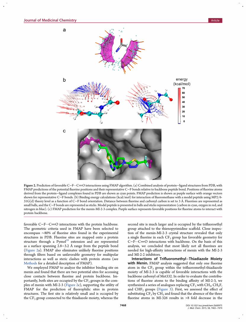
favorable C−F···CO interactions with the protein backbone.The geometric criteria used in FMAP have been selected toencompass ∼80% of fluorine sites found in the experimentalstructures in PDB. Fluorine sites are mapped onto a proteinstructure through a Pymol29 extension and are representedas a surface spanning 2.8−3.2 Å range from the peptide bond(Figure 2a). FMAP also eliminates unlikely fluorine positionsthrough filters based on unfavorable geometry for multipolarinteractions as well as steric clashes with protein atoms (seeMethods for a detailed description of FMAP).We employed FMAP to analyze the inhibitor binding site on
menin and found that there are two potential sites for accessingclose contacts between fluorine and protein backbone. Im-portantly, both sites are occupied by the CF3 groups in the com-plex of menin with MI-2-3 (Figure 2c), supporting the utility ofFMAP for the prediction of fluorophilic sites in proteinstructures. The first site is relatively small and is occupied bythe CF3 group connected to the thiadiazole moiety, whereas the
second site is much larger and is occupied by the trifluoroethylgroup attached to the thienopyrimidine scaffold. Close inspec-tion of the menin-MI-2-3 crystal structure revealed that onlya single fluorine in each CF3 group has favorable geometry forC−F···CO interactions with backbone. On the basis of thisanalysis, we concluded that most likely not all fluorines areneeded for high-affinity interactions of menin with the MI-2-3and MI-2-2 inhibitors.
Interactions of Trifluoromethyl−Thiadiazole Moietywith Menin. FMAP analysis suggested that only one fluorineatom in the CF3 group within the trifluoromethyl-thiadiazolemoiety of MI-2-3 is capable of favorable interactions with thebackbone carbonyl of Met332. In order to evaluate the contribu-tions of fluorine atoms to the binding affinity of MI-2-3, wesynthesized a series of analogues replacing CF3 with CH3, CH2F,and CHF2 groups (Figure 3). First, we assessed the effect ofsubstituting CF3 by CH3 and found that the absence of the threefluorine atoms in MI-326 results in >8 fold decrease in the
Figure 2. Prediction of favorable C−F···CO interactions using FMAP algorithm. (a) Combined analysis of protein−ligand structures from PDB, withFMAP predictions of the potential fluorine positions and their representative C−F bonds relative to backbone peptide bond. Positions of fluorine atomsderived from the protein−ligand complexes found in PDB are shown as cyan points. FMAP prediction is shown as purple surface with orange vectorsshown for representative C−F bonds. (b) Binding energy calculations (kcal/mol) for interaction of fluoromethane with a model peptide using MP2/6-31G(d) theory level as a function of C−F bond orientation. Distance between fluorine and carbonyl carbon is set to 3 Å. Fluorines are represented assmall balls, and the C−F bonds are represented as sticks. Model peptide is presented in balls and sticks representation (carbon in cyan, oxygen in red, andnitrogen in blue). (c) FMAP prediction for the menin-MI-2-3 complex. Purple surface represents favorable positions for fluorine atoms to interact withprotein backbone.
Journal of Medicinal Chemistry Article
DOI: 10.1021/acs.jmedchem.5b00975J. Med. Chem. 2015, 58, 7465−7474
7468

inhibitory activity (Figure 3a). The crystal structure revealedthat MI-326 binds to menin in a very similar manner as MI-2-3(Figure 3b), and the difference in the binding affinity pre-dominantly results from the loss of the fluorine atoms. We thensynthesized and tested two additional analogues with two(MI-319) and single (MI-333) fluorines. The inhibitory activityof MI-319 is very similar to MI-2-3 indicating no differencesbetween CF3 and CHF2 groups (Figure 3a). Surprisingly,MI-333, which harbors the CH2F group, has about 20-foldweaker activity than MI-2-3 and is even 2-fold less potent thanMI-326 with no fluorines (Figure 3a). To explain this effect, wedetermined the crystal structures of MI-333 and MI-319 boundto menin. The CHF2 group in MI-319 binds in a very similarmanner as CF3 with one of the fluorine atoms in a short, 3.2 Å,distance to the backbone carbonyl of Met322 (Figure 3b). Onthe contrary, the single fluorine in MI-333 adopts a position thatis tilted approximately 38.5° from the plane of the thiadiazolering and points away from the protein backbone (3.7 Å distanceto CO ofMet322) (Figure 3b). As a consequence, the fluorineis too far to be involved in a favorable multipolar C−F···COinteractions, and no gain in the activity is observed for MI-333(Figure 3a).The orientation of the CH2F group relative to the thiadiazole
ring was unexpected, emphasizing a strong conformational effectof the fluorine atom. As previously observed, substitution of Hby F can profoundly change the conformational preferences of asmall molecule because of the size and stereoelectronic effects.2
Although we were able to predict the position of fluorine
required for favorable interactions with the protein back-bone using FMAP, we did not anticipate that CH2F can adoptan orientation where the fluorine points away from the backbone.Introduction of the second fluorine into the CHF2 group wasnecessary to achieve an orientation of the C−F bond allowing forfavorable C−F···CO interactions and substantial improve-ment in activity. Analysis of the crystal structure of MI-333shows that S−C−C−F dihedral adapts 38.5° angle. Quantummechanical energy calculations of rotational energy barrier forCFH2 group in MI-333 shows two minima at −55 and 55°, andthe conformation in the crystal structure is disfavored by about0.5 kcal/mol (Figure S1). On the contrary, in MI-319, onefluorine is positioned in the energetical minimum (S−C−C−Fdihedral angle equal to −49°) while the second fluorine whichhas less favorable geometry (S−C−C−F dihedral angle equalto 71°) can interact with backbone. This example demonstratesthat while only single fluorine may interact with backbone, otherfluorines might be needed to stabilize the appropriate rotamericstate.
Interactions of Trifluoroethyl Group in Thienopyrimi-dine Core with Menin. Comparison of the activities of MI-2-2and MI-19 indicates that the trifluoroethyl group contributessignificantly to the high activity of MI-2-2, and replacement ofCF3 with CH3 results in over 20-fold loss in inhibitory activity(Figure 4a). FMAP analysis for the menin binding site suggeststhat only single fluorine in CF3 group can form C−F···COinteractions with the backbone. To test the contributions ofindividual fluorines in the CF3 group of MI-2-2, we synthesizedtwo compounds with CH2F (MI-836) or CHF2 (MI-859) groups.When compared to MI-19, addition of the first fluorine enhancedthe activity nearly 5-fold, whereas addition of the second fluorineincreased the activity further by about 4-fold, making it comparableto MI-2-2 with CF3 group (Figure 4a). To rationalize the effectof these modifications, we determined the crystal structures ofMI-836 andMI-859 bound to menin (Figure 4b). We found thatthe single fluorine in MI-836 points toward a hydrophobic siteformed by the side chains of Leu177, Phe238, Ala182, andSer155, and therefore, the 5-fold gain in the activity likely resultsfrom favorable hydrophobic contacts. Introduction of CHF2 inMI-859 allows for the second fluorine to be involved in theC−F···CO interactions with the backbone of His181,accounting for an additional 4-fold improvement in activity.Very similar IC50 values of MI-859 (with CHF2) and MI-2-2
(with CF3) indicates that the third fluorine is dispensable forbinding. Furthermore, the cLogP value for MI-859 is approx-imately 0.6 unit lower than forMI-2-2 (cLogP = 3.89 and 3.32 forMI-2-2 and MI-859, respectively). Therefore, our approachbased on the FMAP calculations may be used not only to predictfluorine substitutions in ligand molecules but also to designcompounds with fewer number of fluorine atoms and reducedlipophilicity without compromising ligand binding affinity.Analysis of the MI-2-2-menin structure revealed that the
methylene group in the CH2CF3 moiety is positioned closely tothe backbone carbonyl groups of Ser178 and Glu179 and mayconstitute a further site for fluorine substitutions. However, theFMAP analysis revealed that introduction of the CF2 group atthis site would not be favorable due to poor geometry of thetwo fluorines with respect to the carbonyl groups of Ser178 andGlu179. To test this hypothesis, we synthesized MI-273 withCF2CF3 group and found that such a substitution results in a∼15-fold decrease in the activity when compared to MI-2-2(Figure 4a). We determined the crystal structure of MI-273bound to menin and found that it binds in an identical manner as
Figure 3. Effect of fluorine substitutions in thiadiazole moiety on activityof menin−MLL inhibitors. (a) Structures and activities of inhibitors.ΔΔG values are calculated relative to MI-326. (b) Crystal structures ofinhibitors bound to menin showing the shortest distances betweenfluorine and menin backbone. FMAP prediction is shown as purplesurface.
Journal of Medicinal Chemistry Article
DOI: 10.1021/acs.jmedchem.5b00975J. Med. Chem. 2015, 58, 7465−7474
7469
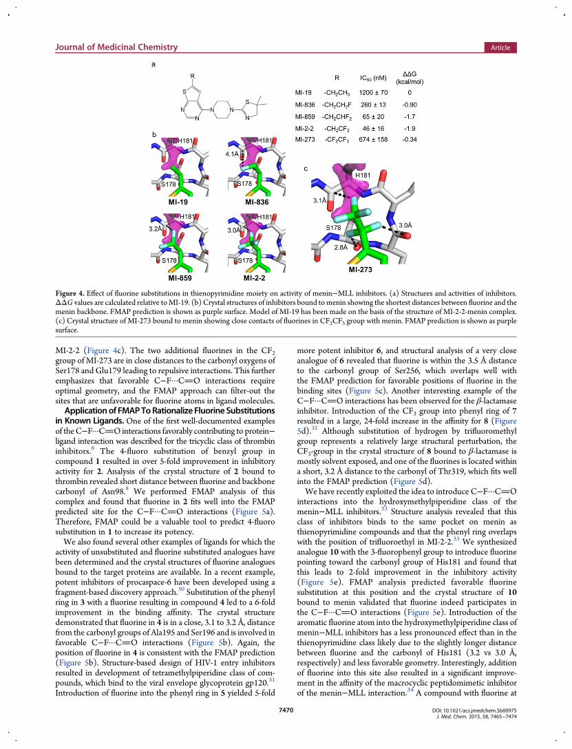
MI-2-2 (Figure 4c). The two additional fluorines in the CF2group of MI-273 are in close distances to the carbonyl oxygens ofSer178 and Glu179 leading to repulsive interactions. This furtheremphasizes that favorable C−F···CO interactions requireoptimal geometry, and the FMAP approach can filter-out thesites that are unfavorable for fluorine atoms in ligand molecules.Applicationof FMAPToRationalize FluorineSubstitutions
in Known Ligands. One of the first well-documented examplesof theC−F···CO interactions favorably contributing to protein−ligand interaction was described for the tricyclic class of thrombininhibitors.8 The 4-fluoro substitution of benzyl group incompound 1 resulted in over 5-fold improvement in inhibitoryactivity for 2. Analysis of the crystal structure of 2 bound tothrombin revealed short distance between fluorine and backbonecarbonyl of Asn98.8 We performed FMAP analysis of thiscomplex and found that fluorine in 2 fits well into the FMAPpredicted site for the C−F···CO interactions (Figure 5a).Therefore, FMAP could be a valuable tool to predict 4-fluorosubstitution in 1 to increase its potency.We also found several other examples of ligands for which the
activity of unsubstituted and fluorine substituted analogues havebeen determined and the crystal structures of fluorine analoguesbound to the target proteins are available. In a recent example,potent inhibitors of procaspace-6 have been developed using afragment-based discovery approach.30 Substitution of the phenylring in 3 with a fluorine resulting in compound 4 led to a 6-foldimprovement in the binding affinity. The crystal structuredemonstrated that fluorine in 4 is in a close, 3.1 to 3.2 Å, distancefrom the carbonyl groups of Ala195 and Ser196 and is involved infavorable C−F···CO interactions (Figure 5b). Again, theposition of fluorine in 4 is consistent with the FMAP prediction(Figure 5b). Structure-based design of HIV-1 entry inhibitorsresulted in development of tetramethylpiperidine class of com-pounds, which bind to the viral envelope glycoprotein gp120.31
Introduction of fluorine into the phenyl ring in 5 yielded 5-fold
more potent inhibitor 6, and structural analysis of a very closeanalogue of 6 revealed that fluorine is within the 3.5 Å distanceto the carbonyl group of Ser256, which overlaps well withthe FMAP prediction for favorable positions of fluorine in thebinding sites (Figure 5c). Another interesting example of theC−F···CO interactions has been observed for the β-lactamaseinhibitor. Introduction of the CF3 group into phenyl ring of 7resulted in a large, 24-fold increase in the affinity for 8 (Figure5d).32 Although substitution of hydrogen by trifluoromethylgroup represents a relatively large structural perturbation, theCF3-group in the crystal structure of 8 bound to β-lactamase ismostly solvent exposed, and one of the fluorines is located withina short, 3.2 Å distance to the carbonyl of Thr319, which fits wellinto the FMAP prediction (Figure 5d).We have recently exploited the idea to introduce C−F···CO
interactions into the hydroxymethylpiperidine class of themenin−MLL inhibitors.33 Structure analysis revealed that thisclass of inhibitors binds to the same pocket on menin asthienopyrimidine compounds and that the phenyl ring overlapswith the position of trifluoroethyl in MI-2-2.33 We synthesizedanalogue 10 with the 3-fluorophenyl group to introduce fluorinepointing toward the carbonyl group of His181 and found thatthis leads to 2-fold improvement in the inhibitory activity(Figure 5e). FMAP analysis predicted favorable fluorinesubstitution at this position and the crystal structure of 10bound to menin validated that fluorine indeed participates inthe C−F···CO interactions (Figure 5e). Introduction of thearomatic fluorine atom into the hydroxymethylpiperidine class ofmenin−MLL inhibitors has a less pronounced effect than in thethienopyrimidine class likely due to the slightly longer distancebetween fluorine and the carbonyl of His181 (3.2 vs 3.0 Å,respectively) and less favorable geometry. Interestingly, additionof fluorine into this site also resulted in a significant improve-ment in the affinity of the macrocyclic peptidomimetic inhibitorof the menin−MLL interaction.34 A compound with fluorine at
Figure 4. Effect of fluorine substitutions in thienopyrimidine moiety on activity of menin−MLL inhibitors. (a) Structures and activities of inhibitors.ΔΔG values are calculated relative toMI-19. (b) Crystal structures of inhibitors bound tomenin showing the shortest distances between fluorine and themenin backbone. FMAP prediction is shown as purple surface. Model of MI-19 has been made on the basis of the structure of MI-2-2-menin complex.(c) Crystal structure of MI-273 bound to menin showing close contacts of fluorines in CF2CF3 group with menin. FMAP prediction is shown as purplesurface.
Journal of Medicinal Chemistry Article
DOI: 10.1021/acs.jmedchem.5b00975J. Med. Chem. 2015, 58, 7465−7474
7470
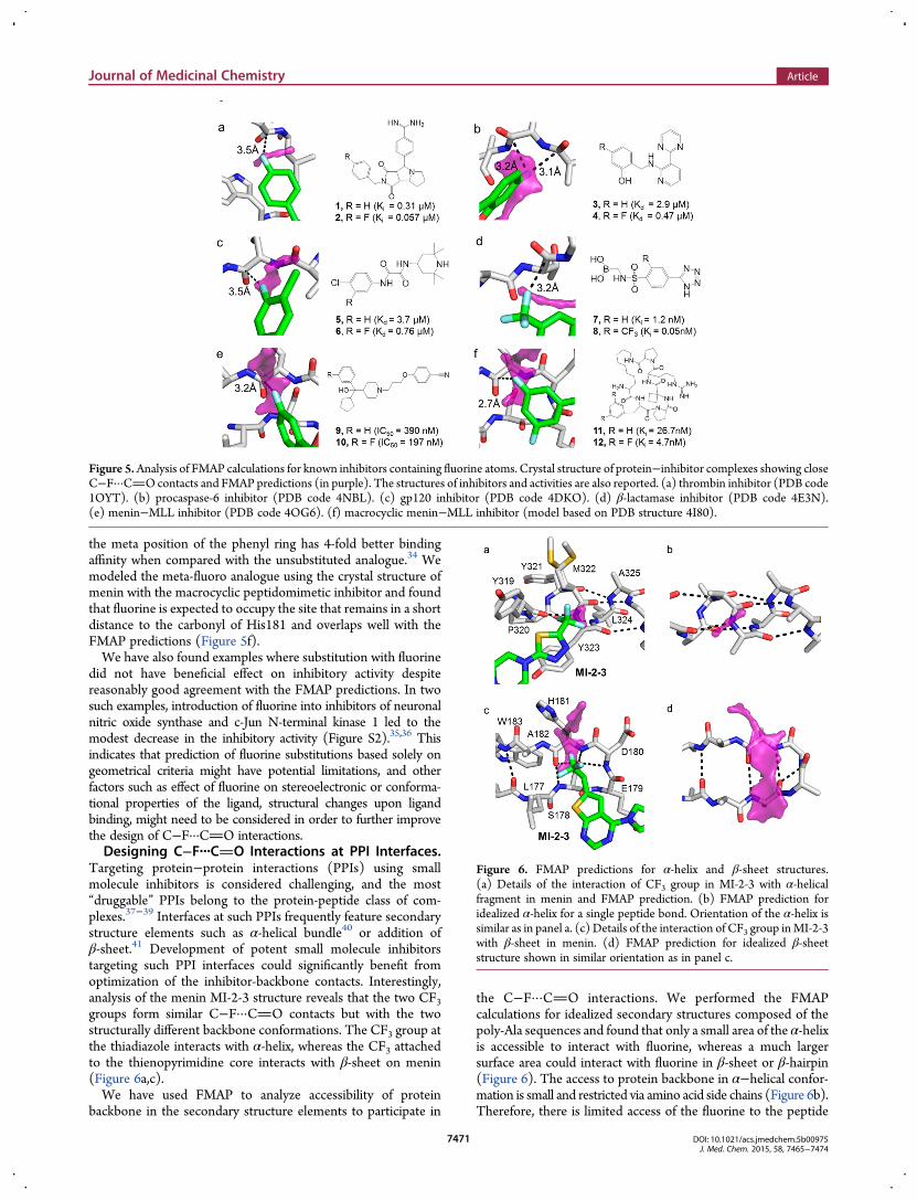
the meta position of the phenyl ring has 4-fold better bindingaffinity when compared with the unsubstituted analogue.34 Wemodeled the meta-fluoro analogue using the crystal structure ofmenin with the macrocyclic peptidomimetic inhibitor and foundthat fluorine is expected to occupy the site that remains in a shortdistance to the carbonyl of His181 and overlaps well with theFMAP predictions (Figure 5f).We have also found examples where substitution with fluorine
did not have beneficial effect on inhibitory activity despitereasonably good agreement with the FMAP predictions. In twosuch examples, introduction of fluorine into inhibitors of neuronalnitric oxide synthase and c-Jun N-terminal kinase 1 led to themodest decrease in the inhibitory activity (Figure S2).35,36 Thisindicates that prediction of fluorine substitutions based solely ongeometrical criteria might have potential limitations, and otherfactors such as effect of fluorine on stereoelectronic or conforma-tional properties of the ligand, structural changes upon ligandbinding, might need to be considered in order to further improvethe design of C−F···CO interactions.Designing C−F···CO Interactions at PPI Interfaces.
Targeting protein−protein interactions (PPIs) using smallmolecule inhibitors is considered challenging, and the most“druggable” PPIs belong to the protein-peptide class of com-plexes.37−39 Interfaces at such PPIs frequently feature secondarystructure elements such as α-helical bundle40 or addition ofβ-sheet.41 Development of potent small molecule inhibitorstargeting such PPI interfaces could significantly benefit fromoptimization of the inhibitor-backbone contacts. Interestingly,analysis of the menin MI-2-3 structure reveals that the two CF3groups form similar C−F···CO contacts but with the twostructurally different backbone conformations. The CF3 group atthe thiadiazole interacts with α-helix, whereas the CF3 attachedto the thienopyrimidine core interacts with β-sheet on menin(Figure 6a,c).We have used FMAP to analyze accessibility of protein
backbone in the secondary structure elements to participate in
the C−F···CO interactions. We performed the FMAPcalculations for idealized secondary structures composed of thepoly-Ala sequences and found that only a small area of the α-helixis accessible to interact with fluorine, whereas a much largersurface area could interact with fluorine in β-sheet or β-hairpin(Figure 6). The access to protein backbone in α−helical confor-mation is small and restricted via amino acid side chains (Figure 6b).Therefore, there is limited access of the fluorine to the peptide
Figure 5.Analysis of FMAP calculations for known inhibitors containing fluorine atoms. Crystal structure of protein−inhibitor complexes showing closeC−F···COcontacts and FMAP predictions (in purple). The structures of inhibitors and activities are also reported. (a) thrombin inhibitor (PDB code1OYT). (b) procaspase-6 inhibitor (PDB code 4NBL). (c) gp120 inhibitor (PDB code 4DKO). (d) β-lactamase inhibitor (PDB code 4E3N).(e) menin−MLL inhibitor (PDB code 4OG6). (f) macrocyclic menin−MLL inhibitor (model based on PDB structure 4I80).
Figure 6. FMAP predictions for α-helix and β-sheet structures.(a) Details of the interaction of CF3 group in MI-2-3 with α-helicalfragment in menin and FMAP prediction. (b) FMAP prediction foridealized α-helix for a single peptide bond. Orientation of the α-helix issimilar as in panel a. (c) Details of the interaction of CF3 group inMI-2-3with β-sheet in menin. (d) FMAP prediction for idealized β-sheetstructure shown in similar orientation as in panel c.
Journal of Medicinal Chemistry Article
DOI: 10.1021/acs.jmedchem.5b00975J. Med. Chem. 2015, 58, 7465−7474
7471
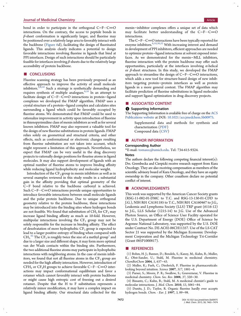
bond in order to participate in the orthogonal C−F···COinteractions. On the contrary, the access to peptide bonds inβ-sheet conformation is significantly larger, and fluorine maybe positioned over a relatively large area to favorably interact withthe backbone (Figure 6d), facilitating the design of fluorinatedligands. This analysis clearly indicates a potential to designfavorable interactions involving fluorine in ligands that bind atPPI interfaces. Design of such interactions should be particularlyfeasible for interfaces involving β-sheets due to the relatively largeaccessibility of protein backbone.
■ CONCLUSIONSFluorine scanning strategy has been previously proposed as aneffective approach to improve the activity of small moleculeinhibitors.2,8,10 Such a strategy is synthetically demanding andrequires synthesis of multiple analogues.8,10 In an attempt tofacilitate design of C−F···CO interactions in protein−ligandcomplexes we developed the FMAP algorithm. FMAP uses acrystal structure of a protein−ligand complex and calculates sitessurrounding a ligand which could be favorably occupied byfluorine atoms. We demonstrated that FMAP could be used torationalize improvement in activity upon introduction of fluorinein thienopyrimidine class of menin inhibitors as well as for severalknown inhibitors. FMAP may also represent a valuable tool forthe design of new fluorine substitutions in protein ligands. FMAPrelies solely on geometrical and structural criteria, and othereffects, such as conformational or electronic changes resultingfrom fluorine substitution are not taken into account, whichmight represent a limitation of this approach. Nevertheless, weexpect that FMAP can be very useful in the drug discoveryprojects to rationally design positions for flourine atoms in ligandmolecules. It may also support development of ligands with anoptimal number of fluorine atoms to improve binding affinitywhile reducing ligand hydrophobicity and molecular weight.Introduction of the CF3 group in menin inhibitors as well as in
several examples reviewed in this study results in a substantialgain in the affinity providing that optimal geometry of theC−F bond relative to the backbone carbonyl is achieved.Such C−F···CO interactions provide unique opportunities tointroduce favorable interactions between small molecule ligandsand the polar protein backbone. Due to unique orthogonalgeometry relative to the protein backbone, these interactionsmay be introduced into the binding sites where hydrogen bondsare not feasible. We found that substitution of CH3 for CF3 mayincrease ligand binding affinity as much as 10-fold. However,multipolar interactions involving the CF3 group may not besolely responsible for the increase in binding affinity. The effectof desolvatation of more hydrophobic CF3 group is expected tolead to a larger positive entropy of binding when compared withCH3.
42 The CF3 is roughly twice the size of a methyl group1 anddue to a larger size and different shape, it may formmore optimalvan der Waals contacts within the binding site. Furthermore,the two additional fluorine atomsmay participate in hydrophobicinteractions with neighboring atoms. In the case of menin inhib-itors, we found that not all fluorine atoms in the CF3 group areneeded for the high affinity interaction. However, introduction ofCFH2 or CF2H groups to achieve favorable C−F···CO inter-actions may impact conformational equilibrium and favor arotamer which cannot favorably interact with protein backboneor might cause high entropic cost of freezing out a desiredrotamer. Despite that the H to F substitution represents arelatively minor modification, it may have a complex impact onligand binding affinity. Our structural data collected for the
menin−inhibitor complexes offers a unique set of data whichmay facilitate better understanding of the C−F···COinteractions.TheC−F···CO interactions have been typically reported for
enzyme inhibitors.8,10,30,32 With increasing interest and demandin development of PPI inhibitors, efficient approaches are neededto optimize protein−ligand interactions at solvent exposed inter-faces. As we demonstrated for the menin−MLL inhibitors,fluorine interaction with the protein backbone may offer suchopportunities, particularly at the interfaces involving α-helicalor β-sheet structures. In this study, we developed the FMAPapproach to streamline the design of C−F···CO interactions,which adds a new tool for structure-based design of new inhib-itors targeting protein−protein interfaces as well as proteinligands in a more general context. The FMAP algorithm mayfacilitate prediction of fluorine substitutions in ligand moleculesand support ligand optimization in drug discovery projects.
■ ASSOCIATED CONTENT*S Supporting InformationThe Supporting Information is available free of charge on the ACSPublications website at DOI: 10.1021/acs.jmedchem.5b00975.
Supplemental data and methods for synthesis andcharacterization (PDF)Compound data (CSV)
■ AUTHOR INFORMATIONCorresponding Author*E-mail: [email protected]. Tel: 734-615-9324.NotesThe authors declare the following competing financial interest(s):Drs. Grembecka and Cierpicki receive research support from KuraOncology. They are also receiving compensation as members of thescientific advisory board of Kura Oncology, and they have an equityownership in the company. Other coauthors declare no potentialconflict of interest.
■ ACKNOWLEDGMENTSThis work was supported by the American Cancer Society grants(RSG-11-082-01-DMC to T.C. and RSG-13-130-01-CDD toJ.G.), NIH R01 CA181185 to T.C., NIH R01 CA160467 to J.G.,Leukemia and Lymphoma Society (LLS) TRP grant (6116-12)to J.G., LLS Scholar (1215-14) to J.G. Use of the AdvancedPhoton Source, an Office of Science User Facility operated forthe U.S. Department of Energy (DOE) Office of Science byArgonne National Laboratory, was supported by the U.S. DOEunder Contract No. DE-AC02-06CH11357. Use of the LS-CATSector 21 was supported by the Michigan Economic Develop-ment Corporation and the Michigan Technology Tri-Corridor(Grant 085P1000817).
■ REFERENCES(1) Bohm, H. J.; Banner, D.; Bendels, S.; Kansy, M.; Kuhn, B.; Muller,K.; Obst-Sander, U.; Stahl, M. Fluorine in medicinal chemistry.ChemBioChem 2004, 5, 637−43.(2) Muller, K.; Faeh, C.; Diederich, F. Fluorine in pharmaceuticals:looking beyond intuition. Science 2007, 317, 1881−6.(3) Purser, S.; Moore, P. R.; Swallow, S.; Gouverneur, V. Fluorine inmedicinal chemistry. Chem. Soc. Rev. 2008, 37, 320−30.(4) Bissantz, C.; Kuhn, B.; Stahl, M. A medicinal chemist’s guide tomolecular interactions. J. Med. Chem. 2010, 53, 5061−84.(5) Dunitz, J. D.; Taylor, R. Organic fluorine hardly ever acceptshydrogen bonds. Chem. - Eur. J. 1997, 3, 89−98.
Journal of Medicinal Chemistry Article
DOI: 10.1021/acs.jmedchem.5b00975J. Med. Chem. 2015, 58, 7465−7474
7472

(6) Parlow, J. J.; Kurumbail, R. G.; Stegeman, R. A.; Stevens, A. M.;Stallings, W. C.; South, M. S. Design, synthesis, and crystal structure ofselective 2-pyridone tissue factor VIIa inhibitors. J. Med. Chem. 2003, 46,4696−701.(7) Kim, D.; Wang, L.; Beconi, M.; Eiermann, G. J.; Fisher, M. H.; He,H.; Hickey, G. J.; Kowalchick, J. E.; Leiting, B.; Lyons, K.; Marsilio, F.;McCann, M. E.; Patel, R. A.; Petrov, A.; Scapin, G.; Patel, S. B.; Roy, R.S.; Wu, J. K.; Wyvratt, M. J.; Zhang, B. B.; Zhu, L.; Thornberry, N. A.;Weber, A. E. (2R)-4-oxo-4-[3-(trifluoromethyl)-5,6-dihydro[1,2,4]-triazolo[4,3-a]pyrazin-7(8H)- yl]-1-(2,4,5-trifluorophenyl)butan-2-amine: a potent, orally active dipeptidyl peptidase IV inhibitor for thetreatment of type 2 diabetes. J. Med. Chem. 2005, 48, 141−51.(8) Olsen, J. A.; Banner, D. W.; Seiler, P.; Sander, U. O.; D’Arcy, A.;Stihle, M.; Muller, K.; Diederich, F. A fluorine scan of thrombininhibitors to map the fluorophilicity/fluorophobicity of an enzymeactive site: Evidence for C-F center dot center dot center dot COinteractions. Angew. Chem., Int. Ed. 2003, 42, 2507−2511.(9) Paulini, R.; Muller, K.; Diederich, F. Orthogonal multipolarinteractions in structural chemistry and biology. Angew. Chem., Int. Ed.2005, 44, 1788−1805.(10) Olsen, J. A.; Banner, D. W.; Seiler, P.; Wagner, B.; Tschopp, T.;Obst-Sander, U.; Kansy, M.; Muller, K.; Diederich, F. Fluorineinteractions at the thrombin active site: protein backbone fragmentsH-C(alpha)-CO comprise a favorable C-F environment andinteractions of C-F with electrophiles. ChemBioChem 2004, 5, 666−75.(11) Hao, G. F.; Wang, F.; Li, H.; Zhu, X. L.; Yang, W. C.; Huang, L. S.;Wu, J. W.; Berry, E. A.; Yang, G. F. Computational discovery ofpicomolar Q(o) site inhibitors of cytochrome bc1 complex. J. Am. Chem.Soc. 2012, 134, 11168−76.(12) Vulpetti, A.; Hommel, U.; Landrum, G.; Lewis, R.; Dalvit, C.Design and NMR-based screening of LEF, a library of chemicalfragments with different local environment of fluorine. J. Am. Chem. Soc.2009, 131, 12949−59.(13) Vulpetti, A.; Schiering, N.; Dalvit, C. Combined use ofcomputational chemistry, NMR screening, and X-ray crystallographyfor identification and characterization of fluorophilic protein environ-ments. Proteins: Struct., Funct., Genet. 2010, 78, 3281−91.(14) Grembecka, J.; He, S.; Shi, A.; Purohit, T.; Muntean, A. G.;Sorenson, R. J.; Showalter, H. D.; Murai, M. J.; Belcher, A. M.; Hartley,T.; Hess, J. L.; Cierpicki, T. Menin-MLL inhibitors reverse oncogenicactivity of MLL fusion proteins in leukemia. Nat. Chem. Biol. 2012, 8,277−84.(15) Shi, A.; Murai, M. J.; He, S.; Lund, G.; Hartley, T.; Purohit, T.;Reddy, G.; Chruszcz, M.; Grembecka, J.; Cierpicki, T. Structural insightsinto inhibition of the bivalent menin-MLL interaction by smallmolecules in leukemia. Blood 2012, 120, 4461−9.(16) Hamelryck, T.; Manderick, B. PDB file parser and structure classimplemented in Python. Bioinformatics 2003, 19, 2308−10.(17) Cock, P. J.; Antao, T.; Chang, J. T.; Chapman, B. A.; Cox, C. J.;Dalke, A.; Friedberg, I.; Hamelryck, T.; Kauff, F.; Wilczynski, B.; deHoon, M. J. Biopython: freely available Python tools for computationalmolecular biology and bioinformatics. Bioinformatics 2009, 25, 1422−3.(18) Boys, S.; Bernardi, F. The calculation of small molecularinteractions by the differences of separate total energies. Someprocedures with reduced errors. Mol. Phys. 1970, 19, 553−566.(19) Giedroyc-Piasecka, W.; Dyguda-Kazimierowicz, E.; Beker, W.;Mor, M.; Lodola, A.; Sokalski, W. A. Physical Nature of Fatty AcidAmide Hydrolase Interactions with Its Inhibitors: Testing a SimpleNonempirical Scoring Model. J. Phys. Chem. B 2014, 118, 14727−14736.(20) Frisch, M. J.; Trucks, G. W.; Schlegel, H. B.; Scuseria, G. E.; Robb,M. A.; Cheeseman, J. R.; Scalmani, G.; Barone, V.; Mennucci, B.;Petersson, G. A.; Nakatsuji, H.; Caricato, M.; Li, X.; Hratchian, H. P.;Izmaylov, A. F.; Bloino, J.; Zheng, G.; Sonnenberg, J. L.; Hada, M.;Ehara, M.; Toyota, K.; Fukuda, R.; Hasegawa, J.; Ishida, M.; Nakajima,T.; Honda, Y.; Kitao, O.; Nakai, H.; Vreven, T.; Montgomery Jr., J. A.;Peralta, J. E.; Ogliaro, F.; Bearpark, M. J.; Heyd, J.; Brothers, E. N.;Kudin, K. N.; Staroverov, V. N.; Kobayashi, R.; Normand, J.;Raghavachari, K.; Rendell, A. P.; Burant, J. C.; Iyengar, S. S.; Tomasi,
J.; Cossi, M.; Rega, N.; Millam, N. J.; Klene, M.; Knox, J. E.; Cross, J. B.;Bakken, V.; Adamo, C.; Jaramillo, J.; Gomperts, R.; Stratmann, R. E.;Yazyev, O.; Austin, A. J.; Cammi, R.; Pomelli, C.; Ochterski, J. W.;Martin, R. L.; Morokuma, K.; Zakrzewski, V. G.; Voth, G. A.; Salvador,P.; Dannenberg, J. J.; Dapprich, S.; Daniels, A. D.; Farkas, O.; Foresman,J. B.; Ortiz, J. V.; Cioslowski, J.; Fox, D. J. Gaussian 09; Gaussian, Inc.:Wallingford, CT, 2009.(21) Grembecka, J.; Belcher, A.M.; Hartley, T.; Cierpicki, T.Molecularbasis of the mixed lineage leukemia-menin interaction: implications fortargeting mixed lineage leukemias. J. Biol. Chem. 2010, 285, 40690−8.(22) Otwinowski, Z.; Minor, W. Processing of X-ray diffraction datacollected in oscillation mode. Methods Enzymol. 1997, 276, 307−326.(23) Murshudov, G. N.; Vagin, A. A.; Dodson, E. J. Refinement ofmacromolecular structures by the maximum-likelihood method. ActaCrystallogr., Sect. D: Biol. Crystallogr. 1997, 53, 240−55.(24) Emsley, P.; Cowtan, K. Coot: model-building tools for moleculargraphics. Acta Crystallogr., Sect. D: Biol. Crystallogr. 2004, 60, 2126−32.(25) Collaborative Computational Project, N. The CCP4 suite:programs for protein crystallography. Acta Crystallogr., Sect. D: Biol.Crystallogr. 1994, 50, 760−763.10.1107/S0907444994003112.(26) Painter, J.; Merritt, E. A. TLSMD web server for the generation ofmulti-group TLS models. J. Appl. Crystallogr. 2006, 39, 109−111.(27) Davis, I. W.; Leaver-Fay, A.; Chen, V. B.; Block, J. N.; Kapral, G. J.;Wang, X.; Murray, L. W.; Arendall, W. B., 3rd; Snoeyink, J.; Richardson,J. S.; Richardson, D. C. MolProbity: all-atom contacts and structurevalidation for proteins and nucleic acids. Nucleic Acids Res. 2007, 35,W375−83.(28) Yang, H.; Guranovic, V.; Dutta, S.; Feng, Z.; Berman, H. M.;Westbrook, J. D. Automated and accurate deposition of structuressolved by X-ray diffraction to the Protein Data Bank. Acta Crystallogr.,Sect. D: Biol. Crystallogr. 2004, 60, 1833−9.(29) The PyMOL Molecular Graphics System, Version 1.2r3pre,Schrodinger, LLC.(30) Murray, J.; Giannetti, A. M.; Steffek, M.; Gibbons, P.; Hearn, B.R.; Cohen, F.; Tam, C.; Pozniak, C.; Bravo, B.; Lewcock, J.; Jaishankar,P.; Ly, C. Q.; Zhao, X.; Tang, Y.; Chugha, P.; Arkin, M. R.; Flygare, J.;Renslo, A. R. Tailoring small molecules for an allosteric site onprocaspase-6. ChemMedChem 2014, 9 (73−7), 2.(31) LaLonde, J. M.; Kwon, Y. D.; Jones, D. M.; Sun, A. W.; Courter, J.R.; Soeta, T.; Kobayashi, T.; Princiotto, A. M.; Wu, X.; Schon, A.; Freire,E.; Kwong, P. D.; Mascola, J. R.; Sodroski, J.; Madani, N.; Smith, A. B.,3rd Structure-based design, synthesis, and characterization of dualhotspot small-molecule HIV-1 entry inhibitors. J. Med. Chem. 2012, 55,4382−96.(32) Eidam, O.; Romagnoli, C.; Dalmasso, G.; Barelier, S.; Caselli, E.;Bonnet, R.; Shoichet, B. K.; Prati, F. Fragment-guided design ofsubnanomolar beta-lactamase inhibitors active in vivo. Proc. Natl. Acad.Sci. U. S. A. 2012, 109, 17448−53.(33) He, S.; Senter, T. J.; Pollock, J.; Han, C.; Upadhyay, S. K.; Purohit,T.; Gogliotti, R. D.; Lindsley, C. W.; Cierpicki, T.; Stauffer, S. R.;Grembecka, J. High-affinity small-molecule inhibitors of the menin-mixed lineage leukemia (MLL) interaction closely mimic a naturalprotein-protein interaction. J. Med. Chem. 2014, 57, 1543−56.(34) Zhou, H.; Liu, L.; Huang, J.; Bernard, D.; Karatas, H.; Navarro, A.;Lei, M.; Wang, S. Structure-based design of high-affinity macrocyclicpeptidomimetics to block the menin-mixed lineage leukemia 1 (MLL1)protein-protein interaction. J. Med. Chem. 2013, 56, 1113−23.(35) Li, B.; Cociorva, O. M.; Nomanbhoy, T.; Weissig, H.; Li, Q.;Nakamura, K.; Liyanage, M.; Zhang, M. C.; Shih, A. Y.; Aban, A.; Hu, Y.;Cajica, J.; Pham, L.; Kozarich, J. W.; Shreder, K. R. Hit-to-leadoptimization and kinase selectivity of imidazo[1,2-a]quinoxalin-4-aminederived JNK1 inhibitors. Bioorg. Med. Chem. Lett. 2013, 23, 5217−22.(36) Xue, F.; Li, H.; Delker, S. L.; Fang, J.; Martasek, P.; Roman, L. J.;Poulos, T. L.; Silverman, R. B. Potent, highly selective, and orallybioavailable gem-difluorinated monocationic inhibitors of neuronalnitric oxide synthase. J. Am. Chem. Soc. 2010, 132, 14229−38.(37) Petsalaki, E.; Russell, R. B. Peptide-mediated interactions inbiological systems: new discoveries and applications. Curr. Opin.Biotechnol. 2008, 19, 344−50.
Journal of Medicinal Chemistry Article
DOI: 10.1021/acs.jmedchem.5b00975J. Med. Chem. 2015, 58, 7465−7474
7473

(38) Smith, M. C.; Gestwicki, J. E. Features of protein-proteininteractions that translate into potent inhibitors: topology, surface areaand affinity. Expert Rev. Mol. Med. 2012, 14, e16.(39) Cierpicki, T.; Grembecka, J. Targeting protein-proteininteractions in hematologic malignancies: still a challenge or a greatopportunity for future therapies? Immunol Rev. 2015, 263, 279−301.(40) Jochim, A. L.; Arora, P. S. Assessment of helical interfaces inprotein-protein interactions. Mol. BioSyst. 2009, 5, 924−6.(41) Remaut, H.; Waksman, G. Protein-protein interaction throughbeta-strand addition. Trends Biochem. Sci. 2006, 31, 436−44.(42) Biffinger, J. C.; Kim, H. W.; DiMagno, S. G. The polarhydrophobicity of fluorinated compounds. ChemBioChem 2004, 5,622−7.
Journal of Medicinal Chemistry Article
DOI: 10.1021/acs.jmedchem.5b00975J. Med. Chem. 2015, 58, 7465−7474
7474