Embed Size (px)
Citation preview

antibodies
Review
Rational Design of Constrained Peptides as ProteinInterface Inhibitors
Ramachandran Murali 1,*, Hongtao Zhang 2 , Zheng Cai 2, Lian Lam 2 and Mark Greene 2,*
�����������������
Citation: Murali, R.; Zhang, H.; Cai,
Z.; Lam, L.; Greene, M. Rational
Design of Constrained Peptides as
Protein Interface Inhibitors.
Antibodies 2021, 10, 32. https://
doi.org/10.3390/antib10030032
Received: 27 May 2021
Accepted: 10 August 2021
Published: 16 August 2021
Publisher’s Note: MDPI stays neutral
with regard to jurisdictional claims in
published maps and institutional affil-
iations.
Copyright: © 2021 by the authors.
Licensee MDPI, Basel, Switzerland.
This article is an open access article
distributed under the terms and
conditions of the Creative Commons
Attribution (CC BY) license (https://
creativecommons.org/licenses/by/
4.0/).
1 Cedars-Sinai Medical Center, Department of Biomedical Science, Research Division of Immunology,Los Angeles, CA 90211, USA
2 Department of Pathology and Laboratory of Medicine, University of Pennsylvania,Philadelphia, PA 19104, USA; [email protected] (H.Z.); [email protected] (Z.C.);[email protected] (L.L.)
* Correspondence: [email protected] (R.M.); [email protected] (M.G.)
Abstract: The lack of progress in developing targeted therapeutics directed at protein–proteincomplexes has been due to the absence of well-defined ligand-binding pockets and the extensiveintermolecular contacts at the protein–protein interface. Our laboratory has developed approaches todissect protein–protein complexes focusing on the superfamilies of erbB and tumor necrosis factor(TNF) receptors by the combined use of structural biology and computational biology to facilitatesmall molecule development. We present a perspective on the development and application ofpeptide inhibitors as well as immunoadhesins to cell surface receptors performed in our laboratory.
Keywords: ErbB; TNF; mimetic; receptor; protein-engineering; immunoadhesion; cancer
1. Introduction
Therapeutic development, particularly small molecule drug creation, has focused onenzymes. Other efforts have included natural compounds that are screened for the abilityto induce phenotypic changes of transformed cells [1]. When human genome sequencingwas undertaken and completed, expectations in the scientific community were that manymore therapeutic targets might be identified. However, few “druggable” candidates werecharacterized [2–4]. This failure to identify new targets motivated many to develop newtools for “undruggable targets” [5–10].
Molecular assembly is a key step in many biological functions. Molecular ensembleformation is dynamic and can vary from simple dimerization to the assembly of largemolecular complexes. Molecular ensemble formation may comprise protein-only, protein–DNA, or protein–lipid complexes. A variety of methods have been developed to targetcertain molecular complexes and include high throughput screening, phage display againstprotein complexes, and alteration of biological activities using antibodies and recombinantproteins [11–13].
Antibody still remains a preferred choice of therapeutic targeting agents especially bypharmaceutical enterprises. Size features affect certain functional aspects, including the lackof tissue penetration for imaging and crossing the blood–brain barrier. Humanization oftherapeutic antibodies remains a time and resource consuming process. Peptide inhibitorsare ideally suited to overcome these limitations and to create novel molecules beyondmonoclonal antibodies. Furthermore, due to their small size, peptides are amenable forchemical modifications to attain clinically relevant pharmacological features, and theirstructural features may also facilitate small non-peptidic inhibitor development.
Small peptides are ideal molecules to explore protein–protein interactions due totheir intermediate size (1–2 kD), ease of synthesis, and ability to bind to protein surfaces.More and more peptides are now used in the clinic [14,15]. This progress is due to im-provement in technologies including cost-effective large-quantity peptide synthesis and
Antibodies 2021, 10, 32. https://doi.org/10.3390/antib10030032 https://www.mdpi.com/journal/antibodies

Antibodies 2021, 10, 32 2 of 10
metabolic stability [16,17]. Increasingly, in addition to using peptides as therapeutics perse, novel approaches using peptide inhibitors are being adopted. These approaches in-clude cell-permeable peptides [18], peptide-based cancer vaccines, and peptide-decoratednanoparticles for diagnostics and drug-delivery. Thus, peptide agents are versatile and canbe adopted for diagnosis and therapy of diseases.
X-ray crystallography has helped to resolve molecular features of many protein com-plexes ranging from spherical viruses, cell surface receptors, intact antibodies, and (Fab)-antigen complexes at atomic resolution [19,20]. Based on the fundamental structuralfeatures of protein–protein complexes, we have developed a method to design and de-velop constrained peptide mimics specific for interaction surfaces using computational andstructural biology approaches.
2. Antibody–Antigen Complex
Atomic structures of antibody–antigen complexes have aided the study of protein–protein interactions [21–23]. The Fab fragments of an antibody can recognize bindingdeterminants such as small organic molecules (which includes haptens that can bind tolarger proteins), carbohydrates, peptides, or large macromolecular complexes. Antibody–antigen complexes, in particular Fab–hapten complexes, have provided insight into aspectsof molecular recognition [24]. Atomic analysis of antibody–hapten complexes has revealedthat an antibody can recognize the three-dimensional structures of antigens. An antibodycan distinguish between antigens of different enantiomers [25]. Thus, the ability of antibodyto recognize “hot spots” on protein can be useful to identify critical residues in a putativetargeted protein.
We used these basic notions to study antibodies that bound to oncoproteins andto develop small peptidic loops from the antigen binding site that could act as smallmolecular peptidomimetics. To develop small peptides targeting the dimerization ofHer2/neu receptor, we used the structure of a crystallographically resolved anti-Her2/neuantibody called Herceptin. Herceptin binds to the ectodomain of the HER2/neu protein.This antibody binding surface was used as a template to develop a small exocyclic peptidemimicking the antibody [26]. A detailed set of methods developing small antibody-likemimetics has been described previously [27].
In this review, we will present methodologies used in the development of peptideinhibitors that limit protein–protein interactions. We will discuss how peptide inhibitors arefacile in developing therapeutic strategies. We focus on the development of antibody-likepeptide mimics to cell surface receptors.
3. Structural Features of Protein–Protein Complexes
The enhanced understanding of protein structures at atomic resolution allowed Jonesand Thornton [28] to propose a set of general parameters that govern protein–proteininteraction. They identified three key features: (1) large protein–protein interface (>1000Å2), (2) secondary structural elements (i.e., helix, beta-sheet, and flexible loops), and(3) molecular surface complementarity that included chemical/electrostatic charge anddiscrete geometrical features.
The precise contribution of amino acids at the protein–protein interface throughmutation studies expanded the concept of molecular recognition in terms of enthalpicparameters (i.e., polar and non-polar interactions such as hydrogen bond and hydrophobiccontacts) to “shape complementarity” and “binding surface area” [29].
Large surfaces can be decorated with solvents which can contribute entropic compo-nents allowing weaker protein–protein interaction or enhanced enthalpic interactions byextending the hydrogen bond network [30–33]. Unlike enzyme–substrate complexes, struc-tural understanding of protein–protein interfaces does not readily reveal critical contactsor a defined targetable site for developing small molecule ligands. These features havelimited development of small molecules to target protein–protein/DNA interactions.
Antibodies 2021, 10, 32 3 of 10
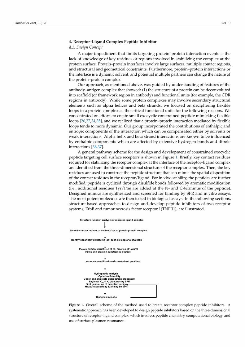
4. Receptor–Ligand Complex Peptide Inhibitor4.1. Design Concept
A major impediment that limits targeting protein–protein interaction events is thelack of knowledge of key residues or regions involved in stabilizing the complex at theprotein surface. Protein–protein interfaces involve large surfaces, multiple contact regions,and structural and geometrical constraints. Furthermore, protein–protein interactions orthe interface is a dynamic solvent, and potential multiple partners can change the nature ofthe protein–protein complex.
Our approach, as mentioned above, was guided by understanding of features of theantibody–antigen complex that showed: (1) the structure of a protein can be deconvolutedinto scaffold (or framework region in antibody) and functional units (for example, the CDRregions in antibody). While some protein complexes may involve secondary structuralelements such as alpha helices and beta strands, we focused on deciphering flexibleloops in a protein complex as the critical functional units for the following reasons. Weconcentrated on efforts to create small exocyclic constrained peptide mimicking flexibleloops [26,27,34,35], and we realized that a protein–protein interaction mediated by flexibleloops tends to more dynamic. Our group incorporated the contributions of enthalpic andentropic components of the interaction which can be compensated either by solvents orweak interactions. Alpha helix and beta strand interactions are known to be influencedby enthalpic components which are affected by extensive hydrogen bonds and dipoleinteractions [36,37].
A general pathway scheme for the design and development of constrained exocyclicpeptide targeting cell surface receptors is shown in Figure 1. Briefly, key contact residuesrequired for stabilizing the receptor complex at the interface of the receptor–ligand complexare identified from the three-dimensional structure of the receptor complex. Then, the keyresidues are used to construct the peptide structure that can mimic the spatial dispositionof the contact residues in the receptor/ligand. For in vivo stability, the peptides are furthermodified; peptide is cyclized through disulfide bonds followed by aromatic modification(i.e., additional residues Tyr/Phe are added at the N- and C-terminus of the peptide).Designed mimics are synthesized and screened for binding by SPR and in vitro assays.The most potent molecules are then tested in biological assays. In the following sections,structure-based approaches to design and develop peptide inhibitors of two receptorsystems, ErbB and tumor necrosis factor receptor 1(TNFR1), are illustrated.
Antibodies 2021, 10, x FOR PEER REVIEW 4 of 11
Figure 1. Overall scheme of the method used to create receptor complex peptide inhibitors. A sys-
tematic approach has been developed to design peptide inhibitors based on the three-dimensional
structure of receptor–ligand complex, which involves peptide chemistry, computational biology,
and use of surface plasmon resonance.
4.2. P185neu/her2 (Her2) Receptor Complex Targeted Peptidomimetics:
The ErbB receptor superfamily consists of four members: EGFR, Her2/neu, Her3, and
Her4. Except for Her2/neu, all the members are known to be activated by ligands. EGF
and TGFβ can interact with EGFR. Neuregulin (NRG) can interact with Her3 and Her4.
ErbB receptors are transmembrane proteins which possess extracellular or ligand
binding domains, as well as transmembrane and tyrosine kinase domain in the cytoplasm.
The extracellular domain consists of four subdomains including Leucine-rich repeat do-
mains (SbdI and SbdIII) and cysteine-rich domains (SbdII and SbdIV) arranged in an al-
ternate manner. Crystal structure studies of this family of receptors has revealed that the
overall topology is conserved across ErbB receptors [38–40]. Furthermore, crystal struc-
tures show that Her2/neu exists as tethered [41] dimer and constitutively active. Other
members undergo larger conformational rearrangement upon ligand binding for activa-
tion (formation of stable dimers) of the receptors [39]. The key feature of the erbB receptors
is two long loops emanating from domain II (S21) and domain IV (S22) that exist in an
extended state in Her2 and closed/open state in other erbB receptors; the closed state is
considered inactive, and the extended state is considered as an active form (Figure 2). No-
tably, the Her2/neu receptor remains extended and is not known to exist in a closed form,
suggesting that Her2/neu preferably forms a stable dimeric complex.
Figure 1. Overall scheme of the method used to create receptor complex peptide inhibitors. Asystematic approach has been developed to design peptide inhibitors based on the three-dimensionalstructure of receptor–ligand complex, which involves peptide chemistry, computational biology, anduse of surface plasmon resonance.

Antibodies 2021, 10, 32 4 of 10
4.2. P185neu/her2 (Her2) Receptor Complex Targeted Peptidomimetics
The ErbB receptor superfamily consists of four members: EGFR, Her2/neu, Her3, andHer4. Except for Her2/neu, all the members are known to be activated by ligands. EGFand TGFβ can interact with EGFR. Neuregulin (NRG) can interact with Her3 and Her4.
ErbB receptors are transmembrane proteins which possess extracellular or ligand bind-ing domains, as well as transmembrane and tyrosine kinase domain in the cytoplasm. Theextracellular domain consists of four subdomains including Leucine-rich repeat domains(SbdI and SbdIII) and cysteine-rich domains (SbdII and SbdIV) arranged in an alternatemanner. Crystal structure studies of this family of receptors has revealed that the overalltopology is conserved across ErbB receptors [38–40]. Furthermore, crystal structures showthat Her2/neu exists as tethered [41] dimer and constitutively active. Other membersundergo larger conformational rearrangement upon ligand binding for activation (forma-tion of stable dimers) of the receptors [39]. The key feature of the erbB receptors is twolong loops emanating from domain II (S21) and domain IV (S22) that exist in an extendedstate in Her2 and closed/open state in other erbB receptors; the closed state is consideredinactive, and the extended state is considered as an active form (Figure 2). Notably, theHer2/neu receptor remains extended and is not known to exist in a closed form, suggestingthat Her2/neu preferably forms a stable dimeric complex.
Antibodies 2021, 10, x FOR PEER REVIEW 5 of 11
Figure 2. Structural features of erbB receptor activation. (a) Three-dimensional structure of ecto-domain of erbB receptor
in resting state. Extracellular ErbB receptors consist of four distinct domains, SbdI–SbdIV. Each domain is shown in sepa-
rate colors. In the resting state, the domains are held in closed or locked conformation through two long loops, S21 and
S22 from domains SbdII and SbdIV. (b) Native state of Her2/neu is shown. In contrast to other members of erbB receptors,
Her2 is observed only in an open or extended conformation. (c) Active state of erbB receptors is shown. Ligands binding
to ErbB receptors induce dimer formation. Ligand binding site is shown by red line. ErbB dimers are stabilized by inter-
locking loops near ligand-binding domain and at the membrane proximal (SbdIV) domains.
To develop dimeric inhibitors of the Her2 /neu associated ErbB receptor complex, we
used the crystal structures of ErbB receptors [38,42] to create the small exocyclic peptide
S22-AFA to target Her2-Her3 heterodimer formation [43]. This S22 peptide also blocked
neuregulin mediated dimerization of the Her2-Her3 receptor complex. A dimeric erbB1-
EGF model was developed, based on the existing experimental evidence that erbB1-EGF
complex has a 2:2 stoichiometry [40]; the C-terminal part of subdomain IV is a dimeric
interaction site [43]. Since the S22 loop stabilizes the active state of Her2-associated di-
meric receptors, we focused on generating other peptides based on the structural features
of S22.
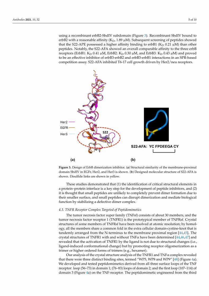
To verify whether the peptides derived from S22 display binding to erbB receptors,
we studied the interaction of the S22-APE peptide by surface plasmon resonance (SPR)
using a recombinant erbB2-SbdIV subdomain (Figure 3). Recombinant SbdIV bound to
erbB2 with a reasonable affinity (KD, 1.89 μM). Subsequent screening of peptides showed
that the S22-APE possessed a higher affinity binding to erbB1 (KD 0.21 μM) than other
peptides. Notably, the S22-AFA showed an overall comparable affinity to the three erbB
receptors (ErbB1: KD 0.41 μM, ErbB2: KD 0.30 μM, and ErbB3: KD 0.43 μM) and proved to
be an effective inhibitor of erbB3-erbB2 and erbB3-erbB1 interactions in an SPR-based
competition assay. S22-AFA inhibited T6-17 cell growth driven by Her2/neu receptors.
Figure 2. Structural features of erbB receptor activation. (a) Three-dimensional structure of ecto-domain of erbB receptor inresting state. Extracellular ErbB receptors consist of four distinct domains, SbdI–SbdIV. Each domain is shown in separatecolors. In the resting state, the domains are held in closed or locked conformation through two long loops, S21 and S22 fromdomains SbdII and SbdIV. (b) Native state of Her2/neu is shown. In contrast to other members of erbB receptors, Her2 isobserved only in an open or extended conformation. (c) Active state of erbB receptors is shown. Ligands binding to ErbBreceptors induce dimer formation. Ligand binding site is shown by red line. ErbB dimers are stabilized by inter-lockingloops near ligand-binding domain and at the membrane proximal (SbdIV) domains.
To develop dimeric inhibitors of the Her2 /neu associated ErbB receptor complex, weused the crystal structures of ErbB receptors [38,42] to create the small exocyclic peptideS22-AFA to target Her2-Her3 heterodimer formation [43]. This S22 peptide also blockedneuregulin mediated dimerization of the Her2-Her3 receptor complex. A dimeric erbB1-EGF model was developed, based on the existing experimental evidence that erbB1-EGFcomplex has a 2:2 stoichiometry [40]; the C-terminal part of subdomain IV is a dimericinteraction site [43]. Since the S22 loop stabilizes the active state of Her2-associated dimericreceptors, we focused on generating other peptides based on the structural features of S22.
To verify whether the peptides derived from S22 display binding to erbB receptors,we studied the interaction of the S22-APE peptide by surface plasmon resonance (SPR)
Antibodies 2021, 10, 32 5 of 10
using a recombinant erbB2-SbdIV subdomain (Figure 3). Recombinant SbdIV bound toerbB2 with a reasonable affinity (KD, 1.89 µM). Subsequent screening of peptides showedthat the S22-APE possessed a higher affinity binding to erbB1 (KD 0.21 µM) than otherpeptides. Notably, the S22-AFA showed an overall comparable affinity to the three erbBreceptors (ErbB1: KD 0.41 µM, ErbB2: KD 0.30 µM, and ErbB3: KD 0.43 µM) and provedto be an effective inhibitor of erbB3-erbB2 and erbB3-erbB1 interactions in an SPR-basedcompetition assay. S22-AFA inhibited T6-17 cell growth driven by Her2/neu receptors.
Antibodies 2021, 10, x FOR PEER REVIEW 6 of 11
Figure 3. Design of ErbB dimerization inhibitor. (a) Structural similarity of the membrane-proxi-
mal domain SbdIV in EGFr, Her2, and Her3 is shown. (b) Designed molecular structure of S22-
AFA is shown. Disulfide links are shown in yellow.
These studies demonstrated that (1) the identification of critical structural elements
in a protein–protein interface is a key step for the development of peptide inhibitors, and
(2) it is thought that small peptides are unlikely to completely prevent dimer formation
due to their smaller surface, and small peptides can disrupt dimerization and mediate
biological function by stabilizing a defective dimer complex.
4.3. TNFR Receptor Complex Targeted of Peptidomimetics
The tumor necrosis factor super family (TNFsf) consists of about 30 members, and
the tumor necrosis factor receptor 1 (TNFR1) is the prototypical member of TNFRsf. Crys-
tal structures of some members of TNFRsf have been resolved at atomic resolution; by
homology, all the members share a common fold in the extra cellular domain-cystine-knot
that is tandemly arranged from the N-terminus to the membrane proximal region [44,45].
The crystal structures of TNFR1 with and without TNFα have been determined [44,46,47]
and revealed that the activation of TNFR1 by the ligand is not due to structural changes
(i.e., ligand-induced conformational change) but by promoting receptor oligomerization
as a trimer or higher ordered forms of trimers (e.g., hexamer).
Our analysis of the crystal structure analysis of the TNFR1 and TNFα complex re-
vealed that there were three distinct binding sites, termed “WP5, WP8 and WP9”[48] (Fig-
ure 4a). We developed and tested peptidomimetics derived from all three surface loops of
the TNF receptor: loop (56–73) in domain 1; (76–83) loops of domain 2; and the first loop
(107–114) of domain 3 (Figure 4a) on the TNF-receptor. The peptidomimetic engineered
from the third domain (WP9QY: Tyr-Cys-Trp-Ser-Gln-Tyr-Leu-Cys-Tyr) inhibited TNFα
binding (IC50 = 75 μM) to its receptor (Figure 4c). Additionally, the peptidomimetic pro-
tected cells against TNFα-induced cell death when apoptosis was induced with 7 pg of
TNFα, suggesting that the peptide specifically binds to TNFα (Figure 4c). Kojima et al.
showed that the peptidomimetic (WP9QY) reduced the clinical score of inflammation in
a collagen-induced arthritis (CIA) mouse model [49], mimicking anti-TNF antibody func-
tion in reducing inflammation. This is one of the first peptide inhibitors, based on the
Figure 3. Design of ErbB dimerization inhibitor. (a) Structural similarity of the membrane-proximaldomain SbdIV in EGFr, Her2, and Her3 is shown. (b) Designed molecular structure of S22-AFA isshown. Disulfide links are shown in yellow.
These studies demonstrated that (1) the identification of critical structural elements ina protein–protein interface is a key step for the development of peptide inhibitors, and (2)it is thought that small peptides are unlikely to completely prevent dimer formation due totheir smaller surface, and small peptides can disrupt dimerization and mediate biologicalfunction by stabilizing a defective dimer complex.
4.3. TNFR Receptor Complex Targeted of Peptidomimetics
The tumor necrosis factor super family (TNFsf) consists of about 30 members, and thetumor necrosis factor receptor 1 (TNFR1) is the prototypical member of TNFRsf. Crystalstructures of some members of TNFRsf have been resolved at atomic resolution; by homol-ogy, all the members share a common fold in the extra cellular domain-cystine-knot that istandemly arranged from the N-terminus to the membrane proximal region [44,45]. Thecrystal structures of TNFR1 with and without TNFα have been determined [44,46,47] andrevealed that the activation of TNFR1 by the ligand is not due to structural changes (i.e.,ligand-induced conformational change) but by promoting receptor oligomerization as atrimer or higher ordered forms of trimers (e.g., hexamer).
Our analysis of the crystal structure analysis of the TNFR1 and TNFα complex revealedthat there were three distinct binding sites, termed “WP5, WP8 and WP9” [48] (Figure 4a).We developed and tested peptidomimetics derived from all three surface loops of the TNFreceptor: loop (56–73) in domain 1; (76–83) loops of domain 2; and the first loop (107–114) ofdomain 3 (Figure 4a) on the TNF-receptor. The peptidomimetic engineered from the third

Antibodies 2021, 10, 32 6 of 10
domain (WP9QY: Tyr-Cys-Trp-Ser-Gln-Tyr-Leu-Cys-Tyr) inhibited TNFα binding (IC50 =75 µM) to its receptor (Figure 4c). Additionally, the peptidomimetic protected cells againstTNFα-induced cell death when apoptosis was induced with 7 pg of TNFα, suggestingthat the peptide specifically binds to TNFα (Figure 4c). Kojima et al. showed that thepeptidomimetic (WP9QY) reduced the clinical score of inflammation in a collagen-inducedarthritis (CIA) mouse model [49], mimicking anti-TNF antibody function in reducinginflammation. This is one of the first peptide inhibitors, based on the structure of theTNFR1 receptor complex, targeting the receptor complex function. Subsequently, themethodology has been validated by developing peptide inhibitors to Fas-FasL and RANKreceptor complexes [50,51].
Antibodies 2021, 10, x FOR PEER REVIEW 7 of 11
structure of the TNFR1 receptor complex, targeting the receptor complex function. Subse-
quently, the methodology has been validated by developing peptide inhibitors to Fas-
FasL and RANK receptor complexes [50,51].
Figure 4. Design and development of anti-TNFR1 complex peptide inhibitor. (a) Three-dimensional structure of TNFR1-
TNF receptor complex in ribbon representation. TNFR1 is shown in blue; each protomer of TNFα is shown in three-dif-
ferent colors. Three main contact loops, WP5, WP9, and WP8, involved in forming stable trimeric complex are indicted.
The major contact, WP9, is highlighted in yellow. (b) Solution structure of WP9 is shown. (c) Inhibitory effects of peptides
inhibitors derived from the structures of WP9 and WP5 loop in a cell survival assay. WP9QY inhibits TNFα-induced
apoptosis in L929 cells. At 75 μM, WP9QY protected nearly 90% of cells from TNFα-induced cell death.
5. Applications of Constrained Peptidomimetics and Creation of Immunoadhesins
Antibody engineering is an active area of research to replace or to create therapeutic
antibody in a facile manner [52–54]. Conventional antibody generation involves immun-
izing animals with antigen, identification of therapeutic antibody, and creating monoclo-
nal antibody species after affinity maturation. These processes are not only time-consum-
ing but also resource intensive [55,56].
We have developed a method to engineer antibody-like binding proteins using our
designed peptidic loops. The goal is to create a modular protein molecule where one or
more structure-based constrained peptides can be tailored to create protein-scaffolds.
These modular protein scaffolds, which we term “loop-bodies”, contain functional bind-
ing loops derived from receptors or antibodies and can include the Fc domain of immu-
noglobulin, and a supporting linker sequence around the loop to create novel immunoad-
hesins.
We chose to engineer the S22 peptide, which, as mentioned above, is derived from
the HER2/neu receptor and interacts with all members of the ErbB receptors. By grafting
the S22 peptide onto a unique linker sequence that is fused to Fc, we have observed greatly
improved binding of the S22 sequence to HER2 as well as to EGFR and HER3. The in vivo
activity of S22Fc loop bodies has been demonstrated by examining the effects on growing
xenograft tumors. The same strategy can be expanded to establish a protein platform to
produce a series of novel therapeutic proteins targeting receptors (e.g., EGFR, Her2,
HER3, TNFR, RANK, etc.) or ligands (e.g., EGF and ligands for ErbB receptors, TNF,
RANKL, etc.).
We have investigated several protein scaffolds as a platform to create more stable
forms of the peptide. One scaffold is the streptavidin tetramer, which, when fused to the
anti-p185her2/neu peptide AHNP, improved the association rate for binding to the target [52].
A second construct was developed by us using the Z domain of Staphylococcus to engi-
neer proteins [57]. This class of novel proteins is an alternative to Fc fusion proteins and
is able to interact with circulating IgGs while binding to a target antigen.
We have found a class of immunoadhesin that functioned well for the S22 species of
peptides. The new S22-Fc species was developed to replace the combination of two differ-
ent antibodies with one engineered fusion protein that can bind to multiple members of
Figure 4. Design and development of anti-TNFR1 complex peptide inhibitor. (a) Three-dimensional structure of TNFR1-TNFreceptor complex in ribbon representation. TNFR1 is shown in blue; each protomer of TNFα is shown in three-differentcolors. Three main contact loops, WP5, WP9, and WP8, involved in forming stable trimeric complex are indicted. The majorcontact, WP9, is highlighted in yellow. (b) Solution structure of WP9 is shown. (c) Inhibitory effects of peptides inhibitorsderived from the structures of WP9 and WP5 loop in a cell survival assay. WP9QY inhibits TNFα-induced apoptosis inL929 cells. At 75 µM, WP9QY protected nearly 90% of cells from TNFα-induced cell death.
5. Applications of Constrained Peptidomimetics and Creation of Immunoadhesins
Antibody engineering is an active area of research to replace or to create therapeuticantibody in a facile manner [52–54]. Conventional antibody generation involves immuniz-ing animals with antigen, identification of therapeutic antibody, and creating monoclonalantibody species after affinity maturation. These processes are not only time-consumingbut also resource intensive [55,56].
We have developed a method to engineer antibody-like binding proteins using ourdesigned peptidic loops. The goal is to create a modular protein molecule where one ormore structure-based constrained peptides can be tailored to create protein-scaffolds. Thesemodular protein scaffolds, which we term “loop-bodies”, contain functional binding loopsderived from receptors or antibodies and can include the Fc domain of immunoglobulin,and a supporting linker sequence around the loop to create novel immunoadhesins.
We chose to engineer the S22 peptide, which, as mentioned above, is derived from theHER2/neu receptor and interacts with all members of the ErbB receptors. By grafting theS22 peptide onto a unique linker sequence that is fused to Fc, we have observed greatlyimproved binding of the S22 sequence to HER2 as well as to EGFR and HER3. The in vivoactivity of S22Fc loop bodies has been demonstrated by examining the effects on growingxenograft tumors. The same strategy can be expanded to establish a protein platform toproduce a series of novel therapeutic proteins targeting receptors (e.g., EGFR, Her2, HER3,TNFR, RANK, etc.) or ligands (e.g., EGF and ligands for ErbB receptors, TNF, RANKL,etc.).
We have investigated several protein scaffolds as a platform to create more stableforms of the peptide. One scaffold is the streptavidin tetramer, which, when fused to the

Antibodies 2021, 10, 32 7 of 10
anti-p185her2/neu peptide AHNP, improved the association rate for binding to the target [52].A second construct was developed by us using the Z domain of Staphylococcus to engineerproteins [57]. This class of novel proteins is an alternative to Fc fusion proteins and is ableto interact with circulating IgGs while binding to a target antigen.
We have found a class of immunoadhesin that functioned well for the S22 speciesof peptides. The new S22-Fc species was developed to replace the combination of twodifferent antibodies with one engineered fusion protein that can bind to multiple membersof the ErbB receptors. The S22 was first designed to be expressed as a fusion protein withthe intact Fc with both CH2 and CH3 domains from a human IgG1.
The Fc fusion protein was expressed in bacteria, in which glycosylation is deficient. It isreported that the aglycosylated Fc is very inefficient to bind to its receptors. Sazinsky et al. [58]described a T299A mutation near the glycosylation site (N297) that dramatically improvedthe interaction between aglycosylated Fc fragments and Fc receptors. We introduced thismutation to our recombinant protein that was expressed in E. coli.
We discovered that a short linker at the N-terminal improved the binding of the Fcfusion protein to receptors on T6-17 and NE91 cells. This novel short linker may represent away to improve the stability of the construct for proper binding activity. The fusion proteinproduced from bacteria was named LS22FcT322G7.
Binding of the S22 peptide to ErbB receptors (EGFR, HER2, and HER3) has beendetermined with the dissociation constant (KD) in the 0.3–0.43 µM range [59]. We performsimilar SPR assay to verify that the fusion protein, LS22FcT322G7, retains affinity for allthese receptors.
We tested the activity of LS22FcT322G7 in the inhibition of in vivo tumor growth.Briefly, we studied two models: (1) T6-17 tumors, which comprise the murine tumor cellline that expresses human HER2/neu, and (2) M1 tumors, which comprise the murinetumor cell line driven by both EGFR and HER2. As shown in Figure 5, LS22FcT322G7dose-dependently inhibits the tumor growth in both models. At the 10 mg/kg dosage,tumors in the treated group were very significantly smaller than those in the control groups.
Antibodies 2021, 10, x FOR PEER REVIEW 8 of 11
the ErbB receptors. The S22 was first designed to be expressed as a fusion protein with the
intact Fc with both CH2 and CH3 domains from a human IgG1.
The Fc fusion protein was expressed in bacteria, in which glycosylation is deficient.
It is reported that the aglycosylated Fc is very inefficient to bind to its receptors. Sazinsky
et al. [58] described a T299A mutation near the glycosylation site (N297) that dramatically
improved the interaction between aglycosylated Fc fragments and Fc receptors. We intro-
duced this mutation to our recombinant protein that was expressed in E. coli.
We discovered that a short linker at the N-terminal improved the binding of the Fc
fusion protein to receptors on T6-17 and NE91 cells. This novel short linker may represent
a way to improve the stability of the construct for proper binding activity. The fusion
protein produced from bacteria was named LS22FcT322G7.
Binding of the S22 peptide to ErbB receptors (EGFR, HER2, and HER3) has been de-
termined with the dissociation constant (KD) in the 0.3–0.43 μM range [59]. We perform
similar SPR assay to verify that the fusion protein, LS22FcT322G7, retains affinity for all
these receptors.
We tested the activity of LS22FcT322G7 in the inhibition of in vivo tumor growth.
Briefly, we studied two models: (1) T6-17 tumors, which comprise the murine tumor cell
line that expresses human HER2/neu, and (2) M1 tumors, which comprise the murine tu-
mor cell line driven by both EGFR and HER2. As shown in Figure 5, LS22FcT322G7 dose-
dependently inhibits the tumor growth in both models. At the 10 mg/kg dosage, tumors
in the treated group were very significantly smaller than those in the control groups.
Figure 5. Inhibition of tumor growth. In the animal model of athymic mice carrying T6-17 or M1 xenografts, LS22FcT322A
demonstrated dose-dependent activity. T6-17 and M1 tumors were established by subcutaneously implanting 5 × 10⁴ and
1 × 10⁶ cells, respectively, into the flank of nude mice. For T6-17 tumors, treatment started the next day after tumor inocu-
lation. For M1 tumors, treatment started 10 days after. Mice were treated three times per week at two different dosages (3
mg/kg or 10mg/kg, i.p.). t test was performed to compare the difference between the size of tumors in the treated group
and the control group. *: p < 0.05; **: p < 0.01.
6. Conclusions
We reviewed our approach to create protein–protein inhibitors targeting the erbB
and TNF receptors. Our methods have utilized structural information that was combined
with computational and biophysical tools. Additionally, we have shown that receptor
complex peptide inhibitors are antibody-like protein molecules. Receptor peptide inhibi-
tors developed by us are widely used by the research community, which has expanded
our approach [60–63]. It is expected that these developments may lead to clinically viable
candidates in future.
Author Contributions: Conceptualization, M.I.G.; software, R.M.; writing—original draft prepara-
tion, R.M. and M.I.G.; writing—review and editing, H.Z., Z.C., and L.L.; visualization, R.M.; funding
Figure 5. Inhibition of tumor growth. In the animal model of athymic mice carrying T6-17 or M1 xenografts, LS22FcT322Ademonstrated dose-dependent activity. T6-17 and M1 tumors were established by subcutaneously implanting 5 × 10⁴
and 1 × 10⁶ cells, respectively, into the flank of nude mice. For T6-17 tumors, treatment started the next day after tumorinoculation. For M1 tumors, treatment started 10 days after. Mice were treated three times per week at two different dosages(3 mg/kg or 10mg/kg, i.p.). t test was performed to compare the difference between the size of tumors in the treated groupand the control group. *: p < 0.05; **: p < 0.01.
6. Conclusions
We reviewed our approach to create protein–protein inhibitors targeting the erbBand TNF receptors. Our methods have utilized structural information that was combined

Antibodies 2021, 10, 32 8 of 10
with computational and biophysical tools. Additionally, we have shown that receptorcomplex peptide inhibitors are antibody-like protein molecules. Receptor peptide inhibitorsdeveloped by us are widely used by the research community, which has expanded ourapproach [60–63]. It is expected that these developments may lead to clinically viablecandidates in future.
Author Contributions: Conceptualization, M.G.; software, R.M.; writing—original draft preparation,R.M. and M.G.; writing—review and editing, H.Z., Z.C., and L.L.; visualization, R.M.; funding acqui-sition, H.Z. and M.G. All authors have read and agreed to the published version of the manuscript.
Funding: We acknowledge grant supports from the Breast Cancer Research Foundation and theNational Institutes of Health to M.I.G. (R01 CA219034) and partially by NIH grant P50 CA142509and the 2016 PCMD Pilot and Feasibility Grant Program to H.Z.
Institutional Review Board Statement: The study was conducted according to the guidelines of theDeclaration of Helsinki, and approved by the Institutional Review Board of University of Pennsylva-nia (protocol number 804008, 10/5/2011; protocol number 803383, 11/15/2013; 806450, 2/23/2018).
Informed Consent Statement: Not applicable.
Data Availability Statement: Not applicable for a review article.
Conflicts of Interest: The authors declare no conflict of interest.
References1. Newman, D.J.; Cragg, G.M. Natural products as sources of new drugs over the 30 years from 1981 to 2010. J. Nat. Prod. 2012, 75,
311–335. [CrossRef] [PubMed]2. Hopkins, A.L.; Groom, C.R. The druggable genome. Nat. Rev. Drug Discov. 2002, 1, 727–730. [CrossRef] [PubMed]3. Makley, L.N.; Gestwicki, J.E. Expanding the number of ‘druggable’ targets: Non-enzymes and protein-protein interactions. Chem.
Biol. Drug Des. 2013, 81, 22–32. [CrossRef] [PubMed]4. Shendure, J.; Findlay, G.M.; Snyder, M.W. Genomic Medicine-Progress, Pitfalls, and Promise. Cell 2019, 177, 45–57. [CrossRef]5. Hennemann, H.; Wirths, S.; Carl, C. Cell-based peptide screening to access the undruggable target space. Eur. J. Med. Chem. 2015,
94, 489–496. [CrossRef]6. Neklesa, T.K.; Winkler, J.D.; Crews, C.M. Targeted protein degradation by PROTACs. Pharmacol. Ther. 2017, 174, 138–144.
[CrossRef] [PubMed]7. Ran, X.; Gestwicki, J.E. Inhibitors of protein-protein interactions (PPIs): An analysis of scaffold choices and buried surface area.
Curr. Opin. Chem. Biol. 2018, 44, 75–86. [CrossRef]8. Ali, A.M.; Atmaj, J.; Van Oosterwijk, N.; Groves, M.R.; Dömling, A. Stapled Peptides Inhibitors: A New Window for Target Drug
Discovery. Comput. Struct. Biotechnol. J. 2019, 17, 263–281. [CrossRef] [PubMed]9. Kessler, D.; Gmachl, M.; Mantoulidis, A.; Martin, L.J.; Zoephel, A.; Mayer, M.; Gollner, A.; Covini, D.; Fischer, S.; Gerstberger, T.;
et al. Drugging an undruggable pocket on KRAS. Proc. Natl. Acad. Sci. USA 2019, 116, 15823–15829. [CrossRef]10. Huang, A.; Garraway, L.A.; Ashworth, A.; Weber, B. Synthetic lethality as an engine for cancer drug target discovery. Nat. Rev.
Drug Discov. 2020, 19, 23–38. [CrossRef]11. Cunningham, A.D.; Qvit, N.; Mochly-Rosen, D. Peptides and peptidomimetics as regulators of protein-protein interactions. Curr.
Opin. Struct. Biol. 2017, 44, 59–66. [CrossRef]12. Cochran, A.G. Antagonists of protein-protein interactions. Chem. Biol. 2000, 7, R85–R94. [CrossRef]13. Liu, B.A.; Engelmann, B.W.; Nash, P.D. High-throughput analysis of peptide-binding modules. Proteomics 2012, 12, 1527–1546.
[CrossRef]14. Lien, S.; Lowman, H.B. Therapeutic peptides. Trends Biotechnol. 2003, 21, 556–562. [CrossRef] [PubMed]15. Vlieghe, P.; Lisowski, V.; Martinez, J.; Khrestchatisky, M. Synthetic therapeutic peptides: Science and market. Drug Discov. Today
2010, 15, 40–56. [CrossRef]16. Morrison, C. Constrained peptides’ time to shine? Nat. Rev. Drug Discov. 2018, 17, 531–533. [CrossRef]17. Rastogi, S.; Shukla, S.; Kalaivani, M.; Singh, G.N. Peptide-based therapeutics: Quality specifications, regulatory considerations,
and prospects. Drug Discov. Today 2019, 24, 148–162. [CrossRef]18. Nagai, Y.; Lam, L.; Greene, M.I.; Zhang, H. FOXP3 and Its Cofactors as Targets of Immunotherapies. Engineering 2019, 5, 115–121.
[CrossRef]19. Burley, S.K.; Berman, H.M.; Kleywegt, G.J.; Markley, J.L.; Nakamura, H.; Velankar, S. Protein Data Bank (PDB): The Single Global
Macromolecular Structure Archive. Methods Mol. Biol. 2017, 1607, 627–641. [CrossRef]20. Berman, H.M.; Vallat, B.; Lawson, C.L. The data universe of structural biology. IUCrJ 2020, 7, 630–638. [CrossRef] [PubMed]21. Jones, S.; Thornton, J.M. Principles of protein-protein interactions. Proc. Natl. Acad. Sci. USA 1996, 93, 13–20. [CrossRef]

Antibodies 2021, 10, 32 9 of 10
22. Sundberg, E.J.; Mariuzza, R.A. Molecular recognition in antibody-antigen complexes. Adv. Protein Chem. 2002, 61, 119–160.[CrossRef]
23. Lafont, V.; Schaefer, M.; Stote, R.H.; Altschuh, D.; Dejaegere, A. Protein-protein recognition and interaction hot spots in anantigen-antibody complex: Free energy decomposition identifies “efficient amino acids”. Proteins 2007, 67, 418–434. [CrossRef][PubMed]
24. Strong, R.K.; Campbell, R.; Rose, D.R.; Petsko, G.A.; Sharon, J.; Margolies, M.N. Three-dimensional structure of murine anti-p-azophenylarsonate Fab 36-71. 1. X-ray crystallography, site-directed mutagenesis, and modeling of the complex with hapten.Biochemistry 1991, 30, 3739–3748. [CrossRef]
25. Parkkinen, T.; Nevanen, T.K.; Koivula, A.; Rouvinen, J. Crystal structures of an enantioselective fab-fragment in free and complexforms. J. Mol. Biol. 2006, 357, 471–480. [CrossRef] [PubMed]
26. Park, B.W.; Zhang, H.T.; Wu, C.; Berezov, A.; Zhang, X.; Dua, R.; Wang, Q.; Kao, G.; O’Rourke, D.M.; Greene, M.I.; et al. Rationallydesigned anti-HER2/neu peptide mimetic disables P185HER2/neu tyrosine kinases in vitro and in vivo. Nat. Biotechnol. 2000, 18,194–198. [CrossRef]
27. Murali, R.; Greene, M.I. Structure based antibody-like peptidomimetics. Pharmaceuticals 2012, 5, 209–235. [CrossRef]28. Jones, S.; Marin, A.; Thornton, J.M. Protein domain interfaces: Characterization and comparison with oligomeric protein interfaces.
Protein Eng. 2000, 13, 77–82. [CrossRef] [PubMed]29. Vaughan, C.K.; Buckle, A.M.; Fersht, A.R. Structural response to mutation at a protein-protein interface. J. Mol. Biol. 1999, 286,
1487–1506. [CrossRef] [PubMed]30. Bhat, T.N.; Bentley, G.A.; Boulot, G.; Greene, M.I.; Tello, D.; Dall’Acqua, W.; Souchon, H.; Schwarz, F.P.; Mariuzza, R.A.; Poljak,
R.J. Bound water molecules and conformational stabilization help mediate an antigen-antibody association. Proc. Natl. Acad. Sci.USA 1994, 91, 1089–1093. [CrossRef]
31. Braden, B.C.; Poljak, R.J. Structural features of the reactions between antibodies and protein antigens. FASEB J. 1995, 9, 9–16.[CrossRef]
32. Yan, C.; Wu, F.; Jernigan, R.L.; Dobbs, D.; Honavar, V. Characterization of protein-protein interfaces. Protein J. 2008, 27, 59–70.[CrossRef]
33. Visscher, K.M.; Kastritis, P.L.; Bonvin, A.M. Non-interacting surface solvation and dynamics in protein-protein interactions.Proteins 2015, 83, 445–458. [CrossRef] [PubMed]
34. Kieber-Emmons, T.; Murali, R.; Greene, M.I. Therapeutic peptides and peptidomimetics. Curr. Opin. Biotechnol. 1997, 8, 435–441.[CrossRef]
35. Murali, R.; Greene, M.I. Structure-based design of immunologically active therapeutic peptides. Immunol. Res. 1998, 17, 163–169.[CrossRef] [PubMed]
36. von Grafenstein, S.; Wallnoefer, H.G.; Kirchmair, J.; Fuchs, J.E.; Huber, R.G.; Schmidtke, M.; Sauerbrei, A.; Rollinger, J.M.; Liedl,K.R. Interface dynamics explain assembly dependency of influenza neuraminidase catalytic activity. J. Biomol. Struct. Dyn. 2015,33, 104–120. [CrossRef]
37. Watkins, A.M.; Wuo, M.G.; Arora, P.S. Protein-Protein Interactions Mediated by Helical Tertiary Structure Motifs. J. Am. Chem.Soc. 2015, 137, 11622–11630. [CrossRef]
38. Franklin, M.C.; Carey, K.D.; Vajdos, F.F.; Leahy, D.J.; de Vos, A.M.; Sliwkowski, M.X. Insights into ErbB signaling from thestructure of the ErbB2-pertuzumab complex. Cancer Cell 2004, 5, 317–328. [CrossRef]
39. Garrett, T.P.; McKern, N.M.; Lou, M.; Elleman, T.C.; Adams, T.E.; Lovrecz, G.O.; Kofler, M.; Jorissen, R.N.; Nice, E.C.; Burgess,A.W.; et al. The crystal structure of a truncated ErbB2 ectodomain reveals an active conformation, poised to interact with otherErbB receptors. Mol. Cell 2003, 11, 495–505. [CrossRef]
40. Garrett, T.P.; McKern, N.M.; Lou, M.; Elleman, T.C.; Adams, T.E.; Lovrecz, G.O.; Zhu, H.J.; Walker, F.; Frenkel, M.J.; Hoyne, P.A.;et al. Crystal structure of a truncated epidermal growth factor receptor extracellular domain bound to transforming growth factoralpha. Cell 2002, 110, 763–773. [CrossRef]
41. Cho, H.S.; Leahy, D.J. Structure of the extracellular region of HER3 reveals an interdomain tether. Science 2002, 297, 1330–1333.[CrossRef] [PubMed]
42. Cho, H.S.; Mason, K.; Ramyar, K.X.; Stanley, A.M.; Gabelli, S.B.; Denney, D.W., Jr.; Leahy, D.J. Structure of the extracellular regionof HER2 alone and in complex with the Herceptin Fab. Nature 2003, 421, 756–760. [CrossRef]
43. Berezov, A.; Chen, J.; Liu, Q.; Zhang, H.T.; Greene, M.I.; Murali, R. Disabling receptor ensembles with rationally designedinterface peptidomimetics. J. Biol. Chem. 2002, 277, 28330–28339. [CrossRef]
44. Naismith, J.H.; Devine, T.Q.; Kohno, T.; Sprang, S.R. Structures of the extracellular domain of the type I tumor necrosis factorreceptor. Structure 1996, 4, 1251–1262. [CrossRef]
45. Naismith, J.H.; Sprang, S.R. Modularity in the TNF-receptor family. Trends Biochem. Sci. 1998, 23, 74–79. [CrossRef]46. Baeyens, K.J.; De Bondt, H.L.; Raeymaekers, A.; Fiers, W.; De Ranter, C.J. The structure of mouse tumour-necrosis factor at 1.4 A
resolution: Towards modulation of its selectivity and trimerization. Acta Cryst. D Biol. Cryst. 1999, 55, 772–778. [CrossRef]47. Ono, M.; Horita, S.; Sato, Y.; Nomura, Y.; Iwata, S.; Nomura, N. Structural basis for tumor necrosis factor blockade with the
therapeutic antibody golimumab. Protein Sci. 2018, 27, 1038–1046. [CrossRef]48. Takasaki, W.; Kajino, Y.; Kajino, K.; Murali, R.; Greene, M.I. Structure-based design and characterization of exocyclic pep-
tidomimetics that inhibit TNF.alpha. binding to its receptor. Nat. Biotechnol. 1997, 15, 1266–1270. [CrossRef]

Antibodies 2021, 10, 32 10 of 10
49. Kojima, T.; Aoki, K.; Nonaka, K.; Saito, H.; Azuma, M.; Iwai, H.; Varghese, B.J.; Yoshimasu, H.; Baron, R.; Ohya, K.; et al.Subcutaneous injections of a TNF-alpha antagonistic peptide inhibit both inflammation and bone resorption in collagen-inducedmurine arthritis. J. Med. Dent. Sci. 2005, 52, 91–99. [PubMed]
50. Cheng, X.; Kinosaki, M.; Takami, M.; Choi, Y.; Zhang, H.; Murali, R. Disabling of receptor activator of nuclear factor-kappaB(RANK) receptor complex by novel osteoprotegerin-like peptidomimetics restores bone loss in vivo. J. Biol. Chem. 2004, 279,8269–8277. [CrossRef]
51. Hasegawa, A.; Cheng, X.; Kajino, K.; Berezov, A.; Murata, K.; Nakayama, T.; Yagita, H.; Murali, R.; Greene, M.I. Fas-disablingsmall exocyclic peptide mimetics limit apoptosis by an unexpected mechanism. Proc. Natl. Acad. Sci. USA 2004, 101, 6599–6604.[CrossRef]
52. Masuda, K.; Richter, M.; Song, X.; Berezov, A.; Masuda, K.; Murali, R.; Greene, M.I.; Zhang, H. AHNP-streptavidin: A tetramericbacterially produced antibody surrogate fusion protein against p185her2/neu. Oncogene 2006, 25, 7740–7746. [CrossRef]
53. Mitran, B.; Andersson, K.G.; Lindström, E.; Garousi, J.; Rosestedt, M.; Tolmachev, V.; Ståhl, S.; Orlova, A.; Löfblom, J. Affibody-mediated imaging of EGFR expression in prostate cancer using radiocobalt-labeled DOTA-ZEGFR:2377. Oncol. Rep. 2019, 41,534–542. [CrossRef] [PubMed]
54. Simeon, R.; Chen, Z. In vitro-engineered non-antibody protein therapeutics. Protein Cell 2018, 9, 3–14. [CrossRef] [PubMed]55. Hanack, K.; Messerschmidt, K.; Listek, M. Antibodies and Selection of Monoclonal Antibodies. Adv. Exp. Med. Biol. 2016, 917,
11–22. [CrossRef]56. Thakur, A.; Huang, M.; Lum, L.G. Bispecific antibody based therapeutics: Strengths and challenges. Blood Rev. 2018, 32, 339–347.
[CrossRef] [PubMed]57. Cai, Z.; Fu, T.; Nagai, Y.; Lam, L.; Yee, M.; Zhu, Z.; Zhang, H. scFv-based “Grababody” as a general strategy to improve
recruitment of immune effector cells to antibody-targeted tumors. Cancer Res. 2013, 73, 2619–2627. [CrossRef] [PubMed]58. Sazinsky, S.L.; Ott, R.G.; Silver, N.W.; Tidor, B.; Ravetch, J.V.; Wittrup, K.D. Aglycosylated immunoglobulin G1 variants
productively engage activating Fc receptors. Proc. Natl. Acad. Sci. USA 2008, 105, 20167–20172. [CrossRef] [PubMed]59. Berezov, A.; Zhang, H.T.; Greene, M.I.; Murali, R. Disabling erbB receptors with rationally designed exocyclic mimetics of
antibodies: Structure-function analysis. J. Med. Chem. 2001, 44, 2565–2574. [CrossRef]60. Kato, G.; Shimizu, Y.; Arai, Y.; Suzuki, N.; Sugamori, Y.; Maeda, M.; Takahashi, M.; Tamura, Y.; Wakabayashi, N.; Murali, R.; et al.
The inhibitory effects of a RANKL-binding peptide on articular and periarticular bone loss in a murine model of collagen-inducedarthritis: A bone histomorphometric study. Arthritis Res. Ther. 2015, 17, 251. [CrossRef]
61. Ding, H.; Gangalum, P.R.; Galstyan, A.; Fox, I.; Patil, R.; Hubbard, P.; Murali, R.; Ljubimova, J.Y.; Holler, E. HER2-positive breastcancer targeting and treatment by a peptide-conjugated mini nanodrug. Nanomedicine 2017, 13, 631–639. [CrossRef] [PubMed]
62. Haque Bhuyan, M.Z.; Tamura, Y.; Sone, E.; Yoshinari, Y.; Maeda, C.; Takahashi, M.; Tabata, Y.; Murali, R.; Waki, Y.; Aoki, K.The intra-articular injection of RANKL-binding peptides inhibits cartilage degeneration in a murine model of osteoarthritis. J.Pharmacol. Sci. 2017, 134, 124–130. [CrossRef] [PubMed]
63. Idress, M.; Milne, B.F.; Thompson, G.S.; Trembleau, L.; Jaspars, M.; Houssen, W.E. Structure-Based Design, Synthesis andBioactivity of a New Anti-TNFα Cyclopeptide. Molecules 2020, 25, 922. [CrossRef] [PubMed]