Embed Size (px)
Citation preview
JOURNAL OF CLINICAL MICROBIOLOGY,0095-1137/00/$04.0010
Mar. 2000, p. 1286–1289 Vol. 38, No. 3
Copyright © 2000, American Society for Microbiology. All Rights Reserved.
Rapid Subtyping of Dengue Virus Serotypes 1 and 4 byRestriction Site-Specific PCR
MARIZE P. MIAGOSTOVICH,1 FLAVIA B. DOS SANTOS,1 C. MILENA GUTIERREZ,2 LEE W. RILEY,3
AND EVA HARRIS3*
Department of Virology, Oswaldo Cruz Institute, Oswaldo Cruz Foundation, Rio de Janeiro, Brazil1; Department ofVirology, Centro Nacional de Diagnostico y Referencia, Ministry of Health, Managua, Nicaragua2; and Infectious
Diseases Unit, Division of Public Health Biology and Epidemiology, School of Public Health,University of California, Berkeley, California3
Received 7 October 1999/Returned for modification 22 November 1999/Accepted 24 December 1999
We previously reported a simple subtyping method, restriction site-specific PCR (RSS-PCR), for denguevirus serotypes 2 and 3; here we describe its application for subtyping dengue virus serotypes 1 and 4. Threemajor RSS-PCR types were observed for dengue virus serotype 1 and two types were observed for dengue virusserotype 4, in agreement with previous strain classifications based on sequence analysis. Because of itssimplicity, this method is amenable to rapid subtyping and application to epidemiological studies of dengue incountries where dengue is endemic.
Dengue viruses are single-stranded, enveloped RNA flavivi-ruses which are traditionally classified into four serotypes, des-ignated dengue-1, -2, -3, and -4, based on antigenic character-istics (2, 19). Numerous methods, including RNase T1oligonucleotide fingerprinting, restriction enzyme analysis, andnucleotide sequencing of different genomic fragments, havedemonstrated strain variation within each dengue serotype,dividing them into distinct genetic subtypes (4, 5, 10, 11, 12, 13,16, 17, 18). Dengue virus classification into subtypes is usefulfor studying the global distribution and movement of dengueserotypes, which contributes to the identification of viral fac-tors that influence disease severity and risk factors associatedwith the transmission of particular strains.
Accurate characterization of strain difference usually re-quires labor-intensive typing procedures, which are difficult toperform in a timely manner during epidemic periods. Recently,a new PCR-based approach to rapidly subtype dengue viruseswas developed (8) and consists of a single reverse transcriptasePCR (RT-PCR) amplification using four primers that targetregions spanning polymorphic endonuclease restriction siteswithin the envelope (E) gene. This method, called restrictionsite-specific PCR (RSS-PCR), is simple, rapid, requires mini-mal laboratory equipment, and uses widely available reagents.The successful results obtained with dengue-2 and dengue-3led us to develop the RSS-PCR method for dengue-1 anddengue-4 strains.
FIG. 1. RSS-PCR patterns of dengue-1 strains of different geographic origins. (A) Agarose gel electrophoresis of RSS-PCR products. Lane 1, dengue-2 Brazil 1998(64022); lane 2, dengue-3 Indonesia 1989 (430); lane 3, dengue-4 Puerto Rico 1986 (081); lane M, 100-bp ladder (Gibco BRL); lanes 4 to 16, dengue-1 [lane 4, Indonesia1977 (407-1); lane 5, Philippines 1984 (901); lane 6, Thailand 1979 (213); lane 7, Thailand 1973 (735); lane 8, Thailand 1974 (106); lane 9, Western Pacific 1980; lane10, Nigeria 1985; lane 11, Sri Lanka 1985; lane 12, Mexico 1982 (086); lane 13, Brazil 1986 (543); lane 14, Brazil RJ 1995 (49217); lane 15, Brazil ES 1998 (60548);lane 16, Brazil BA 1996 (57291)]. Lanes 4, 5, and 9, pattern A1; lane 6, pattern A2; lanes 7 to 8, pattern B; lanes 10 to 16, pattern C. (B) Schematic diagram representingthe different RSS-PCR patterns.
* Corresponding author. Mailing address: Infectious Diseases Unit,Division of Public Health Biology and Epidemiology, School of PublicHealth, University of California, Berkeley, 140 Warren Hall, Berkeley,CA 94720-7360. Phone: (510) 642-4845. Fax: (510) 642-6350. E-mail:[email protected].
1286
on June 6, 2018 by guesthttp://jcm
.asm.org/
Dow
nloaded from

Dengue-1 (Table 1) and dengue-4 (Table 2) strains repre-senting a broad geographical distribution were obtained fromexisting collections; Brazilian and Nicaraguan viruses were iso-lated from sera by inoculation into the Aedes albopictus cellline C6/36 (9) and were identified by immunofluorescence us-ing type-specific monoclonal antibodies (6). Viral seeds werepropagated once in C6/36 cells grown in Leibovitz-15 or min-imal essential medium (Gibco BRL, Grand Island, N.Y.) con-taining 10% fetal bovine serum. Primers were designed basedon the sequence around polymorphic restriction sites in the Egene region of dengue-1 and dengue-4 as described previously
TABLE 1. Dengue-1 strains used in this studya
Strain Location Yr RSSpattern Source
(475) Philippines 1983 A1 1(500) Philippines 1983 A1 1(506) Philippines 1983 A1 1(290) Philippines 1984 A1 1(684) Philippines 1984 A1 1(901) Philippines 1984 A1 1(933) Philippines 1984 A1 1(094) Thailand 1973 B 1(735) Thailand 1973 B 1(106) Thailand 1974 B 1(595) Thailand 1975 A1 1(213) Thailand 1979 A2 1(101) Thailand 1980 A2 1(213) Thailand 1980 A2 1(266) Thailand 1980 B 1(878) Thailand 1980 B 1(190-1) Indonesia 1976 A1 1(448-1) Indonesia 1976 A1 1(406-1) Indonesia 1977 A1 1(407-1) Indonesia 1977 A1 1(077-1) Indonesia 1978 A1 1(159-1) Indonesia 1978 A1 1(086) Mexico 1982 C 1(894) Mexico 1982 C 1(679) Mexico 1983 C 1(598) Mexico 1984 C 1(022) Mexico 1985 C 1(350) Mexico 1985 C 1(210) Mexico 1986 C 1(454) Mexico 1986 C 1(666) Mexico 1988 C 1(461) Brazil 1986 C 1(543) Brazil 1986 C 1(578) Brazil 1986 C 1(426) Aruba 1985 C 1(495) Aruba 1985 C 1(810) Aruba 1986 C 1(648) Puerto Rico 1985 C 1(405) Puerto Rico 1986 C 1(009) Puerto Rico 1986 C 1(454) Puerto Rico 1986 C 1
Sri Lanka 1985 C 1Nigeria 1985 C 1Jamaica 1981 C 1Western Pacific 1980 A1 1
28605 RJ (Brazil) 1986 C 228641 RJ (Brazil) 1986 C 227923 RJ (Brazil) 1986 C 232426 RJ (Brazil) 1986 C 239372 RJ (Brazil) 1990 C 239474 RJ (Brazil) 1990 C 249657 RJ (Brazil) 1995 C 253378 RJ (Brazil) 1995 C 264313 RJ (Brazil) 1999 C 264286 RJ (Brazil) 1999 C 264305 RJ (Brazil) 1999 C 256722 RJ (Brazil) 1996 C 256717 RJ (Brazil) 1996 C 258610 RJ (Brazil) 1997 C 258542 RJ (Brazil) 1997 C 260984 RJ (Brazil) 1998 C 260741 RJ (Brazil) 1998 C 264289 RJ (Brazil) 1999 C 264316 RJ (Brazil) 1999 C 251325 RJ (Brazil) 1995 C 249217 RJ (Brazil) 1995 C 2
Continued
TABLE 1—Continued
Strain Location Yr RSSpattern Source
57399 ES (Brazil) 1996 C 257217 ES (Brazil) 1996 C 259835 ES (Brazil) 1997 C 259934 ES (Brazil) 1997 C 260548 ES (Brazil) 1998 C 260538 ES (Brazil) 1998 C 257291 BA (Brazil) 1996 C 258067 BA (Brazil) 1997 C 258724 RN (Brazil) 1997 C 258438 RN (Brazil) 1997 C 258485 MG (Brazil) 1997 C 258522 MG (Brazil) 1997 C 260616 MG (Brazil) 1998 C 260619 MG (Brazil) 1998 C 260440 CE (Brazil) 1998 C 260447 CE (Brazil) 1998 C 2
a Source 1, School of Public Health, University of California, Berkeley. Strainswere originally obtained from the Division of Vector-Borne Infectious Diseases,Centers for Disease Control and Prevention, Fort Collins, Colo., and the ArmedForces Research Institute of the Medical Sciences, Bangkok, Thailand, and werekindly donated by S. Kliks, University of California, San Francisco. Source 2,Flavivirus Laboratory, Department of Virology, Oswaldo Cruz Institute, Os-waldo Cruz Foundation, Rio de Janeiro, Brazil. All strains were isolated duringepidemics in the following states of the country: Bahia (BA), Ceara (CE),Espırito Santo (ES), Minas Gerais (MS), Rio de Janeiro (RJ), and Rio Grandedo Norte (RN).
TABLE 2. Dengue-4 strains used in this study
Strain Location Yr RSSpattern Sourcea
(H241) Philippines 1956 A2 1(377) Thailand 1977 A2 1(561) Thailand 1977 A2 1(664) Thailand 1979 A2 1(840) Thailand 1980 A2 1(961) Thailand 1984 A2 1(171) Thailand 1984 A2 1(013) Thailand 1984 A2 1(789) Thailand 1985 A1 1(511) Thailand 1985 A2 1(280) Thailand 1988 A2 1(554) Mexico 1984 B 1(410) Mexico 1984 B 1(292) Mexico 1984 B 1(081) Puerto Rico 1986 B 1(437) Puerto Rico 1986 B 1(281) Puerto Rico 1986 B 1(703) Nicaragua 1999 B 2
a Source 1, School of Public Health, University of California, Berkeley. Source2, Department of Virology, Centro Nacional de Diagnostico y Referencia, Min-istry of Health, Managua, Nicaragua.
VOL. 38, 2000 NOTES 1287
on June 6, 2018 by guesthttp://jcm
.asm.org/
Dow
nloaded from

(8). The sequences and genomic positions of primers RSS9 toRSS12 (dengue-1) and RSS21 to RSS24 (dengue-4) are listedin Table 3.
Viral RNAs were extracted from the supernatants of in-fected cells using a QIAamp Viral RNA Mini Kit (Qiagen,Inc., Valencia, Calif.) according to the manufacturer’s protocolor by lysis with guanidine isothiocyanate, extraction with or-ganic solvents, and ethanol precipitation (7). The reaction mix-ture and electrophoresis conditions were as described previ-ously (8), except that 25-ml reaction volumes were used.Briefly, 2.5 ml of viral RNA was added to 22.5 ml of an RT-PCR mixture consisting of 50 mM potassium chloride, 10 mMTris (pH 8.5), 0.01% gelatin, 200 mM concentrations of each ofthe four deoxynucleoside triphosphates, 1.5 mM magnesiumchloride, 30 mM tetramethylammonium chloride, 0.5 M be-taine, 5 mM dithiothreitol, 0.5 mM concentrations of each offour RSS-PCR primers (RSS9 to -12 for dengue-1 and RSS21to -24 for dengue-4), 0.025 U of RT RAV-2 (Amersham Corp.,Arlington Heights, Ill.) per ml and 0.025 U of Taq DNA poly-merase (AmpliTaq; Perkin-Elmer Corp., Foster City, Calif.)per ml. Reverse transcription was conducted at 42°C for 60min, followed directly by 30 amplification cycles of 94°C for30 s, 60°C for 1 min, and 72°C for 2 min, with a final extensionat 72°C for 5 min. Amplification was conducted with 0.5-mltubes (USA Scientific, Ocala, Fla.) in a model PTC-200 ther-mocycler (MJ Research, Inc., Watertown, Mass.).
We selected sets of strains in our collection that representedeach of three dengue-1 genotypes previously described by E-NS1 sequence pairwise comparison (13), with one set includingstrains from Southeast Asia and the South Pacific, anothercontaining viruses from Thailand and Taiwan, and a thirdcontaining isolates from the Americas, Africa, and SoutheastAsia. The bands generated by the RSS-PCR assay using prim-ers RSS9 to -12 showed distinct patterns for American andAsian strains. Figure 1 shows representative examples of eachRSS-PCR pattern for dengue-1 viruses, along with a schematicdiagram summarizing the results. The first group (type A),which includes viruses from the Philippines (1983 to 1984),Indonesia (1976 to 1978), Thailand (1975, 1979, and 1980), andthe western Pacific (1980), was divided into two subgroups (A1and A2), depending on the presence of an ;200-bp fragment.The second group (type B) contains isolates from Thailand(1973, 1974, and 1980), and the third group (type C) is com-posed of strains from the Americas, Africa, and Sri Lanka.These RSS primers are specific for dengue-1, as dengue-2, -3,and -4 did not generate amplified products in this assay (Fig. 1,lanes 1 to 3).
As with dengue-1, several primer sets were tested with den-gue-4 strains from our collection that matched strains previ-ously classified by sequence analysis of the E gene (10) with
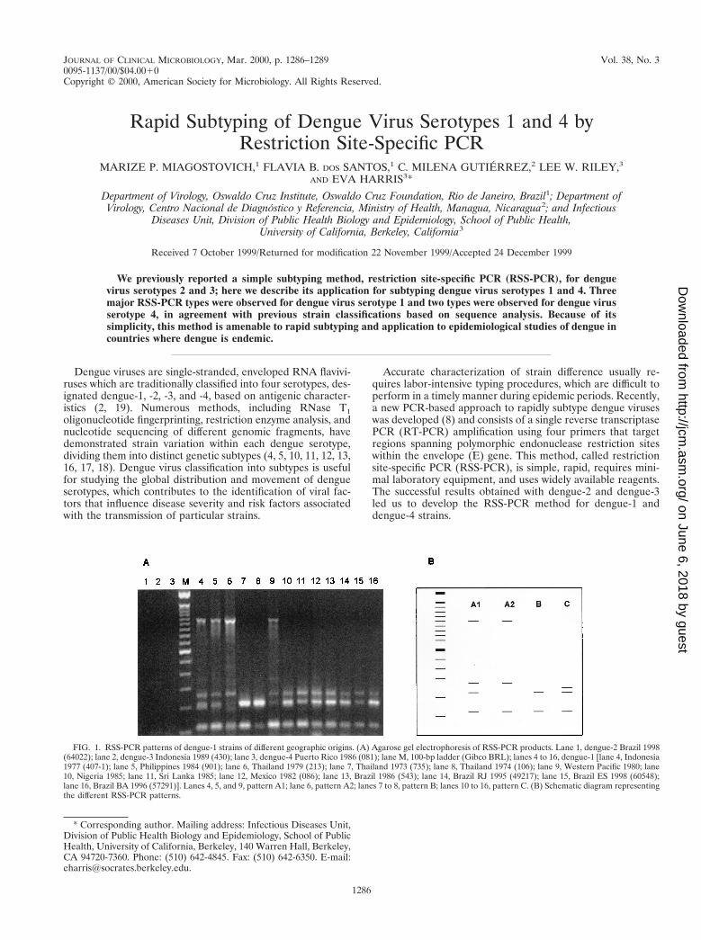
respect to location and date of isolation. The best results wereobtained with primers RSS21 to -24 (Table 3), which were usedto analyze the rest of our dengue-4 strains. Two patterns weregenerated, as shown in Fig. 2 and Table 2, one (type A) rep-resenting strains from Thailand (1977 to 1988) and the other(type B) representing viruses isolated in Mexico and the Ca-ribbean. Type A was divided into two subgroups due to varia-tion in one of the amplified fragments from a Thai isolate from1984. Again, no product was obtained with the other threeserotypes (Fig. 2).
The RSS-PCR method for dengue-1 and -4 fulfills the req-uisites of a molecular typing assay: the primers were specific tothe serotype to which they were developed, the patterns werestable over time, repeated amplification of the same specimensreproducibly yielded the same results, and geographic andtemporal clustering was observed. In addition, in areas withextensive dengue transmission, such as Thailand, the cocircu-lation of two distinct subtypes and of genetic variants withinthe same subtype was observed, which is consistent with pre-vious reports (13, 15).
The dengue-1 RSS-PCR results, categorized according tocountry and year of isolation, are essentially the same as thesequence analysis results showing geographic and temporalclustering. A phylogenetic analysis of 40 dengue-1 strains fromdifferent geographic areas based on a 240-nt region from theE-NS1 junction defined three main genotypes and two addi-tional ones, each represented by a single virus isolate (13).Similarly, another study comparing the sequences of a 179-ntregion of the E genes of 35 dengue-1 isolates yielded threegenotypes (4). The largest genotypic group in both analysesconsisted of dengue-1 strains from the Americas, Africa, andSoutheast Asia, and it corresponds to our RSS-PCR type C.The second genotype, containing viruses from the Philippines,Indonesia, Thailand, and the South Pacific, coincides withRSS-PCR type A, and the third group, consisting of Thai andTaiwanese viruses, corresponds to RSS-PCR type B. RSS-PCRrevealed two distinct subtypes (A and B) circulating simulta-neously in Thailand, in agreement with previous observations(13).
The dengue-4 isolates we analyzed by RSS-PCR fell into twosubtypes, consistent with the two genotypes that resulted fromsequence analysis of the entire E gene (10). By both methods,American isolates were contained in a different group thanthat of Thai viruses. Another phylogenetic analysis derivedfrom sequence comparison of a small 179-nt region of the Egene revealed two subgroups that differed in sequence by4.9%, again grouping the isolates from the Americas, SouthPacific, and Indonesia separately from those from the Philip-pines, Sri Lanka, and Thailand (4). That dengue-4 yieldedfewer subtypes than dengue-1, -2, and -3 is not surprising, since
TABLE 3. Sequences and positions of oligonucleotide primers used to amplify dengue-1 and dengue-4 strains
Primer Sequence Genome positiona Strand
RSS9 59-CTG TTC TAG TGC AGG TTA 1897–1914 1RSS10 59-CAT TTT CCC TAT ACT GCT TCC 2124–2144 2RSS11 59-GTC ACA AAC CCT GCC GTC CT 1089–1108 1RSS12 59-CGC AGC TTC CAT GCT CCA AT 1013–1032 2RSS21 59-GGA C/TCA ACA GTA CAT TTG CCG GA 1196–1218 1RSS22 59-GTT TTC ATG CTC GGG GAA GAT 1292–1313 1RSS23 59-CTT CTG ATG TGT CTG CTC CT 1604–1623 2RSS24 59-GAG AAC TTT CCT GAA/G CAC ATC GT 1836–1858 2
a The sequences of primers RSS9 to -12 are from Caribbean strain CV1636/77 (3) (NCBI accession no. D00501), while their genome positions are given accordingto the nucleotide sequence numbering of strain 45AZ5 (accession no. DVU88537). The sequence of primers RSS21 to -24 are from Philippine strain H-241 (accessionno. U18433), while their genome positions are given according to the nucleotide sequence numbering of Dominican strain 814669 (accession no. M14931).
1288 NOTES J. CLIN. MICROBIOL.
on June 6, 2018 by guesthttp://jcm
.asm.org/
Dow
nloaded from
dengue-4 is reported to have less sequence variation in the Egene than the other dengue serotypes (4). A minor differencewas observed in pattern A (the absence of a 420 bp-fragmentin type A1 compared to A2), indicating a certain degree ofgenetic variation in Thai isolates (1984).
The occurrence of new dengue epidemics every year empha-sizes the need for a simple assay that can facilitate analysis ofa large number of samples in order to obtain more detailedepidemiologic information during epidemic periods. The rela-tion between dengue subtype and disease severity has not beenextensively studied for dengue-1 or dengue-4 strains, but itwarrants further investigation since an association betweenviruses of Southeast Asian origin and dengue hemorrhagicfever has been reported for dengue-2 and -3 (4, 11, 14). RSS-PCR generates a classification of dengue virus subtypes similarto that obtained using the more labor-intensive and costlysequence analysis approach. In this report, RSS-PCR proveduseful in quickly identifying recent isolates of dengue-1 fromBrazil (type C) and dengue-4 from Nicaragua (type B). Thistechnique should be valuable as a simple alternative for therapid characterization of viral isolates and for epidemiologicanalysis.
We thank Srisakul Kliks (University of California, San Francisco),Angel Balmaseda (Ministry of Health, Managua, Nicaragua), and Her-mann Schatzmayr (Instituto Oswaldo Cruz, Rio de Janeiro, Brazil) forviral strains.
This research was supported by Fogarty International Center grantTW-00905.
REFERENCES
1. Brazilian Ministry of Health. 1998. Gerencia tecnica de febre amarela edengue, Dez 1998. Informe tecnico. National Foundation of Health, Brazil-ian Ministry of Health, Brazilia, Brazil.
2. Chambers, T. J., C. S. Hahn, R. Galler, and C. M. Rice. 1990. Flavivirusgenome organization, expression, and replication. Annu. Rev. Microbiol.44:649–688.
3. Chu, M. C., E. J. O. Rourke, and D. W. Trent. 1989. Genetic relatednessamong structural protein genes of dengue 1 virus strains. J. Gen. Virol.70:1701–1712.
4. Chungue, E., O. Cassar, M. T. Drouet, M. G. Guzman, M. Laille, L. Rosen,and V. Deubel. 1995. Molecular epidemiology of dengue-1 and dengue-4viruses. J. Gen. Virol. 76:1877–1884.
5. Deubel, V., R. M. Nogueria, M. T. Drouet, H. Zeller, J. M. Reynes, and D. Q.Ha. 1993. Direct sequencing of genomic cDNA fragments amplified by thepolymerase chain reaction for molecular epidemiology of dengue-2 viruses.Arch. Virol. 129:197–210.
6. Gubler, D. J., G. Kuno, E. Sather, M. Valez, and A. Olivre. 1984. Mosquitocell cultures and specific monoclonal antibodies in surveillance for dengueviruses. Am. J. Trop. Med. Hyg. 33:158–165.
7. Harris, E., T. G. Roberts, L. Smith, J. Selle, L. D. Kramer, S. Valle, E.Sandoval, and A. Balmaseda. 1998. Typing of dengue viruses in clinicalspecimens and mosquitoes by single-tube multiplex reverse transcriptasePCR. J. Clin. Microbiol. 36:2634–2639.
8. Harris, E., E. Sandoval, M. Johnson, A. M. Xet-Mull, and L. W. Riley. 1999.Rapid subtyping of dengue viruses by restriction site-specific (RSS)-PCR.Virology 253:86–95.
9. Igarashi, A. 1985. Mosquito cell cultures and the study of arthropod-bornetogaviruses. Adv. Virus Res. 30:21–39.
10. Lanciotti, R. S., D. J. Gubler, and D. W. Trent. 1997. Molecular evolutionand phylogeny of dengue-4 viruses. J. Gen. Virol. 78:2279–2286.
11. Lanciotti, R. S., J. G. Lewis, D. J. Gubler, and D. W. Trent. 1994. Molecularevolution and epidemiology of dengue-3 viruses. J. Gen. Virol. 75:65–75.
12. Lewis, J. G., G.-J. Chang, R. S. Lanciotti, R. M. Kinney, L. M. Mayer, andD. W. Trent. 1993. Phylogenetic relationships of dengue-2 viruses. Virology197:216–224.
13. Rico-Hesse, R. 1990. Molecular evolution and distribution of dengue virusestype 1 and 2 in nature. Virology 174:479–493.
14. Rico-Hesse, R., L. M. Harrison, R. Alba Salas, D. Tovar, A. Nisalak, C.Ramos, J. Boshell, M. T. R. De Mesa, R. M. R. Nogueira, and A. TravassosDa Rosa. 1997. Origins of dengue type 2 viruses associated with increasedpathogenicity in the Americas. Virology 230:244–251.
15. Rico-Hesse, R., L. M. Harrison, A. Nisalak, D. W. Vaughn, S. Kalayanarooj,S. Green, A. L. Rothman, and F. A. Ennis. 1998. Molecular evolution ofdengue type 2 virus in Thailand. Am. J. Trop. Med. Hyg. 58:96–101.
16. Trent, D. W., J. A. Grant, T. P. Monath, C. L. Manske, M. Corina, and G. E.Fox. 1989. Genetic variation and microevolution of dengue 2 virus in South-east Asia. Virology 172:523–535.
17. Vorndam, V., G. Kuno, and N. Rosado. 1994. A PCR-restriction enzymetechnique for determining dengue virus subgroups within serotypes. J. Virol.Methods 48:237–244.
18. Vorndam, V., R. M. R. Nogueira, and D. W. Trent. 1994. Restriction enzymeanalysis of American region dengue viruses. Arch. Virol. 136:191–196.
19. Westaway, E. G., M. A. Brinton, S. Y. Gaidamovich, M. C. Horzinek, A.Igarashi, L. Kaariainen, D. K. Lvov, J. E. Porterfield, P. K. Russell, andD. W. Trent. 1985. Flaviviridae. Intervirology 24:183–192.
FIG. 2. RSS-PCR patterns of dengue-4 strains of different temporal and geographic origins. (A) Agarose gel electrophoresis of RSS-PCR products. Lane 1,dengue-1 Brazil 1986 (543); lane 2, dengue-2 Brazil 1998 (63731); lane 3, dengue-3 Indonesia 1989 (430); lane M, 100-bp ladder (Gibco BRL); lanes 4 to 9, dengue-4[lane 4, Thailand 1985 (789); lane 5, Thailand 1977 (377); lane 6, Thailand 1988 (280); lane 7, Puerto Rico 1986 (437); lane 8, Mexico 1984 (440); lane 9, Nicaragua1999 (703)]. Lane 4, pattern A1; lanes 5 to 6, pattern A2; lanes 7 to 9, pattern C. (B) Schematic diagram representing the different RSS-PCR patterns. The dotted linedesignates bands that display sample-to-sample variation.
VOL. 38, 2000 NOTES 1289
on June 6, 2018 by guesthttp://jcm
.asm.org/
Dow
nloaded from


![Dengue: A Minireview · Dengue is the most important mosquito-borne viral disease in humans [8] and is caused by infection with any of four DENV serotypes (DENV-1 to DENV-4) [8]](https://img.dokumen.tips/doc/110x75/60262b4663ec447f70385233/dengue-a-minireview-dengue-is-the-most-important-mosquito-borne-viral-disease-in.jpg)
![In Journal of Infectious Disease and Wangikar et al., J ... · distinct serotypes of the dengue virus (DENV1, DENV2, DENV3, DENV4, DENV5) [4,5]. Dengue is endemic in more than 100](https://img.dokumen.tips/doc/110x75/5f9f1776e353c353df3b105d/in-journal-of-infectious-disease-and-wangikar-et-al-j-distinct-serotypes-of.jpg)


![Virology Journal BioMed Central - Universiti Malaysia … · Virology Journal Research Open Access ... (LAMR1) interaction with dengue virus serotypes 1, 2 and 3 ... [20]. A schematic](https://img.dokumen.tips/doc/110x75/5adab9c27f8b9a6d7e8d116c/virology-journal-biomed-central-universiti-malaysia-journal-research-open.jpg)