Embed Size (px)
Citation preview
HAL Id: hal-00856770https://hal.inria.fr/hal-00856770
Submitted on 2 Sep 2013
HAL is a multi-disciplinary open accessarchive for the deposit and dissemination of sci-entific research documents, whether they are pub-lished or not. The documents may come fromteaching and research institutions in France orabroad, or from public or private research centers.
L’archive ouverte pluridisciplinaire HAL, estdestinée au dépôt et à la diffusion de documentsscientifiques de niveau recherche, publiés ou non,émanant des établissements d’enseignement et derecherche français ou étrangers, des laboratoirespublics ou privés.
Rapid Mode Estimation for 3D Brain MRI TumorSegmentation
Haithem Boussaid, Iasonas Kokkinos, Nikos Paragios
To cite this version:Haithem Boussaid, Iasonas Kokkinos, Nikos Paragios. Rapid Mode Estimation for 3D Brain MRITumor Segmentation. Energy Minimization Methods in Computer Vision and Pattern Recognition,Aug 2013, Lund, Sweden. 2013. <hal-00856770>

Rapid Mode Estimation for 3D Brain MRI
Tumor Segmentation
Haithem Boussaid, Iasonas Kokkinos, Nikos Paragios
Center for Visual Computing, Ecole Centrale de Paris, FranceGalen, INRIA Saclay, France
{haithem.boussaid,iasonas.kokkinos,nikos.paragios}@ecp.fr
Abstract. In this work we develop a method for the efficient auto-mated segmentation of brain tumors by developing a rapid initializationmethod. Brain tumor segmentation is crucial for brain tumor resectionplanning, and a high-quality initialization may have a significant impacton segmentation quality.The main contribution of our work is an efficient method to initialize thesegmentation by casting it as nonparametric density mode estimation,and developing a Branch and Bound-based method to efficiently find themode (maximum) of the density function. Our technique is exact, hasguaranteed convergence to the global optimum, and scales logarithmi-cally in the volume dimensions by virtue of recursively subdividing thesearch space through Branch-and-Bound. Our method employs the DualTree data structure originally developed for nonparametric density esti-mation, and recently used for object detection with branch-and-bound.In this work we ‘close the loop’, and use the Dual Tree data structurefor finding the mode of a density.This estimated mode provides our system with an initial tumor hypoth-esis which is then refined by graph-cuts to provide a sharper outline ofthe tumor area. We demonstrate a 12-fold acceleration with respect to astandard mean-shift implementation, allowing us to accelerate tumor de-tection to a level that would facilitate high-quality brain tumor resectionplanning.
1 Introduction
The most common treatment of brain tumors is surgical resection, where thequality of the intervention can be largely affected by the efficient identificationof the surgical margins during planning. Conventional segmentation techniquesrely on prior knowledge and smoothness constraint to overcome the enormousvariability of tumors both in terms of location as well as in terms of geometriccharacteristics. Even though recent studies [1] indicate statistically preferablelocations for tumors in the brain and [2] proved that using this informationimproves substantially the results, in our work we take a more agnostic approach,using a clustering-based method for tumor detection.
Clustering, segmentation and nonparametric density mode estimation arerelated problems whose combination has been particularly studied in 2D images

Fig. 1. First row: patient MRI annotated with ground truth segmentations, Secondrow: Adaboost scores, Third row: detection bounding boxes are shown in yellow, oursegmentations in red and ground truth segmentations in green. From left to right:horizontal plane, sagittal plane and coronal plane.
in the thread of works developed around the Mean-Shift method [3]. This methodis also used as a component in a number of diagnosis tools such as vessel tracking[4], Multiple Sclerosis brain segmentation [5] and MRI brain clustering [6], and isa general tool that applies transversally to a host of problems in medical imaging.
In our work we develop a method to rapidly initialize a segmentation byrelying on nonparametric density mode estimation. The mode of a nonparametricdensity estimate is used to pinpoint the center of the tumor, and thereby initializea 3D segmentation implemented using graph-cuts. Our main contribution lies inaddressing the computational complexity of the mode estimation problem.
The original Mean Shift method [3] is iterative, scales linearly in the numberof points used in the Kernel Density Estimation (KDE) (as it follows the tra-jectory of every one of them) and can require careful checking of convergence.Faster variants of Mean Shift exist including Medoid Shift [7], Quick Shift [8],Fast Gauss transforms [9] as well as the Dual Tree variant of Mean Shift [10].However, those of them that are exact [9, 10] are ‘dense’ i.e. evaluate the KDEover a dense set of locations; as such they may be inappropriate for applicationto 3D medical image volumes. Alternatively, those that focus on the modes [7,8] are only approximate and have complexity proportional to O(KN) where N
is the number of pixels and K is the typical neighborhood size.
The main contribution of our work is a rapid mode estimation technique for3D MRI image segmentation. Dealing with three dimensional data challenges
several algorithms which are reasonably efficient for 2D medical image analysis.In this paper, we leverage upon recent developments using Branch-and-Bound(BB) in object detection [11], which demonstrated that detection is possible intime sub-linear in the image size.
The main thrust of our work is the adaptation of this idea to the modefinding problem in KDE, typically addressed through Mean Shift. We proposean algorithm that can find the mode of the density with best-case complexitybeing logarithmic in the size of the search space.
We apply our algorithm to the setting of 3D brain tumor segmentation. Ouralgorithm takes the scores of a discriminatively trained classifier for tumor voxelsand uses them to construct weights for a KDE-based estimate of the tumorlocation. Using standard mean shift would require tracking the trajectory ofeach voxel, and identifying the largest basin of attraction. Instead our algorithmnarrows down the location of the maximum through an iterative branch-andbound algorithm. In specific, we construct bounds on the value of the KDEover intervals of the search space, and use these bounds to devise a prioritizedsearch strategy for the density’s mode. We demonstrate substantial speedupswhen comparing to the standard mean-shift algorithm.
Furthermore, we couple the mode estimation results with a post-processingstep using graph-cuts, which allows us to boost the segmentation performance ofour algorithm. Systematic evaluations are performed on clinical datasets demon-strating a 12-fold acceleration in speed over classical Mean-Shift while at thesame time achieving robust tumor detection and segmentation.
2 3D Structure Detection
Our goal is to devise an algorithm that can quickly detect the largest regioncorresponding to a class (tumor in our case) within a 3D volume. This problemis particularly challenging for standard segmentation algorithms as it is hard todefine exact boundaries between tumor and normal tissue [12]. Moreover, relyingonly on a classifier to separate the tumor class from the remaining structures inthe MRI volume is challenging, due to the similarity between tumor and normaltissue and the high diversity in appearance of tumor tissue among differentpatients [12].
We start by phrasing our problem as mode seeking for a Kernel DensityEstimate and then proceed to describing our Branch-and-Bound based opti-mization algorithm. We note that even though we focus on tumor segmentation,the same approach could potentially be useful to other maximization problemsin 3D space.
2.1 Problem Formulation
We consider that we are provided with a scoring function that estimates theprobability wi with which a voxel xi in ℜ3 can belong to the considered class(i.e. a tumor vs non-tumor classifier). Namely, we have a mapping:
f : ℜ3 → [0, 1] , xi 7→ wi, (1)
where f encapsulates the feature extraction around xi and the subsequent for-mation of the class posterior. In specific, this score can be the output of a softclassifier or a likelihood function on the density distribution of the tumor class.
In order to pool information from multiple voxels and obtain a regularizedlabeling of the 3D volume, we phrase our problem in terms of a Kernel-basedDensity Estimator of the form:
KDE(x) =n∑
i=1
wiKh (x− xi) (2)
We consider that Kh is a separable decreasing kernel, with the parameter hdetermining the used amount of smoothing. In the context of our application,we work with the finite-support Epanechnikov kernel [13]:
Kh(x− xi) =
0 if ‖x− xi‖ > h
34
(
1−(
‖x−xi‖h
)2)
else,(3)
even though any other separable decreasing kernel could be used, e.g. an uniformor a Gaussian kernel [2]. We also note that in principle we should normalize thewi elements to have unit sum, but the subsequent tasks are unaffected by thisnormalization. We address the problem of region detection in terms of modeestimation for the KDE above, namely we set out to find:
x∗ = argmaxℜ3
S(x) = argmaxℜ3
n∑
i=1
wiKh (x− xi) (4)
Instead of the iterative procedure employed by Mean-Shift, we now describe howBrand-and-Bound can be used to directly recover the solution of Eq.4.
2.2 Branch and Bound Algorithm
Branch-and-Bound is an optimization method that searches for the global maxi-mum of a function S(x). To this end, the algorithm employs a recursive subdivi-sion of an interval of solutions X in its prioritized search for the maximum. Thepriority of an interval is determined by the function’s upper bound S within it.So, if we consider the maximum value of function S within the interval X, sayS(X) = maxx∈X S(x), we bound it with S(X) ≥ S(X). Moreover, we requireS({x}) = S(x)
At each iteration a candidate domain X is popped from the priority queue,and split into subintervals. A new upper bound for each subinterval is computedand inserted in the priority queue. The bounding function drives BB to themost promising intervals until the first singleton interval, say x, is reached. Sincethe bound is tight for singletons, we know that the solutions contained in theremaining intervals of the priority queue will score below x, since the upperbound of their scores is below the returned singleton’s score. This guaranteesthat once a singleton is popped from the priority queue, it will be the globalmaximum of S.

2.3 Bounding the KDE score
Having phrased the general setting of Branch-and-Bound, we now turn to howwe can apply it to mode finding for Kernel Density Estimation; the main math-ematical construct that we need is a bound on the score of a KDE within aninterval of solutions. Namely, we need a function S(X) which gives us for aninterval X an upper bound to the score of the KDE score within X:
S(X) ≥ maxxj∈X
n∑
xi∈X′
wiKh (xj − xi) = S(X). (5)
We call points contained in X ′ the source locations and points in X the domainlocations, with the intuition that the points in X ′ contribute to a score in X
[14].We now decompose the computation of the upper bound in Eq. 5 into smaller
parts by using the partitionsX = ∪d∈DXd andX ′ = ∪s∈SX. Our decompositionis based on the fact that maxx f(x) + g(x) ≤ maxx f(x) + maxx g(x). For Eq. 5this means that if we separately maximize some of the summands and add themback, this gives us something that will be larger than S(X) (and as such, a validcandidate for S(X)).
Based on this observation we introduce the quantity:
µsd = max
xj∈Xd
∑
xi∈Xs
wiKh (xj − xi) (6)
and upper bound the right-hand side of Eq. 5 as:
S(X) ≤ maxd
∑
s
µsd (7)
where we have brought the summation over s outside the maximization overxj . This means that if we can construct separately upper bounds to µs
d, we canadd them up and obtain a valid upper bound for S(X). This will then be usedby Branch-and-Bound to prioritize the search over intervals that contain thedensity’s mode.
In particular, we can upper bound µX′
X with µX′
X as follows:
µX′
X
.= (
∑
i∈X′
s
wi)maxi∈X
maxj∈X′
K(xi, xj) (8)
Thus, the upper bound S(X) for S(X) can be written as:
S(X).= max
X
∑
X′
µX′
X ≥ S(X) (9)
The first term in Eq. 8 can be computed rapidly over large intervals using fine-to-coarse summation. The second term can also be efficiently computed by ex-ploiting the fact that X and X ′ are cubes, as also illustrated in the right of Fig.2, and detailed in [14, 11].

Fig. 2. Left: an example of Dual Trees interaction: points belonging to Source node6 have insignificant contribution to the objective sum computed in Domain points innode A. Right: Distance bounds between nodes in dual trees.
Our algorithm largely follows the one in [11]; it consists in performing a dualrecursion, where the domain intervals X and source intervals X ′ are simultane-ously refined in a coarse-to-fine manner. Namely starting with two intervals X
and X ′ that correspond to the whole signal domain, at each iteration of the dualrecursion these are split, bounded, and pruned if possible. A conservative prun-ing criterion can guarantee that there is no (or controllable loss in accuracy),while at the same time ensuring the number of bounds computed at every level ofthe refinement. The data structure used to implement this is called a dual-tree,as it involves two kd-trees corresponding to the two domain and source intervalsrespectively.
3 3D Brain Tumor Segmentation
Once a region of interest is efficiently selected, we proceed to segmentation inorder to delineate the tumor from the normal tissue. Many segmentation meth-ods have been proposed in the literature for tumor segmentation. MRF based-segmentation [15] has proved its performance and robustness in many appli-cations. Therefore, we formulate the task of tumor segmentation from the 3Dvolume of interest as a discrete energy minimization problem. The 3D volumeV is viewed as a lattice {ϑ, ε} where each voxel vp forms a node in the graph.The MRF energy is written as:
E(V ) =∑
p∈ϑ
Θp(vp) +∑
(p,q)∈ε
Θpq(vp, vq) (10)
The function f serves as the unary potential energy. In order to improve theclassification results, we use a regularization expressed by the binary potentialenergy. The conventional 4-neighborhood system is extended in 3D so that eachvoxel has 8 neighbors. We consider, in this work, the Potts model modulated bythe contrast of normalized intensities as our regularization function.

Fig. 3. First row: a) patient MRI, b) adaboost segmentation result, c) graph cut resultconsidering the whole volume. Second row: a) our method: bounding box b) our methodsegmentation results, c) ground truth segmentation.
This global criterion measures both the total dissimilarity among the twogroups and the total similarity inside them. Global minimum of the consideredenergy is efficiently computed with the graph cut/max flow minimization algo-rithm[15, 16].
4 Experimental Evaluation
image size profile our method Mean-Shift exhaustive search
256x256x24detection 2.5s 31s 60s
overall time 17s 46.5s 75.5s
512x512x33detection 8s 223s 319s
overall time 93s 293.5s 389s
Table 1. Average computational time comparison
To evaluate our method on a real dataset, we use adaboost to provide uswith the scores of individual voxels. It is based on the idea that a combinationof weak classifiers such as decision trees can create a strong classifier. We use40 randomly selected images to train the classifier with the following features:normalized intensities, locations (x,y,z), intensities of smoothed image at 3 halfoctave scales, gradient magnitude, Laplacian of Gaussian features at 3 half oc-tave scales, absolute of Laplacian of Gaussian features at 3 different scales. Ourclassifier was trained with 50 rounds of boosting and we employed Decision Tressof Depth 3.
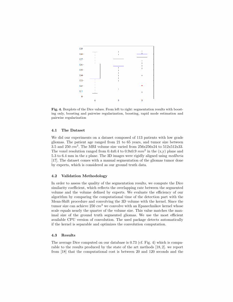
Fig. 4. Boxplots of the Dice values. From left to right: segmentation results with boost-ing only, boosting and pairwise regularization, boosting, rapid mode estimation andpairwise regularization
4.1 The Dataset
We did our experiments on a dataset composed of 113 patients with low gradegliomas. The patient age ranged from 21 to 65 years, and tumor size between3.5 and 250 cm3. The MRI volume size varied from 256x256x24 to 512x512x33.The voxel resolution ranged from 0.4x0.4 to 0.9x0.9 mm2 in the (x,y) plane and5.3 to 6.4 mm in the z plane. The 3D images were rigidly aligned using medInria
[17]. The dataset comes with a manual segmentation of the gliomas tumor doneby experts, which is considered as our ground truth data.
4.2 Validation Methodology
In order to assess the quality of the segmentation results, we compute the Dicesimilarity coefficient, which reflects the overlapping rate between the segmentedvolume and the volume defined by experts. We evaluate the efficiency of ouralgorithm by comparing the computational time of the detection part with theMean-Shift procedure and convolving the 3D volume with the kernel. Since thetumor size can achieve 250 cm3 we convolve with an Epanechnikov kernel whosescale equals nearly the quarter of the volume size. This value matches the max-imal size of the ground truth segmented gliomas. We use the most efficientavailable CPU version of convolution. The used package detects automaticallyif the kernel is separable and optimizes the convolution computation.
4.3 Results
The average Dice computed on our database is 0.73 (cf. Fig. 4) which is compa-rable to the results produced by the state of the art methods [18, 2]. we reportfrom [18] that the computational cost is between 20 and 120 seconds and the

average DICE coefficient is 0.77. Our average computational time is 19 seconds.Mode estimation is a principal ingredient of the proposed method, as the re-sults become less accurate if we only use either adaboost classifier or graph-cuts(cf. Fig. 4, Fig. 4). We compare the computational time between our work, astandard implementation of Mean Shift and an exhaustive search over volumelocations after evaluating KDE in all locations cf. Table 1. We run the algorithmson a 4-core Intel Xeon computer which frequence is 2.67GHz. We use, though,a single core in the computation.
5 Discussion
While our method was inspired from previous work [14, 11], we recall that[14]does not use Branch-and-Bound and provides a technique for the efficient com-putation of a KDE score everywhere, on all ‘domain points’. Similarly, the multi-pole method [19] evaluates a KDE on all candidate locations, and is thus linearin the number of points. The aforementioned methods are excellent for KDEevaluation- but for mode estimation they perform an ‘overkill’, since they ex-actly evaluate the score everywhere, while we only want the location of the max-imum. Instead our technique searches directly for the maximum location, andeffectively ‘ignores’ less promising locations. In particular we discard chunks ofpoints quickly by using cheaply computable upper bounds to their score. Thisallows us to work in time sublinear (practically logarithmic) in the number ofpossible locations. This is crucial for 3D medical data, where increasing the res-olution by a factor of 2 will result in an 8-fold slowdown for Multipole/DualTrees, but will require only 3 more iterations for our method (cf. Table. 1).
To the best of our knowledge, branch and bound has not been used beforefor mode estimation of KDEs. It was used in [11] for Object Detection, but witha different score function. We thus expect that our work will be of interest toother researchers working on mode estimation.
6 Conclusion
In this paper, we have presented a Branch-and-Bound based method for efficientmode estimation in KDE. We used our method for brain tumor detection andsegmentation of 3D MR images. We demonstrate that our method results in a12-fold speedup over standard Mean-Shift. Our approach is more robust thanapplying graph cut on the whole volume. The largest part of the computationaltime is taken by feature computation which can easily accelerated with graphicprocessing units. Future directions include evaluating and adapting the proposedapproach to the 3D liver tumor tracking in radiation therapy where real time iscrucial.
7 Acknowledgements
This work has been funded by grant ANR-10-JCJC-0205; and by the EU ProjectMOBOT FP7-ICT-2011-600796. We would like to thank Hugues Duffau for pro-viding us with the 3D MRI brain tumor dataset.
References
1. Duffau, H., C.L.: Preferential brain locations of low-grade gliomas. Cancer 100(2004) 2622–2626.
2. Parisot, S., Duffau, H., Chemouny, S., Paragios, N.: Graph-based detection, seg-mentation & characterization of brain tumors. In: CVPR. (2012) 988–995.
3. Comaniciu, D., Meer, P.: Mean shift: a robust approach toward feature spaceanalysis. IEEE Trans. Pattern Anal. Mach. Intell. 24(5) (2002) 603 –619.
4. van Walsum, T., Schaap, M., Metz1, C.T., van der Giessen, A.G., Niessen, W.J.:Averaging centerlines mean shift on paths. (MICCAI 2008.)
5. Prima, D.G.L.S., Collins, D.L., Arnold, D.L., Morrissey, S.P., Barillot, C.: Com-bining robust expectation maximization and mean shift algorithms for multiplesclerosis brain segmentation. (MICCAI workshop on Medical Image Analysis onMultiple Sclerosis 2008.)
6. Mayer, A., Greenspan, H.: An adaptive mean-shift framework for mri brain seg-mentation. IEEE TRANSACTIONS ON MEDICAL IMAGING (2009.)
7. Sheikh, Y.A., E.Khan, Kanade, T.: Mode-seeking by medoidshifts. In: ICCV 2007.(Number 141.)
8. Vedaldi, A., Soatto, S.: Quick shift and kernel methods for mode seeking. (In:ECCV 2008.)
9. Yang, C., Duraiswami, R., Gumerov, N.A., Davis, L.S.: Improved fast gauss trans-form and efficient kernel density estimation. In: ICCV. (2003) 464–471.
10. Wang, P., Lee, D., Gray, A.G., Rehg, J.M.: Fast mean shift with accurate andstable convergence. Journal of Machine Learning Research - Proceedings Track 2
(2007) 604–611.11. Kokkinos, I.: Rapid deformable object detection using dual-tree branch-and-bound.
In Shawe-Taylor, J., Zemel, R., Bartlett, P., Pereira, F., Weinberger, K., eds.: NIPS.(2011) 2681–2689.
12. Birkbeck, N., Cobzas, D., Jagersand, M., Murtha, A., Kesztyues, T.: An interactivegraph cut method for brain tumor segmentation. In: WACV. (2009) 1–7.
13. Scott, D.: Multivariate density estimation: theory, practice, and visualization.Wiley series in probability and mathematical statistics: Applied probability andstatistics. (Wiley. 1992.)
14. Gray, A.G.: Nonparametric density estimation: toward computational tractability.(In: In SIAM International Conference on Data Mining 2003.)
15. Boykov, Y., Kolmogorov, V.: An experimental comparison of min-cut/max-flowalgorithms for energy minimization in vision. IEEE Trans. Pattern Anal. Mach.Intell. 26 (2004) 1124–1137
16. Kolmogorov, V., Zabih, R.: What energy functions can be minimized via graphcuts. PAMI 26 (2004) 65–81.
17. Toussaint, N., Souplet, J.C., Fillard, P.: Medinria: Medical image navigation andresearch tool by inria. (In Proc. of MICCAI’07 Workshop on Interaction in medicalimage analysis and visualization (2007).)
18. Bauer, S., Nolte, L.P., Reyes, M.: Fully automatic segmentation of brain tumorimages using support vector machine classification in combination with hierar-chical conditional random field regularization. In: MICCAI. MICCAI’11, Berlin,Heidelberg, Springer-Verlag (2011) 354–361.
19. Engheta, N., Murphy, W.D., Rokhlin, V., Vassiliou, M.: The fast multipole methodfor electromagnetic scattering computation. IEEE Transactions on Antennas andPropagation 40 (1985) 634–641.
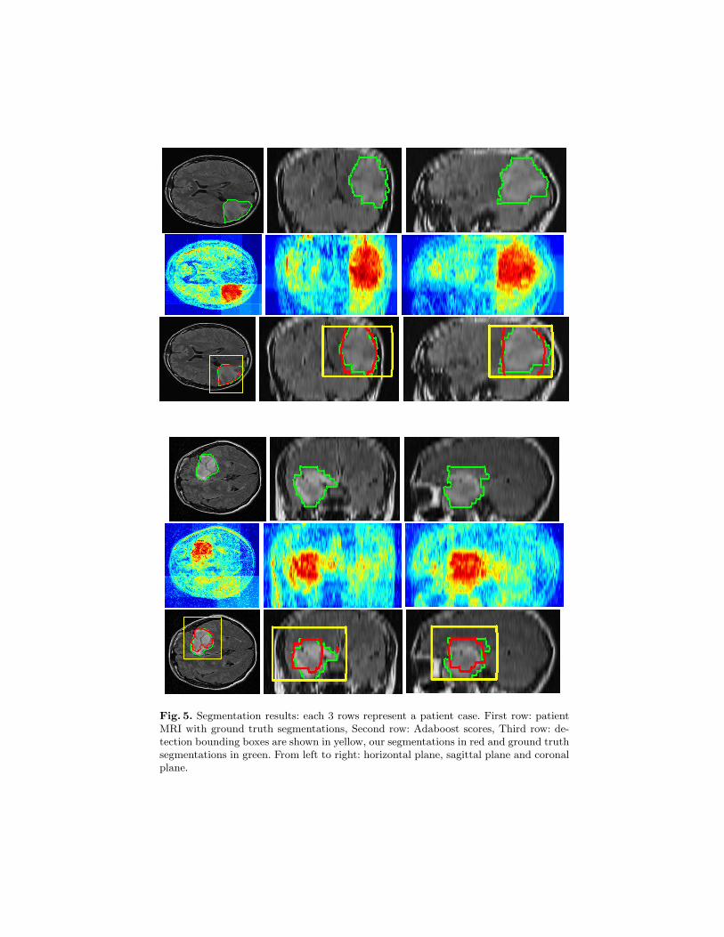
Fig. 5. Segmentation results: each 3 rows represent a patient case. First row: patientMRI with ground truth segmentations, Second row: Adaboost scores, Third row: de-tection bounding boxes are shown in yellow, our segmentations in red and ground truthsegmentations in green. From left to right: horizontal plane, sagittal plane and coronalplane.


