Embed Size (px)
Citation preview

Hum Genet (1992) 88 : 457-462
�9 Springer-Verlag 1992
Rapid generation of chromosome-specific alphoid DNA probes using the polymerase chain reaction lan Dunham 1., Christoph Lengauer 2, Thomas Cremer 2, and Terry Featherstone 1
1 Department of Genetics, Howard Hughes Medical Institute, Washington University School of Medicine, St. Louis, MO 63110, USA 2Institut fOr Humangenetik und Anthropologie der Universit~it, Im Neuenheimer Feld 328, W-6900 Heidelberg, Federal Republic of Germany
Received April 16, 1991 / Revised August 29, 1991
Summary. Non-isotopic in situ hybridization of chromo- some-specific alphoid D N A probes has become a potent tool in the study of numerical aberrations of specific human chromosomes at all stages of the cell cycle. In this paper, we describe approaches for the rapid generation of such probes using the polymerase chain reaction (PCR), and demonstrate their chromosome specificity by fluo- rescence in situ hybridization to normal human meta- phase spreads and interphase nuclei. Oligonucleotide primers for conserved regions of the alpha satellite mono- mer were used to generate chromosome-specific D N A probes from somatic hybrid cells containing various hu- man chromosomes, and from D N A libraries from sorted human chromosomes. Oligonucleotide primers for chro- mosome-specific regions of the alpha satellite monomer were used to generate specific D N A probes for the peri- centromeric heterochromatin of human chromosomes 1, 6, 7, 17 and X directly from human genomic DNA.
Introduction
Alpha satellite D N A is a primate-specific family of tan- demly repeated sequences present in the centromeric re- gions of all human chromosomes (Manuelidis 1978; Ro- senberg et al. 1978; Willard 1985). The basic unit is a monomer repeat of approximately 170 bp. In addition to regions that are strongly conserved among the different chromosomes, this monomer also contains variable re- gions. Blocks of consecutive monomers comprise a higher- order repeat of up to several kilobases in size. Specific multimeric higher-order repeat units have been described for most human chromosomes (for review see Willard and Waye 1987a).
In situ hybridization of alphoid probes cloned from conserved regions of the alphoid monomer has been applied to pinpoint centromeric regions (Mitchell et al.
* Present address: Paediatric Research Unit, The Prince Philip Research Laboratories, Guy's Tower, London Bridge SE1 9RT, UK Offprint requests to: T. Cremer
1985). In clinical cytogenetics, chromosome-specific al- phoid probes have become an important aid for the de- tection of specific numerical chromosome aberrations and for the definition of marker chromosomes (Cremer et al. 1986, 1988b; Hopman et al. 1989; Anastasi et al. 1990; Poddighe et al. 1991). Recently, Koch et al. (1989) have used the polymerase chain reaction (PCR) for the rapid generation of alphoid D N A probes directly f rom somatic hybrid cells containing the human chromosomes X and 11. In their experiments, oligonucleotide primers were applied that anneal to conserved regions of the al- phoid monomer.
In this study, we extend these observations and de- scribe new possibilities for the rapid generation of al- phoid D N A probes by PCR. First, in addition to the use of somatic hybrid cell lines, we demonstrate that primers for conserved regions are useful for the generation of chromosome-specific probes from D N A libraries con- structed from sorted human chromosomes. Secondly, we demonstrate that oligonucleotide primers directed to the chromosome-specific variable regions of alphoid mono- mers can be used to generate chromosome-specific al- phoid probes for in situ hybridization directly from hu- man genomic DNA.
Materials and methods
Cell lines
The human-hamster hybrid cell line C121 was established in Dr. P. Goodfellow's laboratory (Imperial Cancer Research Fund Labora- tories, London, UK) and kindly provided by Dr. P. Vogt (Univer- sity of Heidelberg, FRG). This cell line contains an apparently normal human X chromosome as the only known human chromo- some material, in practically all cells. The human-mouse cell line Rurag 6 was kindly provided by Dr. K. M. Grzesehik (University of Marburg, FRG) and contains human chromosomes 11 and 20, the long arm of human chromosome 7, and the long arm of the human X chromosome (Lengauer et al. 1990).
Chromosome preparations
Metaphase chromosome spreads were obtained from the above hybrid cell lines and from phytohemagglutinin(PHA)-stimulated

458
normal male and female human lymphocytes using standard proce- dures (Cremer et al. 1988a). Preparations were stored in 70% ethanol at 4~ until use.
with denaturation at 92~ for lmin, annealing at 65~ for 2rain, and extension at 72~ for 2 min.
DNA preparations
Genomic DNA was prepared from male human blood and from the hybrid cell lines according to Maniatis et al. (1982). The plas- mid DNA libraries from sorted human chromosomes 1, 2 and 8 were the generous gift of Dr. J. W. Gray (Livermore, USA). Lib- raries were constructed from HindIII-digested DNA cloned in the Bluescribe vector (Stratagene).
Total genomic DNA from a yeast artificial chromosome (YAC)- clone (yHPRT) with a 680-kb long human sequence containing the HPRT-gene (Imai and Olson 1990; Huxley et al. 1991) was pre- pared as described by Green and Olson (1990).
Oligonucleotide primers
The first two primers were directed to a conserved region of the 17l-bp monomer of human alpha satellite DNA (Willard and Waye 1987b) and were: ct27: 5' CAT CAC A A A GAA GTT TCT GAG AAT GCT TC 3' and ct30: 5' TGC ATT CAA CTC ACA GAG TI 'G AAC CTT CC 3'. The second group of primers were directed to chromosome-specific regions of alphoid monomers and were: (1) chromosome-l-specific primers (Willard and Waye 1987b), alE6: 5' GGC CTA TGG CAG CAG AGG ATA TAA CTG CC 3' and cdA7: 5' GTG AGT TTT CTC CCG TAT CCA ACG A A A TCC 3' (the length of the amplification product was 201 bp); (2) chromosome-6-specific primers (Jabs and Persico 1987), a6E: 5' ACT GTG GGC TTC AAT GCC GC 3' and ct6F: 5' GCC TAC GGC A G A A A A A G A AAC C 3' (the length of the amplification product was 182bp); (3) chromosome-7-specific primers (Waye et al. 1987), a7A10: 5' TTC ATT GGA ATC GCG AAT AC 3' and ~7A12: 5' CAA GAA GGC TTC A A A GCA CC 3' (the length of the amplification product was 348bp); (4) chro- mosome-17-specific primers (Waye and Willard 1986; Willard and Waye 1987b), a17Al: 5' AAT TCG TTG GAA ACG GGA TAA TTT CAG CTG 3' and cd7B2: 5' CTT CTG AGG ATG CTT CTG TCT A G A TGG C 3' (the length of the amplification pro- duct was 227bp); and (5) chromosome-X-specific primers (Waye and Willard 1985), aXC11: 5' ATT TCT TTG GAA TCG GGA ATA T I T CCA CAG 3' and aXD12: 5' CTC TCG TCT TTC TGT G A A GAT A A A G 3' (the length of the amplification pro- duct was 212bp).
PCR-conditions and probe labeling
PCR with primers o27 and 030. Samples (100ng) of male human genomic DNA, hybrid cell line DNA or chromosome-specific DNA libraries were used in 5-lal assays containing 5 laM of each of the two primers, 10 mM TRIS-HC1 (pH 8.3), 50 mM KC1, 1.5 mM MgCI2, 0.001% gelatin, 200 laM of each of the four dNTPs, and 2.5 units of Thermus aquaticus DNA polymerase (Perkin Elmer/Cetus). After an initial denaturation at 92~ for 3 rain, 30 cycles were car- ried out with denaturation at 92~ for 50 s, annealing at 64~ for 2 min and extension at 72~ for 3 rain. Amplified DNA products were biotinylated with biotin-ll-dUTP by nick translation (Langer et al. 1981).
PCR with chromosome-specific alphoid primers. Samples (500 ng) of male human genomic DNA were used in 25 lal assays containing i laM each of appropriate primer pair, 10mM TRIS-HC1 (pH 8.3), 50mM KC1, 2.5mM MgC12, 170pg/ml bovine serum albumin, 200laM each dATP, dCTP, dGTP, 1001-tM each dTTP, bio-11- dUTP, and 1.25 units of Thermus aquaticus DNA polymerase overlaid with 25 lal mineral oil. A total of 25 cycles was carried out
In situ hybridization and probe detection
In situ hybridization and washing procedures were performed es- sentially as described previously (Cremer et al. 1988b) with the fol- lowing modifications. Between 5 to 20rig biotinylated alphoid PCR-amplified DNA was applied per slide using 24 • 50 mm cover glasses. If necessary, a higher stringency was achieved by increas- ing the formamide concentration in the hybridization mixture from 50% to 70%, and by an additional washing step with 0.1 • SSC (1 x SSC = 150 mM NaC1/15 mM sodium citrate, pH 7.0) at 60~ for 7rain. For dual in situ hybridizations with the YAC-clone yHPRT (see above) and alphoid DNA specifically amplified from the X-chromosome, 100 ng of biotin-labeled DNA from the YAC- containing strain was used with approximately 20 ng biotinylated alphoid DNA, 50pg unlabeled total human DNA, 5lag salmon sperm DNA, and 50 lag total yeast DNA.
Probe detection with avidin conjugated with fluorescein-iso- thiocyanate (FITC) was carried out as described previously (Cre- mer et al. 1988b). Signals were amplified otlce according to the protocol of Pinkel et al. (1986). Chromosome preparations were counterstained with 1 I~g/ml 4,6 diamidino-2-phenylindole (DAPI) and 0.2 lag/ml propidium iodide mounted in fluroescence antifad- ing buffer ( l ing p-phenylendiamine in i ml glycerine buffer, pH 8.0). Microphotographs were taken with a Zeiss photomicroscope III equipped for epifluorescence or a Zeiss Axiophot. Agfachrom 1000 RS color slide films were used. Chromosomes were identifed by routine GTG-banding either before or after in situ hybridiza- tion (Klever et al. 1991; Gnirke et al. 1991) or by DAPI-banding applied after in situ hybridization (Cremer et al. 1988a).
Results
Fluorescence in situ hybridization with alphoid probes generated by PCR with oligonucleotide primers from alphoid consensus regions
Male h u m a n genomic D N A and genomic D N A f rom the hyb r id cell l ines R u R a g 6 and C121 were ampl i f i ed using the p r i m e r pa i r a27/a30. F igure 1 shows e x a m p l e s of in situ hybr id iza t ion expe r imen t s with b io t iny la t ed amplif i - ca t ion p roduc t s to n o r m a l male and female h u m a n me ta - phase sp reads and in t e rphase nuclei . A mpl i f i c a t i on pro- ducts of male h u m a n genomic D N A p r o d u c e d signals on all c en t romer i c regions in n o r m a l c h r o m o s o m e comple - men t s (Fig. 1 a - c ) . In cont ras t , p r o b e s o b t a i n e d by am- pl i f ica t ion of hybr id cell D N A s ta ined the c e n t r o m e r i c reg ions of specific h u m a n c h r o m o s o m e s , i .e . , the chro- m o s o m e s tha t were con ta ined in the respec t ive hybr id cell l ines. A s expec t ed , P C R of the C121-hybrid D N A y ie lded a p r o b e that l a be l e d only the X - c e n t r o m e r i c h e t e r o c h r o m a t i n (Fig. l d ) . A c c o rd ing ly , on ly one cen- t r omer i c reg ion was s ta ined in n o r m a l male l ymphocy t e nucle i (da ta not shown) , whe reas in female nuclei , two signals could be d e t e c t e d (Fig. l d ) . P C R p roduc t s f rom the R u R a g 6 hybr id cell l ine l abe l ed the c e n t r o m e r i c h e t e r o c h r o m a t i n of h u m a n c h r o m o s o m e s 7, 11 and 20 (Fig. l e , f). The X -c e n t rome r i c h e t e r o c h r o m a t i n was no t s t a ined by this p r o b e , a l though the p re sence of h u m a n X - c h r o m o s o m e ma te r i a l had prev ious ly been con f i rmed in R u R a g 6 cells using c h r o m o s o m a l in situ suppress ion hybr id iza t ion with a b a c t e r i o p h a g e D N A l ib ra ry f rom

Fig.la-f. In situ hybridization experiments of PCR-amplified probes generated from human genomic and somatic hybrid cell DNA with oligonucleotide primers ct27 and a30. Cells were counterstained with propidium iodide (Fig. 2c-e) and DAPI (see below), a In situ hybridization of a PCR-amplified probe generated from male hu- man genomic DNA. Note FITC-signals on the centromeric regions of all chromosomes in a male human lymphocyte metaphase spread. b The same metaphase spread counterstained with DAPI. c Lym- phocyte nuclei from the same experiment show numerous clusters of hybridization signals, d In situ hybridization of a PCR-amplified
probe generated from the somatic hybrid C121. Hybridization sig- nals on a normal female lymphocyte metaphase spread and an ad- jacent interphase nucleus mark the centromeric region of the two X-chromosomes. e Staining of the centromeric regions of chromo- somes 7, 11 and 20 in a normal female lymphocyte metaphase spread obtained after in situ hybridization of a PCR-amplified probe generated from Rurag 6 hybrid cell DNA. In the adjacent nucleus, five signals can be clearly detected, the sixth signal, al- though present in this nucleus, is located at another focal plane (not shown), f The same metaphase spread counterstained with DAPI

460
Fluorescence in situ hybridization with alphoid probes generated by PCR with oligonucleotide primers from chromosome-specific alphoid regions
In order to generate chromosome-specific alphoid probes of human genomic DNA by PCR, it is necessary to de- fine oligonucleotide primer pairs that hybridize to chro- mosome-specific regions of alphoid DNA. Figure 3 shows the results of fluorescent in situ hybridization experi- ments with probes generated from human genomic DNA with the chromosome-specific primer pairs for human chromosomes 1, 6, 7, 17 and X. Specific signals could be seen on the centromeric regions of the respective chromo- somes in normal human lymphocyte metaphase spreads. In the fluorescence microscope, chromosomes could gen- erally be identified by fluorescence banding after DAPI or propidium iodide staining (not shown). In addition, chromosomes were identified using G-banding as de- scribed by Gnirke et al. (1991) (see Fig. 3f). Figure 3d (insert) demonstrates the simultaneous localization of the YAC-clone yHPRT at Xq26 (Imai and Olson 1990; Huxley et al. 1991) together with the PCR-generated X- specific alphoid probe. In some experiments (not shown), cross-hybridization to other centromeres could be seen; however, the specific signal was easily identified from the considerably weaker cross-hybridization signals. This problem was readily alleviated by the addition of either unlabeled total human DNA or alphoid DNA amplified with the primer pair a27/a30 as competitor DNA. These PCR-generated chromosome-specific alphoid probes were also useful for interphase cytogenetics (see Fig. 3d, e).
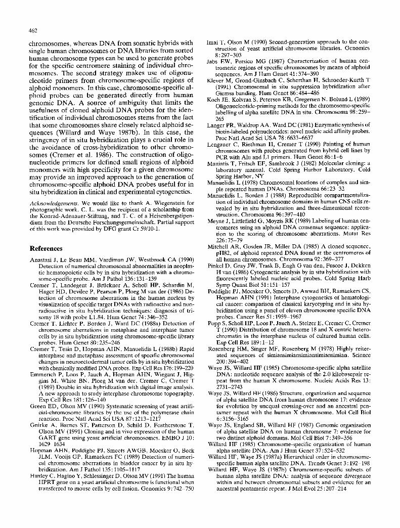
Fig. 2a-c. In situ hybridization of PCR-amplified probes gener- ated from a plasmid DNA library of sorted human chromosomes 8 with primer pair ct27/a30. Note that FITC-signals are restricted to the centromeric regions of the two chromosomes 8 in a male hu- man lymphocyte metaphase spread (a). Chromosomes were coun- terstained with propidium iodide, b Two other specifically labeled chromosomes 8 that were GTG-banded before in situ hybridiza- tion (c)
sorted human X chromosomes (Lengauer et al. 1990). Our present data suggest that the aberrant X-chromo- some contained in this line had lost X-specific alphoid sequences.
DNA prepared from plasmid DNA libraries con- structed from sorted human chromosomes 1, 2 and 8 was also used as a source for PCR with the a27/~30 primers. The resulting probes gave strong in situ hybridization signals on the respective chromosomes (see Fig. 2a, b for chromosome 8). The specificity of the hybridization sig- nals was dependent on the stringency of the in situ hy- bridization (see Methods). Under conditions of high stringency, cross-hybridization signals on centromeric regions of other chromosomes were absent or very weak, even in the absence of competitor DNA (see below).
Discussion
The definition of the centromeric regions of all chromo- somes by specific hybridization signals may become of great importance in automated chromosome analysis, e.g., in the automated evaluation of radiation-induced dicentric chromosomes (Meyne et al. 1989). Staining of the centromeric regions from individual chromosomes has made possible the evaluation of numerical chromo- some aberrations directly in the interphase nucleus, and has brought about completely new diagnostic possibilities in clinical and tumor cytogenetics (see Introduction for re- ferences). In addition, such probes have been exploited in studies of chromosome topography in interphase nu- clei, and in nuclei of terminally differentiated cells (Ma- nuelidis and Borden 1988; Emmerich et ai. 1989; Popp et al. 1990).
So far, alphoid DNA probes have been generated by cloning procedures that are both time consuming and difficult to perform in all cytogenetic laboratories. PCR provides a rapid and easy alternative for the generation of alphoid DNA probes. In this study, two strategies have been followed. The first strategy makes use of oligonucleotide primers for conserved regions of the alpha satellite monomer (Koch et al. 1989). In this ap- proach, the specificity of PCR-generated alphoid probes varies with the source of the DNA used for amplifica- tion. Amplification of human genomic DNA provides probes that hybridize to the centromeric regions of all

Fig. 3a-L Complete metaphase spreads from normal PHA-stimu- lated human lymphocytes after in situ hybridization with PCR- amplified biotinylated alphoid DNA probes specific for chromo- some 1 (a), chromosome 6 (b), chromosome 7 (e), chromosome X (d) and chromosome 17 (e). Probes were generated from total human genomic DNA with oligonucleotide primers directed to chromosome-specific variable regions of alphoid monomers of the respective chromosomes. Metaphase spreads were counterstained with propidium iodide (a-e) and DAPI (not shown). For chromo- some identification, G-banding was performed after in situ hybrid-
ization (f). A r r o w s indicate specific labeling of the centromeric heterochromatin a of both chromosomes 1, b of both chromo- somes 6, c of both chromosomes 7, d of the X chromosome in a male cell. Note also a single signal in the accompanying interphase nucleus. Insert. Simultaneous hybridization of the X-centromere- specific probe with YAC-clone yHPRT. The latter clone yields ad- ditional signals on both chromatids in Xq26 (banding not shown). e A r r o w s indicate specific labeling of the centromeric hetero- chromatin of both chromosomes 17. f Metaphase spread shown in e after G-banding
462
c h r o m o s o m e s , whe rea s D N A f rom somat ic hybr ids with single h u m a n c h r o m o s o m e s or D N A l ibrar ies f rom sor ted h u m a n c h r o m o s o m e types can be used to genera te p robes for the specif ic c e n t r o m e r e s ta ining of ind iv idua l chro- m o s o m e s . The s econd s t ra tegy m a k e s use of o l igonu- c l eo t ide p r ime r s f rom c h r o m o s o m e - s p e c i f i c reg ions of a lpho id m o n o m e r s . In this case, ch romosome-spec i f i c al- p h o i d p r o b e s can be g e n e r a t e d d i rec t ly f rom h u m a n g e n o m i c D N A . A source o f amb igu i ty tha t l imits the usefu lness of c loned a lpho id D N A p r o b e s for the iden- t i f ica t ion of ind iv idua l c h r o m o s o m e s s tems f rom the fact tha t some c h r o m o s o m e s share c losely r e l a t ed a lpho id se- quences (Wi l l a rd and W a y e 1987b). In this case, the s t r ingency of in situ hyb r id i za t i on p lays a crucial role in the avo idance of c ros s -hybr id i za t ion to o t h e r ch romo- somes ( C r e m e r et al. 1986). T h e cons t ruc t ion of ol igo- nuc l eo t ide p r ime r s for de f i ned smal l reg ions of a lpho id m o n o m e r s wi th high specif ic i ty for a g iven c h r o m o s o m e m a y p r o v i d e an i m p r o v e d a p p r o a c h to the gene ra t i on of c h r o m o s o m e - s p e c i f i c a l pho id D N A p r o b e s useful for in situ hybridizat ion in clinical and exper imenta l cytogenetics.
Acknowledgements. We would like to thank A. Wiegenstein for photographic work. C. L. was the recipient of a scholarship from the Konrad-Adenauer-Stiftung, and T. C. of a Heisenbergstipen- dium from the Deutsche Forschungsgemeinschaft. Partial support of this work was provided by DFG grant Cr 59110-1.
References
Anastasi J, Le Beau MM, Vardiman JW, Westbrook CA (1990) Detection of numerical chromosomal abnormalities in neoplas- tic hematopoietic cells by in situ hybridization with a chromo- some-specific probe. Am J Pathol 136:131-139
Cremer T, Landegent J, Briickner A, Scholl HP, Schardin M, Hager HD, Devilee P, Pearson P, Ploeg M van der (1986) De- tection of chromosome aberrations in the human nucleus by visualization of specific target DNAs with radioactive and non- radioactive in situ hybridization techniques: diagnosis of tri- somy 18 with probe L1.84. Hum Genet 74:346-352
Cremer T, Lichter P, Borden J, Ward DC (1988a) Detection of chromosome aberrations in metaphase and interphase tumor cells by in situ hybridization using chromosome-specific library probes. Hum Genet 80 : 235-246
Cremer T, Tesin D, Hopman AHN, Manuelidis L (1988b) Rapid interphase and metaphase assessment of specific chromosomal changes in neuroectodermal tumor cells by in situ hybridization with chemically modified DNA probes. Exp Cell Res 176:199-220
Emmerich P, Loos P, Jauch A, Hopman AHN, Wiegant J, Hig- gins M, White BN, Ploeg M van der, Cremer C, Cremer T (1989) Double in situ hybridization with digital image analysis. A new approach to study interphase chromosome topography. Exp Cell Res 181 : 126-140
Green ED, Olson MV (1990) Systematic screening of yeast artifi- cial-chromosome libraries by the use of the polymerase chain reaction. Proc Natl Acad Sci USA 87 : 1213-1217
Gnirke A, Barnes ST, Patterson D, Schild D, Featherstone T, Olson MV (1991) Cloning and in vivo expression of the human GART gene using yeast artificial chromosomes. EMBO J 10: 1629-1634
Hopman AHN, Poddighe PJ, Smeets AWGB, Moesker O, Beck JLM, Vooijs GP, Ramaekers FC (1989) Detection of numeri- cal chromosome aberrations in bladder cancer by in situ hy- bridization. Am J Pathol 135 : 1105-1117
Huxley C, Hagino Y, Schlessinger D, Olson MV (1991) The human HPRT gene on a yeast artificial chromosome is functional when transferred to mouse cells by cell fusion. Genomics 9 : 742-750
Imai T, Olson M (1990) Second-generation approach to the con- struction of yeast artificial chromosome libraries. Genomics 8 : 297-303
Jabs EW, Persico MG (1987) Characterization of human cen- tromeric regions of specific chromosomes by means of alphoid sequences. Am J Hum Genet 41 : 374-390
Klever M, Grond-Ginsbach C, Scherthan H, Schroeder-Kurth T (1991) Chromosomal in situ suppression hybridization after Giemsa banding. Hum Genet 86: 484-486
Koch JE, Kolvraa S, Petersen KB, Gregersen N, Bolund L (1989) Oligonucleotide-priming methods for the chromosome-specific labelling of alpha satellite DNA in situ. Chromosoma 98 : 259- 265
Langer PR, Waldrop AA, Ward DC (1981) Enzymatic synthesis of biotin-labeled polynucleotides: novel nucleic acid affinity probes. Proc Natl Acad Sci USA 78 : 6633-6637
Lengauer C, Riethman H, Cremer T (1990) Painting of human chromosomes with probes generated from hybrid cell lines by PCR with Alu and L1 primers. Hum Genet 86:1-6
Maniatis T, Fritsch EF, Sambrook J (1982) Molecular cloning: a laboratory manual. Cold Spring Harbor Laboratory, Cold Spring Harbor, NY
Manuelidis L (1978) Chromosomal locations of complex and sim- ple repeated human DNAs. Chromosoma 66: 23-32
Manuelidis L, Borden J (1988) Reproducible compartmentaliza- tion of individual chromosome domains in human CNS cells re- vealed by in situ hybridization and three-dimensional recon- struction. Chromosoma 96 : 397-410
Meyne J, Littlefield G, Moyzis RK (1989) Labeling of human cen- tromeres using an alphoid DNA consensus sequence: applica- tion to the scoring of chromosome aberrations. Mutat Res 226 : 75-79
Mitchell AR, Gosden JR, Miller DA (1985) A cloned sequence, pH82, of alphoid repeated DNA found at the centromeres of all human chromosomes. Chromosoma 92 : 369-377
Pinkel D, Gray JW, Trask B, Engh G van den, Fuscoe J, Dekken H van (1986) Cytogenetic analysis by in situ hybridization with fluorescently labeled nucleic acid probes. Cold Spring Harb Symp Quant Biol 51 : 151-157
Poddighe P J, Moesker O, Smeets D, Awwad BH, Ramaekers CS, Hopman AHN (1991) Interphase cytogenetics of hematologi- cal cancer: comparison of classical karyotyping and in situ hy- bridization using a panel of eleven chromosome specific DNA probes. Cancer Res 51 : 1959-1967
Popp S, Scholl HP, Loos P, Jauch A, Stelzer E, Cremer C, Cremer T (1990) Distribution of chromosome 18 and X centric hetero- chromatin in the interphase nucleus of cultured human cells. Exp Cell Res 189:1-12
Rosenberg HM, Singer MF, Rosenberg M (1978) Highly reiter- ated sequences of simiansimiansimiansimiansimian. Science 200 : 394-402
Waye JS, Willard HF (1985) Chromosome-specific alpha satellite DNA: nucleotide sequence analysis of the 2.0 kilobasepair re- peat from the human X chromosome. Nucleic Acids Res 13: 2731-2743
Waye JS, Willard HF (1986) Structure, organization and sequence of alpha satellite DNA from human chromosome 17: evidence for evolution by unequal crossing-over and an ancestral pen- tamer repeat with the human X chromosome. Mol Cell Biol 6:3156-3165
Waye JS, England SB, Willard HF (1987) Genomic organization of alpha satellite DNA on human chromosme 7: evidence for two distinct alphoid domains. Mol Cell Biol 7 : 349-356
Willard HF (1985) Chromosome-specific organization of human alpha satellite DNA. Am J Hum Genet 37 : 524-532
Willard HF, Waye JS (1987a) Hierarchical order in chromosome- specific human alpha satellite DNA. Trends Genet 3 : 192-198
Willard HF, Waye JS (1987b) Chromosome-specific subsets of human alpha satellite DNA: analysis of sequence divergence within and between chromosomal subsets and evidence for an ancestral pentameric repeat. J Mol Evol 25 : 207-214