Embed Size (px)
Citation preview
Vol. 30, No. 3JOURNAL OF CLINICAL MICROBIOLOGY, Mar. 1992, p. 540-5440095-1137/92/030540-05$02.00/0Copyright © 1992, American Society for Microbiology
Rapid Detection of Respiratory Viruses by Shell Vial Cultureand Direct Staining by Using Pooled and Individual
Monoclonal AntibodiesSANDRA MATTHEY,* DONALD NICHOLSON, SANDY RUHS, BETH ALDEN,
MICHELLE KNOCK, KIM SCHULTZ, AND ANGELA SCHMUECKERDepartment of Pathology, 265 MRC, The University ofIowa
Hospitals and Clinics, Iowa City, Iowa 52242
Received 5 July 1991/Accepted 2 December 1991
The Bartels respiratory virus panel detection kit is an indirect fluorescent-antibody (IFA) method that usespooled and individual antisera for tissue culture confirmation of seven respiratory viruses. We evaluated thesereagents for detecting viral antigen in shell vial cultures and by direct staining of cells from respiratoryspecimens. The isolation from 254 specimens of respiratory viruses in shell vial cultures compared withstandard tube cultures was highly sensitive (94%) and specific (97.3%). The numbers of viral isolates detectedin three consecutive years of testing with shell vial cultures were 68 of 254 (26.8%), 101 of 381 (26.5%), and122 of 430 (28.4%). IFA direct staining of all 1,065 specimens resulted in 183 (17.2%) being uninterpretablebecause of inadequate numbers of cells or interfering fluorescence. The sensitivity and specificity of theinterpretable IFA direct stains in comparison with shell vial cultures were 85.9 and 87.1%, respectively. Fordetection of 881 adequate specimens, Bartels respiratory syncytial virus IFA direct staining compared with anOrtho Diagnostics Systems direct fluorescent-antibody test for respiratory syncytial virus RSV was highlysensitive (95.5%) and specific (97%). Shell vial cultures combined with Bartels IFA reagents are a rapidalternative to standard tube cultures. Bartels IFA direct staining with individual antisera provides usefulsame-day screening of respiratory specimens, but the antiserum pool was not effective in screening for positivespecimens because of excessive amounts of nonspecific fluorescence.
Respiratory viruses cause a variety of human infections,ranging from the common cold to life-threatening pneumonia(10, 14, 15). Over 200 strains of virus can cause respiratorydisease, and the differential diagnosis of respiratory viruseshas often been considered impractical and of limited valuefor patient care. The development of clinically effectiveantiviral agents has changed this perspective and has stimu-lated the development of rapid diagnostic tests for severalviruses, with particular emphasis on respiratory syncytialvirus (RSV) and influenza A virus (1, 2, 7-9, 11-13). Themost common commercially available rapid diagnostic testsuse immunofluorescence or enzyme immunoassay methodswith monoclonal antibodies (1, 3, 7-9).
Cultivation in tissue cultures remains the standard for thedetection of most viruses, although this procedure necessi-tates days to weeks for isolation and identification. The useof centrifugation-enhanced shell vial cultures combined withfluorescein-labelled monoclonal antibodies has significantlyshortened turnaround times (2, 5, 6, 9, 12). Most shell vialtechniques and rapid test kits usually detect only one virus,but rapid diagnosis of respiratory virus infections could beenhanced by testing for several viruses simultaneously. Acommercially available fluorescent-antibody kit manufac-tured by Bartels (Bellevue, Wash.) was designed to confirmseven different viruses in tissue cultures. The kit containsindividual monoclonal antisera to adenovirus, influenza Aand B viruses, parainfluenza viruses 1, 2, and 3, RSV and anantiserum pool containing monoclonal antibodies to allseven viruses. This two-part study evaluated the effective-ness of Bartels individual and pooled reagents for detecting
* Corresponding author.
respiratory viruses directly from respiratory specimens andin shell vial tissue cultures.
MATERIALS AND METHODS
Specimens. Respiratory tract specimens from patients atthe University of Iowa Hospitals and Clinics were collectedby physicians, nurses, and respiratory therapists. Respira-tory specimens from area community hospitals were col-lected by respiratory therapists and laboratory personnel.Nasopharyngeal wash, throat wash, and tracheal aspiratespecimens were obtained by instilling normal saline into theappropriate area and aspirating the specimen into a mucustrap. Bronchial wash and broncheoalveolar lavage speci-mens were collected in sterile containers by physicians.Lung biopsy specimens were obtained by physicians andplaced in sterile containers with 10 to 15 ml of Eagle'sminimum essential medium with Earle's salts (MEM) con-taining 10% fetal bovine serum (FBS) (Sigma Chemical Co.,St. Louis, Mo.), amphotericin B, and gentamicin. Specimenswere transported on ice and processed within 1 h of receiptin the laboratory. Processed specimens which could not beinoculated into tissue cultures within 24 h were mixed withequal amounts of MEM containing 20% FBS and stored at-70°C. Seventy-nine specimens collected from November1987 through April 1988 were stored for up to 12 months andtested along with 175 specimens collected from November1988 through April 1989. Five of these 175 specimens werefrozen prior to inoculation into cell cultures. Three hundredeighty-one specimens were collected from November 1989through April 1990, and 60 of these were frozen prior toinoculation into cell cultures. Four hundred thirty specimenswere collected from December 1990 through April 1991, and
540
on February 17, 2019 by guest
http://jcm.asm
.org/D
ownloaded from

RAPID DETECTION OF RESPIRATORY VIRUSES 541
all were inoculated into cell cultures within 5 h of receipt inthe laboratory.Specimen processing. Specimens were processed as fol-
lows. (i) Nasopharyngeal wash, throat wash, and trachealaspirate specimens were split into equal parts for culturingand direct staining. Specimens for culturing were brought toa 2.0-ml volume with phosphate-buffered saline (PBS), ster-ile glass beads were added, and the mixture was vortexed for30 to 60 s. The specimens were centrifuged at 1,200 x g for10 min, and the supernatant fluid was used to inoculate tissuecultures. Specimens for direct staining were also brought to2.0 ml with PBS and vortexed, and 6.0 ml ofPBS was added.The specimens were centrifuged at 400 x g for 10 min, thesupernatant fluid was decanted, and the wash step wasrepeated one or two more times to remove excess mucus.The cell pellet was resuspended in 0.5 ml of PBS, andapproximately 50 ,ul of the cell suspension was dotted ontotwo-well and eight-well acetone-cleaned slides (Baxter Sci-entific Products, McGaw Park, Ill.). (ii) Bronchial wash andbronchoalveolar lavage specimens were inoculated into tis-sue cultures and dotted onto two-well and eight-well slideswithout additional processing. (iii) Lung biopsy specimenswere divided with sterile scissors, and the freshly cut surfacewas touched directly onto two-well and eight-well slides.The specimens were homogenized to a 5 to 10% suspensionin MEM containing 10% FBS, amphotericin B, and genta-micin in a stomacher blender (Tekmar Co., Cincinnati,Ohio). The suspensions were centrifuged at 1,500 x g for 10min, and the supernatant fluid was filtered through a 0.45-,um-pore-size filter (Millipore Corp., Bedford, Mass.). Thefiltered fluid was used to inoculate cell cultures.
Slides for direct staining were air dried, fixed in acetone,and stored at -70°C when staining was not done within 2 h.
Direct staining. Direct staining of respiratory tract cellsfrom patient specimens was done by indirect fluorescent-antibody (IFA) staining with reagents provided by BartelsImmunodiagnostic Supplies. The two-well slide was stainedwith 50 [lI of the antiserum pool containing monoclonalantibodies to adenovirus, influenza A and B viruses, parain-fluenza viruses 1, 2, and 3, and RSV. The eight-well slidewas stained with approximately 50 RI of individual antiserafor each of the seven viruses. A negative control wasincluded on both the two-well and the eight-well slides. Theslides were incubated for 30 min at 36°C, washed in BartelsPBS for 5 min, and blotted dry, and fluorescein-labelledanti-mouse conjugate was added to each well. The slideswere incubated for 30 min at 36°C, washed in Bartels PBS for5 min, and air dried, and mounting medium and coverslipswere added.
Direct fluorescent-monoclonal-antibody staining for RSV(Ortho RSV staining; Ortho Diagnostics Systems, Raritan,N.J.) was performed on a two-well slide prepared for eachrespiratory specimen. Approximately 50 RI of reagent wasplaced in each well, the slides were incubated at roomtemperature for 30 min, rinsed with PBS, and air dried, andmounting fluid and coverslips were added.
Slides were examined at x100 and x400 magnificationswith an Olympus epifluorescence microscope (Leeds Preci-sion Instruments, Minneapolis, Minn.). Positive direct stainshad two or more fluorescent columnar epithelial cells perwell, in accordance with the manufacturer's specificationsfor both the Bartels IFA and the Ortho RSV procedures.Fluorescence was nuclear, cytoplasmic, or both, dependingon the specific reagent. Slides were considered inadequatewhen there were fewer than 30 intact cells per well and nopositive cells were visualized. Preparations were considered
uninterpretable when nonspecific fluorescence obscured thecell layer of any well or when more than one well on theeight-well slide had specific fluorescence. Inadequate oruninterpretable eight-well slide IFA direct stains were testedagain for the last 430 specimens only. Repeat testing wasdone with the same cell suspensions stored at 4°C. Anadditional wash step was performed with some specimens inan attempt to eliminate nonspecific fluorescence due to thepresence of mucus.Tube culturing. Tube culturing was done for the first 254
specimens only. Rhesus monkey kidney (RMK) and A549tissue culture cells were provided by Bartels Immunodiag-nostic Supplies. The maintenance medium was aspiratedfrom each tube, and the cell monolayers were washed withPBS containing calcium and magnesium. One tube each ofRMK and A549 cells was inoculated with 0.2 ml of speci-men, incubated at 36°C for 30 to 60 min to allow viraladsorption, fed with 2.0 ml of MEM containing 2% FBS,amphotericin B, and penicillin-streptomycin, and incubatedin stationary racks at 36°C. The culture tubes were examineddaily, and slides were prepared when cytopathic effects wereevident or at 7 to 10 days in the absence of cytopathiceffects.
Shell vial culturing. RMK and A549 shell vial tissuecultures were provided by Bartels Immunodiagnostic Sup-plies for the first 254 specimens and purchased from Bartelsor Whittaker Bioproducts (Walkersville, Md.) for the re-maining specimens. Shell vials were prepared in the labora-tory from RMK and A549 tube cultures purchased fromWhittaker Bioproducts when commercial shell vials wereunavailable. Shell vials were prepared by washing confluenttube monolayers with PBS and treating the cells with 0.25%trypsin-EDTA. The cells were suspended in and dilutedapproximately 1:4 with MEM containing 10% FBS, ampho-tericin B, and penicillin-streptomycin. One milliliter of thecell suspension was seeded into sterile shell vials (OrthoDiagnostics Systems), incubated at 36°C in a 5% CO2 atmo-sphere, and used for viral cultures after 2 to 6 days ofincubation.The medium was removed from the shell vials, and the cell
monolayers were washed with PBS containing calcium andmagnesium. One shell vial each of RMK and A549 cells wasinoculated with 0.2 ml of specimen. The vials were centri-fuged at 700 x g for 30 min at room temperature, fed with 1.0ml of MEM containing 2% FBS, amphotericin B, andpenicillin-streptomycin, and incubated at 36°C in a 5% CO2atmosphere. Slides were prepared after 48 to 72 h of incu-bation.
Culture staining. One two-well slide and two eight-wellslides were prepared from each tube and shell vial culture bywashing the monolayers with PBS and scraping the cells into0.5 ml of PBS. Approximately 50 pAl of the cell suspensionwas dotted onto each well of the two-well and eight-wellslides. The slides were air dried and fixed in acetone. Slidesthat could not be stained within 2 h were stored at -70°C.The two-well slides were stained with Bartels IFA antise-
rum pool. The RMK cell eight-well slides were stained withBartels IFA individual reagents as described for directstaining. The A549 cell eight-well slides were stained onlywhen the A549 cell two-well slides were positive and theRMK slides were negative.The second set of eight-well slides prepared from 80 tube
cultures and 64 shell vial cultures were stained with reagentsfrom the Centers for Disease Control (CDC), Atlanta, Ga.,to confirm Bartels staining results. The CDC reagents wereindividual monoclonal antibodies to adenovirus, influenza A
VOL. 30, 1992
on February 17, 2019 by guest
http://jcm.asm
.org/D
ownloaded from
542 MATTHEY ET AL.
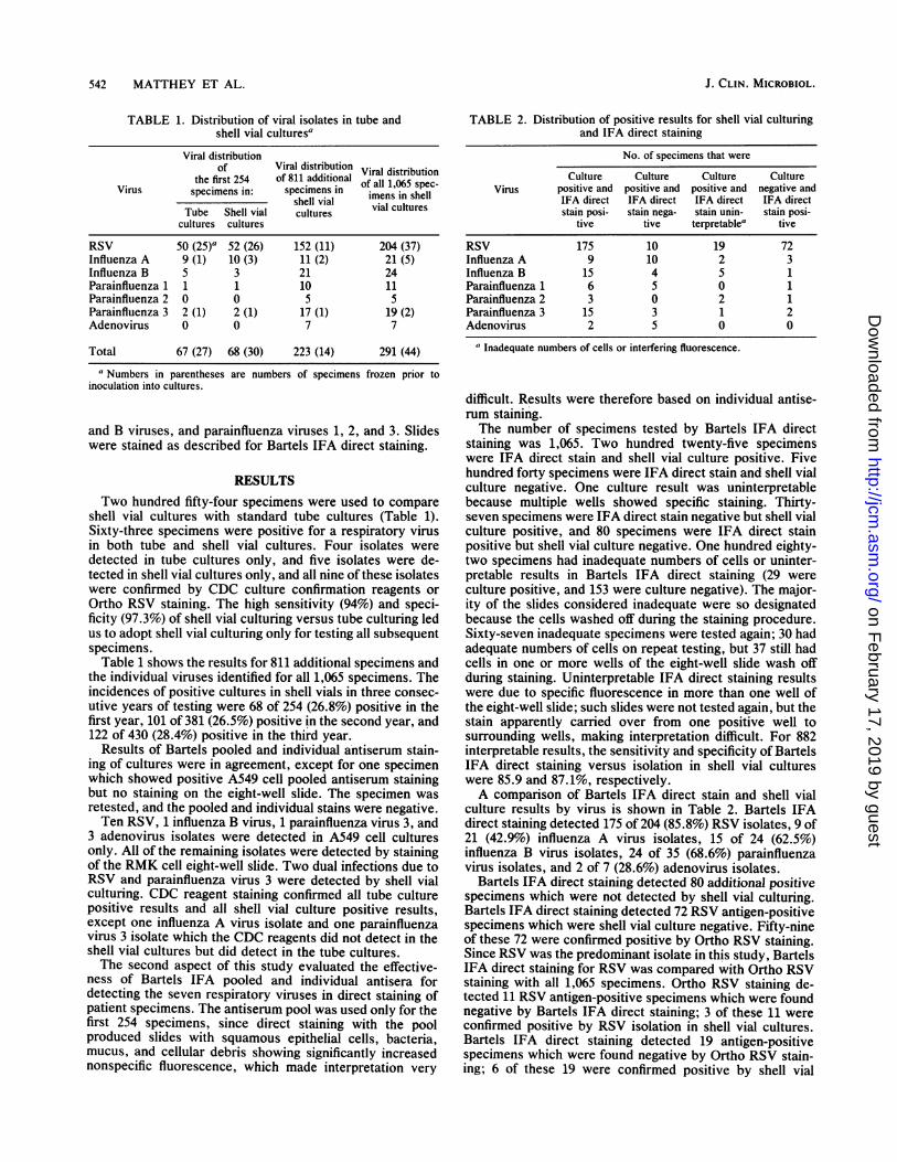
TABLE 1. Distribution of viral isolates in tube andshell vial cultures'
Viral distributionof Viral distribution Viral distribution
the first 254 of 811 additional of all 1,065 spec-Virus specimens in: specimens in imens in shellshell vial vial cultures
Tube Shell vial culturescultures cultures
RSV 50 (25)a 52 (26) 152 (11) 204 (37)Influenza A 9 (1) 10 (3) 11 (2) 21 (5)Influenza B 5 3 21 24Parainfluenza 1 1 1 10 11Parainfluenza 2 0 0 5 5Parainfluenza 3 2 (1) 2 (1) 17 (1) 19 (2)Adenovirus 0 0 7 7
Total 67 (27) 68 (30) 223 (14) 291 (44)
a Numbers in parentheses are numbers of specimens frozen prior toinoculation into cultures.
and B viruses, and parainfluenza viruses 1, 2, and 3. Slideswere stained as described for Bartels IFA direct staining.
RESULTSTwo hundred fifty-four specimens were used to compare
shell vial cultures with standard tube cultures (Table 1).Sixty-three specimens were positive for a respiratory virusin both tube and shell vial cultures. Four isolates weredetected in tube cultures only, and five isolates were de-tected in shell vial cultures only, and all nine of these isolateswere confirmed by CDC culture confirmation reagents orOrtho RSV staining. The high sensitivity (94%) and speci-ficity (97.3%) of shell vial culturing versus tube culturing ledus to adopt shell vial culturing only for testing all subsequentspecimens.Table 1 shows the results for 811 additional specimens and
the individual viruses identified for all 1,065 specimens. Theincidences of positive cultures in shell vials in three consec-utive years of testing were 68 of 254 (26.8%) positive in thefirst year, 101 of 381 (26.5%) positive in the second year, and122 of 430 (28.4%) positive in the third year.
Results of Bartels pooled and individual antiserum stain-ing of cultures were in agreement, except for one specimenwhich showed positive A549 cell pooled antiserum stainingbut no staining on the eight-well slide. The specimen wasretested, and the pooled and individual stains were negative.Ten RSV, 1 influenza B virus, 1 parainfluenza virus 3, and
3 adenovirus isolates were detected in A549 cell culturesonly. All of the remaining isolates were detected by stainingof the RMK cell eight-well slide. Two dual infections due toRSV and parainfluenza virus 3 were detected by shell vialculturing. CDC reagent staining confirmed all tube culturepositive results and all shell vial culture positive results,except one influenza A virus isolate and one parainfluenzavirus 3 isolate which the CDC reagents did not detect in theshell vial cultures but did detect in the tube cultures.The second aspect of this study evaluated the effective-
ness of Bartels IFA pooled and individual antisera fordetecting the seven respiratory viruses in direct staining ofpatient specimens. The antiserum pool was used only for thefirst 254 specimens, since direct staining with the poolproduced slides with squamous epithelial cells, bacteria,mucus, and cellular debris showing significantly increasednonspecific fluorescence, which made interpretation very
TABLE 2. Distribution of positive results for shell vial culturingand IFA direct staining
No. of specimens that were
Culture Culture Culture CultureVirus positive and positive and positive and negative and
IFA direct IFA direct IFA direct IFA directstain posi- stain nega- stain unin- stain posi-
tive tive terpretable" tive
RSV 175 10 19 72Influenza A 9 10 2 3Influenza B 15 4 5 1Parainfluenza 1 6 5 0 1Parainfluenza 2 3 0 2 1Parainfluenza 3 15 3 1 2Adenovirus 2 5 0 0
a Inadequate numbers of cells or interfering fluorescence.
difficult. Results were therefore based on individual antise-rum staining.The number of specimens tested by Bartels IFA direct
staining was 1,065. Two hundred twenty-five specimenswere IFA direct stain and shell vial culture positive. Fivehundred forty specimens were IFA direct stain and shell vialculture negative. One culture result was uninterpretablebecause multiple wells showed specific staining. Thirty-seven specimens were IFA direct stain negative but shell vialculture positive, and 80 specimens were IFA direct stainpositive but shell vial culture negative. One hundred eighty-two specimens had inadequate numbers of cells or uninter-pretable results in Bartels IFA direct staining (29 wereculture positive, and 153 were culture negative). The major-ity of the slides considered inadequate were so designatedbecause the cells washed off during the staining procedure.Sixty-seven inadequate specimens were tested again; 30 hadadequate numbers of cells on repeat testing, but 37 still hadcells in one or more wells of the eight-well slide wash offduring staining. Uninterpretable IFA direct staining resultswere due to specific fluorescence in more than one well ofthe eight-well slide; such slides were not tested again, but thestain apparently carried over from one positive well tosurrounding wells, making interpretation difficult. For 882interpretable results, the sensitivity and specificity of BartelsIFA direct staining versus isolation in shell vial cultureswere 85.9 and 87.1%, respectively.A comparison of Bartels IFA direct stain and shell vial
culture results by virus is shown in Table 2. Bartels IFAdirect staining detected 175 of 204 (85.8%) RSV isolates, 9 of21 (42.9%) influenza A virus isolates, 15 of 24 (62.5%)influenza B virus isolates, 24 of 35 (68.6%) parainfluenzavirus isolates, and 2 of 7 (28.6%) adenovirus isolates.
Bartels IFA direct staining detected 80 additional positivespecimens which were not detected by shell vial culturing.Bartels IFA direct staining detected 72 RSV antigen-positivespecimens which were shell vial culture negative. Fifty-nineof these 72 were confirmed positive by Ortho RSV staining.Since RSV was the predominant isolate in this study, BartelsIFA direct staining for RSV was compared with Ortho RSVstaining with all 1,065 specimens. Ortho RSV staining de-tected 11 RSV antigen-positive specimens which were foundnegative by Bartels IFA direct staining; 3 of these 11 wereconfirmed positive by RSV isolation in shell vial cultures.Bartels IFA direct staining detected 19 antigen-positivespecimens which were found negative by Ortho RSV stain-ing; 6 of these 19 were confirmed positive by shell vial
J. CLIN. MICROBIOL.
on February 17, 2019 by guest
http://jcm.asm
.org/D
ownloaded from
RAPID DETECTION OF RESPIRATORY VIRUSES 543
culturing. Seven of 618 specimens which were found nega-tive by both antigen detection methods were found positivefor RSV in shell vial cultures. One hundred eighty-twospecimens were uninterpretable by Bartels IFA direct stain-ing because of inadequate numbers of cells or interferingfluorescence, and 26 of these specimens also had inadequatenumbers of cells on the Ortho RSV stain slides. Ortho RSVstain specimens which were inadequate were generally dueto poor specimen quality and not to cells washing off theslides. Two specimens which had inadequate numbers ofcells for Ortho RSV staining were acceptable for Bartels IFAdirect staining. The sensitivity, specificity, positive predic-tive value, negative predictive value, and overall agreementof Bartels IFA direct staining versus Ortho RSV staining for881 interpretable specimens were 95.5, 97, 92.5, 98.3, and96.6%, respectively.
DISCUSSION
Rapid diagnosis of respiratory viral infections can have animpact on patient care by indicating appropriate antiviraltherapy, eliminating unnecessary antibacterial therapy, anddetermining patient isolation requirements (10, 14, 15). Thisstudy evaluated rapid diagnosis of respiratory viruses byshell vial culturing and direct staining of cells in respiratorytract specimens. Shell vial cultures combined with BartelsIFA reagents were shown to be equivalent to standard tubecultures in the first phase of this study. Shell vial cultureswere negative for four standard tube culture-positive isolatesbut detected five isolates which were not detected by stan-dard tube cultures. The significant advantage of using shellvial cultures is the decreased turnaround time, from 7 to 10days to 2 to 3 days. Technologist time is also decreased withshell vial cultures, since the vials are not examined daily forcytopathic effect development. Since both methods rely onimmunofluorescence staining for a definitive endpoint, noincreases in costs were realized in the application of shellvials and Bartels IFA reagents. The antiserum pool wasshown to be effective in screening for positive specimens inboth tube and shell vial cultures.Comparable shell vial and tube culture results in the first
group of specimens tested allowed the use of shell vials onlyfor the remaining specimens. Shell vial cultures showedessentially the same isolation rates for all 3 years of testing.As this study demonstrates, the primary respiratory virusisolated at our tertiary-care center is RSV, and the limitednumber of influenza A and B virus, parainfluenza virus, andadenovirus isolates necessitates further study to supportshell vial culturing and Bartels IFA staining for all sevenviruses.The second aspect of this study evaluated Bartels IFA
reagents for direct staining of patient specimens. The sensi-tivity of Bartels IFA direct staining with the individualreagents versus shell vial culturing was 85.9% for the ade-quate specimens. This result is consistent with those ob-tained by other direct antigen detection methods (7, 9, 11).Direct staining with the antiserum pool was not effective inscreening for positive specimens because of the problems ininterpretation caused by all the extraneous fluorescence.
Since RSV was the predominant isolate in this study, itwas of particular interest to compare Bartels IFA directstaining for RSV with Ortho RSV staining, which has beenour standard test for several years. This study showed thatthe two methods were comparable and that no single methoddetected all RSV-positive specimens. Fifty nine specimens(5.5%) were found positive by both RSV antigen detection
methods but were shell vial culture negative. These resultswere attributed to delays in the inoculation of cultures due tostaffing limitations or tissue culture cell shortages and toproblems in the delivery of specimens from outpatient clinicsor outside health care facilities. Also, nonviable RSV hasbeen detected by immunofluorescence in the late stages ofinfection (4).
In this study, the sensitivity of direct staining decreasedfrom 85.9 to 77.3% when the uninterpretable results whichwere culture positive were included. The large number ofspecimens with inadequate numbers of cells for interpreta-tion is a major problem with Bartels IFA direct staining. Theproblem is due to cells washing off one or more wells duringstaining. The washing procedure for Bartels IFA directstaining requires rinsing of the slides with a gentle stream ofPBS directed away from the cell wells and soaking of theslides in PBS for 5 min. Ortho RSV staining does not requiresoaking of the slides, and we did not experience significantproblems with cells washing off with Ortho RSV staining.However, when we tried to minimize the effects of washingwith Bartels IFA direct staining by elimination of the soakingstep or by more gentle manipulation of the PBS rinse, therewere more problems with nonspecific fluorescence and withspecific fluorescence that seemed to carry over from onepositive well into adjacent wells of the eight-well slide. Thepresence of excess mucus in a specimen also appeared tocontribute to cells washing off and to increased nonspecificfluorescence, so the majority of the last 430 specimens testedwere washed a minimum of three times prior to slidepreparation; however, the problem with cells washing off theslides persisted. Repeat testing of inadequate specimens wasless than 50% effective in preventing the cells from washingoff the slides.The need for repeating Bartels IFA direct staining and the
inability to use the antiserum pool as a screening testbecause of interpretation difficulties proved extremelycostly. The Bartels kit has sufficient antisera to screen 100specimens with the antiserum pool but only enough individ-ual reagents to stain 25 slides. Eliminating the pooled anti-serum screen quadruples the cost to almost $32 per test,since only 25 tests per kit can be completed instead of 100tests. The cost of Bartels IFA direct staining would have tobe weighed against the benefits of having same-day testresults for the seven respiratory viruses. We believe thatBartels IFA direct staining could be an extremely effectivesame-day test if the problems with inadequate numbers ofcells could be eliminated, and efforts will be made toovercome these problems.Even with the problems encountered, IFA direct staining
provides a useful screen for our laboratory, which serves a904-bed tertiary-care hospital with a large number of immu-nocompromised pediatric and adult patients. Our protocolfor respiratory virus testing, based on the viral prevalenceand test performance results of this study, will be to screenspecimens for RSV (Ortho RSV staining) and then test allOrtho RSV stain-negative specimens by Bartels IFA individ-ual reagent direct staining and shell vial culturing.
ACKNOWLEDGMENTSWe thank Bartels for providing materials and technical assistance
for this study. We thank Pattie Marshall and Teresa Bradford forhelp in the laboratory and Ronald Jones for reviewing the manu-script.
REFERENCES1. Ahluwalia, G., J. Embree, P. McNicol, B. Law, and G. W.
Hammond. 1987. Comparison of nasopharyngeal aspirate and
VOL. 30, 1992
on February 17, 2019 by guest
http://jcm.asm
.org/D
ownloaded from
544 MATTHEY ET AL.
nasopharyngeal swab specimens for respiratory syncytial virusdiagnosis by cell culture, indirect immunofluorescence assay,and enzyme-linked immunosorbent assay. J. Clin. Microbiol.25:763-767.
2. Bartholoma, N. Y., and B. A. Forbes. 1989. Successful use ofshell vial centrifugation and 16 to 18-hour immunofluorescentstaining for the detection of influenza A and B in clinicalspecimens. Am. J. Clin. Pathol. 92:487-490.
3. Blanding, J. G., M. G. Hoshiko, and H. R. Stutman. 1989.Routine viral culture for pediatric respiratory specimens sub-mitted for direct immunofluorescence testing. J. Clin. Micro-biol. 27:1438-1440.
4. Gardner, P. S., J. McQuillin, and R. McGucken. 1970. The latedetection of respiratory syncytial virus in cells of respiratorytract by immunofluorescence. J. Hyg. 78:575-580.
5. Gleaves, C. A., T. F. Smith, E. A. Shuster, and G. R. Pearson.1985. Comparison of standard tube and shell vial cell culturetechniques for the detection of cytomegalovirus in clinicalspecimens. J. Clin. Microbiol. 21:217-221.
6. Gleaves, C. A., D. J. Wilson, A. D. Wold, and T. F. Smith. 1985.Detection and serotyping of herpes simplex virus in MRC-5 cellsby use of centrifugation and monoclonal antibodies 16 h posti-noculation. J. Clin. Microbiol. 21:29-32.
7. Halstead, D. C., S. Todd, and G. Fritch. 1990. Evaluation of fivemethods for respiratory syncytial virus detection. J. Clin. Mi-crobiol. 28:1021-1025.
8. Hughes, J. H., D. R. Mann, and V. V. Hamparian. 1988.Detection of respiratory syncytial virus in clinical specimens byviral culture, direct and indirect immunofluorescence, and en-zyme immunoassay. J. Clin. Microbiol. 26:588-591.
9. Johnston, S. L., and C. S. Siegel. 1990. Evaluation of directimmunofluorescence, enzyme immunoassay, centrifugation cul-ture, and conventional culture for the detection of respiratorysyncytial virus. J. Clin. Microbiol. 28:2394-2397.
10. Palumbo, P. E., and R. G. Douglas, Jr. 1986. Respiratory tractinfections, p. 263-282. In S. Spector and G. J. Lancz (ed.),Clinical virology manual. Elsevier Science Publishing, Inc.,New York.
11. Ray, C. G., and L. L. Minnich. 1987. Efficiency of immunoflu-orescence for rapid detection of common respiratory viruses. J.Clin. Microbiol. 25:355-357.
12. Smith, M. C., C. Creutz, and Y. T. Huang. 1991. Detection ofrespiratory syncytial virus in nasopharyngeal secretions by shellvial technique. J. Clin. Microbiol. 29:463-465.
13. Stout, C., M. D. Murphy, S. Lawrence, and S. Julian. 1989.Evaluation of a monoclonal antibody pool for rapid diagnosis ofrespiratory viral infections. J. Clin. Microbiol. 27:448-452.
14. Sullivan, C. J., and M. C. Jordan. 1988. Diagnosis of viralpneumonia. Semin. Respir. Infect. 3:148-161.
15. Welliver, R. C. 1988. Detection, pathogenesis, and therapy ofrespiratory syncytial virus infections. Clin. Microbiol. Rev.1:27-39.
J. CLIN. MICROBIOL.
on February 17, 2019 by guest
http://jcm.asm
.org/D
ownloaded from


![arXiv:cond-mat/0102130v1 [cond-mat.soft] 7 Feb 2001 · 2019. 4. 30. · arXiv:cond-mat/0102130v1 [cond-mat.soft] 7 Feb 2001 DNAfolding: structuraland mechanical properties of thetwo-angle](https://img.dokumen.tips/doc/110x75/60e49bdff38c7103d717dc64/arxivcond-mat0102130v1-cond-matsoft-7-feb-2001-2019-4-30-arxivcond-mat0102130v1.jpg)


![arXiv:cs/0701005v1 [cs.PF] 30 Dec 2006arXiv:cs/0701005v1 [cs.PF] 30 Dec 2006 Exact solutionsfor thetwo-and all-terminal reliabilitiesoftheBrecht-Colbournladderand thegeneralizedfan](https://img.dokumen.tips/doc/110x75/5e71e1e4e4926132ab7525ae/arxivcs0701005v1-cspf-30-dec-2006-arxivcs0701005v1-cspf-30-dec-2006-exact.jpg)