Embed Size (px)
DESCRIPTION
Scribd Is Best
Citation preview
COMPARATIVE ANATOMY OF THE MANDIBLE IN NINE BAT SPECIES (MAMMALIA: CHIROPTERA) FROM BRAZIL
(SOUTH AMERICA)
NÃSTASE RÃDULEÞ
Abstract. The drawings and the corresponding explanations of the mandible in nine bat species (OrderChiroptera: families Emballonuridae, Mormoopidae, Furipteridae, Phyllostomidae with thesubfamilies Phyllostominae, Lonchophyllinae, Glossophaginae, Carolliinae, Desmodontinae) arepresented. The material was collected during the expedition made in Brazil (1994) by the scientists of“Grigore Antipa” National Museum of Natural History together with the “Santa Ursula” University(Rio de Janeiro).
Résumé. On présente les dessins et les explications correspondantes sur la mandibule des chauve-souris (ordre Chiroptera: les familles Emballonuridae, Mormoopidae, Furipteridae, Phyllostomidaeavec les sousfamilles Phyllostominae, Lonchophyllinae, Glossophaginae, Carolliinae, Desmodontinae).Le matériel a été capturé pendant l’expédition faite au Brésil (1994) par les spécialistes du MuséumNational d’Histoire Naturelle “Grigore Antipa” et de l’Université “Santa Ursula” (Rio de Janeiro).
Key words: mandible, morphology, description, Mammalia, Chiroptera, Brazil.
Studies on the mandible morphology were made since the Antiquity, but thestudies of comparative anatomy started to be made only in the first part of the 19th
century.Some scientists as Miller (1912), Grassé (1955 a, b), Topál (1969) (for
chiropterans), Eisenberg (1989) presented the drawings of the skull or even of themandible in different mammal genera and species but without discussing themorphological differences between them.
George & Gaughran (1954) made a comparative study of osteology andmiology of the skull and of the cervical region in Blarina brevicauda (Say, 1823)and Scalopus aquaticus (Linnaeus, 1758) where they pointed out also themorphological features of the mandible for each species.
Several studies of comparative anatomy refer to: the spine - Dornescu &Niþescu (1965), Niþescu (1966); pelvic girdle - Heráò (1968); turbinated bones –Andreescu-Niþescu (1970); small mammal skeleton – Niþescu-Andreescu (1971);shoulder blade - Zalman (1971); postcranial skeleton - Èervený & Zalman (1974),Èervený (1978); coxal bone in 6 South-American bat species - Rãduleþ & Murariu(2000); coxal bone in 11 Romanian bat species - Rãduleþ (2003); mandible in 30Romanian mammal species - Rãduleþ (2005). Measurements of the coxal boneswere also made by Heráò (1967).
If, for the time being, the systematists based on the phenotypical, geneticaland serological features of the mammals in identifying the species, the illustration ofthe comparative anatomy of the mandible will make easier the recognition of thespecies from the skeleton remains from the pellets, collections or found in theterrestrial substratum, hollow trees, caves, bridges etc. Thus, the illustration of the
English translation by Mihaela Barcan Achim.
Travaux du Muséum National d’Histoire Naturelle«Grigore Antipa»
Vol. XLIX pp. 375–382© Octobre
2006

comparative anatomy will complete the knowledge of the phenotypical features ofthe species and will improve the identification keys in mammals. The paper isnecessary to the mammalogists, ornithologists and paleontologists.
The mandible morphology in 9 bat species (Order Chiroptera) from Brazil(South America) is presented further on.
MATERIAL AND METHOD
For study I used skulls from the mammal collections of “Grigore Antipa”National Museum of Natural History (Bucharest). Their number being very small, I studied around 20-25 skulls. They were collected during the field trips in differentregions of Brazil (Serra do Navio, Vila Nova, Ilhéus, Olivénça, Andarai, the cavesPratinha and Lapa Doçe, farms Arvoredo and Ponte Alta) made in the 1994expedition by the scientists of “Grigore Antipa” Museum and “Santa Ursula”University. The mandibles were studied with the stereomicroscope and drew usingcamera lucida.
Grassé (1955 b, 1967) named different structures of the mandible processuscoronoideus, processus condyloideus, sigmoid incisura, angular apophysis, etc.
George & Gaughran (1954) used the term angular process for the formationon the posterior side of the mandible ramus and under processus condyloideus(P CON).
For the same formation Pucek (1981) used the term angular processus andmentioned it in the identification key of the genus Plecotus.
Murariu (1999) named it processus angularis, but also angular apophisis (2004).Taking into consideration the scientists inconsistency in naming this
formation I used the term “non nominatus processus” (NNP), according to thenomenclature from “Nomina anatomica veterinaria” (Tudor & Constantinescu,2002) as well as that from “Latin Nomina Anatomica” (modern Latin anatomicalterm – Index and glossary of medical terms, Internet).
Abbreviations:
caput mandibulae CAP Mcorpus mandibulae CORMincisura mandibulae IMlinea obliqua mandibulae LOMnon nominatus processus NNPprocessus condylaris (condyloideus) P CONprocessus coronoideus P CORramus mandibulae RM
RESULTS AND DISCUSSIONS
Description of the mandible morphology in the 9 chiropteran species.
Order Chiroptera
Family Emballonuridae Gervais, 1856
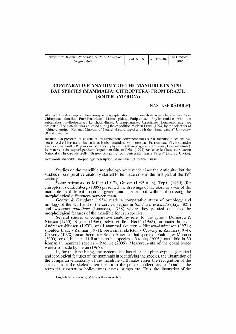
Saccopteryx bilineata (Temminck, 1838) (Fig. 1) has: RM with a very deepcentral concavity from CORM to P CON; P COR is a vertical isosceles triangle-likeblade, with the anterior side widened, thickened and the tip rounded and thickened;
376 NÃSTASE RÃDULEÞ
IM widely semicircular; P CON is triangular with the tip bent to outside; CAP Mlike a wand with rounded ends, and the surface rounded slightly concave in dorsalview; NNP is a trapezial blade perpendicular on RM where the anterior margin isbent innerly, and terminally rounded.
Family Mormoopidae Koch, 1862-63
Pteronotus parnellii (Gray, 1843) (Fig. 2) has: RM with a deeper depressionon the axis P CON – CORM; P COR is a triangular blade, slightly bent to outside,with thickened margines and the tip almost in right angle, but rounded; LOM almostvertical; IM slightly convex; P CON is triangular, innerly bent, with a rounded tipbent outside; CAP M ellipsoidal asymmetrical wand, innerly bent; NNP like a shortarm, with the tip flattened upwardly.
Family Phyllostomidae Gray, 1825
Subfamily Phyllostominae Gray, 1825
In Macrophyllum macrophyllum (Schinz, 1821) (Fig. 3) has: RM has a wideconcavity, deeper towards CORM directed to P CON; IM straight, oblique towardsP CON; P COR has the anterior margin thickened, a prominent rounded tip; P CON
MANDIBLE COMPARATIVE ANATOMY IN BRAZILIAN BATS (CHIROPTERA) 377
Fig. 1 – External lateral view of the mandible in Saccopteryx bilineata (Temminck, 1838).
Fig. 2 – External lateral view of the mandible in Pteronotus parnellii (Gray, 1843).

like an equilateral triangle with the tip bent to outside; CAP M elliptical, innerlybent; NNP an obliquely-laterally directed blade on RM, with a thickened roundedtip, flattened upwardly.
Phyllostomus hastatus (Pallas, 1767) (Fig. 4) has the RM with a deepconcavity towards CORM; P COR is a triangular blade with the anterior marginthickened, an acute angled but rounded tip; IM semicircular; P CON is like anisosceles triangle; CAP M like a wand slightly innerly bent, with a pointed exteriorend; NNP like an obliquely-latyerally directed arm on RM, with thickened margins,and the tip flattened upwardly.
Subfamily Glossophaginae Gray, 1821
In Glossophaga soricina (Pallas, 1766) (Fig. 5), on the outer side: RM has asuperficial central depression; LOM is oblique; P COR is like a triangular blade witha rounded tip posteriorly arched; IM semicircular; P CON like an isosceles triangle;CAP M is oval, asymmetrical, innerly bent; NNP is a short arm with a rounded tipslightly bent upwardly.
378 NÃSTASE RÃDULEÞ
Fig. 3 – External lateral view of the mandible in Macrophyllum macrophyllum (Schinz, 1821).
Fig. 4 – External lateral view of the mandible in Phyllostomus hastatus (Pallas, 1767).

Subfamily Lonchophyllinae Griffiths, 1982
Lonchophylla mordax Thomas, 1903 (Fig. 6) has the RM with an ovaldepression in the lower half of RM, under P COR; IM semicircular; P COR centrallyhas an oblique fold towards P CON and the tip slightly widened, prominent, rounded;P CON has a triangular concavity; CAP M elliptical, asymetrical towards inside;NNP like a straight spine, horizontally directed on RM and with a rounded tip.
Subfamily Carolliinae Miller, 1924
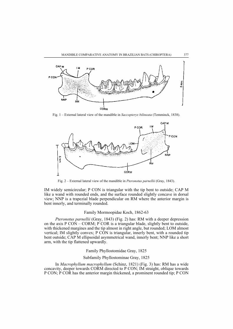
Carollia perspicillata (Linnaeus, 1758) (Fig. 7) has on the external side: RMwith a centrally deeper depression which also penetrates P CON, P COR; IMoblique, semicircular; P COR is a triangular blade with a rounded tip which isthickened together with the anterior half; P CON has a triangular concavitycentrally, with a tip bent outside; CAP M ellipsoidal, asymmetrical towards inside;NNP is an arm with the tip and the lower margin thickened.
Subfamily Desmodontinae Bonaparte, 1845
In Desmodus rotundus (E. Geoffroy, 1810) (Fig. 8) has: RM has a centralconcavity from CORM to P CON; LOM almost vertical; IM straight; P COR is likea right-angled triangle with the anterior margin thickened and a rounded tip; P CON
MANDIBLE COMPARATIVE ANATOMY IN BRAZILIAN BATS (CHIROPTERA) 379
Fig. 5 – External lateral view of the mandible in Glossophaga soricina (Pallas, 1766).
Fig. 6 – External lateral view of the mandible in Lonchophylla mordax Thomas, 1903.
is a triangle with the posterior side thickened; CAP M is oval, asymetriacal towardsinside; NNP like a very short arm with the tip flattened upwardly.
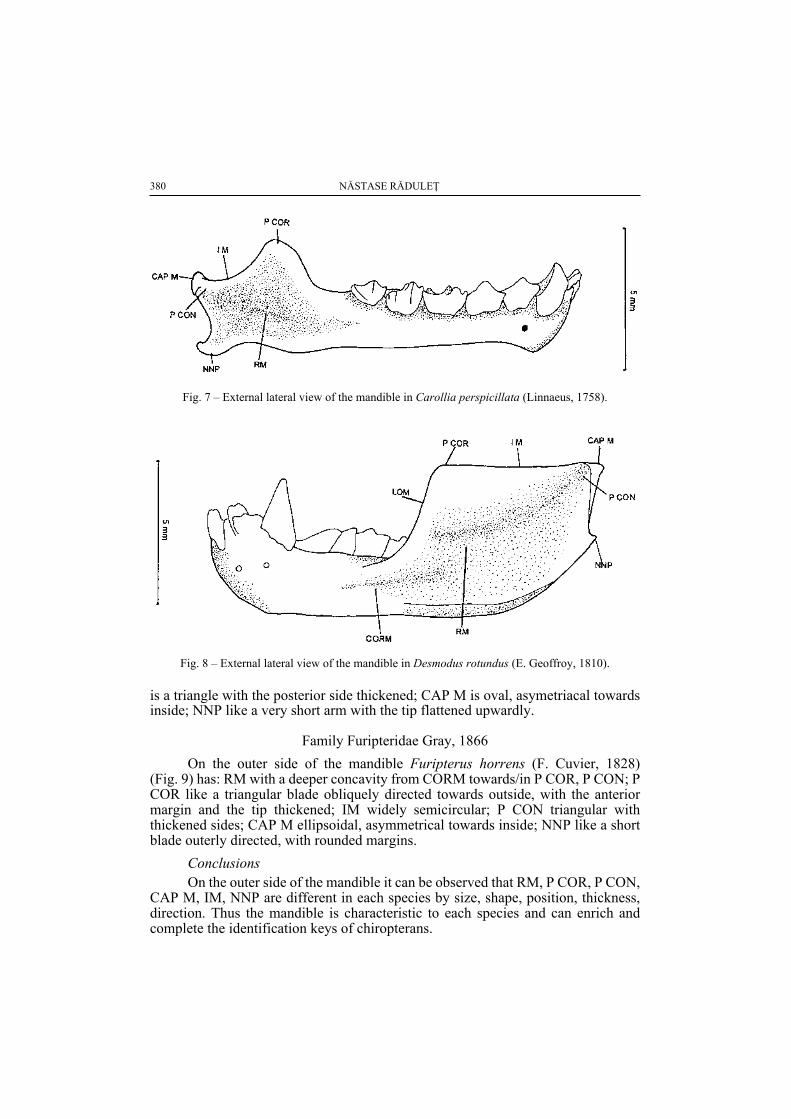
Family Furipteridae Gray, 1866
On the outer side of the mandible Furipterus horrens (F. Cuvier, 1828) (Fig. 9) has: RM with a deeper concavity from CORM towards/in P COR, P CON; PCOR like a triangular blade obliquely directed towards outside, with the anteriormargin and the tip thickened; IM widely semicircular; P CON triangular withthickened sides; CAP M ellipsoidal, asymmetrical towards inside; NNP like a shortblade outerly directed, with rounded margins.
ConclusionsOn the outer side of the mandible it can be observed that RM, P COR, P CON,
CAP M, IM, NNP are different in each species by size, shape, position, thickness,direction. Thus the mandible is characteristic to each species and can enrich andcomplete the identification keys of chiropterans.
380 NÃSTASE RÃDULEÞ
Fig. 7 – External lateral view of the mandible in Carollia perspicillata (Linnaeus, 1758).
Fig. 8 – External lateral view of the mandible in Desmodus rotundus (E. Geoffroy, 1810).
ACKNOWLEDGEMENTS
The author wants to thank to the referees and to Mrs Petruþa Dumitricã and Mrs Aurora Dinufor tracing the drawings in China ink.
ANATOMIA COMPARATÃ A MANDIBULEI LA NOUÃ SPECII DE LILIECI(MAMMALIA: CHIROPTERA) DIN BRAZILIA (AMERICA DE SUD)
REZUMAT
În lucrare este descrisã morfologia mandibulei de la 9 specii de lilieci (Chiroptera), din faunaBraziliei (America de Sud). Materialul este rezultatul colectãrilor de teren efectuate în Brazilia (1994)de cercetãtorii de la Muzeul Naþional de Istorie Naturalã “Grigore Antipa” (Bucureºti) în colaborare cucei de la Universitatea “Santa Ursula” (Rio de Janeiro). Sunt prezentate desene ale mandibulei cupãrþile componente ºi explicaþiile corespunzãtoare. Lucrarea îºi propune sã evidenþieze principalelestructuri (RM, P COR, P CON, IM, CAP M, NNP) care diferã de la o specie la alta prin mãrime, formã,poziþie, grosime, orientare una faþã de cealaltã. Cunoaºterea morfologiei mandibulei va completa cheilede determinare a speciilor de lilieci ºi va fi de un real ajutor pentru mamalogi, ornitologi ºipaleontologi.
LITERATURE CITED
ANDREESCU-NIÞESCU, I., 1970 - Étude comparative des cornetes nasaux chez: Talpa europaea L.,Crocidura leucodon Herm., C. suaveolens Pall., Sorex araneus L., et Neomys fodiensSchreb. (Ord. Insectivora) de Roumanie. Travaux du Muséum d’Histoire Naturelle“Grigore Antipa”, 10: 359–363.
ÈERVENÝ, J., 1978 – Comparative anatomy of large bones in three models of european bats(Rhinolophus, Myotis, Tadarida) Vestnik Èeskoslovenské Spole¤nosti, Zoologické, 42(3): 161–171.
ÈERVENÝ, J., J. ZALMAN, 1974 - Diagnostiké znaky na kostech pøedni kon¤etiny vrápenc°u. LynxMuseum. Nationale, Praha, 16: 86–100.
DORNESCU, TH., I. NIÞESCU, 1965 – Anatomie comparée de la colonne vertébrale chez plusieursespèces de rongeurs de Roumanie. Travaux du Muséum d’Histoire Naturelle “GrigoreAntipa”, 5: 423–441.
EISENBERG, J. F., 1989 – Mammals of the Neotropics. The University of Chicago Press. Chicago andLondon, 1: 449 pp.
GEORGE, R., L. GAUGHRAN, 1954 – A comparative study of the osteology and myology of thecranial and cervical regions of the shrew, Blarina brevicauda, and the mole, Scalopusaquaticus. Miscellaneous. Michigan, 80: 82 pp.
GRASSÉ, P., 1955 a – Traité de zoologie. Anatomie, systematique, biologie. Mammifères. Librairesde l’Academie de Médecine. Paris, 17 (1): 1167 pp.
MANDIBLE COMPARATIVE ANATOMY IN BRAZILIAN BATS (CHIROPTERA) 381
Fig. 9 – External lateral view of the mandible in Furipterus horrens (F. Cuvier, 1828).

GRASSÉ, P., 1955 b – Traité de zoologie. Anatomie, systematique, biologie. Mammifères. Librairesde l’Academie de Médecine. Paris, 17 (2): 2285 pp.
GRASSÉ, P., 1967 – Traité de zoologie. Anatomie, systematique, biologie. Mammifères. Libraires del’Academie de Médecine. Paris, 16 (1): 1162 pp.
HERÁÒ, I., 1967 – K rozdil°um v morfologii pánve svištì horského (Marmota marmota L.) veverkyobecné (Sciurus vulgaris L.) a sysla obecného (Citellus citellus L.). Lynx, ser. nov., 8:7–14. Praha.
HERÁÒ, I., 1968 - Diagnostiké znaky na pánvich šelem. Lynx, ser. nov., 9: 25–33. Praha.MILLER, G. S., 1912 – Catalogue of the Mammals of Western Europe (Europe exclusive of Russia) in
the Collection of the British Museum, London: 1019 pp.MURARIU, D., 1999 –The distribution of the species Microtus agrestis (L., 1761) (Rodentia:
Arvicolidae). Travaux du Muséum National d’Histoire Naturelle “Grigore Antipa”, 41:435–444.
MURARIU, D., 2004 – New reports on the distribution of three bat species (Mammalia: Chiroptera) ofRomania. Travaux du Muséum National d’Histoire Naturelle “Grigore Antipa”, 46:271–279.
NIÞESCU, I., 1966 – Anatomie comparée de la colonne vertébrale chez Ondatra zibethica L.,Apodemus agrarius Pall. et Spalax leucodon Nordmann. Travaux du Muséumd’Histoire Naturelle “Grigore Antipa”, 6: 345–356.
NIÞESCU-ANDREESCU, I., 1971 – Contributions à l’étude de la morphologie du squelet desMammifères de petite taille. Travaux du Muséum d’Histoire Naturelle “GrigoreAntipa”, 11: 417–427.
PUCEK, Z., 1981 – Key to vertebrates of Poland Mammals. PWN – Polish Scientific Publishers,Warszawa. 367 pp.
RÃDULEÞ, N., 2003 – Contributions to the morphological study of the coxal bone of 11 bat species(Mammalia: Chiroptera) from Romania. Travaux du Muséum National d’HistoireNaturelle “Grigore Antipa”, 45: 373–380.
RÃDULEÞ, N., 2005- Comparative anatomy of the mandible in the mammal systematic (Mammalia:Insectivora, Chiroptera, Rodentia) from Romania (I). Travaux du Muséum Nationald’Histoire Naturelle “Grigore Antipa”, 48: 373–380.
RÃDULEÞ, N., D., MURARIU, 2000 – Taxonomical value of the morphological differences of thecoxal bone in six South – American bat species (Chiroptera: Emballonuridae,Mormoopidae and Phyllostomidae). Travaux du Muséum National d’HistoireNaturelle “Grigore Antipa”, 42: 225–234.
TOPÁL, GY., 1969 – Denevérek – Chiroptera, Mammalia. In: Fauna Hungarie. MagyarországÁllatvilága, 22 (2): 281 pp.
TUDOR, D., GH. M. CONSTANTINESCU, 2002 – Nomina anatomica veterinaria. Edit. Vergiliu,Bucureºti: 378 pp. (in Romanian)
ZALMAN, J., 1971 – Diagnostiche merkmale an den schulterblättern einiger fledermäuse der familieRhinolophidae Bell, 1836 und Vespertilionidae Gray, 1821. Vestnik ÈeskoslovenskéSpole¤nosti. Zoologické, 35 (4): 311–319.
*** – Latin Nomina Anatomica (modern Latin anatomical term) –Index and glossary of medical terms.Available online at http://vesalius.northwestern.edu/indexterms.html
Received: February 7, 2006 Muzeul Naþional de Istorie Naturalã “Grigore Antipa”Accepted: June 12, 2006 ªos. Kiseleff 1, 011341 Bucureºti 2, România
e-mail: [email protected]
382 NÃSTASE RÃDULEÞ


