Embed Size (px)
Citation preview
1
Radiotherapy in Lung
CancerAnatomy
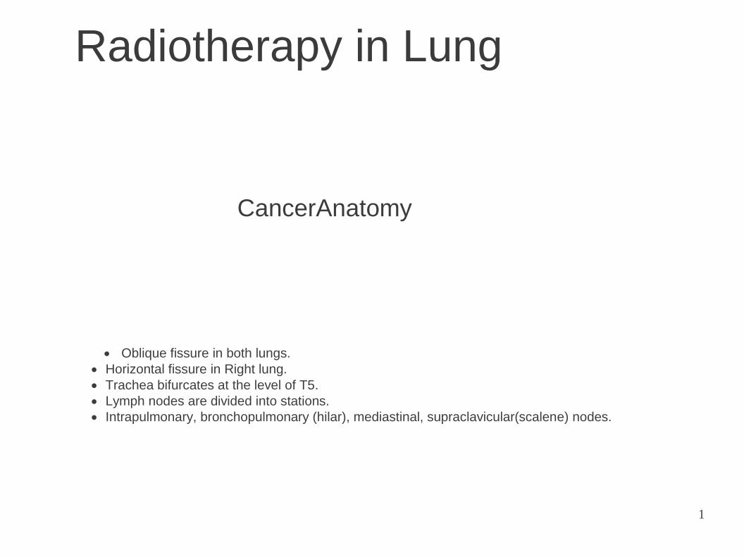
Oblique fissure in both lungs.
Horizontal fissure in Right lung.
Trachea bifurcates at the level of T5.
Lymph nodes are divided into stations.
Intrapulmonary, bronchopulmonary (hilar), mediastinal, supraclavicular(scalene) nodes.
2
3
Epidemiology
● Most common & Deadliest worldwide.
● Survival at 5 years in USA is 15%.
● Primary risk factor- SMOKING (~90%) ● Adenocarcinoma more
than Small/Squamous.
(Filtered cigarette, fine particles reach periphery)
Presentation
● Due to local tumor growth:
– Centrally cough, haemoptysis, obstructive signs. –
Peripherally silent, cough, pleuritic chest pain.
4
– Nerve entrapment (LRLN, phrenic), Vascular obstruction.
– Esophageal narrowing, obstruction, fistula.
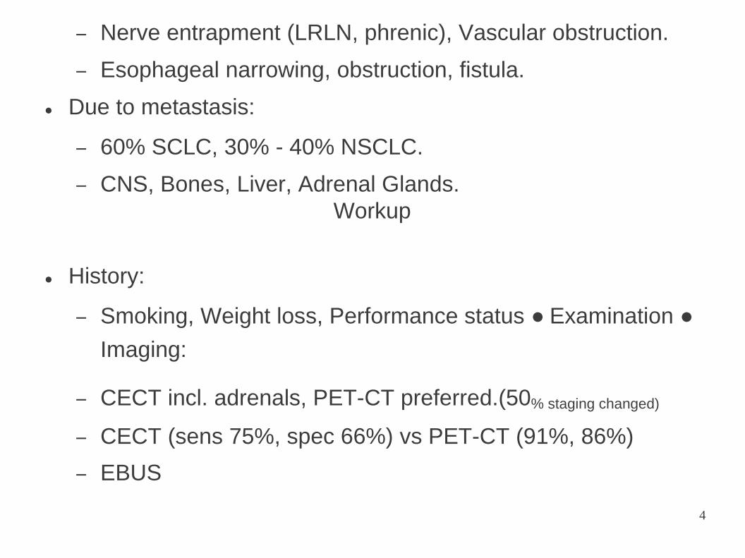
● Due to metastasis:
– 60% SCLC, 30% - 40% NSCLC.
– CNS, Bones, Liver, Adrenal Glands.
Workup
● History:
– Smoking, Weight loss, Performance status ● Examination ●
Imaging:
– CECT incl. adrenals, PET-CT preferred.(50% staging changed)
– CECT (sens 75%, spec 66%) vs PET-CT (91%, 86%)
– EBUS
5
● Tissue:
– FNAC, TBFNA, Mediastinoscopy, VATS
6
7
Overview of management in NSCLC
● Surgery is the main stay for resectable and operable non small cell
lung cancer
● Radiation plays a role in the definitive and adjuvant management of
NSCLC
● Chemotherapy is an important adjuvant treatment modality, often
used with radiation
● Radiation along with chemotherapy are useful for palliation
RT in Lung Cancer: Issues
● NSCLC: A moderately radio-sensitive tumor: dose escalation needed
● Surrounded by organs which are dose limiting: heart, opp. lung,
spinal cord, esophagus ● Respiratory motion: a pertinent factor necessitating motion
management in radiation delivery
8
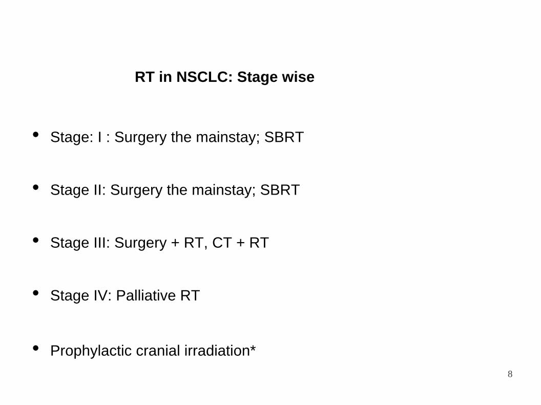
RT in NSCLC: Stage wise
• Stage: I : Surgery the mainstay; SBRT
• Stage II: Surgery the mainstay; SBRT
• Stage III: Surgery + RT, CT + RT
• Stage IV: Palliative RT
• Prophylactic cranial irradiation*
9
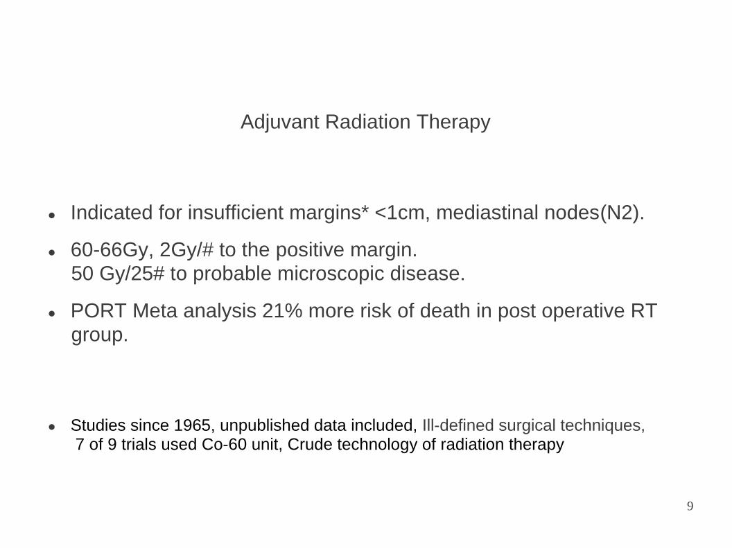
Adjuvant Radiation Therapy
● Indicated for insufficient margins* <1cm, mediastinal nodes(N2).
● 60-66Gy, 2Gy/# to the positive margin.
50 Gy/25# to probable microscopic disease.
● PORT Meta analysis 21% more risk of death in post operative RT
group.
● Studies since 1965, unpublished data included, Ill-defined surgical techniques, 7 of 9 trials used Co-60 unit, Crude technology of radiation therapy
10
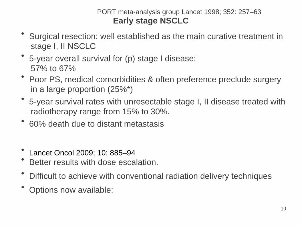
PORT meta-analysis group Lancet 1998; 352: 257–63 Early stage NSCLC
● Surgical resection: well established as the main curative treatment in
stage I, II NSCLC ● 5-year overall survival for (p) stage I disease:
57% to 67% ● Poor PS, medical comorbidities & often preference preclude surgery
in a large proportion (25%*) ● 5-year survival rates with unresectable stage I, II disease treated with
radiotherapy range from 15% to 30%. ● 60% death due to distant metastasis
● Lancet Oncol 2009; 10: 885–94
● Better results with dose escalation.
● Difficult to achieve with conventional radiation delivery techniques
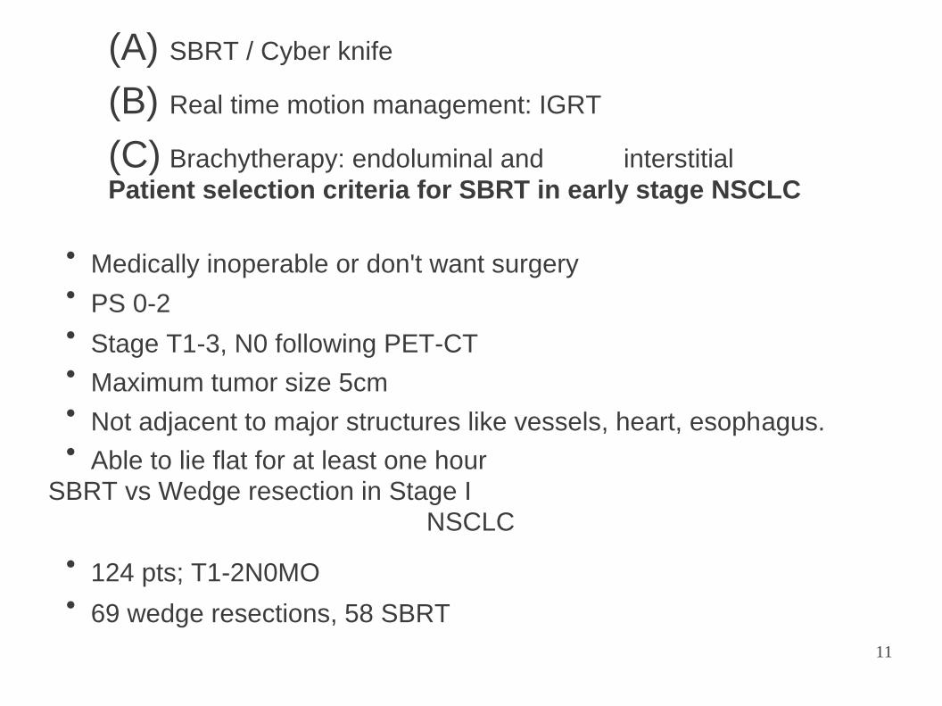
● Options now available:
11
(A) SBRT / Cyber knife
(B) Real time motion management: IGRT
(C) Brachytherapy: endoluminal and interstitial
Patient selection criteria for SBRT in early stage NSCLC
● Medically inoperable or don't want surgery
● PS 0-2
● Stage T1-3, N0 following PET-CT
● Maximum tumor size 5cm
● Not adjacent to major structures like vessels, heart, esophagus.
● Able to lie flat for at least one hour
SBRT vs Wedge resection in Stage I
NSCLC
● 124 pts; T1-2N0MO
● 69 wedge resections, 58 SBRT
12
● SBRT prescribed as 48(T1) or 60(T2) Gy in 4 to 5 fractions
● Median follow up of 2.5 years
● No differences in DM, FFF, or CSS, but OS was higher with wedge
resection at 30 months. (87% vs 72%)
(Distant Metastatis, Freedom from Failure, Case Specific Survival)
Journal of Clinical Oncology, Vol 28, No 6 , 2010: pp. 928-935
13
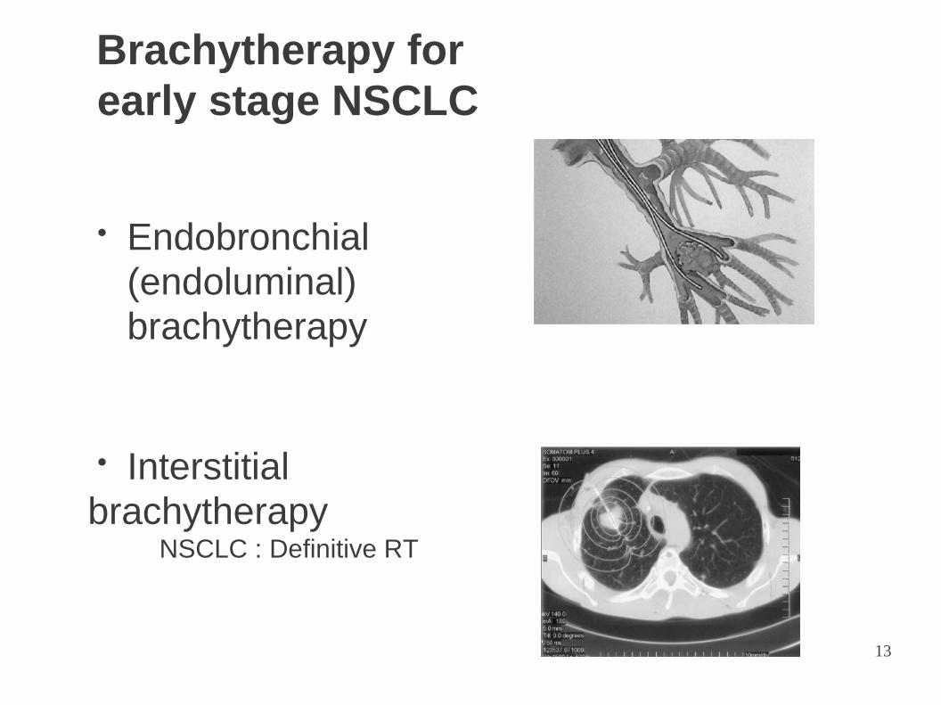
Brachytherapy for
early stage NSCLC
● Endobronchial
(endoluminal)
brachytherapy
● Interstitial
brachytherapy NSCLC : Definitive RT
14
● Stage III:
– Main bulk of the disease.
– 60 – 75 Gy to the gross disease (RTOG 73-01)
– 50 Gy to the microscopic disease – Hyperfractionation showed
better roles.
– 69.6 Gy . 1.2Gy/# , 2#/day. Best survival rates (20% at 3 years)
– CHART (Continuous Hyperfractionated Acclerated Radiotherapy) : 1.5Gy/#. 3#/day. 36#. Total: 54Gy
– RT vs CT/RT Benefit: 2 months (at 3 years)
Ann Intern Med. 1996;125:723-729. RT Techniques: 2D Planning
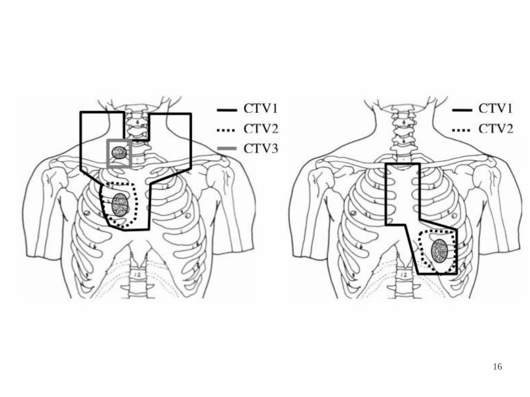
● 2 cm margin around any gross tumor.
● 1 cm margin around regional LN groups.

15
● Upper lobe tumor: B/L supraclav & subcarina.
● Middle lobe tumor: Entire mediastinum
(thoracic inlet to 8 cm below carina)
● Lower lobe tumor: Entire mediastinum from thoracic inlet to
diaphragm.
16

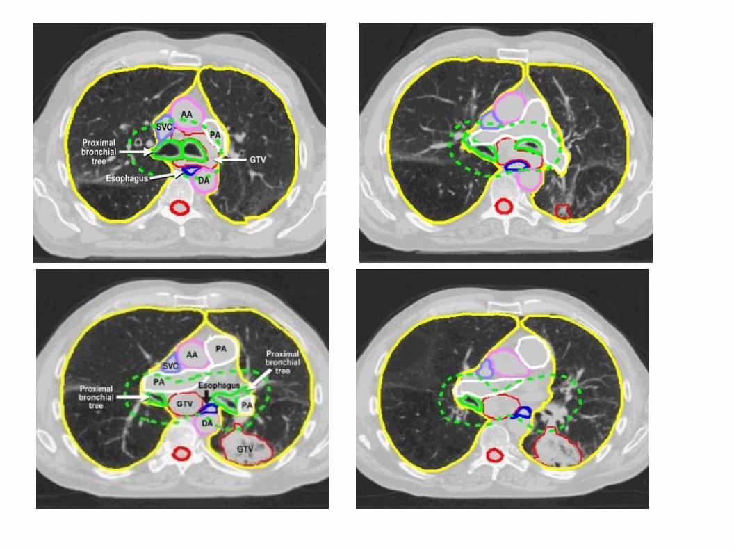
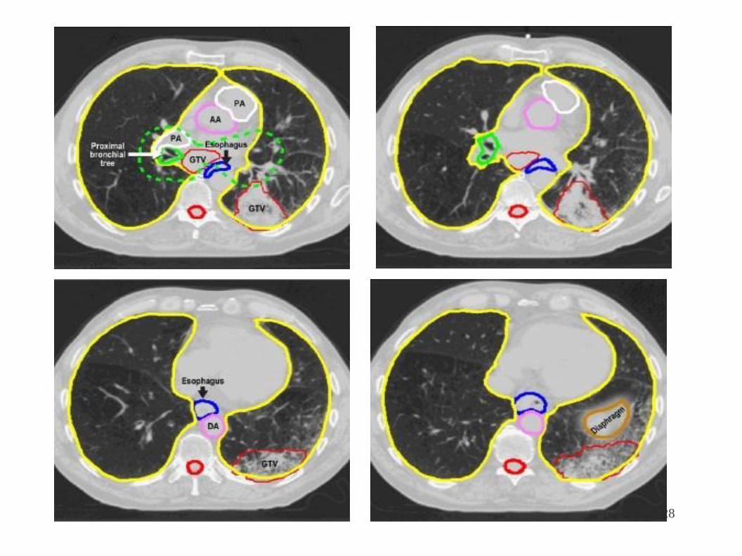
RT Techniques: 3DCRT
● CT Scan in treatment position. (optional Styrofoam)
● GTV : primary tumor & any gross lymph nodes.
● CTV : Area thought to harbor micrometastasis (hilar / mediastinal LN,
Margin).
● PTV: Margin for physiologic organ motion during treatment and daily
inaccuracies.
20 Bethesda, MD: ICRU; 1993:50
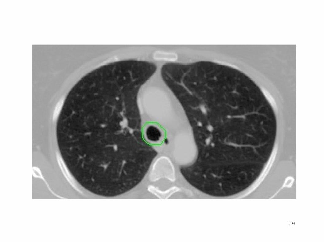
GTV
18
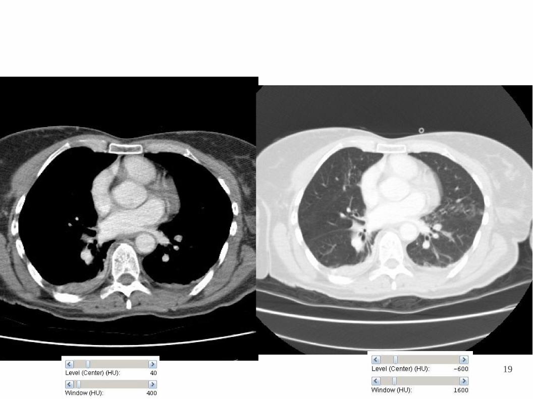
● Visible tumor by any imaging modality.
● Pulmonary extent: on pulmonary windows. ● Mediastinal extent:
mediastinal windows.
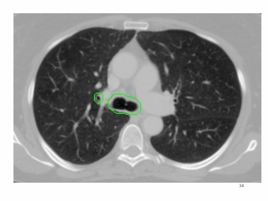
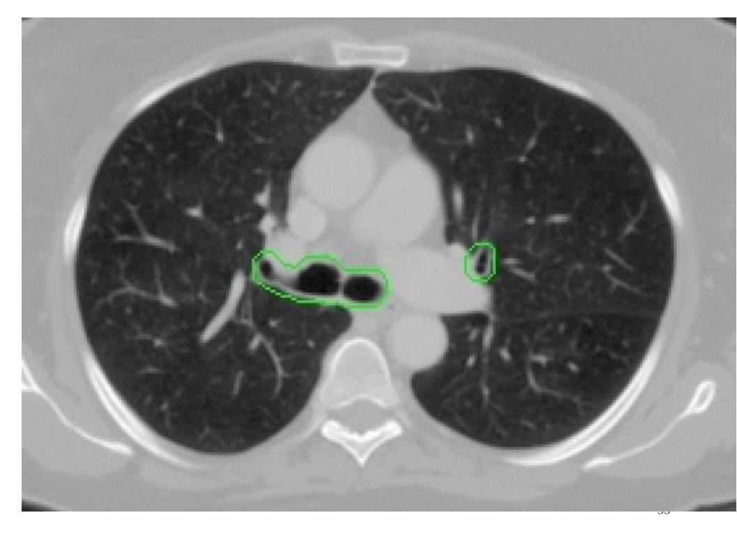
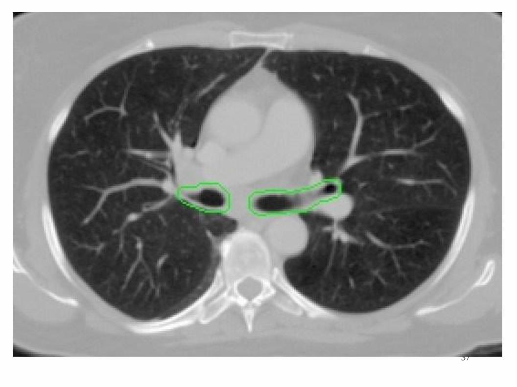
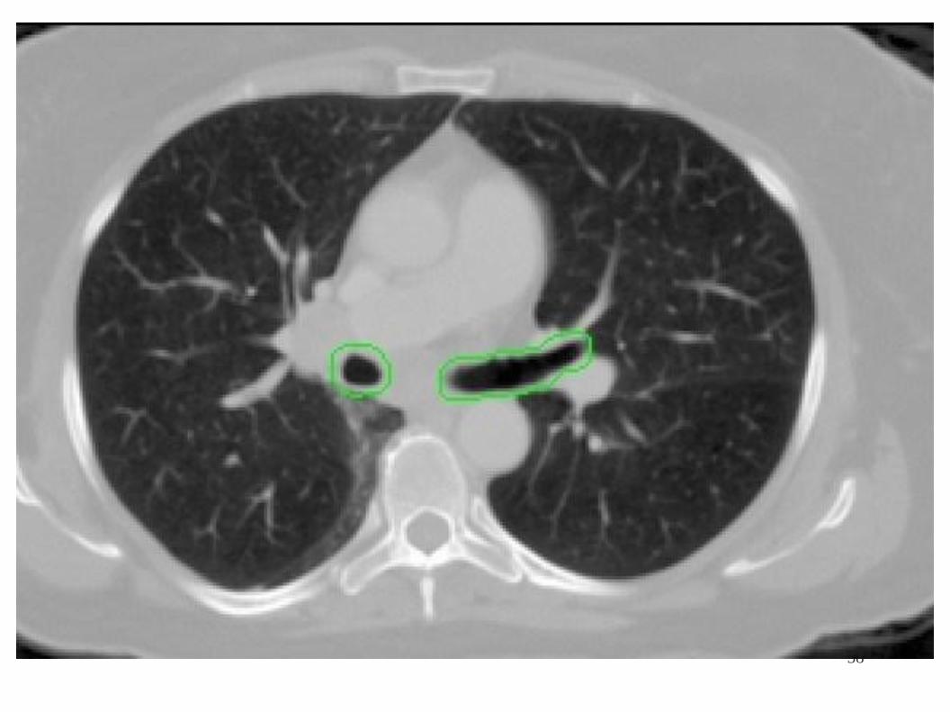
● Lymph node >1 cm in shortest: +ve (15% chance)
● FDG-PET : quite important. (collapse vs tumor, LNs)
19
20
CTV
● Contains gross and microscopic disease.
● GTV-to-CTV : 6 mm for squamous cancers 8 mm for
adenocarcinomas to cover the gross tumor and microscopic disease
with 95% accuracy. For others, 8mm.
● In the absence of radiographic proof of invasion, CTV of primary lesion
should not extend into the chest wall or mediastinum.
● CTV expansions of lymph node disease should not extend into the
major airways or lung.
Giraud P et al. Evaluation of microscopic tumor extension in NSCLC for 3D-CRT planning. Int J Radiat Oncol Biol Phys 2000;48:1015-1024.
PTV
21
● CTV + margin for daily setup error and target motion.
● 4D CT study, 50% of the tumor moves > 5 mm
13% moves > 1 cm (more when near diaphragm)
● Individual assessment is recommended.
● Breath holding, Gated techniques.
● ITV : Only takes the organ movements.
ICRU 62
22
Dose and volume
● Gross disease i.e. primary and involved nodes: 65-70
Gy (+/- CT) ● Elective nodal irradiation not recommended
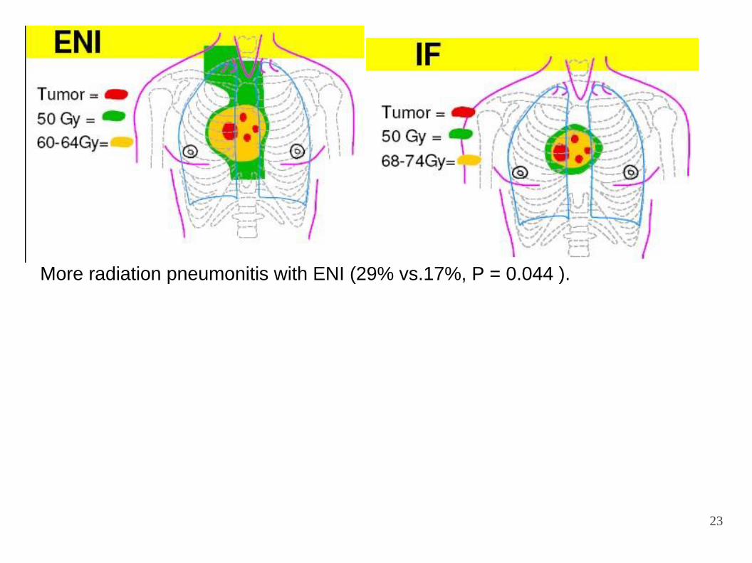
23
More radiation pneumonitis with ENI (29% vs.17%, P = 0.044 ).
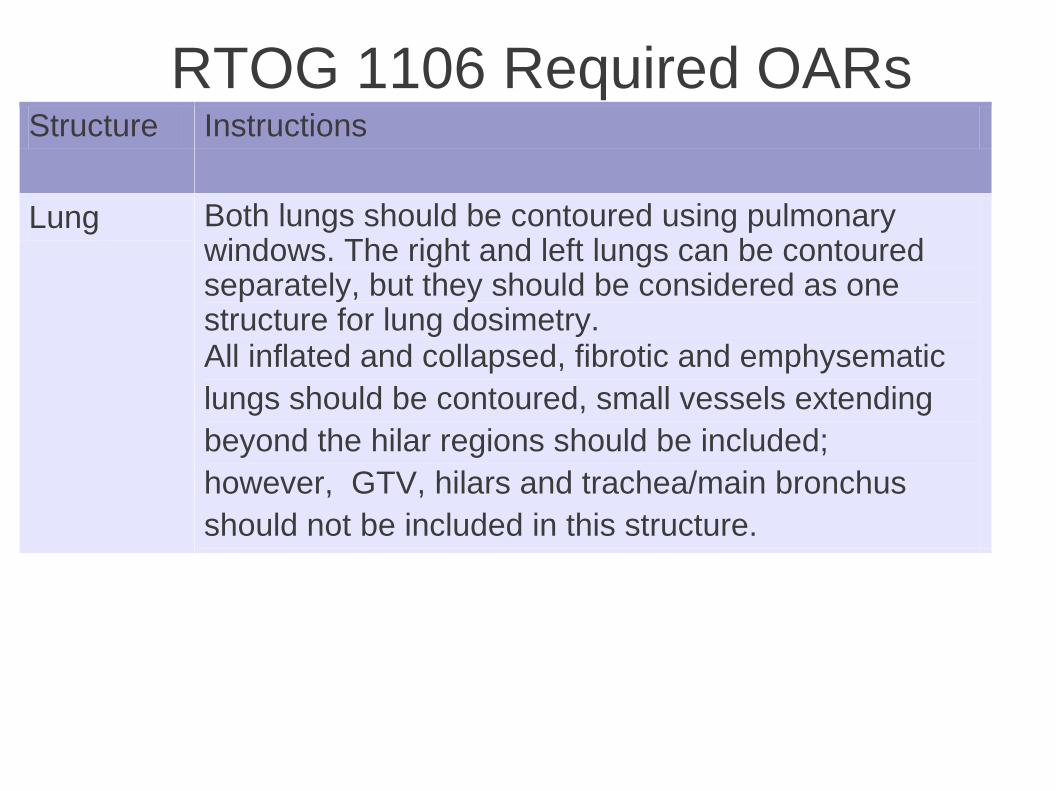
RTOG 1106 Required OARs Structure Instructions
Lung Both lungs should be contoured using pulmonary windows. The right and left lungs can be contoured separately, but they should be considered as one structure for lung dosimetry. All inflated and collapsed, fibrotic and emphysematic
lungs should be contoured, small vessels extending
beyond the hilar regions should be included;
however, GTV, hilars and trachea/main bronchus
should not be included in this structure.
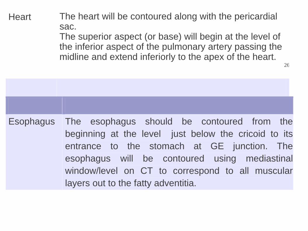
Heart
The heart will be contoured along with the pericardial sac. The superior aspect (or base) will begin at the level of the inferior aspect of the pulmonary artery passing the midline and extend inferiorly to the apex of the heart. 26
Esophagus The esophagus should be contoured from the
beginning at the level just below the cricoid to its
entrance to the stomach at GE junction. The
esophagus will be contoured using mediastinal
window/level on CT to correspond to all muscular
layers out to the fatty adventitia.
26
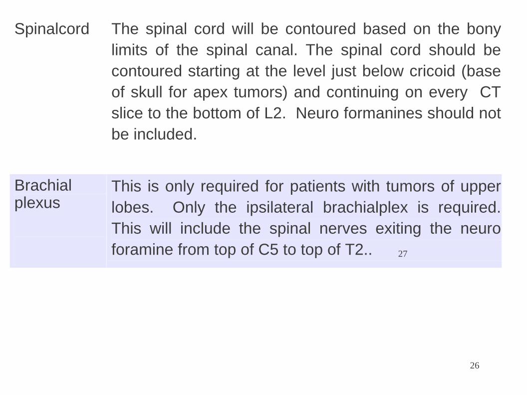
Spinalcord The spinal cord will be contoured based on the bony
limits of the spinal canal. The spinal cord should be
contoured starting at the level just below cricoid (base
of skull for apex tumors) and continuing on every CT
slice to the bottom of L2. Neuro formanines should not
be included.
Brachial plexus
This is only required for patients with tumors of upper
lobes. Only the ipsilateral brachialplex is required.
This will include the spinal nerves exiting the neuro
foramine from top of C5 to top of T2.. 27
28
29
30
31
32
33
34
35

36
37
38
39
40
41
42
43
44
45
46
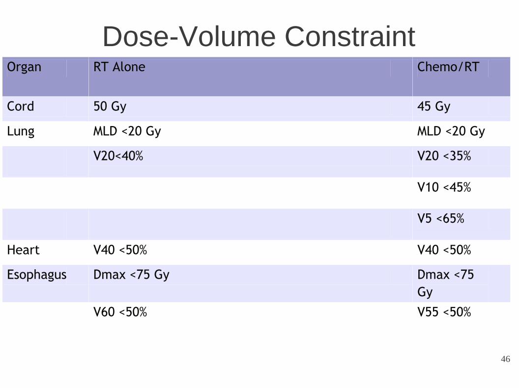
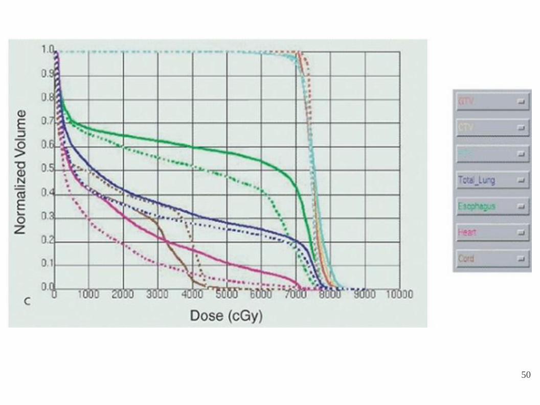
Dose-Volume Constraint Organ RT Alone Chemo/RT
Cord 50 Gy 45 Gy
Lung MLD <20 Gy MLD <20 Gy
V20<40% V20 <35%
V10 <45%
V5 <65%
Heart V40 <50% V40 <50%
Esophagus Dmax <75 Gy Dmax <75
Gy
V60 <50% V55 <50%
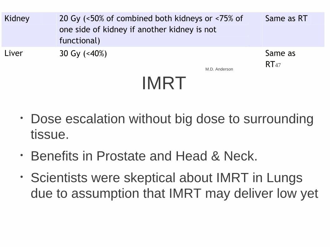
Kidney 20 Gy (<50% of combined both kidneys or <75% of
one side of kidney if another kidney is not
functional)
Same as RT
Liver 30 Gy (<40%) M.D. Anderson
Same as
RT47
IMRT
● Dose escalation without big dose to surrounding
tissue.
● Benefits in Prostate and Head & Neck.
● Scientists were skeptical about IMRT in Lungs
due to assumption that IMRT may deliver low yet
48
damaging doses to a larger volume of normal lung tissue.
● Movement of a tumor because of respiration
introduces another level of complexity to both the
IMRT dosimetry and the technique used.
IMRT
● Found that IMRT may be more suitable than 3D
CT treatment planning for cases of advanced-
stage disease with a larger GTV.
● Median absolute reduction of lung volume
irradiated above 10 and 20 Gy were 7% and 10%, respectively.
● >2 Gy less mean total lung dose and 10%
decrease in the risk of radiation pneumonitis.
● Heart, Esophagus, thoracic tissue dose
decreased. Liu H, Wang X, Dong L, et al. Feasibility of sparing lung and other thoracic structures with intensity-modulated radiotherapy for non-small-cell lung cancer. Int J Radiat Oncol Biol Phys 2004;58:1268-1279. 49 Murshed H, Liu H, Liao Z, et al. Dose and volume reduction for normal lung using intensity-modulated radiotherapy for advanced-stage nonsmall-cell lung cancer. Int J Radiat Oncol Biol Phys 2004;58:1258-1267.
50
51
IMRT
● Tumors in the superior sulcus or close to the
esophagus or spinal cord or patients with positive lymph nodes may benefit more.
● Earlier-stage, small mobile tumors may not be
good candidates for IMRT
52
Chang J, Liu H, Komaki R. Intensity modulated radiation therapy and proton radiotherapy for non-small cell lung cancer. Curr
Oncol Rep 2005;7:255-259.
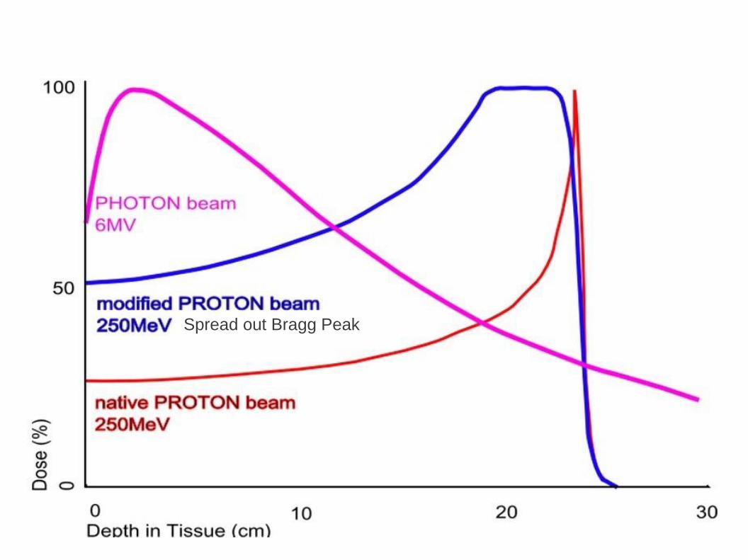
Proton Beam
● Well-defined range of penetration.
● By modulating the Bragg peak across the
target volume, proton beams can deliver a full,
localized, uniform dose of energy to the
treatment site while sparing the surrounding
normal tissues.
● In combination with IGRT.
● Results comparable to surgery in stage IA.
53
Shioyama Y, Tokuuye K, Okumura T, et al. Clinical evaluation of proton radiotherapy for non-small-cell lung cancer. Int J Radiat Oncol Biol Phys 2003;56:7-13.
54
Spread out Bragg Peak
55
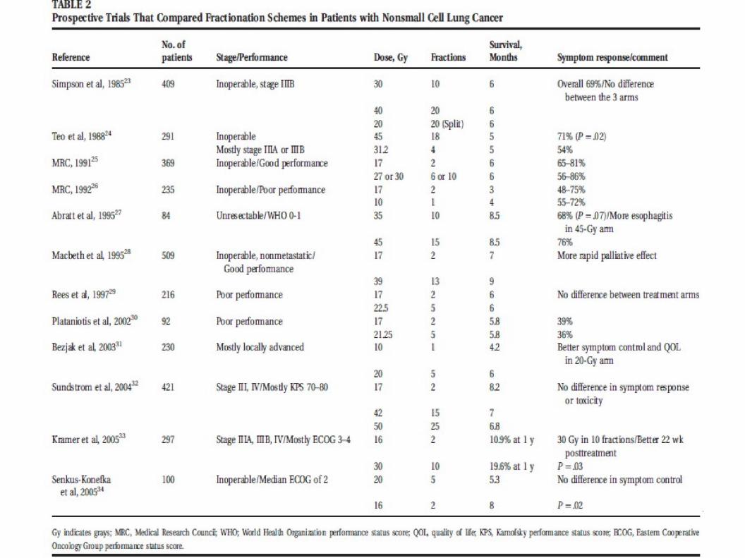
Palliative Radiotherapy
● Stage IIIB/IV
● 40Gy/20# vs 30Gy/10# → No difference.
● 20Gy/10# for short term palliation.

56
57
58
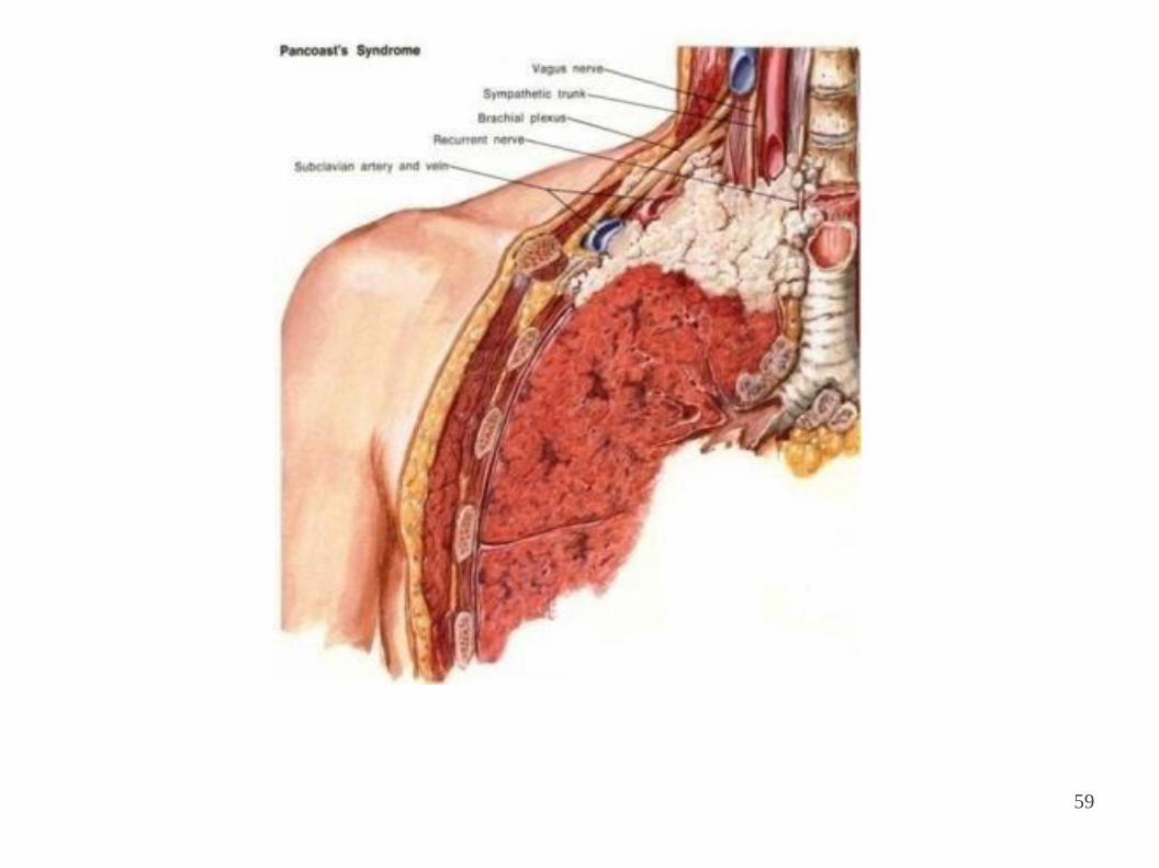
Superior Sulcus Tumor
59
60
Superior sulcus tumors ● Preoperative RT f/b extended surgical
resection: most common treatment. ● Radiotherapy: a primary treatment, for
inoperable superior sulcus tumors ● Palliation of pain in up to 90 percent of the
patients. ● Doses of 20 to 80 Gy have been used
● A dose of at least 60 Gy is recommended for
primary radiotherapy.
61
NEJM, 1997, 337(19): 1370-76
Small Cell Lung Cancer
● Limited disease: confined to the hemithorax. ●
Extensive : extends beyond the hemithorax.
● Most of the improvement in outcome was
attributed to more effective combination chemotherapy regimens.
● Locoregional therapy alone, either surgery or
RT, improved the short-term survival only slightly.
62
● Role of RT proven once distant metastasis was
controlled & local failure was apparent.
Small Cell Lung Cancer
● Thoracic RT and Prophylactic Cranial Irradiation.
● TRT concurrent with chemotherapy.
● Early TRT showed better outcome than late.
● Accelerated hyperfractionation better than daily
fractions ( 5yr survival 28% vs 21%)
● No significant difference in local tumor control or
survival with treatment between 45 Gy and 65 Gy
when effective chemotherapy was given.
Murray N, Coy P, Pater JL, et al. Importance of timing for thoracic irradiation in the combined modality treatment of limited-stage small-cell lung 59 cancer. The National Cancer Institute of Canada Clinical Trials Group. J Clin Oncol 1993;11:336-344.
Prophylactic Cranial Irradiation
Brain metastases -10% at presentation
- 80% at 2 yrs*
Irradiation of entire intracranial contents
Lower border at C2-3 vertebra
Doses 24 – 30 Gy @ 3 Gy/#
Increased the 3 year survival from 18% to 26%#
60 *Cancer 1979:44;1885-1893 #Ann Oncol 2002;13:748-54

61 Thank you
62