Embed Size (px)
Citation preview

Fujisawa et al. Respiratory Research (2021) 22:290 https://doi.org/10.1186/s12931-021-01892-9
RESEARCH
Radiological pleuroparenchymal fibroelastosis-like lesion in idiopathic interstitial pneumoniasTomoyuki Fujisawa1* , Yasuoki Horiike1, Ryoko Egashira2, Hiromitsu Sumikawa3, Tae Iwasawa4, Shoichiro Matsushita5, Hiroaki Sugiura6, Kensuke Kataoka7, Mikiko Hashisako8, Hideki Yasui1, Hironao Hozumi1, Masato Karayama1, Yuzo Suzuki1, Kazuki Furuhashi1, Noriyuki Enomoto1, Yutaro Nakamura1, Naoki Inui1,9 and Takafumi Suda1
Abstract
Background: Pleuroparenchymal fibroelastosis (PPFE) is characterised by predominant upper lobe pleural and subpleural lung parenchymal fibrosis. Radiological PPFE-like lesion has been associated with various types of inter-stitial lung diseases. However, the prevalence and clinical significance of radiological PPFE-like lesion in patients with idiopathic interstitial pneumonias (IIPs) are not fully understood. We aimed to determine the prevalence and clinical impact on survival of radiological PPFE-like lesion in patients with IIPs.
Methods: A post-hoc analysis was conducted using data from the Japanese nationwide cloud-based database of patients with IIPs. All the patients in the database were diagnosed as having IIPs by multidisciplinary discussion. Patients diagnosed with idiopathic PPFE were excluded. Clinical data and chest computed tomography (CT) image of 419 patients with IIPs were analysed. The presence of radiological PPFE-like lesion was independently evaluated by two chest radiologists blind to the clinical data.
Results: Of the 419 patients with IIPs, radiological PPFE-like lesions were detected in 101 (24.1%) patients, mainly in idiopathic pulmonary fibrosis (IPF) and unclassifiable IIPs, but less in idiopathic nonspecific interstitial pneumonia. Prognostic analyses revealed that radiological PPFE-like lesion was significantly associated with poor outcome in patients with IIPs, which was independent of age, IPF diagnosis and %FVC. In survival analyses, the patients with radio-logical PPFE-like lesions had poor survival compared with those without (log-rank, p < 0.0001). Subgroup analyses demonstrated that radiological PPFE-like lesion was significantly associated with poor survival both in patients with IPF and those with unclassifiable IIPs.
Conclusion: Radiological PPFE-like lesion is a condition that could exist in IIPs, mainly in IPF and unclassifiable IIPs. Importantly, the radiological PPFE-like lesion is a non-invasive marker to predict poor outcome in patients with IIPs, which should be carefully considered in clinical practice.
Keywords: Pleuroparenchymal fibroelastosis, Idiopathic interstitial pneumonias, Prognosis, IPF, Unclassifiable IIPs
© The Author(s) 2021. Open Access This article is licensed under a Creative Commons Attribution 4.0 International License, which permits use, sharing, adaptation, distribution and reproduction in any medium or format, as long as you give appropriate credit to the original author(s) and the source, provide a link to the Creative Commons licence, and indicate if changes were made. The images or other third party material in this article are included in the article’s Creative Commons licence, unless indicated otherwise in a credit line to the material. If material is not included in the article’s Creative Commons licence and your intended use is not permitted by statutory regulation or exceeds the permitted use, you will need to obtain permission directly from the copyright holder. To view a copy of this licence, visit http://creativecommons.org/licenses/by/4.0/. The Creative Commons Public Domain Dedication waiver (http://creativecom-mons.org/publicdomain/zero/1.0/) applies to the data made available in this article, unless otherwise stated in a credit line to the data.
BackgroundIdiopathic pleuroparenchymal fibroelastosis (iPPFE) is a condition of idiopathic interstitial pneumonias (IIPs) characterised by fibrosis of the pleura and sub-pleural lung parenchyma accompanied by elastosis of
Open Access
*Correspondence: [email protected] Second Division, Department of Internal Medicine, Hamamatsu University School of Medicine, 1-20-1 Handayama Higashi-ku, Hamamatsu 431-3192, JapanFull list of author information is available at the end of the article

Page 2 of 9Fujisawa et al. Respiratory Research (2021) 22:290
the alveolar walls, predominantly in the upper lobe [1, 2]. iPPFE was categorised as a rare IIPs in the current classification of the American Thoracic Society (ATS)/European Respiratory Society (ERS) Guidelines [3]. Its pathogenesis is not fully understood; however, recent studies have demonstrated that PPFE is observed in vari-ous conditions, including lung and bone marrow trans-plantation, connective tissue diseases (CTD) and history of anticancer/cytotoxic chemotherapy [1, 4–6]. Addi-tionally, radiological PPFE-like lesion on high-resolution computed tomography (HRCT) has been reported in association with several forms of ILDs, including idi-opathic pulmonary fibrosis (IPF), hypersensitivity pneu-monitis and CTD-related interstitial lung disease (ILD) [7–11], with various prevalence and clinical implications.
The clinical significance of radiological PPFE-like lesions in patients with IIPs have not been fully under-stood. No large-scale study in multicenter cohort has assessed the prevalence and clinical impact of radiologi-cal PPFE-like lesions in patients with IIPs. Recently, we have developed the nationwide cloud-based integrated database with the clinical, radiological and pathological data of more than 500 patients with IIPs in Japan. Using the database, we have successfully performed web-based multidisciplinary discussion (MDD) for the 465 cases of IIPs and shown the utility of web-based MDD to increase the accuracy of IIP diagnosis [12]. The nationwide cloud-based database is the largest cohort of patients with MDD diagnosis of IIPs in Japan [12], in which the clini-cal, radiological and pathological data of the enrolled cases are available. Therefore, the database enables us to review radiological PPFE-like lesions on chest HRCT and to evaluate its clinical implications in large number of patients with IIPs.
The aims of the present study were to assess the preva-lence of radiological PPFE-like lesion in patients with IIPs and to clarify its potential impact of survival using the nationwide large cohort of patients with IIPs.
MethodsSubjectA post-hoc analysis was conducted using the data from a retrospective cohort study of the Japanese nationwide cloud-based database of patients with IIPs for web-based MDD [12]. Previously, we built the nationwide cloud-based database containing clinical, radiological and pathological data of consecutive patients with insti-tutional diagnosis of IIPs in 39 institutions (from April 2009 to March 2014) and web-based MDD system. Web-based MDD were performed for the 465 enrolled patients to make accurate diagnosis of IIPs [12]. The IIPs cohort in the nationwide cloud-based database represents a prevalence of IIPs in Japan. In this study, the 465 cases
registered in the database were screened. The patients with MDD diagnosis of IIPs were eligible for inclusion in the absence of the following exclusion criteria: (1) insuf-ficient data; (2) MDD diagnosis of iPPFE.
The study flowchart is presented in Fig. 1. Of the 465 patients with institutional diagnosis of IIPs, 21 were diagnosed as having ILD other than IIPs by web-based MDD and were excluded from this study. Because of insufficient data on HRCT findings, nine patients were excluded. Sixteen patients diagnosed as having iPPFE by web-based MDD were also excluded. Consequently, 419 patients with MDD diagnosis of IIPs (except for iPPFE) were enrolled in this study. Diagnosis/classification of IIPs was categorised according to the ATS/ERS/Japanese Respiratory Society/Latin American Thoracic Associa-tion IPF statements [13] and ATS/ERS IIPs classification [3]. The categories included IPF, idiopathic nonspecific interstitial pneumonia (iNSIP), cryptogenic organizing pneumonia (COP), desquamative interstitial pneumonia (DIP)/respiratory bronchiolitis-ILD (RB-ILD), lymphoid interstitial pneumonia (LIP) and unclassifiable IIPs. This retrospective study was approved by the Institutional Review Board of the Hamamatsu University School of Medicine (approval number E14-360).
Cloud-based integrated database of IIPs(n = 465)
Exclude 21 casesFinal MDD diagnosis was not IIPs
Exclude 9 casesInsufficient data of HRCT findings
Study cohort (n = 419)
Exclude 16 casesFinal MDD diagnosis was iPPFE
MDD diagnosis of IIPs (n = 435)
Fig. 1 Flowchart of case inclusion in the study. Of the 465 patients with institutional diagnosis of IIPs in the nationwide cloud-based integrated database, 21 were diagnosed as having ILD other than IIPs by web-based MDD and were excluded. Because of insufficient data on HRCT findings, nine patients were excluded. Of the 435 patients with web-based MDD diagnosis of IIPs, 16 were diagnosed as having idiopathic PPFE and were excluded. Consequently, 419 patients with MDD diagnosis of IIPs except for iPPFE were enrolled in this study. IIPs idiopathic interstitial pneumonias; ILD interstitial lung disease; MDD multidisciplinary discussion; HRCT high-resolution computed tomography
Page 3 of 9Fujisawa et al. Respiratory Research (2021) 22:290
Data collectionAll the data were collected from the nationwide cloud-based database [12]. The patients’ clinical and HRCT data within 3 months before surgical lung biopsy (SLB), and whole slide images of biopsy specimens were retro-spectively collected and registered in the database to per-formed web-based MDD [12]. The clinical data included age, sex, smoking history, serum Krebs von den Lungen-6 (KL-6), serum surfactant protein D (SP-D), serum lactate dehydrogenase, arterial oxygen pressure, arterial carbon dioxide tension (PaCO2), % FVC, %DLCO and survival outcomes.
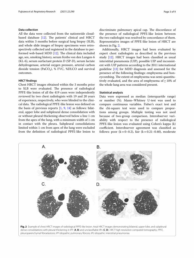
HRCT findingsChest HRCT images obtained within the 3 months prior to SLB were evaluated. The presence of radiological PPFE-like lesion of all the 419 cases were independently reviewed by two chest radiologists with 19 and 20 years of experience, respectively, who were blinded to the clini-cal data. The radiological PPFE-like lesion was defined on the basis of previous reports [1, 9, 14] as follows: bilat-eral, upper lobe and subpleural dense consolidation with or without pleural thickening observed below a line 1 cm from the apex of the lung, with a minimum width of 1 cm in contact with the pleura. Subpleural consolidations limited within 1 cm from apex of the lung were excluded from the definition of radiological PPFE-like lesion to
discriminate pulmonary apical cap. The discordance of the presence of radiological PPFE-like lesion between the two radiologist was resolved by concordance of them. Representative images of PPFE-like lesion of HRCT are shown in Fig. 2.
Additionally, HRCT images had been evaluated by expert chest radiologists as described in the previous study [12]. HRCT images had been classified as usual interstitial pneumonia (UIP), possible UIP and inconsist-ent with UIP patterns according to the 2011 international guideline [13] for MDD diagnosis and assessed for the presence of the following findings: emphysema and hon-eycombing. The extent of emphysema was semi-quantita-tively evaluated, and the area of emphysema of ≥ 10% of the whole lung area was considered present.
Statistical analysisData were expressed as median (interquartile range) or number (%). Mann–Whitney U-test was used to compare continuous variables. Fisher’s exact test and the chi-square test were used to compare propor-tions among groups. Multiple testing was not used because of two-group comparison. Interobserver vari-ability with respect to the presence of radiological PPFE-like lesion was evaluated using Cohen’s kappa (k) coefficient. Interobserver agreement was classified as follows: poor (k = 0–0.2), fair (k = 0.21–0.40), moderate
Fig. 2 Example of chest HRCT images of radiological PPFE-like lesion. Axial HRCT images demonstrating bilateral, upper lobe, and subpleural dense consolidations with pleural thickening in IPF (A, B) and unclassifiable IIPs (C, D). HRCT high-resolution computed tomography; PPFE, pleuroparenchymal fibroelastosis; IPF idiopathic pulmonary fibrosis; IIPs idiopathic interstitial pneumonias

Page 4 of 9Fujisawa et al. Respiratory Research (2021) 22:290
(k = 0.41–0.60), good (k = 0.61–0.80), and excellent (k = 0.81–1.00). The observation period was calculated from the date of the first visit in each institution for an IIP to the last date of contact or the time of death. The vital status of the patients was ascertained on October 2017 for survival analysis. The Cox proportional hazard model was used for univariate and multivariate analyses to identify survival-associated variables. The variable of multivariate Cox-regression analyses in the whole cohort included diagnosis of IPF, pulmonary function (%FVC), both of which were considered as clinically important factors associated with mortality in IIPs [12, 15], as well as patients background (age, sex) and HRCT findings (PPFE-like lesion, emphysema and honeycombing). The Kaplan–Meier method was used to calculate the cumu-lative survival rate. The log-rank test was used to com-pare the survival rate between patient groups. Statistical analyses were performed using commercially available
software (JMP version 9.0: SAS Institute, Inc., Cary, NC, USA). All tests were two-tailed, and a p value of < 0.05 was considered statistically significant.
ResultsPatients’ characteristics and clinical dataThis study included 419 patients with IIPs diagnosed by MDD. The enrolled patients’ characteristics and clinical data are summarised in Table 1. Radiological PPFE-like lesion was detected in 101 (24.1%) patients. Interobserver agreement for the presence of radiological PPFE-like regions was good (k = 0.67). IPF was the most prevalent MDD diagnosis in both groups, followed by unclassifi-able IIPs. Patients with iNSIP had less radiological PPFE-like lesion, with radiological PPFE-like lesion detected in only three patients with iNSIP. Patients with radio-logical PPFE-like lesion were found to have higher levels of PaCO2 and SP-D than those without. On the HRCT
Table 1 Patients’ characteristics, MDD diagnosis and clinical data before surgical lung biopsy, and mortality
Data are presented as n (%) or median (interquartile range)
MDD multidisciplinary discussion; PPFE pleuroparenchymal fibroelastosis; IPF idiopathic pulmonary fibrosis; iNSIP idiopathic nonspecific interstitial pneumonia; COP cryptogenic organizing pneumonia; DIP desquamative interstitial pneumonia; RB-ILD respiratory bronchiolitis-interstitial lung disease; LIP lymphoid interstitial pneumonia; iPPFE idiopathic pleuroparenchymal fibroelastosis; IIPs idiopathic interstitial pneumonias; PaO2 arterial oxygen tension; PaCO2 arterial carbon dioxide tension; LDH lactate dehydrogenase; KL-6 Krebs von den Lungen-6; SP-D surfactant protein D; FVC forced vital capacity; DLCO diffusing capacity for carbon monoxide; HRCT high-resolution computed tomography; UIP usual interstitial pneumonia
Variables Total Without PPFE With PPFE p valuen = 419 n = 318 n = 101
Age, years 65 (59, 70) 65 (58, 70) 67 (61, 71) 0.0319
Sex, male, n (%) 273 (65) 210 (66) 63(62) 0.5493
Never smokers, n (%) 147 (35) 102 (33) 45 (45) 0.0307
IPF, n (%) 199 (47) 145 (46) 54 (53)
iNSIP, n (%) 44 (11) 41 (13) 3 (3)
COP, n (%) 5 (1) 5 (1) 0 (0)
DIP/RB-ILD, n (%) 9 (2) 9 (3) 0 (0)
Unclassifiable IIPs, n (%) 162 (39) 118 (37) 44 (44)
PaO2, mmHg 83.7 (76, 90) 83.5 (76, 90) 85.4 (77, 92) 0.2737
PaCO2, mmHg 40.9 (39, 44) 40.4 (38, 43) 42.7 (39, 45) 0.0006
LDH, U/mL 225 (199, 260) 226 (200, 264) 219 (191, 251) 0.0903
KL-6, U/mL 1088 (671, 1763) 1099 (677, 1894) 1040 (571, 1540) 0.1318
SP-D, ng/mL 198 (131, 322) 178 (118, 305) 248 (156, 373) 0.0013
%predicted FVC (%) 82.2 (70, 94) 83 (71, 96) 79 (67, 91) 0.0661
%predicted DLCO (%) 67.0 (53, 83) 66.2 (53, 81) 70.2 (54, 83) 0.2531
HRCT pattern 0.0041
UIP 38 (9) 25 (8) 13 (13)
Possible UIP 226 (54) 162 (51) 64 (63)
Inconsistent with UIP 155 (37) 131 (41) 24 (24)
HRCT findings
Emphysema 126 (30) 112 (35) 14 (14) < 0.0001
Honeycombing 41 (10) 26 (8) 15 (15) 0.0632
Deceased 116 (28) 76 (24) 40 (40) 0.0027

Page 5 of 9Fujisawa et al. Respiratory Research (2021) 22:290
findings, emphysema was less common in patients with radiological PPFE-like lesion than those without.
Of 101 patients with radiological PPFE-like lesion, 8 patients (7.9%) developed lung cancer during their clinical courses, and 41 patients (12.9%) developed lung cancer in the 318 patients without radiological PPFE-like lesion. In terms of antifibrotic therapy, 41 patients (40.6%; 27 IPF, 13 unclassifiable IIPs, 1 iNSIP) with radio-logical PPFE-like lesion were treated with antifibrotic agents during their clinical courses. Of 318 patients with-out radiological PPFE-like lesion, 101 patients (31.8%; 70 IPF, 28 unclassifiable IIPs, 3 iNSIP) were treated with antifibrotic agents. In this study subject, only one patient with unclassifiable IIPs without radiological PPFE-like lesion underwent lung transplantation during the clinical course.
Prognostic significance of radiological PPFE‑like lesion in patients with IIPsIn the total cohort, 116 patients (28%) died, and mortal-ity was significantly higher in patients with radiological PPFE-like lesion than those without (Table 1). To evaluate prognostic factors in patients with IIPs, Cox proportional hazard regression analyses were performed (Table 2). On the basis of univariate analyses, age, sex, IPF diagnosis, %FVC, UIP pattern on HRCT, PPFE-like lesion, emphy-sema and honeycombing were associated with poor prognosis. Multivariate analyses, however, demonstrated that age, IPF diagnosis, %FVC, the presence of PPFE-like lesion and emphysema on HRCT were significantly asso-ciated with poor outcome in patients with IIPs. When UIP pattern was included in multivariate analyses, we found that UIP pattern was not significantly associated
with poor prognosis (Additional file 1: Table S1). As UIP pattern may be strongly related to IPF diagnosis and hon-eycombing, we performed multivariate analyses with UIP pattern and without IPF diagnosis and honeycombing. In this model, UIP pattern was significantly associated with poor prognosis (Additional file 1: Table S1). Radiologi-cal PPFE-like lesion was strongly associated with poor prognosis in every model, suggesting that presence of radiological PPFE-like lesion is an independent predictor of poor survival in patients with IIPs. Subgroup analyses of prognostic factors in patients with IPF or those with unclassifiable IIPs were performed. Both univariate and multivariate analyses revealed that radiological PPFE-like lesion was significantly associated with poor outcome in patients with IPF (Additional file 1: Table S2). Similarly, radiological PPFE-like lesion was independently related to poor prognosis in patients with unclassifiable IIPs (Additional file 1: Table S3).
Survival analysis comparing patients with PPFE‑like lesion and those without in IIPsThe survival curves comparing patients with radiological PPFE-like lesion and those without in the total cohort are shown in Fig. 3. The survival of patients with radiological PPFE-like lesion was significantly worse than those with-out. Median length of survival were 7.0 years for patients with radiological PPFE-like lesion and 9.9 years for those without. Subgroup analyses of survival curves in patients with MDD diagnosis of IPF or unclassifiable IIPs are shown in Fig. 4. Patients with IPF with radiological PPFE-like lesion were found to have a significantly poor survival than those without. Similarly, survival of patients with unclassifiable IIPs with radiological PPFE-like lesion
Table 2 Analyses of prognostic factors in patients with IIPs (Cox proportional hazards model)
IPF idiopathic pulmonary fibrosis; PaO2 arterial oxygen tension; PaCO2 arterial carbon dioxide tension; KL-6 Krebs von den Lungen-6; FVC forced vital capacity; DLCO diffusing capacity for carbon monoxide; UIP usual interstitial pneumonia; PPFE pleuroparenchymal fibroelastosis; HR hazard ratio
Variable Univariate Multivariate
HR 95% CI p value HR 95% CI p value
Age (years) 1.05 1.03–1.08 < 0.0001 1.05 1.02–1.08 0.0001
Sex (male) 1.84 1.21–2.89 0.0036 1.32 0.82–2.20 0.2593
IPF diagnosis 2.47 1.70–3.65 < 0.0001 2.55 1.68–3.94 < 0.0001
PaO2 (mmHg) 1.00 0.99–1.01 0.5115
PaCO2 (mmHg) 1.01 0.99–1.01 0.1163
KL-6 (U/mL) 1.00 0.999–1.000 0.7180
%FVC, % 0.99 0.977–0.996 0.0043 0.98 0.97–0.99 < 0.0001
%DLCO, % 0.99 0.979–0.998 0.0203
UIP pattern 2.71 1.66–4.25 0.0002
PPFE-like lesion 2.32 1.56–3.38 < 0.0001 2.54 1.64–3.89 < 0.0001
Emphysema 1.61 1.10–2.34 0.0141 1.99 1.29–3.09 0.0021
Honeycombing 2.99 1.86–4.63 < 0.0001 1.50 0.90–2.42 0.1177

Page 6 of 9Fujisawa et al. Respiratory Research (2021) 22:290
was significantly worse than those without. Altogether, these results indicate that radiological PPFE-like lesion is an important prognostic factor for poor outcome in patients with IIPs.
DiscussionThis study demonstrated that radiological PPFE-like lesion was detected in disease entities of IIPs, mainly in patients with IPF and unclassifiable IIPs. Patients with radiological PPFE-like lesion had a worse survival com-pared with those without. Importantly, radiological PPFE-like lesion was an independent predictor of poor outcome in patients with IIPs.
This study is the first to evaluate the prevalence of radiological PPFE-like lesions in patients with IIP dis-ease entities using the nationwide multicenter cohort of IIPs diagnosed by MDD. This study found that radio-logical PPFE-like lesion was detected in 24% of patients with IIPs. Former studies, using single-center cohort, have demonstrated the presence of radiological PPFE-like lesion in various types of ILD. Oda et al. showed that 10% of patients with biopsy-proven IPF met the radiological criteria of PPFE [7]. In a cohort of patients with CTD-related ILD, 19% were found to have radiological PPFE lesions [9]. Recently, Jacob et al. reported that radiologi-cal PPFE was identified in 23% of patients with hypersen-sitivity pneumonitis [8]. The findings of this study using the nationwide large cohort of IIPs indicate that radio-logical PPFE-like lesion is a condition that could exist in IIPs, mainly in IPF and unclassifiable IIPs.
Intriguingly, the prevalence of radiological PPFE-like lesion was different among IIPs disease entities. In this study, radiological PPFE-like lesions were detected in 27% of patients with IPF (54 of 199 patients) and unclas-sifiable IIPs (44 of 162 patients). Contrastingly, only three (6.8%) of the patients with iNSIP had radiological PPFE-like lesion. The presence of PPFE-like lesion in patients with IPF has been described in previous studies [7, 16]; however, no data were available about the prevalence of PPFE-like lesions in patients with iNSIP and those with unclassifiable IIPs. As all the patients enrolled in this study had been performed MDD for their diagnosis [12], IIP diagnoses of this study subjects were highly reliable. Thus, this study have revealed that radiological PPFE-like lesions are reasonably less common in patients with iNSIP. Contrary to iNSIP, radiological PPFE-like lesion was detected in a certain number of patients with unclas-sifiable IIPs. Unclassifiable IIPs are considered to include substantial heterogeneity in their clinical course and out-come and still have varied terminologies [17–19]. Fur-ther studies are needed to elucidate the prevalence and clinical implications of radiological PPFE-like lesion in unclassifiable IIPs.
Most importantly, this study found that the presence of radiological PPFE-like lesion is independently associated with poor prognosis in patients with IIPs. Furthermore, sub-group analyses of patients with IPF and those with unclassifiable IIPs revealed that patients with radiological
Fig. 3 Survival comparison between patients with and without radiological PPFE-like lesion in the whole cohort. The survival of patients with radiological PPFE-like lesion was significantly worse than those without (log-rank, p < 0.0001). PPFE pleuroparenchymal fibroelastosis
Fig. 4 Subgroup survival comparison in IPF or unclassifiable IIPs between patientes with and without radiological PPFE-like lesions. A Patients with IPF with radiological PPFE-like lesion had a significantly worse survival than those without (log-rank, p = 0.0006). B Survival of the patients with unclassifiable IIPs with radiological PPFE-like lesion was significantly worse than those without (log-rank, p = 0.0166). IPF idiopathic pulmonary fibrosis; IIPs idiopathic interstitial pneumonias; PPFE pleuroparenchymal fibroelastosis

Page 7 of 9Fujisawa et al. Respiratory Research (2021) 22:290
PPFE-like lesion had a worse survival than those with-out, with statistical significance in both disease entities. In a cohort of 445 patients with IPF, Lee et al. reported that survival tended to be shorter in patients with radio-logical PPFE finding than those without PPFE [16]. Simi-larly, our previous study demonstrated that the presence of PPFE-like lesion on chest HRCT was an independent poor prognostic factor in patients with CTD-related ILD. Bonifazi et al. have recently shown that the prevalence of radiological PPFE feature was 18% in patients with sys-temic sclerosis, and the presence of PPFE feature was significantly associated with poor survival [10]. These findings including ours indicate that radiological PPFE-like lesion is a potential non-invasive marker to predict poor prognosis in various types of ILD.
The pathophysiology and the exact mechanism for forming radiological PPFE-like lesions ate still unknown. In this study, we did not pathologically examine radio-logical PPFE-like lesions in patients with IIPs, because lower lung fields were usually biopsied in the many cases for diagnosis of IIPs. In addition, since chest HRCT images after SLB was not collected in this cohort, it was difficult to validate whether the biopsy site was the same as the radiological PPFE-like lesion on chest HRCT images obtained before SLB. The assessment of patho-logical mechanisms of radiological PPFE-like lesions is an important topic, which should be addressed in future studies. Given that PPFE was histologically character-ised by fibrosis of the pleura and subpleural lung paren-chyma, radiological PPFE-like lesions may reflect severity of fibrosis in the lungs, which could be related to disease progression and prognoses of ILD. Due to clinical impact on survival of radiological PPFE-like lesion, clinicians should pay attention to the presence of the findings on HRCT as a non-invasive marker to predict worse prog-nosis in patients with ILD.
Distinction between radiological PPFE-like lesion and pulmonary apical cap is clinically important. PPFE-like lesion is usually progressive, whereas apical cap is asymptomatic and not progressive. Pulmonary apical cap is a type of fibroelastotic scar involving the lung apices, which represents an irregular density generally less than 5 mm located over the apex of lung [20]. In this study, radiological PPFE-like lesion was defined as bilateral, upper lobe and subpleural dense consolidations with or without pleural thickening observed below a line 1 cm from the apex of the lung to distinguish pulmonary api-cal cap. Using this definition, we found that radiological PPFE-like lesion was associated with a poor outcome in patients with IIPs. Recently, Sumikawa et al. evalu-ated significance of PPFE-like lesion in 207 patients with ILD including IIPs, hypersensitivity pneumonitis and CTD-related ILD [21]. In their study, the definition of
PPFE-like lesion was different from ours. They defined PPFE-like lesion as subpleural consolidation associated with fibrosis in the upper lobe including apex of the lung, regardless of the extend of the caudal region of the apex of the lung. They reported that PPFE-like lesion was observed in more than 70% of the study subjects. In addi-tion, the broad extent of PPFE like lesion under the aor-tic arch, which probably corresponds to our radiological PPFE-like lesion, were detected in 31% of the patients. Moreover, the broad extent of PPFE like lesion was signif-icantly associated with poor survival; however, the small extent of PPFE-like lesion was not. In the present study, subpleural consolidations limited within 1 cm from apex of the lung were excluded from the definition of radiolog-ical PPFE-like lesion, which might be able to distinguish between PPFE-like lesion and pulmonary apical cap, and elucidate potential impact of radiological PPFE-like lesion on poor outcome in patients with IIPs.
This study has several limitations. First, a retrospective observational study was used. The observation period and treatment provided varied for each patient. The timing of drug administration (e.g., antifibrotic agents) depended on the patient’s clinical situations and was dif-ferent in each case, which might affect the disease pro-gression and prognosis. Second, all the patients enrolled in this study had undergone SLB, thus patients with typi-cal clinical and radiological characteristics of IPF may be excluded, which may cause a selection bias. Third, sequential evaluation of radiological PPFE-like lesion on HRCT and clinical course (e.g., sequential FVC) was not available in the database. The precise distinction between radiological PPFE-like lesions and pulmonary apical cap might be difficult without serial clinical and radiological assessment to confirm disease progression. Further pro-spective multicenter observational studies are needed to validate the clinical implication of radiological PPFE-like lesion in patients with IIPs.
ConclusionsIn conclusion, this study demonstrated that radiologi-cal PPFE-like lesion was a condition that could exist in disease entities of IIPs, especially in IPF and unclassifi-able IIPs. Radiological PPFE-like lesion was a significant marker to predict poor outcome in patients with IIPs. This study demonstrates the importance of evaluating radiological PPFE-like lesion for clinicians to provide optimal assessment of clinical outcome in patients with IIPs.
AbbreviationsCT: Computed tomography; CTD: Connective tissue disease; COP: Cryptogenic organizing pneumonia; DIP: Desquamative interstitial pneumonia; HR: Hazard ratio; HRCT : High-resolution computed tomography; IIPs: Idiopathic interstitial

Page 8 of 9Fujisawa et al. Respiratory Research (2021) 22:290
pneumonias; ILD: Interstitial lung disease; iNSIP: Idiopathic nonspecific interstitial pneumonia; IPF: Idiopathic pulmonary fibrosis; iPPFE: Idiopathic pleuroparenchymal fibroelastosis; LIP: Lymphoid interstitial pneumonia; MDD: Multidisciplinary discussion; RB-ILD: Respiratory bronchiolitis-interstitial lung disease; SLB: Surgical lung biopsy; UIP: Usual interstitial pneumonia.
Supplementary InformationThe online version contains supplementary material available at https:// doi. org/ 10. 1186/ s12931- 021- 01892-9.
Additional file 1: Table S1. Analyses of prognostic factors in patients with IIPs (Cox proportional hazards model). Table S2. Analyses of prognostic factors in patients with IPF (Cox proportional hazards model). Table S3. Analyses of prognostic factors in patients with unclassifiable IIPs (Cox proportional hazards model).
AcknowledgementsNot applicable.
Authors’ contributionsTF: conception and design of the work, acquisition, analysis and interpretation of data, and manuscript writing; YH: acquisition and analysis and interpreta-tion of data; RE, HS: acquisition and analysis of data; TI, SM, HS, KK, MH: analysis of data; HH, MK, YS, KF, NE, YN, NI: analysis and interpretation of data; TS: conception and design of the work, acquisition, analysis and interpretation of data, administrative support, and manuscript writing. All authors read and approved the final manuscript.
FundingThis research is supported by the Practical Research Project for Rare Intractable Diseases from Japan Agency for Medical Research and development, AMED (19ek0109269h0003). The sponsor had no role in the design of the study, the collection and analysis of the data, or preparation of the manuscript.
Availability of data and materialsThe datasets used and/or analyzed during the current study are available from the corresponding author on reasonable request.
Declarations
Ethics approval and consent to participateThis study was approved by the Institutional Review Board of the Hamamatsu University School of Medicine (Approval Number E14-360).
Consent for publicationNot applicable.
Competing interestsThe authors declare that they have no competing interest.
Author details1 Second Division, Department of Internal Medicine, Hamamatsu University School of Medicine, 1-20-1 Handayama Higashi-ku, Hamamatsu 431-3192, Japan. 2 Department of Radiology, Faculty of Medicine, Saga University, Saga, Japan. 3 Department of Diagnostic Radiology, Sakai City Medical Center, Sakai, Japan. 4 Department of Radiology, Kanagawa Cardiovascular and Respiratory Center, Yokohama, Japan. 5 Department of Radiology, St.Marianna University School of Medicine, Kawasaki, Japan. 6 Department of Radiology, National Defense Medical College, Saitama, Japan. 7 Department of Respiratory Medi-cine and Allergy, Tosei General Hospital, Seto, Japan. 8 Department of Ana-tomic Pathology, Graduate School of Medical Sciences, Kyushu University, Fukuoka, Japan. 9 Department of Clinical Pharmacology and Therapeutics, Hamamatsu University School of Medicine, Hamamatsu, Japan.
Received: 13 September 2021 Accepted: 7 November 2021
References 1. Reddy TL, Tominaga M, Hansell DM, von der Thusen J, Rassl D, Parfrey H,
Guy S, Twentyman O, Rice A, Maher TM, et al. Pleuroparenchymal fibroe-lastosis: a spectrum of histopathological and imaging phenotypes. Eur Respir J. 2012;40:377–85.
2. Kusagaya H, Nakamura Y, Kono M, Kaida Y, Kuroishi S, Enomoto N, Fuji-sawa T, Koshimizu N, Yokomura K, Inui N, et al. Idiopathic pleuroparenchy-mal fibroelastosis: consideration of a clinicopathological entity in a series of Japanese patients. BMC Pulm Med. 2012;12:72.
3. Travis WD, Costabel U, Hansell DM, King TE Jr, Lynch DA, Nicholson AG, Ryerson CJ, Ryu JH, Selman M, Wells AU, et al. An official American Thoracic Society/European Respiratory Society statement: update of the international multidisciplinary classification of the idiopathic interstitial pneumonias. Am J Respir Crit Care Med. 2013;188:733–48.
4. von der Thusen JH, Hansell DM, Tominaga M, Veys PA, Ashworth MT, Owens CM, Nicholson AG. Pleuroparenchymal fibroelastosis in patients with pulmonary disease secondary to bone marrow transplantation. Mod Pathol. 2011;24:1633–9.
5. Beynat-Mouterde C, Beltramo G, Lezmi G, Pernet D, Camus C, Fanton A, Foucher P, Cottin V, Bonniaud P. Pleuroparenchymal fibroelastosis as a late complication of chemotherapy agents. Eur Respir J. 2014;44:523–7.
6. Chua F, Desai SR, Nicholson AG, Devaraj A, Renzoni E, Rice A, Wells AU. Pleuroparenchymal fibroelastosis. A review of clinical, radiological, and pathological characteristics. Ann Am Thorac Soc. 2019;16:1351–9.
7. Oda T, Ogura T, Kitamura H, Hagiwara E, Baba T, Enomoto Y, Iwasawa T, Okudela K, Takemura T, Sakai F, Hasegawa Y. Distinct characteristics of pleuroparenchymal fibroelastosis with usual interstitial pneumonia compared with idiopathic pulmonary fibrosis. Chest. 2014;146:1248–55.
8. Jacob J, Odink A, Brun AL, Macaluso C, de Lauretis A, Kokosi M, Devaraj A, Desai S, Renzoni E, Wells AU. Functional associations of pleuroparenchy-mal fibroelastosis and emphysema with hypersensitivity pneumonitis. Respir Med. 2018;138:95–101.
9. Enomoto Y, Nakamura Y, Colby TV, Johkoh T, Sumikawa H, Nishimoto K, Yoshimura K, Matsushima S, Oyama Y, Hozumi H, et al. Radiologic pleuro-parenchymal fibroelastosis-like lesion in connective tissue disease-related interstitial lung disease. PLoS ONE. 2017;12:e0180283.
10. Bonifazi M, Sverzellati N, Negri E, Jacob J, Egashira R, Moser J, Piciucchi S, Mei F, De Lauretis A, Visca D, et al. Pleuroparenchymal fibroelastosis in systemic sclerosis: prevalence and prognostic impact. Eur Respir J. 2020. https:// doi. org/ 10. 1183/ 13993 003. 02135- 2019.
11. Suzuki Y, Fujisawa T, Sumikawa H, Tanaka T, Sugimoto C, Kono M, Hozumi H, Karayama M, Furuhashi K, Enomoto N, et al. Disease course and prognosis of pleuroparenchymal fibroelastosis compared with idiopathic pulmonary fibrosis. Respir Med. 2020;171:106078.
12. Fujisawa T, Mori K, Mikamo M, Ohno T, Kataoka K, Sugimoto C, Kitamura H, Enomoto N, Egashira R, Sumikawa H, et al. Nationwide cloud-based integrated database of idiopathic interstitial pneumonias for multidisci-plinary discussion. Eur Respir J. 2019;53:1802243.
13. Raghu G, Collard HR, Egan JJ, Martinez FJ, Behr J, Brown KK, Colby TV, Cordier JF, Flaherty KR, Lasky JA, et al. An official ATS/ERS/JRS/ALAT state-ment: idiopathic pulmonary fibrosis: evidence-based guidelines for diag-nosis and management. Am J Respir Crit Care Med. 2011;183:788–824.
14. Enomoto Y, Nakamura Y, Satake Y, Sumikawa H, Johkoh T, Colby TV, Yasui H, Hozumi H, Karayama M, Suzuki Y, et al. Clinical diagnosis of idiopathic pleuroparenchymal fibroelastosis: a retrospective multicenter study. Respir Med. 2017;133:1–5.
15. Martinez FJ, Flaherty K. Pulmonary function testing in idiopathic intersti-tial pneumonias. Proc Am Thorac Soc. 2006;3:315–21.
16. Lee SI, Chae EJ, Song JS, Lee JH, Song JW. Pleuroparenchymal fibroelas-tosis in patients with idiopathic pulmonary fibrosis. Respirology. 2020. https:// doi. org/ 10. 1111/ resp. 13796.
17. Ryerson CJ, Urbania TH, Richeldi L, Mooney JJ, Lee JS, Jones KD, Elicker BM, Koth LL, King TE Jr, Wolters PJ, Collard HR. Prevalence and prognosis of unclassifiable interstitial lung disease. Eur Respir J. 2013;42:750–7.
18. Guler SA, Ellison K, Algamdi M, Collard HR, Ryerson CJ. Heterogeneity in unclassifiable interstitial lung disease. A systematic review and meta-analysis. Ann Am Thorac Soc. 2018;15:854–63.
19. Guler SA, Ryerson CJ. Unclassifiable interstitial lung disease: from pheno-typing to possible treatments. Curr Opin Pulm Med. 2018;24:461–8.
20. McLoud TC, Isler RJ, Novelline RA, Putman CE, Simeone J, Stark P. The api-cal cap. AJR Am J Roentgenol. 1981;137:299–306.

Page 9 of 9Fujisawa et al. Respiratory Research (2021) 22:290
• fast, convenient online submission
•
thorough peer review by experienced researchers in your field
• rapid publication on acceptance
• support for research data, including large and complex data types
•
gold Open Access which fosters wider collaboration and increased citations
maximum visibility for your research: over 100M website views per year •
At BMC, research is always in progress.
Learn more biomedcentral.com/submissions
Ready to submit your researchReady to submit your research ? Choose BMC and benefit from: ? Choose BMC and benefit from:
21. Sumikawa H, Johkoh T, Egashira R, Sugiura H, Yamano Y, Kataoka K, Kondoh Y, Arakawa H, Nakamura M, Kuriu A, et al. Pleuroparenchymal fibroelastosis-like lesions in patients with interstitial pneumonia diag-nosed by multidisciplinary discussion with surgical lung biopsy. Eur J Radiol Open. 2020;7:100298.
Publisher’s NoteSpringer Nature remains neutral with regard to jurisdictional claims in pub-lished maps and institutional affiliations.