Embed Size (px)
Citation preview

Radiographic Radiographic Technique Technique EvaluationEvaluation

Radiograph EvaluationRadiograph Evaluation
We understand how radiographs are made.We understand how radiographs are made. We understand how to develop our films to We understand how to develop our films to
have a visible image.have a visible image. Now we will learn how to evaluate the Now we will learn how to evaluate the
finished radiographic product.finished radiographic product. The technician needs to have the ability to The technician needs to have the ability to
properly evaluate a radiograph.properly evaluate a radiograph. If you don’t know what is good, then it will be If you don’t know what is good, then it will be
hard to attain a good quality image on film.hard to attain a good quality image on film. Will help to minimize re-takes.Will help to minimize re-takes.

Assessing a RadiographAssessing a Radiograph Taking a second radiograph is unavoidable.Taking a second radiograph is unavoidable. If you are able to realize what is wrong on the If you are able to realize what is wrong on the
initial radiograph, you can make corrections so initial radiograph, you can make corrections so that the second attempt is the last attempt. that the second attempt is the last attempt.
Radiographic quality depends on the Radiographic quality depends on the technologist’s understanding of the concepts technologist’s understanding of the concepts and variables that produce a good radiograph.and variables that produce a good radiograph.
Quality radiographs are obtained by a Quality radiographs are obtained by a understanding of the variables we have understanding of the variables we have discussed so far.discussed so far.

Let’s ReviewLet’s Review
X-ray generationX-ray generation mA is applied to the filament in the cathode. mA is applied to the filament in the cathode. This generates an electron cloud.This generates an electron cloud. The electrons are moved to the target on the The electrons are moved to the target on the
anode by kV. anode by kV. The collision produces heat and x-radiation.The collision produces heat and x-radiation. mAs controls the total number of x-rays mAs controls the total number of x-rays
produced.produced. kVp controls the penetrating power of the x-kVp controls the penetrating power of the x-
rays.rays.

And more ReviewAnd more Review
Density and ContrastDensity and Contrast Density is defined as amount of Density is defined as amount of
blackness of the radiograph. blackness of the radiograph. Primarily affected by mAs.Primarily affected by mAs.
Contrast is defined as the density Contrast is defined as the density differences between two areas of a differences between two areas of a finished radiograph.finished radiograph. Primarily affected by kVp.Primarily affected by kVp.
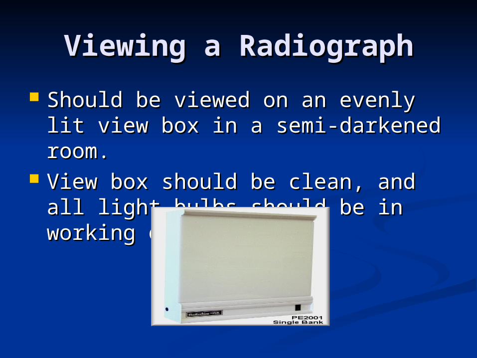
Viewing a RadiographViewing a Radiograph
Should be viewed on an evenly lit Should be viewed on an evenly lit view box in a semi-darkened room.view box in a semi-darkened room.
View box should be clean, and all View box should be clean, and all light bulbs should be in working light bulbs should be in working order. order.
Film View PositionFilm View Position
Film position is also important.Film position is also important. Usually follow medical viewing Usually follow medical viewing
protocol. protocol. V/D or D/V anatomy should be placed V/D or D/V anatomy should be placed
on the top of the view screen.on the top of the view screen. Lateral views should be positioned to Lateral views should be positioned to
face viewer’s left with spine at the top.face viewer’s left with spine at the top.

Correct Lateral Correct Lateral PositioningPositioning

Correct V/D positioningCorrect V/D positioning

Evaluation of Radiographic Evaluation of Radiographic TechniqueTechnique
Ask two basic questions when Ask two basic questions when evaluating a radiograph.evaluating a radiograph. 1. Is the film too light or too dark?1. Is the film too light or too dark?
2. Is there proper penetration?2. Is there proper penetration?

Is the film too light or Is the film too light or too dark?too dark?
The more exposure, the blacker the The more exposure, the blacker the film.film.
The less exposure, the lighter the The less exposure, the lighter the film.film. (opposite from photography). (opposite from photography).
Will most likely need to increase or Will most likely need to increase or decrease kVp or mAs.decrease kVp or mAs. To know which one to adjust, must To know which one to adjust, must
answer second question.answer second question.

Is there proper Is there proper penetration?penetration?
If there is inappropriate penetration If there is inappropriate penetration of the x-radiation, then the kVp of the x-radiation, then the kVp should be changed.should be changed. If film is dark, should be decreased.If film is dark, should be decreased. If film is light, should be increased.If film is light, should be increased.
If penetration of x-radiation is If penetration of x-radiation is satisfactory, then mAs should be satisfactory, then mAs should be adjusted.adjusted.

If film is too lightIf film is too light
Is the film under penetrated?Is the film under penetrated?
If no: Increase mAs 30-50%If no: Increase mAs 30-50%
If yes: Increase the kVp 10-15%If yes: Increase the kVp 10-15%

If film is too darkIf film is too dark
Is the film over penetrated?Is the film over penetrated?If yes: Decrease kVp by 10-15%If yes: Decrease kVp by 10-15%
If no: Decrease mAs by 30-50%.If no: Decrease mAs by 30-50%.

What determines Adequate What determines Adequate Penetration of X-rays?Penetration of X-rays?
When viewing an abdominal When viewing an abdominal radiograph, should be able to see radiograph, should be able to see outlines of liver, spleen, kidneys and outlines of liver, spleen, kidneys and bowel.bowel.
Inadequate penetration has areas Inadequate penetration has areas that appear almost completely white.that appear almost completely white.

Not good penetrationNot good penetration

Good Penetration Good Penetration

Too Dark RadiographsToo Dark Radiographs
If bone tissue is gray and there is not If bone tissue is gray and there is not much contrast between the bone and much contrast between the bone and adjacent soft tissue, there was too adjacent soft tissue, there was too much penetration. much penetration. Decrease kVp by 10-15%Decrease kVp by 10-15%
If bone tissue is relatively white as If bone tissue is relatively white as compared to surrounding tissues, then compared to surrounding tissues, then there was not too much penetration.there was not too much penetration. Decrease mAs 30-50%Decrease mAs 30-50%

Other Error Other Error ConsiderationsConsiderations
Exhausted chemicalsExhausted chemicals Poor development techniquesPoor development techniques Darkroom issuesDarkroom issues
Not lightproofNot lightproof Film issuesFilm issues