Embed Size (px)
Citation preview

1
RADIO TELEMETRY DEVICES TO MONITOR BREATHING IN NON-
SEDATED ANIMALS
Nathalie Samson, Sylvain Dumont, Marie-Laure Specq, Jean-Paul Praud
Neonatal Respiratory Research Unit, Departments of Pediatrics and Physiology,
Université de Sherbrooke, QC, Canada – J1H 5N4
Address for correspondence and proofs:
Jean-Paul Praud MD PhD Phone: 1 (819) 346-1110, ext 14851
Departments of Pediatrics and Physiology Fax: 1 (819) 564-5215 Université de Sherbrooke email: [email protected]
J1H 5N4, QC Canada

2
ABSTRACT
Radio telemetry equipment has significantly improved over the last 10-15 years and is
increasingly being used in research for monitoring a variety of physiological parameters
in non-sedated animals. The aim of this review is to provide an update on the current
state of development of radio telemetry for recording respiration. Our literature review
found only rare reports of respiratory studies via radio telemetry. Much of this article will
hence report our experience with custom-built radio telemetry devices designed for
recording respiratory signals, together with numerous other physiological signals in
lambs. Our current radio telemetry system allows to record 24 simultaneous signals
24h/day for several days. To our knowledge, this is the highest number of physiological
signals, which can be recorded wirelessly. Our devices have been invaluable for
studying respiration in our ovine models of preterm birth, reflux laryngitis, postnatal
exposure to cigarette smoke, respiratory syncytial virus infection and nasal ventilation,
all of which are relevant to neonatal respiratory problems.
Keywords: Wireless recording, polysomnography, lamb, apnea, sleep

3
1. INTRODUCTION
Over the past few decades, scientists have adapted and developed, with the help of
emerging technologies, various radio telemetry systems in their quest of studying and
understanding biological functions. Radio telemetry is now considered state of the art in
collecting numerous physiological signals in non-sedated, unrestrained animals,
compared to conventional measurement techniques using hardwired equipment. It is
generally acknowledged that the quality of physiological measurements collected from
conscious, unstressed animals is superior, since they are collected under conditions
that best represent the normal state of the animal and, when appropriate, are most
predictive of the results that would be achieved in human beings (Kramer and Kinter,
2003).
Radio telemetry systems combine miniature sensors and transmitters to detect and
transmit biopotentials in animals to remote receivers via an antenna. If necessary, the
receiver converts the analog frequency signal into a digital signal to be computerized
into a data acquisition system. There are several reliable, more or less invasive radio
telemetry systems that are commercially available for simultaneous monitoring of up to
5 physiological signals in laboratory animals 1. Radio telemetry systems for recording
cardiovascular variables, including electrocardiogram (ECG), heart rate and blood
pressure were initially developed for drug development companies (Brooks et al., 1996;
Butz and Davisson, 2001; Kramer et al., 1993; Mills et al., 2000), in response to
1 Data Science International, DSI (St-Paul, MN, USA); Emka Technologies (Paris, France); Konigsberg Instruments (Pasadena, CA, USA), Mini Mitter (Sunriver, OR, USA); Remo Technologies Ltd (Salisbury, UK); and Telemetronics Biomedical BV (Wageningen, The Netherlands) (list is not exhaustive).

4
requirements by the FDA to submit continuous measurements of vital functions with
each new drug application. Together with body temperature (Dilsaver et al., 1992) and
activity indices (Mills et al., 2000), they have been the most frequently reported
physiological signals measured via radio telemetry, especially in rodents. In addition,
electroencephalogram (EEG) and electromyogram (EMG) have been recorded in a
number of animal species, including mice, rats, rabbits, cats, dogs, sheep and monkeys
(Antier et al., 1998; Champeroux et al., 1994; Herzog et al., 1993; Kant et al., 1995;
Kawai et al., 2008; Letourneau et al., 1999; Letourneau and Praud, 2003; Livezey and
Sparber, 1990; Meile and Zittel, 2002; Mumford and Wetherell, 2001; Nakajima et al.,
1996; Sei and Morita, 1996; Tang et al., 2005). However, until very recently, the use of
telemetry for studying respiration was restricted to a handful of reports on respiratory
rate derived from pleural pressure (Murphy et al., 1998) or arterial pressure waveforms
(reviewed in Kramer and Kinter, 2003).
A number of respiratory signals can theoretically be considered for inclusion in a radio
telemetry system designed for recording breathing in animals (Baekey et al., 2009; Al-
Khalidi et al., 2011). Airflow limitations and/or apneas can be identified using nasal
thermistor/thermocouple, nasal pressure cannula, nasal capnography, nasal or facial
mask pneumotachography or tracheal sounds via a microphone or a piezoelectric
sensor attached on the upper thorax or neck. Recording of rib cage and abdominal
movements (strain gauge or piezoelectric belts, impedancemetry, inductance
plethysmography), as well as respiratory efforts (diaphragm electromyography, pleural
or esophageal pressure) allows identification of central apneas. In addition, when used
with a sensor aimed at recognizing airflow limitation/apnea recording, such respiratory
5
signals allow apnea characterization as central, obstructive or mixed. Pulse oximetry
and PCO2 monitoring, either via end tidal capnography or transcutaneously, are unique
indices of efficacious breathing. Overall, while respiratory frequency can be derived
from every respiratory signal collected, tidal volume can only be measured by calibrated
impedance plethysmography, pneumotachograph and by respiratory metabolic
plethysmography chamber in small rodents. Each technique has its own distinct
advantages and limitations and should be chosen according to the research question,
the animal species and the experimental conditions. Also, in selected cases, it is
relevant to consider recording of respiratory signals such as electrical activity of
thoracic, abdominal or upper airway muscles.
To our knowledge, it is only very recently that wireless recording of some respiratory
signals has been made commercially available (see table 1). Our group has been
actively studying the perinatal control of breathing in lambs since the late 80’s, in an
attempt to better understand the pathophysiology of apneas of prematurity, apparent-life
threatening events and sudden infant death syndrome, with a special focus on laryngeal
physiology (Praud et al., 2006; Samson et al., 2007, 2008; St-Hilaire et al., 2007).
Although rapidly developing, the sophisticated telemetry systems at the time did not
allow respiration studies. For the purpose of our research needs, it became necessary
to design and build our own radio telemetry system to allow for simultaneous recording
of numerous biopotentials linked to respiration during prolonged polysomnographic
recordings in non-sedated, freely moving lambs. The present article provides an
overview of our telemetered recording systems and reports a few examples of the data

6
obtained in our various lamb models, while underscoring some limitations and our
attempts at improvement.

7
2. DESCRIPTION OF OUR TELEMETERED RECORDING SYSTEM
2.1 SURGICAL INSTRUMENTATION AND RECORDING ELECTRODES / PROBES
Chronic surgical instrumentation under general anesthesia (2% isoflurane, 30% N2O,
68% O2) is performed in the first few days of life in all lambs, in order to implant
indwelling catheters (for monitoring fluid or air pressures) and electrodes (for monitoring
biopotentials). Over the years, we have developed our own recording electrodes and
indwelling catheters. As previously described, two needle-electrodes are inserted into
the parietal cortex directly through the skull for electrocorticogram (ECoG) recording,
while a third needle-electrode is inserted under the scalp as a ground (Carreau et al.,
2011). Eye movements (EOG) are recorded with two custom-built silver electrodes
inserted subcutaneously close to the right eye socket. The electrocardiogram is
recorded with 2 needle-electrodes inserted under the periosteum of the 5th rib, on both
sides of the thorax, and directly glued on the rib (Carreau et al., 2011). Respiratory
muscle EMG activities (including diaphragm, glottal and/or pharyngeal muscles) are
recorded with custom-built bipolar electrodes, made from either right or 180° angled
gold connectors (Sullins Connector Solutions, Digi-Key Corporation, Thief River Falls,
MN, USA). These EMG electrodes are inserted perpendicularly into the muscular fibers
and then secured with a stitch. All indwelling catheters are made from silastic tubing
(0.03 in. ID, Dow Corning, Midland, MI) and inserted directly into either the carotid artery
(arterial pressure measurements) or between two tracheal rings (respiratory pressure
measurements). Leads from all electrodes are subcutaneously tunneled to a common
exit on the lamb’s back. All recording electrodes and catheters are protected by
8
bandages and by a jacket worn by the lamb. Immediately prior to the recordings, further
external instrumentation is added, including a J-type thermocouple for nasal flow
recording, thoracic and abdominal elastic bands for respiratory inductance
plethysmography and a Masimo pulse oximeter probe on the tail root. During
polysomnographic recordings, all catheters, electrodes and probes are connected to our
radio telemetry system housed in the lamb’s jacket. All procedures described, including
surgical instrumentation and subsequent polysomnographic recordings in lambs, have
been approved by the Ethics Committee on Animal Care and Experimentation of our
institution.
2.2 RADIO TELEMETRY SYSTEM
Our radio telemetry system can simultaneously transmit and record up to 24
physiological signals of interest for the study of sleep apneas in freely moving lambs.
These parameters include ECoG and EOG for scoring states of alertness, upper airway
and respiratory muscle EMGs, respiratory pressures, nasal flow and thoraco-abdominal
movements for detection and characterization of apneas, as well as cardio-vascular
data, such as ECG and blood pressure. Finally, we can also record arterial hemoglobin
saturation in O2 using pulse oximetry (SpO2).
2.2.1 Radio telemetry transmitters
Our current radio telemetry system is comprised of 3 different transmitters: 1) one 12-
channel analog transmitter for EMG (x 8), ECG, ECoG (x 2), EOG and ground; 2) one
8-channel digital transmitter for abdominal and thoracic movements, nasal flow (x 2)
and respiratory or arterial pressures (x 4); 3) one digital transmitter for SpO2, pulse rate,
9
signal strength and plethysmographic signal. The transmitters are housed in a jacket
with several pockets; each wire stemming from an implanted electrode or an external
probe, as well as each catheter, is connected to its specific transmitter, while protected
by the jacket in its external course.
Twelve-channel analog transmitter. In the past year, we have extended our initial 8-
channel analog radio telemetry system (Letourneau and Praud, 2003). Indeed, 4
additional biopotentials can now be collected with the latest version. The 12-channel
analog transmitter (dimensions 63 X 52 X 4 mm) is built using surface-mounted
technology and a four-layer printed circuit board (PCB). Each channel has its own
differential preamplifier, high-pass filter and low-pass filter, allowing gain and cutoff
frequency of each channel to be specifically adjusted to the biopotential to be
transmitted. The twelve channels include eight channels for EMG acquisition (frequency
range: 30-500 Hz, gain: 390), two channels for ECoG acquisition (0.16-500 Hz, gain:
10) and two other channels (frequency range 1.6-500 Hz) for EOG (gain: 107) and ECG
(gain: 10) acquisition (see block diagram in Figure1-A). The 12-channel radio telemetry
system uses time division multiplexing to transmit all channels on the same carrier. The
time division multiplexing assigns a different time interval of 70 µs to each channel. At
the end of the 12th channel, a synchronization interval of 140 µs, during which
transmission is interrupted, is added to ensure synchronization between receiver and
transmitter. All biopotentials are thus sampled at a rate of 1 KHz. Finally, the radio-
frequency (RF) carrier is frequency-modulated by this resulting signal, which is called
pulse amplitude modulation/time division multiplexing (PAM/TDM). The transmission
frequency can be adjusted within the biomedical band range for Canada (176-216
10
MHz); the frequency used herein is 183 MHz. The transmitter antenna is 33.5 cm long
and constructed with flexible wire (whip-type antenna). The system is designed to
transmit biopotentials up to a maximal distance of 2 meters.
Eight-channel digital transmitter. The digital transmitter (dimensions 63 X 51 X 14
mm) is built using surface-mounted technology PCB (as described above). Each
channel has its own differential preamplifier, high-pass and low-pass filter, allowing gain
and cutoff frequency of each channel to be specifically adjusted to the biopotential to be
transmitted. It comprises eight channels, including two channels for abdominal and
thoracic movements (frequency range: DC-150 Hz, gain: 300), two channels for nasal
flow (frequency range: DC-150 Hz, gain: 1750) and four channels for respiratory or
arterial pressure (frequency range: DC-150 Hz, gain: 500 and 1000) acquisition (see
block diagram in Figure 1-B). The 8 channels are multiplexed and sequentially sampled
by a 16-bit successive-approximation register (SAR) analog-to-digital converter (ADC)
at a rate of 300 samples per second per channel. ADC results are then sent to the
microcontroller which generates a frame comprising a framing byte (0xAA) followed by
the 16-bit ADC result of each of the eight channels. The frame is then transmitted at a
rate of 56 Kbps using a frequency shift keying (FSK) transmitter operating at 916 MHz,
which falls into the ISM (Industrial, Scientific and Medical) band for North America (902-
928 MHz). The transmitter antenna used is an ultra compact omni directional antenna
(ANT-916-JJB-RA, Antenna Factor, Merlin, OR). The system is designed to transmit
biopotentials at a maximal distance of 10 meters.
Masimo pulse oximetry transmitter. The Masimo pulse oximetry transmitter
(dimensions 63 X 54 X 11 mm) is built using surface mounted technology and includes
11
the MS-13 OEM Board from Masimo Corporation and the RF transmitter (see block
diagram in Figure 1-C). It also includes a 5 V step-up DC/DC converter to power the 5 V
based MS-13 OEM board from the 3.7 V Li-Ion battery. A microcontroller is used to
configure the MS-13 OEM board at power-up. The MS-13 OEM board then outputs a
proprietary message at a rate of 9600 bps that is wirelessly transmitted using a FSK
transmitter operating at 903 MHz, which falls into the ISM band for North America (902-
928 MHz), via an ultra compact omni directional antenna (ANT-916-JJB-RA, Antenna
Factor, Merlin, OR). The four signals transmitted are SpO2, heart rate, pulse wave and
signal strength. The system is designed to transmit the MS-13 information at a maximal
distance of 10 meters.
All transmitters are powered with an Ultralife Lithium-Ion rechargeable battery offering a
capacity of 900 mAh (UBP002, Ultralife Corporation, Newark, NY), which provides up to
16 h for both the 12-channel analog and the 8-channel digital transmitters and up to 8 h
for the Masimo pulse oximetry transmitter. All transmitters are inserted into a custom-
built photopolymer case designed by stereolithography.
2.2.2 Radio telemetry receivers
Twelve-channel analog receiver. As previously described (Letourneau and Praud,
2003), the receiving antenna (RG-59, GE Marquette Canada, Mississauga, ON,
Canada) is placed at about 2 m from the recorded lamb. The receiver demodulates,
synchronizes and demultiplexes the frequency-modulated PAM/TDM signal in order to
rebuild the original signals (Figure 2-A). The superheterodyne receiver provides the
demodulated signal and a receiver signal strength indicator (RSSI). A sudden change in
RSSI signal indicates the synchronization interval sent by the transmitter. This allows
12
synchronization of the demultiplexer clock with that of the multiplexer (transmitter). The
PAM/TDM signal can thus be demultiplexed by selecting the specific time interval
associated with each individual channel and low-pass filtered to recover the original
signal from each channel. For more flexibility, each channel has its own adjustable gain
amplifier. Finally, a low output impedance buffer allows connection of the receiver
outputs to a computer acquisition system (Biopac MP 150 + AcqKnowledge software
version, Biopac Systems, Goleta, CA, USA).
Eight-channel digital receiver. The demodulator unit of the 916 MHz FSK receiver is
combined with the antenna in the lamb’s recording room. The demodulated RF signal is
sent through an RS485 link to the reconstruction unit of the receiver located in an
adjacent room. A microcontroller analyzes the received data to detect the presence of
the framing byte (0xAA), identifying the beginning of a new frame (Figure 2-B). This
allows the synchronization and demultiplexing of the data to the appropriate channel.
The microcontroller then sequentially updates the 16-bit digital-to-analog converter
(DAC) of each channel at a rate of 300 samples per second per channel. Each channel
has its own adjustable gain amplifier and a 4th order, 150 Hz low-pass filter to recover
the original signal. Finally, a low output impedance buffer allows connection of the
receiver outputs to the Biopac computer acquisition system.
Masimo pulse oximetry receiver. The demodulator unit of the 903 MHz FSK receiver
is combined with the antenna in the lamb’s recording room. The demodulated RF signal
is sent through an RS485 link to the reconstruction unit of the receiver located in an
adjacent room. A microcontroller analyzes the received data to extract the desired
information from the Masimo proprietary message (Figure 2-C). This allows extraction of
13
the 8-bit autoscaled IR plethysmographic signal at a rate of 62.5 Hz and the SpO2,
pulse rate and signal strength values at a rate of 1 Hz. The microcontroller then
sequentially updates the 16-bit digital-to-analog converter (DAC) of the corresponding
channel at the appropriate rate. Each channel has its own adjustable gain amplifier.
Additionally, the plethysmographic signal channel has a 4th order, 31 Hz low-pass
reconstruction filter. Finally, a low output impedance buffer allows connection of the
receiver outputs to the Biopac computer acquisition system.
2.3 VIDEO RECORDING
Simultaneous video recordings are systematically performed to monitor the lamb’s
behavior in real time during polysomnographic recordings, using a webcam modified for
infrared vision (Microsoft, Lifecam, VX-3000). Adequate infrared lighting is ensured by
an infrared LED dome illuminator (CV-100 DE/DR, DLi Systems, Dubai, United Arab
Emirates). The video files, which are synchronized with the other physiological data by
the AcqKnowledge software, provide essential information, especially for recognizing
states of alertness and characterizing position and activity.
3. IN VIVO VALIDATION AND USE OF OUR RADIO TELEMETRY
SYSTEM
In vivo validation of the accuracy of each data channel has been performed for the three
radio telemetry transmitters. Indeed, data obtained with our 8/12-channel analog system
(EMG x 4-8, ECoG x 2, EOG x 1, ECG x 1) have been validated by comparing them
with those simultaneously recorded using conventional, hardwired equipment
(Letourneau and Praud, 2003). Results obtained with both systems were virtually

14
identical, especially including sleep stage scoring, bradycardia detection and
laryngeal/respiratory muscle EMG activity. In addition, data obtained with our 8-channel
digital transmitter (thoracic and abdominal movements x 2, nasal flow x 2 and pressure
x 4) were compared with results obtained with conventional, hardwired equipment.
Accordingly, thoracic and abdominal movements were simultaneously recorded with the
original, hardwired respiratory inductance plethysmography equipment (Respitrace,
Monitoring systems, Miami Beach, FL, USA) and with our wireless system in 5 non-
sedated newborn lambs. Results showed excellent and consistent concordance
between both systems for apnea detection and characterization, together with relative
variations in tidal volume amplitude. Nasal flow as well as respiratory or arterial
pressures (RX104A pressure transducer, Biopac Systems, Goleta, CA, USA) were also
compared and validated with standard, hardwired equipment. Indeed, nasal flow signal
was compared with airflow collected using a pneumotachograph (21070B + 47304A
flow transducer, Hewlett Packard, Hewlett-Packard Company, Palo Alto, CA, USA),
whereas respiratory and arterial pressures were compared with calibrated pressure
transducers (respectively Validyne, Northridge, CA and Trantec model 60-800,
American Edwards Laboratories, Santa Anna, CA). Results obtained for each
parameter were virtually identical to conventional measurements (unpublished data).
Finally, validation of our wireless Masimo pulse oximeter was performed similarly to our
previous wireless Nonin pulse oximeter system (Reix et al., 2005). Briefly, the sensors
(LNOP YI reflectance sensor from Masimo, Irvine, CA) of both our wireless Masimo
pulse oximeter and the standard hardwired Masimo Radical pulse oximeter were
attached externally at the root of the lamb’s tail for continuous monitoring of arterial
15
SpO2, heart rate and pulse wave. Under various O2 conditions, our wireless pulse
oximeter was as reliable as the standard Masimo pulse oximeter currently used in our
laboratory, for monitoring and detecting variations in SpO2 variations (unpublished
results).
Furthermore, after validation of the accuracy of each data channel, prolonged and
repeated polysomnographic recordings were conducted in 17 lambs (including 7
preterm lambs), to test whether our telemetry system was well adapted to our needs for
prolonged recordings. Continuous polysomnographic recordings (with battery changes
every 8 to 12h) were performed over a period of up to 7 days, totaling more than 1200 h
of recording. Overall, our radio telemetry system was shown to be particularly well
suited for studying cardio-respiratory function during the various sleep states in our non-
sedated, freely-moving preclinical ovine models, and over a period of at least 7 days.
Examples of polysomnographic recordings obtained in newborn lambs using our radio
telemetry system are illustrated in Figures 3, 4 and 5.
4. DISCUSSION
The present article reports the development and performance of a wireless radio
telemetry system for prolonged polysomnographic recordings of respiration and related
physiological signals in non-sedated, freely moving lambs. This comprehensive set of
devices allows to perform a complete and extensive assessment of breathing during all
states of alertness, including cardio-vascular consequences of apneas in large animals,
in conditions as close to normal as possible. To our knowledge, it offers the highest
number of physiological signals, which can be recorded simultaneously and wirelessly
16
throughout the various states of alertness. Given that restraints prevent the normal
cycling of states of alertness (Hegde et al., 2008; McLeod et al., 1979; Tang et al.,
2007), and can disturb normal breathing (Al-Qarawi, 2005; DeLorme and Moss, 2002),
such a system is an invaluable asset for a research program such as ours.
4.1 IMPORTANCE OF TELEMETRY IN BIOMEDICAL RESEARCH
Telemetry is now considered state of the art in collecting physiological signals in the
absence of sedation, compared to conventional systems in which animals are hardwired
to the recording devices, and consequently more stressed. In addition, telemetry allows
us to reduce animal use by increasing the number of physiological signals collected, as
well as the number of recording sessions in each animal. Hence, in addition to
increasing the odds of recording normal physiology (Kramer et al., 2001), the use of
telemetry in biomedical animal research also constitutes a major asset from an ethical
standpoint. Indeed, the resulting decreased invasiveness, together with the need for
fewer animals per experiment, clearly contributes to the refinement and reduction
components of the now inevitable “3 Rs concept” (refinement, reduction, replacement)
proposed 40 years ago by Russell and Burch (1959). Finally, the use of wireless
telemetry substantially avoids wire setup breakage, which can be frequent with
hardwired recording equipment. The success of radio telemetry, especially with drug
development companies, has led several equipment manufacturers to enter and
develop this specific market, mentioned in the introduction section.
4.2 TELEMETERED RECORDING OF BREATHING

17
As reviewed in the introduction section, an increasing number of physiological signals
can now be recorded via telemetry. Interestingly, while the use of radio telemetry has
initially been largely restricted to cardiovascular function in rodents, a clear trend in
recording additional physiological functions, including respiration, and in applying
telemetry to large animals, has appeared in the last decade.
Review of the literature reveals that only a limited number of research teams have
recorded respiration, using either custom-built or commercially available wireless
equipment. Microwave radiation recording of respiratory rate was first proposed in
rodents (Gordon and Ali, 1984) and was used more recently in hibernating black bears
(Suzuki et al., 2009). Impedance monitoring has recently been used in monkeys
(Authier et al., 2010) and dogs (Kearney et al., 2010). Respiratory rate has also been
derived from arterial pressure waveform in rodents (reviewed in Kramer and Kinter,
2003), and from pleural pressure in rats (Murphy et al., 1998), dogs (Ednick et al., 2007)
and monkeys (Carrier et al., 2010; Layton et al., 2011; Warren et al., 2011). Respiratory
inductive plethysmography has been used in dogs (Murphy et al., 2010). Finally, one
group has reported respiratory rate recording using custom-built equipment, namely an
acoustic sensor in swine and a thoracic or abdominal circumference in cattle (Eigenberg
et al., 2008).
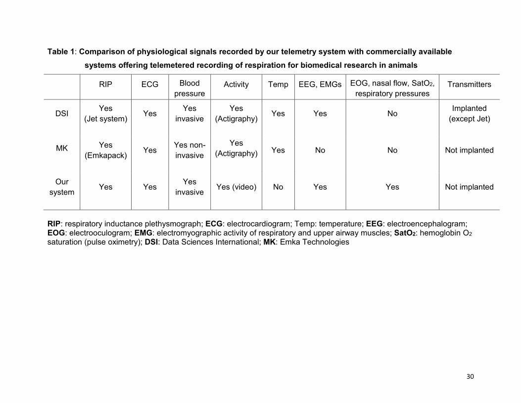
To our knowledge, wireless recording equipment of respiration integrated with other
physiological signals can now be purchased from two manufacturers. The main
differences with our system are presented in table 1. The most significant difference
relates to the greater number of physiological signals, which can be recorded with our
custom-built system (EEG, EOG, EMGs, nasal flow, SpO2, respiratory or arterial
18
pressures), making it uniquely suited to polysomnographic recordings. In addition, our
telemetry system can be used with any common data acquisition system, such as from
Biopac Systems, AD Instruments or National Instruments. This flexibility is a unique
advantage compared to commercially available telemetry systems, which work with their
proprietary software and are often very difficult to interface with common data
acquisition systems.
Throughout the years, our radio telemetry system has allowed us to study numerous
physiological signals related to neonatal breathing. Beyond nasal flow (thermocouples)
and thoraco-abdominal movements (respiratory inductance plethysmograph), we have
been particularly interested in EMG activities of respiratory muscles and upper airway
muscles. Thus, EMG of the diaphragm, laryngeal intrinsic muscles including the
thyroarytenoid muscle (glottal constrictor) and the cricothyroid muscle (glottal dilator), as
well as pharyngeal muscles such as the genioglossus and the levator palatini (figure 4)
have been recorded under various conditions.
The contribution of our telemetry system has proven to be an invaluable tool for
studying breathing anomalies in our various preclinical neonatal ovine models. These
unique models include apneas in premature lambs (Renolleau et al., 1999; St-Hilaire et
al., 2007), application of nasal CPAP (Samson et al., 2005, 2008) or intermittent positive
pressure ventilation (Roy et al., 2008), postnatal exposure to cigarette smoke (St-Hilaire
et al., 2010), reflux laryngitis (Carreau et al., 2011), respiratory syncytial virus infection
(unpublished results), all of which are relevant to neonatal respiratory problems. Overall,
review of the scientific literature suggests that our team has the most extensive and
consistent publication track record on respiration recording via radio telemetry.
19
4.3 LIMITATIONS OF OUR TELEMETRY SYSTEM – THE NEED FOR CONTINUOUS IMPROVEMENT
4.3.1 Surgical chronic instrumentation: a special concern
From our standpoint, while the jacket-worn transmitters fulfill the needs for minimal
invasiveness (and increased possibility of reuse in a number of animals over several
years), our current surgical, chronic instrumentation remains nevertheless too invasive.
This is of special concern within the context of our research program focused on the
early postnatal period. Indeed, in order to study this critical period, we are often obliged
to start recordings as early as 2 days after the 2-3 hour-long surgery necessary to
implant our indwelling catheters and electrodes, at a time where some post-surgery
stress is still present and could interfere with the results. We believe that a significant
improvement in our research design should come from the ability to decrease and even
avoid surgical stress whenever possible. Some progress has been made in the last
years to decrease this stress, e.g. by using thoracic videosurgery instead of
conventional thoracotomy (Roy et al., 2008; Samson et al., 2008). However, we are
continually committed to finding new ways to replace surgery by non-invasive
instrumentation as often as possible, without decreasing the quality of recorded
physiological signals. This is already virtually up and running for the respiratory
component of our recordings, by using thermocouples glued on the skin for nasal flow,
bands of respiratory inductance plethysmography stretched around the rib cage and
abdomen for respiratory movements and an oximeter probe attached to the lamb’s tail
root for blood oxygenation. However, as already pointed out, an important aspect of our
recordings is the ability to study respiration within a polysomnography setting. We are
20
thus currently assessing the best means to continuously record and recognize the
states of consciousness using electrophysiological signals (EEG, EOG) via
subcutaneous, needle-electrodes or external cups glued on the skin vs. infrared video
recordings in combination with EOG and regularity of respiration. Furthermore, we are
currently testing the possibility to chronically instrument laryngeal muscles, as well as
the crural diaphragm, using modified transcutaneous techniques (Insalaco et al., 1991;
Trelease et al., 1982).
4.3.2 Lingering technical problems with our telemetry system
While the current 8/12-channel analog transmitters offer good-quality signals in virtually
all conditions in our experimental setting, they provide more unstable/noisy signals than
the digital transmitters because they are more sensitive to the RF physical environment
changes in their close vicinity. Closer supervision is thus necessary, which is unpractical
or even impossible at night during round-the-clock recording. We eventually anticipate
replacing our analog transmitters by digital transmitters as soon as possible. In the
meantime, we are currently optimizing the analog transmitters to be less sensitive to
these physical environment changes.
Finally, the total weight of our 3 transmitters + batteries (340 g) represents a significant
load for the youngest, tiniest preterm lambs, thus generating some discomfort in the
standing position. Further improvements will be needed to decrease the size and weight
of the overall system, now that we intend to study preterm lambs on a more frequent
basis. In its current format, our radiotelemetry could be used in large animals such as
cats, dogs, rabbits, swine, monkeys, sheep and goats, but is not adapted to the study of
small rodents.
21
5. CONCLUSION
Continuous telemetered recording of respiration has been made available commercially
within the last 2 years, especially in response to FDA requirements for new drug
applications. In our laboratory, we have rather progressively developed our own radio
telemetry system over the last 15 years to fulfill our needs for polysomnographic
recordings of neonatal breathing in ovine models. To our knowledge, the system
presented herein allows the simultaneous recording of the highest number of
physiological signals via telemetry, and has allowed us to gain a unique expertise in
radio telemetry recording of breathing. Obviously, telemetered recording of breathing
offers great promise for biomedical animal research and a worldwide spread of this
technology is predictable in the forthcoming years.
ACKNOWLEDGEMENTS
The authors acknowledge the technical assistance of Jean-Philippe Gagné. This work is
funded by the Canadian Institutes of Health (MOP 15558). Jean-Paul Praud is a
member of the FRSQ-funded Clinical Research Center Étienne-Le Bel, Sherbrooke
University Hospital, and the holder of the Canada Research Chair in Neonatal
Respiratory Physiology. Marie-Laure Specq holds a doctoral scholarship from the Lung
Association, Canadian Thoracic Society and a Merit scholarship for foreign students in
University from the Ministère de l'Education, du Loisir et du Sport du Québec.
22
REFERENCES
Al-Khalidi, F.Q., Saatchi, R., Burke, D., Elphick, H., Tan, S., 2011. Respiration rate
monitoring methods: a review. Pediatr. Pulmonol. 46, 523-529.
Al-Qarawi, A.A., 2005. Immobilization (restraint) stress in desert sheep and goats, and
the influence of pretreatment with xylazine or sodium betaine thereon. Pol. J. Vet. Sci.
8, 73-78.
Antier, D., Zhang, B.L., Mailliet, F., Akoka, S., Pourcelot, L., Sannajust, F., 1998. Effects
of neonatal focal cerebral hypoxia-ischemia on sleep-waking pattern, ECoG power
spectra and locomotor activity in the adult rat. Brain Res. 807, 29-37.
Authier, S., Haefner, P., Fournier, S., Troncy, E., Moon, L.B., 2010. Combined
cardiopulmonary assessments with implantable telemetry device in conscious freely
moving cynomolgus monkeys. J. Pharmacol. Toxicol. Methods 62, 6-11.
Baekey, D.M., Feng, P., Decker, M.J., Strohl, K.P., 2009. Breathing and sleep:
measurement methods, genetic influences, and developmental impacts. ILAR J. 50,
248-261.
Brooks, D., Horner, R.L., Kozar, L.F., Waddell, T.K., Render, C.L., Phillipson, E.A.,
1996. Validation of a telemetry system for long-term measurement of blood pressure. J.
Appl. Physiol. 81, 1012-1018.
Butz, G.M., Davisson, R.L., 2001. Long-term telemetric measurement of cardiovascular
parameters in awake mice: a physiological genomics tool. Physiol. Genomics 5, 89-97.
23
Carreau, A-M., Patural, H., Samson, N., Doueik, A.A., Hamon, J., Fortier, P-H., Praud,
J-P., 2011. Effects of Simulated Reflux Laryngitis on Laryngeal Chemoreflexes in
Newborn Lambs. J. Appl. Physiol. In press.
Carrier, C.A., Elliott, T.B., Ledney, G.D., 2010. Real-time telemetric monitoring in whole-
body 60Co gamma-photon irradiated rhesus macaques (Macaca mulatta). J. Med.
Primatol. 39, 399-407.
Champeroux, P., Lala, P., Richard, S., 1994. Use of telemetry for electroencephalogram
monitoring in conscious rabbits. Edinburgh: Br. Toxicol. Soc.
DeLorme, M.P., Moss, O.R., 2002. Pulmonary function assessment by whole-body
plethysmography in restrained versus unrestrained mice. J. Pharmacol. Toxicol.
Methods 47, 1-10.
Dilsaver, S.C., Overstreet, D.H., Peck, J.A., 1992. Measurement of temperature in the
rat by rectal probe and telemetry yields compatible results. Pharmacol. Biochem.
Behav. 42, 549-552.
Ednick, M.D., Pagala, M., Barakat, J.P., Nino, G., Shah, P., Cunningham, J.N.,Jr,
Vaynblat, M., Kazachkov, M., 2007. Telemetric recording of intrapleural pressure. J.
Surg. Res. 138, 10-14.
Eigenberg, R.A., Brown-Brandl, T.M., Nienaber, J.A., 2008. Sensors for dynamic
physiological measurements. Comput. Electron. Agric. 62: 41-47.
24
Gordon, C.J., Ali, J.S., 1984. Measurement of ventilatory frequency in unrestrained
rodents using microwave radiation. Respir. Physiol. 56, 73-79.
Hegde, P., Singh, K., Chaplot, S., Shankaranarayana Rao, B.S., Chattarji, S., Kutty,
B.M., Laxmi, T.R., 2008. Stress-induced changes in sleep and associated neuronal
activity in rat hippocampus and amygdala. Neuroscience 153, 20-30.
Herzog, W., Stano, A., Leonard, T.R., 1993. Telemetry system to record force and EMG
from cat ankle extensor and tibialis anterior muscles. J. Biomech. 26, 1463-1471.
Insalaco, G., Kuna, S.T., Costanza, B.M., Catania, G., Cibella, F., Bellia, V., 1991.
Thyroarytenoid muscle activity during loaded and nonloaded breathing in adult humans.
J. Appl. Physiol. 70, 2410-2416.
Kant, G.J., Pastel, R.H., Bauman, R.A., Meininger, G.R., Maughan, K.R., Robinson,
T.N.,3rd, Wright, W.L., Covington, P.S., 1995. Effects of chronic stress on sleep in rats.
Physiol. Behav. 57, 359-365.
Kawai, N., Tanaka, E., Langenbach, G.E., van Wessel, T., Sano, R., van Eijden, T.M.,
Tanne, K., 2008. Jaw-muscle activity changes after the induction of osteoarthrosis in the
temporomandibular joint by mechanical loading. J. Orofac. Pain 22, 153-162.
Kearney, K., Metea, M., Gleason, T., Edwards, T., Atterson, P., 2010. Evaluation of
respiratory function in freely moving Beagle dogs using implanted impedance
technology. J. Pharmacol. Toxicol. Methods 62, 119-126.
25
Kramer, K., van Acker, S.A., Voss, H.P., Grimbergen, J.A., van der Vijgh, W.J., Bast, A.,
1993. Use of telemetry to record electrocardiogram and heart rate in freely moving
mice. J. Pharmacol. Toxicol. Methods 30, 209-215.
Kramer, K., Kinter, L., Brockway, B.P., Voss, H.P., Remie, R., Van Zutphen, B.L., 2001.
The use of radio telemetry in small laboratory animals: recent advances. Contemp. Top.
Lab. Anim. Sci. 40, 8-16.
Kramer, K., Kinter, L.B., 2003. Evaluation and applications of radio telemetry in small
laboratory animals. Physiol. Genomics 13, 197-205.
Layton, R.C., Brasel, T., Gigliotti, A., Barr, E., Storch, S., Myers, L., Hobbs, C., Koster,
F., 2011. Primary pneumonic plague in the African Green monkey as a model for
treatment efficacy evaluation. J. Med. Primatol. 40, 6-17.
Letourneau, P., Dumont, S., Kianicka, I., Diaz, V., Dorion, D., Drolet, R., Praud, J-P.,
1999. Radio telemetry system for apnea study in lambs. Respir. Physiol. 116, 85-93.
Letourneau, P., Praud, J-P., 2003. A radio telemetry system for polysomnographic
recordings in lambs. Methods 30, 115-121.
Livezey, G.T., Sparber, S.B., 1990. Hyperthermia sensitizes rats to cocaine's
proconvulsive effects and unmasks EEG evidence of kindling after chronic cocaine.
Pharmacol. Biochem. Behav. 37, 761-767.
26
McLeod, C.N., Harding, R., Johnson, P., McClelland, M.E., Whyte, P.L., 1979. Studies
on the control of respiration and behaviour during development in ewe-reared lambs.
Biotelem. Patient Monit. 6, 171-175.
Meile, T., Zittel, T.T., 2002. Telemetric small intestinal motility recording in awake rats: a
novel approach. Eur. Surg. Res. 34, 271-274.
Mills, P.A., Huetteman, D.A., Brockway, B.P., Zwiers, L.M., Gelsema, A.J., Schwartz,
R.S., Kramer, K., 2000. A new method for measurement of blood pressure, heart rate,
and activity in the mouse by radio telemetry. J. Appl. Physiol. 88, 1537-1544.
Mumford, H., Wetherell, J.R., 2001. A simple method for measuring EEG in freely
moving guinea pigs. J. Neurosci. Methods 107, 125-130.
Murphy, D.J., Renninger, J.P., Gossett, K.A., 1998. A novel method for chronic
measurement of pleural pressure in conscious rats. J. Pharmacol. Toxicol. Methods 39,
137-141.
Murphy, D.J., Renninger, J.P., Schramek, D., 2010. Respiratory inductive
plethysmography as a method for measuring ventilatory parameters in conscious, non-
restrained dogs. J. Pharmacol. Toxicol. Methods 62, 47-53.
Nakajima, M., Sakai, T., Mizumoto, A., Itoh, Z., 1996. Development of a new telemetry
recording system for measuring of gastrointestinal contractile activity in unrestrained
and conscious small animals. J. Smooth Muscle Res. 32, 1-7.

27
Praud, J-P., Samson, N., Moreau-Bussiere, F., 2006. Laryngeal function and nasal
ventilatory support in the neonatal period. Paediatr. Respir. Rev. 7 Suppl 1, S180-2.
Reix, P., Dumont, S., Duvareille, C., Cyr, J., Moreau-Bussiere, F., Arsenault, J., Praud,
J-P., 2005. Monitoring pulse oximetry via radio telemetry in freely-moving lambs. Respir.
Physiol. Neurobiol. 147, 65-72.
Renolleau, S., Letourneau, P., Niyonsenga, T., Praud, J-P., 1999. Thyroarytenoid
muscle electrical activity during spontaneous apneas in preterm lambs. Am. J. Respir.
Crit. Care Med. 159, 1396-1404.
Roy, B., Samson, N., Moreau-Bussiere, F., Ouimet, A., Dorion, D., Mayer, S., Praud, J-
P., 2008. Mechanisms of active laryngeal closure during noninvasive intermittent
positive pressure ventilation in nonsedated lambs. J. Appl. Physiol. 105, 1406-1412.
Russell, W.M.S., Burch, R.L., 1959. The Principles of Humane Experimental Technique.
London: Methuen.
Samson, N., St-Hilaire, M., Nsegbe, E., Reix, P., Moreau-Bussiere, F., Praud, J-P.,
2005. Effect of nasal continuous or intermittent positive airway pressure on nonnutritive
swallowing in the newborn lamb. J. Appl. Physiol. 99, 1636-1642.
Samson, N., Lafond, J.R., Moreau-Bussiere, F., Reix, P., Praud, J-P., 2007.
Cricothyroid muscle electrical activity during respiration and apneas in lambs. Respir.
Physiol. Neurobiol. 155, 147-155.
28
Samson, N., Roy, B., Ouimet, A., Moreau-Bussiere, F., Dorion, D., Mayer, S., Praud, J-
P., 2008. Origins of the inhibiting effects of nasal CPAP on nonnutritive swallowing in
newborn lambs. J. Appl. Physiol. 105, 1083-1090.
Sei, H., Morita, Y., 1996. Acceleration of EEG theta wave precedes the phasic surge of
arterial pressure during REM sleep in the rat. Neuroreport 7, 3059-3062.
St-Hilaire, M., Samson, N., Nsegbe, E., Duvareille, C., Moreau-Bussiere, F., Micheau,
P., Lebon, J., Praud, J-P., 2007. Postnatal maturation of laryngeal chemoreflexes in the
preterm lamb. J. Appl. Physiol. 102, 1429-1438.
St-Hilaire, M., Duvareille, C., Avoine, O., Carreau, A-M., Samson, N., Micheau, P.,
Doueik, A., Praud, J-P., 2010. Effects of postnatal smoke exposure on laryngeal
chemoreflexes in newborn lambs. J. Appl. Physiol. 109, 1820-1826.
Suzuki, S., Matsui, T., Kawahara, H., Gotoh, S., 2009. Development of a noncontact
and long-term respiration monitoring system using microwave radar for hibernating
black bear. Zoo Biol. 28, 259-270.
Tang, X., Xiao, J., Parris, B.S., Fang, J., Sanford, L.D., 2005. Differential effects of two
types of environmental novelty on activity and sleep in BALB/cJ and C57BL/6J mice.
Physiol. Behav. 85, 419-429.
Tang, X., Yang, L., Sanford, L.D., 2007. Interactions between brief restraint, novelty and
footshock stress on subsequent sleep and EEG power in rats. Brain Res. 1142, 110-
118.

29
Trelease, R.B., Sieck, G.C., Harper, R.M., 1982. A new technique for acute and chronic
recording of crural diaphragm EMG in cats. Electroencephalogr. Clin. Neurophysiol. 53,
459-462.
Warren, R., Lockman, H., Barnewall, R., Krile, R., Blanco, O.B., Vasconcelos, D., Price,
J., House, R.V., Bolanowksi, M.A., Fellows, P., 2011. Cynomolgus macaque model for
pneumonic plague. Microb. Pathog. 50, 12-22.
30
Table 1: Comparison of physiological signals recorded by our telemetry system with commercially available systems offering telemetered recording of respiration for biomedical research in animals
RIP ECG Blood pressure
Activity Temp EEG, EMGs EOG, nasal flow, SatO2, respiratory pressures
Transmitters
DSI Yes (Jet system) Yes Yes
invasive Yes
(Actigraphy) Yes Yes No Implanted (except Jet)
MK Yes (Emkapack) Yes Yes non-
invasive Yes
(Actigraphy) Yes No No Not implanted
Our system Yes Yes Yes
invasive Yes (video) No Yes Yes Not implanted
RIP: respiratory inductance plethysmograph; ECG: electrocardiogram; Temp: temperature; EEG: electroencephalogram; EOG: electrooculogram; EMG: electromyographic activity of respiratory and upper airway muscles; SatO2: hemoglobin O2 saturation (pulse oximetry); DSI: Data Sciences International; MK: Emka Technologies

31
FIGURE LEGENDS
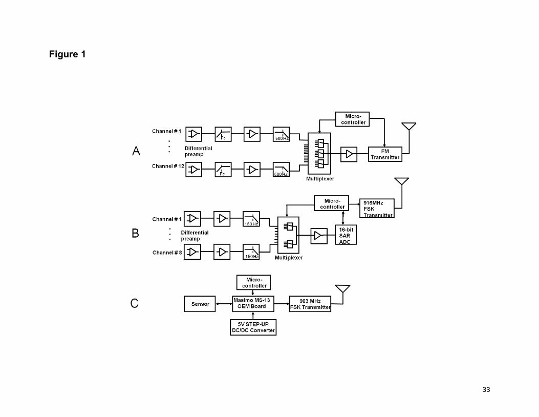
Figure 1: Block diagram of the transmitters
(A) 12-channel analog, (B) 8-channel digital and (C) Masimo pulse oximetry
transmitters.
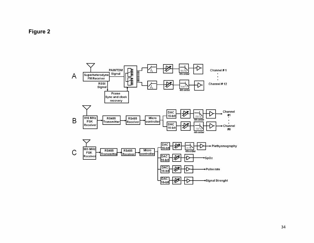
Figure 2: Block diagram of the receivers
(A) 12-channel analog, (B) 8-channel digital and (C) Masimo pulse oximetry receivers.
Figure 3: Respiration during quiet sleep in a newborn lamb
ECoG (electrocorticogram), EOG (electrooculogram), TA [electrical activity (EMG) of
the thyroarytenoid muscle, a glottal constrictor muscle], ∫TA (integrated TA EMG) and
ECG (electrocardiogram) were transmitted via our 12-channel analog system. Thorax
and Abdomen (thoracic and abdominal respiratory movements recorded via respiratory
inductance plethysmography, with inspiration upward), AP (arterial pressure, mmHg,
recorded via a liquid-filled carotid catheter-transducer system) were transmitted via our
8-channel digital system; SpO2 (oxygen hemoglobin saturation, %) and Pulse wave
(pulse wave recorded by a Masimo pulse oximetry probe) were transmitted via our 4-
channel Masimo digital system.
32
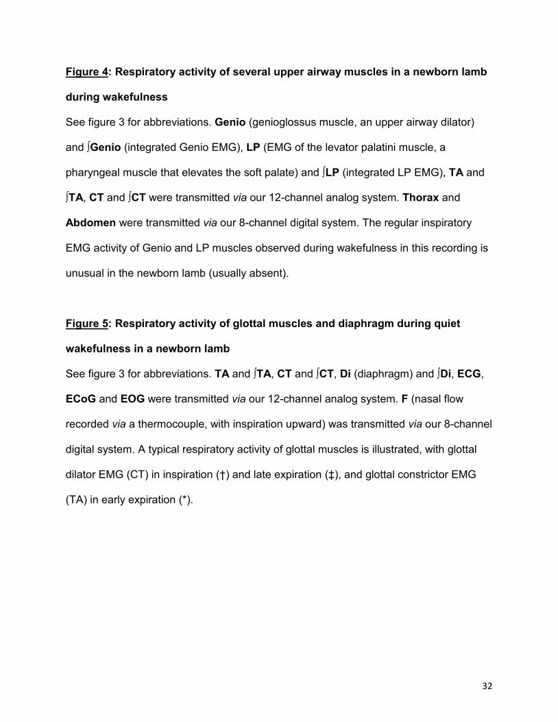
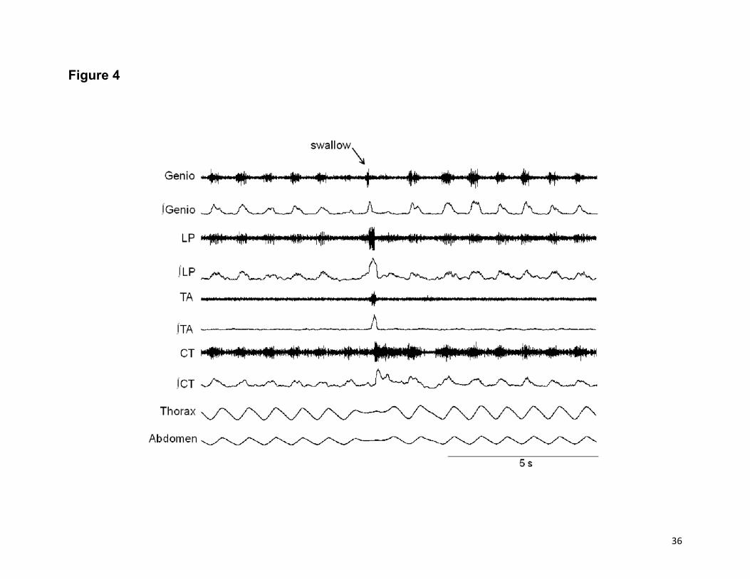
Figure 4: Respiratory activity of several upper airway muscles in a newborn lamb
during wakefulness
See figure 3 for abbreviations. Genio (genioglossus muscle, an upper airway dilator)
and ∫Genio (integrated Genio EMG), LP (EMG of the levator palatini muscle, a
pharyngeal muscle that elevates the soft palate) and ∫LP (integrated LP EMG), TA and
∫TA, CT and ∫CT were transmitted via our 12-channel analog system. Thorax and
Abdomen were transmitted via our 8-channel digital system. The regular inspiratory
EMG activity of Genio and LP muscles observed during wakefulness in this recording is
unusual in the newborn lamb (usually absent).
Figure 5: Respiratory activity of glottal muscles and diaphragm during quiet
wakefulness in a newborn lamb
See figure 3 for abbreviations. TA and ∫TA, CT and ∫CT, Di (diaphragm) and ∫Di, ECG,
ECoG and EOG were transmitted via our 12-channel analog system. F (nasal flow
recorded via a thermocouple, with inspiration upward) was transmitted via our 8-channel
digital system. A typical respiratory activity of glottal muscles is illustrated, with glottal
dilator EMG (CT) in inspiration (†) and late expiration (‡), and glottal constrictor EMG
(TA) in early expiration (*).
33
Figure 1
34
Figure 2

35
Figure 3
36
Figure 4

37
Figure 5
TA
∫TA
∫CT
∫Di
CT
Di
F
ECG
ECoG
EOG