Embed Size (px)
Citation preview

Radiation Safety● Atomic and Nuclear Structure● Radioactive Decay● Interaction of Radiation with Matter● Time, Distance and Shielding● Quantities and Units● Internal Radiation Dose● Annual Dose Limits● Natural Background and Average Population Doses● Biological Effects of Ionizing Radiation● Radiation Protection Procedures

Atomic and Nuclear Structure
● Atom● Nucleus
– Nucleons● Electrons
● Atomic Number, Z, and Atomic Mass Number, A● Nomenclature: ● Nuclide, Element, Isotope, Isobar
● Energy Units● eV
– Most nuclear interactions keV - MeV

Radioactive Decay IRadionuclide releasing (or capturing) particles
and energy in transitioning to a more stable state● Nuclear stability depends in a complex way on
atomic mass, neutron/proton ratio, number (evenness; “magic”) of nucleons

Radioactive Decay II
● Decay rate● Decay Constant,● Half-life,
● Activity● Curie: 1 Ci = 3.7 x 10¹⁰ disintegrations per second
(dps) ● Becquerel: 1 Bq = 1 dps [SI unit, but too small]
● Random process; number depends on number of atoms

Radioactive Decay III
● Types of Radiation● Particulate
– Alpha– Beta(-/+)
● Electromagnetic– X-ray– Gamma ray

Radioactive Decay IV
● Types of Radioactive Decay● Alpha (α): ΔZ = -2; ΔA = -4
● Beta (β-): ΔZ = +1; ΔA = 0
● Electron Capture: ΔZ = -1; ΔA = 0; typically X-ray● Positron (β+) Emission: ΔZ = -1; ΔA = 0;
annihilation => two photons● Internal Transition or Gamma (γ): ΔZ = 0; ΔA = 0● Internal Conversion: ΔZ = 0; ΔA = 0;γ knocks out
electron => X-ray

Interaction of Radiation with Matter I● Ionizing vs Non-Ionizing Radiation
● Ionizing radiation can remove orbital electrons● Non-ionizing radiation cannot modify atoms
● Consequences of Interactions● Ionizing
– Creation of ion pairs● Electrons may be energetic enough to cause ionization● Such delta rays may break molecular bonds and create free
radicals
– Excitation raises temperatures and may break bonds● Non-ionizing
– Heat– Ultraviolet can induce damaging photochemical reactions
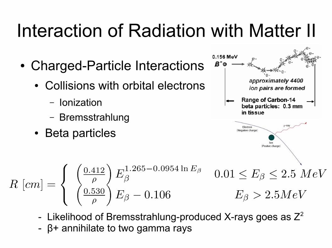
Interaction of Radiation with Matter II
● Charged-Particle Interactions● Collisions with orbital electrons
– Ionization– Bremsstrahlung
● Beta particles
- Likelihood of Bremsstrahlung-produced X-rays goes as Z2
- β+ annihilate to two gamma rays

Interaction of Radiation with Matter III
● Alpha particles● Massive; tend to be slow● Leave dense, straight, short paths of ions

Interaction of Radiation with Matter IV
● Electromagnetic Radiation, gamma and X-Rays● Indirect Ionization● Photoelectric Effect
– Probability goes as Z4 and 1/E3
● Compton Scattering– Weak inverse dependence on Z
● Pair Production– Probability goes as Z2

Radiation Protection:Time, Distance, and Shielding
● Time● Length of exposure minimized● Radioactive sources stored and allowed to decay
● Distance● Intensity decreases as the inverse square of the
distance from a point source● Never hold sources directly

Radiation Protection:Time, Distance, and Shielding
● Shielding—to reduce, not eliminate exposure● In shield design, measure effectiveness under
worst-case working conditions. Never assume shielding is correct unless tested.
● Attenuation depends on material and radiation type and energy
● Charged Particle Attenuation– Charged particles (especially alpha particles) tend to
have relatively short range, but this is energy dependent

Shielding, cont.
– Beta particles● Shielding thickness usually full range, but beta energy spread
(50% have energies < Emax allow for thinner shielding against beta particles. However, high-energy beta particles can scatter with significant energy and bremsstrahlung.
Bare source
**
*

Shielding, cont.
– X-Rays and Gamma Rays● No fixed range● Interaction (photoelectric absorption, Compton scattering, pair
production) probability dependent on photon energy and Z of shielding material
● Characterized by an attenuation coefficient, μ, which has units of L-1, per unit thickness of material
● Ignoring (very prevalent) scattering:

Quantities and Units I
● Exposure: ionization in air● The charge produced per unit mass of air by x-rays
or gamma rays as they traverse a collecting volume● Defined only for x-rays and low energy gamma rays
when measured in air.● Roentgen, R, (traditional) and C/kg

Quantities and Units II
● Absorbed Dose: energy deposition in material● Energy absorbed per unit mass
– Traditional: 1 rad = 100 erg/g– SI: gray: 1 Gy = 1 J/kg = 100 rad
● Varies with energy, type of radiation, and depth of penetration– Reference depths: 0.007 cm for skin, 0.3 cm for eye lens,
1 cm for total body (deep dose)● For low Z material, exposure ≈ absorbed does
– 1 R ≈ 0.87 rad in air; 1 R ≈ 0.92 rad in small tissue volume
Quantities and Units III
● Quality Factor, Q● The relative effectiveness of a form of radiation to
produce biological damage● Accounts for differing degrees of hazard from the
different types of radiation● alphas and heavy particles : neutrons : betas and
gammas :: 20 : 10 : 1● Damage related to both absorbed dose and quality
factor

Quantities and Units IV
● Dose Equivalent: risk of harm● = absorbed dose x quality factor● Traditional unit: roentgen equivalent man, rem● SI unit: Seivert, Sv
1 Sv = 100 rem

Rates
● Exposure per unit time: mR/hr● Absorbed does per unit time: mrad/hr● Dose equivalent per unit time: mrem/hr● Counts per unit time: cpm (counts per minute)

Estimating Radiation Levels I
● Gamma exposure● Characterized by the specific gamma ray constant, Γ
X is the gamma exposure rate, A the gamma source activity, and d the distance from the gamma source

Estimating Radiation Levels II
● Beta dose rates● Beta particles deposit their energy in a short
distance– Easy to shield– Energy deposition very efficient
● Beta emitters of > 0.2 MeV readily produce a superficial dose to the skin.

Internal Radiation Dose I
● Involves both dosimetry and physiology; large uncertainties
● Uptake● inhalation, ingestion, absorption, or injection● Estimated from animal studies and uptake or
transfer coefficients model a “standard” man
● Inter-organ transfer● Again, may be substantial individual differences

Internal Radiation Dose II
● Residence time● Effective decay constant: combination of biological
elimination and physical decay
● Energy deposition● Essentially all particulate radiation energy created
in an organ will be absorbed in that organ.● Electromagnetic radiation absorbed fraction
depends on the organ size and the radiation energy

Annual Dose Limits● Occupational
● Total Effective Dose Equivalent (“Whole-body”)– Combines external and internal– 0.05 Sv = 5 rem
● Eye lens: 0.15 Sv = 15 rem● Skin and extremity (0.007 cm) and all other organs:
0.5 Sv = 50 rem● For minors, 10% of above● Pregnant woman who has declared pregnancy: 0.5
mSv = 0.5 rem
● Public (due to industrial sources): 1 mSv = 0.1 rem/yr; 0.02 mSv = 2 mrem/hr

Natural Background and Average Population Doses I
● Cosmic radiation● Sea level: 30 mrem/yr● 1 km: 40 mrem/yr● Long-haul jet: 1 mrem/hr● C-14: 1 mrem/yr
● Terrestrial● External: 30 – 140 mrem/yr● Radon (inhale): 2.4 rem/yr

Natural Background and Average Population Doses II
● Internally Deposited (in muscle) Radionuclides, 40K: 40 mrem/yr
● Subtotal (Natural Background Radiation Dose Equivalent): ~300 mrem/yr
● Medical Radiations: ~50 mrem/yr● Miscellaneous Dose: ~10 mrem/yr● Total Average Population Dose: ~360 mrem/yr

Biological Effects of Ionizing Radiation I
● Sequential Pattern● Latency: broad range, minutes – generations● Demonstrable effects: cessation of or abnormal
mitosis, chromosome clumping, and giant cell formation
● Recovery: Usually apparent only after short-term (< months) effects; irreparable damage → long-term effects

Biological Effects of Ionizing Radiation II
● Determinants● Dose response curve● Absorption rate● Area exposed● Species and individual sensitivity variation
– LD50/30 (kill 50% in 30 days; for humans 450 rad)
● Cell sensitivity variation– (most) white blood cells; red blood cells; epithelial cells
(especially GI tract); muscle cells; nerve cells (least)

Biological Effects of Ionizing Radiation III
● Short-term Effects● Acute radiation syndrome: dose related to type
– Prodrome: nausea, vomiting, malaise– Latency: symptoms subside, but effects accumulating– Manifestation: hair loss (epilation), fever, infection,
hemorrhage, severe diarrhea, prostration, disorientation, and cardiovascular collapse.
– Recovery or death

Exposure (Roentgen)
Effects 0 100 400 600 1000 5000
Organs Affected Nil Hemopoiesis GI Tract CNS
Signs Nil Leukopenia, Hemorrhages, Infection
Diarrhea, Electrolyte,Imbalance
Convulsions,Tremors,
Ataxia
Critical Period 4-6 Weeks 1-2 Weeks 0-2 Days
Cause and Time of Death
Hemorrhage, Infection2 months
Circulatory Collapse 2 weeks
Respiratory Failure,Cerebral Edema2 days
Prognosis Good 50% Deaths 100% Deaths
Acute Radiation Syndrome

Biological Effects of Ionizing Radiation IV
● Long-Term Effects● Expressed as a statistical increase in the incidence
of certain already-existing conditions– Somatic damage (cancers)– embryological defects– Cataracts– shortened lifespans– genetic mutations

Radiation Protection I
● Accidents typically result from● Human factors
– Lack of knowledge, judgment, experience, training– Fatigue, emotional issues, motivation, responsibility
● Environmental factors– Lighting– Temperature– Working conditions
● Toxic Agents

Radiation Protection II
● Plan for safety● Campus Phone: 911● Otherwise: 703-993-2810
● Establish laboratory protection procedures● Layout● Protective clothing● Storage● Records● Labeling

Radiation Protection III
● Anticipating Accidents● Stay aware● Put nothing into your mouth● Institute protective measures
– Plan the work– Know the isotopes– Follow procedures
Radiation Protection IV
● Decontamination Procedures● Wash with warm water and non-abrasive soap● Avoid organic solvents or acid or alkaline solutions● Scrub with a soft brush or cloth without abrading
skin● Pay special attention to creases, folds, hair,
fingernails, inter-finger spaces and the outer edges of the hands. If there is a risk of spread, mask the non-contaminated adjacent areas of the body.
● Wash for only a few minutes, dry with clean cloth or swab (now contaminated) and then monitor

Radiation Protection V
● Radiation Detection● Photographic media● Gas media
– Ionization chamber– Proportional chamber– Geiger-Mueller counter
● Scintillation media● Solid-state media

Radiation Protection VI
● Detection Instruments● Personal dosimeters
– Required if > 10%
of annual dose rate● Survey instruments

Radiation Protection VII● Mitigating External Radiation Hazards
● Reduce exposure time● Increase distance to source (r-2)● Shield
● Mitigating Internal Radiation Hazards● Maintain good hygiene● Control contamination● Prevent inhalation● Wear protective clothing
● ALARA