Embed Size (px)
Citation preview

BioMed CentralRadiation Oncology
ss
Open AcceResearch3-D reconstruction of anterior mantle-field techniques in Hodgkin's disease survivors: doses to cardiac structuresDirk Vordermark*1, Ines Seufert1, Franz Schwab1, Oliver Kölbl1, Margret Kung2, Christiane Angermann2 and Michael Flentje1Address: 1Dept. of Radiation Oncology, University of Würzburg, Germany and 2Dept. of Cardiology, University of Würzburg, Germany
Email: Dirk Vordermark* - [email protected]; Ines Seufert - [email protected]; Franz Schwab - [email protected]; Oliver Kölbl - [email protected]; Margret Kung - [email protected]; Christiane Angermann - [email protected]; Michael Flentje - [email protected]
* Corresponding author
AbstractBackground: The long-term dose-effect relationship for specific cardiac structures in mediastinalradiotherapy has rarely been investigated. As part of an interdisciplinary project, the 3-D dosedistribution within the heart was reconstructed in all long-term Hodgkin's disease survivors (n =55) treated with mediastinal radiotherapy between 1978 and 1985. For dose reconstruction,original techniques were transferred to the CT data sets of appropriate test patients, in whom left(LV) and right ventricle (RV), left (LA) and right atrium (RA) as well as right (RCA), left anteriordescending (LAD) and left circumflex (LCX) coronary arteries were contoured. Dose-volumehistograms (DVHs) were generated for these heart structures and results compared betweentechniques.
Results: Predominant technique was an anterior mantle field (cobalt-60). 26 patients (47%) weretreated with anterior mantle field alone (MF), 18 (33%) with anterior mantle field and monoaxial,bisegmental rotation boost (MF+ROT), 7 (13%) with anterior mantle field and dorsal boost(MF+DORS) and 4 (7%) with other techniques. Mean ± SD total mediastinal doses for MF+ROT(41.7 ± 3.5 Gy) and for MF+DORS (42.7 ± 7.4) were significantly higher than for MF (36.7 ± 5.2Gy). DVH analysis documented relative overdosage to right heart structures with MF (medianmaximal dose to RV 129%, to RCA 127%) which was siginificantly reduced to 117% and 112%,respectively, in MF+ROT. Absolute doses in right heart structures, however, did not differ betweentechniques. Absolute LA doses were significantly higher in MF+ROT patients than in MF patientswhere large parts of LA were blocked. Median maximal doses for all techniques ranged between48 and 52 Gy (RV), 44 and 46 Gy (LV), 47 and 49 Gy (RA), 38 and 45 Gy (LA), 46 and 50 Gy (RCA),39 and 44 Gy (LAD) and 34 and 42 Gy (LCX).
Conclusion: In patients irradiated with anterior mantle-field techniques, high doses to anteriorheart portions were partly compensated by boost treatment from non-anterior angles. As thethreshold doses for coronary artery disease, cardiomyopathy, pericarditis and valvular changes areassumed to be 30 to 40 Gy, cardiac toxicity must be anticipated in these patients. Thus, dosedistributions in individual subjects should be correlated to the corresponding cardiovascularfindings in these long-term survivors, e. g. by cardiovascular magnetic resonance imaging.
Published: 20 April 2006
Radiation Oncology2006, 1:10 doi:10.1186/1748-717X-1-10
Received: 10 November 2005Accepted: 20 April 2006
This article is available from: http://www.ro-journal.com/content/1/1/10
© 2006Vordermark et al; licensee BioMed Central Ltd.This is an Open Access article distributed under the terms of the Creative Commons Attribution License (http://creativecommons.org/licenses/by/2.0), which permits unrestricted use, distribution, and reproduction in any medium, provided the original work is properly cited.
Page 1 of 8(page number not for citation purposes)

Radiation Oncology 2006, 1:10 http://www.ro-journal.com/content/1/1/10
BackgroundThe risk of cardiac toxicity associated with mediastinalradiotherapy is well known. Multiple studies haveaddressed the prevalence of valvular disease, myocardialchanges, coronary artery disease and the resulting risk ofmyocardial infarction or death from cardiac disease after
thoracic radiotherapy, in particular after mantle-field irra-diation in Hodgkin's disease [1-4]. Whereas the introduc-tion of large radiation portals for concomitant irradiationof adjacent nodal sites by Kaplan [5] and implementationof effective multi-agent chemotherapy regimens by DeVita [6] led to a significant increase in cure rates in the1970s and 1980s, clinical trials in the 1990s focussed onthe reduction of radiotherapy doses and irradiated vol-umes. For instance, a multi-center trial of the GermanHodgkin's Lymphoma Study Group (GHSG) establishedthat in intermediate-risk patients cure rates are identicalafter extended-field and involved-field radiotherapy with30 Gy, each following chemotherapy with COPP/ABVD[7]. In a subsequent series of trials, involved-field doses of20 Gy and 30 Gy were compared in low-risk and interme-diate-risk groups and early analyses in intermediate-riskpatients suggest equivalence [8].
Despite these efforts to reduce radiation-induced late tox-icity, of which heart disease is one aspect, oncologists andcardiologist are still seeing survivors of Hodgkin's diseasetreated in earlier decades, e. g. with extended-field radio-therapy of approximately 40 Gy, with or without chemo-therapy.
Although some information is available on thresholdradiation doses for certain cardiac toxicities such as coro-nary artery disease, pericarditis, or valvular changes, esti-mating that a critical dose range is between 30 and 40 Gy[9], a correlation of damage to particular cardiac struc-tures and dose to the corresponding region has rarely beenattempted, due to a lack of 3-D computed tomographydata sets for patients reported in published series. Someauthors have calculated the heart dose at a certain depthand used this value for statistical analysis [3].
In the present analysis, we reconstructed the dose to car-diac structures in patients treated with anterior mantle-field techniques, with or without boost, for Hodgkin's dis-ease between 1978 and 1985. Dose-volume histogramswere generated for heart cavities and coronary arteries byapplying the information on original radiotherapy tech-nique to CT data sets of test patients. The informationthus obtained will form the basis of a detailed analysis ofpatterns of cardiac damage within an interdisciplinaryproject of cardiologists and radiation oncologists.
ResultsOf the 55 patients, 26 (47%) were treated with anteriormantle field (MF) alone, 18 (33%) with anterior mantlefield and monoaxial, bisegmental rotation boost(MF+ROT), 7 (13%) with anterior mantle field and dorsalboost (MF+DORS) and 4 (7%) with other techniques, e.g. three-field technique (OTHER). Typical reconstructeddose distributions of anterior mantle field, rotation boost
Reconstruction of dose distribution for typical cases of ante-rior mantle field (A), monoaxial, bisegmental rotation boost (B) and dorsal boost (C)Figure 1Reconstruction of dose distribution for typical cases of ante-rior mantle field (A), monoaxial, bisegmental rotation boost (B) and dorsal boost (C). Note the contours of heart cavities and position of coronary arteries (RA: right atrium, LA: left atrium, RV: right ventricle, LV: left ventricle, yellow arrow: right coronary artery, red arrow: left anterior descending (LAD) artery, green arrow: left circumflex (LCX) artery).
RA
RA
RA
LA
LA
LA
RV
RV
RV
LV
LV
LV
A
B
C
Page 2 of 8(page number not for citation purposes)
Radiation Oncology 2006, 1:10 http://www.ro-journal.com/content/1/1/10
and dorsal boost are shown in Fig. 1. Anterior mantlefields were typically treated at 120 cm source-skin dis-tance, single surface dose 1.3 Gy, single dose at prescrip-tion point (at approximately 7 cm depth) 2.0 Gy. Fordorsal and rotation boost, the source-isocenter distancewas 60 cm and typical single doses were 2 Gy at isocenter.
Mean ± SD total prescribed doses in the mid-third of themediastinum were 36.7 ± 5.2 Gy for MF, 41.7 ± 3.5 Gy forMF+ROT (p = 0.003), 42.7 ± 7.4 for MF+DORS (p = 0.01)and 41 ± 2 Gy for OTHER. The mean contribution ofboost doses to these total doses were 9.8 ± 2.2 Gy inMF+ROT and 10 ± 2.3 Gy in MF+DORS.
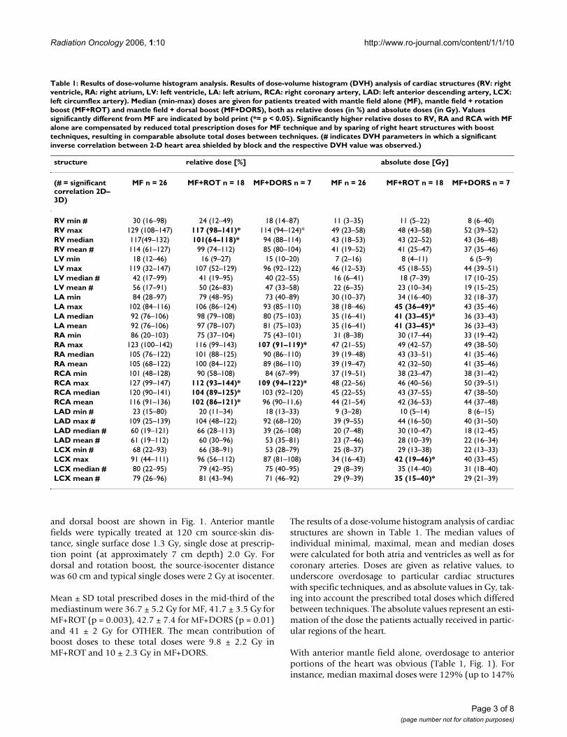
The results of a dose-volume histogram analysis of cardiacstructures are shown in Table 1. The median values ofindividual minimal, maximal, mean and median doseswere calculated for both atria and ventricles as well as forcoronary arteries. Doses are given as relative values, tounderscore overdosage to particular cardiac structureswith specific techniques, and as absolute values in Gy, tak-ing into account the prescribed total doses which differedbetween techniques. The absolute values represent an esti-mation of the dose the patients actually received in partic-ular regions of the heart.
With anterior mantle field alone, overdosage to anteriorportions of the heart was obvious (Table 1, Fig. 1). Forinstance, median maximal doses were 129% (up to 147%
Table 1: Results of dose-volume histogram analysis. Results of dose-volume histogram (DVH) analysis of cardiac structures (RV: right ventricle, RA: right atrium, LV: left ventricle, LA: left atrium, RCA: right coronary artery, LAD: left anterior descending artery, LCX: left circumflex artery). Median (min-max) doses are given for patients treated with mantle field alone (MF), mantle field + rotation boost (MF+ROT) and mantle field + dorsal boost (MF+DORS), both as relative doses (in %) and absolute doses (in Gy). Values significantly different from MF are indicated by bold print (*= p < 0.05). Significantly higher relative doses to RV, RA and RCA with MF alone are compensated by reduced total prescription doses for MF technique and by sparing of right heart structures with boost techniques, resulting in comparable absolute total doses between techniques. (# indicates DVH parameters in which a significant inverse correlation between 2-D heart area shielded by block and the respective DVH value was observed.)
structure relative dose [%] absolute dose [Gy]
(# = significant correlation 2D–3D)
MF n = 26 MF+ROT n = 18 MF+DORS n = 7 MF n = 26 MF+ROT n = 18 MF+DORS n = 7
RV min # 30 (16–98) 24 (12–49) 18 (14–87) 11 (3–35) 11 (5–22) 8 (6–40)RV max 129 (108–147) 117 (98–141)* 114 (94–124)* 49 (23–58) 48 (43–58) 52 (39–52)RV median 117(49–132) 101(64–118)* 94 (88–114) 43 (18–53) 43 (22–52) 43 (36–48)RV mean # 114 (61–127) 99 (74–112) 85 (80–104) 41 (19–52) 41 (25–47) 37 (35–46)LV min 18 (12–46) 16 (9–27) 15 (10–20) 7 (2–16) 8 (4–11) 6 (5–9)LV max 119 (32–147) 107 (52–129) 96 (92–122) 46 (12–53) 45 (18–55) 44 (39–51)LV median # 42 (17–99) 41 (19–95) 40 (22–55) 16 (6–41) 18 (7–39) 17 (10–25)LV mean # 56 (17–91) 50 (26–83) 47 (33–58) 22 (6–35) 23 (10–34) 19 (15–25)LA min 84 (28–97) 79 (48–95) 73 (40–89) 30 (10–37) 34 (16–40) 32 (18–37)LA max 102 (84–116) 106 (86–124) 93 (85–110) 38 (18–46) 45 (36–49)* 43 (35–46)LA median 92 (76–106) 98 (79–108) 80 (75–103) 35 (16–41) 41 (33–45)* 36 (33–43)LA mean 92 (76–106) 97 (78–107) 81 (75–103) 35 (16–41) 41 (33–45)* 36 (33–43)RA min 86 (20–103) 75 (37–104) 75 (43–101) 31 (8–38) 30 (17–44) 33 (19–42)RA max 123 (100–142) 116 (99–143) 107 (91–119)* 47 (21–55) 49 (42–57) 49 (38–50)RA median 105 (76–122) 101 (88–125) 90 (86–110) 39 (19–48) 43 (33–51) 41 (35–46)RA mean 105 (68–122) 100 (84–122) 89 (86–110) 39 (19–47) 42 (32–50) 41 (35–46)RCA min 101 (48–128) 90 (58–108) 84 (67–99) 37 (19–51) 38 (23–47) 38 (31–42)RCA max 127 (99–147) 112 (93–144)* 109 (94–122)* 48 (22–56) 46 (40–56) 50 (39–51)RCA median 120 (90–141) 104 (89–125)* 103 (92–120) 45 (22–55) 43 (37–55) 47 (38–50)RCA mean 116 (91–136) 102 (86–121)* 96 (90–11,6) 44 (21–54) 42 (36–53) 44 (37–48)LAD min # 23 (15–80) 20 (11–34) 18 (13–33) 9 (3–28) 10 (5–14) 8 (6–15)LAD max # 109 (25–139) 104 (48–122) 92 (68–120) 39 (9–55) 44 (16–50) 40 (31–50)LAD median # 60 (19–121) 66 (28–113) 39 (26–108) 20 (7–48) 30 (10–47) 18 (12–45)LAD mean # 61 (19–112) 60 (30–96) 53 (35–81) 23 (7–46) 28 (10–39) 22 (16–34)LCX min # 68 (22–93) 66 (38–91) 53 (28–79) 25 (8–37) 29 (13–38) 22 (13–33)LCX max 91 (44–111) 96 (56–112) 87 (81–108) 34 (16–43) 42 (19–46)* 40 (33–45)LCX median # 80 (22–95) 79 (42–95) 75 (40–95) 29 (8–39) 35 (14–40) 31 (18–40)LCX mean # 79 (26–96) 81 (43–94) 71 (46–92) 29 (9–39) 35 (15–40)* 29 (21–39)
Page 3 of 8(page number not for citation purposes)
Radiation Oncology 2006, 1:10 http://www.ro-journal.com/content/1/1/10
in individual patients) in the right ventricle, 123% (up to142%) in the right atrium and 127% (up to 147%) in theright coronary artery. Compared with anterior mantlefield alone, median maximal doses were significantlyreduced to 117% in the right ventricle and 112% in theright coronary artery by combining the anterior mantlefield technique with a monoaxial, bisegmental rotationboost technique (Table 1). Patients in whom the anteriormantle field was combined with a dorsal boost had signif-icantly reduced median maximal doses the right atrium,107%, and the right coronary artery, 109%.
When absolute doses in Gy were compared, the signifi-cant differences in dose to the right heart structures werelost, as patients treated with anterior mantle field alonereceived lower total mediastinal doses (mean 36.7 Gy)than patients in the MF+ROT (41.7 Gy) and MF+DORS(42.7 Gy) groups, indicating that the use of these boosttechniques permitted dose increases in the mediastinumwhile maintaining the dose levels in the right heart struc-tures seen with reduced-dose anterior mantle field alone.However, the use of a rotation boost technique led to sig-nificantly increased median absolute doses to the leftatrium and the left circumflex artery, compared to mantlefield alone. With the different techniques, median maxi-mum doses to the right and left ventricle were between 48and 52 Gy and between 44 and 46 Gy, respectively. In thecoronary arteries, median maximal doses ranged between46 and 50 Gy for the right coronary artery, 39 and 44 Gyin the left anterior descending artery and 34 and 42 Gy inthe left circumflex artery.
To investigate the effect of shielding part of the heart withlead blocks in anterior mantle fields on the DVH results ofcardiac structures (all patients and techniques consideredtogether), we first quantified the percentage of 2-D heartarea on the portal film shielded by lead blocks. The mean(± SD) percentage of the heart contour shielded by blockswas 36.3 ± 8.9% (range 18.2–53%). This percentage cor-related inversely with several DVH parameters, e. g. themean doses to both ventricles (Table 1), indicating spar-ing of the respective structures. All significant correlationsapplied equally to relative doses (in %) and absolute totaldoses (in Gy).
DiscussionIrradiation of the mediastinum with anterior mantle fieldtechniques, alone or followed by boost techniques, repre-sents an outdated treatment technique that was, however,state of the art in the 1970s. The mantle field, defined as"a single anteroposterior radiation therapy field designedto treat in continuity the major lymph node-bearing areasabove the diaphragm while maximally shielding the lungsin patients with lymphoma" [10] was first introduced byKaplan in 1956 [5] and replaced the irradiation of individ-
ual nodal sites with separate fields. Extended-field radio-therapy of Hodgkin's lymphoma with megavoltageequipment, using e. g. the mantle field, was introduced incenters in Germany in the early to mid-1970s [11,12].Concerns about the dose to the spinal cord and technicalreasons led several centers to use anterior mantle fieldsalone or anteriorly weighted opposing fields rather thanequally weighted opposing fields for mantle treatment[13]. The limited source-to-isocenter distance of cobaltmachines and the necessity of large irradiation fieldsrequired extended source-to-skin distances which wereachieved by positioning the patient on the floor in thesupine position and placing blocks on a block holderabove the patient. Introducing a dorsal mantle fieldwould have required changing the patient position toprone and introduced unwanted dosimetric uncertainties.In cases where underdosage to the posterior mediastinumwith an anterior mantle field was a concern, lateraloblique, dorsal or rotation boost techniques wereemployed [13]. Boost treatments could be performed onthe treatment table, as fields were shorter due to limitedtreated mediastinal volumes. Although most reportsfocussed on cardiac toxicity after treatment with opposingphoton beams, anterior mantle field technique in particu-lar has been associated with high rates of late toxicity suchas constrictive pericarditis [14-16].
Apart from a historic interest, a 3-D reconstruction of dosedistributions achieved with such techniques is relevantbecause patients thus treated may require special cardio-logical attention. In the present study, 39% of the initialstudy cohort are alive today. Thus, detailed knowledge ofthe individual cardiac dose distribution in long-term sur-vivors of mediastinal radiotherapy may help to identifypatients at risk and aid the cardiologist in the interpreta-tion of the patients' complaints and findings. Besides, thecorrelation of the reconstructed formerly applied 3-Ddose distribution with the corresponding cardiovascularfindings as obtained with non-invasive cardiac imagingtechniques (e. g. cardiovascular magnetic resonance imag-ing [17]), may expand our knowledge of dose-effect rela-tionships of cardiac structures such as the myocardium,coronary arteries or heart valves. It should be noted thatsolely studying the long-term survivors could potentiallyintroduce a bias regarding the cardiac dose distribution inthe overall cohort of n = 143 patients treated in the studyperiod. The causes of death of the n = 88 non-survivors arenot known and it can not be excluded that the cardiacdoses differed between survivors and non-survivors. How-ever, in preparation of a clinical investigation of long-termsurvivors, the dose reconstruction now presented was lim-ited to this subgroup.
Although the cardiac effects of thoracic radiotherapy havebeen extensively studied in the 1980s and 1990s [3,4],
Page 4 of 8(page number not for citation purposes)

Radiation Oncology 2006, 1:10 http://www.ro-journal.com/content/1/1/10
direct comparisons between the dose applied to specificstructures of the heart and consecutive patterns of cardio-vascular structural and functional abnormalities are scarcein the literature. In a report on 144 survivors of mediasti-nal radiotherapy for Hodgkin's lymphoma, Glanzmann etal. calculated the total doses to the "anterior heart region"to be between 30 and 42 Gy in 93% of patients [3]. Whilethe relative risk for myocardial infarction and for infarc-tion or sudden death was 4.2 and 6.7, respectively, andvalvular thickening was observed in 60% of patients after30 years of follow-up, patients without additional cardiacrisk factors (smoking, hypertension, hypercholesterine-mia) were found to have no increased risk of cardiacevents. In a recent systematic evaluation of Hodgkin's dis-ease survivors 14 years (median) after chest radiotherapy,42% had significant valvular defects, 75% conductiondefects, and 30% a reduced peak oxygen uptake, a predic-tor of mortality in heart failure [1]. Further, a review ofradiation-associated cardiovascular disease by Adams etal. suggests the following dose levels ("total cumulativeradiation exposure to the chest") as a guideline for theselection of patients to be screened for cardiovascular dis-ease [9]: pericarditis above 35 Gy, cardiomyopathy above35 Gy (or above 25 Gy if anthracyclines were used), coro-nary artery disease above 30 Gy, valvular disease above 40Gy. These data indicate that it may be of importance toreconstruct the local dose distribution and especially thelocalization of dose peaks if the total prescription dosewas in the range of 30 to 40 Gy, in such patients in orderto estimate the patients' individual cardiovascular risk,facilitating timely cardiovascular diagnostic proceduresand adequate treatment before the occurrence of compli-cations.
Cardiac structures now chosen for contouring and DVHanalysis were limited by their visibility on standard non-enhanced planning CT scans. The heart cavities were con-toured due to practicality although one could argue thatthe target tissue is the myocardium and not the heart con-tent. Such considerations are unresolved until today inother body regions, e. g. concerning contouring of thewhole rectum vs. the rectal surface in pelvic radiotherapy.Despite poor visibility, we contoured the major coronaryarteries as we expected these to be major target organsmediating radiation-induced cardiac toxiticity, hypothe-sizing that with the anterior mantle technique, coronaryarteries would differ in total dose resulting in specific pat-terns of coronary artery disease, as suggested in the olderliterature [18].
In the 55 long-term survivors now analyzed, anteriormantle-field technique alone was associated with amedian maximal dose to the right ventricle of 128% andto the right coronary artery of 127%, with even higherdoses in individual patients. Obviously, delivering part of
the radiation dose via other techniques with lateral or dor-sal beam entry reduced this relative overdosage in anteriorportions of the heart. In patients treated with anteriormantle field followed by rotation or dorsal boost, signifi-cantly higher total doses could be delivered than withmantle field alone, without further increases in dose toanterior heart structures. This was achieved at the expenseof irradiating parts of the left atrium which were eitherblocked or outside the high-dose region in the mantle-field technique. It is currently unclear if patterns of radia-tion-associated coronary artery disease differ from thoseseen in unirradiated patients. An older angiographic studyin 15 patients with coronary artery disease after thoracicradiotherapy for different tumor entities (nine Hodgkin'slymphomas) found predominant left main and rightostial coronary artery disease [18]. Although the left mainartery was not analyzed in the present study due to itsshort presentation on CT, high doses to the anterior heart,as resulting from anterior mantle field technique, couldexplain these locations.
Despite these technical efforts, the median maximal totaldoses to the right heart and right coronary artery were 48Gy and 46 Gy, respectively. These structures also receivedthe highest maximal doses in individual patients (58 Gyand 56 Gy, respectively). With a more modern opposed-field technique using a linear accelerator, in comparison,the overdosage to anterior heart structures should not bemore than 6% of a manually calculated dose [19]. Theadoption of involved-field radiotherapy with doses nothigher than 30 Gy in favorable and intermediate Hodg-kin's disease [7], e.g. in the current and previous genera-tions of trials in Germany, has therefore reduced themaximum dose to any cardiac structure to slightly over 30Gy. While the combination with chemotherapy may pro-vide additional cardiotoxic effects, patients without medi-astinal involvement, who previously would have beentreated with an extended field in the form of a mantlefield, would receive no dose to the heart in involved-fieldradiotherapy.
The 2-D percentage of shielded heart area correlatedinversely with several DVH parameters. Especially the leftand right ventricle as well as the branches of the left coro-nary artery benefitted from blocking. In structures thatwere partially blocked in most patients, such as left andright ventricle, mean and median dose but not minimumor maximum dose were correlated with the blocked heartarea. The dose to the left and right atrium as well as theright coronary artery was not associated with heart shield-ing, suggesting that these structures are exposed to highradiation doses even with extensive heart shielding. Thisknowledge may be of interest even in the days of lower-dose involved-field radiotherapy when the position ofcoronary arteries is not usually considered.
Page 5 of 8(page number not for citation purposes)

Radiation Oncology 2006, 1:10 http://www.ro-journal.com/content/1/1/10
The present analysis is limited by the lack of individual 3-D imaging studies in these patients. Variations in cardiacanatomy, e. g. branching of coronary arteries, could not beconsidered in the present design. The current data do notprovide precise quantitative data on dose distribution inindividual patients. However, for the whole cohort, thecalculated doses should be a very good approximation. Aspart of an ongoing interdisciplinary study between cardi-ologists, radiologists and radiation oncologists, Hodg-kin's disease survivors will be invited for extensivecardiologic tests including cardiac MRI [17]. As previouslysuggested [20], this project opens up the possibility toreconstruct the radiation dose in the individual patient's3-D data set and directly correlate this with individual car-diac pathology. Even this approach, although consideringindividual anatomy, will not be able to take into accountchanges in cardiac morphology between the time of treat-ment (about 25 years ago) and clinical reevaluation.
ConclusionOur investigation documented the 3-D dose distributionof patients treated with anterior mantle-field technique 25
years ago. This technique, in some patients followed bymediastinal boost irradiation from non-anterior direc-tions, was associated with dose peaks in anterior portionsof the heart, especially the right ventricle and right coro-nary artery. Given the relatively high total radiation dosesprescribed in this treatment era, our data set should pro-vide a solid base for a detailed analysis of cardiac dose-effects in long-term Hodgkin's disease survivors.
MethodsPatientsBetween 1978 and 1985, 143 patients were treated withmediastinal radiotherapy for Hodgkin's disease at theUniversity of Würzburg. Of these, n = 55 (38.5%) werealive at the time of analysis in March of 2003 and wereincluded in the present analysis, as these patients arepotentially available for future cardiologic evaluation.Mean ± SD age at the time of treatment was 25 ± 10 years(range 6 to 49 years). A review of patient charts yielded thepatient characteristics shown in Table 2.
Dose reconstructionFor 2-D evaluation of the portal films of mantle field irra-diation, heart borders were contoured and films scanned.Images were quanititatively analyzed using image process-ing software Scion Image vs. 4.0.2 (Scion Co., Frederick/MA, USA). The 2-D heart area was measured as well as theportion of the heart shielded by lead blocks.
For 3-D dose reconstruction, each treated patient was firstassigned to one of four test patients (treated more recentlyfor Hodgkin's disease) most closely resembling the anat-omy of the treated patient, in particular with regard toheart shape. In each of the test patients, for whom plan-ning CT studies in 1-cm slice thickness and spacing wereavailable, the following structures were contoured inHelax TMS (Nucletron, Veenendal, Netherlands) treat-ment planning system on the basis of a digital atlas of tho-racic CT anatomy [21] and of recent publications on cross-sectional heart anatomy [22,23]: left atrium, left ventricle,right atrium, right ventricle, left anterior descending(LAD) artery, left circumflex (LCX) artery, right coronaryartery (RCA).
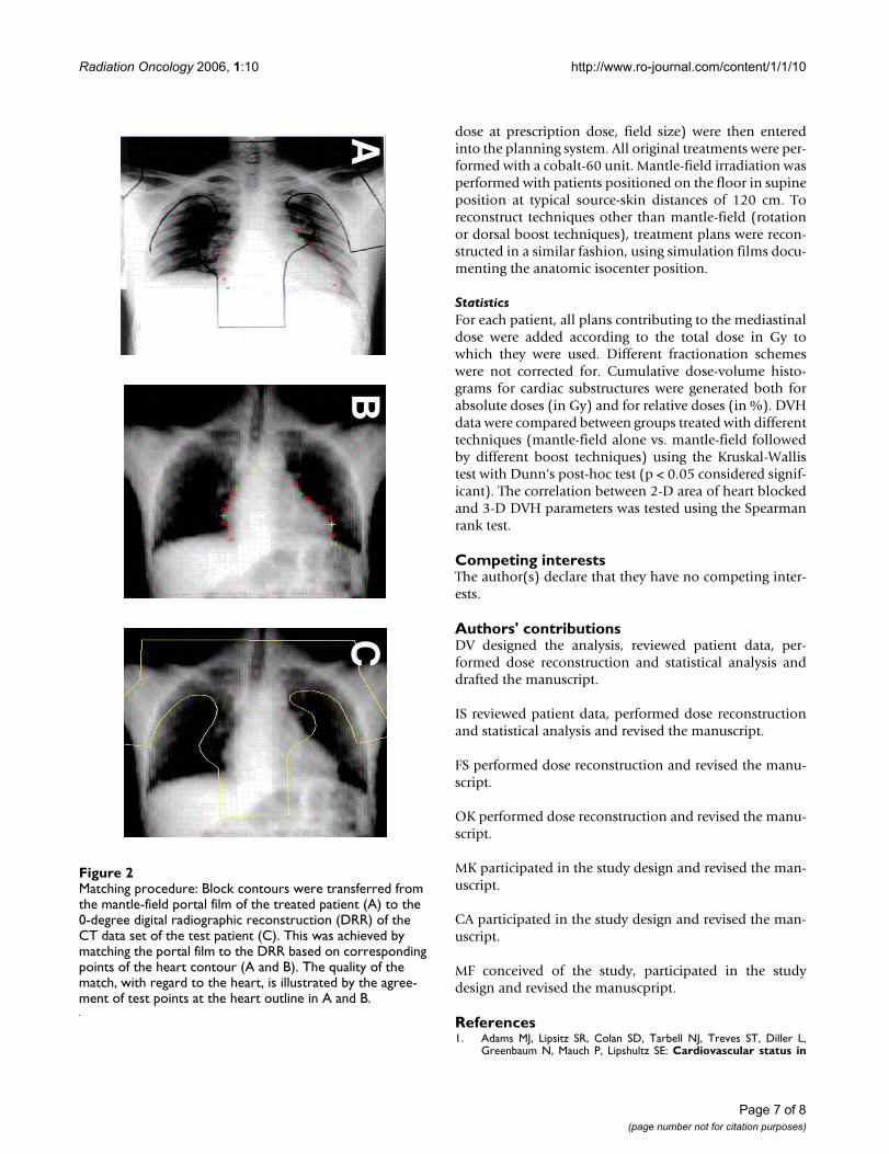
For reconstruction of the cardiac dose distribution, theoriginal mantle-field portal film of the treated patient wasmatched to a digital radiographic reconstruction from a 0degree angle of the test patient (Fig. 2). This matching pro-cedure was based on specific corresponding landmarks onthe heart outline of the treated patient and test patient,resulting in good matching results with regard to theheart, sometimes at the expense of incorrect matchingconcerning the lungs or bony structures, which were notpart of the current analysis. All treatment details (source-skin distance, depth of prescription point, surface dose,
Table 2: Patient Characteristics of n = 55 living patients treated with mediastinal radiotherapy for Hodgkin's disease between 1978 and 1985.
characteristics n (%)
sex male 29 (53%)female 26 (47%)
stage I 13 (24%)II 28 (51%)III 14 (25%)
B symptoms 16 (29%)risk factors a 0 (0%)
b 1 (2%)c 13 (24%)d 10 (18%)
histology lymphocyte-predominant 7 (13%)nodular sclerosing 36 (65%)mixed 10 (18%)not available 2 (4%)
involved regions cervical 27 (49%)supra-/infraclavicular 31 (56.4%)axilla 13 (23,7%)mediastinum 30 (54.5%)paraaortic 2 (4%)inguinal 3 (5%)spleen 11 (20%)
treatment radiotherapy alone 33 (60%)chemoradiation 22 (40%)
Page 6 of 8(page number not for citation purposes)
Radiation Oncology 2006, 1:10 http://www.ro-journal.com/content/1/1/10
dose at prescription dose, field size) were then enteredinto the planning system. All original treatments were per-formed with a cobalt-60 unit. Mantle-field irradiation wasperformed with patients positioned on the floor in supineposition at typical source-skin distances of 120 cm. Toreconstruct techniques other than mantle-field (rotationor dorsal boost techniques), treatment plans were recon-structed in a similar fashion, using simulation films docu-menting the anatomic isocenter position.
StatisticsFor each patient, all plans contributing to the mediastinaldose were added according to the total dose in Gy towhich they were used. Different fractionation schemeswere not corrected for. Cumulative dose-volume histo-grams for cardiac substructures were generated both forabsolute doses (in Gy) and for relative doses (in %). DVHdata were compared between groups treated with differenttechniques (mantle-field alone vs. mantle-field followedby different boost techniques) using the Kruskal-Wallistest with Dunn's post-hoc test (p < 0.05 considered signif-icant). The correlation between 2-D area of heart blockedand 3-D DVH parameters was tested using the Spearmanrank test.
Competing interestsThe author(s) declare that they have no competing inter-ests.
Authors' contributionsDV designed the analysis, reviewed patient data, per-formed dose reconstruction and statistical analysis anddrafted the manuscript.
IS reviewed patient data, performed dose reconstructionand statistical analysis and revised the manuscript.
FS performed dose reconstruction and revised the manu-script.
OK performed dose reconstruction and revised the manu-script.
MK participated in the study design and revised the man-uscript.
CA participated in the study design and revised the man-uscript.
MF conceived of the study, participated in the studydesign and revised the manuscpript.
References1. Adams MJ, Lipsitz SR, Colan SD, Tarbell NJ, Treves ST, Diller L,
Greenbaum N, Mauch P, Lipshultz SE: Cardiovascular status in
Matching procedure: Block contours were transferred from the mantle-field portal film of the treated patient (A) to the 0-degree digital radiographic reconstruction (DRR) of the CT data set of the test patient (C)Figure 2Matching procedure: Block contours were transferred from the mantle-field portal film of the treated patient (A) to the 0-degree digital radiographic reconstruction (DRR) of the CT data set of the test patient (C). This was achieved by matching the portal film to the DRR based on corresponding points of the heart contour (A and B). The quality of the match, with regard to the heart, is illustrated by the agree-ment of test points at the heart outline in A and B.
AB
C
Page 7 of 8(page number not for citation purposes)

Radiation Oncology 2006, 1:10 http://www.ro-journal.com/content/1/1/10
Publish with BioMed Central and every scientist can read your work free of charge
"BioMed Central will be the most significant development for disseminating the results of biomedical research in our lifetime."
Sir Paul Nurse, Cancer Research UK
Your research papers will be:
available free of charge to the entire biomedical community
peer reviewed and published immediately upon acceptance
cited in PubMed and archived on PubMed Central
yours — you keep the copyright
Submit your manuscript here:http://www.biomedcentral.com/info/publishing_adv.asp
BioMedcentral
long-term survivors of Hodgkin's disease treated with chestradiotherapy. J Clin Oncol 2004, 22:3139-3148.
2. Aleman BM, van den Belt-Dusebout AW, Klokman WJ, van't VeerMB, Bartelink H, van Leeuwen FE: Long-term cause-specific mor-tality of patients treated for Hodgkin's disease. J Clin Oncol2003, 21:3431-3439.
3. Glanzmann C, Kaufmann P, Jenni R, Hess OM, Huguenin P: Cardiacrisk after mediastinal irradiation for Hodgkin's disease. Radi-other Oncol 1998, 46:51-62.
4. Lund MB, Ihlen H, Voss BM, Abrahamsen AF, Nome O, Kongerud J,Stugaard M, Forfang K: Increased risk of heart valve regurgita-tion after mediastinal radiation for Hodgkin's disease: anechocardiographic study. Heart 1996, 75:591-595.
5. Kaplan HS: The radical radiotherapy of regionally localizedHodgkin's disease. Radiology 1962, 78:553-561.
6. De Vita VT, Serpick AA, Carbone PP: Combination chemother-apy in the treatment of advanced Hodgkin's disease. AnnIntern Med 1970:881-895.
7. Engert A, Schiller P, Josting A, Herrmann R, Koch P, Sieber M, Bois-sevain F, De Wit M, Mezger J, Duhmke E, Willich N, Muller RP,Schmidt BF, Renner F, Muller-Hermelink HK, Pfistner B, Wolf J,Hasenclever D, Loffler M, Diehl V: German Hodgkin's Lym-phoma Study Group. Involved-field radiotherapy is equallyeffective and less toxic compared with extended-field radio-therapy after four cycles of chemotherapy in patients withearly-stage unfavorable Hodgkin's lymphoma: results of theHD8 trial of the German Hodgkin's Lymphoma StudyGroup. J Clin Oncol 2003, 21:3601-3608.
8. Eich HT, Müller RP, Hansemann K: Results of the 4th interimanalysis of the HD 11 trial of the GHSG for intermediatestage Hodgkin's lymphoma: 30 Gy vs. 20 Gy involved fieldradiotherapy [abstract]. Strahlenther Onkol 2005, 181(suppl1):49.
9. Adams MJ, Hardenbergh PH, Constine LS, Lipshultz SE: Radiation-associated cardiovascular disease. Crit Rev Oncol Hematol 2003,45:55-75.
10. Carmel RJ, Kaplan HS: Mantle irradiation in Hodgkin's disease.An analysis of technique, tumor eradication and complications. Cancer1976, 37:2813-2825.
11. Musshoff K, Weidkuhn V, Bammert J, Felker HU: Diagnosis andtherapy of Hodgkin's disease in Freiburg in Breisgau 1964 to1976 [German]. Strahlenther 1985, 161:596-614.
12. Schulz U, Malewski U, Alberti W: Prognostic factors during con-trol of the course of lymhogranulomatosis: comparison of 2therapy technics and analysis of serologic parameters [Ger-man]. Strahlenther 1985, 161:221-224.
13. Willich N, Bayer M, Krimmel K, Lengsfeld M, Rohloff R, Wendt T: Aventral mantle technic with dorsolateral mediastinal satura-tion in the radiotherapy of Hodgin's disease [German].Strahlenther Onkol 1988, 164:393-401.
14. Byhardt R, Brace K, Ruckdeschel J, Chang P, Martin R, Wiernik P:Dose and treatment factors in radiation-related pericardialeffusion associated with the mantle technique for Hodgkin'sdisease. Cancer 1975, 35:795-802.
15. Coltart RS, Roberts JT, Thom CH, et al.: Severe constrictive peri-carditis after single 16 MeV anterior mantle irradiation forHodgkin's disease. Lancet 1985, 1:488-489.
16. Gottdiener JS, Katin MJ, Borer JS, Bacharach SL, Green MV: Late car-diac effects of therapeutic mediastinal irradiation. New Engl JMed 1983, 308:569-572.
17. McCrohon JA, Moon JC, Prasad SK, McKenna WJ, Lorenz CH, CoatsAJ, Pennel DJ: Differentiation of heart failure related to dilatedcardiomyopathy and coronary artery disease using gadolin-ium-enhanced cardiovascular magnetic resonance. Circulation2003, 108:54-59.
18. McEniery PT, Dorosti K, Schiavone W, Pedrick TJ, Sheldon WC:Clinical and angiographic features of coronary artery diseaseafter chest irradiation. Am J Cardiol 1987, 60:1020-1024.
19. Glanzmann C, Huguenin P, Lütolf UM, Maire R, Jenni R, GumppenbergV: Cardiac lesions after mediastinal irradiation for Hodgkin'sdisease. Radiother Oncol 1994, 30:43-54.
20. Vordermark D, Seufert I, Schwab F, Flentje M, Kung M, AngermannC: Cardiac toxicity of mediastinal radiotherapy: which arethe critical structures? [letter]. J Clin Oncol 2005, 23:3966-3967.
21. Küper K: MR/CT-Atlas der Anatomie. Digitale Schnittbilddi-agnostik [German]. Thieme, Stuttgart 2001.
22. Rabin DN, Rabin S, Mintzer RA: A pictorial review of coronaryartery anatomy on spiral CT. Chest 2000, 118:488-491.
23. Sevrukov A, Jelnin V, Kondos GT: Electron beam CT of the cor-onary arteries: cross-sectional anatomy for calcium scoring.Am J Roentgenol 2001, 177:1437-1445.
Page 8 of 8(page number not for citation purposes)