Embed Size (px)
Citation preview
RAD 240 PathologyRadiological Sciences, Department of Health Sciences
Week of November 17, 2013
Dr Shai
Lecture 7
Pathology of the
Kidney & Urinary
Tract
overview
Kidney structural abnormalities
Diseases of the glomerulus
Systemic diseases
Diseases of tubules, vessels, & interstitium
Neoplasms
Pathologies of renal tract

Renal anatomy

Structural – congenital kidney disorders
1. AGENESIS of the kidneyUNILATERAL in 1/1000 births
Solitary kidney undergoes hypertrophy
Susceptible to infections
Males 2:1 females, left kidney usually absent
BILATERAL in 1/3000 births, part of Potter’s syndrome
Abnormal face, urinary tract and nervous system
Oligohydramnios* in pregnancy as kidneys not present to contribute amniotic fluid (absence of fetal urine)
Not compatible with post natal life (DEATH)

CONGENITAL CON’TB. HYPOPLASIA
Kidneys fail to reach normal adult size (congenitally or from shrinkage), often a result of early life chronic infection
C. ECTOPIC KIDNEYS1 or 2 kidneys in abnormal position (usually pelvis)
D. HORSESHOE KIDNEYPoles of kidneys are fused, usually inferiorly, to form a large U-shaped (horseshoe) kidney
1/500 people, asymptomatic as collecting systems are normal
2 to 8 times more likely to develop Wilm’s tumours in children

Horseshoe kidneys
Animated view CT abdomen – horseshoe kidney

Pathology specimen – horseshoe kidney
Ultra sound horseshoe kidney

Cystic Kidney DiseasesIncludes: Hereditary, developmental and acquired disorders
Important: 1. appropriate patient management to delay onset of renal failure
2. Appropriate genetic counseling to relatives at risk
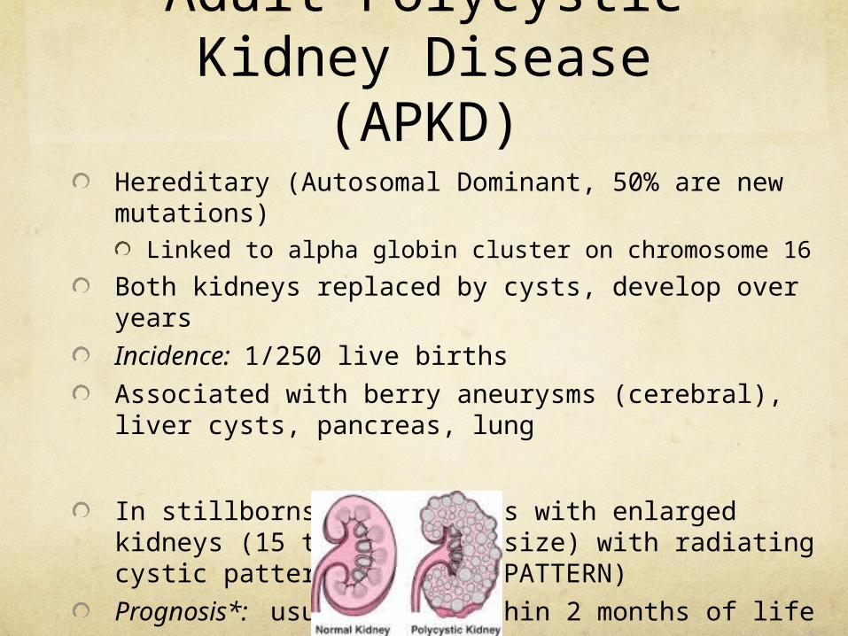
Adult Polycystic Kidney Disease (APKD)
Hereditary (Autosomal Dominant, 50% are new mutations)
Linked to alpha globin cluster on chromosome 16
Both kidneys replaced by cysts, develop over years
Incidence: 1/250 live births
Associated with berry aneurysms (cerebral), liver cysts, pancreas, lung
In stillborns* or neonates with enlarged kidneys (15 times normal size) with radiating cystic pattern (SUNBURST PATTERN)
Prognosis*: usually die within 2 months of life
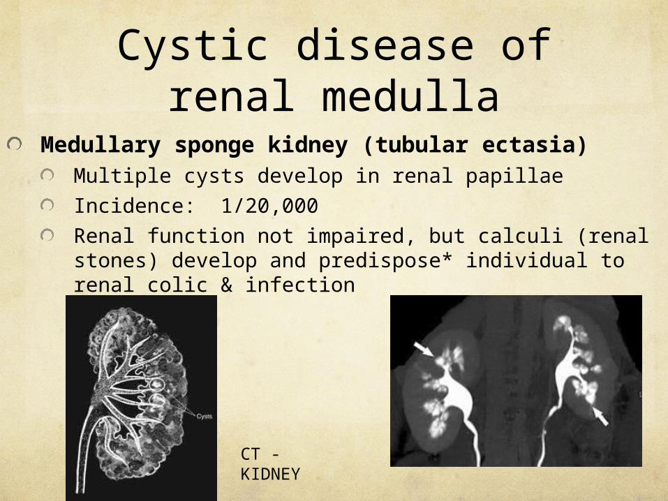
Cystic disease of renal medulla
Medullary sponge kidney (tubular ectasia)Multiple cysts develop in renal papillae
Incidence: 1/20,000
Renal function not impaired, but calculi (renal stones) develop and predispose* individual to renal colic & infection
CT - KIDNEY
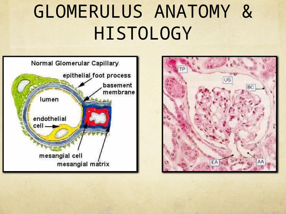
GLOMERULUS ANATOMY & HISTOLOGY
Glomerular Diseases
Typically caused by structural abnormalities
4 significant components may be damaged:i) endothelial cells lining the capillary
Ii) glomerular basement membrane (BM)
Iii) mesangium: supporting mesentery to the capillary comprised of mesangial cells (phagocytic cells) & associated matric
Epithelial cells or podoctyes, coat outer layer of BM


Patterns of glomerular disease
Most diseases affect different glomeruli to varying degrees
Global: affecting entire glomerulus uniformly
Segmental: affecting one glomerular segment, sparing others
Diffuse: affecting all glomeruli in both kidneys
Focal: affecting proportion of glomeruli, sparing others
Eg. “diffuse global” or “ focal segmental”

Aetiology of glomerular diseases
Primary (majority): disease begins in glomerululusTypes: proliferative*, membrnaous, glomerulosclerotic, minimal change lesions
Secondary: 2nd ary to systemic disease (immune complex mediated, metabolic, vascular)
Hereditary: Alport’s Syndrome, Fabry’s disease, congenital nephortic syndrome

Diagnosis & SymptomsHistologically
Glomerular response to injury
Percutaneous* needle biopsy
Immunological comple deposition investigation
Symptoms• Aymptomatic haematuria*
• Haematuria without proteinaemia, continuous or intermittent, does not cause renal failure
• Aymptomatic proteinuria*• Proteinuria >0.3 g / 24 hours, without haematuria,
continuous, orthostatic (postural), or transient• Acute Nephritic syndrome
• Sudden haematuria, proteinuria, hypertension• Loin pain, headache, orbital oedema*

• Nephrotic syndrome• Proteinuria >3.5 g / 24 hrs, with hypoproteinaemia,
oedema, hypercholesterolaemia
• Chronic Renal failure• Irreversible deterioration in renal function caused by
the destruction of nephrons over time• Impairment of excretory, metabolic, endocrine functions
of kidney• Management: excretory function may be replaced by
dialysis, metabolic and endcrine functions need transplant to re initiate

Proliferative glomerulonephritis
Group of disorders characterized by histological degrees of proliferation in mesangial & epithelial cells in glomerulus
TypesDiffuse proliferative
Rapidly progressive
Focal proliferative
Membranoproliferative
* these are patterns of reactions, not diagnoses

Diffuse proliferative glomerulonephropathie
sDiffuse, global, acute inflammation
From deposition of immune complexes in glomeruli
Stimulated by infection
Aetiology: post streptococcal infection , or less frequently viruses, protozoas and other bacterial infection

Rapdily progressive glomerulonephritis
CRESCENTIC GLOMERULONEPHRITIS (RPGN)
From severe glomerular injury characterized by formation of cellular crescent shaped masses within Bowman’s space
Often post streptococcal
Crescent shaped masses are composed of epithelial cells & macrophages, causing glomerular ischaemia
Focal proliferative glomerulonephritis
Acute inflammation within cellular proliferation in a few glomeruli (focal), affecting one segment (segmental)
Better known as focal segmental proliferative glomerulonephritis
Primary: mesangial IgA disease, Goodpasteur’s syndrome
Secondary: systemic endocarditis, vasculitis, connective tissue disease

Membranoproliferative glomerulonephritis
MPGN
Diffuse, global pattern of glomerulonephritis with thickening of membrane
Primary: idiopathic
Secondary: associated with SLE, endocarditis, cerebral shunts
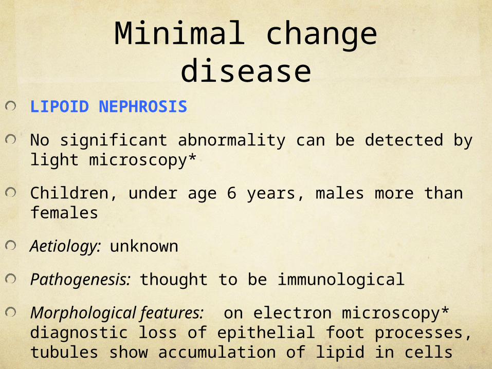
Minimal change disease
LIPOID NEPHROSIS
No significant abnormality can be detected by light microscopy*
Children, under age 6 years, males more than females
Aetiology: unknown
Pathogenesis: thought to be immunological
Morphological features: on electron microscopy* diagnostic loss of epithelial foot processes, tubules show accumulation of lipid in cells
Prognosis: good, no permanent renal damage

Minimal change disease – electron microscopy

Hereditary glomerulonephritis
1. Alport’s SyndromeClinical triad: deafness, glomerulonephritis, ocular lesionsX linked inherited*, mutatin of COLIValpha5 gene
Clinical features: glomerulonephritis (microscopic haematuria & proteinuria in childhood, later nephrotic syndrome), ocular disease & deafness to high pitched sounds
2. Fabry’s syndromeRare, X linked recessive syndrome
Glycosphingolipid metabolism, resulting in painful extermities, red hyperkeratotic papules on skin*, proteinura, renal failure
Congenital Nephrotic Syndrome: rare, mesangial proliferation or glomerulosclerosis

Chronic Glomerulonephritis
Chronic renal failure with small contracted kidneys, and all glomeruli are hyalinized (end stage kidneys)
Macroscopically: affected kidneys are small, granularity of external surface, scarring from nephron hyalinization
Microscopically: hyalinization of glomeruli

Systemic diseases
Systemic lupus erythematosusRenal involvement causes diffuse or focal glomerular disease
Basis of glomerular damage is immune complex deposition in basement membrane, BM thickens
Henoch Schonlein purpuraImmune complex mediated systemic vasculitis
Affects small arteries in skin, joints, gut, kidneys

Bacterial EndocarditisRenal lesions in infective endocarditis from immune complexes or embolism infarction of heart valve vegetations
Diabetic Glomerulosclerosis
Diabetic glomerular damage causes increased permeability of capillaries in the BM, leading to proteinuris and nephrotic syndrome
Pathogenesis: not fully understood, involves deficiency in proteoglycans, glomerular hypertrophy, BM thickening, mesangial cell hypertrophy
Histologically
1) capillary wall thickening – mild proteinuria
2) diffuse glomerulosclerosis
3) nodular glomerulosclerosis

AmyloidosisAmyloid, an extra cellular fibrillar protein* is deposited in tissues, including kidney
Amyloid fibrils deposited in BM, and mesangium
Proteinuria & nephrotic syndrome & chronic renal failure
Wegener’s GranulomatosisImmune complex, systemic, necrotizing vasculitis, affecting nose, respiratory and renal tracts
Polyarteritis nodosaSystemic disease, inflammatory necrosis of walls of small & mediums sized arteries
Necrosis of medium sized arteries causes infarcts in the kidney, as fibrinoid necrosis

Vascular DiseasesBenign Nephrosclerosis
Hyaline arteriosclerosis of the kidney, associated with benign hypertension*
It is an important complication of long term benign hypertension, chronic renal failure being the most important outcome
It is the commonest nephropathy, found in about 75% of autopsies over the age of 60 years
Long standing hypertension cuases reduced blood flow to glomeruli, caused by vascular changes, branches of renal artery thicken with hypertrophy of muscular media, results in ischaemia with scarring.
Afferent arterioloes undergo hyalinization (arteriosclerosis)
Clinically: increase blood urea & reduction in creatinine clearance
Prognosis: <5% die from renal failure, death from congestive heart failure associated with renal failure

nephrosclerosis

Malignant nephrosclerosis
Renal disease associated with malignant hypertension
Pathogenesis: in accelerated hypertension, the rise in blood pressure is rapid, large muscular vessels undergo fibroplastic proliferation, BUT NO MUSCULAR HYPERTROPHY
Afferent arterioles undergo necrosis, and necrosis of glomerular capillary network.
90% of cases untreated cause death.

Renal artery stenosis
Narrowing of renal arteries, from atherosclerosis or arterial fibromuscular dysplasia
OutcomesChronic ischaemia of affected kidneyRenovascular hypertension
Inadequate perfusion of kidney caused by renal artery stenosis leads to hypertension, and abnormal activation of the renin-angiotensin system


Neoplasms of the kidney
Benign tumoursCortical adenoma: epithelial tumours of renal tubular epithelium
Macroscopically: discreet nodules < 20mm diameter in cortex
Microscopically: well differentiated large clear cells
Renal hamartoma: commonest benign tumour of kidney, made of spindle cells
Macroscopically: firm white nodules in medulla, 3-10mm size
Microscopically: spindle cells
Angiomyolipoma: hamartoma* composed of smooth muscle, blood vessels, and fate.

Renal hamartoma
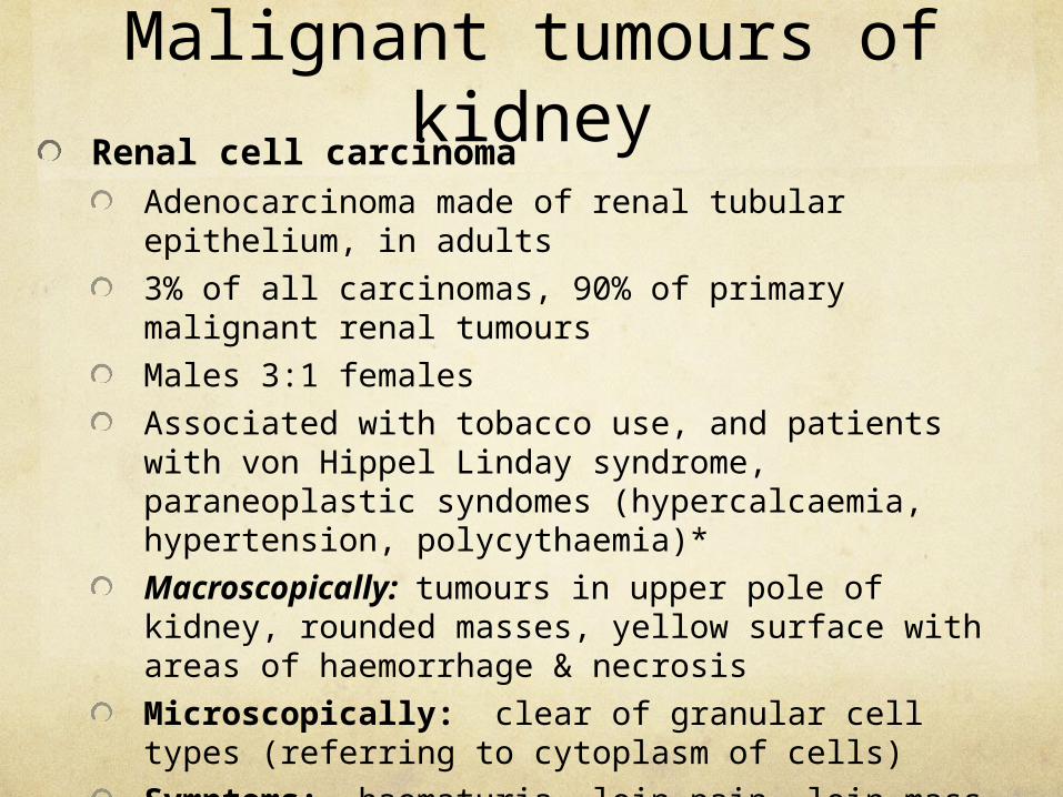
Malignant tumours of kidney
Renal cell carcinomaAdenocarcinoma made of renal tubular epithelium, in adults
3% of all carcinomas, 90% of primary malignant renal tumours
Males 3:1 females
Associated with tobacco use, and patients with von Hippel Linday syndrome, paraneoplastic syndomes (hypercalcaemia, hypertension, polycythaemia)*
Macroscopically: tumours in upper pole of kidney, rounded masses, yellow surface with areas of haemorrhage & necrosis
Microscopically: clear of granular cell types (referring to cytoplasm of cells)
Symptoms: haematuria, loin pain, loin mass
Prognosis: 70% chance 10 year survival


Renal cell carcinoma imaging

Wilm’s TumourNephroblastoma
Malignant embryonal tumour derived from primitive metanephros*
Common in childhood, peak 1 to 4 yrs age
3 types identifiedCommonest : tumour suppresor gene WT1 located on chromosome 11
Macroscopically: large rounded masses of solid, fleshy white lesions with necrosis. Tumour is aggressive, rapidly growing, extends beyond capsules into mesentery
Microscopically: composed of up to 4 elements {primitive blastematous tissue, immature glomerular structures, epithelial tubes, stroma of spindle cells)
Presents with abdominal mass, treat with chemo and radio therapy

Wilm’s tumour
Macroscopical changes in Wilms tumour and CT imaging of Wilm’s tumour

Urinary tract obstruction
Obstruction of urine drainage from kidney at ANY level within urinary tract
Intrinsic lesions: within ureteric wall or lumen (eg urinary calculus*, necrotic debris*, fibrosis after trauma or infection)
Extrinsic lesions: from external pressue (eg tumours of rectum or bladder, pregnancy, retroperitoneal fibrosis)
LocationsRenal pelvis: calculi, tumours
Pelviureteric junction: strictures, calculi, ext compression
Ureter: calculi, ext compression (pregnancy, fibrosis, tumour)
Bladder neck: tumour, calculi
Urethra: porstatic hyperplasia or carcinoma, stricture


Pathogenesis: obstruction at any point causes increased presure, superior to blockage, with dilatation of renal pelvis and calyces (hydronephrosis)
Obstruction ate pelviureteric junction > hydronephrosis@ ureter> hydro ureter with subsequent hydronephrosis@ bladder neck> bladder distension with hypertrophy of bladder muscle > hydroureter and hydronephrosis
Hydronephrosis effects:Fluid entering the collecting ducts cannot empty into renal pelvis, and intra renal resorption of fluid occursIf the obstruction is removed at this stage: normal renal function resumes

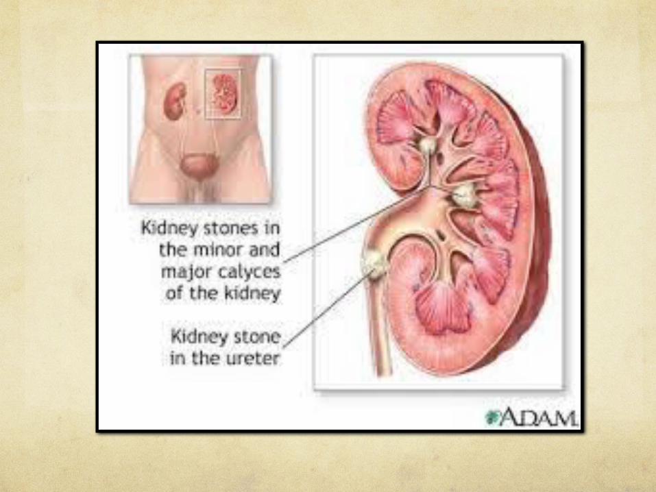
Urolithiasis (urinary calculi)
Formation of stones in the urinary tract (U/S)
1-5% population in UK, usually after 30 years age, males> females
Can form anywhere in urinary tract, usually in renal pelvisComposition of stones
Calcium oxalate (80%)
Triple phosphates (15%) magnesium ammonium phosphate stones
Uric acid (5%)
Calculi in cystinuria and oxalosis
Aetiology: acquired (urinary tract obstruction, persistent urinary tract infections, reduced urine volume from dehydration; or inherited 1ary metabolic disorders, cystinuria, etc

Mechanism of stone formation involves excess solute in urine (primary increase in metabolite or stasis) or due to reduced solubility of solute in urine (persistently low pH)
Calculi vary in size, from sand like particles to large staghorn stones which fit the whole renal pelvis and branch into calyces
If calcium deposition occurs, nephrocalcinosis
Symptoms:Renal colic* with nausesa & vomitting caused by passage of small stones along ureter
Dull ache in loins
Strangury* desire to pass something that will not pass (stones in bladder)
Recurrent urinary tract infections (UTI)
Management: rest, analgesia, warmth, fluid intake to pass small stones <5mm, or ureteroscopy or lithotripsy

Urinary calculi imaging

CYSTITIS
Inflammation of bladder
Extremely common, mostly women because of shorter urethra (less traveling distance for bacteria)
AetiologyInfection or irritants (radiation)
Bacterial (E Coli, proteus, strep faecalis, staphylococcus)
Viral (adenovirus), fungal (candida), parasites (schistosoma)

Macroscopically: acute inflammation with oedema, erythema & ulceration of bladder mucosa
Microscopically: infiltration of mucosa with acute inflammatory cells

Risk factors:Urinary retention (due to obstruction, bladder paralysis, calculi, foreign bodies/diverticuli, uterine prolapse)
Infection of adjacent structures (prastatitis, urethritis, colon disease)
Diabetes mellitus
Pregnancy
Trauama (catherization)
Routes of infectionAscending (commonest) urethra then bladder infection (catheterization)
Descending from kidney (eg renal tuberculosis)
Direct spread or hameatogenous, or lymphatic
Clinical features: pyrexia*, pain in rt iliac fossa*, loin and supra pubic pain, micturition disorders (frequency, urgency, dysuria*, haematuria, incontinence*)
Examination: mid stream urine analysis
Treatment: anit bacterial, antibiotics, flluid therapy
Sequelae:Resolution
Chronicity
Pyelonephritis*

Bladder metaplasia
GLANDULAR METAPLASIA (CYSTITIS GLANDULARIS)
Small, rounded urothelial cells under urothelial surface (Brunn’s nests)
Develop central lumen surrounded by cuboidal cells
ADENOMATOUS METAPLASIABenign, metaplsia or urothelium to cuboidal epithelium
SQUAMOUS METAPLASIAKeratinizing or non keratinizing

BLADDER TUMOURS
TRANSITIONAL CELL CARCINOMA
Incidence: 1/5000 in UK, 3% of all cancer deaths
Males 3: 1 females, 60-70 years
Aetiology
Chemical: eposure to environmental agents (cigarette smoking, aniline dyes, rubber industry)
Leucoplakia: keratinizing metaplsia, white plaques and calculi
Bladder diverticuli*
Most occur at base of trigone*

Transitional cell CT and histology

MORPHOLOGYPapillary (commonest) warty masses, projecting into lumen with little invasion of bladder wall
Solid: tumours grow directly into bladder wall, often ulcerated
Mixed papillary and solid
Flat in situ carcinoma: reddened mucosa
GRADINGTCC grades I-III
I: well differentiated, no invasion
II: moderately well differentiated, papillary ,atypical cells
III: poorly differentaited, pleomorphic* cells, invasive
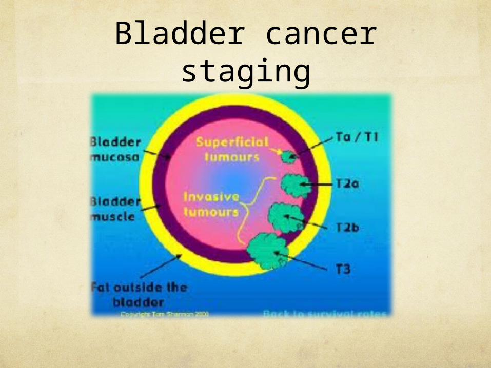
Bladder cancer staging

TNM
Tumour-node-metastasis Staging
T1: tumour confined to mucosa or submucosa
T2: superficial muscle involved
T3: deep muscle involved
T4: invasion beyond the bladder
Spread: local to pelvic structures, lymph or haematogenous
Treatment: stage dependeant, diathermy +- radiotheapy, cystectomy or palliative* radiotherapy
Prognosis: depends on histoogical type

Try…
Search online and app based radiology slides and identify both normal and abnormal structures
2ND MID TERM EXAMINATION:
COVER CARDIOVASCULAR, RESPIRATORY, URINARY, GASTROINTESTINAL PATHOLGIES
MULTIPLE CHOICE AND SHORT ANSWERS
REVIEW CLASS BEFORE THE EXAM: please start studying, so you can optimize the review time, good luck’