Embed Size (px)
Citation preview
Rac1-Dependent Lamellipodial Motility in ProstateCancer PC-3 Cells Revealed by Optogenetic Control ofRac1 ActivityTakuma Kato1, Katsuhisa Kawai2, Youhei Egami2, Yoshiyuki Kakehi1, Nobukazu Araki2*
1 Department of Urology, School of Medicine, Kagawa University, Miki, Kagawa, Japan, 2 Department of Histology and Cell Biology, School of Medicine, Kagawa
University, Miki, Kagawa, Japan
Abstract
The lamellipodium, an essential structure for cell migration, plays an important role in the invasion and metastasis of cancercells. Although Rac1 recognized as a key player in the formation of lamellipodia, the molecular mechanisms underlyinglamellipodial motility are not fully understood. Optogenetic technology enabled us to spatiotemporally control the activityof photoactivatable Rac1 (PA-Rac1) in living cells. Using this system, we revealed the role of phosphatidylinositol 3-kinase(PI3K) in Rac1-dependent lamellipodial motility in PC-3 prostate cancer cells. Through local blue laser irradiation of PA-Rac1-expressing cells, lamellipodial motility was reversibly induced. First, outward extension of a lamellipodium parallel to thesubstratum was observed. The extended lamellipodium then showed ruffling activity at the periphery. Notably, PI(3,4,5)P3
and WAVE2 were localized in the extending lamellipodium in a PI3K-dependent manner. We confirmed that the inhibition ofPI3K activity greatly suppressed lamellipodial extension, while the ruffling activity was less affected. These results suggestthat Rac1-induced lamellipodial motility consists of two distinct activities, PI3K-dependent outward extension and PI3K-independent ruffling.
Citation: Kato T, Kawai K, Egami Y, Kakehi Y, Araki N (2014) Rac1-Dependent Lamellipodial Motility in Prostate Cancer PC-3 Cells Revealed by Optogenetic Controlof Rac1 Activity. PLoS ONE 9(5): e97749. doi:10.1371/journal.pone.0097749
Editor: Daotai Nie, Southern Illinois University School of Medicine, United States of America
Received January 8, 2014; Accepted April 24, 2014; Published May 21, 2014
Copyright: � 2014 Kato et al. This is an open-access article distributed under the terms of the Creative Commons Attribution License, which permitsunrestricted use, distribution, and reproduction in any medium, provided the original author and source are credited.
Funding: This study was supported by Japan Society for the Promotion of Science (JSPS) KAKENHI (http://www.jsps.go.jp/english/e-grants/index.html) 25861427to TK; 24659087 and 23390039 to NA; 26860136 and 24890154 to KK; 25860142 to YE; 24390369 to YK. The funders had no role in study design, data collectionand analysis, decision to publish, or preparation of the manuscript.
Competing Interests: The authors have declared that no competing interests exist.
* E-mail: [email protected]
Introduction
Cell migration plays an important role in embryonic organo-
genesis; wound healing and immune responses; and the patho-
genesis of several diseases including cancer invasion and metastasis
[1,2]. Therefore, an understanding of the molecular mechanisms
underlying cell migration is important for developing new
therapeutic strategies for preventing tumor invasion and metasta-
sis. Cell migration involves the processes of polarized cellular
protrusion and adhesion in the direction of movement, cell
contraction, disassembly of adhesive foci, and retraction at the
periphery of the cell’s trailing edge [1]. During the tumor cell
migration that is associated with cancer metastasis and invasion,
metastatic cells exhibit drastic changes in shape. This deformation
is caused by actin cytoskeletal remodeling, which is regulated by
Rho family GTPases such as Cdc42 and Rac1. Rho family
GTPases behave as molecular switches, cycling between active
GTP-bound forms and inactive GDP-bound forms. Rho family
GTPases are activated by guanine nucleotide exchange factors
(GEFs) and inactivated by GTPase-activating proteins (GAPs) [3].
Rac1, a member of the Rho family GTPases, leads to the
production of sheet-like protrusions referred to as lamellipodia or
membrane ruffles, while Cdc42, another member of the Rho
family, creates spike-like protrusions called filopodia [3]. Rac1 is
hyperactivated in metastatic prostate cancer cells [4]. Additionally,
the inhibition of Rac1 activity blocks the migration and invasion of
prostate cancer cells [5]. These studies suggest that Rac1-mediated
lamellipodial formation plays an important role in prostate cancer
metastasis.
To date, the expression of Rac1 mutants such as the
constitutively active (CA) Rac1Q61L and the dominant negative
(DN) Rac1T17N has been widely used for investigating the
involvement of Rac1 in lamellipodial formation and ruffling [6].
However, the cell phenotype data obtained using Rac1 mutants
must be interpreted with caution. Due to the effects of irreversible,
permanent and global expression in the cells, it is hard to say that
the phenotypes of cells expressing Rac1 mutants exactly reflect the
protein’s action as a molecular switch. To elucidate the precise
role of the spatiotemporal activation of Rac1, Wu et al. [7,8]
recently developed a photo-activatable Rac1 (PA-Rac1) system by
fusing a light-oxygen-voltage (LOV) domain and a carboxy-
terminal helical extension (Ja) sequence to the amino terminus of a
constitutively active Rac1. LOV is a protein light-switch domain of
Avena sativa phototropin 1. In the dark, the flavin-binding LOV
domain interacts with Ja and blocks the effector binding site of PA-
Rac1 by configuring into its closed conformation. Irradiation with
light at 400–500 nm light induces the dissociation of LOV domain
and Ja helix, and leads to Rac1 activation. This photo-induced
activation is reversible. Using this system, localized Rac1
activation was shown to be sufficient to induce cell motility and
determine the direction of cell movement [7,8].
PLOS ONE | www.plosone.org 1 May 2014 | Volume 9 | Issue 5 | e97749

The relationship between Rac1 and phosphatidylinositol 3-
kinase (PI3K) in the formation of lamellipodia is complicated
because PI3K functions both upstream and downstream of Rac1
[9]. Phosphatidylinositol 3,4,5-triphosphate (PI(3,4,5)P3) is known
to be bind Rac GEFs and then accelerate actin polymerization
through Rac1 activation [10]. Additionally, a positive feedback
loop has been reported between PI(3,4,5)P3 and Rac for cell
polarity during eukaryotic chemotaxis [11,12]. However, in the
regulation of cell protrusion and polarity, reports regarding the
function of PI3K downstream of Rac1 are mixed [13,14]. Thus,
the precise role of PI3K downstream of Rac1 remains a
controversial issue.
To clarify the relevance of PI3K to Rac1-dependent lamellipo-
dial motility, we applied the PA-Rac1 system to prostate cancer
cells. Photomanipulation of PA-Rac1 activity using a blue laser
enabled us to distinguish two lamellipodial motile processes in
living cells: lamellipodial extension and peripheral ruffling.
Notably, we found that PI3K inhibitors suppressed the initiation
of lamellipodial extension but had little effect on peripheral
ruffling. The present study revealed that Rac1-dependent
lamellipodial motile processes consist of two dissociable activities:
PI3K-dependent lamellipodial outward extension and PI3K-
independent peripheral ruffling.
Materials and Methods
Reagents and cDNA ConstructsFetal bovine serum (FBS) and RPMI-1640 were obtained from
the Sigma Chemical Co. (St. Louis, MO). The X-tremeGENE HP
DNA Transfection Reagent was acquired from Roche Diagnostic
Systems (Basel, Switzerland). The other reagents were purchased
from Wako Pure Chemicals (Osaka, Japan) or Nacalai Tesque
(Kyoto, Japan), unless otherwise indicated.
pTriEx/mCherry-PA-Rac1Q61L (plasmid #22027) and
pTriEx/mCherry-PA-Rac1 T17N (plasmid #22029) were ob-
tained from Addgene (Cambridge, MA). Dr. Joel A. Swanson
(University of Michigan) kindly provided the pmCitrine-AKT-
pleckstrin homology domain (PH) and pmCitrine-Rac1Q61L. The
pEGFP-N1-WAVE2 constructs were generous gifts of Dr.
Tadaomi Takenawa (Kobe University).
Cell Culture and TransfectionPC-3 human prostate cancer cells were purchased from the
American Type Culture Collection (Rockville, MD) and were
maintained in RPMI medium containing 10% heat-inactivated
FBS, 100 U/ml penicillin, and 100 mg/ml streptomycin. The cells
were maintained at 37uC in a humidified 5% CO2 incubator. For
live-cell imaging, the cells were seeded on 25 mm coverslips in
35 mm dishes at a density of 2.06104 cells/dish and were
incubated overnight before transfection.
The X-tremeGENE HP DNA Transfection Reagent was used
for plasmid transfection according to the manufacturer’s instruc-
tions. pTriEx/mCherry-PA-Rac1Q61L was added to the 35 mm
dishes. In the co-transfection treatments, 0.01–0.5 mg of the
appropriate plasmids was added to the 35 mm dishes together
with 0.3 mg of the PA-Rac1 plasmid.
Figure 1. Photo-manipulation of PA-Rac1 induces lamellipodial extension and subsequent ruffling. PC-3 cells were transientlytransfected with pTriEx/mCherry-PA-Rac1 and subjected to local photoactivation of PA-Rac1 (rectangular area outlined by blue dots). Time-lapseimages of a PC-3 cell expressing mCherry-PA-Rac1 were acquired during photoactivation using 445-nm laser irradiation. The upper and middlepanels show phase-contrast and mCherry fluorescence images, respectively. Elapsed times after the initiation of photoactivation are shown at thetop. In the bottom panel, the contours of the cell shape at the indicated elapsed times are drawn in blue (0 min, original), yellow (3.5 min, extensionphase), and red (6 min, ruffling phase). The black profiles indicate the membrane ruffles. Scale bar, 10 mm.doi:10.1371/journal.pone.0097749.g001
PI3K in Rac1-Mediated Lamellipodial Motility
PLOS ONE | www.plosone.org 2 May 2014 | Volume 9 | Issue 5 | e97749
Drug TreatmentsTo determine the role of PI3K in Rac1-induced cell motility,
the cells were treated with PI3K inhibitors during irradiation. We
used 50 mM LY294002 (Sigma), 100 nM wortmannin (Sigma),
and 1 mM ZSTK474 (Active Biochemicals) as PI3K inhibitors.
LY294002 and wortmannin, which are pan-PI3K inhibitors, are
widely used as tools for investigating diverse signal transduction
processes involving PI3K. ZSTK474 also inhibits all four PI3K
isoforms but does not inhibit PI3K-related kinases such as mTOR
and DNA-dependent protein kinase. These inhibitors were
dissolved in dimethyl sulfoxide (DMSO), stored at 220uC, and
applied to the cells at the indicated final concentrations. These
inhibitors were usually added to cells 30 min before photoactiva-
tion, but in some experiments, they were added during photoac-
tivation. For the control treatments, 0.1% DMSO was applied to
the cells.
Photoactivation and Live-cell ImagingAt 12–24 hours after transfection, the culture medium was
replaced with Ringer’s buffer (RB) consisting of 155 mM NaCl,
5 mM KCl, 2 mM CaCl2, 1 mM MgCl2, 2 mM Na2HPO4,
10 mM glucose, 10 mM HEPES pH 7.2, and 0.5 mg/ml bovine
serum albumin. The 25 mm cover slips were placed in an RB-
filled chamber on a 37uC thermo-controlled stage (Tokai Hit INU-
ONI, Shizuoka, Japan). Photoactivation of PA-Rac1 and live-cell
imaging were performed using an Axio Observer Z1 inverted
microscope equipped with a laser scanning unit (LSM700, Zeiss),
as previously described [15]. To photoactivate PA-Rac1, the
indicated area of the prostate cancer cells expressing mCherry-PA-
Rac1 was repeatedly irradiated using a 5 mW 445 nm laser at
0.2% power for the indicated periods in a photobleaching mode.
Live-cell images were acquired through a 63x Plan-Apochromat/
N. A. 1.4 lens every 15 sec using a 10 mW 555 nm laser at 0.5%–
2.0% power to obtain mCherry fluorescence and bright-field
phase-contrast images. To visualize PI(3,4,5)P3 or WAVE2, the
cells were cotransfected with pmCherry-PA-Rac1 and the
pmCitrine-AKT-PH domain or pEGFP-WAVE2, respectively.
EGFP or mCitrine fluorescence images were acquired only at the
first and last time points to avoid unintended photoactivation by
the 488 nm laser, as this excitation wavelength slightly overlaps
with the photoactivation spectrum [8]. We adjusted the power of
488 nm laser as low as possible, so that the acquisition of the first
frame by the laser would not impact PA-Rac1 activity.
Time-lapse images using phase-contrast and fluorescence
microscopy were taken at 15 sec intervals and assembled into
QuickTime movies using Zen 2009 software (Carl Zeiss).
Kymograph analyses were performed using MetaMorph imaging
software (Molecular Devices). The image data presented here are
representative of the results of at least three independent
experiments.
Quantitative Image AnalysisFor quantitative image analysis of the lamellipodial extension
due to PA-Rac1 photoactivation in the absence or presence of
PI3K inhibitors, we took measurements of cell area increases by
subtracting the areas of the cells before photoactivation from those
after photoactivation using MetaMorph imaging software.
For quantitative analysis of mCitrine-AKT-PH and EGFP-
WAVW2 recruitment to photoactivated areas, the fluorescence
intensities of the regions of interest were measured using
MetaMorph imaging software. The fluorescence intensities of
mCitrine and EGFP after 5 min of photoactivation were
compared with the fluorescence intensities in the corresponding
area before photoactivation using at least 16 cells.
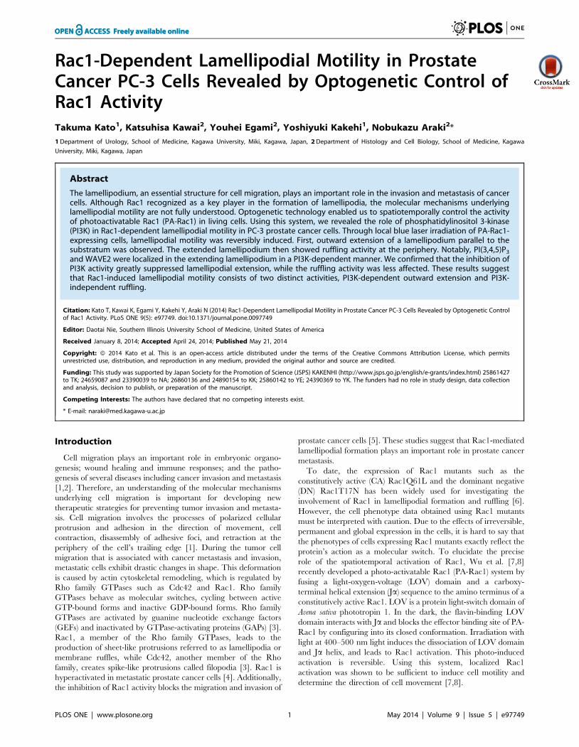
Figure 2. Local and reversible control of lamellipodial dynamics by photomanipulation of PA-Rac1 activity. Time-lapse images of a PC-3 cell expressing mCherry-PA-Rac1 were acquired during PA-Rac1 photo-manipulation by local laser irradiation of different areas. Selected phase-contrast and mCherry fluorescence images are shown. First, region 1 was irradiated for 10 min. The irradiation was then moved to region 2. At25 min, the irradiation was turned off. Selected time-lapse images of phase-contrast and mCherry fluorescence are shown. The extending andretracting lamellipodia are outlined in red and yellow, respectively. Scale bar, 10 mm.doi:10.1371/journal.pone.0097749.g002
PI3K in Rac1-Mediated Lamellipodial Motility
PLOS ONE | www.plosone.org 3 May 2014 | Volume 9 | Issue 5 | e97749
To quantify the effects of PI3K inhibitors on extended
lamellipodia and ruffling activity in mCitrine-Rac1Q61L-express-
ing cells, the cells were fixed with 4% paraformaldehyde at 30 min
after the addition of the PI3K inhibitors (50 mM LY294002,
100 nM wortmannin, or 1 mM ZSTK474) or the vehicle only
(0.1% DMSO). Using the MetaMorph imaging system, the
maximum diameters of the cells expressing Rac1Q61L were
measured before and after the drug treatments to evaluate the
effect of PI3K inhibition on the extended lamellipodial. Similarly,
the peripheral ruffles per cell were counted using a fluorescence
microscope to evaluate the impact of PI3K inhibition on ruffling
activity. Thirty cells in each group were subjected to the
quantitative image analysis.
Data are presented as the means 6 standard error (SE) for the
number of cells indicated in the text. Statistical analysis was
performed using the Wilcoxon t-test feature of Excel 2012.
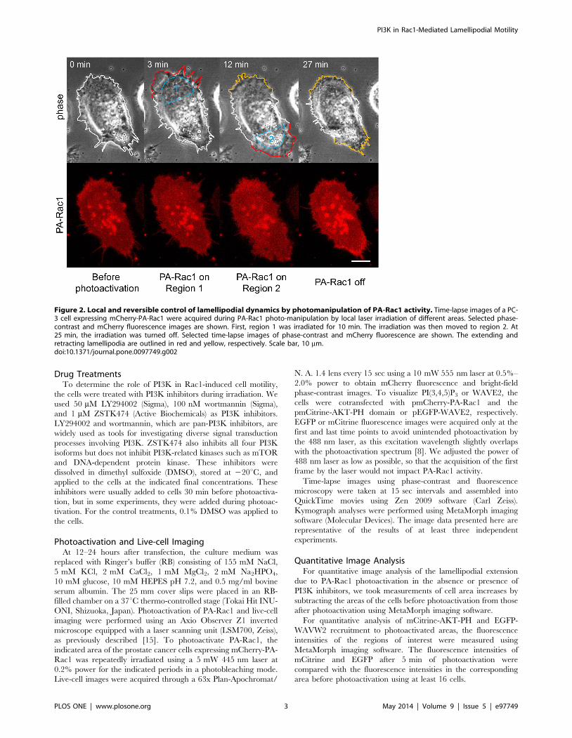
Figure 3. PI3K is required for lamellipodial extension but not for peripheral ruffling. (A) PC-3 cells were transiently transfected withpTriEx/mCherry-PA-Rac1. The cells were subjected to repeated photoactivation in the absence (control) or presence of 50 mM LY294002. The leadingedge of the extending lamellipodium is outlined in red. Scale bar, 10 mm. (B) The increased cell area due to lamellipodial extension was quantified bysubtracting the area of the cell before photoactivation from the area of the cell 7 min after the beginning of PA-Rac1 activation. This increase wasverified in 22 PC-3 cells. The data plot shows the increased area due to lamellipodial expansion that was induced by PA-Rac1 photoactivation in theabsence [LY(2)] or presence of LY294002 [LY(+)]. The significance of the differences between LY(2) and LY(+) was confirmed with the Wilcoxon t-test(right). The increase in the lamellipodial area in the presence of LY294002 was significantly lower than that of the control cells (p,0.01). (C)Kymographic analysis was performed at a two-headed arrow placed across the lamellipodium of a PC-3 cell expressing mCherry-PA-Rac1 before andafter the addition of LY294002. The laser-irradiated area is indicated with a blue rectangle. The white line outlines the extending lamellipodium, andthe dotted line outlines the original cell shape. The lower panel shows the kymograph of a lamellipodium undergoing changes in length. Thekymograph demonstrates the extension and retraction of a lamellipodium during PA-Rac1 activation (blue 1) and deactivation (black 1), respectively.The green arrow indicates the addition of LY294002. The PI3K inhibitor had less of an inhibitory effect on lamellipodial extension and ruffling (blue 2).However, the initiation of lamellipodial extension was drastically inhibited (black 2–blue 3). Scale bars, 10 mm.doi:10.1371/journal.pone.0097749.g003
PI3K in Rac1-Mediated Lamellipodial Motility
PLOS ONE | www.plosone.org 4 May 2014 | Volume 9 | Issue 5 | e97749
Differences between the analyzed samples were considered
significant at p,0.05.
Results
Local Activation of PA-Rac1 Reversibly InducesLamellipodial Extension and Ruffling
To elucidate the relationship between Rac1 activation and
lamellipodial dynamics, we introduced mCherry-fused PA-Rac1
into PC-3 cells. After confirming the expression of mCherry-PA-
Rac1 based on the mCherry signal, we irradiated a peripheral
region of the cells using a 445 nm laser during the intervals of
image acquisition and acquired phase-contrast images of the live
cells every 15 sec by confocal microscopy. Typically, after 1–4 min
of irradiation with the 445 nm laser, a thin sheet-like protrusion
(i.e., a lamellipodium) extending parallel to the substratum was
observed at the cell peripheral site that was irradiated by the
445 nm laser. The lamellipodium reached its maximum length
after 5–6 min of PA-Rac1 activation. At that time, following the
outward extension, the fully extended lamellipodium curled up its
leading edge to show a peripheral ruffling movement. The ruffling
movements continued for the duration of irradiation. After
irradiation ceased, both the lamellipodial extension and the
peripheral ruffling promptly receded (Fig. 1 and Movie S1). In
our previous study, dorsal ruffling was induced in RAW 264
macrophages by PA-Rac1 activation [15,16], but dorsal ruffling
was not prominent in PC-3 cells.
When we irradiated a different area of the same cell, a
lamellipodial extension was generated at the newly irradiated area,
suggesting that these phenomena were dependent on local PA-
Rac1 activation by light irradiation (Fig. 2). The overexpression of
a GDP-bound (dominant negative) mutant of PA-Rac1 (PA-Rac1
T17N) did not induce either lamellipodial extension or peripheral
ruffling after 445-nm laser irradiation (not shown). This finding
indicated that the morphological changes induced by 445-nm laser
irradiation are dependent on GTP-loaded Rac1.
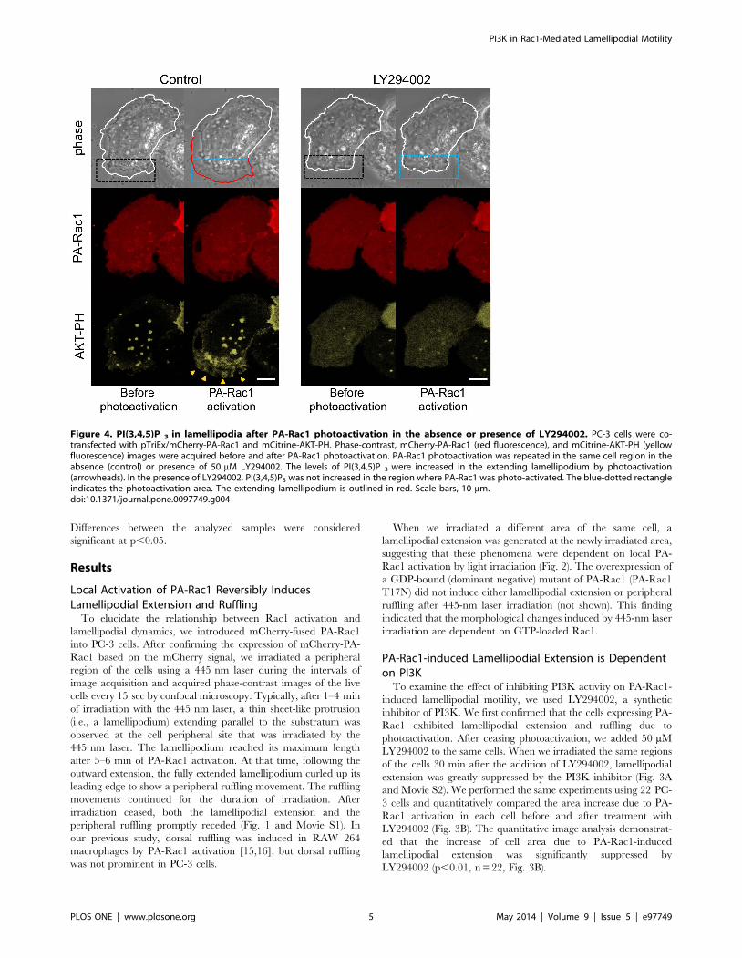
PA-Rac1-induced Lamellipodial Extension is Dependenton PI3K
To examine the effect of inhibiting PI3K activity on PA-Rac1-
induced lamellipodial motility, we used LY294002, a synthetic
inhibitor of PI3K. We first confirmed that the cells expressing PA-
Rac1 exhibited lamellipodial extension and ruffling due to
photoactivation. After ceasing photoactivation, we added 50 mM
LY294002 to the same cells. When we irradiated the same regions
of the cells 30 min after the addition of LY294002, lamellipodial
extension was greatly suppressed by the PI3K inhibitor (Fig. 3A
and Movie S2). We performed the same experiments using 22 PC-
3 cells and quantitatively compared the area increase due to PA-
Rac1 activation in each cell before and after treatment with
LY294002 (Fig. 3B). The quantitative image analysis demonstrat-
ed that the increase of cell area due to PA-Rac1-induced
lamellipodial extension was significantly suppressed by
LY294002 (p,0.01, n = 22, Fig. 3B).
Figure 4. PI(3,4,5)P 3 in lamellipodia after PA-Rac1 photoactivation in the absence or presence of LY294002. PC-3 cells were co-transfected with pTriEx/mCherry-PA-Rac1 and mCitrine-AKT-PH. Phase-contrast, mCherry-PA-Rac1 (red fluorescence), and mCitrine-AKT-PH (yellowfluorescence) images were acquired before and after PA-Rac1 photoactivation. PA-Rac1 photoactivation was repeated in the same cell region in theabsence (control) or presence of 50 mM LY294002. The levels of PI(3,4,5)P 3 were increased in the extending lamellipodium by photoactivation(arrowheads). In the presence of LY294002, PI(3,4,5)P3 was not increased in the region where PA-Rac1 was photo-activated. The blue-dotted rectangleindicates the photoactivation area. The extending lamellipodium is outlined in red. Scale bars, 10 mm.doi:10.1371/journal.pone.0097749.g004
PI3K in Rac1-Mediated Lamellipodial Motility
PLOS ONE | www.plosone.org 5 May 2014 | Volume 9 | Issue 5 | e97749
Additionally, we conducted kymographic analysis to determine
the changes in length of the lamellipodia. After confirming PA-
Rac1-induced lamellipodial extension and ruffling due to PA-Rac1
photoactivation, we added LY294002 to the cells during PA-Rac1
photoactivation. Although the lengths of the lamellipodia were
changed due to peripheral ruffling, the inhibitory effect of
LY294002 on lamellipodial extension was small. Peripheral
ruffling persisted for the duration of irradiation. Immediately after
photoactivation ceased, both lamellipodial extension and periph-
eral ruffling receded completely. When we re-irradiated the same
region, the cells did not show lamellipodial extension (Fig. 3C).
These results suggest that the formation of a lamellipodium is
more sensitive to PI3K inhibitors than is the maintenance of
extended lamellipodia and peripheral ruffling.
Similar results were obtained using 100 nM wortmannin or
1 mM ZSTK474 as PI3K inhibitors (Fig. S1). As a control, the
same PA-Rac1 activation experiments were performed using cells
treated with 0.1% DMSO, the vehicle of the inhibitors. PA-Rac1-
induced lamellipodial formation was not inhibited by DMSO (Fig.
S2).
PA-Rac1 Photoactivation can Locally Activate PI3K andRecruit WAVE2
Because PA-Rac1-induced lamellipodial extension was inhibited
by the PI3K inhibitors, we attempted to clarify that PA-Rac1
activation led to PI3K activation. To monitor the production of
PI(3,4,5)P3 by PI3K activity, PC-3 cells were cotransfected with
pmCitrine-AKT-PH and pTriEx/mCherry-PA-Rac1Q61L and
observed using a Zeiss LSM700 (Fig. 4). The fluorescence intensity
of mCitrine-AKT-PH at the lamellipodial leading edge after 5 min
of PA-Rac1photoactivation was measured using MetaMorph
imaging software and was quantitatively compared with that of
the same region before photoactivation. After 5 min of local
activation of PA-Rac1, the fluorescence intensity of mCitrine-
AKT-PH showed a 125.4% 622.3% increase (n = 16) compared
with that measured before photoactivation, suggesting that the
levels of PI(3,4,5)P3 in the extending lamellipodia were greatly
increased by the local photoactivation of PA-Rac1. In the presence
of LY294002, no cells showed an increase in the fluorescence
intensity of mCitrine-AKT-PH after irradiation. This finding
suggests that Rac1 photoactivation activates PI3K to produce
PI(3,4,5)P3 from PI(4,5)P2 at the membrane of the extending
lamellipodia.
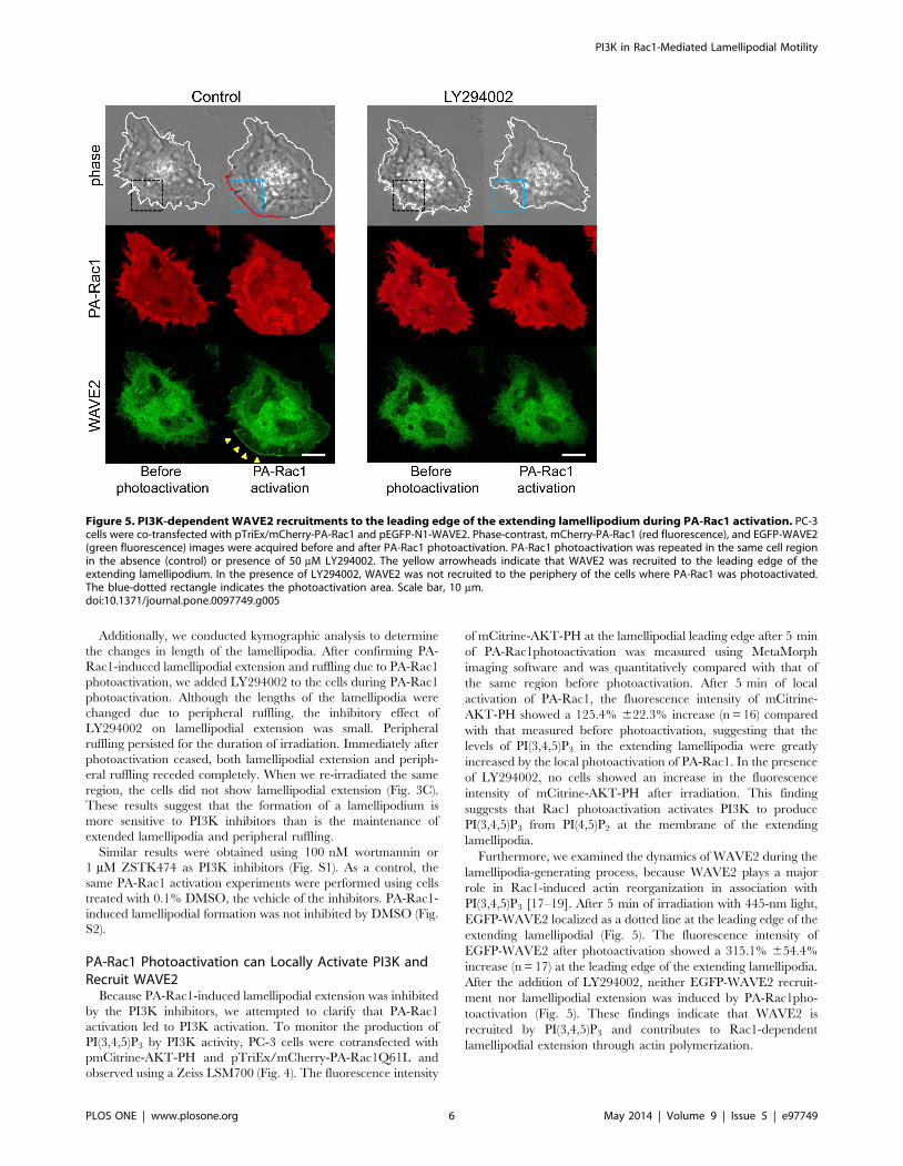
Furthermore, we examined the dynamics of WAVE2 during the
lamellipodia-generating process, because WAVE2 plays a major
role in Rac1-induced actin reorganization in association with
PI(3,4,5)P3 [17–19]. After 5 min of irradiation with 445-nm light,
EGFP-WAVE2 localized as a dotted line at the leading edge of the
extending lamellipodial (Fig. 5). The fluorescence intensity of
EGFP-WAVE2 after photoactivation showed a 315.1% 654.4%
increase (n = 17) at the leading edge of the extending lamellipodia.
After the addition of LY294002, neither EGFP-WAVE2 recruit-
ment nor lamellipodial extension was induced by PA-Rac1pho-
toactivation (Fig. 5). These findings indicate that WAVE2 is
recruited by PI(3,4,5)P3 and contributes to Rac1-dependent
lamellipodial extension through actin polymerization.
Figure 5. PI3K-dependent WAVE2 recruitments to the leading edge of the extending lamellipodium during PA-Rac1 activation. PC-3cells were co-transfected with pTriEx/mCherry-PA-Rac1 and pEGFP-N1-WAVE2. Phase-contrast, mCherry-PA-Rac1 (red fluorescence), and EGFP-WAVE2(green fluorescence) images were acquired before and after PA-Rac1 photoactivation. PA-Rac1 photoactivation was repeated in the same cell regionin the absence (control) or presence of 50 mM LY294002. The yellow arrowheads indicate that WAVE2 was recruited to the leading edge of theextending lamellipodium. In the presence of LY294002, WAVE2 was not recruited to the periphery of the cells where PA-Rac1 was photoactivated.The blue-dotted rectangle indicates the photoactivation area. Scale bar, 10 mm.doi:10.1371/journal.pone.0097749.g005
PI3K in Rac1-Mediated Lamellipodial Motility
PLOS ONE | www.plosone.org 6 May 2014 | Volume 9 | Issue 5 | e97749
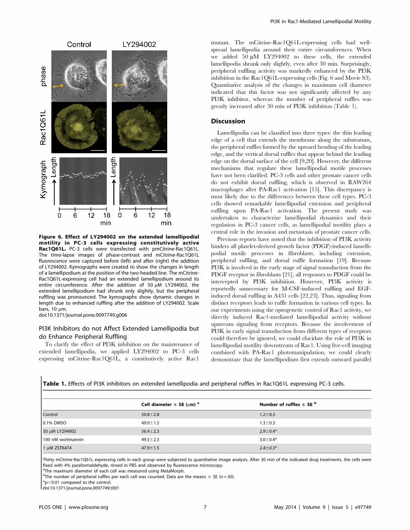
PI3K Inhibitors do not Affect Extended Lamellipodia butdo Enhance Peripheral Ruffling
To clarify the effect of PI3K inhibition on the maintenance of
extended lamellipodia, we applied LY294002 to PC-3 cells
expressing mCitrine-Rac1Q61L, a constitutively active Rac1
mutant. The mCitrine-Rac1Q61L-expressing cells had well-
spread lamellipodia around their entire circumferences. When
we added 50 mM LY294002 to these cells, the extended
lamellipodia shrank only slightly, even after 30 min. Surprisingly,
peripheral ruffling activity was markedly enhanced by the PI3K
inhibition in the Rac1Q61L-expressing cells (Fig. 6 and Movie S3).
Quantitative analysis of the changes in maximum cell diameter
indicated that this factor was not significantly affected by any
PI3K inhibitor, whereas the number of peripheral ruffles was
greatly increased after 30 min of PI3K inhibition (Table 1).
Discussion
Lamellipodia can be classified into three types: the thin leading
edge of a cell that extends the membrane along the substratum,
the peripheral ruffles formed by the upward bending of the leading
edge, and the vertical dorsal ruffles that appear behind the leading
edge on the dorsal surface of the cell [9,20]. However, the different
mechanisms that regulate these lamellipodial motile processes
have not been clarified. PC-3 cells and other prostate cancer cells
do not exhibit dorsal ruffling, which is observed in RAW264
macrophages after PA-Rac1 activation [15]. This discrepancy is
most likely due to the differences between these cell types. PC-3
cells showed remarkable lamellipodial extension and peripheral
ruffling upon PA-Rac1 activation. The present study was
undertaken to characterize lamellipodial dynamics and their
regulation in PC-3 cancer cells, as lamellipodial motility plays a
central role in the invasion and metastasis of prostate cancer cells.
Previous reports have noted that the inhibition of PI3K activity
hinders all platelet-derived growth factor (PDGF)-induced lamelli-
podial motile processes in fibroblasts, including extension,
peripheral ruffling, and dorsal ruffle formation [19]. Because
PI3K is involved in the early stage of signal transduction from the
PDGF receptor in fibroblasts [21], all responses to PDGF could be
intercepted by PI3K inhibition. However, PI3K activity is
reportedly unnecessary for M-CSF-induced ruffling and EGF-
induced dorsal ruffling in A431 cells [22,23]. Thus, signaling from
distinct receptors leads to ruffle formation in various cell types. In
our experiments using the optogenetic control of Rac1 activity, we
directly induced Rac1-mediated lamellipodial activity without
upstream signaling from receptors. Because the involvement of
PI3K in early signal transduction from different types of receptors
could therefore be ignored, we could elucidate the role of PI3K in
lamellipodial motility downstream of Rac1. Using live-cell imaging
combined with PA-Rac1 photomanipulation, we could clearly
demonstrate that the lamellipodium first extends outward parallel
Figure 6. Effect of LY294002 on the extended lamellipodialmotility in PC-3 cells expressing constitutively activeRac1Q61L. PC-3 cells were transfected with pmCitrine-Rac1Q61L.The time-lapse images of phase-contrast and mCitrine-Rac1Q61Lfluorescence were captured before (left) and after (right) the additionof LY294002. Kymographs were created to show the changes in lengthof a lamellipodium at the position of the two-headed line. The mCitrine-Rac1Q61L-expressing cell had an extended lamellipodium around itsentire circumference. After the addition of 50 mM LY294002, theextended lamellipodium had shrunk only slightly, but the peripheralruffling was pronounced. The kymographs show dynamic changes inlength due to enhanced ruffling after the addition of LY294002. Scalebars, 10 mm.doi:10.1371/journal.pone.0097749.g006
Table 1. Effects of PI3K inhibitors on extended lamellipodia and peripheral ruffles in Rac1Q61L expressing PC-3 cells.
Cell diameter ± SE (mm) a Number of ruffles ± SE b
Control 50.862.8 1.260.3
0.1% DMSO 49.061.5 1.360.3
50 mM LY294002 56.462.3 2.960.4*
100 nM wortmannin 49.562.3 3.060.4*
1 mM ZSTK474 47.961.5 2.460.3*
Thirty mCitrine-Rac1Q61L expressing cells in each group were subjected to quantitative image analysis. After 30 min of the indicated drug treatments, the cells werefixed with 4% paraformaldehyde, rinsed in PBS and observed by fluorescence microscopy.aThe maximum diameter of each cell was measured using MetaMorph.bThe number of peripheral ruffles per each cell was counted. Data are the means 6 SE (n = 30).*p,0.01 compared to the control.doi:10.1371/journal.pone.0097749.t001
PI3K in Rac1-Mediated Lamellipodial Motility
PLOS ONE | www.plosone.org 7 May 2014 | Volume 9 | Issue 5 | e97749
to the substratum, and that the fully extended lamellipodium then
shows ruffling activity by curling up the leading edge. Further-
more, we found that the lamellipodial extension induced by PA-
Rac1 activation was severely perturbed by PI3K inhibitors while
peripheral ruffling was not inhibited. These results suggest that two
types of lamellipodial motility, extension and ruffling, are
differentially regulated by PI3K-dependent and PI3K-indepen-
dent signaling pathways.
Wiskott-Aldrich syndrome protein (WASP) and WASP-family
verprolin-homologous protein (WAVE) family proteins are acti-
vators of Arp2/3-dependent polymerization [17]. WAVE family
proteins are associated with lamellipodial formation through the
Rac1 signaling pathway. To prevent disordered actin polymeri-
zation, WAVE family proteins exist as heteropentameric protein
complexes that hinder their own active sites. Although WAVEs are
functionally activated by GTP-bound Rac1 when actin polymer-
ization is initiated, WAVEs cannot bind directly to GTP-bound
Rac1. Instead, IRSp53 (insulin receptor tyrosine kinase substrate
p53) works as a linker molecule to connect Rac1 and the WAVE
complex [18]. GTP-bound Rac1 induces an allosteric change in
the WAVE complex that exposes its active site; WAVE2 then
activates the Arp2/3 complex, which becomes a nucleus for actin
polymerization at the leading edge of the lamellipodium [24–26].
In our PA-Rac1-activation experiments, WAVE2 was localized
to the leading edges of extending lamellipodia. However, neither
WAVE2 recruitment nor lamellipodial extension was observed
when PI3K activity was inhibited. These results suggest that
PI(3,4,5)P3 is required for WAVE2 recruitment and for lamelli-
podial extension. Because WAVE2 has a PI(3,4,5)P3-binding
sequence [19], PI(3,4,5) P3 may recruit WAVE2 to the leading
edge. Suetsugu et al. [18] reported that the activity of the WAVE2
complex was optimized by IRSp53 in association with activated
Rac1 and PI(3,4,5)P3. Furthermore, the simultaneous binding of
GTP-bound Rac and acidic phospholipids such as PI(3,4,5)P3 to
WAVE2 is required for the efficient recruitment and activation of
WAVE2 [17]. These previous reports strengthen our assertion that
the inhibition of lamellipodial extension by PI3K inhibitors results
from the perturbation of WAVE2 recruitment.
In this study, the activation of PA-Rac1 induced the production
of PI(3,4,5)P3 and the recruitment of WAVE2 when lamellipodial
extension was initiated. Furthermore, PI3K inhibitors hindered
the recruitment of WAVE2 and PI(3,4,5)P3 and suppressed
lamellipodial extension. These findings indicate that PI3K plays
an essential role in initiating lamellipodial extension. Furthermore,
we employed constitutively active Rac1Q61L-expressing cells to
observe the response of the extended lamellipodia to the inhibition
of PI3K activity. In cells expressing Rac1Q61L, the extended
lamellipodia were relatively resistant to PI3K inhibitors. In
addition, the PI3K inhibitors actually enhanced the peripheral
ruffling activity of the lamellipodia, but rather enhanced this
activity. Thus, while PI3K may not be crucial for the maintenance
of extended lamellipodia or for ruffling activity, the initiation of
lamellipodial extension is highly dependent on PI3K. Notably,
PI3K inhibitors also enhanced peripheral ruffling activity in
Rac1Q61L-expressing PC-3 cells, although the mechanism for this
phenomenon remains unclear. Using EGF-stimulated A431 cells,
we have previously shown that PI(4,5)P2 is enriched in the
membrane of ruffles, however, PI(3,4,5)P3 levels are elevated only
at the closing of circular ruffles into macropinosomes [23].
Therefore, ruffle formation is likely more dependent on
PI(4,5)P2. Because PI3K inhibition results in an increase in
PI(4,5)P2 levels, it may be hypothesized that this increased
PI(4,5)P2 enhances ruffling activity. We recently reported that
the sequential breakdown of PI(3,4,5)P3 is also important for the
completion of macropinosome formation from membrane ruffling
[27]. Thus, the roles of phosphoinositide metabolism in membrane
ruffling and lamellipodial extension are more complicated and
important than we previously predicted. Future studies should
conduct more detailed examinations of the interactions of each
phosphoinositide with its effectors and/or other signaling path-
ways.
Recently, the overexpression of a Rac1 activator protein (14-3-3
protein zeta) and several GEFs (VAV3, P-Rex1) was identified in
prostate cancer [28–30]. Moreover, castration-resistant prostate
cancer cells, which have a high malignant potential associated with
invasion and metastasis, overexpress Rac1 [31]. These findings
suggest that Rac1 overexpression affects the progression of
prostate cancer. Although several studies have shown that PI3K
inhibitors obstruct the migration of prostate cancer cells as
induced by chemical mediators, those studies assumed that PI3K
affected the upstream signal transduction of Rac1 [32–34]. To our
knowledge, no previous report has examined the relationship
between PI3K and Rac1 downstream signal transduction in
prostate cancer. In this study, we clearly showed that PI(3,4,5)P3
and the Rac1 downstream effector protein WAVE2 act in a
coordinated manner in lamellipodial extension, which contributes
to the migration of prostate cancer cells. Therefore, the inhibition
of PI3K activity effectively obstructs the Rac1-overexpression-
mediated migration of prostate cancer cells.
Conclusions
Optogenetic technology enabled us to spatiotemporally control
PA-Rac1 activity in prostate cancer cells. We demonstrated that
the inhibition of PI3K activity suppressed lamellipodial extension
but had less of an inhibitory effect on peripheral ruffling. The
present study indicates that PI3K, acting downstream of Rac1, has
an essential role in the initiation of lamellipodial extension, which
underlies prostate cancer cell invasion and metastasis. The better
understanding and further characterization of the molecular
regulation of the lamellipodial motile processes of metastatic
prostate cancer cells will provide new insights for the development
of cancer therapies.
Supporting Information
Figure S1 Effects of other PI3K inhibitors on lamelli-podial extension induced by photoactivation. PC-3 cells
were transiently transfected with pTriEx/mCherry-PA-Rac1. The
cells were subjected to repeated photoactivation in the absence
(control) or presence of 100 nM wortmannin or 1 mM ZSTK474.
The leading edge of the extending lamellipodium is outlined in
red. Both wortmannin and ZSTK474 obstructed lamellipodial
extension. Scale bars, 10 mm.
(TIF)
Figure S2 PA-Rac1-induced lamellipodial extension wasnot influenced by dimethyl sulfoxide. PC-3 cells were
transiently transfected with pTriEx/mCherry-PA-Rac1 and sub-
jected to local photoactivation of PA-Rac1 (rectangular area
outlined by blue dots). The cells were subjected to repeated
photoactivation in the absence (control) or presence of 0.1%
dimethyl sulfoxide (DMSO). Kymographic analysis was performed
at a line placed across a lamellipodium. After 30 min of treatment
with 0.1% DMSO, the cell showed lamellipodial extension to the
same extent as in the absence of DMSO. Scale bars, 10 mm.
(TIF)
Movie S1 Photoactivation of PA-Rac1 induces lamelli-podial extension and subsequent ruffling. This movie
PI3K in Rac1-Mediated Lamellipodial Motility
PLOS ONE | www.plosone.org 8 May 2014 | Volume 9 | Issue 5 | e97749
shows that local PA-Rac1 activation induced lamellipodial
extension and subsequent ruffling. PC-3 cells were transiently
transfected with pmCherry-PA-Rac1 (shown in red). The 445-nm
laser-irradiated area is indicated with a blue rectangle. This movie
corresponds to the images shown in Fig. 1. Scale bar, 10 mm.
(MP4)
Movie S2 PI3K is required for lamellipodial extensionbut not for peripheral ruffling. This movie shows that the
lamellipodial extension induced by PA-Rac1 activation was
suppressed by LY294002. The PA-Rac1 signal is shown as red.
The 445 nm laser-irradiated area is indicated with a blue
rectangle. This movie corresponds to the images shown in
Fig. 3A. Scale bar, 10 mm.
(MP4)
Movie S3 Effect of LY294002 on the extended lamelli-podial motility in PC-3 cells expressing constitutivelyactive Rac1Q61L. This movie shows that the extended
lamellipodium is not shortened but is actively ruffled by PI3K
inhibition. PC-3 cells were transiently transfected with pmCitrine-
Rac1Q61L (shown in green). This movie corresponds to the
images shown in Fig. 6. Scale bar, 10 mm.
(MP4)
Acknowledgments
The authors would like to thank Dr. Joel A. Swanson (University of
Michigan) and Dr. Tadaomi Takenawa (Kobe University) for kindly
providing plasmids. The authors would also like to thank Dr. Xia Zhang,
Dr. Katsuya Miyake, Ms. Yukiko Iwabu, and Mr. Kazuhiro Yokoi for their
assistance and advice.
Author Contributions
Conceived and designed the experiments: TK YK NA. Performed the
experiments: TK. Analyzed the data: TK. Contributed reagents/
materials/analysis tools: TK KK YE. Wrote the paper: TK NA.
References
1. Ridley AJ, Schwartz MA, Burridge K, Firtel RA, Ginsberg MH, et al. (2003)
Cell migration: Integrating signals from front to back. Science 302: 1704–1709.2. Vicente-Manzanares M, Webb DJ, Horwitz AR (2005) Cell migration at a
glance. J Cell Sci 118: 4917–4919.3. Jaffe AB, Hall A (2005) Rho GTPases: Biochemistry and biology. Annu Rev Cell
Dev Biol 21: 247–269.
4. Knight-Krajewski S, Welsh CF, Liu Y, Lyons LS, Faysal JM, et al. (2004)Deregulation of the Rho GTPase, Rac1, suppresses cyclin-dependent kinase
inhibitor p21(CIP1) levels in androgen-independent human prostate cancer cells.Oncogene 23: 5513–5522.
5. Gao Y, Dickerson JB, Guo F, Zheng J, Zheng Y (2004) Rational design andcharacterization of a Rac GTPase-specific small molecule inhibitor. Proc Natl
Acad Sci U S A 101: 7618–7623.
6. Spiering D, Hodgson L (2011) Dynamics of the Rho-family small GTPases inactin regulation and motility. Cell Adh Migr 5: 170–180.
7. Wu YI, Frey D, Lungu OI, Jaehrig A, Schlichting I, et al. (2009) A geneticallyencoded photoactivatable Rac controls the motility of living cells. Nature 461:
104–108.
8. Wu YI, Wang X, He L, Montell D, Hahn KM (2011) Spatiotemporal control ofsmall GTPases with light using the LOV domain. Methods Enzymol 497: 393–
407.9. Rikitake Y, Takai Y (2011) Directional cell migration regulation by small G
proteins, nectin-like molecule-5, and afadin. Int Rev Cell Mol Biol 287: 97–143.10. Costa C, Germena G, Hirsch E (2010) Dissection of the interplay between class I
PI3Ks and Rac signaling in phagocytic functions. ScientificWorldJournal 10:
1826–1839.11. Weiner OD, Neilsen PO, Prestwich GD, Kirschner MW, Cantley LC, et al.
(2002) A PtdInsP(3)- and Rho GTPase-mediated positive feedback loop regulatesneutrophil polarity. Nat Cell Biol 4: 509–513.
12. Inoue T, Meyer T (2008) Synthetic activation of endogenous PI3K and Rac
identifies an AND-gate switch for cell polarization and migration. PLoS One 3:e3068.
13. Welf ES, Ahmed S, Johnson HE, Melvin AT, Haugh JM (2012) Migratingfibroblasts reorient directionality by a metastable, PI3K-dependent mechanism.
J Cell Biol 197: 105–114.14. Yoo SK, Deng Q, Cavnar PJ, Wu YI, Hahn KM, et al. (2010) Differential
regulation of protrusion and polarity by PI3K during neutrophil motility in live
zebrafish. Dev Cell 18: 226–236.15. Fujii M, Kawai K, Egami Y, Araki N (2013) Dissecting the roles of Rac1
activation and deactivation in macropinocytosis using microscopic photo-manipulation. Sci Rep 3: 2385.
16. Araki N, Ikeda Y, Kato T, Kawai K, Egami Y, et al. (2014) Development of an
automated fluorescence microscopy system for photomanipulation of geneticallyencoded photoactivatable proteins (optogenetics) in live cells. Microscopy 63:
doi: 10.1093/jmicro/dfu003.17. Lebensohn AM, Kirschner MW (2009) Activation of the WAVE complex by
coincident signals controls actin assembly. Mol Cell 36: 512–524.
18. Suetsugu S, Kurisu S, Oikawa T, Yamazaki D, Oda A, et al. (2006)
Optimization of WAVE2 complex-induced actin polymerization by mem-brane-bound IRSp53, PIP(3), and Rac. J Cell Biol 173: 571–585.
19. Suetsugu S, Yamazaki D, Kurisu S, Takenawa T (2003) Differential roles ofWAVE1 and WAVE2 in dorsal and peripheral ruffle formation for fibroblast cell
migration. Dev Cell 5: 595–609.
20. Abercrombie M, Heaysman JE, Pegrum SM (1970) The locomotion offibroblasts in culture. II. ‘‘RRuffling’’. Exp Cell Res 60: 437–444.
21. Claesson-Welsh L (1994) Signal transduction by the PDGF receptors. ProgGrowth Factor Res 5: 37–54.
22. Araki N, Johnson MT, Swanson JA (1996) A role for phosphoinositide 3-kinasein the completion of macropinocytosis and phagocytosis by macrophages. J Cell
Biol 135: 1249–1260.
23. Araki N, Egami Y, Watanabe Y, Hatae T (2007) Phosphoinositide metabolismduring membrane ruffling and macropinosome formation in EGF-stimulated
A431 cells. Exp Cell Res 313: 1496–1507.24. Ridley AJ (2011) Life at the leading edge. Cell 145: 1012–1022.
25. Kurisu S, Takenawa T (2009) The WASP and WAVE family proteins. Genome
Biol 10: 226.26. Oda A, Eto K (2013) WASPs and WAVEs: From molecular function to
physiology in hematopoietic cells. Semin Cell Dev Biol 24: 308–313.27. Maekawa M, Terasaka S, Mochizuki Y, Kawai K, Ikeda Y, et al. (2014)
Sequential breakdown of 3-phosphorylated phosphoinositides is essential for thecompletion of macropinocytosis. Proc Natl Acad Sci U S A 111: doi: 10.1073/
pnas.1311029111.
28. Goc A, Abdalla M, Al-Azayzih A, Somanath PR (2012) Rac1 activation drivenby 14–3-3zeta dimerization promotes prostate cancer cell-matrix interactions,
motility and transendothelial migration. PLoS One 7: e40594.29. Qin J, Xie Y, Wang B, Hoshino M, Wolff DW, et al. (2009) Upregulation of
PIP3-dependent Rac exchanger 1 (P-Rex1) promotes prostate cancer metastasis.
Oncogene 28: 1853–1863.30. Lin KT, Gong J, Li CF, Jang TH, Chen WL, et al. (2012) Vav3-Rac1 signaling
regulates prostate cancer metastasis with elevated Vav3 expression correlatingwith prostate cancer progression and posttreatment recurrence. Cancer Res 72:
3000–3009.31. Kobayashi T, Inoue T, Shimizu Y, Terada N, Maeno A, et al. (2010) Activation
of Rac1 is closely related to androgen-independent cell proliferation of prostate
cancer cells both in vitro and in vivo. Mol Endocrinol 24: 722–734.32. Frankenberry KA, Somasundar P, McFadden DW, Vona-Davis LC (2004)
Leptin induces cell migration and the expression of growth factors in humanprostate cancer cells. Am J Surg 188: 560–565.
33. Monet M, Gkika D, Lehen’kyi V, Pourtier A, Vanden Abeele F, et al. (2009)
Lysophospholipids stimulate prostate cancer cell migration via TRPV2 channelactivation. Biochim Biophys Acta 1793: 528–539.
34. Yu HM, Frank DE, Zhang J, You X, Carter WG, et al. (2004) Basal prostateepithelial cells stimulate the migration of prostate cancer cells. Mol Carcinog 41:
85–97.
PI3K in Rac1-Mediated Lamellipodial Motility
PLOS ONE | www.plosone.org 9 May 2014 | Volume 9 | Issue 5 | e97749