Embed Size (px)
Citation preview
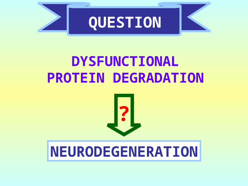
QUESTION
DYSFUNCTIONALPROTEIN DEGRADATION
NEURODEGENERATION
?
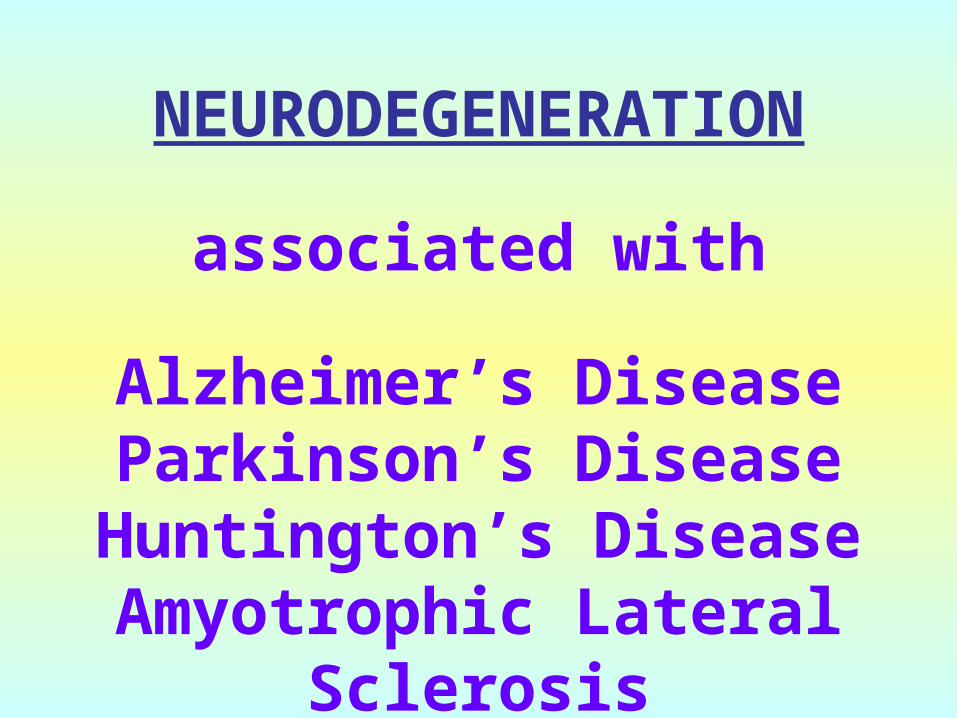
NEURODEGENERATION
associated with
Alzheimer’s DiseaseParkinson’s Disease
Huntington’s DiseaseAmyotrophic Lateral
Sclerosis
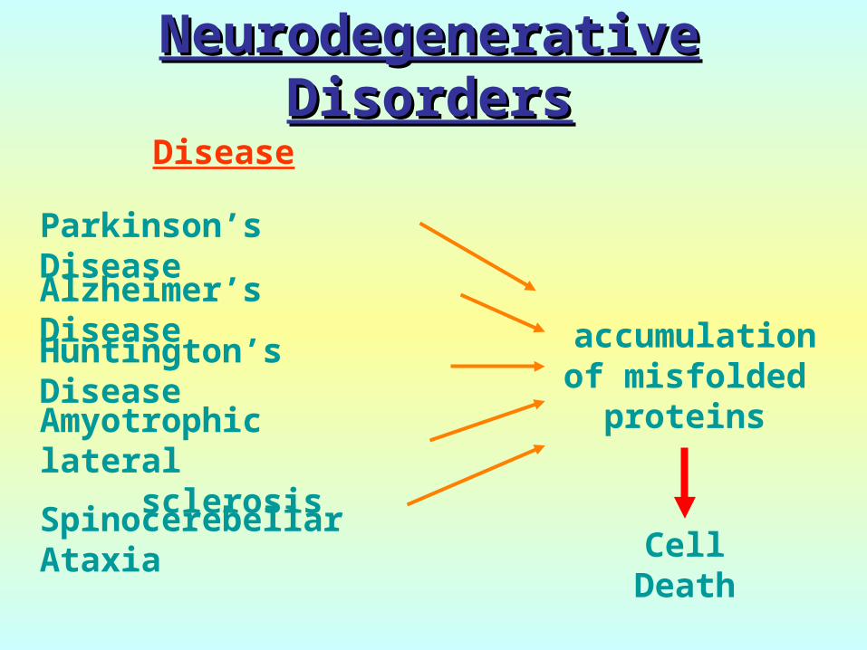
Parkinson’s Disease
Alzheimer’s Disease
Huntington’s Disease
Amyotrophic lateral sclerosis
Spinocerebellar Ataxia
Disease
accumulation of misfolded proteins
Cell Death
Neurodegenerative Neurodegenerative DisordersDisorders
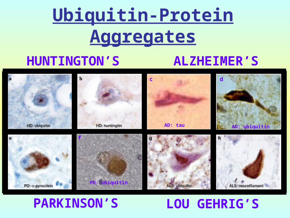
Ubiquitin-Protein Aggregates
HUNTINGTON’S ALZHEIMER’S
PARKINSON’S LOU GEHRIG’S
f
PD: ubiquitin
c
AD: tau
d
AD: ubiquitin
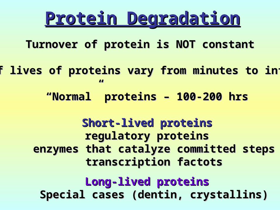
Protein DegradationProtein DegradationTurnover of protein is NOT constantTurnover of protein is NOT constant
Half lives of proteins vary from minutes to infinityHalf lives of proteins vary from minutes to infinity
““Normal” proteins – 100-200 hrsNormal” proteins – 100-200 hrs
Short-lived proteinsShort-lived proteinsregulatory proteinsregulatory proteins
enzymes that catalyze committed stepsenzymes that catalyze committed stepstranscription factotstranscription factots
Long-lived proteinsLong-lived proteinsSpecial cases (dentin, crystallins)Special cases (dentin, crystallins)
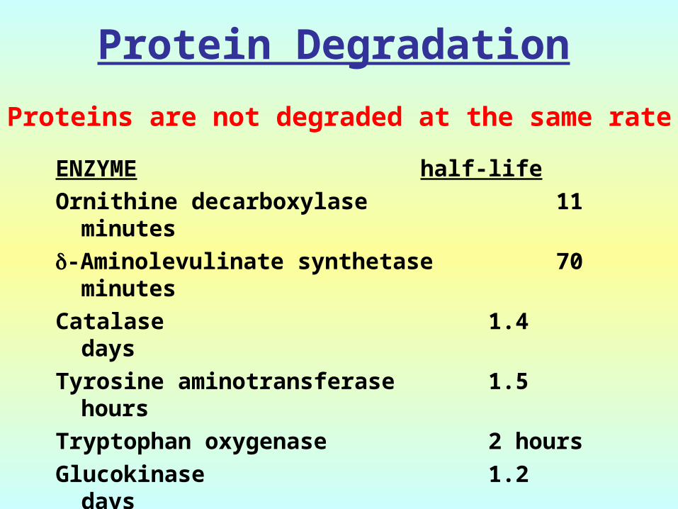
Protein Degradation
ENZYME half-lifeOrnithine decarboxylase 11
minutes-Aminolevulinate synthetase 70
minutesCatalase 1.4
daysTyrosine aminotransferase 1.5
hoursTryptophan oxygenase 2 hoursGlucokinase 1.2
daysLactic dehydrogenase 16 daysHMG CoA reductase 3 hour
Proteins are not degraded at the same rate
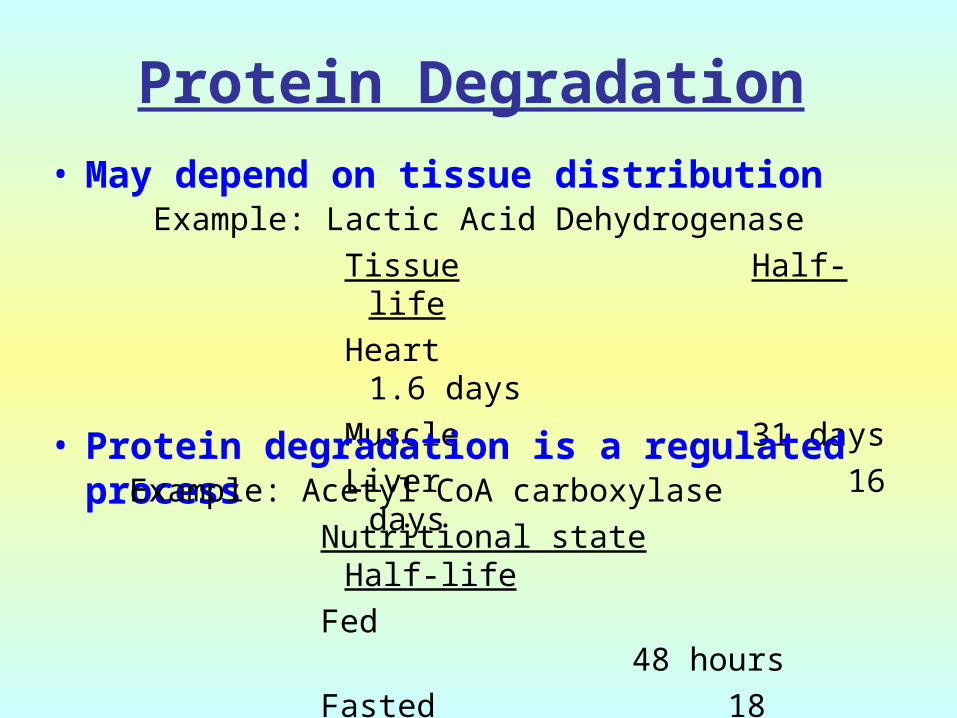
Protein Degradation
Example: Lactic Acid DehydrogenaseTissue Half-lifeHeart 1.6
daysMuscle 31 daysLiver 16
days
• May depend on tissue distribution
• Protein degradation is a regulated processExample: Acetyl CoA carboxylase
Nutritional state Half-lifeFed
48 hoursFasted 18
hours
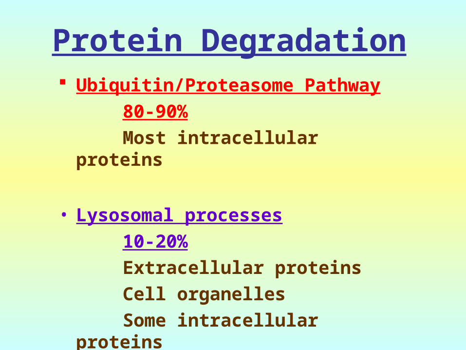
Protein Degradation Ubiquitin/Proteasome Pathway
80-90%Most intracellular proteins
• Lysosomal processes10-20%
Extracellular proteinsCell organellesSome intracellular proteins

Two Sites for Protein Degradation
Proteasomes
Large (26S) multiprotein complex (28 subunits)
Degrades ubiquitinated proteins
Lysosomes
Basal degradation – non-selective
Degradation under starvation – selective for “KFERQ” proteins

The Ubiquitin/ProteasomeThe Ubiquitin/Proteasome
PATHWAYPATHWAY

UBIQUITIN
KK
GG
Small peptide that is a “TAG” 76 amino acids C-terminal glycine - isopeptide
bond with the -amino group of lysine residues on the substrate
Attached as monoubiquitin or polyubiquitin chains
Three genes in humans:
Two are stress genes (B and C)
One, UbA as a fusion protein
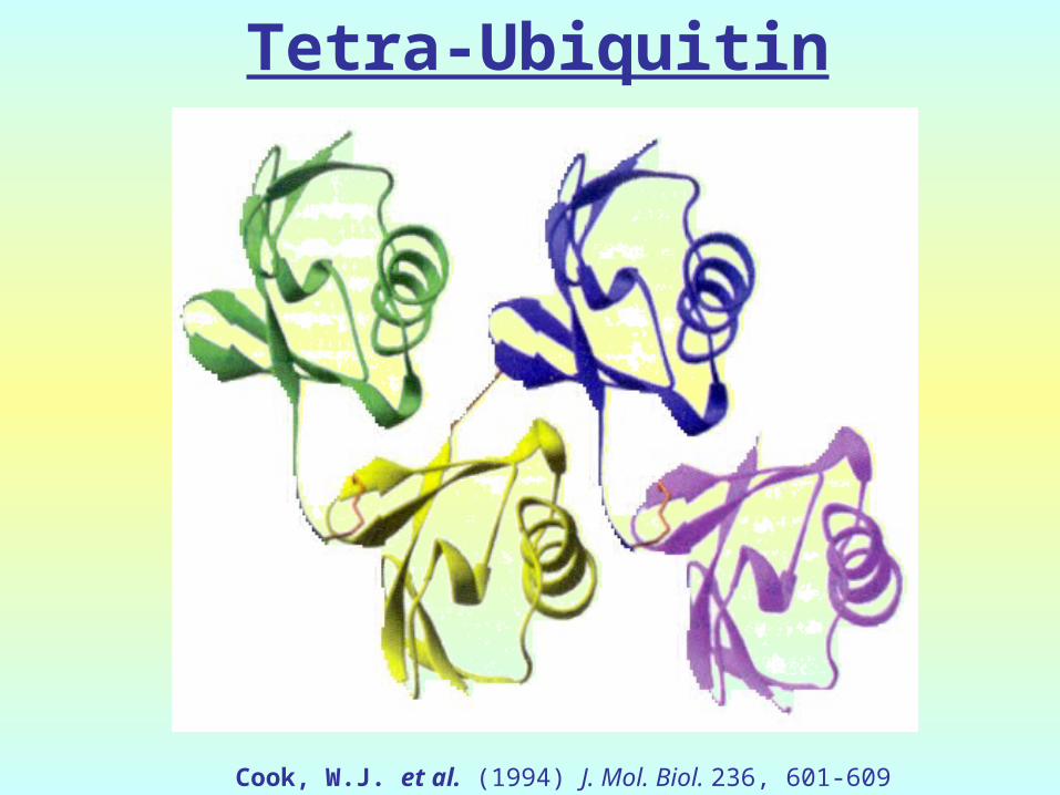
Tetra-Ubiquitin
Cook, W.J. et al. (1994) J. Mol. Biol. 236, 601-609

UBIQUITIN GENES
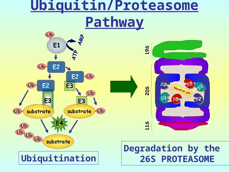
Degradation by the 26S PROTEASOME
Ubiquitin/Proteasome Pathway
UbiquitinationUbiquitination
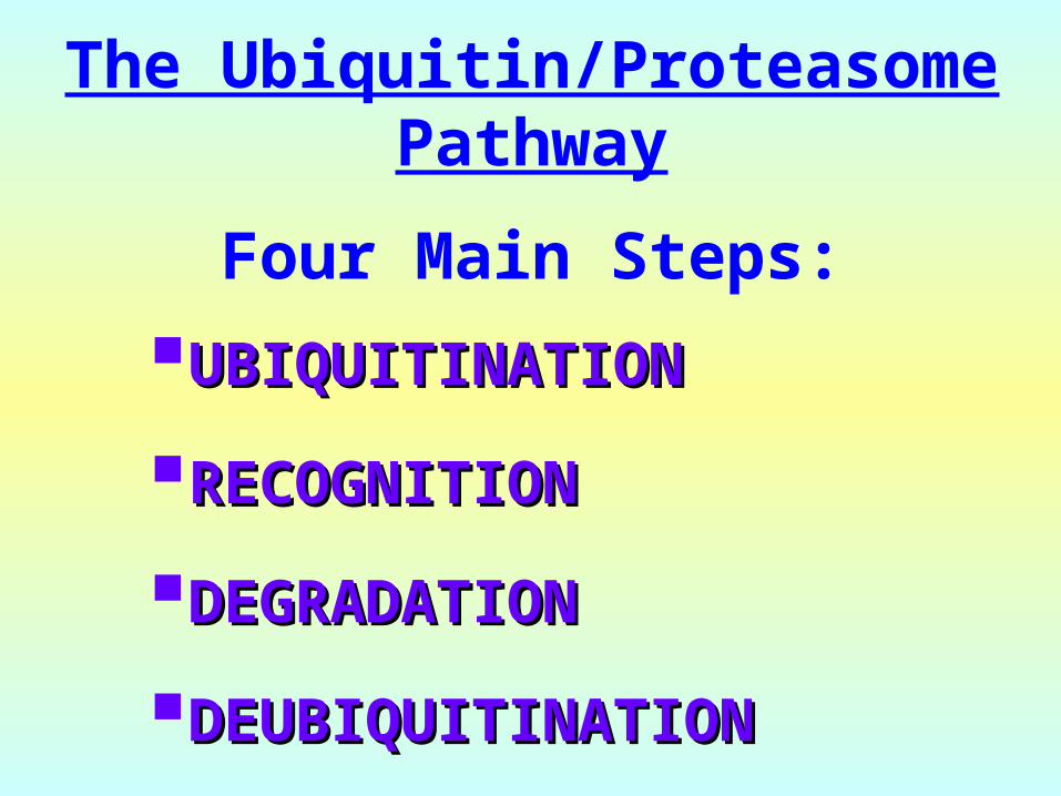
The Ubiquitin/Proteasome Pathway
Four Main Steps:
UBIQUITINATIONUBIQUITINATION
RECOGNITIONRECOGNITION
DEGRADATIONDEGRADATION
DEUBIQUITINATIONDEUBIQUITINATION

UBIQUITINATED PROTEINSUBIQUITINATED PROTEINS

UBIQUITIN UBIQUITIN CHAINSCHAINS
MQIFVKTLTGKTITLEVESSDTIDNVKAKIQDKEGIPPDQ
QRLIFAGKQLEDGRTLADYNIQKESTLHLVLRLRGG
6 11 29
48 63
27 33
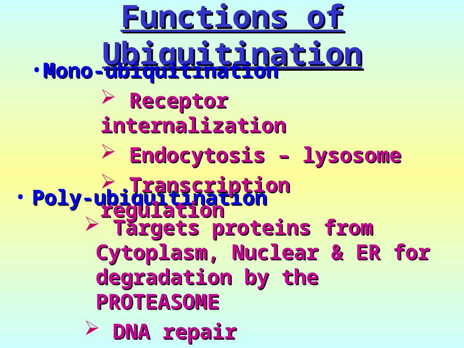
Functions of Functions of UbiquitinationUbiquitination
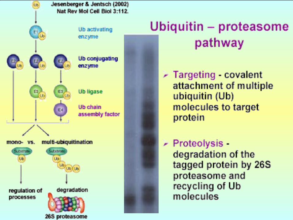
• Poly-ubiquitinationPoly-ubiquitination Targets proteins from Targets proteins from Cytoplasm, Nuclear & ER for Cytoplasm, Nuclear & ER for degradation by the degradation by the PROTEASOME PROTEASOME
DNA repairDNA repair
•Mono-ubiquitinationMono-ubiquitination Receptor internalizationReceptor internalization Endocytosis – lysosomeEndocytosis – lysosome Transcription regulationTranscription regulation
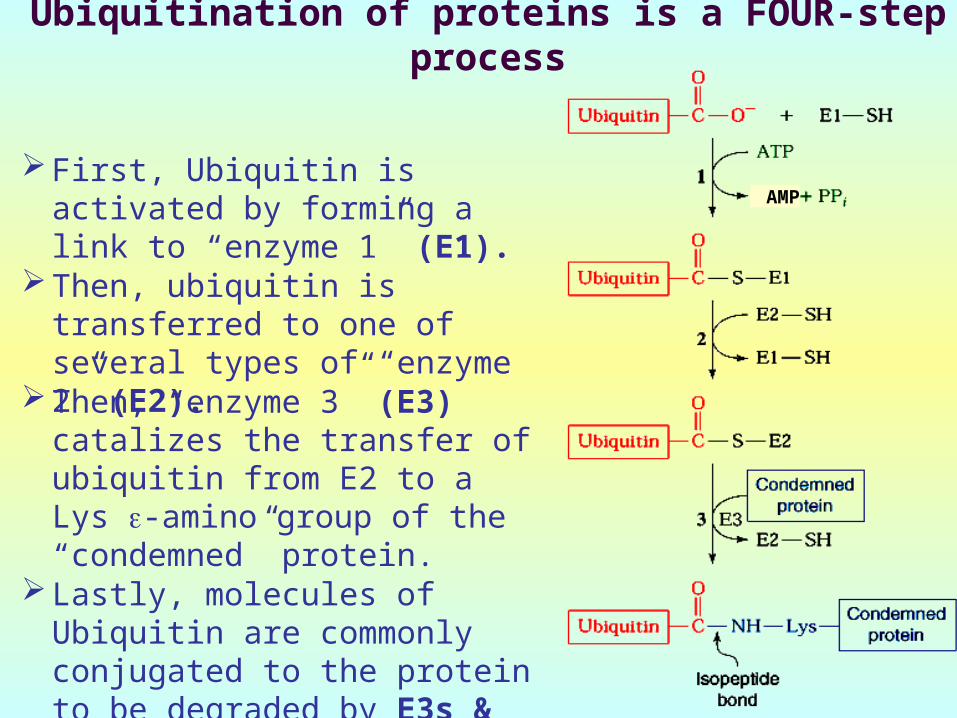
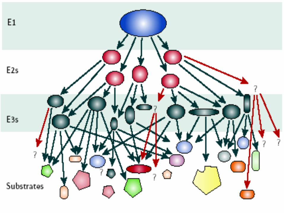
Ubiquitination of proteins is a FOUR-step process
First, Ubiquitin is activated by forming a link to “enzyme 1” (E1).
Then, ubiquitin is transferred to one of several types of “enzyme 2” (E2).
Then, “enzyme 3” (E3) catalizes the transfer of ubiquitin from E2 to a Lys -amino group of the “condemned” protein.
Lastly, molecules of Ubiquitin are commonly conjugated to the protein to be degraded by E3s & E4s
AMP
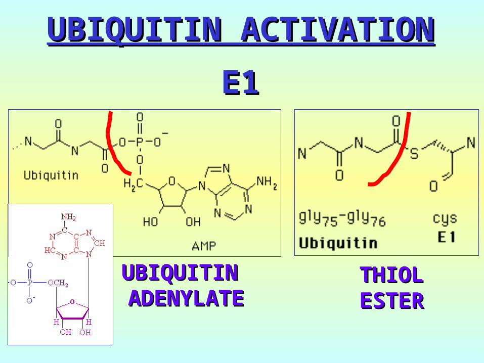
UBIQUITIN ACTIVATIONUBIQUITIN ACTIVATION
E1E1
UBIQUITIN UBIQUITIN ADENYLATEADENYLATE
THIOLTHIOLESTERESTER
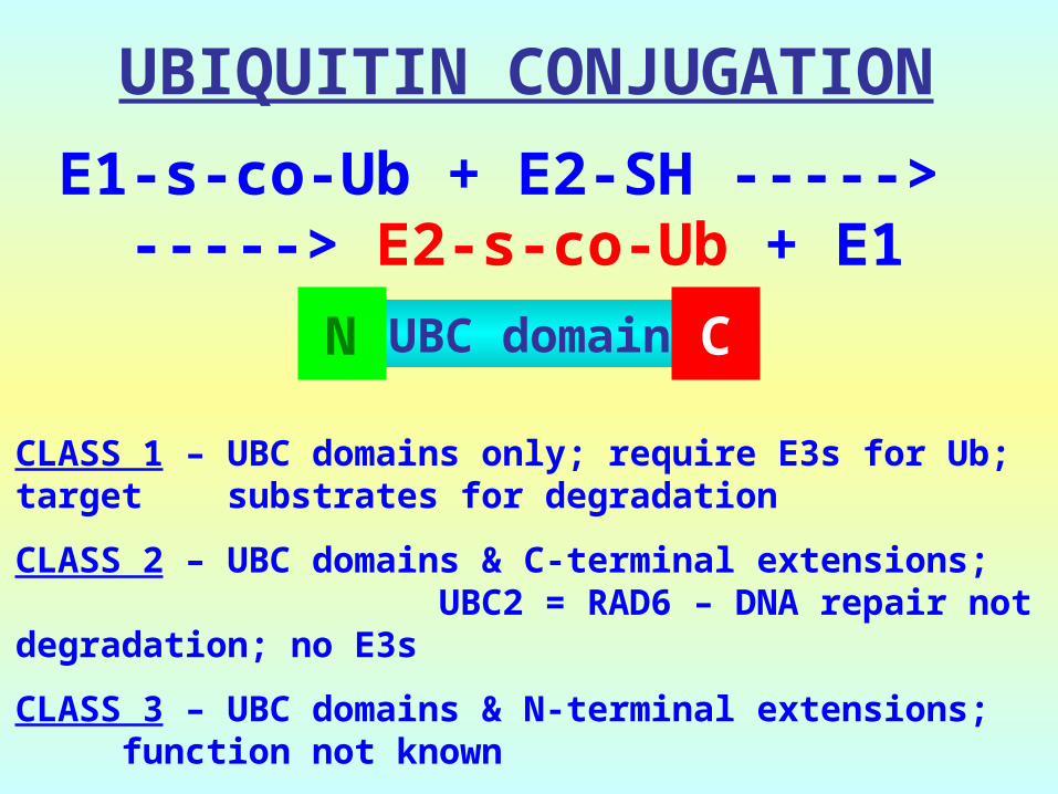
E1-s-co-Ub + E2-SH -----> -----> E2-s-co-Ub + E1
UBIQUITIN CONJUGATION
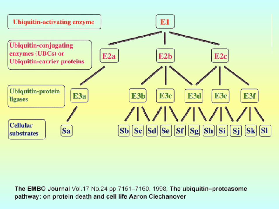
CLASS 1 – UBC domains only; require E3s for Ub; target substrates for degradation
CLASS 2 – UBC domains & C-terminal extensions; UBC2 = RAD6 – DNA repair not degradation; no E3s
CLASS 3 – UBC domains & N-terminal extensions; function not known
UBC domainN C
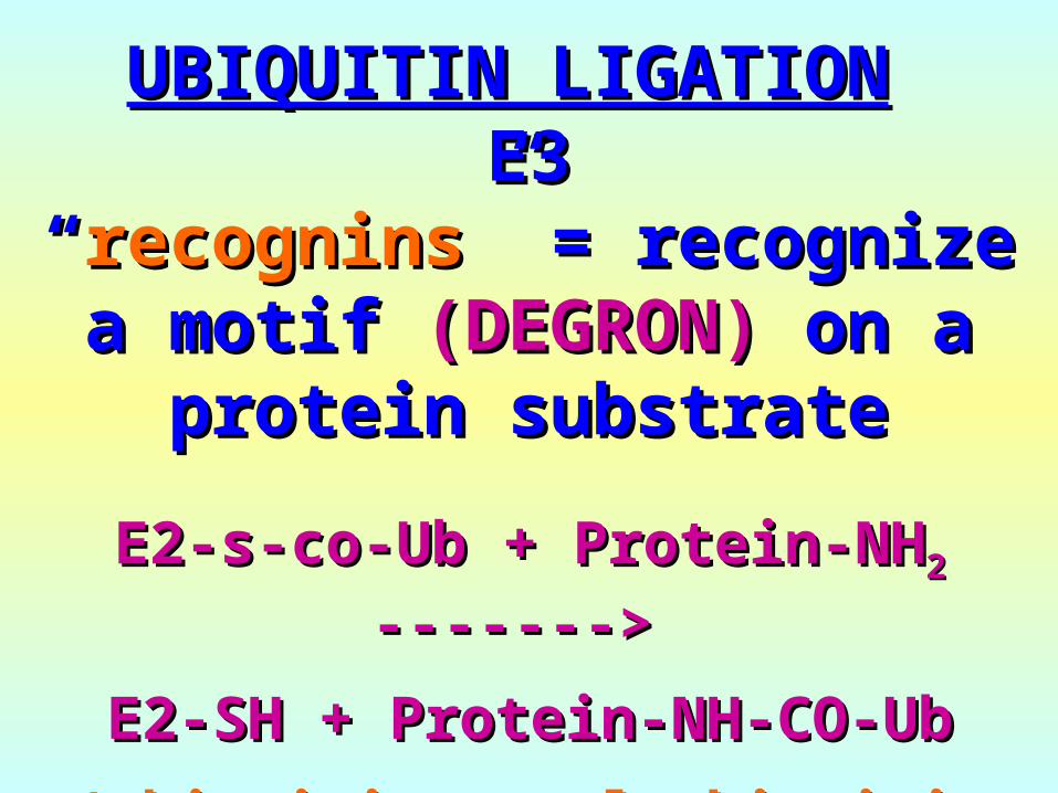
E2-s-co-Ub + Protein-NHE2-s-co-Ub + Protein-NH22 -------> ------->
E2-SH + Protein-NH-CO-UbE2-SH + Protein-NH-CO-Ub
(ubiquitin = polyubiquitin (ubiquitin = polyubiquitin chains)chains)
UBIQUITIN LIGATIONUBIQUITIN LIGATION E3E3
““recogninsrecognins” =” = recognize recognize a motifa motif (DEGRON)(DEGRON) on a on a
protein substrateprotein substrate
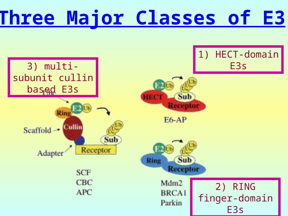
Three Major Classes of E3
3) multi-subunit cullin based E3s
1) HECT-domain E3s
2) RING finger-domain E3s
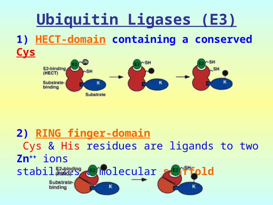
Ubiquitin Ligases (E3)1) HECT-domain containing a conserved Cys
2) RING finger-domain Cys & His residues are ligands to two Zn++ ionsstabilizes a molecular scaffold

Ubiquitin Ligases (E3) (cont.)
3) Complex E3s: Multiple subunitsEx: SCF-type E3, VBC-Cul2 E3 and other cullin based E3s, Anaphase promoting complex (APC)
-they provide a Scaffold for Ub transfer
-F-box – substraterecognition
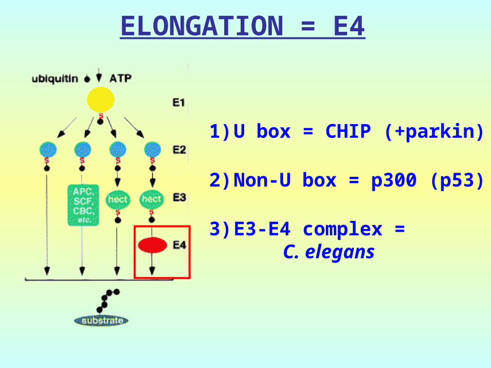
ELONGATION = E4
1)U box = CHIP (+parkin)
2)Non-U box = p300 (p53)
3)E3-E4 complex = C. elegans
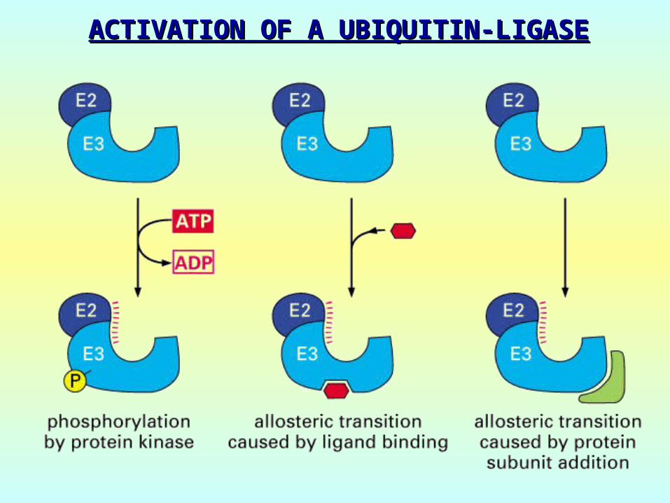
ACTIVATION OF A UBIQUITIN-LIGASEACTIVATION OF A UBIQUITIN-LIGASE
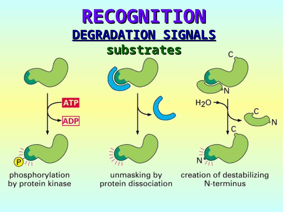
DEGRADATION SIGNALSDEGRADATION SIGNALSsubstratessubstrates
RECOGNITIONRECOGNITION

N-end RULE
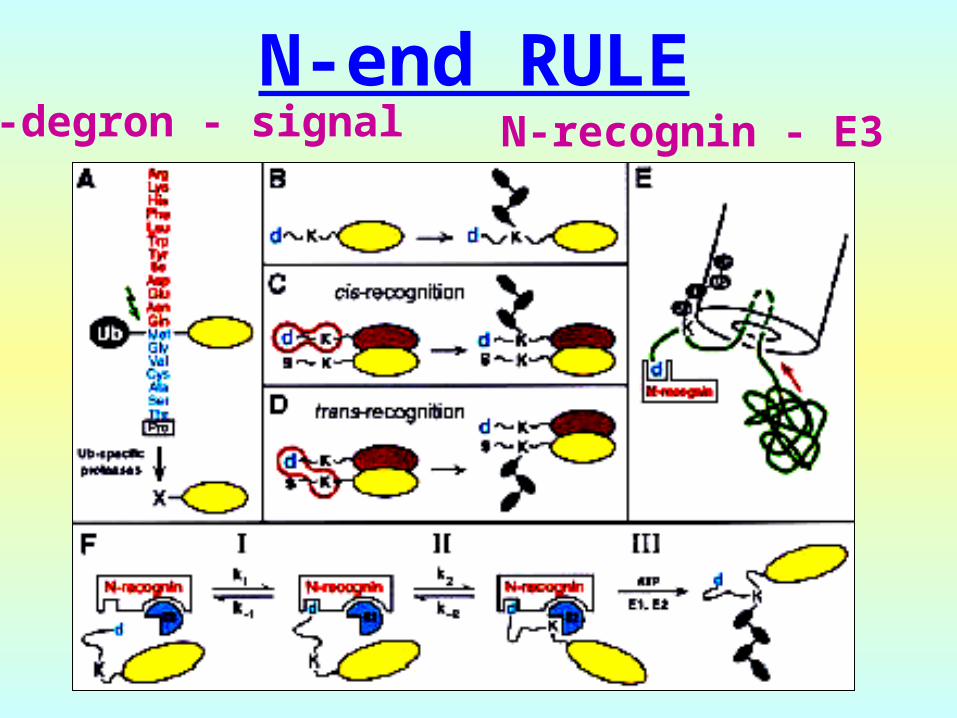
N-end RULEN-degron - signal N-recognin - E3


DEGRADATIONDEGRADATION
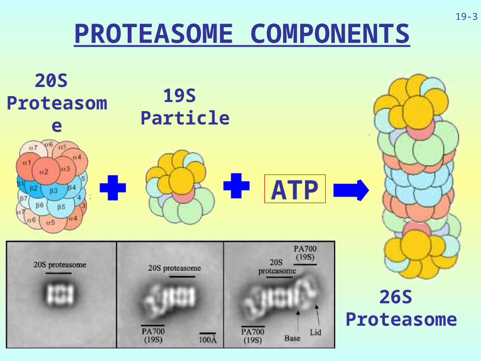
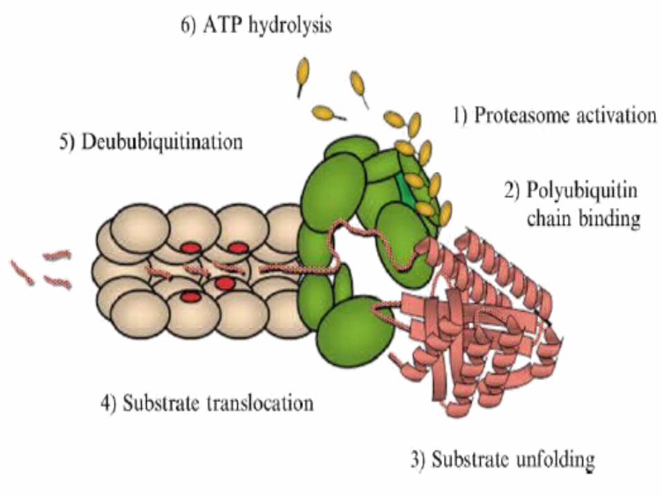
PROTEASOME COMPONENTS
19-3
20S Proteaso
me
19S Particle
26S Proteasome
ATP
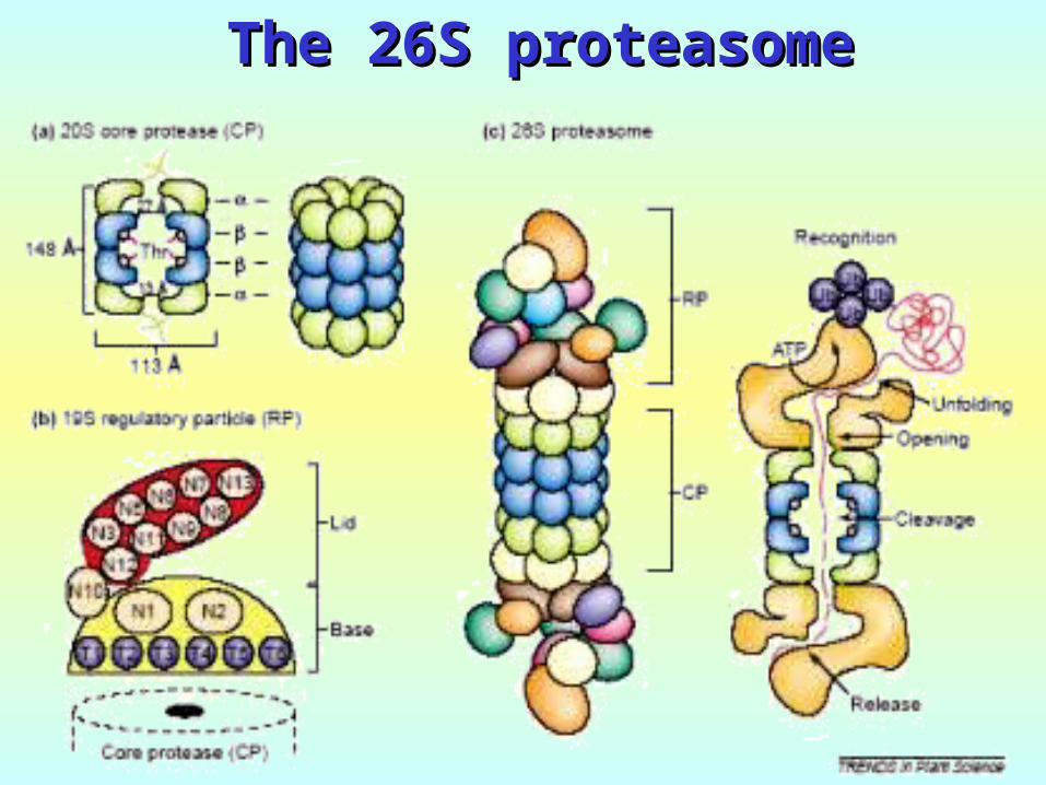
The 26S proteasomeThe 26S proteasome
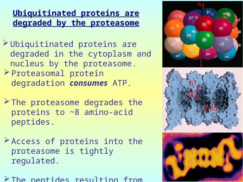
Ubiquitinated proteins are degraded by the proteasome
Proteasomal protein degradation consumes ATP.
The proteasome degrades the proteins to ~8 amino-acid peptides.
Access of proteins into the proteasome is tightly regulated.
The peptides resulting from the proteasome activity diffuse out of the proteasome freely.
Ubiquitinated proteins are degraded in the cytoplasm and nucleus by the proteasome.

Hydrolysis peptide bonds after:Hydrolysis peptide bonds after:
hydrophobic a.a.hydrophobic a.a. = = CHYMOTRYPSIN-CHYMOTRYPSIN-LIKE - LIKE - 55
acidic a.a.acidic a.a. = (-) = (-)CASPASE-LIKE CASPASE-LIKE --11
basic a.a.basic a.a. = (+) = (+)TRYPSIN-LIKE TRYPSIN-LIKE --22
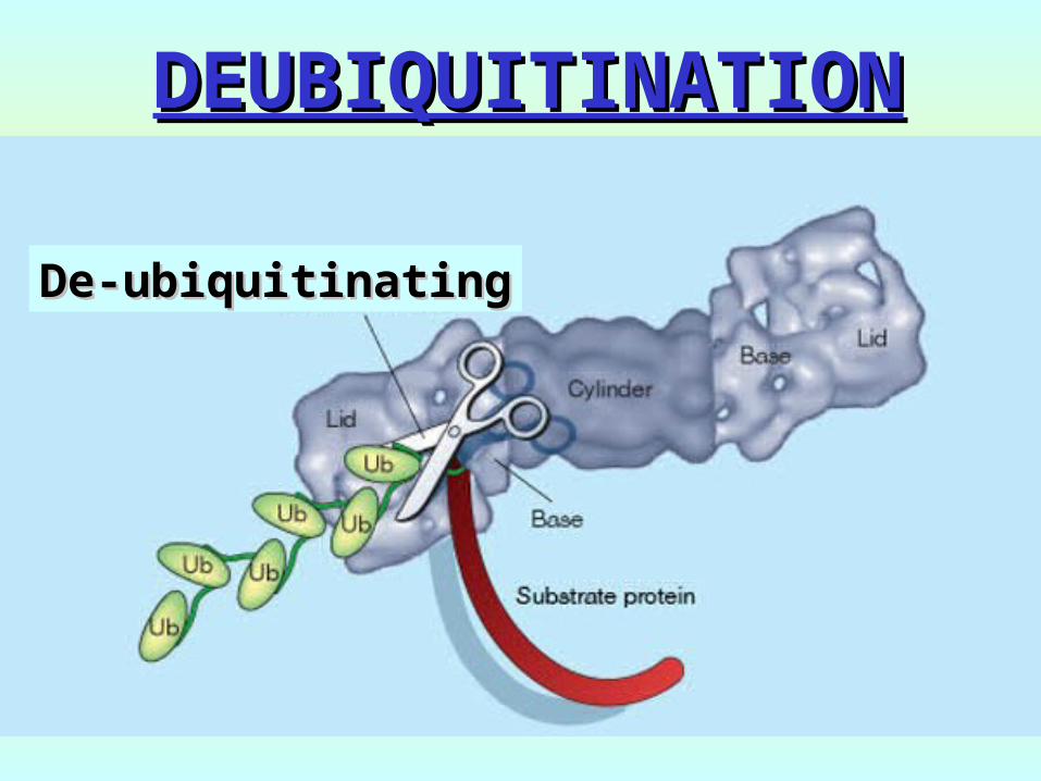
DEUBIQUITINATIONDEUBIQUITINATION
De-ubiquitinatingDe-ubiquitinating
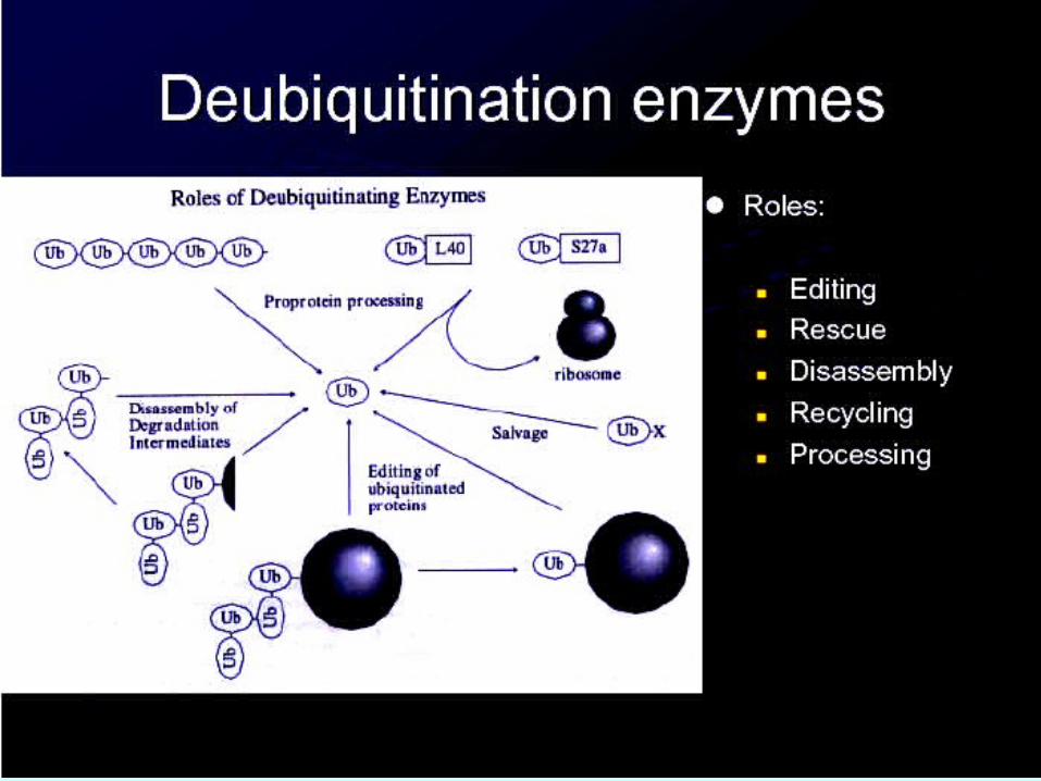
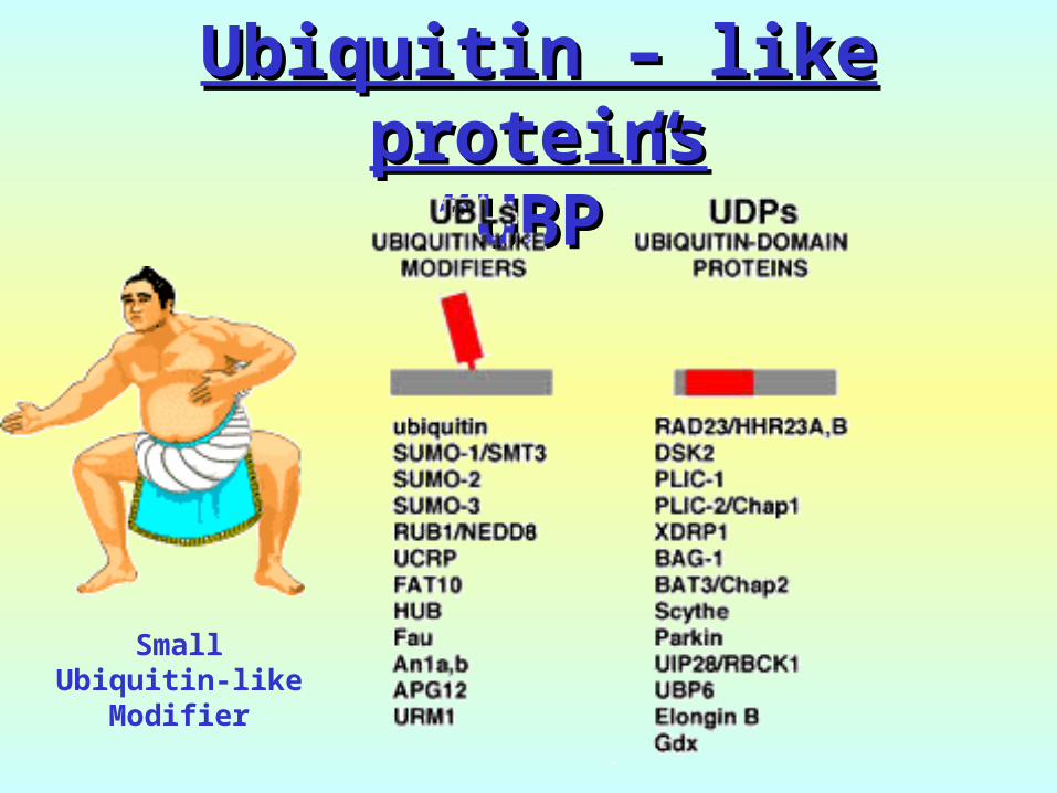
Ubiquitin – like Ubiquitin – like proteinsproteins““UBP”UBP”
SmallUbiquitin-like
Modifier
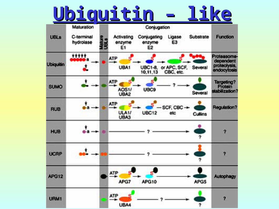
Ubiquitin – like Ubiquitin – like modifiersmodifiers

LYSOSOMESLYSOSOMES
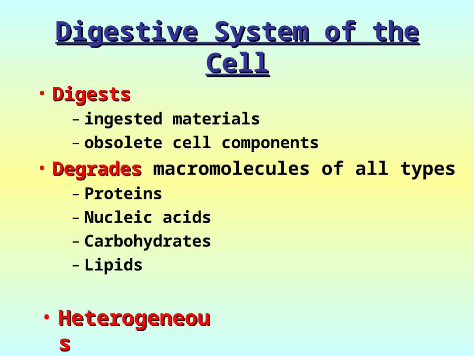
Digestive System of the Digestive System of the CellCell
• DigestsDigests – ingested materials– obsolete cell components
• DegradesDegrades macromolecules of all types
– Proteins– Nucleic acids– Carbohydrates– Lipids
• HeterogeneHeterogeneousous
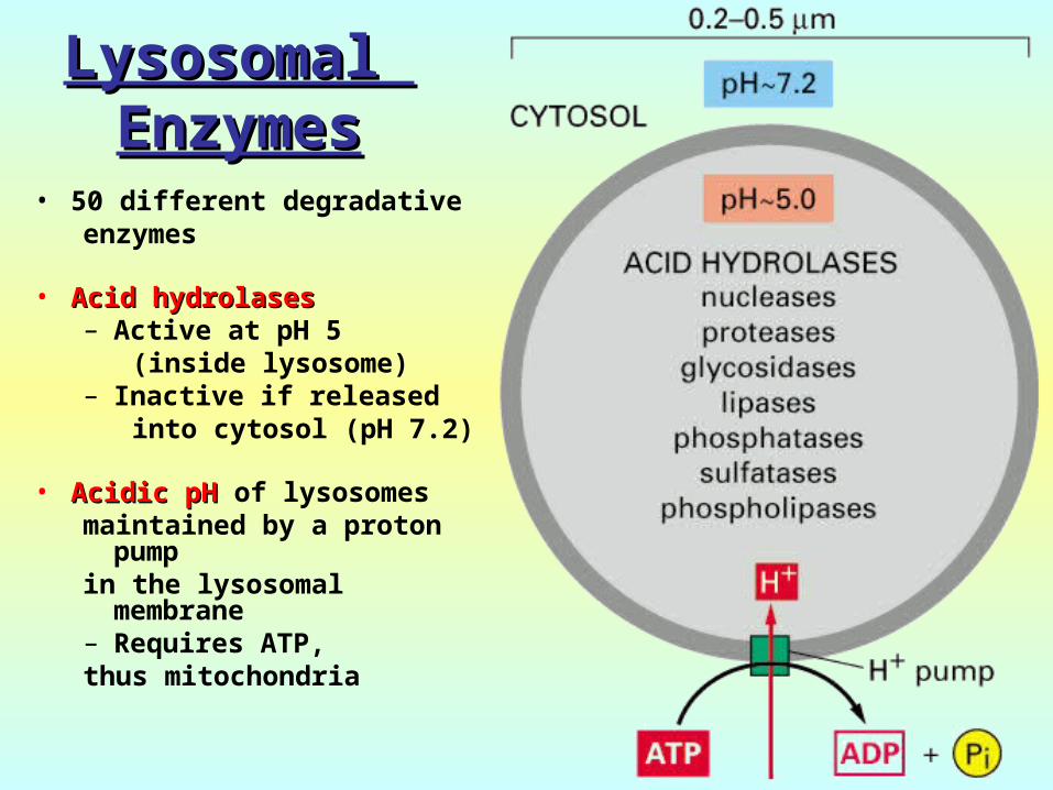
Lysosomal Lysosomal EnzymesEnzymes
• 50 different degradative enzymes
• Acid hydrolasesAcid hydrolases– Active at pH 5
(inside lysosome)– Inactive if released
into cytosol (pH 7.2)
• Acidic pHAcidic pH of lysosomes maintained by a proton
pump in the lysosomal
membrane– Requires ATP, thus mitochondria
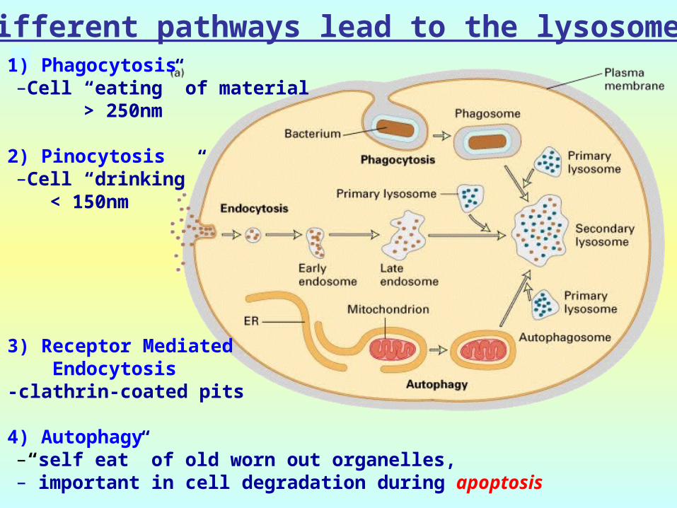
Different pathways lead to the lysosome 1) Phagocytosis–Cell “eating” of material
> 250nm
2) Pinocytosis–Cell “drinking” < 150nm
3) Receptor Mediated Endocytosis-clathrin-coated pits
4) Autophagy–“self eat” of old worn out organelles, – important in cell degradation during apoptosis
Protein degradation in the Protein degradation in the lysosomeslysosomes
Lysosomes degrade extracellular proteins that the cell incorporates by endocytosis.
Lysosomes can also degrade intracellular proteins that are enclosed in other membrane-limited organellas.
In well-nourished cells, lysosomal protein degradation is non-selective (non-regulated).
In starved cells, lysosomes degrade preferentially proteins containing a KFERQKFERQ “signal” peptide.
The regression of the uterus after childbirth is mediated largely by lysosomal protein degradation

AUTOPHAGYAUTOPHAGY
- Macroautophagy – inducible (mTOR)- Macroautophagy – inducible (mTOR)(autophagy)(autophagy)
- Microautophagy - constitutive- Microautophagy - constitutive
- Chaperone-mediated autophagy - Chaperone-mediated autophagy (CMA) – KFERQ motif(CMA) – KFERQ motif

AUTOPHAGY

AUTOPHAGY (MACRO) PATHWAYOxidative stress Infection Protein aggregates

AUTOPHAGY (MACRO) PATHWAY
Lysosome
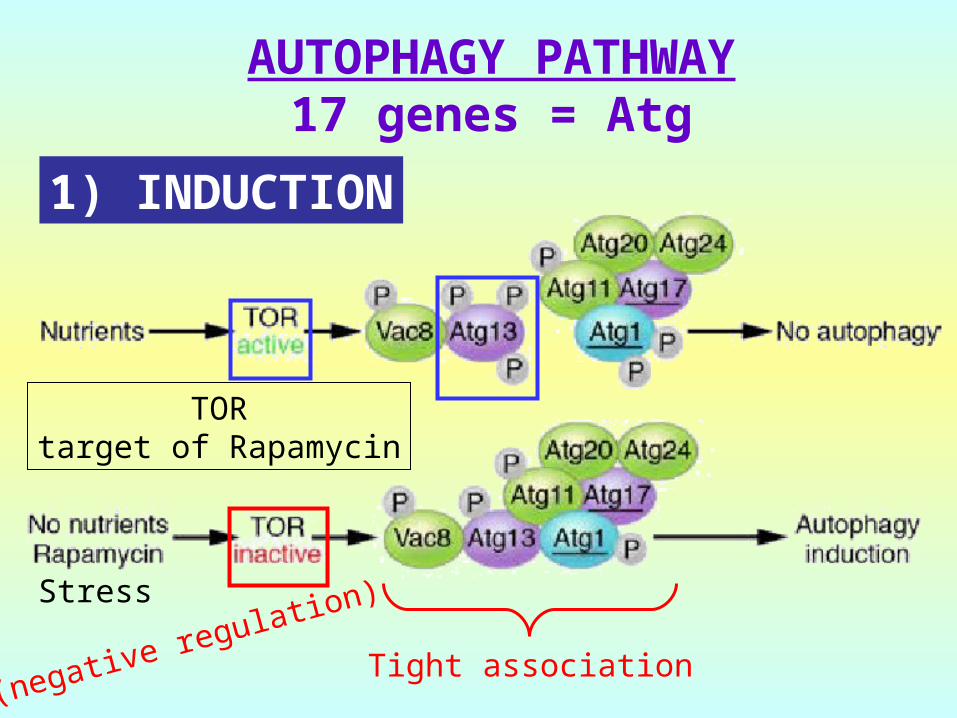
AUTOPHAGY PATHWAY17 genes = Atg
Stress
TORtarget of Rapamycin
Tight association(negative regulation)
1) INDUCTION
AUTOPHAGY PATHWAY
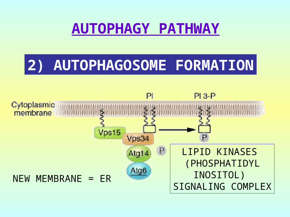
2) AUTOPHAGOSOME FORMATION
NEW MEMBRANE = ER
LIPID KINASES (PHOSPHATIDYL
INOSITOL) SIGNALING COMPLEX
AUTOPHAGY PATHWAY
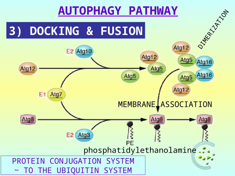
PROTEIN CONJUGATION SYSTEM~ TO THE UBIQUITIN SYSTEM
3) DOCKING & FUSION
DIM
ERIZ
ATIO
N
MEMBRANE ASSOCIATION
phosphatidylethanolamine
AUTOPHAGY PATHWAY
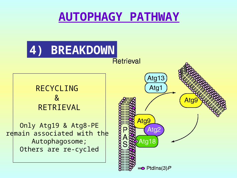
4) BREAKDOWN
RECYCLING &
RETRIEVAL
Only Atg19 & Atg8-PEremain associated with the
Autophagosome;Others are re-cycled

AUTOPHAGY PATHWAY
Neurodegenerative diseasesNeurodegenerative diseases- PD, AD, HD and TSE- aggregate removal
Infectious diseasesInfectious diseases- remove pathogens
CancerCancer- sequester damaged organelles- promote autophagic death
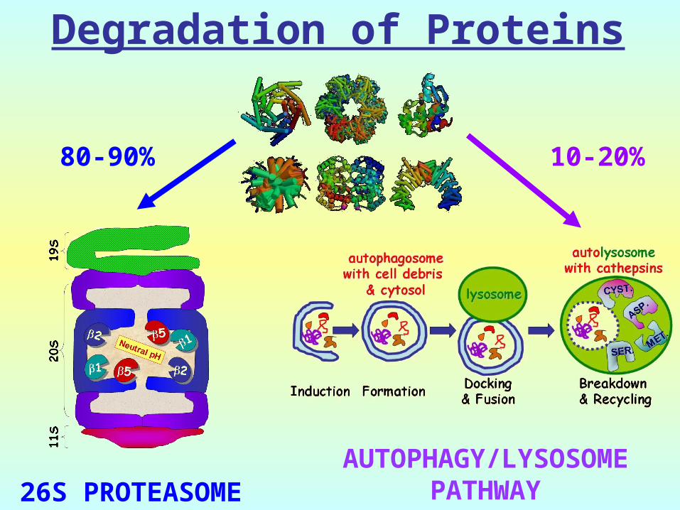
80-90%
26S PROTEASOME
10-20%
AUTOPHAGY/LYSOSOMEPATHWAY
Degradation of Proteins
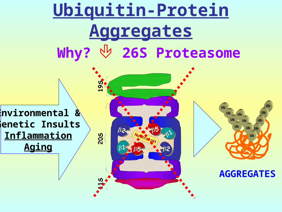
Why? 26S Proteasome
AGGREGATES
Environmental &Genetic InsultsInflammation
Aging
Ub
Ub
Ub
Ub
Ub
Ub
Ub
Ub
Ub
Ub
Ub
Ub
Ubiquitin-Protein Aggregates
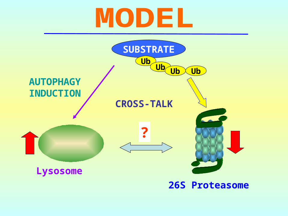
Lysosome
26S Proteasome
?
SUBSTRATEUb
Ub UbUb
CROSS-TALK
AUTOPHAGYINDUCTION

The End
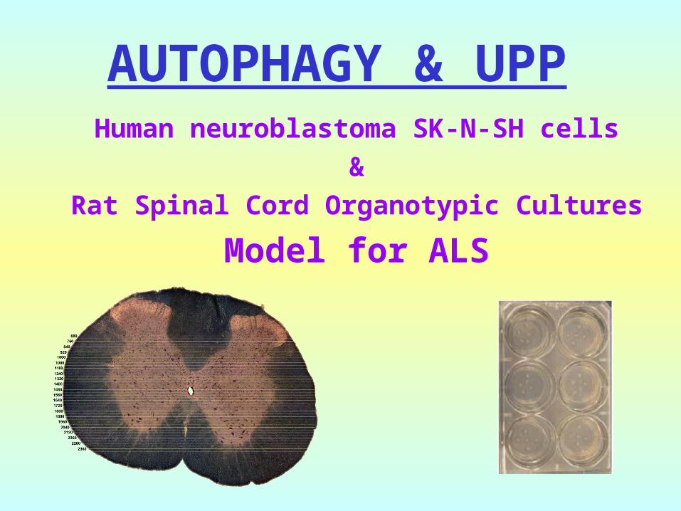
AUTOPHAGY & UPPHuman neuroblastoma SK-N-SH cells
&Rat Spinal Cord Organotypic Cultures
Model for ALS
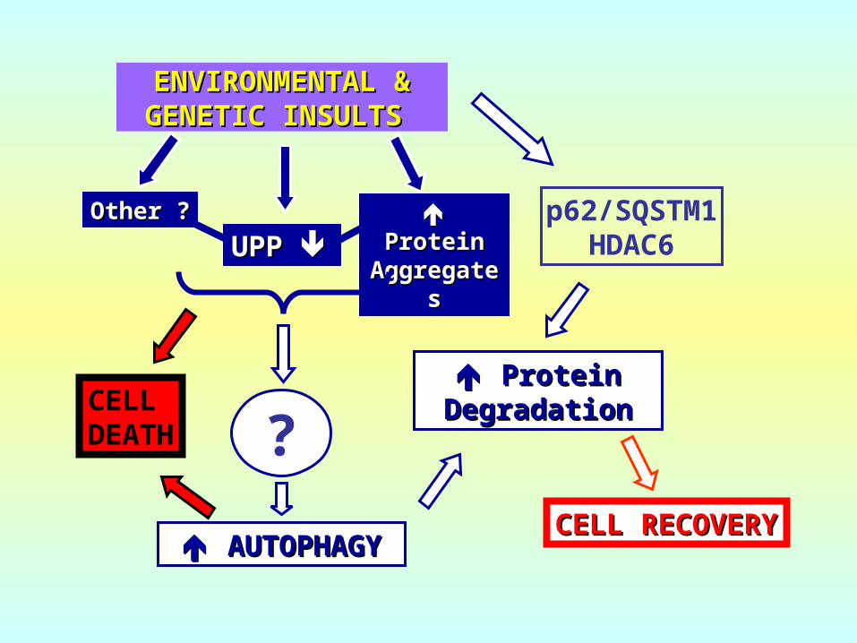
CELL RECOVERYCELL RECOVERY AUTOPHAGYAUTOPHAGY
UPP UPP
CELL DEATH
ENVIRONMENTAL & ENVIRONMENTAL & GENETIC INSULTSGENETIC INSULTS
ProteinProteinAggregatAggregat
eses
Other ?Other ?
? Protein Protein
DegradationDegradation
p62/SQSTM1HDAC6
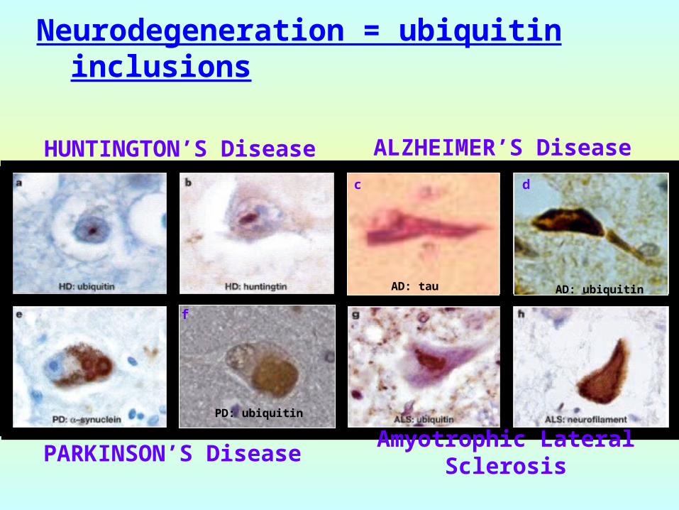
Neurodegeneration = ubiquitin inclusions
HUNTINGTON’S Disease
ALZHEIMER’S Disease
PARKINSON’S DiseaseAmyotrophic Lateral
Sclerosis
f
PD: ubiquitin
c
AD: tau
d
AD: ubiquitin