Embed Size (px)
Citation preview

Quality of Long LSO/LYSO Crystals
This article has been downloaded from IOPscience. Please scroll down to see the full text article.
2012 J. Phys.: Conf. Ser. 404 012026
(http://iopscience.iop.org/1742-6596/404/1/012026)
Download details:
IP Address: 131.215.71.79
The article was downloaded on 14/03/2013 at 15:18
Please note that terms and conditions apply.
View the table of contents for this issue, or go to the journal homepage for more
Home Search Collections Journals About Contact us My IOPscience
Quality of Long LSO/LYSO Crystals
Ren-Yuan Zhu256-48, HEP, Caltech, Pasadena, CA 91125, USA
E-mail: [email protected]
Abstract. Because of their high stopping power (X0 = 1.14 cm) and fast (t = 40 ns) andbright (4 times of BGO) scintillation cerium doped silicate based heavy crystal scintillators(LSO and LYSO) have attracted a broad interest in the high energy physics community pursuingprecision electromagnetic calorimeter for future high energy physics experiments. Their excellentradiation hardness against γ-rays, neutrons and charged hadrons also makes them a preferredmaterial for calorimeters to be operated in a severe radiation environment, such as the HL-LHC.The optical and scintillation properties and its radiation hardness against γ-ray irradiations upto 1 Mrad are presented for the first 2.5 × 2.5 × 28 cm LYSO sample. An absorption band wasfound at the seed end of this sample and three other long samples, which was traced back toa bad seed crystal used in the corresponding crystal growth process. Significant progresses inoptical and scintillation properties were achieved for large size LYSO crystals after eliminatingthis absorption band. Their application in future HEP experiments at HL-LHC are discussed.
1. IntroductionIn the last two decades, cerium doped silicate based heavy crystal scintillators have beendeveloped for the medical industry. As of today, mass production capabilities of lutetiumoxyorthosilicate (LSO) [1] and lutetium-yttrium oxyorthosilicate (LYSO) [2, 3] are established.Because of their high stopping power (ρ = 7.4 g/cm3, X0 = 1.14 cm and RMoliere = 2.07cm), high light yield (about 4 times of BGO) and fast decay time (τ = ∼40 ns) these crystalshave also attracted a broad interest in the physics community for future high energy physicsexperiments [4], such as the proposed SuperB forward endcap calorimeter [5], the KLOEexperiment [6] and the Mu2e experiment [7]. Investigations on large size (2.5 × 2.5 × 20 cm)LSO/LYSO crystals also show that this new generation of inorganic crystal scintillators hasexcellent radiation hardness against γ-rays [8], neutrons [9] and charged hadrons [10]. Thismaterial thus may also be considered for calorimeters to be operated in a severe radiationenvironment, such as the HL-LHC, and seems the only suitable scintillator for which mass-production capability is presently available.
The main obstacles of using this material in the experimental physics are two fold: theavailability of high quality crystals in sufficiently large size and the high cost associated withtheir raw material cost and high melting point (>2,000oC). An effort has been made at SichuanInstitute of Piezoelectric and Acousto-optic Technology (SIPAT), Chongqing, China, to growlarge size (up to 28 cm or 25 X0 long) LSO and LYSO crystals for high energy physicsapplications. In this work, the first 2.5 × 2.5 × 28 cm LYSO sample from SIPAT is evaluated,including its optical and scintillation properties and its radiation hardness against γ-rays up to1 Mrad. Poor longitudinal transmittance was observed in this sample, which was found to becaused by an absorption band at the seed end. After removing this absorption band both the
XVth International Conference on Calorimetry in High Energy Physics (CALOR2012) IOP PublishingJournal of Physics: Conference Series 404 (2012) 012026 doi:10.1088/1742-6596/404/1/012026
Published under licence by IOP Publishing Ltd 1
optical and scintillation properties are improved significantly for large size samples. This reportis a part of an on-going R&D program to further develop this material to be of practical use atthe HL-LHC.
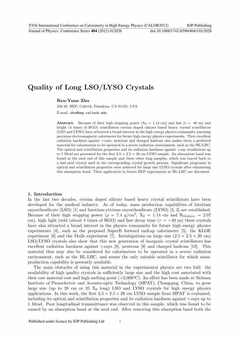
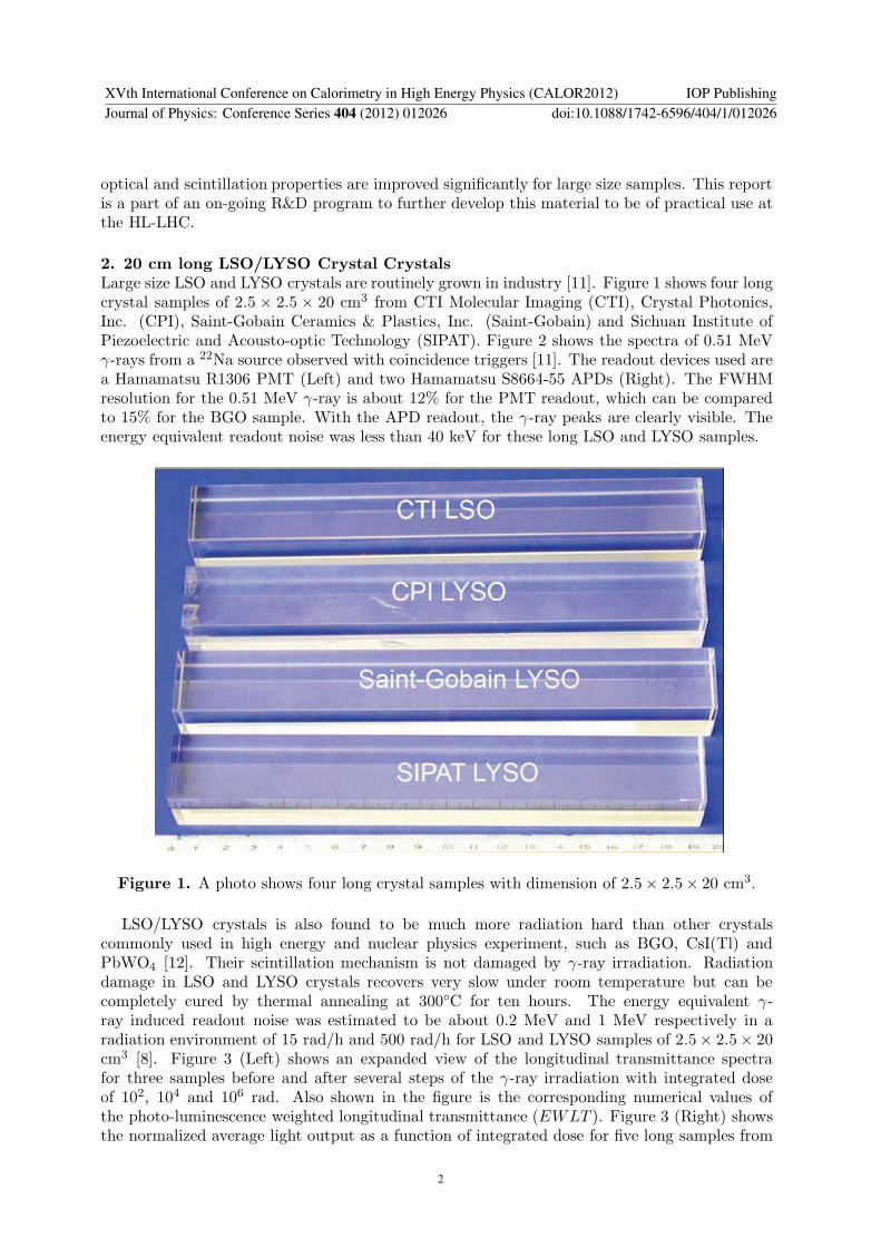
2. 20 cm long LSO/LYSO Crystal CrystalsLarge size LSO and LYSO crystals are routinely grown in industry [11]. Figure 1 shows four longcrystal samples of 2.5 × 2.5 × 20 cm3 from CTI Molecular Imaging (CTI), Crystal Photonics,Inc. (CPI), Saint-Gobain Ceramics & Plastics, Inc. (Saint-Gobain) and Sichuan Institute ofPiezoelectric and Acousto-optic Technology (SIPAT). Figure 2 shows the spectra of 0.51 MeVγ-rays from a 22Na source observed with coincidence triggers [11]. The readout devices used area Hamamatsu R1306 PMT (Left) and two Hamamatsu S8664-55 APDs (Right). The FWHMresolution for the 0.51 MeV γ-ray is about 12% for the PMT readout, which can be comparedto 15% for the BGO sample. With the APD readout, the γ-ray peaks are clearly visible. Theenergy equivalent readout noise was less than 40 keV for these long LSO and LYSO samples.
Figure 1. A photo shows four long crystal samples with dimension of 2.5× 2.5× 20 cm3.
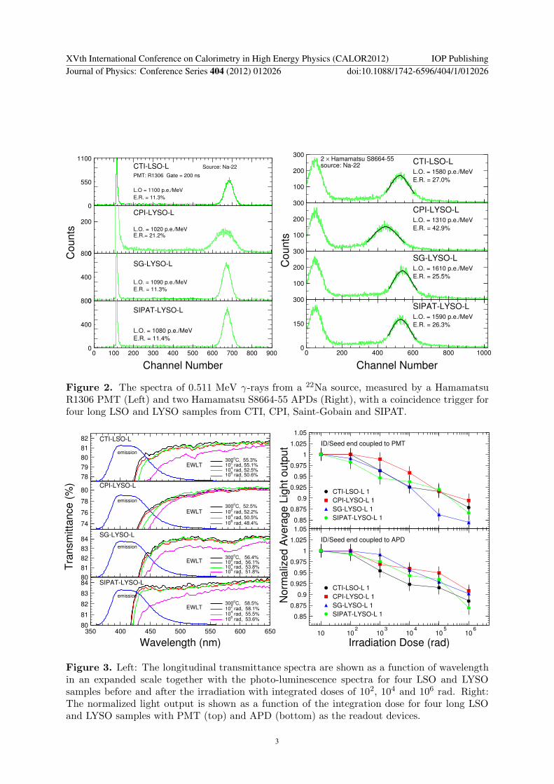
LSO/LYSO crystals is also found to be much more radiation hard than other crystalscommonly used in high energy and nuclear physics experiment, such as BGO, CsI(Tl) andPbWO4 [12]. Their scintillation mechanism is not damaged by γ-ray irradiation. Radiationdamage in LSO and LYSO crystals recovers very slow under room temperature but can becompletely cured by thermal annealing at 300◦C for ten hours. The energy equivalent γ-ray induced readout noise was estimated to be about 0.2 MeV and 1 MeV respectively in aradiation environment of 15 rad/h and 500 rad/h for LSO and LYSO samples of 2.5× 2.5× 20cm3 [8]. Figure 3 (Left) shows an expanded view of the longitudinal transmittance spectrafor three samples before and after several steps of the γ-ray irradiation with integrated doseof 102, 104 and 106 rad. Also shown in the figure is the corresponding numerical values ofthe photo-luminescence weighted longitudinal transmittance (EWLT ). Figure 3 (Right) showsthe normalized average light output as a function of integrated dose for five long samples from
XVth International Conference on Calorimetry in High Energy Physics (CALOR2012) IOP PublishingJournal of Physics: Conference Series 404 (2012) 012026 doi:10.1088/1742-6596/404/1/012026
2
0
550
1100
CTI-LSO-L Source: Na-22
PMT: R1306 Gate = 200 ns
L.O = 1100 p.e./MeV
E.R. = 11.3%
0
200
CPI-LYSO-L
L.O. = 1020 p.e./MeVE.R.= 21.2%
0
400
800
SG-LYSO-L
L.O. = 1090 p.e./MeV
E.R. = 11.3%
0
400
800
0 100 200 300 400 500 600 700 800 900
SIPAT-LYSO-L
L.O. = 1080 p.e./MeV
E.R. = 11.4%
Channel Number
Co
un
ts
100
200
300
source: Na-222 × Hamamatsu S8664-55 CTI-LSO-L
E.R. = 27.0%
L.O. = 1580 p.e./MeV
100
200
300CPI-LYSO-L
E.R. = 42.9%
L.O. = 1310 p.e./MeV
100
200
300SG-LYSO-L
E.R. = 25.5%
L.O. = 1610 p.e./MeV
0
150
300
0 200 400 600 800 1000
SIPAT-LYSO-L
E.R. = 26.3%
L.O. = 1590 p.e./MeV
Channel Number
Counts
Figure 2. The spectra of 0.511 MeV γ-rays from a 22Na source, measured by a HamamatsuR1306 PMT (Left) and two Hamamatsu S8664-55 APDs (Right), with a coincidence trigger forfour long LSO and LYSO samples from CTI, CPI, Saint-Gobain and SIPAT.
78
79
80
81
82 CTI-LSO-L
EWLT
emission
300oC, 55.3%
102 rad, 55.1%
104 rad, 52.5%
106 rad, 50.6%
74
76
78
80CPI-LYSO-L
EWLT
emission300
oC, 52.5%
102 rad, 52.2%
104 rad, 50.5%
106 rad, 48.4%
80
81
82
83
84SG-LYSO-L
EWLT
emission
300oC, 56.4%
102 rad, 56.1%
104 rad, 53.8%
106 rad, 51.8%
T
ran
sm
itta
nce
(%
)
80
81
82
83
84
350 400 450 500 550 600 650
SIPAT-LYSO-L
EWLT
emission
300oC, 58.5%
102 rad, 58.1%
104 rad, 55.5%
106 rad, 53.6%
Wavelength (nm)
0.85
0.875
0.9
0.925
0.95
0.975
1
1.025
1.05
ID/Seed end coupled to PMT
CTI-LSO-L 1
CPI-LYSO-L 1
SG-LYSO-L 1
SIPAT-LYSO-L 1
0.85
0.875
0.9
0.925
0.95
0.975
1
1.025
1.05
10 102
103
104
105
106
ID/Seed end coupled to APD
CTI-LSO-L 1
CPI-LYSO-L 1
SG-LYSO-L 1
SIPAT-LYSO-L 1
Norm
aliz
ed A
vera
ge L
ight outp
ut
Irradiation Dose (rad)
Figure 3. Left: The longitudinal transmittance spectra are shown as a function of wavelengthin an expanded scale together with the photo-luminescence spectra for four LSO and LYSOsamples before and after the irradiation with integrated doses of 102, 104 and 106 rad. Right:The normalized light output is shown as a function of the integration dose for four long LSOand LYSO samples with PMT (top) and APD (bottom) as the readout devices.
XVth International Conference on Calorimetry in High Energy Physics (CALOR2012) IOP PublishingJournal of Physics: Conference Series 404 (2012) 012026 doi:10.1088/1742-6596/404/1/012026
3
various vendors. It is interesting to note that all samples show consistent radiation resistancewith degradations of both the light output and transmittance at 10 to 15% level after γ-rayirradiation with an integrated dose of 1 Mrad.
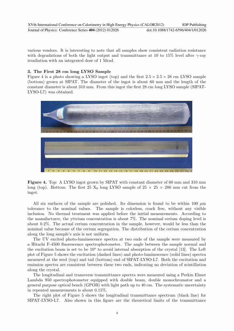
3. The First 28 cm long LYSO SampleFigure 4 is a photo showing a LYSO ingot (top) and the first 2.5 × 2.5 × 28 cm LYSO sample(bottom) grown at SIPAT. The diameter of the ingot is about 60 mm and the length of theconstant diameter is about 310 mm. From this ingot the first 28 cm long LYSO sample (SIPAT-LYSO-L7) was obtained.
Figure 4. Top: A LYSO ingot grown by SIPAT with constant diameter of 60 mm and 310 mmlong (top). Bottom: The first 25 X0 long LYSO sample of 25 × 25 × 280 mm cut from theingot.
All six surfaces of the sample are polished. Its dimension is found to be within 100 µmtolerance to the nominal values. The sample is colorless, crack free, without any visibleinclusion. No thermal treatment was applied before the initial measurements. According tothe manufacturer, the yttrium concentration is about 7%. The nominal cerium doping level isabout 0.2%. The actual cerium concentration in the sample, however, would be less than thenominal value because of the cerium segregation. The distribution of the cerium concentrationalong the long sample’s axis is not uniform.
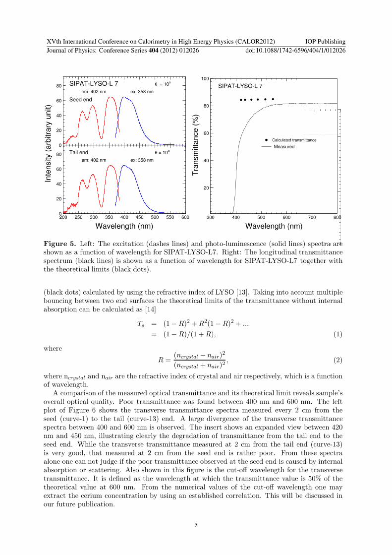
The UV excited photo-luminescence spectra at two ends of the sample were measured bya Hitachi F-4500 fluorescence spectrophotometer. The angle between the sample normal andthe excitation beam is set to be 10o to avoid internal absorption of the crystal [13]. The Leftplot of Figure 5 shows the excitation (dashed lines) and photo-luminescence (solid lines) spectrameasured at the seed (top) and tail (bottom) end of SIPAT-LYSO-L7. Both the excitation andemission spectra are consistent between these two ends, indicating no deviation of scintillationalong the crystal.
The longitudinal and transverse transmittance spectra were measured using a Perkin ElmerLambda 950 spectrophotometer equipped with double beam, double monochromator and ageneral purpose optical bench (GPOB) with light path up to 40 cm. The systematic uncertaintyin repeated measurements is about 0.15%.
The right plot of Figure 5 shows the longitudinal transmittance spectrum (black line) forSIPAT-LYSO-L7. Also shown in this figure are the theoretical limits of the transmittance
XVth International Conference on Calorimetry in High Energy Physics (CALOR2012) IOP PublishingJournal of Physics: Conference Series 404 (2012) 012026 doi:10.1088/1742-6596/404/1/012026
4
0
20
40
60
80 SIPAT-LYSO-L 7
Seed end
θ = 10o
em: 402 nm ex: 358 nm
0
20
40
60
80
200 250 300 350 400 450 500 550 600
Tail end θ = 10o
em: 402 nm ex: 358 nm
Wavelength (nm)
In
ten
sity (
arb
itra
ry u
nit)
20
40
60
80
100
300 400 500 600 700 800
SIPAT-LYSO-L 7
Calculated transmittance
Measured
Wavelength (nm)
Tra
nsm
itta
nce (
%)
Figure 5. Left: The excitation (dashes lines) and photo-luminescence (solid lines) spectra areshown as a function of wavelength for SIPAT-LYSO-L7. Right: The longitudinal transmittancespectrum (black lines) is shown as a function of wavelength for SIPAT-LYSO-L7 together withthe theoretical limits (black dots).
(black dots) calculated by using the refractive index of LYSO [13]. Taking into account multiplebouncing between two end surfaces the theoretical limits of the transmittance without internalabsorption can be calculated as [14]
Ts = (1− R)2 + R2(1 − R)2 + ...
= (1− R)/(1 + R), (1)
where
R =(ncrystal − nair)
2
(ncrystal + nair)2, (2)
where ncrystal and nair are the refractive index of crystal and air respectively, which is a functionof wavelength.
A comparison of the measured optical transmittance and its theoretical limit reveals sample’soverall optical quality. Poor transmittance was found between 400 nm and 600 nm. The leftplot of Figure 6 shows the transverse transmittance spectra measured every 2 cm from theseed (curve-1) to the tail (curve-13) end. A large divergence of the transverse transmittancespectra between 400 and 600 nm is observed. The insert shows an expanded view between 420nm and 450 nm, illustrating clearly the degradation of transmittance from the tail end to theseed end. While the transverse transmittance measured at 2 cm from the tail end (curve-13)is very good, that measured at 2 cm from the seed end is rather poor. From these spectraalone one can not judge if the poor transmittance observed at the seed end is caused by internalabsorption or scattering. Also shown in this figure is the cut-off wavelength for the transversetransmittance. It is defined as the wavelength at which the transmittance value is 50% of thetheoretical value at 600 nm. From the numerical values of the cut-off wavelength one mayextract the cerium concentration by using an established correlation. This will be discussed inour future publication.
XVth International Conference on Calorimetry in High Energy Physics (CALOR2012) IOP PublishingJournal of Physics: Conference Series 404 (2012) 012026 doi:10.1088/1742-6596/404/1/012026
5

0
20
40
60
80
100
400 450 500 550 600 650
SIPAT - LYSO - L7
Cutoff wavelength:
1: 387.6 nm
2: 387.7 nm
3: 387.6 nm
4: 386.6 nm
5: 387.7 nm
6: 387.8 nm
7: 387.9 nm
8: 388.0 nm
9: 388.1 nm
10: 388.9 nm
11: 389.2 nm
12: 389.6 nm
13: 389.9 nm
Wavelength (nm)
Tra
nsm
itta
nce (
%)
81
82
83
84
420 440
Tra
nsm
itta
nce
(%
)
Wavelength (nm)
12
34
56789
1011
1213
SIPAT-LYSO-L7
# 1 E.R. = 12.6%
# 2 E.R. = 12.6%
# 3 E.R. = 12.5%
# 4 E.R. = 12.5%
# 5 E.R. = 12.3%
# 6 E.R. = 12.3%
100 200 300 400 500 600 700 800 900 1000
# 7 E.R. = 12.2%
Channel Number
Figure 6. Left: The transverse transmittance spectra measured every 2 cm from the seed(curve-1) to the tail (curve-13) end are shown as a function of wavelength for SIPAT-LYSO-L7.The inset is an expanded view between 420 nm and 450 nm. Right:The pulse height spectra of0.511 MeV γ-ray peaks (solid lines) and corresponding Gaussian fits measured by a HamamatsuR1306 PMT are shown at seven points evenly distributed along SIPAT-LYSO-L7. Also shownare the numerical values of the FWHM energy resolutions (E.R.).
Despite the poor transmittance observed at the seed end this 28 cm long LYSO sampleprovides adequate light output and energy resolution. Two photodetectors, a HamamatsuR1306 PMT and a Hamamatsu S8664-1010 APD with 1 cm2 area, were used in the light outputmeasurement. In these measurements the seed end of the sample was coupled to the PMTvia Dow Corning 200 fluid, while all other faces of the sample were wrapped with two layers ofTyvek paper. To reduce the effect of the intrinsic natural radioactivity, a collimated 22Na sourcewas used with a coincidence trigger provided by a BaF2 crystal. The detail of the setup usedin these measurements is discussed in reference [4]. The γ-ray peak position was determined bya simple Gaussian fit. The right plot of Figure 6 shows the pulse height spectra measured bythe Hamamatsu R1306 PMT at seven points evenly distributed along SIPAT-LYSO-L7. TheFWHM resolutions obtained for 0.511 MeV γ-rays from the 22Na source are about 12.5%, whichis quite good for crystals of such length.
The γ-ray peak positions obtained by sending a collimated beam of γ-ray at seven pointsevenly distributed along the crystal were used to extract the light response uniformity of thecrystal. A linear fit is used to fit the normalized response
LO
LOmid
= 1 + δx
xmid − 1, (3)
where LOmid represents the average light output at the middle of the sample, x is the distancefrom the end coupled to the readout device and δ represents the deviation of the light responseuniformity.
This sample was measured with both PMT and APD readout. The average light outputis 1,380 p.e./MeV and 1,510 p.e./MeV for the PMT and APD readout respectively. The
XVth International Conference on Calorimetry in High Energy Physics (CALOR2012) IOP PublishingJournal of Physics: Conference Series 404 (2012) 012026 doi:10.1088/1742-6596/404/1/012026
6

corresponding light response uniformities (δ values) are 0.7 ± 1% and 2.8 ± 1.5%. Despiteits poor transmittance observed at the seed end this 28 cm long LYSO sample shows rathergood longitudinal light response uniformity.
4. Radiation Resistance of the First 28 cm long sample against γ-ray IrradiationsRadiation damage of this sample against γ-rays was investigated. A 137Cs source was used forthe irradiations with a dose rate of 7,500 rad/h. The irradiation was carried out step by stepto integrated doses of 102, 104 and 106 rad. Since radiation damage in LYSO crystals does notrecover, and is not dose rate dependent [8], the total integrated dose is used to represent thelevel of the radiation applied to this sample in this study.
72
74
76
78
80
82
350 400 450 500 550 600 650
SIPAT-LYSO-L7
EWLT (from top to bottom):
emission
Pre-irradiation, 48.8%
102 rad, 47.7%
104 rad, 46.9%
106 rad, 44.1%
Wavelength (nm)
Tra
nsm
itta
nce
(%
)
0.8
0.9
1
1.1
δ = (2.8±1.5) Average L.O. = 1510 p.e./MeV
0.8
0.9
1
1.1
δ = (2.4±1.5) Average L.O. = 1490 p.e./MeV
0.8
0.9
1
1.1
δ = (3.0±1.5) Average L.O. = 1410 p.e./MeV
0.8
0.9
1
1.1
δ = (2.6±1.5) Average L.O. = 1300 p.e./MeV
35 70 105 140 175 210
245
0
280
SIPAT-LYSO-L7 Seed end coupling As received
102 rad
104 rad
106 rad
No
rma
lize
d L
igh
t O
utp
ut
Distance from the end coupled to APD (mm)
Figure 7. Left: An expanded view of the longitudinal transmittance spectra measured beforeand after γ-ray irradiations in several steps up to 1 Mrad is shown for SIPAT-LYSO-L7. Alsoshown is the emission spectrum and the values of EWLT defined in the text. Right: Normalizedlight output and light response uniformity measured by Hamamatsu S8664-1010 APD beforeand after γ-ray irradiations in several steps up to 1 Mrad are shown for SIPAT-LYSO-L7.
The left plot of Figure 7 shows an expanded view of the longitudinal transmittance spectrabefore and after the γ-ray irradiations with an integrated dose of 102, 104 and 106 rad. Alsoshown in the figure is the emission spectrum and the values of the emission weighted longitudinaltransmittance (EWLT), which is defined as
EWLT =
∫LT (λ)Em(λ)dλ∫
Em(λ)dλ(4)
The numerical value of EWLT represents how transparent the crystal is to the scintillation lightso is a good measure of its transparency. Its degradation represents the radiation damage effecton transparency. For SIPAT-LYSO-L7 its radiation damage in EWLT is 9.6% after 1 Mradirradiations.
The right plot of Figure 7 shows the normalized light output and response uniformitymeasured by the Hamamatsu APD before and after γ-ray irradiations with an integrated dose of
XVth International Conference on Calorimetry in High Energy Physics (CALOR2012) IOP PublishingJournal of Physics: Conference Series 404 (2012) 012026 doi:10.1088/1742-6596/404/1/012026
7
102, 104 and 106 rad. The damage on the light output was found to be about 13% after 1 Mradirradiations. This is much better than 26% light output loss measured for typical lead tungstate(PWO) crystals after 10 krad with a dose rate of 400 rad/h [15]. We also note that the lightresponse uniformity of SIPAT-LYSO-L7 does not change, indicating that its energy resolutionmay be maintained even after 1 Mrad irradiations. Despite the poor transmittance at the seedend the radiation resistance of this 28 cm long LYSO sample SIPAT-LYSO-L7 is quite good.
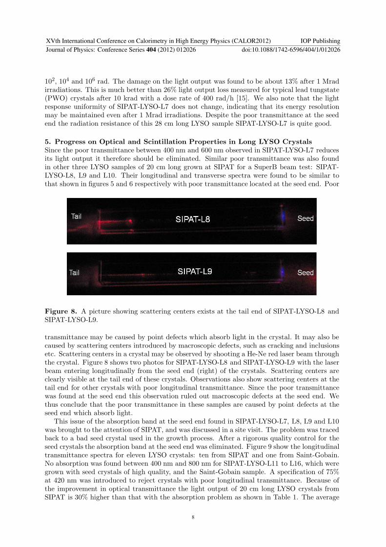
5. Progress on Optical and Scintillation Properties in Long LYSO CrystalsSince the poor transmittance between 400 nm and 600 nm observed in SIPAT-LYSO-L7 reducesits light output it therefore should be eliminated. Similar poor transmittance was also foundin other three LYSO samples of 20 cm long grown at SIPAT for a SuperB beam test: SIPAT-LYSO-L8, L9 and L10. Their longitudinal and transverse spectra were found to be similar tothat shown in figures 5 and 6 respectively with poor transmittance located at the seed end. Poor
Figure 8. A picture showing scattering centers exists at the tail end of SIPAT-LYSO-L8 andSIPAT-LYSO-L9.
transmittance may be caused by point defects which absorb light in the crystal. It may also becaused by scattering centers introduced by macroscopic defects, such as cracking and inclusionsetc. Scattering centers in a crystal may be observed by shooting a He-Ne red laser beam throughthe crystal. Figure 8 shows two photos for SIPAT-LYSO-L8 and SIPAT-LYSO-L9 with the laserbeam entering longitudinally from the seed end (right) of the crystals. Scattering centers areclearly visible at the tail end of these crystals. Observations also show scattering centers at thetail end for other crystals with poor longitudinal transmittance. Since the poor transmittancewas found at the seed end this observation ruled out macroscopic defects at the seed end. Wethus conclude that the poor transmittance in these samples are caused by point defects at theseed end which absorb light.
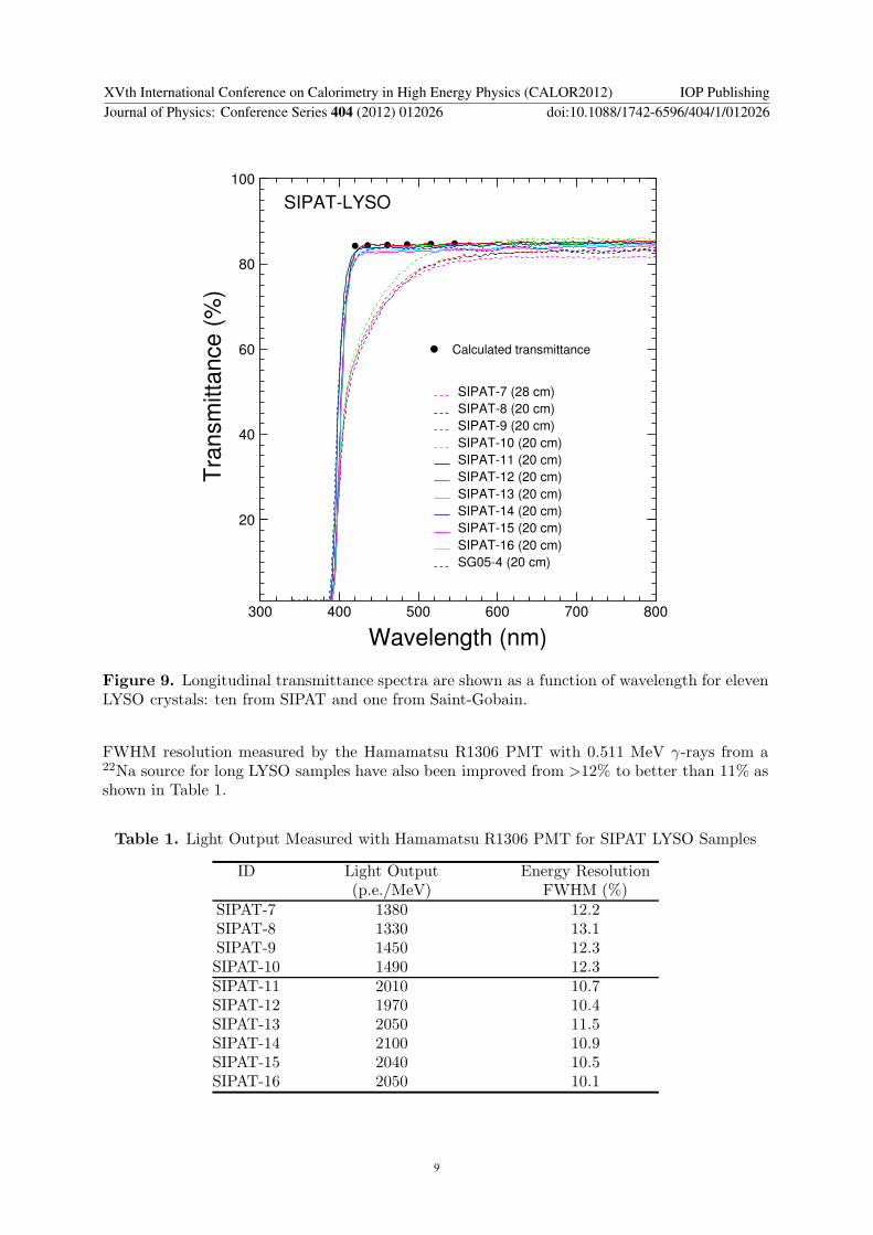
This issue of the absorption band at the seed end found in SIPAT-LYSO-L7, L8, L9 and L10was brought to the attention of SIPAT, and was discussed in a site visit. The problem was tracedback to a bad seed crystal used in the growth process. After a rigorous quality control for theseed crystals the absorption band at the seed end was eliminated. Figure 9 show the longitudinaltransmittance spectra for eleven LYSO crystals: ten from SIPAT and one from Saint-Gobain.No absorption was found between 400 nm and 800 nm for SIPAT-LYSO-L11 to L16, which weregrown with seed crystals of high quality, and the Saint-Gobain sample. A specification of 75%at 420 nm was introduced to reject crystals with poor longitudinal transmittance. Because ofthe improvement in optical transmittance the light output of 20 cm long LYSO crystals fromSIPAT is 30% higher than that with the absorption problem as shown in Table 1. The average
XVth International Conference on Calorimetry in High Energy Physics (CALOR2012) IOP PublishingJournal of Physics: Conference Series 404 (2012) 012026 doi:10.1088/1742-6596/404/1/012026
8
20
40
60
80
100
300 400 500 600 700 800
SIPAT-LYSO
Calculated transmittance
SIPAT-7 (28 cm)
SIPAT-8 (20 cm)
SIPAT-9 (20 cm)
SIPAT-10 (20 cm)
SIPAT-11 (20 cm)
SIPAT-12 (20 cm)
SIPAT-13 (20 cm)
SIPAT-14 (20 cm)
SIPAT-15 (20 cm)
SIPAT-16 (20 cm)
SG05-4 (20 cm)
Wavelength (nm)
Tra
nsm
itta
nce
(%
)
Figure 9. Longitudinal transmittance spectra are shown as a function of wavelength for elevenLYSO crystals: ten from SIPAT and one from Saint-Gobain.
FWHM resolution measured by the Hamamatsu R1306 PMT with 0.511 MeV γ-rays from a22Na source for long LYSO samples have also been improved from >12% to better than 11% asshown in Table 1.
Table 1. Light Output Measured with Hamamatsu R1306 PMT for SIPAT LYSO Samples
ID Light Output Energy Resolution(p.e./MeV) FWHM (%)
SIPAT-7 1380 12.2SIPAT-8 1330 13.1SIPAT-9 1450 12.3SIPAT-10 1490 12.3SIPAT-11 2010 10.7SIPAT-12 1970 10.4SIPAT-13 2050 11.5SIPAT-14 2100 10.9SIPAT-15 2040 10.5SIPAT-16 2050 10.1
XVth International Conference on Calorimetry in High Energy Physics (CALOR2012) IOP PublishingJournal of Physics: Conference Series 404 (2012) 012026 doi:10.1088/1742-6596/404/1/012026
9

6. SummaryIn a brief summary, LSO/LYSO crystals are an excellent material for a total absorptionelectromagnetic calorimeter for a future high-energy physics experiment in a severe radiationenvironment. The first 2.5×2.5×28 cm (25 X0) LYSO sample was successfully grown at SIPAT. Ithas consistent emission, adequate light response uniformity and good radiation resistance againstγ-rays up to 1 Mrad. This sample and three other large size samples from SIPAT, however,showed poor longitudinal transmittance between 400 nm and 600 nm as well as poor transversetransmittance at the seed end. This poor transmittance at the seed end was understood as beingcaused by point defects which absorb light, and was traced back to a bad seed crystal used intheir growth. With rigorous quality control on seed crystals recently grown LYSO crystals showno absorption band at the seed end and have a light output of 30% more than those with thisproblem. The corresponding average FWHM resolution measured by using 0.511 MeV γ-raysfrom a 22Na source is also improved from >12% to better than 11%. Because of the crystalsexcellent radiation hardness, a LSO/LYSO crystal calorimeter is capable of making precisionmeasurement for electrons, photons and jets and thus provides a great physics discovery potentialin a severe radiation environment, like the HL-LHC.
AcknowledgmentsThis work is partially supported by the U.S. Department of Energy Grant No. DE-FG03-92-ER40701 and the U.S. National Science Foundation Award PHY-0612805 and PHY-0516857.
References[1] C. Melcher and J. Schweitzer, “Cerium-doped lutetium oxyorthosilicate: a fast, efficient new scintillator,”
IEEE Trans. Nucl. Sci. 39 (1992) 502–505.[2] D.W. Cooke, K.J. McClellan, B.L. Bennett, J.M. Roper, M.T. Whittaker and R.E. Muenchausen, “Crystal
growth and optical characterization of cerium-doped Lu1.8Y0.2SiO5,” J. Appl. Phys. 88 (2000) 7360–7362.[3] T. Kimble, M Chou and B.H.T. Chai, “Scintillation properties of LYSO crystals,” in NSS conference Record
2002 IEEE, Vol 3, 1434-1437.[4] J.M. Chen, R.H. Mao, L.Y. Zhang and R.Y. Zhu., “Large size LSO and LYSO crystal for future high energy
and nuclear physics experiments,” IEEE Trans. Nucl. Sci. 54 (2007) 718–724.[5] C. Cecchi, A LYSO Calorimeter for the super B factory, 2011 J. Phys.: Conf. Ser. 293 012066.[6] F. Happacher , CCALT: Crystal Calorimeter at KLOE2, 2011 J. Phys.: Conf. Ser.[7] The Mu2e Experiment, see http://mu2e.fnal.gov/.[8] J.M. Chen, R.H. Mao, L.Y. Zhang and R.Y. Zhu, “Gamma-ray induced radiation damage in large size LSO
and LYSO crystal samples,” IEEE Trans. Nucl. Sci., 54 (2007) 1319–1326.[9] Rihua Mao, Liyuan Zhang and Ren-yuan Zhu, “Effects of neutron irradiations in various crystal samples of
large size for future crystal calorimeter,” 2009 IEEE NUCLEAR SCIENCE SYMPOSIUM CONFERENCE
RECORD, VOLS 1-5 (2009) 2041-2044.[10] F. Nessi-Tedaldi, G. Dissertori, P. Lecomte, D. Luckey and F. Pauss, “Studies of Cerium Fluoride, LYSO and
Lead Tungstate Crystals Exposed to High Hadron Fluences”,Paper N32-3, 2009 IEEE NSS Conference.[11] J.M. Chen, R.H. Mao, L.Y. Zhang and R.-Y. Zhu, IEEE Trans. Nucl. Sci. 54 (2007) 718.[12] J.M. Chen, R.H. Mao, L.Y. Zhang and R.-Y. Zhu, IEEE Trans. Nucl. Sci. 54 (2007) 1319.[13] Rihua Mao, Liyuan Zhang and Ren-yuan Zhu, “Optical and Scintillation Properties of Inorganic Scintillators
in High Energy Physics,” IEEE Trans. Nucl. Sci. 55 (2008) 2425–2431.[14] D. A. Ma and R. Y. Zhu, “Light attenuation length of barium fluoride crystals,” Nucl. Instr. and Meth.
A333, (1993) 422–424.[15] Rihua Mao, Liyuan Zhang and Ren-yuan Zhu, “Gamma-ray induced radiation damage in PWO and
LSO/LYSO crystals,” 2009 IEEE NUCLEAR SCIENCE SYMPOSIUM CONFERENCE RECORD, VOLS1-5 (2009) 2045-2049.
[16] R.H. Mao, L.Y. Zhang and R.-Y. Zhu, Optical and Scintillation Properties of Inorganic Scintillators in High
Energy Physics, IEEE Trans. Nucl. Sci. NS-55 (2008) 2425–2431.[17] D.A. Ma and R.-Y. Zhu, Nucl. Instr. and Meth. A333 (1993) 422.
XVth International Conference on Calorimetry in High Energy Physics (CALOR2012) IOP PublishingJournal of Physics: Conference Series 404 (2012) 012026 doi:10.1088/1742-6596/404/1/012026
10