Embed Size (px)
Citation preview

© 2014 Alam and Khan. This work is published by Dove Medical Press Limited, and licensed under Creative Commons Attribution – Non Commercial (unported, v3.0) License. The full terms of the License are available at http://creativecommons.org/licenses/by-nc/3.0/. Non-commercial uses of the work are permitted without any further
permission from Dove Medical Press Limited, provided the work is properly attributed. Permissions beyond the scope of the License are administered by Dove Medical Press Limited. Information on how to request permission may be found at: http://www.dovepress.com/permissions.php
Drug Design, Development and Therapy 2014:8 183–195
Drug Design, Development and Therapy Dovepress
submit your manuscript | www.dovepress.com
Dovepress 183
O r i g i n a l r e s e a r c h
open access to scientific and medical research
Open access Full Text article
http://dx.doi.org/10.2147/DDDT.S51577
Qsar and docking studies on xanthone derivatives for anticancer activity targeting Dna topoisomerase iiα
sarfaraz alamFeroz KhanMetabolic and structural Biology Department, central institute of Medicinal and aromatic Plants, council of scientific and industrial research, lucknow, Uttar Pradesh, india
correspondence: Feroz Khan Metabolic and structural Biology Department, central institute of Medicinal and aromatic Plants, council of Scientific and Industrial Research, PO-ciMaP, Kukrail Picnic spot road, lucknow-226015, Uttar Pradesh, india Tel +91 522 271 7668 Fax +91 522 234 2666 email [email protected]
Abstract: Due to the high mortality rate in India, the identification of novel molecules is
important in the development of novel and potent anticancer drugs. Xanthones are natural
constituents of plants in the families Bonnetiaceae and Clusiaceae, and comprise oxygenated
heterocycles with a variety of biological activities along with an anticancer effect. To explore the
anticancer compounds from xanthone derivatives, a quantitative structure activity relationship
(QSAR) model was developed by the multiple linear regression method. The structure–activity
relationship represented by the QSAR model yielded a high activity–descriptors relationship
accuracy (84%) referred by regression coefficient (r2=0.84) and a high activity prediction accu-
racy (82%). Five molecular descriptors – dielectric energy, group count (hydroxyl), LogP (the
logarithm of the partition coefficient between n-octanol and water), shape index basic (order 3),
and the solvent-accessible surface area – were significantly correlated with anticancer activity.
Using this QSAR model, a set of virtually designed xanthone derivatives was screened out.
A molecular docking study was also carried out to predict the molecular interaction between
proposed compounds and deoxyribonucleic acid (DNA) topoisomerase IIα. The pharmacokinet-
ics parameters, such as absorption, distribution, metabolism, excretion, and toxicity, were also
calculated, and later an appraisal of synthetic accessibility of organic compounds was carried
out. The strategy used in this study may provide understanding in designing novel DNA topoi-
somerase IIα inhibitors, as well as for other cancer targets.
Keywords: drug likeness, ADMET, regression model, HeLa cell line
IntroductionDrug discovery and development is not only a time-consuming process, but also a costly
procedure. Therefore, we wanted to apply computational methods for lead generation
and lead optimization in the drug discovery process. This emerging trend has immense
importance in reducing the phase time, as well as in amplifying the design of small
molecule-based leads with better biological activity and minimal side effects for a
disease-specific target. After the development of the first peptide-based HIV protease
inhibitors,1 followed by a target for antihypertension2 and inhibitors of the H5N1 avian
influenza,3 scientists are paying more attention to the in silico approach. Even with such
improvements, the design of a novel anticancer drug that works effectively on a patient
is still out of reach. Cancer, which is the uncontrolled growth and proliferation of cells
due to mutation of genes which accelerate cell division rates and evade the programmed
cell death, is the leading cause of death in the world.4 The frequency of one particular
manifestation of cancer, cervical cancer, is dramatically increasing. A link between
cancer and human deoxyribonucleic acid (DNA) topoisomerase type IIα (Top2A)

Drug Design, Development and Therapy 2014:8submit your manuscript | www.dovepress.com
Dovepress
Dovepress
184
alam and Khan
(enzyme commission number [EC]: 5.99.1.3)5 has already
been ascribed, and there is an interest in developing a specific
inhibitor as a new therapeutic regimen for the cancer.
Xanthones, which are used in this study, comprise a large
number of oxygenated heterocycles which play an important
role in medicinal chemistry. Their derivatives are widely dis-
tributed in various plants, and they have a variety of biological
properties, such as antioxidant, hepato- protective, anti-
inflammatory, anti-α-glucosidase, and anticancer properties.6
Due to their antitumor effect, xanthones are attracting more
interest. Until now, there have been only a few computational
studies on xanthone; also, the protein targets of xanthones
have not yet received a great deal of attention.6
Traditionally it is difficult to select the best chemical
moiety of compound that plays an effective role in treating or
preventing cancer, so we used computational strategies that
include quantitative structure activity relationship (QSAR)
modeling, virtual screening, shape similarity screening,
pharmacophore searching, molecular docking, and ADMET
(absorption, distribution, metabolism, excretion, and toxic-
ity properties of a molecule within an organism) studies
to identify potential protein targets of xanthone and other
phytochemicals.7 Using these computational methodolo-
gies, we demonstrate a multiple linear regression QSAR
model for activity prediction that successfully predicts the
anticancer activities of newly designed xanthone derivatives.
In the QSAR model, the regression coefficient (r2), which
indicates the relationship correlation, was 0.84, while the
cross-validation regression coefficient (r2CV), which indi-
cates the prediction accuracy of the model, was 0.82. The
QSAR study indicates that dielectric energy, group count
(hydroxyl), LogP, shape index basic (order 3), and solvent-
accessible surface area were significantly correlated with
anticancer activity. After successful validation of this model,
it was then used to design and virtually screen 50 compounds
and identify 39 with IC50
values of #20 µM. Lipinski’s rule
of five was used to filter the compounds and was further
accompanied by molecular docking studies, which were
performed for predicting active compounds against highly
promising anticancer drug targeting (Figure 1). Since in
humans the drug target protein for doxorubicin (DrugBank
ID: DB00997) is Top2A, we selected it as a target protein.
This target is widely used for existing anticancer agents: eg,
etoposide; anthracyclines (doxorubicin, daunorubicin); and
mitoxantrone. These drugs work either through the poison
of topoisomerase II cleavage complexes or by inhibiting
the ATPase activity by acting as noncompetitive inhibitors
of adenosine triphosphate (ATP).8 A docking study was
carried out to identify the putative binding site of active
xanthone derivatives (which could be helpful in explaining
the underlying structure–activity relationship), by using a
crystal structure of inhibitor-bound Top2A. Based on the
QSAR model, molecular docking, ADMET, and synthesis
accessibility, we then identified four inhibitors with IC50
values of 7.94 µM, 0.63 µM, 2.51 µM, and 0.16 µM, as
potent inhibitors of Top2A (Figure 1). This study is a sig-
nificant approach in the identification of hits compounds
with structural diversity, which may provide further helpful
insights to screening and designing novel anticancer com-
pounds and their respective protein targets. Moreover, this
study is also projected to explore the molecular mechanism
by which xanthone derivatives can be further utilized with
better activity by rational modifications.
Methods and computational detailsstructure cleaningDrawing and geometry cleaning of compounds with anti-
cancer activity was performed using ChemBioDraw Ultra
version 12.0 (2010) software (PerkinElmer Informatics,
Waltham, MA, USA). The two-dimensional (2D) structures
were transformed into three-dimensional (3D) structures
using the converter module of ChemBio3D Ultra. The 3D
structures were then subjected to energy minimization,
which was performed in two steps. The first step was energy
minimization using molecular mechanics-2 (MM2) until the
root mean square (RMS) gradient value became smaller than
Xanthone derivatives
Compounds with predictedIC50=20 µM
Rule of five filter
Molecular docking
ADMET
Syntheticaccessibility
Hitcompounds
50
39
34
11
10
4
4
Figure 1 Virtual screening protocol for the identification of novel DNA Top2A inhibitors.Abbreviations: aDMeT, absorption, distribution, metabolism, excretion, and toxicity properties of a molecule within an organism; Dna, deoxyribonucleic acid; ic50, inhibitory concentration to 50% of the population; Top2a, topoisomerase type iiα.

Drug Design, Development and Therapy 2014:8 submit your manuscript | www.dovepress.com
Dovepress
Dovepress
185
Qsar and docking studies on xanthone derivatives
0.100 kcal/mol/Å; then in a second step, minimized MM2
(dynamics) compounds were subjected to reoptimization
through the MOPAC (Molecular Orbital Package, Chem-
BioDraw Ultra version 12.0 [2010] software; PerkinElmer
Informatics, Waltham, MA, USA) method, until the RMS
gradient attained a value smaller than 0.0001 kcal/mol/Å.
Parameters for Qsar model developmentIn the present study, cancer cell line-based QSAR modeling
was performed. Initially, a total of 64 compounds with reported
anticancer activity against the human cervical cancer cell line
(HeLa) were used as a training data set while developing the
QSAR model (Tables 1 and S1).6,9–16 The anticancer activity
was in IC50
form. A total of 52 chemical descriptors (physico-
chemical properties) were calculated for each compound. The
selection was made on the basis of structural/pharmacophore
or chemical class similarity. Similarly, in order to select the
best subset of descriptors, highly correlated descriptors were
excluded. Finally, a model was developed based on the forward
stepwise multiple linear regression method. The resulting
QSAR model exhibited a high regression coefficient. The model
was successfully validated using random test set compounds
(Table S2), and was evaluated for the robustness of its predic-
tions via the cross-validation coefficient.
Various descriptors like steric, electronic, and thermo-
dynamic were calculated by the Scigress Explorer software
(Fujitsu, Tokyo, Japan). For the validation of QSAR models,
the leave one out method was used;17 the best model was
selected on the basis of various statistical parameters, such as
a square of the correlation coefficient (R2), and the quality of
each model was estimated from the cross-validated squared
correlation coefficient (rCV2).
statistical calculations used in Qsar modelingThe stepwise multiple linear regression method calculates
QSAR equations by adding one variable at a time and testing
each addition for significance. Only variables that are found
to be significant are used in the QSAR equation. This regres-
sion method is especially useful when the number of variables
is large and when the key descriptors are not known. In the
forward mode, the calculation begins with no variables and
builds a model by entering one variable at a time into the
equation. In the backward mode, the calculation begins with
all variables included and drops variables one at a time until
the calculation is complete; however, backward regression
calculations can lead to overfitting.
Table 1 comparison of experimental and predicted activities of training data set molecules based on Qsar model
Serial number
Compound ID
Experimental activity6,9–16,*
Predicted activity*
Error factor**
1 Xtr-1 3.7 3.466 -0.232 Xtr-2 3.8 3.92 0.123 Xtr-3 3.9 3.549 -0.354 Xtr-5 3.78 3.701 -0.085 Xtr-6 3.87 3.952 0.086 Xtr-7 3.7 3.833 0.1337 Xtr-8 3.7 3.782 0.088 Xtr-10 3.7 3.604 -0.069 Xtr-11 3.7 4.322 0.62210 Xtr-12 4.21 4.313 0.10311 Xtr-13 3.7 3.493 -0.20712 Xtr-14 3.7 4.172 0.47213 Xtr-15 3.8 4.57 0.7714 Xtr-16 3.7 3.642 -0.05815 Xtr-17 3.71 3.84 0.1316 Xtr-18 4.24 3.857 -0.38317 Xtr-20 3.75 3.693 -0.05718 Xtr-21 5.37 5.266 -0.10919 Xtr-22 3.74 3.607 -0.13320 Xtr-23 5.46 5.381 -0.07921 Xtr-24 5.46 6.313 0.85322 Xtr-25 5.46 5.94 0.4823 Xtr-27 5.18 4.866 -0.31424 Xtr-31 5.18 5.467 0.28725 Xtr-32 5.18 4.957 -0.22326 Xtr-34 5.18 5.443 0.35727 Xtr-35 8.38 7.63 -0.7528 Xtr-36 4.98 4.476 -0.50429 Xtr-37 5.2 4.769 -0.43130 Xtr-38 4 4.259 0.25931 Xtr-40 4.23 4.54 0.3132 Xtr-41 5.33 5.096 -0.23433 Xtr-42 4.84 4.546 -0.29434 Xtr-43 3.7 3.626 -0.07435 Xtr-44 4.17 4.421 0.25136 Xtr-45 5.82 5.879 0.05937 Xtr-47 4 4.619 0.61938 Xtr-50 4.16 4.145 0.01539 Xtr-52 4.21 4.401 0.19140 Xtr-53 4 4.466 0.46641 Xtr-54 4 3.988 -0.01242 Xtr-55 4.01 3.9 -0.1143 Xtr-56 4.63 4.851 0.22144 Xtr-57 3.86 3.76 -0.145 Xtr-58 4.3 4.129 0.17146 Xtr-59 4.62 4.814 0.19447 Xtr-61 4.59 4.783 0.19348 Xtr-62 4 4.005 0.00549 Xtr-63 4 4.198 0.19850 Xtr-64 4.6 4.633 0.03351 Xtr-65 5 4.973 -0.02752 Xtr-66 4 4.254 0.25453 Xtr-67 3.83 3.853 0.023
(Continued)

Drug Design, Development and Therapy 2014:8submit your manuscript | www.dovepress.com
Dovepress
Dovepress
186
alam and Khan
Multiple regression correlation coefficientVariations in the data are quantified by the correlation
coefficient (r), which measures how closely the observed
data track the fitted regression line. This is a measure of
how well the equation fits the data (ie, it measures how good
the correlation is). A perfect relation has r=+1 (positively
correlated) or -1 (negatively correlated); no correlation has
r=0. The regression coefficient, r2, is sometimes quoted, and
this gives the fraction of the variance (in percentage) that is
explained by the regression line. The more scattered the data
points, the lower the value of r. A satisfactory explanation
of the data is usually indicated by an r2. Errors in either the
model or in the data will lead to a bad fit. This indicator of
fit to the regression line is calculated as:
R2= (sum of the squares of the deviations from the
regression line)/(sum of the squares of the
deviations from the mean) (1)
R2= (regression variance)/(original variance) (2)
where the regression variance is defined as the original
variance minus the variance around the regression line. The
original variance is the sum of the squares of the distances
of the original data from the mean.
Validating Qsar equations and dataThe cross-validation coefficient, rCV2, can be calculated as
rCV 22
21= −
−−
ΣΣ
( )
( ).
y y
y yi j
i z
(3)
Here, yi and y
j are the measured and predicted values of
dependent variables, respectively. yz is the averaged value
of dependent variable of the training set.
leave one out cross-validationLeave one out cross-validation (LOOCV) is one of the most
effective methods for validation of a model with a small
training dataset. Here, training is done with a data size of
(N–1) and tested the remaining one, where N represents the
complete set of data. In the LOOCV method, the training and
testing are repeated for N amount of time, so as to pass each
individual data through the testing process.
Virtual designing of novel xanthone derivativesThe 50 compounds (Table S3) were virtually designed and
then validated. The QSAR model was used to predict the
biological responses to these chemical structures.
Rule of five filtersAll the chemical structures are evaluated for good oral bio-
availability in order to be an effective drug-like compound,
subject to Lipinski’s rule of five.18 According to this rule,
a drug-like molecule should have not more than one of the fol-
lowing violations: no more than five hydrogen bond donors;
no more than ten hydrogen bond acceptors; molecular weight
no more than 500; and LogP no more than 5.
Protein preparationThe protein preparation protocol is used to perform tasks
such as inserting missing atoms in incomplete residues,
deleting alternate conformations (disorder), removing
waters, standardizing the names of the atoms, modeling
missing loop regions, and protonating titratable residues by
using predicted pKs (negative logarithmic measure of acid
dissociation constant). CHARMM (Chemistry at HARvard
Macromolecular Mechanics; Cambridge, MA, USA) is used
for protein preparation with an energy of -31.1116, initial
RMS gradient energy of 181.843, and grid spacing of 0.5
angstrom (Å). The hydrogen atoms were added before the
processing. Protein coordinates from the crystal structure
of Top2A (PDB [Protein Data Bank] ID: 1ZXM) Chain A
determined at a resolution of 1.87 Å were used (Figure 2).
Protein–ligand dockingMolecular docking studies were performed to generate the bio-
active binding poses of inhibitors in the active site of enzymes
by using the LibDock program from Discovery Studio,
Table 1 (Continued)
Serial number
Compound ID
Experimental activity6,9–16,*
Predicted activity*
Error factor**
54 Xtr-69 5.66 4.967 -0.69355 Xtr-71 5.82 5.367 -0.45356 Xtr-73 5.38 4.869 -0.51157 Xtr-74 5.11 5.551 0.44158 Xtr-75 5.07 5.032 -0.03859 Xtr-76 3.78 3.802 0.02260 Xtr-78 4.91 4.734 -0.17661 Xtr-80 5.04 4.963 -0.07762 Xtr-81 5 4.344 -0.65663 Xtr-82 4.81 4.864 0.05464 Xtr-83 6.05 5.505 -0.545
Notes: *Measured and predictive value is in pic50; **the difference between predicted activity values and experimental activity values is represented as error (ratio between the predicted and experimental activity), with a negative sign if the actual activity is higher than that of the predicted activity.Abbreviations: pic50, negative of the log inhibitory concentration to 50% of the population; QSAR, quantitative structure activity relationship; ID, identification.

Drug Design, Development and Therapy 2014:8 submit your manuscript | www.dovepress.com
Dovepress
Dovepress
187
Qsar and docking studies on xanthone derivatives
version 3.5 (Accelrys, San Diego, CA, USA). LibDock uses
protein site features, referred to as hot spots, consisting of
two types (polar and apolar). The ligand poses are placed into
the polar and apolar receptor interactions site. In the current
study, the Merck Molecular Force Field was used for energy
minimization of the ligands. The binding sphere was primarily
defined as all residues of the target within 5 Å from the first
binding site. Here, the ATP binding site was used to define the
active site, referred to as the hot spots (Figure 2). Conformer
Algorithm based on Energy Screening And Recursive build-up
(CAESAR) was used for generating conformations. Then, the
smart minimizer was used for in situ ligand minimization. All
other docking and consequent scoring parameters used were
kept at their default settings.
We also analyzed the protein ligand complexes to better
understand the interactions between protein residues and
bound ligands, along with the binding site residues of the
defined receptor. The 2D diagrams helped to identify the
binding site residue, including amino acid residues, waters,
and metal atoms.
The score ligand poses protocol was used for the scor-
ing functions, such as LibDock score, Jain, LigScore 1,
LigScore 2, piecewise linear potential (PLP) and potential
of mean force (PMF) 04, to evaluate ligand binding in a
receptor cavity.
Validation using autoDock VinaAutoDock Vina19 software (Scripps Research Institute, La
Jolla, CA, USA) was also used for molecular docking stud-
ies to validate the LibDock score. For this, the designed
compounds were optimized and then used for docking
experiments. The same binding site and receptor used in
the LibDock program are used for this study. The docking
program takes the PDBQT file format of ligands and receptor,
A
B
Ligand binding site
Figure 2 (A) structural model of human Dna Top2a (PDB iD: 1ZXM) with aTP binding site (yellow); (B) aTP binding site pocket residues.Abbreviations: aTP, adenosine triphosphate; Dna, deoxyribonucleic acid; Top2a, topoisomerase type iiα.
Drug Design, Development and Therapy 2014:8submit your manuscript | www.dovepress.com
Dovepress
Dovepress
188
alam and Khan
a modified PDB file, which has added polar hydrogens and
partial charges. Other docking parameters were set to the
software’s default values.
Pharmacokinetics parametersADMET refers to the absorption, distribution, metabolism,
excretion, and toxicity properties of a molecule within an
organism, and were predicted using ADMET descriptors in
Discovery Studio 3.5 (Accelrys). In this module, six math-
ematical models (aqueous solubility, blood–brain barrier
penetration, cytochrome P450 2D6 inhibition, hepatotoxicity,
human intestinal absorption, and plasma protein binding) are
used to quantitatively predict properties of a set of rules that
specify ADMET characteristics of the chemical structure of
the molecules. These ADMET descriptors allow us to elimi-
nate compounds with unfavorable ADMET characteristics
early on to avoid expensive reformulation, preferably before
synthesis, and also help to evaluate proposed structural refine-
ments that are designed to improve ADMET properties.
Validation of synthetic accessibility for hit compounds using sYlViaSynthetic accessibility scores for hit compounds were used to
validate the synthetic possibilities. For this, the SYLVIA-XT
1.4 program (Molecular Networks, Erlangen, Germany) was
used to calculate the synthetic accessibility of these optimized
compounds.20 The appraisal of synthetic accessibility of
organic compounds using SYLVIA provides a score on a scale
from 1 (very easy to synthesize) to 10 (complex and challeng-
ing to synthesize). A number of criteria, such as complexity
of the ring system, complexity of the molecular structure,
number of stereo centers, similarity to commercially avail-
able compounds, and potential for using powerful synthetic
reactions have been independently weighted to provide a
single value for synthetic accessibility.
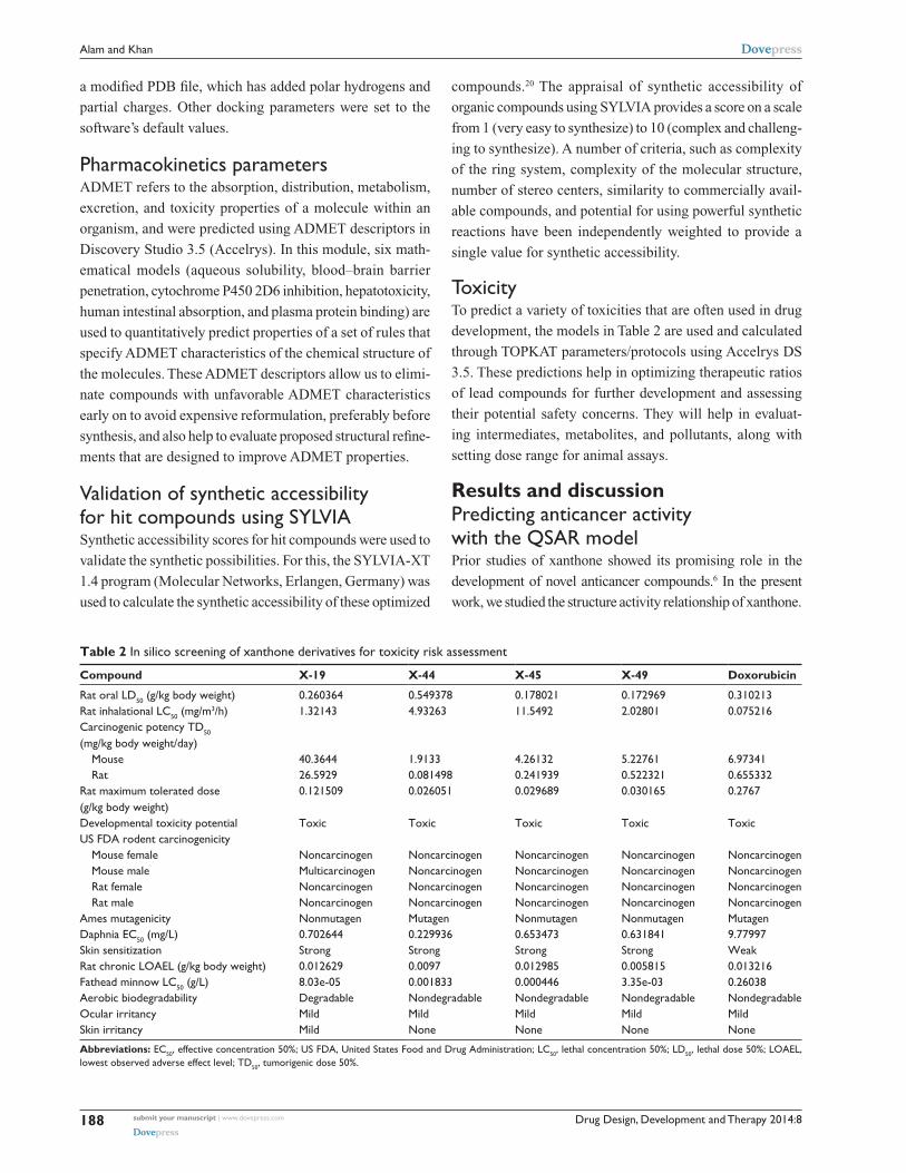
ToxicityTo predict a variety of toxicities that are often used in drug
development, the models in Table 2 are used and calculated
through TOPKAT parameters/protocols using Accelrys DS
3.5. These predictions help in optimizing therapeutic ratios
of lead compounds for further development and assessing
their potential safety concerns. They will help in evaluat-
ing intermediates, metabolites, and pollutants, along with
setting dose range for animal assays.
Results and discussionPredicting anticancer activity with the Qsar modelPrior studies of xanthone showed its promising role in the
development of novel anticancer compounds.6 In the present
work, we studied the structure activity relationship of xanthone.
Table 2 in silico screening of xanthone derivatives for toxicity risk assessment
Compound X-19 X-44 X-45 X-49 Doxorubicin
rat oral lD50 (g/kg body weight) 0.260364 0.549378 0.178021 0.172969 0.310213rat inhalational lc50 (mg/m3/h) 1.32143 4.93263 11.5492 2.02801 0.075216carcinogenic potency TD50 (mg/kg body weight/day) Mouse 40.3644 1.9133 4.26132 5.22761 6.97341 rat 26.5929 0.081498 0.241939 0.522321 0.655332rat maximum tolerated dose (g/kg body weight)
0.121509 0.026051 0.029689 0.030165 0.2767
Developmental toxicity potential Toxic Toxic Toxic Toxic ToxicUs FDa rodent carcinogenicity Mouse female noncarcinogen noncarcinogen noncarcinogen noncarcinogen noncarcinogen Mouse male Multicarcinogen noncarcinogen noncarcinogen noncarcinogen noncarcinogen rat female noncarcinogen noncarcinogen noncarcinogen noncarcinogen noncarcinogen rat male noncarcinogen noncarcinogen noncarcinogen noncarcinogen noncarcinogenames mutagenicity nonmutagen Mutagen nonmutagen nonmutagen MutagenDaphnia ec50 (mg/l) 0.702644 0.229936 0.653473 0.631841 9.77997skin sensitization strong strong strong strong Weakrat chronic lOael (g/kg body weight) 0.012629 0.0097 0.012985 0.005815 0.013216Fathead minnow lc50 (g/l) 8.03e-05 0.001833 0.000446 3.35e-03 0.26038aerobic biodegradability Degradable nondegradable nondegradable nondegradable nondegradableOcular irritancy Mild Mild Mild Mild Mildskin irritancy Mild none none none none
Abbreviations: ec50, effective concentration 50%; Us FDa, United states Food and Drug administration; lc50, lethal concentration 50%; lD50, lethal dose 50%; lOael, lowest observed adverse effect level; TD50, tumorigenic dose 50%.

Drug Design, Development and Therapy 2014:8 submit your manuscript | www.dovepress.com
Dovepress
Dovepress
189
Qsar and docking studies on xanthone derivatives
9
Linear (training)
LOOCV
External test set
Training
Training (R2)=0.84LOOCV (R2)=0.79
7
pIC50 (experimental)
pIC
50 (
pre
dic
ted
)
5
9
8
7
6
5
4
3
3
Figure 3 regression plot representing training, testing, and cross-validation of model.Abbreviations: pic50, negative of the log inhibitory concentration to 50% of the population; lOOcV, leave one out cross-validation; R2, correlation coefficient.
The structure–activity relationship denoted by the QSAR model
yielded a high activity–descriptors relationship accuracy of 84%
referred by regression coefficient (r2=0.84) and a high activity
prediction accuracy of 82%. Five molecular descriptors –
dielectric energy, group count (hydroxyl), LogP, shape index
basic (order 3), and the solvent-accessible surface area – were
significantly correlated with anticancer activity. The QSAR
model equation is given below, showing the relationship between
experimental activity in vitro (ie, the inhibitory concentration
to 50% of the population [IC50
]) as the dependent variable and
five independent variables (chemical descriptors):
Predicted-log IC50
(pIC50
) (µM) = +2.19682 × dielectric energy
+0.22309 × group count (hydroxyl)
-0.543107 × LogP
-0.469003 × shape index basic (order 3)
+0.0175389 × solvent-accessible surface area
+2.57271. (4)
Here, the rCV2 is 0.82, which indicates that the newly
derived QSAR model has a prediction accuracy of 82%, and
the r2 is 0.84, which indicates that the correlation between
the activity (dependent variable) and the descriptors (inde-
pendent variables) for the training data set compounds was
84% (Figure 3); the LOOCV R2 is 0.79. It is evident from the
above equation that among the molecular descriptors, dielec-
tric energy, group count (hydroxyl), and solvent-accessible
surface area are positively correlated, meaning the biologi-
cal activity increases when the values of these descriptors is
positively increased. On other hand, the descriptors LogP and
Shape index basic (order 3), are both negatively correlated
with activity; the activity decreases when the values of these
descriptors increases. Thus, we successfully developed a
QSAR model for prediction of in vitro anticancer activity.
A multiple linear regression QSAR mathematical model was
developed for activity prediction that successfully and accu-
rately predicted the anticancer activities of newly designed
xanthone derivatives.
experimental validation of Qsar modelThe multiple linear regression-based QSAR model for the
inhibitory activity of xanthone derivatives against HeLa cell
lines has been validated with four compounds, Xan-1, Xan-2,
Xan-3, and Xan-4 (Table 3).21 It was found that the predicted
results through the QSAR model show compliance with their
experimental results.
Virtually designing and filtering of novel xanthone derivativesUsing this multiple linear regression QSAR mathematical
model, which was developed for activity prediction against
HeLa cell line, we predicted the anticancer activities of some
newly designed xanthone derivatives (Table S3). The pre-
dicted IC50
value of final hit compounds X-19, X-44, X-45,
and X-49 are 7.94 µM, 0.63 µM, 2.51 µM, and 0.16 µM,
respectively. The QSAR model quantified the activity-
dependent chemical descriptors and predicted the inhibitory
concentration (log IC50
) of each derivative, thus indicating
its potential range of inhibition (Table 3).
Protein–ligand docking studiesFollowing development of the model and filtering through
Lipinski’s rule of five, we first analyzed Top2A, and five
active sites were obtained. We chose one of these with ATP
binding sites, shown in Figure 2. In order to understand the
ligand recognition in Top2A, we initially carried out docking
with the known Top2A inhibitor/anticancer drug doxorubicin,
and later with the most active 34 among the designed and

Drug Design, Development and Therapy 2014:8submit your manuscript | www.dovepress.com
Dovepress
Dovepress
190
alam and Khan
Tab
le 3
scr
eene
d ou
t xa
ntho
ne d
eriv
ativ
es t
arge
ting/
inhi
bitin
g T
op2a
and
hel
a hu
man
can
cer
cell
line
Com
poun
d
IDSt
ruct
ure
Exp
erim
enta
l act
ivit
y
pIC
50 (-l
og IC
50) μM
Pre
dict
ed a
ctiv
ity
pI
C50
(-l
og IC
50) μM
Die
lect
ric
en
ergy
(k
cal/m
ole)
Gro
up
coun
t
(hyd
roxy
l)
LogP
Shap
e in
dex
(b
asic
kap
pa,
orde
r 3)
Solv
ent-
acce
ssib
le
surf
ace
area
(a
ngst
rom
squ
are)
Xan
-14.
93.
8-0
.612
24.
927
4.92
440
2.6
Xan
-24.
04.
3-0
.503
24.
132
3.88
369.
4
Xan
-33.
84.
3-0
.716
31.
545
2.39
126
2.8
Xan
-43.
83.
8-0
.552
22.
082
2.06
223
0.4
X-1
9–
5.1
-0.9
32
1.71
85.
087
428.
5
X-4
4–
6.2
-0.7
70
1.39
24.
3946
9.7
X-4
5–
5.6
-0.6
80
2.69
94.
519
466

Drug Design, Development and Therapy 2014:8 submit your manuscript | www.dovepress.com
Dovepress
Dovepress
191
Qsar and docking studies on xanthone derivatives
X-4
9–
6.8
-0.6
21
1.97
64.
411
486.
4
con
trol
5.8
5.9
-0.6
020
1.50
53.
863
413.
8
Abb
revi
atio
ns: h
ela,
hum
an c
ervi
cal c
ance
r ce
ll lin
e; p
ic50
, neg
ativ
e of
the
log
inhi
bito
ry c
once
ntra
tion
to 5
0% o
f the
pop
ulat
ion;
Top
2a, t
opoi
som
eras
e ty
pe ii
α; ID
, ide
ntifi
catio
n.
filtered compounds. Out of 34, 20 failed to dock and three
showed lower scores than the control. The docking program
produces several poses with different orientations within the
defined active site. All poses produce a different LibDock
score. The best score was taken into account for further
study. The compounds X-12, X-19, X-29, X-32, X-35, X-39,
X-40, X-44, X-45, X-48, and X-49 (Table S3) were selected
as candidate compounds based on their high docking score
compared to doxorubicin. The analysis of the protein ligand
complexes revealed binding site residue, including amino
acid residues, waters, and metal atoms. A 2D diagram show-
ing various interactions, such as hydrogen bonds, atomic
charge interactions, and Pi-sigma interactions between the
surrounding residues and the ligand, was also displayed.
Different interactions were represented by different colors:
eg, pink indicates electrostatic interaction; purple indicates
covalent bond; and green indicates van der–Waals molecular
interaction. Solvent accessibility of the ligand atom and the
amino acid residues are shown in light blue shading sur-
rounding the atom or residue. High shading indicates more
exposure to solvent. The inhibitory activity of xanthone has
been explained by two major factors: H-bond and pi-sigma
interactions (Figure 4).
To evaluate ligand binding in a receptor cavity, the score
ligand poses protocol was used for the scoring functions
for LibDock score, Jain, LigScore 1, LigScore 2, PLP, and
PMF 04. The H-bond and pi-sigma interactions residues are
also provided (Table 4).
assessment through pharmacokinetic parametersSince the docking studies were found to be promising, the
chemical descriptors for the pharmacokinetic properties were
also calculated, so as to check the compliance of study com-
pounds with standard range. For this, the aqueous solubility,
blood–brain barrier penetration, cytochrome P450 2D6 bind-
ing, hepatotoxicity, intestinal absorption, and plasma protein
binding were calculated. Calculating these properties was
intended as the first step toward analyzing the novel chemi-
cal entities in order to check the failure of lead candidates,
which may cause toxicity or be metabolized by the body
into an inactive form or one unable to cross the membranes.
The results of this analysis are reported in Table 5, together
with a biplot (Figure 5). The pharmacokinetic profiles of
all the compounds under investigation were predicted by
means of six precalculated ADMET models provided by the
Accelrys Discovery Studio 3.5 program. The biplot shows
the two analogous 95% and 99% confidence ellipses for

Drug Design, Development and Therapy 2014:8submit your manuscript | www.dovepress.com
Dovepress
Dovepress
192
alam and Khan
TRPA:119
ALAA:92 ASN
A:91
ARGA:98
THRA:215
ASNA:120
ASNA:95
ASPA:94 SER
A:149
ASNA:150
THRA:147
SERA:148
3.3
3.7
GLYA:164
ILEA:118
TRPA:119
CYSA:216
ASNA:91
PHEA:142
ALAA:167
ASNA:150
GLYA:166 GLY
A:161
ALAA:167
GLYA:166
TYRA:165
LYSA:168
5.1
PHEA:142 SER
A:148
LEUA:140
SERA:149
HISA:130
VALA:137
PROA:126
ILEA:125
ARGA:98
ASPA:94THR
A:215
ALAA:92
ASNA:95
ASNA:91
SERA:149ILE
A:125
GLYA:124ARG
A:98
THRA:181
ASNA:95
ASNA:120
THRA:215
PHEA:142
ALAA:92
ILEA:217
ASNA:91ILE
A:88
GLYA:166
GLUA:87
TYRA:165
ALAA:167 6
GLNA:376
ARGA:162
GLYA:161
GLYAl164
ASNA:150 SER
A:148
LYSA:168
5.1
ILEA:141
THRA:147
LEUA:140
VALA:137
LYSA:123
ILEA:217
ASNA:120
LYSA:123
4.8
ILEA:141
GLYA:164
THRA:147
GLYA:164
TYRA:165
LYSA:168
ARGA:162
SERA:148
THRA:147
ILEA:141
SERA:149
VALA:137
ILEA:217
ILEA:88
ARGA:98
LYSA:123
ASNA:95
THRA:181
ILEA:125
GLYA:124
ASNA:120
ALAA:92
THRA:215
ASNA:163
ARGA:162
TYRA:165
GLNA:376GLY
A:166
GLYA:161
ALAA:167
LYSA:168
GLUA:87
ILEA:125PHE
A:142ILEA:88
ILEA:217
LEUA:89
ILEA:118
ILEA:141
CYSA:216
SigmaPi
4.1
Sigma
Pi
Pi
Sigma
A B
C D
Figure 4 2D diagrams illustrating protein–ligand interactions: (A) compound X-19; (B) compound X-44; (C) compound X-45; (D) compound X-49.Abbreviation: 2D, two-dimensional.
Table 4 LibDock scoring functions, SYLVIA synthetic accessibility scores, and AutoDock binding affinity of identified potential xanthone derivatives inhibitors for Dna Top2a
Compounds LibDock score
Jain LigScore 1 LigScore 2 PLP 1 PLP 2 PMF 04 SYLVIA score
H-bonding analysis
Pi-sigma interaction analysis
AutoDock binding energy (kcal/mol)
X-19 138.108 5.8 0.28 -2.26 99.22 99.26 -66.4 6.35 asn-91, ser-148, ser-149
asn-91 -7.3
X-44 133.709 4.57 1.18 2.11 107.27 99.08 -9.01 6.35 ala-167 ile-141 -7.2X-45 120.382 5.63 -0.89 -3.12 103.5 107.69 -24.12 5.91 TYr-165,
lYs-168(2)no Pi-sigma interaction found
-7.1
X-49 137.133 4.34 0.78 0.35 109.22 100.56 8.32 6.98 lYs-168, his-130
ser-149 -7.3
Doxorubicin 71.47 2.62 -4.87 -8.71 45.3 48.91 -9 6.16 PrO-371, lYs-378
TYr-151 -6.4
Abbreviations: Dna, deoxyribonucleic acid; PlP, piecewise linear potential; PMF, potential of mean force; Top2a, topoisomerase type iiα.

Drug Design, Development and Therapy 2014:8 submit your manuscript | www.dovepress.com
Dovepress
Dovepress
193
Qsar and docking studies on xanthone derivatives
Tab
le 5
com
plia
nce
of c
ompo
unds
to
the
theo
retic
al p
aram
eter
s of
ora
l bio
avai
labi
lity
and
drug
like
ness
pro
pert
ies
Com
poun
d
nam
eA
queo
us
solu
bilit
yB
lood
–bra
in
barr
ier
pene
trat
ion
Cyt
ochr
ome
P
450
2D6
bind
ing
Hep
atot
oxic
ity
Inte
stin
al
abso
rpti
onP
lasm
a pr
otei
n
bind
ing
PSA
Alo
gP98
X-1
22
(low
)4
(und
efine
d)Fa
lse
(non
inhi
bito
r)T
rue
(tox
ic)
2 (p
oor)
Tru
e (h
ighl
y bo
unde
d)14
4.09
43.
876
X-1
92
(low
)4
(und
efine
d)Fa
lse
(non
inhi
bito
r)T
rue
(tox
ic)
1 (m
oder
ate)
Tru
e (h
ighl
y bo
unde
d)11
1.39
34.
343
X-2
92
(low
)4
(und
efine
d)Fa
lse
(non
inhi
bito
r)T
rue
(tox
ic)
1 (m
oder
ate)
Tru
e (h
ighl
y bo
unde
d)13
6.12
12.
112
X-3
22
(low
)3
(low
)Fa
lse
(non
inhi
bito
r)T
rue
(tox
ic)
0 (g
ood)
Tru
e (h
ighl
y bo
unde
d)98
.004
3.30
9X
-35
3 (g
ood)
3 (lo
w)
Fals
e (n
on in
hibi
tor)
Tru
e (t
oxic
)0
(goo
d)T
rue
(hig
hly
boun
ded)
106.
934
2.15
8X
-39
2 (lo
w)
3 (lo
w)
Fals
e (n
on in
hibi
tor)
Tru
e (t
oxic
)0
(goo
d)T
rue
(hig
hly
boun
ded)
98.0
043.
309
X-4
02
(low
)4
(und
efine
d)Fa
lse
(non
inhi
bito
r)T
rue
(tox
ic)
0 (g
ood)
Tru
e (h
ighl
y bo
unde
d)10
3.42
3.58
X-4
42
(low
)3
(low
)Fa
lse
(non
inhi
bito
r)T
rue
(tox
ic)
0 (g
ood)
Tru
e (h
ighl
y bo
unde
d)79
.811
2.73
X-4
52
(low
)2
(med
ium
)Fa
lse
(non
inhi
bito
r)T
rue
(tox
ic)
0 (g
ood)
Tru
e (h
ighl
y bo
unde
d)70
.881
3.95
2X
-48
2 (lo
w)
3 (lo
w)
Fals
e (n
on in
hibi
tor)
Tru
e (t
oxic
)0
(goo
d)T
rue
(hig
hly
boun
ded)
100.
626
3.09
3X
-49
2 (lo
w)
2 (m
ediu
m)
Fals
e (n
on in
hibi
tor)
Tru
e (t
oxic
)0
(goo
d)T
rue
(hig
hly
boun
ded)
91.6
963.
595
Dox
orub
icin
2 (lo
w)
4 (u
ndefi
ned)
Fals
e (n
on in
hibi
tor)
Tru
e (t
oxic
)3
(ver
y po
or)
Fals
e (p
oorl
y bo
unde
d)20
9.31
–0.0
44
Abb
revi
atio
ns: A
logP
, the
loga
rith
m o
f the
par
titio
n co
effic
ient
bet
wee
n n-
octa
nol a
nd w
ater
; PSA
, pol
ar s
urfa
ce a
rea.
the blood–brain barrier penetration and human intestinal
absorption models, respectively. The polar surface area
(PSA) was shown to have an inverse relationship with percent
human intestinal absorption, and thus cell wall permeability,
although a relationship between PSA and permeability has
been demonstrated. Moreover, when we calculated the PSA
as a chemical descriptor for passive molecular transport
through membranes, the results showed a lower PSA value
of hit compounds than doxorubicin, but still within the limit;
ie, ,140 Å2. The aqueous solubility predictions (defined in
water at 25°C) show that hit compounds are soluble in water.
LogP value, which is a measure of lipophilicity and is the
ratio of the solubility of the compound in octanol compared
to its solubility in water, was found to be in range of the hit
compounds and follows Lipinski’s rule of five, implicating a
better oral bioavailability. The excretion process that elimi-
nates the compound from the human body also depends on
LogP. The hit compounds are highly ($90%) bound to car-
rier proteins in the blood. This binding shows the efficiency
of drugs. The drugs which are orally administered must be
absorbed by the intestine; here the predicted result shows that
all the compounds can be easily absorbed by the intestine,
in comparison to doxorubicin (Table 5). The hit compounds
are found to be noninhibitors of cytochrome P450 2D6
(CYP2D6), which indicates that all compounds may be well
metabolized in Phase I metabolism. The CYP2D6 enzyme
is one of the important enzymes involved in drug metabo-
lism.22 Obtained results (Table 5) were cross-checked with
the standard levels listed in Table S4.
Molecular docking validationTo validate the LibDock score, a further docking study
through AutoDock Vina was completed. The docking
study with DNA Top2A (PDB:1ZXM) revealed that the
final hit compounds have shown a high binding affinity,
as compared to the standard anticancer drug doxorubicin
(Table 4).
Validation of final hits using SYLVIATo further validate our compounds, the synthetic accessibility
of the compounds was also measured using the SYLVIA-XT
1.4 program. The synthetic accessibility of known drug
doxorubicin was also calculated for comparison purposes.
The SYLVIA score of hit compounds and doxorubicin is
given in Table 4 for comparison. The SYLVIA score for
the final hits illustrates that these compounds may be syn-
thesized easily.

Drug Design, Development and Therapy 2014:8submit your manuscript | www.dovepress.com
Dovepress
Dovepress
194
alam and Khan
Toxicity risk assessment screeningToxicity risk assessment screening was performed for all
the hit compounds. Results showed that all the compounds
are noncarcinogenic. There is mild ocular irritancy for
all the compounds. Likewise, there is no skin irritancy,
with the exception of X-19, which has mild irritancy. The
other properties, such as rat oral LD50
, Ames mutagenicity,
developmental toxicity potential, rat inhalational LC50
, rat
maximum tolerated dose, fathead minnow LC50
, and aerobic
biodegradability, are also provided in Table 2.
ConclusionXanthones are natural constituents of plants which contain
a variety of biological activities, along with anticancer
effects. The present study deals with the multiple linear
regression-QSAR modeling for xanthone derivatives
against human cancer cell line HeLa and anticancer
target Top2A. Four compounds (X-19, X-44, X-45, and
X-49) were screened out through the QSAR model, dock-
ing, ADMET screening, and synthetic accessibility. The
screened leads can be used for further analysis and drug
development. Aside from this, this study also provided
a significant approach in the identification of novel and
potent anticancer compounds from xanthone derivatives,
and can be utilized as a guide for future studies for screen-
ing and designing the structurally diverse compounds from
the xanthone family.
AcknowledgmentsWe acknowledge the Council of Scientific and Industrial
Research, New Delhi and the Council of Science and
Technology, Uttar Pradesh, Lucknow for financial support
through the GAP247 project at CSIR-Central Institute of
Medicinal and Aromatic Plants, Lucknow, India.
DisclosureThe authors report no conflicts of interest in this work.
References1. Roberts NA, Martin JA, Kinchington D, et al. Rational design of peptide-
based HIV proteinase inhibitors. Science. 1990;248(4953):358–361.2. Blundell T, Sibanda BL, Pearl L. Three-dimensional structure, specificity
and catalytic mechanism of renin. Nature. 1983;304(5923):273–275.3. von Itzstein M, Wu WY, Kok GB, et al. Rational design of potent
sialidase-based inhibitors of influenza virus replication. Nature. 1993; 363(6428):418–423.
8
6
4
2
0
−2
−50 0
ADMET_AlogP98Absorption-95Absorption-99BBB-95BBB-99
50 100
ADMET_PSA_2D
AD
ME
T_A
log
P98
150 200
X-44
X-45X-48 X-19
X-40X-49
X-39
X-32X-29
X-12
Doxorubicin
X-35
Figure 5 Plot of PSA versus LogP for candidate compounds showing the 95% and 99% confidence limit ellipses corresponding to the blood–brain barrier and intestinal absorption models.Abbreviations: aDMeT, absorption, distribution, metabolism, excretion, and toxicity properties of a molecule within an organism; alogP, the logarithm of the partition coefficient between n-octanol and water; BBB, blood–brain barrier; PSA, polar surface area.

Drug Design, Development and Therapy
Publish your work in this journal
Submit your manuscript here: http://www.dovepress.com/drug-design-development-and-therapy-journal
Drug Design, Development and Therapy is an international, peer-reviewed open-access journal that spans the spectrum of drug design and development through to clinical applications. Clinical outcomes, patient safety, and programs for the development and effective, safe, and sustained use of medicines are a feature of the journal, which
has also been accepted for indexing on PubMed Central. The manu-script management system is completely online and includes a very quick and fair peer-review system, which is all easy to use. Visit http://www.dovepress.com/testimonials.php to read real quotes from published authors.
Drug Design, Development and Therapy 2014:8 submit your manuscript | www.dovepress.com
Dovepress
Dovepress
Dovepress
195
Qsar and docking studies on xanthone derivatives
4. Centers for Disease Control and Prevention. Cancer data and Statistics (webpage on the Internet). Available from http://www.cdc.gov/cancer/dcpc/data/. Accessed December 5, 2013.
5. Fortune JM, Osheroff N. Topoisomerase II as a target for anticancer drugs: when enzymes stop being nice. Prog Nucleic Acid Res Mol Biol. 2000;64:221–253.
6. Su QG, Liu Y, Cai YC, Sun YL, Wang B, Xian LJ. Anti-tumour effects of xanthone derivatives and the possible mechanisms of action. Invest New Drugs. 2011;29(6):1230–1240.
7. Yadav DK, Meena A, Srivastava A, Chanda D, Khan F, Chattopadhyay SK. Development of QSAR model for immunomodulatory activity of natural coumarinolignoids. Drug Des Devel Ther. 2010;4:173–186.
8. Pommier Y, Leo E, Zhang H, Marchand C. DNA topoisomerases and their poisoning by anticancer and antibacterial drugs. Chem Biol. 2010;17(5):421–433.
9. Pouli N, Marakos P. Fused xanthone derivatives as antiproliferative agents. Anticancer Agents Med Chem. 2009;9(1):77–98.
10. Kuete V, Wabo HK, Eyong KO, et al. Anticancer activities of six selected natural compounds of some Cameroonian medicinal plants. PLoS One. 2011;6(8):e21762.
11. Koizumi Y, Tomoda H, Kumagai A, Zhou XP, Koyota S, Sugiyama T. Simaomicin α, a polycyclic xanthone, induces G
1 arrest with suppression
of retinoblastoma protein phosphorylation. Cancer Sci. 2009;100(2): 322–326.
12. Woo S, Kang DH, Nam JM, et al. Synthesis and pharmacological evalu-ation of new methyloxiranylmethoxyxanthone analogues. Eur J Med Chem. 2010;45(9):4221–4228.
13. Woo S, Jung J, Lee C, Kwon Y, Na Y. Synthesis of new xanthone analogues and their biological activity test – cytotoxicity, topoisomerase II inhibition, and DNA cross-linking study. Bioorg Med Chem Lett. 2007;17(5):1163–1166.
14. Cho HJ, Jung MJ, Woo S, et al. New benzoxanthone derivatives as topoisomerase inhibitors and DNA cross-linkers. Bioorg Med Chem. 2010;18(3):1010–1017.
15. Gao XM, Yu T, Cui MZ, et al. Identification and evaluation of apoptotic compounds from Garcinia oligantha. Bioorg Med Chem Lett. 2012;22(6):2350–2353.
16. Jun KY, Lee EY, Jung MJ, et al. Synthesis, biological evaluation, and molecular docking study of 3-(3′-heteroatom substituted-2′-hydroxy-1′-propyloxy) xanthone analogues as novel topoisomerase IIα catalytic inhibitor. Eur J Med Chem. 2011;46(6):1964–1971.
17. Qidwai T, Yadav DK, Khan F, Dhawan S, Bhakuni RS. QSAR, docking and ADMET studies of artemisinin derivatives for antimalarial activ-ity targeting plasmepsin II, a hemoglobin-degrading enzyme from P. falciparum. Curr Pharm Des. 2012;18(37):6133–6154.
18. Lipinski CA, Lombardo F, Dominy BW, Feeney PJ. Experimental and computational approaches to estimate solubility and permeability in drug discovery and development settings. Adv Drug Deliv Rev. 2001;46(1–3):3–26.
19. Trott O, Olson AJ. AutoDock Vina: improving the speed and accuracy of docking with a new scoring function, efficient optimization, and multithreading. J Comput Chem. 2010;31(2):455–461.
20. Boda K, Seidel T, Gasteiger J. Structure and reaction based evalua-tion of synthetic accessibility. J Comput Aided Mol Des. 2007;21(6): 311–325.
21. Ee GC, Lim CK, Rahmat A, Lee HL. Cytotoxic activities of chemi-cal constituents from Mesua daphnifolia. Trop Biomed. 2005;22(2): 99–102.
22. Lynch T, Price A. The effect of cytochrome P450 metabolism on drug response, interactions, and adverse effects. Am Fam Physician. 2007;76(3):391–396.