Embed Size (px)
Citation preview
HYPOTHESIS AND THEORYpublished: 26 December 2016
doi: 10.3389/fphys.2016.00653
Frontiers in Physiology | www.frontiersin.org 1 December 2016 | Volume 7 | Article 653
Edited by:
T. Alexander Quinn,
Dalhousie University, Canada
Reviewed by:
Konstantinos Letsas,
Evangelismos Hospital, Greece
Markus Zabel,
University Hospital Göttingen,
Germany
Michele Schiariti,
University of Rome Sapienza, Italy
*Correspondence:
M. Anette E. Haukilahti
Specialty section:
This article was submitted to
Cardiac Electrophysiology,
a section of the journal
Frontiers in Physiology
Received: 18 August 2016
Accepted: 12 December 2016
Published: 26 December 2016
Citation:
Haukilahti MAE, Eranti A, Kenttä T and
Huikuri HV (2016) QRS Fragmentation
Patterns Representing Myocardial
Scar Need to Be Separated from
Benign Normal Variants: Hypotheses
and Proposal for Morphology based
Classification. Front. Physiol. 7:653.
doi: 10.3389/fphys.2016.00653
QRS Fragmentation PatternsRepresenting Myocardial Scar Needto Be Separated from Benign NormalVariants: Hypotheses and Proposalfor Morphology based Classification
M. Anette E. Haukilahti 1*, Antti Eranti 2, Tuomas Kenttä 1 and Heikki V. Huikuri 1
1 Research Unit of Internal Medicine, Medical Research Center, University Hospital of Oulu, Oulu, Finland, 2Department of
Internal Medicine, Päijät-Häme Central Hospital, Lahti, Finland
The presence of a fragmentedQRS complex (fQRS) in two contiguous leads of a standard
12-lead electrocardiogram (ECG) has been shown to be an indicator of myocardial
scar in multiple different populations of cardiac patients. QRS fragmentation is also a
predictor of adverse prognosis in acute myocardial infarction, coronary artery disease,
and ischemic cardiomyopathy and a prognostic tool in structural heart diseases. An
increased risk of sudden cardiac death associated with fQRS has been documented
in patients with ischemic cardiomyopathy and hypertrophic cardiomyopathy. However,
fQRS is also frequently observed in apparently healthy subjects. Thus, a more detailed
classification of different QRS fragmentations is needed to identify the pathological
fragmentation patterns and refine the role of fQRS as a risk marker of adverse cardiac
events and sudden cardiac death. In most studies fQRS has been defined by the
presence of an additional R wave (R′), or notching in the nadir of the S wave, or the
presence of >1 R′ in two contiguous leads corresponding to a major coronary territory.
However, this approach does not discriminate between minor and major fragmentations
and the location of the fQRS is also neglected. In addition to this, the method is
susceptible to large interobserver variability. We suppose that some fQRS subtypes
result from conduction delays in the His-Purkinje system, which is a benign finding
and thus can weaken the prognostic values of fQRS. The classification of fQRSs
to subtypes with unambiguous definitions is needed to overcome the interobserver
variability related issues and to separate fQRSs caused by myocardial scarring from
benign normal variants. In this paper, we review the anatomic correlates of fQRS and
the current knowledge of prognostic significance of fQRS. We also propose a detailed
fQRS classification for research purposes which can later be simplified after the truly
pathological morphologies have been identified. The research material of our study
consist of 15,245 ECGs from the random general population and approximately six
thousands (n = 6,241) ECGs from subjects with a known cardiac disease.
Keywords: fragmented QRS, electrocardiography, prognosis, diagnosis, cardiac arrhythmias, cardiac diseases,
myocardial scar, sudden cardiac death
Haukilahti et al. The Modified fQRS Criteria: Hypotheses
BACKGROUNDS
The Formation of the QRS FragmentationPatternsThe occurrence of fQRS was first noticed in 1973 by Boineauand Cox (1973) when experimenting on acute ischemia oncanine hearts. The mechanism of formation of the fQRS can beexplained by significant scarring and necrosis of the myocardium(Zipes and Das, 2009; Fares et al., 2013; Jain et al., 2014). In2006 Das et al. proved by myocardial single photon emissiontomography (SPECT) in a population of patients referred toa nuclear stress test that the presence of fQRS correlates toa regional myocardial scar and the sensitivity of fQRS todetect myocardial scar is significantly greater (85.6%) than thesensitivity the Q wave has (36.3%) (Das et al., 2006). The locationof fQRS in the ECG has also generally correlated well with thelocation of myocardial scar (Zipes and Das, 2009). Lorgis et al.(2014) showed by cardiac magnetic resonance imaging (cMRI)that among patients with ST elevation myocardial infarction(STEMI) fQRS is a marker of infarct size and acute ventricularremodeling. In their studies, the presence of fQRS was associatedwith a larger infarct size and peri-infarct zone, myocardialperfusion abnormalities, increased heart volumes on the leftside of the heart and lower left ventricular ejection fraction(LVEF). In a population of stable coronary artery disease (CAD)patients with a chronic total occlusion the prevalence of fQRShas been shown to be greater among those with poor collateralcirculation or myocardial scar (Bonakdar et al., 2016). Scar tissueand ischemic regions in the myocardium cause nonhomogenousactivation due to regional conduction slowing or block that canlead to the ECG findings considered as fQRS (Das et al., 2007).
In patients with hypertrophic cardiomyopathy (HCM) thenumber of leads with fQRS morphology and also the locationof fragmentation has been shown to correlate with the amountand location of areas showing late gadolinium enhancement incMRI indicating myocardial scarring (Konno et al., 2015). HCMis not only characterized with cellular hypertrophy but also tissueheterogeneity such as myofiber disarray or interstitial, diffusefibrosis. According to one study fQRS in lateral leads showedthe highest accuracy for myocardial fibrosis. Debonnaire et al.(2015). In addition, it has been noticed (Adar et al., 2015) thatradiotherapy for breast cancer induces formation of fQRS onthe ECG with an independent contribution of cardiac radiationdose. One of the well-known side effects of the radiotherapyis myocardial fibrosis. In other words, fQRS changes can alsooriginate from more diffuse scarring processes than myocardialinfarction (MI).
In cardiac sarcoidosis there is formation of myocardialgranuloma and scarring, and an increased prevalence of fQRShas been observed in patients suffering the disease (Homsi et al.,2009). Thus, fQRS may represent a local conduction slowingin the ventricular myocardium caused by either scar, fibrosis,inflammation, or ischemia. These actions shift the QRS vectors asa distribution of the normal electrical activation of the ventricleswhich may produce the different RSR′-patterns (Das et al., 2007;Sha et al., 2011). However, some of the pathologic processesthat result in fQRS are reversible. Among the scarred tissue
there can be regions of viable myocardial tissue (Zipes and Das,2009; Fares et al., 2013) and ischemia decelerates the conductionvelocity in the peri-infarct zone (Mann et al., 2015). It hasbeen shown that fQRS may diminish from the ECG during acardiac rehabilitation program after a STEMI and patients withhypertension (Bulut et al., 2015). Resolution of fQRS has alsobeen detected among patients responding favorable to cardiacresynchronization therapy (Celikyurt et al., 2015). This can beexplained by improved electrical stability in the myocardium andit is also related to increase in survival and decrease in majorcardiac events.
Prognostic Significance of the FragmentedQRS Complex in Cardiomyopathies andStructural Heart DiseasesfQRS is frequently observed in subjects with ischemic and non-ischemic cardiomyopathy and in subjects with other structuralcardiac diseases. For example, fQRS can be exploited inidentification of the arrhythmogenic right ventricular dysplasia-cardiomyopathy and detection of left ventricle aneurysms (Fareset al., 2013). When occurring ≥3 major coronary arteryterritories fQRS is independently associated with ventriculartachyarrhythmias (VTA) and sudden cardiac death (SCD) inpatients with HCM (Debonnaire et al., 2015). In this study75% of the HCM patients displayed fQRS at least one coronaryartery territory, most commonly in the inferior territory. Itseems that fQRS in HCM patients could reflect the vulnerablestructural substrate that is needed for incidence of re-entry VTA(Debonnaire et al., 2015).
Especially when occurring in the lateral territory fQRS hasbeen noticed to increase not only the non-fatal cardiac events andlong-term cardiovascular events but also mortality in patientswith CAD (Jain et al., 2014; Gong and Li, 2016; Güngör et al.,2016). It is possible that fQRS could detect some subclinicalregional left ventricle dysfunction in patients with CAD (Yanet al., 2012). In patients with acute coronary syndrome fQRS hasshown to be an independent predictor for mortality (Das et al.,2009).When studying patients with non-ST elevationmyocardialinfarction (NSTEMI) (Das et al., 2009) fQRS was detected on theECG of 51% of MI patients and only 3.7% of the patients withunstable angina who served as a control group. fQRS can alsobe associated with low LVEF. (Fares et al., 2013; Lorgis et al.,2014; Gong and Li, 2016; Güngör et al., 2016; Ma et al., 2016) andmulti-vessel CAD in patients with STEMI (Ma et al., 2016) andNSTEMI (Güngör et al., 2016) which may explain the associationwith adverse prognosis. In silent MIs, which are problematic todiscover in diabetics, women with unusual chest pain and insenile dementia, fQRS can be the only evidence of the ongoingevent (Fares et al., 2013).
QRS Fragmentation in the Prediction ofArrhythmiasfQRS can be used as an ECG tool in prediction of arrhythmias.According to the previous studies (Das and El Masry, 2010; Daset al., 2010; Jain et al., 2014) of fragmented QRS as a risk markerin cardiovascular diseases, fQRS predicts ventricular arrhythmia
Frontiers in Physiology | www.frontiersin.org 2 December 2016 | Volume 7 | Article 653
Haukilahti et al. The Modified fQRS Criteria: Hypotheses
in patients with CAD. Some studies have identified fQRS as anindependent predictor of SCDs (Das et al., 2010; Terho et al.,2014). In idiopathic dilated cardiomyopathy fQRS has shownto predict both ventricular tachycardias and all-cause mortality(Sha et al., 2011). Also a greater risk of developing Torsades despointes has been shown to be associated with fQRS in patientswith acquired long QT syndrome (Fares et al., 2013). In addition,fQRS predicts both arrhythmic events and mortality in patientswith implantable cardioverter defibrillator (ICD) (Jain et al.,2014).
Also the relationship between fQRS and monomorphicventricular tachycardia has been observed. In patients withventricular tachycardias fragmented QRS complexes have longerdurations which may be due to the correspondence with thefQRS site and the regions where re-entry occurs. This couldbe one explanation for the underlying mechanisms of theventricular tachycardia (Fares et al., 2013). A higher amount offragmented QRS complexes are also seen in Brugada syndromepatients with SCN5A mutation compared to those lackingthe mutation (Fares et al., 2013). Therefore, Morita et al.(2008) suggested that fQRS may be an useful ECG tool foridentifying Brugada syndrome. However, another review (Jainet al., 2014) claims fQRS reflecting poor prognosis in patientswith Brugada syndrome and arrhythmogenic right ventricularcardiomyopathy. Now, it is currently studied if fQRS couldbe used as a risk stratification tool when evaluating the needfor invasive therapy or device therapy. Especially left ventricleaneurysms are prone to ventricular arrhythmias and treatablewith ablation. fQRS can be taken advantage of identifying thepotential areas for the treatment. fQRS is also a non-invasivemethod when making the decision for the ICD implantation(Fares et al., 2013).
Reasons for the Morphology Based QRSFragmentation ClassificationMost research groups who have studied the link between theSCD and the fQRS have used the fQRS criteria proposed by Daset al. (2006). Das et al. defined fQRS as an additional R wave(R′) or notched S wave. According to the fQRS criteria proposedby Das et al. there can be one or more additional R waves inthe fragmented QRS complex (R′
> 1). The R′ can be spiked orslurred. The conditions for fQRS in the criteria of Das et al. arethat fQRS have to exist in two or more contiguous leads and thewidth of QRS complex must be <120 ms. However, there are noconditions for the amplitude of the fQRS. Also the location ofthe fQRS in the QRS complex has been neglected. Broader QRScomplexes have their own fQRS criteria, also developed by Daset al. (2006).
We aim at developing a more specific and unambiguous fQRScriteria. With the current fQRS criteria the repeatability of themeasuring between different subjects and clinics is quite weak(Malik, 2013). With the current criteria the utility of fQRS as arisk marker alternates considering the population studied and theincidence of ventricular disease in it (MacAlpin, 2010). When wetake a look at some of the studies considering fQRS as a risk factorof SCDwith study populations similar to the original study of Das
et al. (2006) the findings are contradictory. Wang et al. (2010)studied ECG and nuclear perfusion images of 460 patients witha known CAD. In their study the sensitivity of fQRS was worse(1.7%) than the sensitivity of Q wave (31.7%) for identifyingmyocardial scar. Their recent findings (Wang et al., 2014) arealso similar: fQRS cannot be supported as a reliable predictor forSPECTmyocardial scar, major adverse cardiac events or all-causemortality based on a study with along follow-up. In addition tothis, another study (Ahn et al., 2013) claims that neither fQRSor Q waves are valuable prognostic tools when diagnosing thetransmural irreversible myocardial injury in the case of acutemyocardial infarction.
Thus, the diagnostic accuracy of fQRS in detecting myocardialscar and prognostic significance of fQRS in different populationshave varied substantially. In addition to differences betweenpopulations studied, one conceivable reason for the controversiescould be the measuring differences between the clinics (Malik,2013). However, we believe that the most important reason forthe variation is that some fQRS morphologies are benign normalvariants whereas other morphologies represent myocardialscarring. This is supported by the high prevalence of fQRS inapparently healthy populations. Terho et al. (2014) observedinferior fQRS in 15.6%, anterior in 2.9% and lateral in 0.5% of thesubjects without a known cardiac disease in a general populationsample of over 10,000 middle-aged subjects. Respectively, inpatients with a known cardiac disease 16.7% had inferior, 3.8%anterior and 1.8% lateral fQRS. It is possible (Terho et al.,2014) that some fQRS morphologies could be an early markerof subclinical cardiac disease in those without a known cardiacdisease. Therefore, there is a need for a more detailed fQRSclassification which takes into account the amplitudes of thefragmentations and the location of the fragmentations in the QRScomplex. Das et al. (2006) already presented various differentfQRS morphologies, but they have not been used.
In experimenting with different fQRS classification criteria,we observed that the fQRS morphology classes presented by Daset al. (2006) (Figure 1) as especially susceptible to interobserverand even intraobserver variability. It even lacks some of thefQRS morphologies we would consider worth to familiarize with.Das et al. do not require change of slope’s sign in their fQRScriteria. We found especially the inclusion of slurred changes asindicators of fQRS conflicting in ECGs recorded at paper speedof 50mm/s. Also the separation of early repolarization patterns(ER) from fragmentation in the terminal part of the QRS complexrequires attention (Macfarlane et al., 2015). Furthermore, anyconsensus has not been done to these fQRS criteria proposed byDas et al.
Even though, fQRS criteria proposed by Das et al. may bethe mostly used criteria for fragmented QRS complex, there arealso other fQRS criteria. Torigoe et al. studied fQRS consideringthe number of leads (Torigoe et al., 2012). The number ofleads with fQRS in patients with prior myocardial infarction,especially when occurring in three or more leads, seemed to bean independent predictor for cardiac death or hospitalization forheart failure. However, the research materials in both of Das et al.and Torigoe et al. have been analyzed by on-off-criteria: leadseither contain any kind of fQRS or no fQRS at all. Neither of
Frontiers in Physiology | www.frontiersin.org 3 December 2016 | Volume 7 | Article 653

Haukilahti et al. The Modified fQRS Criteria: Hypotheses
FIGURE 1 | Different morphologies of fQRS on 12-lead ECG describing the fQRS criteria proposed by Das et al. Reprinted from the article “Prevalence and
prognostic significance of fragmented QRS complex in middle-aged subjects with and without clinical or electrocardiographic evidence of cardiac disease” (Terho
et al., 2014) with permission from Elsevier.
these research groups have evaluated the prognostic values of thedifferent fQRS morphologies.
Previously one research group has had a largely similarapproach to ours. Maheshwari et al. (2013) have proposedthat fQRS should be assessed in greater detail also payingattention to the morphology of the fQRS. However, becausethere were no standardized fQRS morphologies, Maheshwariet al. avoided publishing their morphological results obtainedfrom an automated algorithm-based detection of fQRS. In otherwords any data from the prognostic values of the differentfQRS morphologies have not yet been published. Maheshwariet al. classified 10 different fQRS morphologies based on earlierliterature and some other variations: (A) rSr′, (B) rsR′ (notchedR), (C) RsR′ with ST-elevation, (D) rSR′, E) RsR′ without STelevation, (F) Rsr′, G) RSr′, (H) notched S, (I) RSR′, (J) f-QRS.In their study the measurements obtained with an automatedalgorithm correlated well with those made by two experiencedcardiologists.
Even though the fQRS criteria of Maheshwari et al. (2013)are largely similar with our modified fQRS criteria alsoincluding a morphology-based classification, we think that someamendments could be beneficial. The main difference betweenour modified fQRS criteria to the criteria of Maheswhari et al.is that we do not observe fragmentations only time-basedbut also amplitude-based. We believe that fQRSs with a highamplitude deflection between the R′s would have a greaterprognostic value for SCD than minor-fQRSs have. However,this approach provides a chance to study whether two minor-fQRS in contiguous leads would predict the greater risk for SCDthan one major-fQRS. The time-based observation enables us toexamine if the location of the fragmentation has an influence tothe prognostic values.
Based on our experiences in testing different fQRS criteria, afragmentation of the Q wave should also be documented.We alsoassume that it would make sense to differentiate between twoor more fragmentations occurring in the different parts of theQRS complex (for example fragmented R wave and fragmentedS wave) and multiple fragmentations in a single wave of the QRScomplex. We have paid attention specially in naming of different
fQRS morphologies to avoid as many misunderstandings aspossible. Maheshwari et al. (2013) have considered always thelast R wave as additional R wave. To our mind the additional Rwave, however, can occur before the original R wave. This maynot always produce the largest amplitude change but illustratesthe myocardial conduction delay better. However, they examinefQRSs with and without Q wave and S wave as we are going to do.They also consider fragmentation present in wideQRS complexes(>120 ms) which is a great extra. We have thought to analyze>120 ms fragmented QRS complexes in the later studies andcontemplate a detailed criteria for them, too.
THE INTRODUCTION OF THE MODIFIEDFQRS CRITERIA
To clarify present fQRS criteria we have developed modifiedfQRS criteria which we believe to be well adaptable to researchuse, especially concerning searching the associations from thebroad cohort studies. In the modified fQRS criteria we requireoccurring of a change in the sign of the slope that theECG alteration could be considered as fragmentation. Alsofragmentation in the downslope of the R wave should occur inthe upper 50% of the R wave to be considered as fQRS instead ofER. In the consensus paper of early repolarization by Macfarlanepublished in 2015 (Macfarlane et al., 2015), ER has been definedas a positive ECG change at the end of the QRS complex. ER canbe slurred or notched and it should occur on the final 50% of thedownslope of an R wave. The fragmentation can also occur in theascending part of the R wave, at the R peak or in any part of the Swave or Q wave.
While doing literature searching we did not come acrosswith the accurate definition of R′ and r′. We defined theseterms to clarify our criteria for fQRS. R′ is an additional Rwave which amplitude is higher than 50% of amplitude ofthe highest amplitude wave (by absolute value) of the QRScomplex (Q wave, R wave, or S wave). Similarly, r′ is anadditional R wave which amplitude is lower than 50% of theamplitude of the highest amplitude wave (by absolute value) of
Frontiers in Physiology | www.frontiersin.org 4 December 2016 | Volume 7 | Article 653

Haukilahti et al. The Modified fQRS Criteria: Hypotheses
the QRS complex (Q wave, R wave, or S wave). In this paperR is standardizing for the main deflection and R’ the lesserdeflection (fragmentation) regardless of the order they presentthemselves. The same regulation concerns other additional waves(S′ and Q′). In the modified fQRS criteria we require that thewidth of the QRS complex to be <120 ms. The fQRS doesnot need to occur in at least two contiguous leads becausewe want to study, for example, if one major-fQRS could havea greater prognostic value than two contiguous minor-fQRSs.The fQRS amplitudes will be measured by using EASE ECG-analysis computer program developed to the need in question.In every fQRS there are tagged three points: first to the notchof fragmentation, second to the peak of fragmentation and thethird to the peak of the wave in which fQRS occurs (Figure 2).The amplitude differences will be measured between thesepoints.
Instead of the six-class fQRS morphology of Das et al. (2006)and the ten-class morphology of Maheshwari et al. (2013) wehave developed more detailed morphology classification of fiveclasses and 15 subgroups. We separate fQRSs into fragmentationof Q wave, R wave and S wave. In addition to this there arefour different RSR-patterns. Other forms of fragmentation areconsidered as a separate class. We believe this morphologyclassification will help us to identify the most malign fQRSmorphologies form those which are supposedly benign findings.The morphology classification of the modified fQRS criteria isdescribed below.
Fragmentation in the Q WaveWhen there is notching in a Q wave, the Q wave is consideredas a fragmented Q wave (Figure 3). The additional Q wave (Q′)can occur in any part of descending or ascending Q wave or atthe Q peak. Fragmentation must always be located under thebaseline and therefore be negative. In addition, the fragmentationcan occur in the borderline of the Q wave and the R wave. In thiscase, we call the fragmentation Q-R-borderline-fQRS (Figure 3).Now the fragmentation has both a positive and a negative change(+/−) because it is observed on the both sides of the baseline.
FIGURE 2 | An example of the measurement of amplitude changes and
location of the fragmentations in QRS complex. One point is tagged to
the fQRS notch, the second to the fQRS peak and the third to the peak of the
wave in which fQRS occurs.
When measuring fQRS amplitude, the third point will be taggedto the peak of the Q wave.
Fragmentation in the R WaveThis morphology class includes different RsR-morphologies(Figure 4). We call it by common noun of “notched R.” Nowfragmentation is always positive (does not go to the negative sideof the baseline) and can occur in any part of ascending R wave(R′sR or r′sR), in the R peak (RsR′ or Rsr′) or in the upper part(>50%) of descending R wave (RsR′ or Rsr′). When occurringat the R peak the amplitude difference between the peak of theadditional R wave and the peak of the original R wave cannotbe over 1,0 mm/0,1 mV. In other words we separate notchedR into the three subgroups based on the location in the timedomain. In our research we will also consider Rsr′ in the lowerpart of downsloping R wave (in the absence of S-wave, (<50% ofR amplitude) as one additional subgroup, as Das et al. did, but inthe reality we perceive it more as an ER. When measuring fQRSamplitude, the third point will be tagged to the peak of the Rwave.
Fragmentation in the S WaveWe divide the notched S-morphology of Das et al. into twoseparate classes: notched S and R-S-borderline-fQRS (Figure 5).We believe this will help us to compare the prognostic valuesof different fQRS morphologies better. When the fragmentationoccurs in the descending S wave, at the S peak or in the ascendingpart of S wave, we call it notched S. Notched S is always a negativefQRS.We divide this class into three subgroups mentioned aboveconsidering the location of the fQRS in the time domain. Whenoccurring at the S peak, the amplitude difference between thepeak of the additional S wave and the peak of the original Swave can be, on top, 1,0mm/0,1mV. R-S-borderline-fQRS occursin the borderline of the R wave and the S wave. It goes to theboth sides of the baseline so that the fragmentation gets bothpositive and negative values. When observed from the beginningof the QRS complex the first S wave must have smaller amplitudechange than the second S wave. Also the first R wave must be>50% of the amplitude of the fragmentation’s R wave. Theseadditions will help us to separate R-S-borderline morphologyfrom the different RSR-patterns with an additional S wave. Whenmeasuring fQRS amplitude, the third point will be tagged to thepeak of the S wave.
Different RSR-PatternsIn addition to fragmentations of the different waves of QRScomplex described above, to our mind, there can be four differentRSR-patterns separated. We will analyze the effect of a Q waveand an additional S wave (RSRS-patterns) considering these fQRSmorphologies.
RSR′
In this RSR pattern, the S wave goes below the baseline, thusseparating it from the notched R-morphology, where the S′ waveremains entirely positive. The RSR pattern must fulfill the criteriafor R′. An additional R wave can occur in any part of ascending Rwave (R′SR), in the R peak (RSR) or in the upper part (>50%) ofthe descending Rwave (RSR′) (Figure 6).When occuring at the R
Frontiers in Physiology | www.frontiersin.org 5 December 2016 | Volume 7 | Article 653

Haukilahti et al. The Modified fQRS Criteria: Hypotheses
FIGURE 3 | The modified fQRS criteria: different morphologies of the fragmented Q wave and a Q-R-borderline-fQRS morphology on the right.
FIGURE 4 | The modified fQRS criteria: different RsR-morphologies.
FIGURE 5 | The modified fQRS criteria: different morphologies of the notched S on the left and an example of the R-S-borderline-fQRS on the right.
peak the amplitude difference between the peak of the additionalR wave and the peak of the original R wave cannot be over 1,0mm/0,1 mV.
If the additional R wave occurs in the ascending part ofthe R wave with a Q wave must the amplitude of R′ beobserved precisely. If the amplitude of the R′ is smaller than
50% of the highest amplitude wave the fQRS is considered as aQ-R-borderline-fQRS instead of RSR-morphology. Again, if theadditional R wave occurs in the lower part of the downslopingR wave (<50%) and the S wave between the two R waves goesunder the baseline the fQRS morphology can be considered as aRSr′. The separation between these morphologies is also based on
Frontiers in Physiology | www.frontiersin.org 6 December 2016 | Volume 7 | Article 653

Haukilahti et al. The Modified fQRS Criteria: Hypotheses
FIGURE 6 | The modified fQRS criteria: different RSR-morphologies.
the amplitude of the additional R wave. When measuring fQRSamplitude, measurement points are tagged to the both peaks of Rwaves and to the S peak.
RSr′
When a small r′ occurs after a large R wave and a considerable Swave (gets negative values), we categorize the fQRS to the RSr′-morphology (Figure 7). Additional r′ must fulfill the criteria of r′.Also the original R wavemust be over 50% of the amplitude of theof the highest amplitude wave of the QRS complex. Q wave can bepresent.We expect thismorphology to be a benign finding.Whenmeasuring amplitude, measurement points are tagged to the peakof the r′, the S peak and the baseline after the QRS complex or tothe peak of the following S′ wave.
rSr′
In this fQRS morphology S wave is the highest amplitude wave.The both amplitude of r wave and the amplitude of the additionalr′ must be smaller than the 50% of the amplitude of S wave(Figure 7). Thus, criteria for r′ must be fulfilled. Q wave can bepresent. As the previous morphology, also this fQRS morphologyis supposed to lack prognostic value.Whenmeasuring amplitude,measurement points are tagged similarly to RSr′-morphology.
r′SR
r′SR or mini-r-fQRS, as we like to call this morphology, is a fQRSwhere a small r′ (the amplitude is smaller than 50% of the highestamplitude wave) occurs in the beginning of the QRS complex andis followed immediately by S wave that goes under the baseline(Figure 7). Criteria for r′ must be fulfilled. There cannot be aQ wave before the r′. If so, the fQRS is classified as a Q-R-borderline-fQRS. Our hypothesis is that this morphology dosnot have much of prognostic value. When measuring amplitude,measurement points are tagged to the baseline before QRScomplex, the peak of the r′ and the S peak.
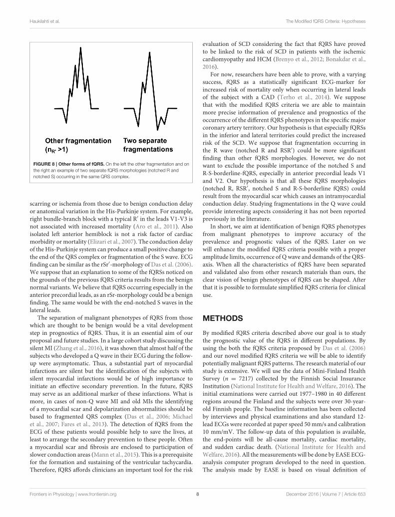
Other Forms of FragmentationThe fQRS will be considered as the other fragmentation if itdiffers from the fQRS morphologies described above. We areaimed at developing the modified fQRS criteria so that only
FIGURE 7 | The morphologies of the modified fQRS criteria from left to
right: (1) RSr′ (2) rSr′ (3) r′SR.
fQRSs which have more than one additional R waves (R′> 1) in
the same wave could be considered as the other fragmentation(Figure 8). When measuring the amplitude differences twopoints are tagged to measure the largest amplitude differenceof the fragmentation and the third point to measure the largeramplitude difference next to the largest change. However, thismorphology class includes also fragmented QRS complexeswhich include more than one fragmentation in separate wavesof the QRS complex (Figure 8). In these cases the both differentfQRS morphologies will be examined separately to ensure thatany potential fQRS morphology finding will not go unnoticed.
HYPOTHESIS
We expect that by using more detailed modified fQRS criteriait would be possible to separate fQRSs that are developed dueto intramyocardial conduction delay caused by the myocardial
Frontiers in Physiology | www.frontiersin.org 7 December 2016 | Volume 7 | Article 653
Haukilahti et al. The Modified fQRS Criteria: Hypotheses
FIGURE 8 | Other forms of fQRS. On the left the other fragmentation and on
the right an example of two separate fQRS morphologies (notched R and
notched S) occurring in the same QRS complex.
scarring or ischemia from those due to benign conduction delayor anatomical variation in the His-Purkinje system. For example,right bundle-branch block with a typical R′ in the leads V1-V3 isnot associated with increased mortality (Aro et al., 2011). Alsoisolated left anterior hemiblock is not a risk factor of cardiacmorbidity or mortality (Elizari et al., 2007). The conduction delayof the His-Purkinje system can produce a small positive change tothe end of the QRS complex or fragmentation of the S wave. ECGfinding can be similar as the rSr′-morphology of Das et al. (2006).We suppose that an explanation to some of the fQRSs noticed onthe grounds of the previous fQRS criteria results from the benignnormal variants. We believe that fQRS occurring especially in theanterior precordial leads, as an rSr-morphology could be a benignfinding. The same would be with the end-notched S waves in thelateral leads.
The separation of malignant phenotypes of fQRS from thosewhich are thought to be benign would be a vital developmentstep in prognostics of fQRS. Thus, it is an essential aim of ourproposal and future studies. In a large cohort study discussing thesilent MI (Zhang et al., 2016), it was shown that almost half of thesubjects who developed a Q wave in their ECG during the follow-up were asymptomatic. Thus, a substantial part of myocardialinfarctions are silent but the identification of the subjects withsilent myocardial infarctions would be of high importance toinitiate an effective secondary prevention. In the future, fQRSmay serve as an additional marker of these infarctions. What ismore, in cases of non-Q wave MI and old MIs the identifyingof a myocardial scar and depolarization abnormalities should bebased to fragmented QRS complex (Das et al., 2006; Michaelet al., 2007; Fares et al., 2013). The detection of fQRS from theECG of these patients would possible help to save the lives, atleast to arrange the secondary prevention to these people. Oftena myocardial scar and fibrosis are enclosed to participation ofslower conduction areas (Mann et al., 2015). This is a prerequisitefor the formation and sustaining of the ventricular tachycardia.Therefore, fQRS affords clinicians an important tool for the risk
evaluation of SCD considering the fact that fQRS have provedto be linked to the risk of SCD in patients with the ischemiccardiomyopathy and HCM (Brenyo et al., 2012; Bonakdar et al.,2016).
For now, researchers have been able to prove, with a varyingsuccess, fQRS as a statistically significant ECG-marker forincreased risk of mortality only when occurring in lateral leadsof the subject with a CAD (Terho et al., 2014). We supposethat with the modified fQRS criteria we are able to maintainmore precise information of prevalence and prognostics of theoccurrence of the different fQRS phenotypes in the specific majorcoronary artery territory. Our hypothesis is that especially fQRSsin the inferior and lateral territories could predict the increasedrisk of the SCD. We suppose that fragmentation occurring inthe R wave (notched R and RSR′) could be more significantfinding than other fQRS morphologies. However, we do notwant to exclude the possible importance of the notched S andR-S-borderline-fQRS, especially in anterior precordial leads V1and V2. Our hypothesis is that all these fQRS morphologies(notched R, RSR′, notched S and R-S-borderline fQRS) couldresult from the myocardial scar which causes an intramyocardialconduction delay. Studying fragmentations in the Q wave couldprovide interesting aspects considering it has not been reportedpreviously in the literature.
In short, we aim at identification of benign fQRS phenotypesfrom malignant phenotypes to improve accuracy of theprevalence and prognostic values of the fQRS. Later on wewill enhance the modified fQRS criteria possible with a properamplitude limits, occurrence of Q wave and demands of the QRS-axis. When all the characteristics of fQRS have been separatedand validated also from other research materials than ours, theclear vision of benign phenotypes of fQRS can be shaped. Afterthat it is possible to formulate simplified fQRS criteria for clinicaluse.
METHODS
By modified fQRS criteria described above our goal is to studythe prognostic value of the fQRS in different populations. Byusing the both the fQRS criteria proposed by Das et al. (2006)and our novel modified fQRS criteria we will be able to identifypotentially malignant fQRS patterns. The researchmaterial of ourstudy is extensive. We will use the data of Mini-Finland HealthSurvey (n = 7217) collected by the Finnish Social InsuranceInstitution (National Institute for Health andWelfare, 2016). Theinitial examinations were carried out 1977–1980 in 40 differentregions around the Finland and the subjects were over 30-year-old Finnish people. The baseline information has been collectedby interviews and physical examinations and also standard 12-lead ECGs were recorded at paper speed 50 mm/s and calibration10 mm/mV. The follow-up data of this population is available,the end-points will be all-cause mortality, cardiac mortality,and sudden cardiac death. (National Institute for Health andWelfare, 2016). All themeasurements will be done by EASE ECG-analysis computer program developed to the need in question.The analysis made by EASE is based on visual definition of
Frontiers in Physiology | www.frontiersin.org 8 December 2016 | Volume 7 | Article 653

Haukilahti et al. The Modified fQRS Criteria: Hypotheses
fragmentation amplitude and morphology. All the data will beanalyzed by Statistical Package for Social Studies 21.0 program(SPSS).
In addition, research permission to another research materialfrom the Institute of Health and Welfare, Health 2000 data(National Institute for Health and Welfare, 2009), is currentlyadmitted. Health 2000 data will increase the coverage of ourrandom general population. This study is very similar to Mini-Finland Health Survey. The initial examinations were carried out2000–2001 and the material contains over 7217 standard ECGsfrom over 30-year-old Finnish subjects. The baseline informationhas been collected by interviews, health information comes fromthe updated registers and ECGs are recorded in the physicalexaminations. ECGs are similar compared to Mini-Finland data.The endpoint of the follow-up will be the same as above.(National Public Health Institute, 2000).
In addition, we are going to extend our research materialto ARTEMIS and EU-CERT-ICD data. ARTEMIS data contains1950 CAD patients with type 2 diabetes (National Institutes ofHealth, 2016). This case-control study data is collected during2007–2014 in 2 and 5 year follow-ups and will help us to estimatethe prognostic values of the fQRS in patients with CAD. InEU-CERT-ICD data all the subjects fulfill the criteria for theICD placement and ICD have also been/will be implanted tosome of the subjects. This data contains both a retrospective
database (n = 2041) and the prospective material (EU-CERT-ICD, 2016). All the ECGs have been/will be recorded before ICD-implantation. The size of the prospective material is estimated tobe approximately 2250 ECGs.We plan to compare the prevalenceand prognostic values of fQRS in ARTEMIS and EU-CERT-ICDdata (total n = 6241) to those in Mini-Finland and Health 2000data (total n= 15,245). Later on the new fQRS criteria should alsobe validated by comparing the QRS fragmentation with cardiacimaging, e.g., magnetic resonance imaging with gadolinium lateenhancement.
AUTHOR CONTRIBUTIONS
MH wrote the article and developed the modified fQRS-criteria.AE participated in drafting the article and revising it critically andis also made substantial contributions to acquisition of data andanalysis and interpretation of data. TK has developed the ECGanalysis computer programme used analyzing the data and is alsoparticipated revising article. HH has, as the group leader, helpedin drafting the article, and revising it critically.
FUNDING
Funded in part by the grant no 602299 of FP 7 Health Programof EC (EU-CERT-ICD).
REFERENCES
Adar, A., Canyılmaz, E., Kiris, A., Ilter, A., Serdar, L., Memis, Y., et al. (2015).Radiotherapy induces development of fragmented QRS in patients with breastcancer. Breast Care 10, 4. doi: 10.1159/000431030
Ahn, M. S., Kim, J. B., Yoo, B. S., Lee, J. W., Lee, J. H., Youn, Y. J., et al. (2013).Fragmented QRS complexes are not hallmarks of myocardial injury as detectedby cardiac magnetic resonance imaging in patients with acute myocardialinfarction. Int. J. Cardiol. 168, 3. doi: 10.1016/j.ijcard.2012.12.086
Aro, A. L., Anttonen, O., Tikkanen, J. T., Junttila, M. J., Kerola, T., Rissanen,H. A., et al. (2011). Intraventricular conduction delay in a standard 12-leadelectrocardiogram as a predictor of mortality in the general population. Circ.Arrhythm. Electrophysiol. 4, 5. doi: 10.1161/CIRCEP.111.963561
Boineau, J. P., and Cox, J. L. (1973). Slow ventricular activation in acute myocardialinfarction. A source of re-entrant premature ventricular contractions.Circulation 48, 702–713. doi: 10.1161/01.CIR.48.4.702
Bonakdar, H., Moladoust, H., Kheirkhah, J., Abbaspour, E., Assadian Rad, M., et al.(2016). Significance of a fragmented QRS complex in patients with chronictotal occlusion of coronary artery without prior myocardial infarction. Anatol.J. Cardiol. 16, 2. doi: 10.5152/akd.2015.5887
Brenyo, A., Pietrasik, G., Barsheshet, A., Huang, D. T., Polonsky, B.,McNitt, S., et al. (2012). QRS fragmentation and the risk of suddencardiac death in MADIT II. J Cardiovasc. Electrophysiol. 23, 12.doi: 10.1111/j.1540-8167.2012.02390.x
Bulut, M., Deniz Acar, R., Ergün, S., Geçmen, Ç., and Akçakoyun, M.(2015). Cardiac rehabilitation improves the QRS fragmentation in patientswith ST elevation myocardial infarction. J. Cardiovasc. Thorac. Res. 7, 3.doi: 10.15171/jcvtr.2015.21
Celikyurt, U., Karauzum, K., Sahin, T., Agacdiken, A., Vural, A., and Ural, D.(2015). Association between resolution of fragmented QRS and response tocardiac resynchronization therapy. Ann. Noninvasive Electrocardiol. 20, 2.doi: 10.1111/anec.12186
Das, M. K., and ElMasry, H. (2010). Fragmented, Q. R. S., and other depolarizationabnormalities as a predictor of mortality and sudden cardiac death. Curr. Opin.Cardiol. 25, 1. doi: 10.1097/HCO.0b013e328333d35d
Das, M. K., Khan, B., Jacob, S., Kumar, A., andMahenthiran, J. (2006). Significanceof a fragmented Q8RS complex versus a Qwave in patients with coronary arterydisease. Circulation 113, 21. doi: 10.1161/CIRCULATIONAHA.105.595892
Das, M. K., Maskoun, W., Shen, C., Michael, M. A., Suradi, H., Desai, M., et al.(2010). FragmentedQRS on twelve-lead electrocardiogram predicts arrhythmicevents in patients with ischemic and nonischemic cardiomyopathy. HeartRhythm 7, 1. doi: 10.1016/j.hrthm.2009.09.065
Das, M. K., Michael, M. A., Suradi, H., Peng, J., Sinha, A., Shen, C., et al.(2009). Usefulness of fragmented QRS on a 12-lead electrocardiogram inacute coronary syndrome for predicting mortality. Am. J. Cardiol. 104, 12.doi: 10.1016/j.amjcard.2009.07.046
Das, M. K., Saha, C., El Masry, H., Peng, J., Dandamudi, G., Mahenthiran, J., et al.(2007). Fragmented QRS on a 12-lead ECG: a predictor of mortality and cardiacevents in patients with coronary artery disease. Heart Rhythm 4, 1385–1392.doi: 10.1016/j.hrthm.2007.06.024
Debonnaire, P., Katsanos, S., Joyce, E., Van Den Brink, O. V. W., Atsma, D.E., Schalij, M.e t al. (2015). QRS fragmentation and QTc duration relateto malignant ventricular tachyarrhythmias and sudden cardiac death inpatients with hypertrophic cardiomyopathy. J. Cardiovasc. Electrophysiol. 26,5. doi: 10.1111/jce.12629
EU-CERT-ICD (2016). EU-CERT-ICD Website. Available online at:http://www.eu-cert-icd.eu/
Elizari, M. V., Acunzo, R. S., and Ferreiro, M. (2007). Hemiblocks revisited.Circulation 115, 9. doi: 10.1161/CIRCULATIONAHA.106.637389
Fares, H., Heist, K., Lavie, C. J., Kumbala, D., Ventura, H., Meadows, R.,et al. (2013). Fragmented QRS Complexes—a novel but underutilizedelectrocardiograhic marker of Heart Disease. Crit. Pathw. Cardiol. 12, 4.doi: 10.1097/HPC.0b013e31829e005d
Gong, B., and Li, Z. (2016). Total mortality, major adverse cardiac events, andechocardiographic-derived cardiac parameters with fragmented QRS complex.Ann. Noninvasive Electrocardiol. 21, 4. doi: 10.1111/anec.12325
Güngör, B., Özcan, K. S., Karatas, M. B., Sahin, I., Öztürk, R., and Bolca,O. (2016). Prognostic value of QRS fragmentation in patients with acutemyocardial infarction: a meta-analysis. Ann. Noninvasive Electrocardiol. 6,604–612. doi: 10.1111/anec.12357
Frontiers in Physiology | www.frontiersin.org 9 December 2016 | Volume 7 | Article 653

Haukilahti et al. The Modified fQRS Criteria: Hypotheses
Homsi, M., Alsayed, L., Safadi, B., Mahenthiran, J., and Das, M. K. (2009).Fragmented QRS complexes on 12-lead ECG: a marker of cardiac sarcoidosis asdetected by gadolinium cardiac magnetic resonance imaging.Ann. NoninvasiveElectrocardiol. 14, 4. doi: 10.1111/j.1542-474X.2009.00320.x
Jain, R., Singh, R., Yamini, S., and Das, M. K. (2014). Fragmented ECG asa risk marker in cardiovascular diseases. Curr. Cardiol. Rev. 10, 277–286.doi: 10.2174/1573403X10666140514103451
Konno, T., Hayashi, K., Fujino, N., Oka, R., Nomura, A., Nagata, Y., et al.(2015). Electrocardiographic QRS fragmentation as a marker for myocardialfibrosis in hypertrophic cardiomyopathy. J. Cardiovasc. Electrophysiol. 26, 10.doi: 10.1111/jce.12742
Lorgis, L., Cochet, A., Chevallier, O., Angue, M., Gudjoncik, A., Lalande, A., et al.(2014). Relationship between fragmented QRS and no-reflow, infarct size, andperi-infarct zone assessed using cardiac magnetic resonance in patients withmyocardial infarction. Can. J. Cardiol. 30, 2. doi: 10.1016/j.cjca.2013.11.026
Ma, X., Duan,W., Poudel, P., Ma, J., and Sharma, D. X. Y. (2016). Fragmented QRScomplexes have predictive value of imperfect ST-segment resolution in patientswith STEMI after primary percutaneous coronary intervention. Am. J. Emerg.
Med. 34, 3. doi: 10.1016/j.ajem.2015.11.010MacAlpin, R. N. (2010). The fragmented QRS: does it really indicate a ventricular
abnormality? J. Cardiovasc. Med. 11, 11. doi: 10.2459/JCM.0b013e32833b9816Macfarlane, P. W., Antzelevitch, C., Haissaguerre, M., Huikuri, H. V., Potse, M.,
Rosso, R., et al. (2015). The early repolarization pattern: a consensus paper. J.Am. Coll. Cardiol. 66, 4. doi: 10.1016/j.jacc.2015.05.033
Maheshwari, S., Acharyya, A., Puddu, P. E., Mazomenos, E. B., Leekha, G.,Maharatna, K., et al. (2013). An automated algorithm for online detectionof fragmented QRS and identification of its various morphologies. J. R. Soc.Interface 10, 89. doi: 10.1098/rsif.2013.0761m
Malik, M. (2013). Electrocardiographic smoke signals of fragmentedQRS complex.J. Cardiovasc. Electrophysiol. 24, 11. doi: 10.1111/jce.12226
Mann, D. L., Zipes, D. P., Libby, P., and and, O., Bonow, R. (2015). “Arrhythmias,sudden death, and syncope,” in Braunwald’s Heart Disease: A Textbook
of Cardiovascular Medicine, 10th Edn., ed D. L. Mann (Philadelphia, PA:Saunders), 5.
Michael, M. A., El Masry, H., Khan, B. R., and Das, M. K. (2007).Electrocardiographic signs of remote myocardial infarction. Prog. Cardiovasc.Dis. 50, 198–208. doi: 10.1016/j.pcad.2007.05.003
Morita, H., Kusano, K. F., Miura, D., Nagase, S., Nakamura, K., Morita, S.T., et al. (2008). Fragmented QRS as a marker of conduction abnormalityand a predictor of prognosis of Brugada syndrome. Circulation 118, 17.doi: 10.1161/CIRCULATIONAHA.108.770917
National Institutes of Health, P. (2016). Prognosis of Type 2 Diabetic
Patients (ARTEMIS). Available online at: https://clinicaltrials.gov/ct2/show/NCT01426685?term=NCT01426685&rank=1
National Public Health Institute, P. (2000). The Report of the Methods. The
execution, data andmethods of the Health 2000 study. Available online at: http://www.julkari.fi/bitstream/handle/10024/78181/2005b6.pdf?sequence,=1
National Institute for Health and Welfare (2009). Health 2000.Available online at:http://www.terveys2000.fi/indexe.html
National Institute for Health and Welfare (2016). Mini-Finland Health
Survey. Available online at: https://www.thl.fi/en/web/thlfi-en/research-and-expertwork/population-studies/finnish-mobile-clinic/mini-finland-health-survey
Sha, J., Zhang, S., Tang, M., Chen, K., Zhao, X., and Wang, F. (2011). FragmentedQRS is associated with all-cause mortality and ventricular arrhythmiasin patient with idiopathic dilated cardiomyopathy. Ann. Noninvasive
Electrocardiol. 16, 3. doi: 10.1111/j.1542-474X.2011.00442.xTerho, H. K., Tikkanen, J. T., Junttila, J. M., Anttonen, O., Kenttä, T.
V., Aro, A. L., et al. (2014). Prevalence and prognostic significance offragmented QRS complex in middle-aged subjects with and without clinicalor electrocardiographic evidence of cardiac disease. Am. J. Cardiol. 114, 1.doi: 10.1016/j.amjcard.2014.03.066
Torigoe, K., Tamura, A., Kawano, Y., Shinozaki, K., Kotoku, M., and Kadota, J.(2012). The number of leads with fragmented QRS is independently associatedwith cardiac death or hospitalization for heart failure in patients with priormyocardial infarction. J. Cardiol. 59, 1. doi: 10.1016/j.jjcc.2011.09.003
Wang, D. D., Buerkel, D. M., Corbett, J. R., and Gurm, H. S. (2010).Fragmented QRS complex has poor sensitivity in detecting myocardialscar. Ann. Noninvasive Electrocardiol. 15, 4. doi: 10.1111/j.1542-474X.2010.00385.x
Wang, D. D., Tibrewala, A., Nguygen, P., Swadia, T., Jacobsen, G., Khan,A., et al. (2014). Fragmented QRS on surface electrocardiogram isnot a reliable predictor of myocardial scar, angiographic coronarydisease or long term adverse outcomes. Cardiovasc. Diagn. Ther. 4, 4.doi: 10.3978/j.issn.2223-3652.2014.08.03
Yan, G. H., Wang, M., Yiu, K. H., Lau, C. P., Zhi, G., Lee, S. W., et al. (2012).Subclinical left ventricular dysfunction revealed by circumferential 2D strainimaging in patients with coronary artery disease and fragmented QRS complex.Heart Rhythm 9, 6. doi: 10.1016/j.hrthm.2012.01.007
Zhang, Z.-M., Rautaharju, P. M., Prineas, R. J., Rodriguez, C. J., Loehr,L., Rosamond, W. D., et al. (2016). Race and sex differences in theincidence and prognostic significance of silent myocardial infarction in theAtherosclerosis Risk in Communities (ARIC) Study. Circulation 133, 22.doi: 10.1161/CIRCULATIONAHA.115.021177
Zipes, D. P., and Das, M. K. (2009). Fragmented QRS: a predictor of mortalityand sudden cardiac death. Heart Rhythm 6, 3. doi: 10.1016/j.hrthm.2008.10.019
Conflict of Interest Statement: The authors declare that the research wasconducted in the absence of any commercial or financial relationships that couldbe construed as a potential conflict of interest.
Copyright © 2016 Haukilahti, Eranti, Kenttä and Huikuri. This is an open-access
article distributed under the terms of the Creative Commons Attribution License (CC
BY). The use, distribution or reproduction in other forums is permitted, provided the
original author(s) or licensor are credited and that the original publication in this
journal is cited, in accordance with accepted academic practice. No use, distribution
or reproduction is permitted which does not comply with these terms.
Frontiers in Physiology | www.frontiersin.org 10 December 2016 | Volume 7 | Article 653


