Embed Size (px)
Citation preview
Q-SWITCHED Nd:YAG LASER INDUCED PHOTODISRUPTION IN
AN EYE MODEL
WAN RASHIDAH BINTI WAN MAJID
A thesis submitted in fulfilment of the
requirements for the award of the degree of
Master of Science (Physics)
Faculty of Science
Universiti Teknologi Malaysia
JANUARY 2010

iii
Dedicated to:
My Parent: Wan Majid Wan Idris & Hasimah Awang,
Siblings: Redhuan, Rodzli, Rodhiah, Ridzaudin, Robiatul Adawiyah
Husband and son: Abd Rahman Tamuri and Abdullah Uwais Abd Rahman
Thanks for the endless love, advices and supports

iv
ACKNOWLEDGEMENTS
In the name of Allah, Most Gracious, Most Merciful
Alhamdulillah, all the praise to Allah the Almighty, for giving me the
strength, patience and time for completing this study.
A million of thanks to my supervisors; Prof. Dr.Noriah Bidin and Dr. Jasman
Zainal for their help and guidance throughout the work. Their encouragements really
help me to stand up and continue the research to the end.
A special thanks also go to the lab assistants, En. Ab. Rasid Isnin and Pn.
Ruzilah Omar for helping me in preparing for the experimental work and also in
finishing my writing process. To all my labmates, especially Aishah, Nazariah, Aizi
and Fairuz, thanks for the helps during the hard and joyful times in the laser
technology lab.
Last but not least, thanks to Universiti Teknologi Malaysia (UTM) and
MOSTI for the scholarship award and financial help which is really supporting me in
the 2 years of study in UTM.

v
ABSTRACT
This study attempts to characterize the photodisruption in simulated eye model induced by Nd:YAG laser. To simulate the eye environment, saline solution was chosen as vitreous filled pyrex cuvette which acted like eye ball. Polymethylmethacrylate (PMMA) plate later on was placed in the pyrex cuvette to be as an intraocular lens. The laser beam was focused into saline solution using two techniques. The first technique uses single camera lens and the second technique uses combination of negative and positive lenses. Activities at the focal region were visualized by means of CCD video camera and interfaced with image processing system via Matrox Inspector sofware. The pressure wave induced at the focal region was detected using hydrophone and the plasma temperature was measured and estimated using Langmuir probe. The damage induced after exposure of laser on PMMA was observed using optical microscope. By focusing light pulses lasting in nanoseconds to a spot size, this laser can create an optical breakdown associated with plasma formation. Multiple breakdowns were observed when the laser was focused using single lens. A single ellipsoidal plasma configuration was generated with a combination of lenses. A series of acoustic-shockwave signals representing the pressure waves produced at the focal region had also been recorded. From the measurement, a maximum pressure of 0.0254 bar was obtained. The temperature raised at the plasma region was estimated to be 12,064 K or 1.04 eV. The damage threshold was obtained at a fluence of 6.86 x 10² Jcm-2 on the PMMA with various damage formations. Severe damage was observed as the number of laser pulses increases. In short, all the mechanisms involved have been successfully characterized. These information can be very useful in recognizing the opportunities and limitations of the Nd:YAG laser in medical applications.

vi
ABSTRAK
Kajian ini bertujuan untuk mencirikan fotopenghancuran dalam sampel simulasi mata yang dijana oleh laser Nd:YAG. Untuk menyediakan sampel mata, larutan garam dipilih untuk menggantikan cecair dalam mata yang diisi di dalam bekas pyrex yang bertindak sebagai bebola mata. Kepingan perspeks (PMMA) kemudiannya diletakkan sebagai kanta intraokular. Alur laser difokuskan ke dalam larutan garam dengan menggunakan dua teknik. Teknik pertama adalah menggunakan satu kanta kamera dan yang kedua menggunakan kombinasi kanta negatif dan kanta positif. Aktiviti pada kawasan pemfokusan diperhatikan menggunakan kamera video CCD yang diantaramuka dengan sistem pemprosesan imej melalui perisisian Matrox Inspector. Gelombang tekanan yang dijana pada kawasan pemfokusan dikesan menggunakan hidrofon dan suhu plasma diukur dan dianggarkan menggunakan penduga Langmuir. Kerosakan yang dijana selepas dedahan laser ke atas PMMA diperhatikan menggunakan mikroskop optik. Dengan memfokuskan denyut cahaya nanosaat kepada satu saiz titik, laser ini boleh menghasilkan keruntuhan optik diikuti dengan pembentukan plasma. Keruntuhan berganda dapat diperhatikan semasa laser difokuskan menggunakan satu kanta. Satu plasma berbentuk elipsoid dijana dengan kombinasi kanta. Beberapa siri isyarat gelombang akustik-kejutan yang mewakili tekanan gelombang yang dijana pada kawasan pemfokusan juga dirakamkan. Berdasarkan pengukuran, tekanan maksimum sebanyak 0.0254 bar diperolehi. Peningkatan suhu pada kawasan plasma dianggarkan sebanyak 12,064 K atau 1.04 eV. Kerosakan ambang berlaku pada 6.86 x 10² Jcm-2 di atas permukaan PMMA dengan beberapa bentuk kerosakan. Kerosakan yang berlaku didapati meningkat dengan peningkatan kuantiti denyut laser yang digunakan. Secara ringkas, semua mekanisma ini telah berjaya dicirikan. Semua maklumat ini boleh menjadi sangat berguna dalam mengenalpasti peluang dan had dalam mengaplikasikan laser Nd:YAG dalam perubatan.

vii
TABLE OF CONTENTS
CHAPTER TITLE PAGE
Title page
Declaration of originality
Dedication
Acknowledgements
Abstract
Abstrak
Table of Contents
List of Tables
List of Figures
List of Symbols
List of Appendices
i
ii
iii
iv
v
vi
vii
x
xi
xvi
xviii
1 INTRODUCTION 1
1.1 Overview
1.2 Problem Statement
1.3 Research Objective
1.4 Research Scope
1.5 Thesis Outline
1
2
3
4
4
2 THEORY 6
2.1 Introduction
2.2 Laser Beam Focusing
2.3 Photodisruption
6
7
9

viii
2.3.1 Optical Breakdown
2.3.2 Plasma
2.3.2.1 Plasma Formation
2.3.2.2 Plasma Temperature
2.3.3 Acoustic Shockwave Generation
2.4 Laser Interaction with Transparent Material
2.5 Conclusion
11
14
14
15
18
20
22
3 METHODOLOGY 23
3.1 Introduction
3.2 Samples
3.2.1 Saline Solution
3.2.2 Polymethylmethacrylate (PMMA)
3.3 Nd:YAG Laser System
3.3.1 Pockels Cell
3.3.2 External Triggering Circuit
3.4 Measurement Equipment
3.4.1 Power Meter
3.4.2 Photodetector
3.4.3 Langmuir Probe
3.4.4 Pressure Sensor
3.5 Imaging Equipment
3.6 Image Calibration
3.7 Experimental Setup
3.7.1 Observation of Plasma Formation
3.7.2 Plasma Temperature Measurement
3.7.3 Detection of Pressure Waves
3.7.4 Photodisruption Effects on PMMA
23
24
24
25
25
27
28
30
30
31
31
33
33
36
37
37
39
40
41
4 PLASMA FORMATION 43
4.1 Introduction
4.2 Plasma Formation Induced by Single Lens Technique
4.3 Plasma Formation Induced by Combination Lenses
43
44
48
ix
Technique
4.4 Measurement of the Plasma Length
50
5 PLASMA TEMPERATURE 54
5.1 Introduction
5.2 Plasma Temperature
54
55
6 GENERATION OF PRESSURE WAVES 62
6.1 Introduction
6.2 Pressure Measurement
6.3 Pressure Profile
62
63
67
7 PHOTODISRUPTION EFFECTS ON PMMA 70
7.1 Introduction
7.2 Photodisruption Effects
70
71
8 CONCLUSION 79
8.1 Introduction
8.2 Conclusion
8.3 Recommendations
79
80
81
REFERENCES
Appendices A - G
83
89 - 98

x
LIST OF TABLES
TABLE NO.
TITLE
PAGE
3.1 Values of laser beam parameters for different focusing
techniques.
38
4.1 Plasma length measured for both techniques. 52
5.1 Data obtained from the Langmuir probe signal detected
by oscilloscope.
59
6.1 Amplitude of the signals detected for different oscillator
voltages.
67
6.2 Pressure amplitude as a function of laser energy at
various distances.
67
7.1 Damaged area measured for different laser energy for 1,
5 and 10 pulses.
77
7.2 Damaged area measured for various number of laser
pulses.
77
xi
LIST OF FIGURES
FIGURE NO.
TITLE
PAGE
2.1 The depth of focus of the laser light [11].
7
2.2 Beam diameter of a Gaussian beam as fundamental mode
TEM00 and function of z [11].
8
2.3 Mechanism of photodisruption induced by Q-switched
Nd:YAG laser [21].
11
2.4 (a) Initiation, (b) electron avalanche growth and (c) plasma
formation by optical breakdown. The dominant mechanism
of initiation of ionization by a Q-switched pulse is
thermionic emission [21].
13
2.5 Current-voltage (I-V) characteristic curve of plasma [40].
16
2.6 Schematic diagram of breakdown due to Q-switched laser
pulse in PMMA. f denotes the position of the focus [57].
22
3.1 Samples used in the experiment:
(a) Saline solution
(b) PMMA
24

xii
3.2 Photograph of HY200 Nd:YAG laser.
26
3.3 HY200 Nd:YAG laser component layout [61].
26
3.4 Simplified four level system for solid-state Nd:YAG
laser [11].
27
3.5 Schematic diagram of the external trigger circuit.
29
3.6 Output pulse of the external trigger circuit.
29
3.7 Time delay between the external trigger and the laser.
30
3.8 The Langmuir probe
32
3.9 The Langmuir probe and its detection circuit:
(a) The detection circuit of the Langmuir probe
(b) Schematic diagram of Langmuir probe detection
circuit [68].
32
3.10 The voltage mode pressure sensor used to detect the
pressure waves signals.
33
3.11 Photographs of imaging equipments:
(a) CCD Camera
(b) Photomicroscope
34
3.12 Interface of the Matrox Inspector software.
35
3.13 VideoTest 5.0 software used to analyze the laser beam on
burn paper.
35
3.14 Image of wire taken using CCD camera. 36

xiii
3.15 Single lens focusing technique.
37
3.16 Combination of two lenses to focus the laser beam.
37
3.17 Experimental setup to study the generation of plasma in
saline using combination of two lenses.
39
3.18 Schematic diagram of experimental setup
40
3.19 Experimental arrangement for pressure wave detection
41
3.20 Schematic diagram of experimental setup to study the
damage on PMMA.
42
4.1 Plasma produced when single lens technique used.
Magnification of 6x. The direction of laser beam is
toward the right.
46
4.2 Growth of plasma anterior to the predicted focal point
[21]:
(a) a threshold pulse with spherical breakdown at the
beam waist;
(b) a greatly suprathreshold pulse attains breakdown
threshold anterior to the minimal spot size
(c) a moderately suprathreshold pulse extends toward the
laser source in a multilobed configuration
47
4.3 Multiple breakdown due to longer focal region [56].
47
4.4 Plasma formed in saline solution. Magnification factor is
8x. The laser is incident from the left.
49
xiv
4.5 Observation of plasma using different focusing
techniques:
(a) Single lens focusing technique
(b) Combination lenses focusing technique
51
4.6 The distribution of plasma beam along the x-axis [11]:
(a) Gaussian beam profile
(b) Plasma configuration
52
4.7 Plasma length with respect to laser energy.
53
5.1 Typical signals collected by Langmuir probe as a
function of positive bias voltage.
56
5.2 Typical signals collected by Langmuir probe as a
function of negative bias voltage.
57
5.3 I-V characteristic curve of Langmuir probe.
60
5.4 Linear part of the I-V characteristic curve.
61
6.1 Typical acoustic shockwave signal detected at different
voltage at a distance of 1.87 mm.
64
6.2 Typical acoustic shockwave signal detected at different
voltage at a distance of 2.56 mm.
65
6.3 Typical acoustic shockwave signal detected at different
voltage at a distance of 5.76 mm.
66
6.4 Acoustic shockwave pressure as a function of laser
energy at three different distances.
69

xv
6.5 Acoustic shockwave pressure plotted against various
distances.
69
7.1 Damage induced by a single laser pulse on PMMA
(Magnification of 10x).
72
7.2 Damage induced by 5 pulses of Q-Switched laser on
PMMA (Magnification of 10x).
73
7.3 Effects on PMMA which has been exposed to 10 pulses
of Q-switched Nd:YAG laser (Magnification of 10x).
74
7.4 Target irradiated at different number of pulses at laser
energy of 93.0 mJ. (Magnification of 10x).
75
7.5 Damaged area as a function of laser energy for different
number of pulses.
78
7.6 Damaged area versus number of laser pulses taken at
laser energy of 93.0 mJ.
78

xvi
LIST OF SYMBOLS
a - Radius of the aperture
Cp - Specific heat
d,D - Distance
E - Laser energy
Ea - Absorbed laser energy
Eo - Electric field strength
f - Focal length
I - Current
Is - Electron saturation current
L - Lens
M - Magnification factor
ne - Electron density
P - Pressure
Pd - Power density
Rb - Radius of the optical beam
RL - Resistor
Rt - Acoustic source radius
r - Radius of the beam spot
Te - Electron temperature
V - Voltage amplitude
V - Optical absorbed volume
Vf - Floating potential
Vs - Plasma potential
Vpp - Probe potential
W - Laser power

xvii
w - Beam radius
w0 - Beam waist
z - Depth of focus
z0 - Focal point
zR - Rayleigh region
- Absorption coefficient of the liquid
â - Thermal expansion coefficient
∆T - Temperature rise
- Wavelength
eff - Penetration coefficient
v - Speed of sound
- Density of the liquid
xviii
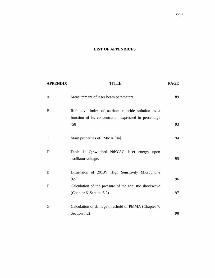
LIST OF APPENDICES
APPENDIX
TITLE PAGE
A Measurement of laser beam parameters
89
B Refractive index of natrium chloride solution as a
function of its concentration expressed in percentage
[58].
93
C Main properties of PMMA [84].
94
D Table 1: Q-switched Nd:YAG laser energy upon
oscillator voltage.
95
E Dimension of 2013V High Sensitivity Microphone
[65].
96
F Calculation of the pressure of the acoustic shockwave
(Chapter 6, Section 6.2)
97
G Calculation of damage threshold of PMMA (Chapter 7,
Section 7.2)
98

CHAPTER 1
INTRODUCTION
1.1 Overview
The remarkable properties of laser radiation make it such a useful tool to be
applied for medical applications. The laser beam can be controlled, focused and
manipulated to give precise, specific and localized effects in tissues [1]. The
applications of lasers and other optical technology in biomedicine is a rapidly
growing field. These applications can be classified as diagnostic or therapeutic. In a
diagnostic application, the goal is to learn something about the physiology or
pathology of the tissue through its interaction with light. On the other hand, for
therapeutic use, it is involved with permanent modification of tissue. This can range
from simple cutting associated with surgery to the initiation of cytotoxic chemical
reactions in photodynamic therapy [2].
The most widespread medical application for laser technology in medicine
has occurred in ophthalmology. Ophthalmic laser applications have experienced
rapid growth with the use of argon, krypton, argon pumped dye, Nd:YAG and most
recently, near-IR diode lasers [3] since the introduction of ruby laser in 1960s.

2
In 1961, Zaret [4] employed a ruby laser for iris and retinal photocoagulation
in rabbits. Delivery systems for retinal photocoagulation employing ruby laser had
been developed by Campbell and Koester as well as Zweng and his associates in
1963 [5, 6]. The ruby laser was a valuable tool, but it is quickly supplanted with the
introduction of the argon laser photocoagulator. It was because the output of the
argon laser was a steady continuous wave instead of a short pulse and it could be
moved by existing fiber optic technology into slit lamp. The argon laser is the most
widely used to treat extrafoveal chorioretinal diseases such as age-related macular
degeneration and diabetic retinophathy, and also been successfully used to treat
glaucoma by iridectomy or trabeculoplasty [7].
Ophthalmology offers wide application of lasers since eye is one of the most
accessible human organs, and its media (cornea, aqueous humor, lens and vitreous)
are transparent to visible light, allowing direct inspection of its internal structures for
diagnosis and treatment [3].
1.2 Problem Statement
Photodisruptor laser applications are very useful for cutting, incising or
vaporizing intraocular tissue [8]. When laser is deposited on a tissue as thermal
energy, there are several mechanisms that may occur such as optical breakdown
associated with plasma and acoustic-shockwave generation. Effects generated by this
laser-tissue interaction depend on the target material (gas, liquid or solid). Biological
tissues are more complex and variable.
In this study, saline solution and polymethylmethacrylate (PMMA) are used
to simulate the eye condition. Some experimental work has been setup to observe the
photodisruption mechanism induced by Q-switched Nd:YAG laser. The mechanism
is studied based on laser parameters (energy, number of pulses and distance of
observation). It is very crucial to study plasma formation and acoustic-shockwave
3
generation as they are the main processes of the photodisruption. The investigation
on damages induced by photodisruption on the target is crucial as it can be very
useful or can be a very destructive. These observations are required to ensure a safety
use of laser as a photodisruptor in ophthalmology.
Therefore, the characterization of the photodisruption induced by Q-switched
Nd:YAG laser would provide some useful information on how the mechanism of
photodisruption depends on the laser parameters. This information also can be very
useful indications for clinician and for the system designer to recognize the
opportunities and limitations of lasers in applying these devices in medicines.
1.3 Research Objective
The main objective of the research is to characterize the mechanism of
photodisruption induced by Q-Switched Nd:YAG laser. This goal can be achieved as
the following:
a) Observation of plasma formation in saline water
b) Measurement of plasma temperature using Langmuir probe
c) Measurement of acoustic-shockwave generation in saline water using
piezoelectric transducer
d) Investigation of photodisruption effects on transparent material
(PMMA) using image analysis.
4
1.4 Research Scope
In this study, a Q-switched Nd:YAG laser with a fundamental wavelength of
1064 nm and 10 ns pulse duration has been employed as a source to generate
photodisruption. The laser beam has been focused using two focusing techniques.
One is a single lens technique and the other is combination of two lenses technique.
The plasma formation and the generation of acoustic-shockwave were being studied
in saline solution. PMMA was utilized as a target material to observe the effects of
photodisruption. The dynamic expansion of plasma was observed using CCD camera
which was interfaced to a personal computer. The plasma temperature was measured
using Langmuir probe. Pressure generated by acoustic-shockwave was detected
using piezoelectric transducer which was linked to an oscilloscope. The effects of
photodisruption mechanism were then observed using photomicroscope and analyzed
using image processing software.
1.5 Thesis Outline
This thesis is divided into eight chapters. Chapter 1 describes the general
overview of the research project. The history of laser use in medicine and laser as a
photodisruptor are also reviewed. The theory of photodisruption mechanism induced
by Q-switched laser will be detailed in Chapter 2. The discussions will include
optical focusing technique and laser induced damage on transparent material. The
samples, instruments and the experimental setup used to study the photodisruption
are presented in Chapter 3. The results and findings of this project are being
discussed in Chapter 4 to Chapter 7. The plasma formation and plasma temperature
measurement are discussed in Chapter 4 and Chapter 5, respectively while acoustic-
shockwave generation is described in Chapter 6. In Chapter 7, damage effects
produced by the photodisruption mechanisms on transparent material are discussed.

5
Finally, Chapter 8 comprises the conclusion of the study and recommendations for
future work.