Embed Size (px)
Citation preview

Document for use in Europe, Middle-East and Africa only.This surgical technique is not for use on French territory.
S U RG I C A L T EC H N I Q U E
EN
Integra®
PyroCarbon & Silicone MCP Total Joint

2 Surgical technique PyroCarbon and silicone MCP Total JointDocument for use in Europe, Middle-East and Africa only.
ONE instrumentation for TWO solutions: per operative choice
Silicone MCP total jointPyrocarbone MCP total joint

3Surgical technique PyroCarbon and silicone MCP Total JointDocument for use in Europe, Middle-East and Africa only.
Table of Contents
IntroductionIndications ................................................................................................................................................................................................................ 2Contraindications .................................................................................................................................................................................................... 2Warnings and Precautions ..................................................................................................................................................................................... 2System Overview ..................................................................................................................................................................................................... 3Preoperative Assessment ....................................................................................................................................................................................... 3
Surgical TechniqueStep 1: Initial Incision and Joint Exposure ........................................................................................................................................................... 4Step 2: Opening the Metacarpal Medullary Canal ............................................................................................................................................. 4Step 3: Establishing Metacarpal Medullary Canal Alignment ........................................................................................................................... 4Step 4: Metacarpal Osteotomy .............................................................................................................................................................................. 5Step 5: Opening the Phalangeal Medullary Canal .............................................................................................................................................. 5Step 6: Establishing Phalangeal Medullary Canal Alignment ........................................................................................................................... 6Step 7: Phalangeal Osteotomy ............................................................................................................................................................................... 6Step 8: Distal Component Broaching ................................................................................................................................................................... 7Step 9: Proximal Component Broaching .............................................................................................................................................................. 7Step 10: Trial Insertion and Reduction (and Collateral Ligament Suture) ........................................................................................................ 8Step 11: Implantation of Components ..................................................................................................................................................................9Step 12: Final Reduction and Soft Tissue Closure ...............................................................................................................................................9Step 13: Closure ...................................................................................................................................................................................................... 10Postoperative Therapy Protocol – Osteoarthritis and Trauma Patients ......................................................................................................... 10Postoperative Therapy Protocol – Rheumatoid Patients .................................................................................................................................. 11
Instrumentation ...................................................................................................................................................................................................... 13Ordering Information, Implant Dimensions and Part Numbers ......................................................................................................................14

4 Surgical technique PyroCarbon and silicone MCP Total JointDocument for use in Europe, Middle-East and Africa only.
IndicationsThe PyroCarbon MCP is indicated for use as a total joint replacement of index, long, ring, and small finger metacarpophalangeal (MCP) joints that exhibit symptoms of pain, limited motion, or inadequate bony alignment (i.e., subluxation/dislocation) secondary to articular destruction or degenerative disease related to rheumatoid arthritis, systemic lupus erythematosus, osteoarthritis, or post-traumatic arthritis where soft tissue reconstruction can provide adequate stabilization.
Contraindications
IndicationsThe Ascension® Silicone MCP Finger Joint Prosthesis is intended for cementless replacement of the metacarpophalangeal (MCP) joint, where disabled by rheumotoid, degenerative, or traumatic arthritis.
Pyrocarbone MCP total joint Silicone MCP total joint
• Inadequate bone stock at the implantation site• Active infection in the MCP joint• Nonfunctioning and irreparable MCP musculotendinous
system• Physical interference with or by other prostheses during
implantation or use• Procedures requiring modification of the prosthesis• Skin, bone, circulatory and/or neurological deficiency at the
implantation site
See package insert for full prescribing information.
ContraindicationsActive local or systemic infection ; Destruction of the metacarpal, phalanx, or phalanges or poor bone quality which prevents adequate fixation of the implant ; Loss of musculature, neuromuscular compromise, or vascular deficiency in the affected fingers ; Growing patients with open epiphyses ; Patient with high activity levels ; and patients unwilling or unable to comply with physician's instructions..
WarningsDo not modify the PyroCarbon MCP implant in any manner. Reshaping the implant using cutters, grinders, burrs, or other means will damage the structural integrity of the device and could result in implant fracture and/or particulate debris.Do not mismatch proximal and distal component sizes. For example, a Size 10 proximal component should be matched with only a Size 10 distal component. The wear behavior of mismatched proximal and distal component size combinations has not been evaluated, and is unknown.
Do not grasp the PyroCarbon MCP implant with metal instruments, or instruments with teeth, serrations, or sharp edges. Implants should be handled only with instrumentation provided by Integra. PyroCarbon MCP implants are made of pyrocarbon, which is a ceramic-like material. Mishandling implants could cause surface damage and reduce their strength, and could result in implant fracture and/or particulate debris.Do not use PyroCarbon MCP components in combination with proximal and distal components from other products. The wear behavior of PyroCarbon MCP components against proximal and distal component from other products has not been evaluated, and could damage the structural integrity of the device and result in implant fracture and/or particulate debris.

5Surgical technique PyroCarbon and silicone MCP Total JointDocument for use in Europe, Middle-East and Africa only.
Welcome into the pyrocarbon users group !Before using this product and in addition to all the precautions listed in this document, you have to know:Uniform line doesn’t mean loosening or failure: it’s the radio opaque pyrocarbon property.
0.005
0.004
0.003
0.002
0.001
0.000
0 2 4 6 8 10
Cycles (in Millions)
PyroCarbon on PyroCarbonCoCr on UHMWPE
Wea
r Dep
th (i
n in
ches
)
Graphite Substrate(radiographic)
0.5mm Pure Carbon Coating (radiolucent)
Lucent spaces are normal
Lucent spaces are normal
Never touch Pyc with metal.
Properties of this material:
Cardiac Valve1
• 40+ years use cardiac valve
• 3+ million valves
• 20+ million patient years
MCP Joint2
• 30+ years clinical experience
• No detectable wear
• Low friction bearing
• Cement free fixation
Pyrocarbon is Durable3
Pycmetal
1. Haubold AD, On the durability of pyrolytic carbon in vivo, Medical Progress through Technology 20:201-208, 1994
2. Cook SD, Beckenbaugh RD, Redondo J, Popich LS, Klawitter JJ, Linscheid RL, “Long term follow-up of pyrolytic carbon metacarpophalangeal implants,”
J Bone and Joint Surg, Vol. 81-A, No. 5, 635-648, 1999.
3. Test Report: TR-0008-01-A PyroCarbon Wear. Ascension Orthopedics, 1999.

6 Surgical technique PyroCarbon and silicone MCP Total JointDocument for use in Europe, Middle-East and Africa only.
System OverviewThe PyroCarbon MCP Total Joint is part of an MCP system featuring two implant options that utilize the same instrumentation and surgical techniques: a two-component, anatomically designed PyroCarbon total joint replacement and a one-piece silicone spacer consisting of proximal and distal intramedullary stems and a central flexible hinge. The Silicone MCP is recommended as a salvage procedure when the total joint cannot be used, such as with primary rheumatoid patients. Both implants are available in 6 sizes and use the same color-coded instrumentation, providing an intraoperative choice. Components are press-fit, eliminating the need for cement. Successful use of these implants depends in particular on proper patient selection, surgical technique, and postoperative therapy.
ESSENTIAL PRODUCT USE INFORMATION: For additional important information pertaining to the use of this product, please see product package insert. This information was current at the time of printing, but may have been revised after that date. Do not resterilize this device. Resterilization could lead to mishandling and surface damage that could result in implant fracture. Do not reuse this device. Any implant that has been damaged, mishandled, or removed from the sterile field may have surface damage that could result in implant fracture and should be discarded..
Precautions for pyrocarbone MCP total joint• Do not use the PyroCarbon MCP in a joint where soft tissue
reconstruction cannot provide adequate stabilization. Similar to the natural joint, the PyroCarbon MCP attains stabilization from the surrounding capsuloligamentous structures. Because soft tissue reconstruction may be unable to maintain joint stability, the PyroCarbon MCP is not recommended for use in joints: › where it is not possible to reconstruct the radial-collateral
ligament, or › in joints that exhibit extension lag greater than 45
degrees, › ulnar deviation greater than 30 degrees, or › severe subluxation and/or shortening greater than 1
centimeter. › special attention should be given to soft tissue
reconstruction and joint stability in the ring and small fingers.
• Corrective wrist surgery may be required prior to use of the PyroCarbon MCP. In patients with severe intercarpal supination and radial deviation of the wrist, ulnar deviation of the digits may not be correctable with soft tissue reconstruction at the MCP alone. In these instances, it is recommended that corrective wrist surgery be performed first at a separate setting.
• Obtain proper training prior to use. Surgeons should obtain training from a qualified instructor prior to implanting the PyroCarbon MCP to ensure thorough understanding of the indications, implantation and removal techniques, instrumentation, and postoperative rehabilitation protocol.
• Inspect the articulating surfaces of the PyroCarbon MCP to insure they are clean and free of all debris prior to use. Foreign debris could result in excessive wear.
• Do not resterilize this device. Resterilization could lead to mishandling and surface damage that could result in implant fracture.
• Do not reuse this device. Any implant that has been damaged, mishandled, or removed from the sterile field may have surface damage that could result in implant fracture and should be discarded.
Precautions for Silicone MCP total jointThe following conditions, singularly or concurrently, tend to place excessive loads on the finger joint prosthesis and, thereby, place the patient at higher risk for failure of the prosthesis. If excessive loading of the affected finger joint cannot be prevented, this finger joint prosthesis should not be used.
• Excessive activity of the affected joint;• Uncorrected or recurrent deformity;• Incorrect sizing of the implant;• Inadequate soft tissue or bony support;• Implant malposition.The benefits of finger joint replacement may not meet the patient’s expectations or may deteriorate over time. Pain, swelling, instability, and/or deformity may persist or return after finger joint replacement.

7Surgical technique PyroCarbon and silicone MCP Total JointDocument for use in Europe, Middle-East and Africa only.
Surgical Technique
As the manufacturer of this device, Integra LifeSciences Corporation does not practice medicine and does not recommend this or any other surgical technique for use on a specific patient. The surgeon who performs any procedure is responsible for determining and using the appropriate technique in each patient.
Step 1 • Initial Incision and Joint Exposure
Make a longitudinal incision over the dorsum of the metacarpophalangeal (MCP) joint. Split the capsule longitudinally and dissect to expose the joint, preserving the capsule as much as possible for later repair. Continue the dissection so that the dorsal base of the proximal phalanx and the metacarpal head with the collateral ligaments are visualized. Metacarpal is removed to gain access to phalangeal head which is cut, broached and sized prior to the metacarpal.
The implant is sized by the phalangeal end with the exception of the ring finger, where the metacarpal side is broached first.
1-1
Step 2 • Opening the Metacarpal Medullary Canal
Flex finger to expose the head of the metacarpal. Use a K-wire to make the initial entry point in the metacarpal head confirming alignment on X-ray.
Entry point is made in the dorsal 1/3 of the metacarpal head and centered across the width of the head. Remove K-wire and open with starter awl rotating 360° with the cutting edge until the laser mark is reached. The opening should be aligned with the long axis of the metacarpal’s medullary canal.
2-1
Step 3 • Establishing Metacarpal Medullary Canal Alignment
Attach the Alignment Guide to the Alignment Awl. Insert the Alignment Awl into the initial entry point.
Advance into the medullary canal 1/2 to 2/3 the length of the metacarpal. The Alignment Guide should be parallel to the dorsal surface of the metacarpal and in line with the long axis of the bone. Confirm proper alignment with X-ray.
3-1
1-1
2-1
3-1
1/3

8 Surgical technique PyroCarbon and silicone MCP Total JointDocument for use in Europe, Middle-East and Africa only.
4-2
4-1
5-1
27.5°
Osteotomy Level
Avoid impingement by instrument
Step 4 • Metacarpal Osteotomy
Remove Alignment Guide and place the Proximal Cutting Guide on the Awl. The Cutting Guide provides a 27.5° distal back cut. Advance the Cutting Guide 1.0-2.0 mm distal to the dorsal attachments of the collateral ligaments near the cortical-chondral junction. Withdraw or advance Awl into position, holding steady to avoid toggling. The Cutting Guide should be parallel to the surface of the metacarpal bone. Position saw blade in the Cutting Guide slot and create the initial cut until the rod of the Alignment Awl is reached.
4-1
Surgical Pearls
Use of a small oscillating saw blade (7mm x 29.5 mm x 0.4 mm), such as that in the MCP disposable pack provided, is recommended to perform the osteotomy.Test correct positioning by sliding blade through the cutting guide slot before initiating the saw. The proximal cut should be 1.0-2.0mm distal to the collateral ligaments. Articular cartilage left behind does not need to be removed from the articular head. An initial conservative osteotomy allows for alterations and joint space adjustments during trial insertion.
Surgical Pearls
Flex the joint to avoid damage, by impingement of the K-wire or Starter Awl, to the dorsal edge of the metacarpal osteotomy.
Remove the Alignment Awl and complete the osteotomy freehand by following the previously established plane.
4-2
Step 5 • Opening the Phalangeal Medullary Canal
Use a K-wire to make the initial entry point in the proximal phalanx confirming alignment on X-ray. The entry point is made in the dorsal 1/3 of the base of the proximal phalanx and centered across the width of the base. Remove the K-wire and open with the starter awl rotating 360° with the cutting edge until the laser mark is reached. The opening should be aligned with the long axis of the phalanx’s medullary canal.
5-1

9Surgical technique PyroCarbon and silicone MCP Total JointDocument for use in Europe, Middle-East and Africa only.
Step 6 • Establishing Phalangeal Medullary Canal Alignment
Mount the Alignment Guide on the Alignment Awl and flex the joint. Insert and advance the Alignment Awl into the proximal phalangeal medullary canal approximately 1/2 to 2/3 the length of the phalanx. The Alignment Guide should be parallel to the dorsal surface of the phalanx and in line with the long axis of the bone.
6-1
7-2
6-1
7-1
5°
Step 7 • Phalangeal Osteotomy
Remove Alignment Guide and place the Distal Cutting Guide on to the awl. The Cutting Guide provides a 5° distal back cut. Advance the Cutting Guide 0.5-1.0 mm proximal to the collateral ligament attachments. Advance the awl into position.
The Cutting Guide should be parallel to the surface of the phalanx. Position saw blade in the Cutting Guide slot and create the initial cut until the rod of the Alignment Awl is reached.
7-1
Surgical Pearls
Test correct positioning by sliding blade through cutting guide slot before initiating the saw. The distal cut should remove only the articular surface.If the proximal phalanx is badly deformed or deficient dorsally, the cut may need to be done free hand.For pyrocarbone MCP total joint: do not mismatch proximal and distal component sizes. Example: size 10 proximal component should be matched with only a size 10 distal component.
Matched Component Sizes
Remove the Alignment Awl and complete the osteotomy freehand by following the previously established plane.
7-2

10 Surgical technique PyroCarbon and silicone MCP Total JointDocument for use in Europe, Middle-East and Africa only.
8-1
9-1
9-2
8-2
8-3
Step 8 • Distal Component Broaching
After an entry way is made to allow insertion of the Size 05 Distal Broach, the canal is broached. The goal is to insert the largest implant possible while maintaining a centralized alignment within the canal.
8-1
A burr can be used to remove remaining cortical bone at the opening of the canal. Sculpt opening in the shape of the broach. Do not use burr within the intramedullary canal. It is important to minimize burring within the canal as this will disrupt the press fit of the implant and may damage the endosteal bone. Overheating the bone is to be avoided at all costs. It is strongly suggested to use irrigation while utilizing powered burrs. If burring within the canal is necessary, impaction grafting is highly recommended.
8-2
Begin with the smallest size Distal Broach with the Alignment Guide attached and insert it halfway into the medullary canal. Proper positioning is confirmed with lateral and AP X-rays. If you notice any malalignment, remove the Broach and correct positioning with a side-cutting bur. Re-insert the Broach and confirm position with X-ray. Once proper position is confirmed, the canal may be sequentially enlarged with the broaches until the final broach rests flush to 1mm below the osteotomy level. Incomplete or partial insertion of the Broach should be corrected before the Trial is inserted.
8-3
Seating Plane
Seating Plane
Avoid Impingement
Avoid Impingement
Step 9 • Proximal Component Broaching
The goal is to fill the medullary canal with the same size broach that was used on the proximal phalanx while maintaining centralized alignment within the canal. Be sure to evaluate both the AP and Lateral views on the X-ray before proceeding to the next Broach to ensure proper positioning.
With the Alignment Guide attached, begin with the smallest size Broach and insert it halfway into the medullary canal. Proper positioning is confirmed with lateral and AP X-rays. If you notice any malalignment, remove the broach and correct positioning. Re-insert the Broach and confirm position with X-ray.
Continue upsizing the Broach size until you have matched the broach size used in the proximal phalanx. The final Broach should be seated flush to 1mm below the edge of the osteotomy. Incomplete or partial insertion of the Broach should be corrected prior to Trial insertion.
9-1
9-2

11Surgical technique PyroCarbon and silicone MCP Total JointDocument for use in Europe, Middle-East and Africa only.
Step 10 • Trial Insertion and Reduction
Broaching is complete when both sides have been broached to equal sizes in the distal and proximal ends. Flex the joint.
Pyrocarbone MCP total joint: Insert the Distal Trial. Gently impact with the Distal Impactor until the collar of the trial is flush with the phalangeal osteotomy. After seating Distal Trial, insert the Proximal Trial. Gently impact the Proximal 10-2 Trial with the Proximal Impactor until the collar of the trial seats against the metacarpal osteotomy. Reduce the joint and assess stability, joint laxity, and range of motion. Hyperextension of 5-10° of the joint should be possible.
After seating Distal Trial, insert the Proximal Trial. Gently impact the Proximal Trial with the Proximal Impactor until the collar of the trial seats against the metacarpal osteotomy. Reduce the joint and assess stability, joint laxity, and range of motion. Hyperextension of 5-10° of the joint should be possible.
Surgical Pearls
If you have over-broached or utilized burrs in the intramedullary canal: This may occur inadvertently during attempts at placing larger implants or in patients with very sclerotic bone. In such cases, cancellous allograft may be impaction grafted into the intramedullary canal using the Trial. Grafting is performed using morcellized graft from the proximal phalanx osteotomies. Grafting may be continued until the final Broach or Trial size fits snugly against the osteotomy edge.
Many surgeons frequently use impaction grafting.If a lax joint is encountered: Ensure you have selected the largest size implant that can fit into the medullary canals. Occasionally, a larger implant may be placed, either proximally or distally, by enlarging the intramedullary canal with a burr. If stability is not obtained with a larger implant, closely examine the collateral ligament insertion site. These ligaments may have been inadvertently damaged
during the osteotomy process. If collateral ligament stability has been compromised, a collateral ligament stitch will be required to re-establish lateral stability. Collateral ligament stabilizing sutures may be useful at the MCP joint where there is joint laxity. It is recommended to use this suture for every patient with RA. (See page 10.)
Silicone MCP total joint: The color-coded silicone Trials produce the same fit as the final component. Flex the joint. Insert the silicone Trial, distal stem first until the collars seat against the bones. Reduce the joint and assess stability, joint laxity, and range of motion. Full extension of the joint should be possible. To improve extension or relieve tension, increase the depth of the osteotomies to increase the joint space. Generally the metacarpal osteotomy should be adjusted first. Mount the osteotomy guide on the appropriate broach and reinsert in the canal to make an adjustment cut. Remove bone in small increments to avoid joint laxity or instability. Reinsert the trial. Reduce the joint and assess stability, joint laxity, and range of motion. After a satisfactory reduction, use a pick-up to remove the trial.

12 Surgical technique PyroCarbon and silicone MCP Total JointDocument for use in Europe, Middle-East and Africa only.
A
11-1 11-2 11-3
B
C
Collateral Ligament Suture:
Adequate soft tissue is usually found at the dorsal portion of the metacarpal bone in the vicinity of the accessory collateral ligament. Distally, the insertion areas of the collateral ligament are usually sufficient to support a strong suture. If adequate soft tissue purchase is not possible with a standard suture, a drill hole prepared with a .045” K-wire can be utilized to pass suture through bone for fixation. The drill holes may be created at the lateral margins of the metacarpal and proximal phalanx.
The suture should be placed before the implant is inserted. A strong “0” absorbable suture (such as Dexon or Vicryl) with a large non-cutting needle should be used. The suture is passed in a horizontal mattress fashion dorsally through metacarpal soft tissues (A) and then through the proximal phalanx volarly exiting external to the lateral bands (B).
The two arms of the suture are individually pulled dorsally and proximally (C). If properly placed, the sutures will pull the proximal phalanx to either the respective radial or ulnar direction, and the proximal phalanx will be pulled up to the metacarpal. If the proximal phalanx is not angulated with closure of the joint space by the individual radial and ulnar sutures, the sutures need to be replaced.
Enter dorsally through metacarpal soft tissues.
Exit dorsally through metacarpal soft tissues pulling on two arms of suture.
Pass volarly through lateral bands on proximal phalanx creating horizontal mattress suture.
Step 11 • Implantation of Components
Silicone MCP total joint (fig. 11-3): with the joint flexed, insert the final implant, distal stem first, until the collars seat against the bones.
Pyrocarbone MCP total joint (fig. 11-1 and 11-2): flex the joint. Ensure correct axial rotation of the component by verifying that the dorsal surface of the component is parallel to the dorsal surface of the proximal phalanx. Insert the MCP Distal Component manually guiding implant into place using thumb pressure to seat into place. Gently use impactor for final positioning. Insert the equally sized MCP Proximal Component and gently impact with the Proximal Impactor until the collar of the component is flush with the metacarpal osteotomy.

13Surgical technique PyroCarbon and silicone MCP Total JointDocument for use in Europe, Middle-East and Africa only.
Step 13 • Closure
The extensor tendon is centralized and snug, which can be accomplished by imbrication of the radial hood under the ulnar side of the central tendon then repairing the radial side central tendon to the radial hood. Occasionally, the central tendon can be advanced and sutured into the dorsal base of the phalanx to increase stability of the implant against volar subluxation.
For pyrocarbone MCP total joint: confirm the correct position of the implants with X-ray.
Step 12 • Final Reduction and Soft Tissue Closure
Reduce the joint. Recheck stability, joint axial alignment and range of motion of the components, which should mimic the performance of the trial components. Full digit extension should be possible. If collateral ligament suture has been placed, the radial collateral ligament suture is tied tightly with the MCP fully extended and slight radial deviation.
The ulnar suture is then tied into position with the finger held in the same radial deviated position. Tighten the soft tissue envelope with a capsular repair to provide support and prevent volar subluxation/dislocation of the implant.
12-1
14 Surgical technique PyroCarbon and silicone MCP Total JointDocument for use in Europe, Middle-East and Africa only.
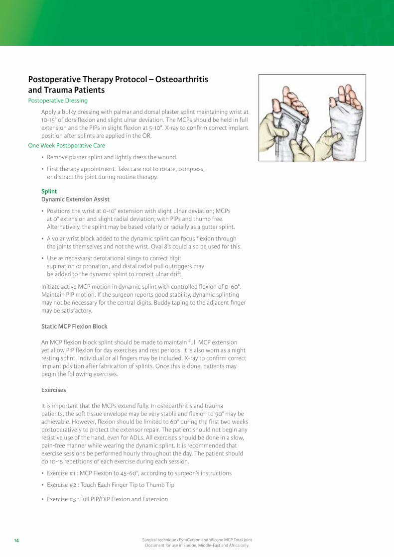
Postoperative Therapy Protocol – Osteoarthritis and Trauma PatientsPostoperative Dressing
Apply a bulky dressing with palmar and dorsal plaster splint maintaining wrist at 10-15° of dorsiflexion and slight ulnar deviation. The MCPs should be held in full extension and the PIPs in slight flexion at 5-10°. X-ray to confirm correct implant position after splints are applied in the OR.
One Week Postoperative Care
• Remove plaster splint and lightly dress the wound.
• First therapy appointment. Take care not to rotate, compress, or distract the joint during routine therapy.
SplintDynamic Extension Assist
• Positions the wrist at 0-10° extension with slight ulnar deviation; MCPs at 0° extension and slight radial deviation; with PIPs and thumb free. Alternatively, the splint may be based volarly or radially as a gutter splint.
• A volar wrist block added to the dynamic splint can focus flexion through the joints themselves and not the wrist. Oval 8’s could also be used for this.
• Use as necessary: derotational slings to correct digit supination or pronation, and distal radial pull outriggers may be added to the dynamic splint to correct ulnar drift.
Initiate active MCP motion in dynamic splint with controlled flexion of 0-60°. Maintain PIP motion. If the surgeon reports good stability, dynamic splinting may not be necessary for the central digits. Buddy taping to the adjacent finger may be satisfactory.
Static MCP Flexion Block
An MCP flexion block splint should be made to maintain full MCP extension yet allow PIP flexion for day exercises and rest periods. It is also worn as a night resting splint. Individual or all fingers may be included. X-ray to confirm correct implant position after fabrication of splints. Once this is done, patients may begin the following exercises.
Exercises
It is important that the MCPs extend fully. In osteoarthritis and trauma patients, the soft tissue envelope may be very stable and flexion to 90° may be achievable. However, flexion should be limited to 60° during the first two weeks postoperatively to protect the extensor repair. The patient should not begin any resistive use of the hand, even for ADLs. All exercises should be done in a slow, pain-free manner while wearing the dynamic splint. It is recommended that exercise sessions be performed hourly throughout the day. The patient should do 10-15 repetitions of each exercise during each session.
• Exercise #1 : MCP Flexion to 45-60°, according to surgeon’s instructions
• Exercise #2 : Touch Each Finger Tip to Thumb Tip
• Exercise #3 : Full PIP/DIP Flexion and Extension

15Surgical technique PyroCarbon and silicone MCP Total JointDocument for use in Europe, Middle-East and Africa only.
Four Weeks Postoperative Care
• Continue wearing splints or use buddy taping and do exercises as previously prescribed.
• Resume light ADL outside of the splint.
• Increase MCP flexion to 90° actively. If 60° of flexion has not been achieved, active assistive ROM exercises and a dynamic MCP flexion assist splint may be required.
• X-ray to confirm correct implant position.
Six+ Weeks Postoperative Care
progress to full activities as tolerated.

16 Surgical technique PyroCarbon and silicone MCP Total JointDocument for use in Europe, Middle-East and Africa only.
Postoperative Therapy Protocol – Rheumatoid PatientsPostoperative Dressing
• Apply a bulky dressing with a palmar and dorsal plaster splint maintaining the wrist at 10-15° of dorsiflexion and slight ulnar deviation. The MCPs should be held in full extension and the PIPs in slight flexion at 5-10°. Position must be checked in the OR with postoperative X-ray.
• If SWAN-neck deformities are present preoperatively, the PIPs should be placed in maximum flexion.
Two Days Postoperative Care
• Remove the bulky dressing and apply a short arm cast that maintains wrist in ulnar deviation at 0-10° and MCP joints in full extension with slight radial deviation (apply moleskin-tape straps to hold digits in radial deviation and extension), and allows full PIP/DIP extension/flexion.
• X-ray to confirm correct implant position after cast is applied.
Two Days–Three Weeks Postoperative Care
• If necessary, minimize edema by elevation, massage, or compression.
• Take care not to rotate, compress, or distract the joints.
• Gentle, active and passive motion of individual PIPs/DIPs can be performed to maintain IP flexibility.
Three Weeks Postoperative Care
• Patient’s first formal therapy appointment.
• Remove sutures, and lightly dress wound.
• Take care not to rotate, compress, or distract the joint during routine therapy.
SplintDynamic Extension Assist:
• Positions the wrist at 0-10° extension with slight ulnar deviation; MCPs at 0° extension and slight radial deviation; with IPs and thumb free. Alternatively, the splint may be based volarly or radially as a gutter splint.
• A volar wrist block added to the dynamic splint can focus flexion through the joints themselves and not the wrist. Oval 8’s could also be used for this.
• Use as necessary: derotational slings to correct digit supination or pronation, and distal radial pull outriggers may be added (as shown) to the dynamic splint to correct ulnar drift.
Static MCP Flexion Block:
This is an exercise splint for intrinsic tightness and/or to maintain IP joint mobility.
• Supports the MCPs volarly and allows IP flexion.
• Forearm-based with wrist neutral and slight ulnar deviation.
• MCPs at 0° of extension, and IPs and thumb are free.
Static Resting Splint:
• Positions the wrist at 0-10 degrees of extension and slight ulnar deviation.
• The MCPs at 0 degrees with finger dividers or otoform to promote slight radial deviation.

17Surgical technique PyroCarbon and silicone MCP Total JointDocument for use in Europe, Middle-East and Africa only.
• The PIPs are held in comfortable flexion, and
• The thumb should be in a position of rest. Confirm proper implant position in splints with x-ray. Once the splints are made and have been checked with x-ray, patients may begin the exercises as described in the next section.
Exercises
It is imperative for the success of this surgery in RA that the MCPs are not allowed to flex past 45° for the first six weeks. More aggressive motion can result in recurrent ulnar deviation or an extension lag of the MCPs. The patient should not begin any resistive use of the hand, even for ADL until week 6, as this can result in subluxation. Basic joint protection principals to prevent recurrent ulnar drift and subluxation should be discussed. A review of appropriate ADL techniques, and adaptive equipment may be necessary to maintain patient’s overall independence. All exercises should be done in a slow and pain-free manner.
It is recommended that exercise sessions be performed hourly throughout the day, with 10-15 repetitions of each exercise during each session. Frequency and number of repetitions depends on soft tissue stability and rate of healing.
Exercise #1: Controlled MCP flexion 0-30° (regulated by lead sinkers on outrigger fish line)
Exercise #2: Touch each finger tip to thumb tip (careful not to flex more than 30°)
Exercise #3: Radial Finger Walking
Exercise #4: PIP/DIP Flexion and Extension
Four Weeks Postoperative Care
• Continue wearing splints and do exercises as previously described.
• If the MCPs cannot actively flex to 45° after the first four weeks, finger slats can be taped either dorsally or volarly to block flexion at the IPs and focus motion at the MCP joints (reverse oval 8’s also work). Dynamic flexion assist splints may be used.
• Continue MCP flexion to 45° and begin light functional activities in dynamic splint.
Six Weeks Postoperative Care
• Continue wearing splints and do exercises as previously prescribed.
• Begin gentle motion outside of splint but continue dynamic splinting to ensure proper alignment.
• Increase MCP flexion to 60° in dynamic splint.
• Resume light ADL only while wearing dynamic splint.
6-12 Weeks Postoperative Care
Gradual weaning from the dynamic splint if alignment maintained; resume light ADLs.
12+ Weeks Postoperative Care
• Continue therapy as needed.
• Night resting splint worn for life.

18 Surgical technique PyroCarbon and silicone MCP Total JointDocument for use in Europe, Middle-East and Africa only.
1
10
1112
8
9
5
67
2 3
4
# Description
1 Starter Awl
2 Alignment Awl
3 Alignment Guide
4 Proximal Osteotomy Guide
5 Distal Osteotomy Guide
6 Distal Broaches
# Description
7 Proximal Broaches
8 Trials
9 Distal Impactor
10 Proximal Impactor
11 Trial Extractor
12 Implant Extractor
Instrumentation
19Surgical technique PyroCarbon and silicone MCP Total JointDocument for use in Europe, Middle-East and Africa only.
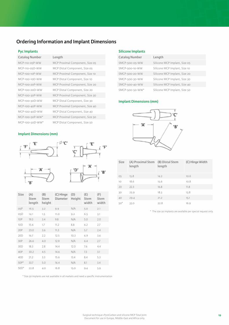
* Size 50 implants are not available in all markets and need a specific instrumentation
* The size 50 implants are available per special request only.
Pyc Implants
Catalog Number Length
MCP-110-05P-WW MCP Proximal Component, Size 05
MCP-110-05D-WW MCP Distal Component, Size 05
MCP-100-10P-WW MCP Proximal Component, Size 10
MCP-100-10D-WW MCP Distal Component, Size 10
MCP-100-20P-WW MCP Proximal Component, Size 20
MCP-100-20D-WW MCP Distal Component, Size 20
MCP-100-30P-WW MCP Proximal Component, Size 30
MCP-100-30D-WW MCP Distal Component, Size 30
MCP-100-40P-WW MCP Proximal Component, Size 40
MCP-100-40D-WW MCP Distal Component, Size 40
MCP-100-50P-WW* MCP Proximal Component, Size 50
MCP-100-50D-WW* MCP Distal Component, Size 50
Implant Dimensions (mm)
Size (A) Stem length
(B) Stem height
(C) Hinge Diameter
(D) Height
(E) Stem width
(F) Stem width
05P 16.5 3.2 9.9 N/A 5.0 2.1
05D 14.1 1.5 11.0 9.2 6.5 3.1
10P 19.3 3.4 9.8 N/A 5.0 2.0
10D 15.6 1.7 11.2 8.8 6.2 2.7
20P 23.0 3.6 11.3 N/A 5.7 2.4
20D 16.7 2.2 12.5 10.3 6.9 3.6
30P 26.6 4.0 12.9 N/A 6.4 2.7
30D 18.3 2.8 14.4 12.0 7.6 4.4
40P 30.2 4.5 14.6 N/A 7.3 3.1
40D 21.2 3.3 15.6 13.4 8.4 5.3
50P* 33.7 5.0 16.4 N/A 8.1 3.4
50D* 22.8 4.0 16.8 15.0 9.4 5.9
Silicone Implants
Catalog Number Length
SMCP-500-05-WW Silicone MCP Implant, Size 05
SMCP-500-10-WW Silicone MCP Implant, Size 10
SMCP-500-20-WW Silicone MCP Implant, Size 20
SMCP-500-30-WW Silicone MCP Implant, Size 30
SMCP-500-40-WW Silicone MCP Implant, Size 40
SMCP-500-50-WW* Silicone MCP Implant, Size 50
Implant Dimensions (mm)
Size (A) Proximal Stem length
(B) Distal Stem length
(C) Hinge Width
05 15.8 14.2 10.6
10 18.6 15.6 10.8
20 22.3 16.8 11.8
30 25.9 18.3 13.8
40 29.4 21.2 15.1
50* 33.0 22.8 16.9
Ordering Information and Implant Dimensions

Ordering Information and Implant Dimensions (following)
Pyc Instruments
Catalog Number Length
CSA-000-01 Lid (Case)
CSA-100-05 MCP Base 05-40 (Case)
CSA-100-06 MCP Trial Caddy 05-40 (Case)
IMP-100-00P MCP Proximal Impactor
IMP-100-00D MCP Distal Impactor
EXT-200-00 PIP Trial Extractor
EXT-100-01 MCP Implant Extractor
ALG-100-00 E/M Alignment Guide
OSG-100-00P MCP Proximal Osteotomy Guide
OSG-100-00D MCP Distal Osteotomy Guide
AWL-200-00 PIP Alignment Awl
AWL-100-01 Starter Awl
TRL-101-05P MCP Proximal Trial Sz. 05 (Metal)
TRL-101-10P MCP Proximal Trial Sz. 10 (Metal)
TRL-101-20P MCP Proximal Trial Sz. 20 (Metal)
TRL-101-30P MCP Proximal Trial Sz. 30 (Metal)
TRL-101-40P MCP Proximal Trial Sz. 40 (Metal)
TRL-101-05D MCP Distal Trial Sz. 05 (Metal)
TRL-101-10D MCP Distal Trial Sz. 10 (Metal)
TRL-101-20D MCP Distal Trial Sz. 20 (Metal)
Silicone Instruments
Catalog Number Length
TRL-101-30D MCP Distal Trial Sz. 30 (Metal)
TRL-101-40D MCP Distal Trial Sz. 40 (Metal)
TRL-500-05 MCP Silicone Trial Sz. 05
TRL-500-10 MCP Silicone Trial Sz. 10
TRL-500-20 MCP Silicone Trial Sz. 20
TRL-500-30 MCP Silicone Trial Sz. 30
TRL-500-40 MCP Silicone Trial Sz. 40
BRH-101-05P MCP Proximal Broach Sz. 05 (Cutting)
BRH-100-10P MCP Proximal Broach Sz. 10
BRH-100-20P MCP Proximal Broach Sz. 20
BRH-100-30P MCP Proximal Broach Sz. 30
BRH-100-40P MCP Proximal Broach Sz. 40
BRH-101-05D MCP Distal Broach Sz. 05 (Cutting)
BRH-100-10D MCP Distal Broach Sz. 10
BRH-100-20D MCP Distal Broach Sz. 20
BRH-100-30D MCP Distal Broach Sz. 30
BRH-100-40D MCP Distal Broach Sz. 40
Integra®
PyroCarbon MCP Total Joint
Integra LifeSciences Services (France) Immeuble Séquoia 2 97 allée Alexandre Borodine Parc technologique de la Porte des Alpes 69800 Saint Priest n France Phone: +33(0)4 37 47 59 10
EC REP
Integra and the Integra logo are registered trademarks of Integra Lifesciences Corporation in the United States and/or other countries. Document for use in Europe, Middle-East & Africa only.This surgical technique is not for use on French territory. ©2020 Integra LifeSciences Corporation. All rights reserved. Last revision date: 2020-06. 0257213-4-EN LC-04-106-023 Rev B
Availability of these products might vary from a given country or region to another, as a result of specific local regulatory approval or clearance requirements for sale in such country or region. n Consult product labels and inserts for any indication, contraindications, hazards, warnings, precautions, and instructions for use.n Non contractual document. Integra reserves the right, without prior notice, to modify the products in order to improve their quality. n Warning: Applicable laws restrict these products to sale by or on the order of a physician.
Products mentioned in this document are CE class I, IIa or IIb devices. Please contact Integra customer service should you need any additional information on devices classification. All the medical devices mentioned on this document are CE marked according to European council directive 93/42/EEC on medical devices and its relatives, unless specifically identified as “NOT CE MARKED”.
Ascension orthopedics, inc.11101 Metric BlvdAustin, Texas 78758 n USAIntegra LifeSciences Services (France) SAS
Immeuble Séquoia 2 n 97 allée Alexandre BorodineParc technologique de la Porte des Alpes69800 Saint Priest n FrancePhone: +33 (0)4 37 47 59 00 n +33 (0)4 37 47 59 99 (fax)
Integra Contact - Sales & MarketingEUROPE, MIDDLE-EAST and AFRICA
International: +33 (0)4 37 47 59 50 n +33 (0)4 37 47 59 25 (fax) n [email protected]: +33 (0) 437 47 59 10 n +33 (0) 437 47 59 29 (Fax) n [email protected]: +32 (0)2 257 41 30 n +32 (0)2 253 24 66 (fax) n [email protected]: +41 (0)22 721 23 00 n +41 (0)22 721 23 99 (fax) n [email protected] Kingdom: +44 (0)1264 312 725 n +44 (0)1264 312 821 (fax) n [email protected]
Integra Contact - Questions & Information